CHAPTER 14 Overview of the Circulation; Biophysics of Pressure, Flow, and Resistance
The main function of the circulation is to serve the needs of the tissues by transporting nutrients to them, transporting away waste products, carrying hormones from one part of the body to another, and in general maintaining homeostatic conditions in the tissue fluids for optimal survival and function of the cells.
Physical Characteristics of the Circulation (p. 157)
The circulation is divided into the pulmonary circulation, which supplies the lungs, and the systemic circulation, which supplies tissues in the remainder of the body. The functional parts of the circulation are the following:
The Circulation Is a Complete Circuit
Contraction of the left heart propels blood into the systemic circulation through the aorta, which empties into smaller arteries, arterioles, and eventually capillaries. Because the blood vessels are distensible, each contraction of the heart distends the vessels; during relaxation of the heart, the vessels recoil, thereby continuing flow to the tissues, even between heartbeats. Blood leaving the tissues enters the venules and then flows into increasingly larger veins, which carry the blood to the right heart.
The right heart then pumps the blood through the pulmonary artery, small arteries, arterioles, and capillaries, where oxygen and carbon dioxide are exchanged between the blood and the tissues. From the pulmonary capillaries, blood flows into venules and large veins and empties into the left atrium and left ventricle before it is again pumped into the systemic circulation.
Because Blood Flows Around the Same Vessels, Any Change in Flow in a Single Part of the Circuit Transiently Alters Flow in Other Parts
For example, strong constriction of the arteries in the systemic circulation can transiently reduce the total cardiac output, in which case blood flow to the lungs decreases equally as much as flow through the systemic circulation.
Another feature of the circulation is that sudden constriction of a blood vessel must always be accompanied by opposite dilation of another part of the circulation because blood volume cannot change rapidly and blood itself is not compressible. For instance, strong constriction of the veins in the systemic circulation displaces blood into the heart, dilating the heart and causing it to pump with increased force. This is one of the mechanisms by which cardiac output is regulated. With prolonged constriction or dilation of a portion of the circulation, changes in total blood volume can occur through exchange with the interstitial fluid or because of changes in fluid excretion by the kidneys.
Most of the Blood Volume Is Distributed in the Veins of the Systemic Circulation
About 84% of the total blood volume is in the systemic circulation, with 64% in the veins, 13% in the arteries, and 7% in the systemic arterioles and capillaries. The heart contains about 7% of the blood volume and the pulmonary vessels 9%.
Velocity of Blood Flow Is Inversely Proportional to the Vascular Cross-Sectional Area
Because approximately the same volume of blood flows through each segment of the circulation, vessels with a large cross-sectional area, such as the capillaries, have slower blood flow velocity. The approximate total cross-sectional areas of the systemic vessels for the average human being are as follows:
| Vessel | Cross Sectional Area (cm2) |
| Aorta | 2.5 |
| Small arteries | 20 |
| Arterioles | 40 |
| Capillaries | 2500 |
| Venules | 250 |
| Small veins | 80 |
| Venae cavae | 8 |
Thus, under resting conditions, the velocity of blood flow in capillaries is only about 1/1000 the velocity of flow in the aorta.
Pressures Vary in the Different Parts of the Circulation
Because the pumping action of the heart is pulsatile, the aortic arterial pressure rises to its highest point, the systolic pressure, during systole and falls to its lowest point, the diastolic pressure, at the end of diastole. In the healthy adult, systolic pressure is approximately 120 mm Hg, and diastolic pressure is 80 mm Hg. This is usually written as 120/80 mm Hg. The difference between systolic and diastolic pressure is called the pulse pressure (120 – 80 = 40 mm Hg). As blood flows through the systemic circulation, its pressure falls progressively to approximately 0 mm Hg by the time it reaches the termination of the venae cavae in the right atrium of the heart.
Pressure in the systemic capillaries varies from as high as 35 mm Hg near the arteriolar ends to as low as 10 mm Hg near the venous ends, but the average functional capillary pressure is about 17 mm Hg.
Pressures in the Pulmonary Circulation Are Much Lower Than Those in the Systemic Circulation
Pressure in the pulmonary arteries is also pulsatile, but systolic arterial pressure is about 25 mm Hg and diastolic pressure 8 mm Hg, with a mean pulmonary artery pressure of only 16 mm Hg. Pulmonary capillary pressure averages only 8 mm Hg, yet the total blood flow through the lungs is the same as that in the systemic circulation because of the lower vascular resistance of the pulmonary blood vessels.
Basic Principles of Circulatory Function (p. 158)
The details of circulatory function are complex and are described later, but there are three basic principles that underlie the major functions of the circulatory system:
Interrelationships of Pressure, Flow, and Resistance (p. 159)
Blood Flow through a Vessel Is Determined by the Pressure Gradient and Vascular Resistance
The flow of blood through a vessel can be calculated by the formula F = ΔP/R, where F is blood flow, ΔP is the pressure difference between the two ends of the vessel, and R is the vascular resistance. Note that it is the difference in pressure between the two ends of the vessel that provides the driving force for flow, not the absolute pressure in the vessel. For example, if the pressure at both ends of the vessel were 100 mm Hg, there would be no flow despite the presence of high pressure.
Because of the extreme importance of the relationship among pressure, flow, and resistance, the reader should become familiar with the other two algebraic forms of this relationship: ΔP = F × R and R = ΔP/F. Blood pressure is usually expressed in millimeters of mercury (mm Hg), and blood flow is expressed in milliliters per minute (ml/min); vascular resistance is expressed as mm Hg/ml per minute. In the pulmonary circulation, the pressure gradient is much lower than that in the systemic circulation, whereas the blood flow is the same as that in the systemic circulation; therefore the total pulmonary vascular resistance is much lower than the systemic vascular resistance.
Vessel Diameter Has a Marked Effect on Resistance to Blood Flow—Poiseuille’s Law
According to the theory of Poiseuille, vascular resistance is directly proportional to the viscosity of the blood and the length of the blood vessel and inversely proportional to the radius of the vessel raised to the fourth power:
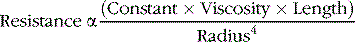
Decreased Radius of a Blood Vessel Markedly Increases Vascular Resistance
Because vascular resistance is inversely related to the fourth power of the radius, even small changes in radius can cause very large changes in resistance. For example, if the radius of a blood vessel increases from one to two (two times normal), resistance decreases to 1/16 of normal (½4) and flow increases to 16 times normal if the pressure gradient remains unchanged. Small vessels in the circulation have the greatest amount of resistance, whereas large vessels have little resistance to blood flow.
For a parallel arrangement of blood vessels, as occurs in the systemic circulation in which different organs are each supplied by an artery that branches into multiple vessels, the total resistance can be expressed as
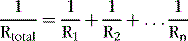
where R1, R2, and Rn, are the resistances of each of the various vascular beds in the circulation. The total resistance is less than the resistance of any of the individual vascular beds.
For a series arrangement of blood vessels, as occurs within a tissue in which blood flows through arteries, arterioles, capillaries, and veins, the total resistance is the sum of the individual resistances, as

where R1, R2, and Rn are the resistances of the various blood vessels in series within the tissues.
Conductance is a measure of the ease of which blood can flow through a vessel and is the reciprocal of resistance.

Increased Blood Hematocrit and Increased Viscosity Raise Vascular Resistance and Decrease Blood Flow
The greater the viscosity, the less is the flow of blood in a vessel if all other factors remain constant. The normal viscosity of blood is about three times as great as the viscosity of water. The main factor that makes blood so viscous is that it has large numbers of suspended red blood cells, each of which exerts frictional drag against adjacent cells and against the wall of the blood vessel.
The percentage of blood comprising cells, called the hematocrit, is normally about 40; this indicates that about 40% of the blood is cells, and the remainder is plasma. The greater the percentage of cells in the blood—that is, the greater the hematocrit—the greater is the viscosity of blood and therefore the greater is the resistance to blood flow.
“Autoregulation” Attenuates the Effect of Arterial Pressure on Tissue Blood Flow
The effect of arterial pressure on blood flow in many tissues is usually far less than one would expect, based on our previous discussion. The reason for this is that an increase in arterial pressure usually initiates compensatory increases in vascular resistance within a few seconds through activation of the local control mechanisms discussed in Chapter 17. Conversely, with reductions in arterial pressure vascular resistance is promptly reduced in most tissues and blood flow is maintained relatively constant. The ability of each tissue to adjust its vascular resistance and to maintain normal blood flow during changes in arterial pressure between approximately 70 and 175 mm Hg is called blood flow autoregulation.
Changes in tissue blood flow rarely last for more than a few hours even when increases in arterial pressure or increased levels of vasoconstrictors or vasodilators are sustained. The reason for the relative constancy of blood flow is that each tissue’s local autoregulatory mechanisms eventually override most of the effects of vasoconstrictors to provide a blood flow that is appropriate for the needs of the tissue.