INSPIRATORY MUSCLE TRAINING

Individuals with chronic respiratory disease, for example COPD, experience inspiratory muscle dysfunction due to a combination of problems, which include hyperinflation, increased work of breathing, hypoxaemia, and hypercapnia (Laghi & Tobin 2003). Other problems that may reduce inspiratory muscle strength and endurance include corticosteroid-induced myopathy, chronic inflammation and chronic gas exchange abnormalities (Maltais et al 2000). Factors that lead to inspiratory muscle dysfunction contribute to the development of dyspnoea and to limitation of exercise tolerance (Solcher & Dechman 1998). Optimizing function through a reduction in dyspnoea is a key aim of therapy and there is now unequivocal evidence that pulmonary rehabilitation is the only treatment shown convincingly to reduce dyspnoea in chronic respiratory disease. A contributory factor in this is through training-induced improvements in aerobic capacity of the skeletal muscles, achieved through increasing mechanical efficiency and lowering blood lactate concentrations (Casaburi et al 1991, O’Donnell et al 1995). However, it is also established that whole-body exercise conditioning fails to improve respiratory muscle strength and endurance (Weiner et al 1992). As inspiratory muscle dysfunction contributes to dyspnoea in COPD, it is evident that specifically targeting the inspiratory muscles may lead to further reductions in dyspnoea (Hill et al 2004).

The inspiratory muscles, like other skeletal muscles, undergo physiological adaptations in response to training. Despite this being evident, the early literature on the use of inspiratory muscle training (IMT) in patients with COPD presented a rather mixed picture. In part this was due to the paucity of controlled clinical trials, but more importantly due to the nature of the training adopted. In general the trials were confounded by the methodology of the training in which the frequency, duration and intensity of training were less than that required to achieve a true training response, as highlighted in an early meta-analysis (Smith et al 1992). Ten years later Lotters et al (2002) published a second meta-analysis, which included only studies in which IMT was controlled in terms of fixing the intensity of IMT in order to achieve a training response. This review concluded that IMT improved respiratory muscle strength and endurance although the effects of IMT on exercise capacity remained to be determined. More recently Geddes et al (2005) published a systematic review in which IMT was shown to be effective, not only in terms of increasing inspiratory muscle strength and endurance but also in improving exercise capacity in adults with COPD. However, this review emphasized that the method of IMT employed is important if clinical benefits are to be obtained and that training of the ventilatory muscles must follow the basic principles of training for any striated muscle with regard to the intensity and duration of the stimulus, the specificity of training and the reversibility of training.

The principle of specificity of training states that the effects of training are very specific to the neural and muscular elements of overload. The overload principle states that overload must be applied to a muscle for a training response to occur (Kennedy 1995). Overload may be applied by increasing the frequency or duration of training or the intensity of the loading, or a combination of these factors. Generally, training theory suggests that inspiratory muscle strength gains can be achieved at intensities of 80–90% of maximal inspiratory pressure. Strength-endurance gains (maximal effective force that can be maintained) can be achieved at 60–80%, and endurance (the ability to continue a dynamic task for a prolonged period) at approximately 60% of peak, which equates with high-intensity training regimens used in systemic exercise (Kraemer et al 2002).

Overload may also include the concept of incremental loading. This involves decreasing the rest periods between muscle contractions (Komi & Hakkinen 1991), which has been shown to recruit a larger proportion of muscle fibres and, hence, a larger pool of fibres are trained for subsequent lower but potentially fatiguing loads (Reid & Samrai 1995, Reid et al 1994). In studies that have used the principle of high-intensity incremental IMT improvements in lung volumes, diaphragm thickness and exercise capacity have been obtained in healthy subjects and in patients with cystic fibrosis (Enright et al 2004, 2006).

  Page 180 

In many previous investigations training methodologies have varied to include loads imposed on the respiratory muscles which can be characterized as flow, pressure and volume loads (McCool 1992). A low-pressure high-flow load was first described in 1976 by Leith & Bradley which involves training the respiratory muscles by voluntarily ventilating at high levels for a prolonged period (usually 15 minutes). It is therefore analogous to the high ventilatory demands imposed on the inspiratory muscles during high-intensity exercise (Belman 1993). The load imposed on the inspiratory muscles requires a high flow rate at low pressure, although it is generally impractical, as it requires the assembly of complex breathing circuits in order to ensure that the individual remains normocapnic. In addition, breathless patients find this method of IMT uncomfortable to maintain and hence this method of IMT tends to be reserved for laboratory-based investigations rather than being used in a clinical setting (Scherer et al 2000). In contrast to a flow load, a high-pressure low-flow load occurs with any process that increases the transpulmonary pressure required to breathe in. These loads can be experimentally imposed by inhaling from a rigid chamber or by breathing through an external resistance. Such loads can be achieved by either resistive or threshold devices. Resistive training devices incorporate a range of apertures, which vary in size in order to apply the prescribed resistive load. Threshold devices impose a threshold or a critical inspiratory opening pressure that the subject is required to overcome before the start of an inspiratory flow. The efficacy of both these modes of IMT have been the subject of some debate, although recently it has been shown that targeted resistive IMT is as effective as threshold IMT for adults with COPD (Hsiao et al 2003). On a practical level some issues require consideration, for example targeted resistive devices provide visual feedback which enhances motivation, although ensuring that the patient is maintaining the required training intensity can be problematic. Conversely, threshold trainers provide a more constant resistance although the loss of visual feedback may result in loss of motivation.

In addition to the mode of training adopted, the frequency of training in IMT interventions, the duration of the training intervention and the issue of reversibility of training requires consideration. Standard guidelines of the American College of Sports Medicine (Kennedy 1995) suggest a training frequency of one to two times per day for a total duration of 20–30 minutes, three to five times per week for 6 weeks. However, functional improvements and adaptive cellular changes in the inspiratory muscles have been shown to occur following 5 weeks of training (Ramirez-Sarmiento et al 2002) although training must be maintained for the cellular training effects to be sustained (McArdle et al 2001). In summary, the optimal frequency of training is thought to be three times weekly, to continue beyond 4 weeks and with maintenance achieved by continuing training at one or two times weekly (Fleck 1994).

In conclusion, although there is much conflicting evidence in the literature, which has cast doubt on the place of IMT in patients with respiratory muscle dysfunction, more recent data that have incorporated the appropriate physiological training principles during IMT look promising (Geddes et al 2005). The use of targeted inspiratory resistive or threshold modes of IMT as opposed to non-targeted inspiratory resistive modes ensures that the training intensity is achieved and maintained. Thus with effective IMT regimens, exercise intolerance, dyspnoea and hypercapnic ventilatory failure may be prevented or alleviated. In addition, another indication for IMT is that weakness of the inspiratory muscles may lead to an inability to generate an adequate flow to assure lung deposition when using dry powder inhalers. Hence strengthening the inspiratory muscles may improve the efficacy of inhaled drug therapy (Weiner & Weiner 2006). These considerations are vital if IMT is to find a proven place in pulmonary rehabilitation programmes (Hill & Eastwood 2005).

INTERMITTENT POSITIVE PRESSURE BREATHING

Intermittent positive pressure breathing (IPPB) is the maintenance of a positive airway pressure throughout inspiration, with airway pressure returning to atmospheric pressure during expiration. The American Association for Respiratory Care (AARC) has developed clinical practice guidelines for the use of IPPB as a hyperinflation and aerosol delivery technique (American Association for Respiratory Care 2003). The literature on IPPB has been reviewed by Bott et al (1992) and Denehy & Berney (2001). Denehy & Berney concluded that although the use of IPPB has declined, it may still have a place in the management of patients with re-duced lung volumes. They also state that the rationale for using IPPB should be based on its known physiological effects, the availability of other treatment modalities, the condition of the patient and the current research knowledge base. The Bird ventilator (Fig. 5.56) is a pressure-cycled device convenient to use for providing IPPB as an adjunct to physiotherapy in the spontaneously breathing patient.

image

Figure 5.56 The Bird Mark 7 ventilator.

IPPB has been shown to augment tidal volume (Stiller et al 1992, Sukumalchantra et al 1965) and using an IPPB device in the completely relaxed subject, the work of breathing during inspiration approaches zero (Ayres et al 1963). These two effects support the use of IPPB to help in the clearance of bronchial secretions when more simple airway clearance techniques alone are not maximally effective, for example in the semi-comatosed patient with chronic bronchitis and sputum retention (Pavia et al 1988), the postoperative patient or in a patient with neuromuscular disease and a chest infection. The reduction in the work of breathing can be used with effect in the exhausted patient with acute severe asthma, but there is no evidence that the effect of bronchodilators delivered by IPPB is greater than from a nebulizer alone (Webber et al 1974).

  Page 181 

An ideal IPPB device for use with physiotherapy should be portable and have simple controls. Other important features are as follows:

Positive pressure.

The range of pressures is likely to be from 0 to 35 cmH2O.

Sensitivity.

The patient should be able to ‘trigger’ the inspiratory phase with minimal effort. Fully automatic control is unpleasant for most patients and unnecessary for physiotherapy. A hand triggering device is useful to test the ventilator and nebulizer.

Flow control.

With ventilators such as the Bird Mark 7, the inspiratory gas is delivered at a flow rate that can be preset by means of a control dial. Optimal distribution of gas to the more peripheral airways is achieved at relatively slow flow rates, but if the patient is very short of breath and has a fast respiratory rate, a slow inspiratory phase may be unacceptable. It is often useful to alter the flow control several times during a single treatment session, providing slow breaths during the periods attempting to mobilize peripheral secretions and a faster flow rate when a patient is recovering his breath after expectoration. Some Bennett positive pressure ventilators do not require flow rate adjustment because automatic variable flow is provided with each breath. This feature is known as ‘flow sensitivity’ and means that the flow of the inspired gas adapts to the resistance of the individual’s airways.

Nebulizer.

An efficient nebulizer in the circuit is necessary to humidify the driving gas and, when appropriate, to deliver bronchodilator drugs.

Air-mix control.

When driven by oxygen, air must be entrained by the apparatus to provide an air/oxygen mixture for the patient. Some Bird ventilator devices have a control that should be set to give a mixture, while others have no control but automatically entrain air. The use of 100% oxygen for a patient is very rare and when it is indicated, an IPPB device with an air-mix control will be needed. When air is not entrained through the apparatus, the flow rate control must be regulated to provide an adequate flow to the patient.

When IPPB (a Bird ventilator) is driven by oxygen and the air-mix control is in use, the percentage of oxygen delivered to the patient is approximately 45% (Starke et al 1979). This percentage will be considerably higher than the controlled percentage delivered by an appropriate Venturi mask, for example to a patient with chronic bronchitis. This higher percentage is rarely dangerous during treatment because the patient’s ventilation is assisted and the removal of secretions as a result of treatment is likely to lead subsequently to an improvement in arterial blood gas tensions (Gormezano & Branthwaite 1972).

It has been suggested that a few patients become more drowsy during or after IPPB as a result of the high percentage of oxygen received. Starke et al (1979) showed that increased drowsiness caused by hypercapnia occurred whether oxygen or air was the driving gas for IPPB and that the deterioration was dependent on inappropriate settings of the ventilator. The pressure and flow controls must be set to provide an adequate tidal volume, this being particularly important when treating patients with a rigid thoracic cage (Starke et al 1979).

Occasionally, IPPB may be powered by Entonox and in this case the air-mix control would need to be in the position to provide 100% of the driving gas with no additional air entrained.

Breathing circuit.

To prevent cross-infection it is essential for each patient to have his own breathing circuit, which consists of tubing, nebulizer, exhalation valve and a mouthpiece or mask. The majority of patients prefer to use a mouthpiece, but a facemask is required when treating confused patients. A flange mouthpiece (Fig. 5.57) is useful for patients who have difficulty making an airtight seal around the mouthpiece.

image

Figure 5.57 Flange mouthpiece for use with IPPB.

The type of breathing circuit used will depend on local infection control guidelines. The circuits can be autoclavable, non-disposable but non-autoclavable, or disposable. Many countries use single patient disposable circuits.

Preparation of the apparatus

1. Normal saline solution or the drug to be nebulized (3–4 ml in total) is inserted into the nebulizer chamber.
2. The breathing circuit is connected to the IPPB ventilator and the ventilator connected to the driving gas source. It can be used from an oxygen or air cylinder if piped compressed gas is unavailable.
3. If there is an air-mix control, this should be in the position for entrainment of air.
4. If there is an automatic control (expiratory timer) this should be turned off to allow the patient to ‘trigger’ the machine at his desired rate.
5. The sensitivity, flow and pressure controls are set appropriately for the individual. With the Bird Mark 7 the sensitivity control is usually adjusted to a low number (5–7) where minimal inspiratory effort is required. The pressure and flow controls are adjusted to provide regular assisted ventilation without discomfort. A patient with a rigid rib cage will require a higher pressure setting to obtain an adequate tidal volume than someone with a more mobile rib cage. When adjusting the settings for a new patient, it may be easiest to start with a pressure at approximately 12 cmH2O and the flow at about ‘10’, then gradually increase the pressure and reduce the flow until the pattern of breathing is the most appropriate for the individual. Some IPPB devices do not have numbered markings, but after finding the most effective settings for a patient during one treatment, it is useful to note the positions of the controls in order to use these as a starting point at the next treatment. The controls to be set on the Bennett PR-1 are the nebulizer, sensitivity and pressure.
6. Before starting a treatment, the hand triggering device is operated to check that there are no leaks in the breathing circuit and that the nebulizer is functioning well.
Treatment of the patient

The position in which IPPB is used depends on the indication for treatment. It may be used in side lying, high side lying or in the sitting position. The patient should be positioned comfortably and encouraged to relax the upper chest and shoulder girdle.

After the purpose of the IPPB treatment has been explained, the patient is asked to close his lips firmly around the mouthpiece and then to make a slight inspiratory effort, which will trigger the device into inspiratory flow. The patient should then relax throughout inspiration, allowing his lungs to be inflated. When the preset pressure is reached at the mouth the ventilator cycles into expiration; the patient should remain relaxed and let the air out quietly.

  Page 183 

If the patient attempts to assist inspiration there will be a delay in reaching the cycling pressure. A delay will also occur if there is a leak around the mouthpiece, at any of the circuit connections or from the patient’s nose. A nose clip may be required until the patient becomes familiar with the technique.

Observation of the manometer on the ventilator should allow detection of any faults in the patient’s technique. At the start of inspiration the needle should swing minimally to a negative pressure and then swing smoothly up to the positive pressure set, before cutting out into expiration and returning to zero. A larger negative swing at the beginning of inspiration shows that the patient is making an unnecessary effort in triggering the device. If the patient makes an active effort throughout inspiration, the needle will rise very slowly to the inspiratory set pressure and if they attempt to start expiration before the preset pressure is reached, the needle will rise sharply above the set pressure and then cut out into expiration.

When IPPB is taught correctly the work of breathing is relieved, but if the patient is allowed to assist either inspiration or expiration there may be an increase in the work of breathing.

A short pause between completion of expiration and the next inspiration avoids hyperventilation and possible dizziness. Occasionally children using IPPB tend to swallow air during treatment. It is important to observe the size of the abdomen before and during IPPB to rec-ognize signs of abdominal distension and discontinue treatment if this occurs.

When IPPB is used to relieve the work of breathing while delivering bronchodilator therapy, for example in the acute severe asthmatic patient, it is often helpful for the physiotherapist to hold the breathing circuit to allow the patient to relax the shoulders and arms as much as possible (Fig. 5.58).

image

Figure 5.58 to reduce the work of breathing during inhalation of nebulized bronchodilator.

A facemask for IPPB is used in the drowsy or confused patient and in those with facial weakness unable to make an airtight seal at the mouth. When using IPPB to assist in mobilizing secretions, the patient should be appropriately positioned to assist loosening and mobilization of secretions, for example in side lying. The patient’s jaw should be elevated and the mask held firmly over the face, ensuring an airtight fit. Chest shaking during the expiratory phase may be used to assist in mobilizing secretions. In a drowsy patient it may be necessary to stimulate coughing using nasotracheal suction if spontaneous coughing is not stimulated by IPPB and chest shaking.

In medical patients with retained secretions and poor respiratory reserve, IPPB may be useful both to mobilize secretions and to relieve the effort of breathing following expectoration. The flow control on a Bird ventilator should be adjusted to give a slow, comfortable breath to mobilize secretions, but following the exertion of expectoration there may be an increase in respiratory requirements, which may necessitate increasing the flow and possibly reducing the pressure until the breathing effort and pattern return to normal or baseline.

  Page 184 

IPPB may be used in patients with chest wall deformity, for example kyphoscoliosis (Sinha & Bergofsky 1972), when there is difficulty with clearing secretions during an infective episode. To achieve an adequate increase in ventilation in patients with a rigid rib cage, the pressure setting needs to be higher than for a more mobile rib cage.

Occasionally, in postoperative patients, IPPB is the adjunct of choice when the patient is unable to augment tidal volume adequately during treatment. In these patients, in contrast to the relaxed technique normally used with IPPB, thoracic expansion may be actively encouraged during the inspiratory phase.

Contraindications for IPPB

image Pneumothorax.
image Large bullae.
image Lung abscess, as the size of the air space may increase.
image Severe haemoptysis, as treatment is inappropriate until the bleeding has lessened.
image Postoperative air leak, unless the advantages of IPPB would outweigh the possibility of increasing the air leak during treatment.
image Bronchial tumour in the proximal airways. Air may flow past the tumour during inspiration and may be trapped on expiration as the airways narrow. There would be no contraindication if the tumour were situated peripherally.

MANUAL HYPERINFLATION

The technique of manual hyperinflation may be indicated to mobilize and assist clearance of excess bronchial secretions and to reinflate areas of lung collapse in the intubated patient. It is described in Chapter 8.

MANUAL THERAPY TECHNIQUES

Musculoskeletal dysfunction is common in people with respiratory disease. People with chronic cardiorespiratory disease may often demonstrate skeletal, musculoskeletal and nervous systems adaptations over time, related to the severity and management of their disease. As age increases the incidence of musculoskeletal deterioration will also increase (Parasa & Maffulli 1999). Any postural or degenerative changes are likely to have implications for physical function and quality of life as well as influencing the cardiorespiratory system.

Postural and skeletal changes occurring over time relate to the overuse of upper chest breathing patterns, lack of lower rib expansion and reduction in the more efficient pattern of diaphragmatic breathing. Chronic hyperinflation typically leads to the development of a barrel-shaped chest with an increase in the anteroposterior diameter of the chest. Pain may limit rib expansion and abdominal breathing, particularly in patients following abdominal surgery.

Secondary malalignment of the scapulae is associated with prolonged coughing using trunk flexion and the increased outward pressure on the chest wall. More sputum may mean more pain and less efficient airway clearance (Massery 2005).

In a study of 143 young adults with cystic fibrosis, Henderson and Specter (1994) found 77% of females and 36% of males over 15 years of age had a kyphosis of more than 40° (the upper limit of normal). Kyphosis tends to worsen with age and disease severity (Massie et al 1998).

The neck and shoulder girdle structures adapt to counterbalance the flexed trunk sitting position. Neutral neck and head position is compromised as the neck and head are drawn forwards by the large superficial muscle groups and hyperactivity of the suboccipital extensors. The greater the thoracic kyphosis, the more likely it is that the middle and upper cervical regions will become lordotic, as the upper cervical spine hyperextends and tilts the head upward to maintain a vertical orientation of the face.

This upper cervical spine hyperextension and forward head posture combine, with a loss of endurance of the deep cervical flexor muscles, to increase the likelihood of cervicogenic headache (Jull et al 2002, Watson & Trott 1993). Chronic headaches may also be associated with medical causes in patients with cardiorespiratory disease (Festini et al 2004, Ravilly et al 1996).

The incidence of musculoskeletal chest pain in people with cystic fibrosis tends to increase as the disease progresses. Painful stiffness in the chest may inhibit airway clearance and increase the work of breathing (Massery 2005). A decrease in muscle strength and mobility in the trunk, chest and shoulders has been demonstrated in people with cystic fibrosis (Ross et al 1987).

In the presence of an inefficient, upper chest breathing pattern, the overactive scalene muscles elevate the first and second ribs while the levator scapulae depress and rotate the lateral shoulder girdle (Fig. 5.59). Shortening of upper trapezius and tightness of pectoralis minor and major elevate and anteriorly tilt the scapulae, respectively. At the same time the antagonist and stabilizing muscles, serratus anterior and the middle and lower fibres of trapezius, lengthen and weaken, causing winging and inferior rotation of the scapulae (Sahrmann 2005). Over time the long thoracic extensors and multifidus lose their segmental stabilizing capacity and endurance, and become less able to sustain the upright sitting neutral posture.

image

Figure 5.59 CJ, aged 16, cystic fibrosis. (A) Relaxed sitting posture (posterior view). Note: forward head position, tight suboccipital and mid-cervical extensors, tight upper and middle fibres of trapezius, asymmetry and abducted and protracted position of the scapulae, increased thoracic kyphosis, reduced upper lumbar lordosis, posterior rotation of pelvis. (B) Relaxed sitting posture (side view). Note: forward head position, increased sternocleidomastoid activity, increased low cervical lordosis and thoracic kyphosis, abducted and protracted scapulae, anterior position of humerus in glenoid fossa, internal rotation of humerus, lax abdominal muscles.

  Page 185 

The sternocleidomastoids are used excessively during coughing. Muscle fatigue related to the excess work of breathing may further accentuate poor posture in people with moderate to severe chronic lung disease. The existing kyphosis of the thoracic spine is increased due to prolonged bed rest and reductions in general exercise tolerance. Habitual slouching due to dyspnoea and feeling unwell will place more kyphotic strain on the thoracic and lumbar spines, and the lumbopelvic angle will be flexed instead of lordotic.

Vertebral intersegmental motion will be gradually lost as the chest becomes fixed in elevation and flexion. Reduced range of thoracic extension will contribute to loss of the final 30 degrees of shoulder flexion and abduction; while tightness in anterior deltoid, teres major and latissimus dorsi muscles and disturbance of normal scapulothoracic rhythm will decrease the free range of external rotation and flexion available at the glenohumeral joint. As a consequence the overstretching of infraspinatus and teres minor, associated with the internally rotated position of the humerus, may lead to poor stability of the humerus in the glenoid fossa (Fig. 5.60).

image

Figure 5.60 (A) Sitting posture (posterior view) following active assisted anterior rotation of pelvis. Note: decreased mid-cervical lordosis, improved position of scapulae, reduced thoracic kyphosis, neutral rotation of pelvis and improved lumbar lordosis. (B) Sitting posture (side view) following active assisted anterior rotation of pelvis. Note: less forward head position, activation of deep cervical flexors, reduced sternocleidomastoid activity, improved scapulae and humeral position and thoracic kyphosis.

These muscular and skeletal aberrations are likely to have consequences on the range and quality of pelvic position in sitting, neck and shoulder motion, and both general and specific trunk and shoulder movement and function. In particular they cause a physical limit to the end range of shoulder elevation and an alteration in muscle recruitment likely to increase the risk of shoulder tendon impingement and wear.

Individuals with respiratory disease may complain of acute or chronic cervical, thoracic or rib joint pain, which may decrease chest expansion as measured by a reduction in vital capacity. Joint manifestations (mainly hypertrophic osteoarthropathy) are common in children with cystic fibrosis, affecting 2–8.5% of patients (Botton et al 2003, Parasa & Maffulli 1999). Back pain may be due to cystic fibrosis-related arthropy, arthritis due to coexistent conditions or drug reactions as well as the more obvious mechanical reasons. Mechanical back pain in people with cystic fibrosis has been described in the literature, but the incidence and prevalence have not been reliably established for the different age groups.

  Page 186 

With increasing longevity in people with cystic fibrosis, musculoskeletal changes will become more important. Decreased bone mineral density is common at all ages but further reduction tends to occur with time, increasing illness and adverse effects of medication. With an increased emphasis on encouraging general exercise to improve respiratory and general health and bone density, musculoskeletal problems may become more prevalent, requiring careful monitoring (Buntain et al 2004).

With chronic respiratory disease, fracture rates are reported to be approximately twice as high in women aged 16 to 32 years and the same increase is observed at a slightly later stage in men (Parasa & Maffulli 1999). Low bone mineral density is related to poor nutrition, reduced weight bearing, resistive muscle activity and the use of corticosteroids (Aris et al 1998, Bachrach et al 1994, Henderson & Madsen 1996). The combination of vertebral wedging, soft tissue contractures, poor posture and coughing may also cause persistent back pain in these patients (Fok et al 2002).

There is a high prevalence of acute episodes of pain in adults with cystic fibrosis (Festini et al 2004). As children with chest pain and cystic fibrosis are more likely to have a lower FEV1 per cent predicted and poorer quality of life, the assessment of musculoskeletal pain in this client group should be routine (Koh et al 2005).

Pain may restrict the ability to attain an upright posture and to use an efficient muscle pattern. Tensioning or compression of the neural tissues, as they exit from the cervical spine and proceed through the axilla, may also occur in some individuals (Butler 1991) associated with chronic overuse of the accessory muscles of respiration, elevated first rib and a depressed lateral shoulder girdle.

Subjective assessment

Assessment of those with chronic respiratory disease, or following heart or chest surgery, should include questioning regarding headache, neck, thoracic or lumbar pain and any upper limb pain or distal arm paraesthesia. The use of valid and reliable outcome measures improves evaluation of the effect of treatment. The area of pain can be recorded on a body chart and the intensity quantified using an absolute visual analogue scale (AVAS). The impact of any pain or movement restriction on activities of daily living may be assessed using a functional disability scale (e.g. Neck Disability Index (Vernon & Mior 1991), Shoulder, Arm and Hand Disability Index (Institute for Work & Health 1996) or headache questionnaire (Niere & Jerak 2004)). Individual involvement in the identification of treatment goals and expectations will enable clearer planning and prioritization. In those with dyspnoea, it will be helpful to quantify dyspnoea intensity using an AVAS or Borg scale (Pfeiffer et al 2002) before any postural correction or treatment. Questioning about the behaviour of musculoskeletal pain, during the night and in the morning, will help to clarify the degree of inflammation involved. Headache may be multifactorial in origin and may be related to upper cervical spine dysfunction or to various other physiological and biochemical changes.

  Page 187 

Activities that aggravate the problem may include sustained end-range postures of the neck or thoracic spine, trunk movements which require a reversal of the thoracic kyphosis or activities involving shoulder elevation. Repetitive coughing will load the costotransverse joints and may result in localized pain. Additionally, the increased abdominal pressure related to persistent cough may result in increased lumbar disc pressure and rupture, while the repetitive flexion and extension of the spine during coughing may aggravate existing cervical pathology or dysfunction.

Physical assessment: posture

Musculoskeletal assessment should proceed in a systematic manner from evaluation of posture to assessment of joint mobility, muscle recruitment patterns, muscle length, strength and endurance, keeping in mind the specific function loss or pain area and type reported. Any change in symptoms during assessment should be noted. In particular any improvement in pain when posture is modified may assist motivation to change.

In the presence of chronic respiratory disease, the physiotherapist will need to keep in mind the possibility of reduced bone density and fragile skin tissue related to age or the long-term use of systemic steroids. Pain, dyspnoea and fatigue will also need to be monitored concurrently and the assessment adjusted as necessary. The presence of wound and drain sites in the postsurgical patient may require modified assessment positions. While the assessment is ideally performed in sitting, supine and prone, examination of those with dyspnoea may need to be conducted in semi-supine, sitting or high side lying.

With the individual in relaxed sitting, the following should be noted:

1. The relaxed posture of the pelvis, lumbar, thoracic and cervical spines
2. The point of maximal curve of each of these segments
3. Whether the spinal posture is fixed or able to be corrected
4. The position of the scapulae and the location of the humeral head within the glenoid
5. The posture of the neck and head and alignment with the trunk and pelvis.

If it is possible to assist the pelvis to roll anteriorly to move the body weight on to the ischial tuberosities, note whether the thoracic kyphosis, cervical lordosis and head forward position all automatically improve (Fig. 5.60). Is the lumbosacral flexed position able to be reversed as the pelvis is assisted to roll forward? The cervical spine and the head may need to be guided to move the centre of gravity of the body over the pelvis and the head placed in a less protracted position to assess the reversibility of the resting posture. The inability to maintain this corrected position will indicate the extent of loss of endurance of the postural muscles.

Observe where the scapulae are resting and how the arms are hanging in standing and then in sitting. Usually if the position of the pelvis and spine is faulty, the scapulae will be elevated or dropped, protracted and winging with either upward or downward rotation. This ‘weak’ position of the scapulae means that stress will be transferred to the shoulder and neck joint structures during overhead activities. Arm elevation will also be weak. If the arms rest in an internally rotated position, this will interfere with smooth coordinated arm elevation and increase the risk of shoulder tendon impingement.

Physical assessment: range of motion

Total range of thoracic motion is dependent on the mobility of the apophyseal, costovertebral, costotransverse joints and ribs and in particular the extensibility of the intercostal, pectoralis and latissimus dorsi muscles. Stiffness in the upper thoracic spine may be indicated by an inability to reverse the kyphosis on request or during cervical extension and shoulder abduction. A flattened or lordotic area in the mid-thoracic region usually indicates hypomobility (Boyling & Palastanga 1994). People with chronic respiratory disease and breathlessness are often unable to lie flat during the night, due to difficulty breathing and/or persistent coughing, and the spine is not rested in extension.

  Page 188 

The major portion of thoracic rotation is expected to be in the middle thoracic spine (T6 to T8) (Gregersen & Lucas 1967) with lateral flexion occurring as a conjunct movement (White & Panjabi 1990). During lateral flexion the ribs should flare and spread on the contralateral side and approximate on the ipsilateral side (Boyling & Palastanga 1994).

The end-feel of normal thoracic rotation is springy due to limitation by ligamentous tissue and joint capsules. Age or postural changes at the costovertebral joints may restrict rib motion and lead to a harder end-feel (Nathan 1962). Gentle overpressure applied at end-range will assist determination of the quality of restriction, but overpressure should be used with caution if the risk of osteoporosis or existing fracture is suspected.

During spinal flexion, the inferior facets of the apophyseal joint of the superior vertebra normally glide superoanteriorly on the facets of the inferior vertebra. In extension the reverse movement occurs. Although the initial limitation to extension is from the anterior ligaments, the anterior annulus and the posterior longitudinal ligament, the normal end-feel is one of bony impingement as the inferior articular facets contact the lamina of the caudad vertebrae (White & Panjabi 1990). The mobility of the upper and middle ribs may be assessed by palpating bilaterally anteriorly and posteriorly during a deep inspiration; the lower ribs are assessed by palpating laterally during a full cycle of inspiration and expiration (Lee 2003).

The range of glenohumeral rotation will depend on the resting position of the humerus and tightness of the anterior and posterior shoulder capsule and muscles. During normal bilateral shoulder flexion and abduction the thoracic spine extends (particularly in the younger age group). Any restriction in the range of thoracic lateral flexion and rotation will limit the range of unilateral shoulder elevation (Boyling & Palastanga 1994). Shortened or overactive latissimus dorsi and teres major will add further limitation.

Observing posteriorly during shoulder elevation should enable assessment of any abnormal patterns of muscle recruitment. The upper trapezius and levator scapulae muscles, sternocleidomastoid and the scalenes tend to be overactive in people with respiratory disease while the deep upper cervical and scapular stabilizers will be underactive (Fig. 5.61). Abnormal scapulohumeral rhythm is usually most obvious as shoulder movement is initiated and then again towards the end of range. Assessing passive shoulder motion in supine (or half sitting in those with dyspnoea) will enable better differentiation between scapulohumeral and scapulo-thoracic motions. The excursion, strength and endurance of specific muscle groups identified as overactive or underactive need to be examined individually, in order to determine the relationship of movement impairment to pain and disability. Neural tissue provocation tests (Butler 1991) and tests for reflexes, power and sensation should be performed if any arm or hand pain or paraesthesia is reported. Thoracic outlet disorder may develop in the chronic respiratory disease due to the fixed and limited posture of the neck and upper body structures.

image

Figure 5.61 Shoulder abduction. Note: overactivity of upper trapezius, poor reversal of thoracic kyphosis, abducted, protracted and rotated scapulae, shortened teres major and latissimus dorsi and absence of lower trapezius activity.

Physiotherapy management

Prioritization of the main problems needs to be identified before treatment is started; the severity of musculoskeletal and cardiorespiratory dysfunction and the chronicity of the pain and disability need to be assessed. The time available and ability to perform home treatment techniques need to be considered in the choice of technique and in estimated prognosis.

  Page 189 
Postural correction and motor control training

Postural correction may change the breathing pattern and the intensity of dyspnoea. These factors need to be monitored carefully during treatment. As the individual becomes familiar with the gentle effort required to activate the correct muscles, oxygen consumption may be reduced. Adherence to a home exercise programme will be improved if a direct link can be demonstrated between improvement in posture and relief of pain or shortness of breath.

The ‘ideal’ posture is one where the body is positioned so that the spine, pelvic and shoulder girdle are in their neutral zone allowing the muscles to work in the most efficient manner. In the ideal posture the deep neck flexors, lower trapezius, transversus abdominus, gluteus medius and the pelvic floor will be softly activated milliseconds before the movement is begun.

Posture may be improved by educating awareness of positioning of the pelvis in sitting and the use of more efficient movement patterns using visual, auditory and sensory feedback. Postural correction utilizes motor learning with training of the holding ability of the postural stabilizers, while avoiding substitution by the stronger prime movers (White & Sahrmann 1994). The principles of motor control require frequent gentle repetition of the corrected movement or position. The initial focus should be on correcting any posterior pelvic rotation in sitting and on reducing the lumbar and thoracic kyphosis to bring the head back over the trunk. A small pillow or lumbar roll may then be used to maintain this position. If necessary, postural correction can be started in semi-supine or high side lying and then incorporated into maintenance of corrected posture during specific activities. The use of the diaphragm, abdominal and neck shoulder muscles will need to be monitored and changed if inappropriate.

Mobilization techniques

Physiotherapy management of joint restriction and pain may include passive mobilizations of cervical and thoracic apophyseal, costotransverse, costochondral and sternochondral joints and the glenohumeral joint (Bray 1994, Vibekk 1991). Manipulation is usually contraindicated. The focus of treatment will most commonly be on improving the range and quality of thoracic extension and rotation and on increasing the mobility of the ribs. Positioning during treatment will need to be carefully selected to minimize dyspnoea or pain. Specific joint restrictions may be treated with passive mobilization techniques in static positions or functional movements, and then optimally followed by active assisted or active exercises. General techniques to the upper, mid or lower regions of the spine or localized techniques to a specific vertebral level or rib can be performed in sitting, forward lean sitting or in high side lying (Lee 2003) (Figs 5.62, 5.63). Mobilization of the ribs may be performed in side lying, with the upper arm elevated to stretch the intercostal muscles or in sitting, using active shoulder abduction combined with lateral flexion. In forward lean sitting with the head and arms supported on pillows, the rib cage will be free to move during mobilization techniques.

image

Figure 5.62 Mobilization of thoracic extension. Passive or active assisted, with fulcrum at T8.

image

Figure 5.63 Mobilization of thoracic rotation. Passive or active assisted, with posteroanterior pressure on ribs 7 and 8.

Active or passive bilateral arm flexion and spine extension may be combined with deep inspiration and expiration to improve rib mobility. In sitting, the active extension or rotation can be performed while the therapist assists the movement to encourage an increase in range. Self-mobilizations can be performed over the back of a chair, in four-point kneel or leaning against a wall using a rolled towel for localization (Fig. 5.64). Home mobilization exercises will be necessary if the respiratory condition is chronic and the musculoskeletal dysfunction long term. A mirror, or training a family member, will assist self-treatment and provide helpful feedback.

image

Figure 5.64 (A) Assisted active exercise for rotation of cervical and thoracic spine. (B) Active assisted exercise for thoracic spine extension. (C) Active exercise for thoracic spine lateral flexion and stretching of the intercostal muscles. (D) Active mobilization exercise for mid-thoracic extension. (E) Passive stretch of anterior shoulder muscles and mobilization of thoracic extension.

  Page 190 

Following surgery via sternotomy or thoracotomy, specific gentle passive mobilizations of the sternocostal joints or costotransverse joints may be required, if localized painful limitation of shoulder or thoracic movement or pain on breathing are present. Following thoracotomy, patients may tend to immobilize the arm on the side of the incision and need to be encouraged to move within pain limits as early as possible to reduce the risk of frozen shoulder. The scapula may be taken through its range of protraction, retraction, elevation and depression while the patient is in side lying. Bilateral arm movements are preferred in the early stage following surgery, initially avoiding abduction and external rotation to reduce stress on the scar.

The long-term ventilated patient may also develop musculoskeletal problems. Routine passive mobilization of the shoulder through its full range of flexion, external rotation and abduction should be mandatory. Lateral flexion and extension of the thoracic spine can be performed via arm elevation when in side lying. Gentle passive rotation of the thoracic spine can also be performed in this position with the upper arm resting on the lateral chest wall.

Muscle-lengthening techniques

Stretching of tight muscle groups may precede or accompany endurance training of the lengthened muscle groups (Janda 1994). Stretching of the anterior deltoid and pectoralis major muscles, using a proprioceptive neuromuscular facilitation hold–relax technique, has been shown to increase vital capacity and shoulder range of movement (Putt & Paratz 1996). Other muscles that may require careful stretching are: sternocleidomastoid, the scalenes, upper and middle fibres of trapezius, levator scapulae, pectoralis minor, teres major, latissimus dorsi, subscapularis and the suboccipital extensors (Table 5.2). Sustained stretches may be facilitated by conscious or reflex relaxation of the muscle during exhalation. Hold–relax techniques using the agonist or contract-relax techniques using the antagonist of the shortened muscle (White & Sahrmann 1994) may augment sustained stretches and myofascial release massage along the line of the muscle fibres. Where possible, individuals should be taught to perform their own stretches and mobilizations as part of long-term maintenance.

Table 5.2 Assessment of muscle length

Muscle Observation if muscle tight Length testing position
Pectoralis major Internal rotation and anterior translation of the humerus Horizontal extension and abduction to 140°
Pectoralis minor Anterior and inferior position of coracoid process and elevation of ribs 3–5 Retraction and depression of scapula
Upper cervical extensors Forward position of head on neck, increased upper cervical lordosis Flexion of the head on the upper cervical spine
Upper trapezius Elevation of scapula, palpable anterior border of trapezius (occiput to distal clavicle) Cervical flexion with contralateral lateral flexion and ipsilateral rotation
Levator scapula Increased muscle bulk anterior to upper trapezius and posterior to sternocleidomastoid from C2–4 to superior angle of scapula Cervical flexion, contralateral lateral flexion and contralateral rotation, keeping the medial superior scapula border depressed
Sternocleidomastoid Forward position of head on neck, elevated 1st rib and prominence at the clavicular insertion of sternocleidomastoid Upper cervical flexion with lower cervical extension
Anterior scalenes Elevation of ribs 1–3, ipsilateral lateral flexion of head on neck Exhalation with depression of ribs 1–3 and upper cervical flexion
Latissimus dorsi Internal rotation of humerus Elevation of shoulder in external rotation with posterior pelvic tilt
Teres major Medial rotation of humerus, protracted and upward rotation of scapula Flex shoulder while sustaining scapular retraction and depression
Diaphragm Flexed thorax and localized lordosis at the thoracolumbar junction Relaxed diaphragmatic breathing
Taping

Taping of the scapula in a more neutral position, or of the thoracic spine in a reduced kyphosis, may temporarily unload the affected tissue to gain pain relief and facilitate healing. It will also provide a feeling as to which posture will assist pain reduction of the thoracic kyphosis and what may need to be assisted until the holding capacity of the thoracic extensors and lower fibres of trapezius has been improved. Strapping tape (over anti-allergy tape), applied in the corrected sitting, may give proprioceptive feedback in the early stages of retraining. It is important to ensure comfort and that cervical motion is freer following taping. Appropriate warnings and instructions regarding removal of the tape should be given.

There are many different approaches to taping. A long piece of tape starting anteriorly above the clavicle and crossing the mid-fibres of trapezius may inhibit overactivity of this muscle. The tape is then crossed over at the peak of the thoracic kyphosis and extended down to the lumbar spine if necessary. It should not be so firm that pain is produced or neural symptoms provoked. A horizontal tape to lift the lateral edge of the acromion and a tape around the inferior border of the scapula to facilitate serratus anterior action may both help. Tape under the axilla to lift the scapula and relieve neural tension may also assist pain reduction, but care needs to be taken with the sensitive skin of the axilla. All taping should be designed and applied related to the individual’s specific and individual needs. Retesting range of motion and pain on aggravating movements will allow direct appraisal of the effectiveness of taping. Warnings regarding possible skin reaction and pain provocation should be given.

  Page 191 
  Page 192 
Muscle retraining (strength and endurance)

Training of scapular retraction and depression using middle and lower fibres of trapezius is important to complement any gain in range of thoracic extension and to improve scapular stability (Fig. 5.65). The holding capacity of the deep upper cervical flexors and cervicothoracic extensors will need to be trained to reduce the degree of forward head posture and to assist relaxation of sternocleidomastoid and the scalene muscles (Table 5.3). The longus colli and rectus capitus anterior major may be trained initially in high sitting, then progressed to supine if shortness of breath allows (Fig. 5.66). Alternatively, gentle nodding of the head on neck against slight self-applied resistance using the thumb can be taught in sitting. The serratus anterior action of holding the scapula against the chest wall will be improved with training using a half push-up action against a wall (taking care that upper trapezius is not overactive) (O’Leary et al 2007).

image

Figure 5.65 (A) Active scapulae retraction/depression (rhomboids, middle and lower trapezius). (B) Active scapulae retraction/depression in shoulder elevation. (C) Active scapulae retraction/depression in shoulder extension.

Table 5.3 Assessment of holding capacity of lengthened muscles

Muscle Test position
Deep upper cervical flexors Half supine, nodding of head on neck. Test holding ability
Middle and lower trapezius With patient prone (or sitting if short of breath), test holding ability by placing scapula in retraction and depression and asking patient to hold
Serratus anterior Note ability to maintain scapula against chest wall during a partial push-up against a wall
Infraspinatus Test strength of external rotation
image

Figure 5.66 Position for training activation of deep upper cervical flexors and lower trapezius and for stretching of upper cervical extensors and pectoralis minor and major.

A gym ball may be useful for encouraging a more upright sitting posture in younger clients. Prone positions over the ball may be used to stimulate the antigravity muscles. Side lying over the ball will assist with rib mobility and stretching of the intercostal muscles if mobility and shortness of breath allow. Thera-Band® can be used to apply resistance to weak motion and to give more specific directional feedback.

  Page 193 
Neural tissue techniques

When neural tissue provocation tests reveal irritation or restriction, the primary aim of treatment will be to mobilize the tight adjacent structures and improve posture to reduce load on the sensitive tissues. The effect on the neural system should be monitored during and after treatment. If progress is inadequate, gentle mobilization (not stretching) of the neural tissues at the site of restriction may be required.

Summary

People with chronic respiratory disease may have postural dysfunction and musculoskeletal pathology in addition to their cardiothoracic disease. Early identification of disability and musculoskeletal limitations will provide the physiotherapist with the opportunity to teach preventative strategies and enable early, more effective intervention. Postural awareness and education with a home mobilizing and strengthening programme may be usefully included in a holistic home programme.

Clinical research is required to evaluate whether the musculoskeletal complications described in this section can be prevented or minimized by an early intervention programme, and whether improving the function of the musculoskeletal system has a positive effect on respiratory function.

  Page 194 

NEUROPHYSIOLOGICAL FACILITATION OF RESPIRATION

The respiration of mammals involves a ventilatory system in which the essential part, the lung, effects exchange between the surrounding air and the blood. Even though this organ is richly supplied with nerves, it does not have an autonomous function. It undertakes this task by the conjoint action of two elements, the rib cage and the diaphragm, which form the chamber enclosing it (Duron & Rose 1997).

Breathing is a complex behaviour. It is governed by a variety of regulating mechanisms under the control of large parts of the central nervous system. Ongoing research into the respiratory ventilatory system (rib cage and diaphragm) has dramatically altered traditional understanding of the respiratory muscles and their neural control. The motor synergy of respiration includes the major and accessory respiratory muscles and motoneuron pools from the level of the fifth cranial nerve down to the upper lumbar segments (Euler 1986).

Respiratory rhythmicity, as with other rhythmical repetitive motor actions (i.e. locomotion and mastication), is supported in the central nervous system by a central pattern generator (CPG). CPGs are neuronal networks capable of generating the characteristic rhythmic patterns in the complete absence of extrinsic reflexes and feedback loops (Atwood & MacKay 1989, Euler 1986, Gordon 1991). However, in order to adapt the ventilatory system to prevailing and anticipated needs and to achieve coordination with the cardiovascular system, breathing is regulated by a multitude of reflexes, negative feedback circuits and feedforward mechanisms (Ainsworth l997, Euler 1986, Koepchen et al 1986).

The purpose of this discussion is to assist the integration of evidence from biological research with clinical practice and to consider the implications of models of respiratory neural control for clinical work. Much biological research now validates empirical practices of earlier years. For example, research into the function of the abdominal muscles now supports empirical practices of the 1940s and 1950s, when abdominal supports were used to assist those with emphysema (Alvarez et al 1981, De Troyer 1997, Grassino 1974). The present models of the neural control of respiration, with their emphasis on the roles of spinal neurons and on the importance of afferent (sensory) input, provide further biological support for neurophysiological facilitation procedures, i.e. clinical use of selective afferent input in respiratory care.

  Page 195 

Neurophysiological facilitation of respiration is the use of selective external proprioceptive and tactile stimuli that produce reflexive movement responses in the ventilatory apparatus to assist respiration. The responses they elicit appear to alter the rate and depth of breathing and can be demonstrated to occur in other mammals (dogs) as well as in humans (Bethune 1975, 1976). These procedures are particularly useful in the chest care of the unconscious patient and of the conscious postsurgical patient who frequently find that the reflexive nature of the respiratory movements reduces the perception of pain (Bethune 1991).

Neural control

Research on ventilatory muscle control presently places considerable emphasis on spinal respiratory motoneurons and their controlling or modifying influence on central respiratory programmes. One of the newer theoretical models of the functional organization of the neural control of breathing identifies three major central nervous system levels: suprabulbar mechanisms, bulbar mechanisms and spinal motoneuron pools and integrating mechanisms (Euler 1986) (Fig. 5.67).

image

Figure 5.67 Functional organization of neural control of breathing.

(Modified from Euler 1986)

Based on a similar model, Miller et al (1997) have considered the many neural structures that can potentially modify the final output of the ventilatory muscles. Input from peripheral sensory structures (proprioceptive, cutaneous, vagal and chemoceptive) and from a variety of brain regions (cerebral cortex, pons, cerebellum and others) is all integrated in the premotor bulbospinal respiratory neurons. Adjustments to the respiratory control of these multifunctional muscles occurs in order to support their many non-respiratory behaviours including speech, swallowing, coughing, vomiting. The motoneuron pools that drive these multifunctional ventilatory muscles are subjected to changes in activity pattern due to their control by the neuronal networks, recruited on the basis of the different incoming stimuli. The spinal respiratory motoneurons are the final common pathway. They determine the output of the major respiratory muscles including their ‘rhythmic breath-by-breath respiratory drive’. The actions of the ventilatory apparatus only during eupneic respiration (normal easy breathing) will be discussed.

Breathing in all mammalian species depends on a bilateral neuronal respiratory network within the lower brainstem, which generates three neural phases: inspiration, post-inspiration and expiration. Inspiration involves augmenting activity in the inspiratory nerves and muscles. The post-inspiratory phase represents declining activity in the inspiratory nerves (early expiration). During late or active expiration the expiratory nerves and muscles exhibit augmenting activity which ends abruptly at the next inspiration. All phase activities are generated without the need for peripheral feedback. Although classical studies proposed a hierarchical organization of various ‘centres’ in the pons and medulla, studies have revealed that supramedullary structures are not essential for the maintenance of respiratory rhythm. Respiratory neurons in the rostral pons, previously known as the ‘pneumotaxic center’ controlling respiratory rhythm, are not necessary for rhythm generation. These pontine neurons are now thought to stabilize the respiratory pattern, slow the rhythm and influence timing. Efferent axons from the medullary neurons project to the inspiratory neurons in the spinal cord (Atwood & MacKay 1989, Bianchi & Pasaro 1997, Richler et al 1997).

  Page 196 

The origin of the respiratory rhythm remains unclear as precise knowledge of all possible interactions among neurons in the respiratory network is incomplete.

‘Respiratory drive is regulated by information from sensory receptors within the airway, lungs and respiratory muscles, as well as central and peripheral chemoceptors’

(Frazier et al 1997)

Proprioceptive information arising from respiratory muscles may regulate the motor activity through long loop reflexes that include the medullary respiratory centres. Proprioceptive information through segmental and intersegmental loops at the spinal level may also influence the motor activity. Although complex spinal circuitry exists for modulating diaphragmatic activity through large and small phrenic afferents, proprioceptive regulation of phrenic motoneurons seems weak or absent. Afferent information from the lower intercostals and the abdominal muscles (T9–10) may facilitate phrenic motoneurons by a spinal reflex. Emerging evidence suggests that phrenic afferents are more involved in respiratory regulation during stressed breathing (Frazier et al 1997, Hilaire & Monteau 1997). There are apparent differences between the neural mechanisms controlling the diaphragm and those controlling the thoracic muscles. While phrenic motoneurons appear mainly under medullary control and seem insensitive to proprioception, thoracic respiratory neurons seem to receive respiratory drive mainly via a network of thoracic interneurons.

Respiratory muscles

The diaphragm

The diaphragm is the major inspiratory muscle in humans. Current understanding of its action suggests that it does not expand the entire chest wall, as previously proposed. Actions of the diaphragm are being investigated with more attention being paid to the direction of the muscle fibres that compose it and the insertional and ‘appositional’ forces that are generated. The insertional forces are the result of muscular attachments. The appositional force is that pleural pressure that develops on the inner aspect of the lower ribs between the ribs and the diaphragm where the diaphragmatic fibres, that are directed cranially, are in direct contact with the rib cage (De Troyer 1997).

Diaphragmatic muscle fibres originate from three major sites: the xiphisternal junction, the costal margin of the lower rib cage and the transverse processes of the lumbar vertebrae. All fibres insert into the central tendon. Thus, the orientation of these fibres differs. For example, midcostal diaphragmatic muscle fibres are perpendicular to midsternal and midcrural fibres. In humans, diaphragmatic muscle fibres have tendinous insertions within the muscle and do not traverse the full length of the muscle from origin to insertion, as in some smaller animals. Therefore, the mechanical action of these fibres is complex, depending on relationships imposed by the specific attachments and the loads imposed by the rib cage and abdominal wall.

Older literature raised the possibility that there might be motor innervation of some parts of the diaphragm from intercostal nerves. It is now clear that the only innervation is the phrenic nerve via the phrenic motoneurons originating in the third, fourth and fifth segments of the cervical cord in humans. Animal studies have demonstrated that the diaphragm is somatotopically innervated. In the cat, C5 innervates the ventral portions of both costal and crural diaphragmatic fibres, while their dorsal portions are innervated by C6. Studies in other animals have produced similar data. The compartmentalization related to these innervation patterns and the further sub-compartmentalization of motor unit territories within these areas ‘provide the potential for differential control’ of different regions of diaphragmatic muscle. The differences between the diaphragmatic fibres from the three sites of origin have prompted some investigators to suggest that the crural portion is a separate muscle, under separate neuromotor control (Sieck & Prakash 1997). The crural portion has no costal attachment. Crural fibres surrounding the oesophagus may be under separate neural control in order to act as a sphincter. Detailed histochemical studies have demonstrated other differences between fibres from the three originating sites. A recognized uniqueness of the diaphragm is that it has few muscle spindles. When they are present, they are found primarily in the crural portion (Agostoni & Sant’ Ambrogio 1970, Sieck & Prakash 1997).

Studies of isolated diaphragmatic contractions, examined by electrical stimulation of the phrenic nerve in dogs, demonstrated that while the lower ribs moved cranially and the cross-sectional area of the lower rib cage increased, the upper ribs moved caudally and the cross-sectional area of the upper rib cage decreased. Similar results have been obtained in human subjects with phrenic nerve pacing following traumatic transaction of the upper cord and during spontaneous breathing in subjects with transaction of the lower cord, who use the diaphragm exclusively. In seated humans (as in the dog) the diaphragm has both an expiratory action on the upper rib cage and an inspiratory action on the lower rib cage, which increases in its transverse diameter.

  Page 197 

It has been established that the inspiratory action of the diaphragm on the rib cage is due in part to the insertional force of its attachment to the lower ribs. During inspiration the muscle fibres of the diaphragm shorten and the dome descends relative to the costal insertions of the muscle. The descent of the dome, which remains relatively constant in size and shape during breathing, expands the thorax vertically, resulting in a fall in pleural pressure. The descent also displaces abdominal viscera caudally, increasing abdominal pressure, which pushes the abdominal wall outwards. The diaphragmatic fibres inserting on the upper borders of the lower six ribs also apply a force on these ribs when they contract. This force equals the force exerted on the central tendon. If the abdominal viscera effectively oppose the diaphragmatic descent, the lower ribs are lifted and rotated outwards.

The inspiratory force of the diaphragm is also related to its apposition to the rib cage. This is best explained in the words of De Troyer (1997):

‘The zone of apposition makes the lower rib cage in effect part of the abdominal container and measurements in dogs have established that during breathing the changes in pressure in the pleural recess between the apposed diaphragm and the rib cage are almost equal to the changes in abdominal pressure. Pressure in the pleural recess rises, rather than falls during inspiration, thus indicating that the rise in abdominal pressure is truly transmitted through the apposed diaphragm to expand the lower rib cage’

The inspiratory efficiency of the insertional and appositional forces is largely dependent on the resistance the abdominal viscera provide to diaphragmatic descent. If the resistance of the abdominal contents was eliminated, the zone of apposition would disappear during inspiration and the contracting diaphragmatic muscle would become oriented transversely at their attachments onto the ribs. In this case, the insertional force would have an expiratory action on the lower ribs. These studies reinforce the view of Goldman (1974) that abdominal muscle contraction, commonly associated only with an expiratory action, appears to have an important role in defending diaphragmatic length during inspiration.

The intercostal muscles

The place of the intercostal muscles has been more difficult to establish. Conventional wisdom regards the external intercostals as inspiratory in function, elevating the ribs, and the internal intercostals as expiratory in function, depressing the ribs. This theory was based on geometric considerations proposed in 1848 (the Hamberger theory) and it has been challenged since 1867, when electrical stimulation of the intercostal muscles was undertaken for the first time. These latter studies suggested that the external and internal intercostal muscles were synergistic in action. The Hamberger theory is regarded as being incomplete. Its theoretical model is planar but real ribs are curved. Therefore, the changes in length of the intercostal muscles (i.e. their mechanical advantage) vary with respect to the position of the muscle fibres along the rib. Also, the Hamberger theory assumed that all ribs rotate by equal amounts around parallel axes. The radii of curvature of the different ribs are different (Duron & Rose 1997).

Histological and electrophysiological studies have disclosed that the rib cage is non-homogeneous. It has motor components that vary with their location in the upper or lower thorax. In addition, each intercostal can be functionally different depending on its position in the same intercostal space (Gray 1973). It is now generally accepted that most of the external intercostal muscles do not participate in the ventilatory process during quiet breathing (De Troyer 1997, Duron & Rose 1997). Unlike the diaphragm, the intercostal muscles also have a postural function. Detailed studies of the respiratory and postural actions of the intercostal muscles have revealed functional differences from segment to segment and between external and internal intercostal muscles within the same segment. The major place of each intercostal muscle in postural activity and/or respiratory cycles has yet to be established. Nevertheless, Duron & Rose (1997) reviewed extensive studies in animal and human subjects and report precise distributions of inspiratory and expiratory activity. A summary of their findings follows:

1. In addition to the diaphragm, the inspiratory muscles active during normal breathing are the ventral intercartilaginous part of the intercostal muscles and the dorsal levator costae muscle.
  Page 198 
2. The lateral part of the external and internal intercostal muscles of the upper rib spaces are synergistic muscles. They often have a postural type of activity. Their motoneurons may be activated by the central inspiratory drive; thus they may participate in respiration.
3. In the four lowest intercostal spaces, the lateral parts of the external and internal intercostal muscles are also synergistic. The lateral part of the internal intercostal is active in expiration during quiet breathing. The lateral part of the external intercostal is also expiratory but only during dyspnoea, similar to abdominal expiratory action.
4. The lateral part of the intercostal muscles are antagonistic in the 5th–8th intercostal spaces. The external intercostals are inspiratory and the internal intercostals are expiratory.
5. In every intercostal space the dorsal part of the external (inspiratory) and the dorsal part of the internal (expiratory) muscles are antagonistic during quiet breathing.
6. All intercostal muscles of the lateral part of the rib cage participate in posture. There appears to be a clear distinction between the dorsal and ventral part of each intercostal space from which phasic respiratory activities are always recorded and the lateral part of each intercostal space where tonic postural activities are observed.

The insertions of both the external and internal intercostal muscles suggest that their orientation would assist rotation of the thorax. Indeed, electromyographical (EMG) studies on normal human subjects have demonstrated that external intercostals on the right were activated when the torso was rotated to the left, but silent when the torso was rotated to the right. On the other hand, the internal intercostals on the right were only active when the torso was rotated to the right. The abundance of muscle spindles and the preponderance of type I (slow) muscle fibres in intercostal muscles are consistent with postural activity. Eighty-five percent of external intercostal muscle fibres in dogs are type 1, a percentage that is higher than that of antigravity limb muscles.

Accessory muscles of inspiration

The scalene muscles in humans have traditionally been considered as accessory inspiratory muscles. However, EMG studies have established that scalene muscles invariably contract with the diaphragm and parasternal intercostals during inspiration. No clinical situation exists in which paralysis of all inspiratory muscles occurs without also affecting the scalene muscles, so it is impossible to accurately define the isolated action of these muscles on the human rib cage. Observations on quadriplegic patients have demonstrated that persistent inspiratory action in scalene muscles is observed in those subjects with a spinal transection at C7 or lower that preserves scalene innervation. In these situations the anteroposterior diameter of the rib cage remains constant or increases, as opposed to the inward displacement of the upper rib cage when the level of transection interferes with scalene innervation (De Troyer 1997). Accessory muscles of the neck assist thoracic respiration by stabilizing the upper rib cage. This is a minor function in normal persons at rest. These muscles become more active during exercise and in the presence of diseases such as asthma and chronic obstructive pulmonary disease. Generally, neck and upper airway muscles have a higher proportion of fast muscle fibres, faster isometric contraction times and lower fatigue resistance than the diaphragm (Lunteren & Dick 1997).

Many other muscles can elevate the ribs when they contract and are therefore truly ‘accessory’ muscles of inspiration. These are muscles running between the head and the rib cage, shoulder girdle and rib cage, spine and shoulder girdle. Such muscles as the sternocleidomastoid, pectoralis minor, trapezius, serrati and erector spinae are primarily postural in function. They are active in respiration in healthy humans only during increased respiratory effort. Of these accessory muscles, only the sternocleidomastoids have been extensively studied. In patients with transection of the upper cord causing paralysis of the diaphragm, intercostals, scalene and abdominal muscles, the sternocleidomastoids (innervation cranial nerve 11) contract forcefully during unassisted inspiration, causing a large increase in the expansion of the upper rib cage but an inspiratory decrease in the transverse diameter of the lower rib cage (De Troyer 1997).

The abdominal muscles

The four muscles of the ventrolateral wall of the abdomen, the rectus abdominis, the external oblique, the internal oblique and the transversus abdominis, have significant respiratory function in humans. The fibres in each of these muscles assume a direction different from each other; consequently, the mechanical action of an abdominal muscle contraction depends on fibre direction and the concurrent action of the other abdominal muscles. Added to this complexity is the fact that the force generated by the abdominal wall is applied to a load that is determined by viscous and non-linear elastic resistances. The capacity of the abdominal wall to function adequately varies markedly among individuals and correlates well with an individual’s activity level, gender, corpulence and age.

  Page 199 

Abdominal muscle fibres are similar to those of other skeletal muscle. Differences in fibre composition between them are minor. Generally speaking, type 1 (slow) muscle fibres predominate. Bishop (1997) reports that although details of the morphology of abdominal motor units are not known and information on the number and distribution of muscle proprioceptors (muscle spindles and tendon organs) in abdominal muscle is scarce, proprioceptive feedback is recognized as an important modulator of abdominal motoneuron excitability. Electrically evoked reflexes studied in cats under the conditions of bilateral rhizotomy of the lumbar segments or C6 spinal cord transection, demonstrated that both segmental feedback and supraspinal signals control abdominal motoneurons. Furthermore, studies on the phasic and tonic abdominal stretch reflexes suggest a special functional significance for the gamma-spindle loop. Normal individuals, when standing, develop tonic abdominal muscle activity unrelated to respiratory phases.

Many brain regions can modify abdominal motoneuron output via multiple descending pathways. Spinal abdominal motoneurons receive strong projections from the brainstem. However, brainstem and spinal abdominal motoneurons receive direct and indirect projections from the premotor cortex, the motor cortex, the cerebellum, the hypothalamus, the pons and many other regions of the brain. The voluntary control over the abdominal muscles via the motor cortex is very similar to control by the cortex over muscles of the limbs and digits.

The respiratory action of the abdominal muscles is first to contract and pull the abdominal wall inward and so increase abdominal pressure. This pressure causes the diaphragm to move upwards into the thoracic cavity, which in turn results in an increase in pleural pressure and a decrease in lung volume. The abdominal muscles also displace the rib cage. By virtue of their insertions on the ribs, it would appear that the action of the abdominal muscles is to pull the lower ribs caudally and thus deflate the rib cage in another expiratory action. However, experiments in dogs have shown that these muscles also have an inspiratory action. Because of the large zone where the diaphragm is directly apposed to the rib cage, the rise in abdominal pressure due to abdominal muscle contraction is transmitted to the lower rib cage. In addition, the rise in abdominal pressure forcing the diaphragm cranially and the consecutive increase in passive diaphragmatic tension also tend to raise the lower ribs and expand the lower rib cage (insertional force of the diaphragm). Regardless of their actions on the ribs, the abdominal muscles are primarily expiratory muscles through their actions on the diaphragm and the lung.

Neurophysiological facilitatory stimuli

The proprioceptive and tactile stimuli selected produce remarkably consistent reflexive responses in the ventilatory muscles. Inspiratory expansion of the ribs, increased epigastric excursion, visibly increased and often palpably increased tone in the abdominal muscles and change in the respiratory rate (usually slower) are among the responses observed. In the clinical setting these res-ponses are often accompanied by involuntary coughing, changes in breath sounds on auscultation, rapid return of mechanical chest wall stability, less necessity for suctioning, a more normal respiratory pattern and retention of the improved breathing pattern for some time after the treatment period. In some unconscious patients there is an apparent increase in the level of consciousness (more reaction to other stimuli). These effects appear to be cumulative. Successive application of the stimuli elicits faster responses and longer retention of the altered pattern. The changes noted during treatment application are frequently dramatic. The responses are most pronounced in the most deeply unconscious. The facilitatory stimuli are:

image intercostal stretch
image vertebral pressure to the upper thoracic spine
image vertebral pressure to the lower thoracic spine
image anterior-stretch lift of the posterior basal area
image moderate manual pressure
image perioral pressure
image abdominal co-contraction.

The foregoing discussion of neural control models, with the emphasis on the importance of afferent input and the place of spinal motoneurons, indicates that the majority of the responses to these stimuli are mediated by muscle stretch receptors via dorsal roots and intersegmental reflexes (Table 5.4).

Table 5.4 Neurophysiological facilitation for the chest

image
Intercostal stretch (Fig. 5.68A)

Intercostal stretch is provided by applying pressure to the upper border of a rib in a direction that will widen the intercostal space above it. The pressure should be applied in a downward direction, not pushing inward into the patient. The application of the stretch is timed with an exhalation and the stretched position is then maintained as the patient continues to breathe in his usual manner. As the stretch is maintained, a gradual increase in inspiratory movements in and around the area being stretched occurs. This may be done as a uni- lateral or bilateral procedure. It should not be performed on fractured or floating ribs. Care must be exercised around sensitive mammary tissue in females. When performed over areas of instability, as in the presence of paradoxical movement of the upper rib cage or over areas of decreased mobility, this procedure is effective in restoring normal breathing patterns. Epigastric excursions can be observed if intercostal stretch is performed over the lower ribs, but above the floating ribs. This may represent the reflexive activation of the diaphragm by the intercostal afferents that innervate its margins.

image

Figure 5.68 (A) Intercostal stretch: pressure down towards the next rib, not ‘in’ towards the patient’s back. (B) Vertebral pressure (i) over T2, 3, 4, (ii) T9, 10, 11. (C) Lifting posterior basal area. (D) Perioral stimulation: moderate pressure on top lip (the airway should not be occluded). (E) Co-contraction of abdominal muscles: pressure over lower ribs and pelvic bone.

  Page 200 
Vertebral pressure (Fig. 5.68B)

Firm pressure applied directly over the vertebrae of the upper and lower thoracic cage activates the dorsal intercostal muscles. Pressure should be applied with an open hand for comfort and must be firm enough to provide some (intrafusal) stretch. For this reason it is easier to apply when the patient is supine, as in the supine position it is not necessary to stabilize the body and one may also observe the patient’s reactions. Afferent input that activates the dorsal intercostal muscles is consistent with the observations of Duron & Rose (1997) that in every intercostal space the dorsal part of the external (inspiratory) and the dorsal part of the internal (expiratory) intercostal muscles are antagonistic during quiet breathing.

Firm pressure over the uppermost thoracic vertebrae results in increased epigastric excursions in the presence of a relaxed abdominal wall. Pressure over the lower thoracic vertebrae results in increased inspiratory movements of the apical thorax. These responses correlate with the observations of Helen Coombs (1918) who demonstrated that section of the thoracic roots diminished costal respiration. She stated:

  Page 201 

‘If the spinal roots are cut in the thoracic region alone there is diminution of costal respiration although abdominal respiration remains unaltered and the rate is very little changed: if the cervical dorsal roots are also involved, independent costal respiration disappears ‘

In 1930, in research with kittens, Coombs and Pike said:

‘… when dorsal roots of spinal nerves are divided in the thoracic region, costal respiration in kittens from birth to ten days old almost ceases … when dorsal roots of cervical nerves are sectioned, the thoracic nerves being intact, the movements of the diaphragm are much cut down and the respiratory rate is slower at no matter what age’

  Page 202 

There is little to be found in the literature defining intersegmental respiratory reflexes. Sieck & Prakash (1997) noted that phrenic motoneurons do not receive a major excitatory input from muscle spindle afferents. However, they recognize that there are extrasegmental reflexes that affect phrenic motoneuron activation. Group I and II afferents from intercostal nerves have been shown to exert both inhibitory and facilitatory influences on phrenic nerve activity.

Anterior-stretch basal lift (Fig. 5.68C)

This procedure is performed by placing the hands under the ribs of the supine patient and lifting gently upwards. The lift is maintained and provides a maintained stretch and pressure posteriorly and an anterior stretch as well. This may be done bilaterally if the patient is small enough. If this is not possible or necessary, it should also be effective when performed unilaterally. As the lift is sustained, stretch is maintained and increasing movement of the ribs in a lateral and posterior direction can be seen and felt. Increased epigastric movements also often become obvious. The lift to the back places some stretch on the dorsal intercostal area and should also stretch the spaces between some of the mid-thoracic ribs (5–8). These are both areas where the intercostal muscles are considered to be antagonistic in action. The epigastric movements suggest that the diaphragm is being activated by intercostal afferents.

Maintained manual pressure

When firm contact of the open hand(s) is maintained over an area in which expansion is desired, gradual increasing excursion of the ribs under the contact will be felt. This is a useful procedure to obtain expansion in any situation where pain is present; for instance, when there are chest tubes or after cardiac surgery which may have required splitting of the sternum. Manual contact over the posterior chest wall is also useful and comfortable for persons with chronic obstructive pulmonary disease. The inspiratory response is thought to be due to cutaneous tactile receptors. The contact should be firm so that it does not tickle.

In 1963 Sumi studied hair, tactile and pressure receptors in the cat and reported thoracic cutaneous fields for both inspiratory and expiratory motoneurons. He proposed that since the excitatory skin fields for inspiratory motoneurons were more extensive than those for expiratory motoneurons, more inspiratory motoneurons could be excited by a single skin stimulus. Local cutaneous stimulation of the thoracic region would then tend to reflexly produce an inspiratory position of the rib cage. Duron & Bars (1986) also studied thoracic cutaneous stimulation in the cat. They directly electrically stimulated desheathed lateral cutaneous nerves in anaesthetized decerebrate cats and cats that were both decerebrate and spinal. Among their findings was widespread inhibition on both inspiratory and expiratory activity after stimulation of the cutaneous nerve. Their observations also suggested that responses from the upper and lower thoracic areas were different. They acknowledge that the place of each of the different cutaneous afferent components needs to be identified.

Perioral pressure (Fig. 5.68D)

Perioral stimulation is provided by applying firm maintained pressure to the patient’s top lip, being careful not to occlude the nasal passage. (The use of surgical gloves is advised to avoid contamination.) The response to this stimulus is a brief (approximately 5 second) period of apnoea followed by increased epigastric excursions. The initial response may frequently be observed as a large maintained epigastric swell. Pressure is maintained for the length of time the therapist wishes the patient to breathe in the activated pattern. As the stimulus is maintained the epigastric excursions may increase so that movement is transmitted to the upper chest and the patient appears to be deep breathing. Respiratory rate is usually slower. The patient may sigh on initiation of the procedure or some time after the response has become established.

The paucity of muscle spindles in the diaphragm determines that phrenic motoneurons which provide its motor activation do not receive any significant excitatory input from muscle spindle afferents and there are few, if any, gamma motoneurons in the phrenic motor nucleus (Sieck & Prakash 1997). Information regarding afferent facilitation of phrenic motoneurons and/or other reflex interactions influencing their excitability is sparse. The responses that are observed on application of this stimulus correlate very well with the work of Peiper (1963).

When this perioral stimulus is applied to the unconscious patient if the mouth is open, it will close. Swallowing is noted and sucking movements are often evident even in the presence of oral airways. Swallowing and sucking may not be evident initially, but may appear in the more deeply unconscious after re-peated stimulation. Occasionally such a patient has been observed to push pursed lips forward in a ‘mouth phenomenon’ or ‘lip phenomenon’ or ‘snout phenomenon’. These observations are similar to observations made by Peiper (1963) while studying the neurology of respiration and the neurology of food intake and the relationship between sucking, swallowing and breathing in infants. The ‘mouth’ or ‘lip’ or ‘snout phenomenon’ has been reported by Peiper and other investigators, as a reflex response to gentle tapping on the upper lip noted in young normal infants and in adults with severe cerebral disorders. Movement of the lips, sucking, swallowing and chewing motions have been reported on stroking the lips of comatose adults and are thought to be related to infantile rooting reflexes.

  Page 203 

Peiper observed that three centrally directed rhythmic movements arise during an infant’s food intake: sucking, breathing and swallowing. Earlier experiments on young animals had established that there was a sucking centre located bilaterally in the medulla. Peiper established the dominance of the sucking centre over respiration. Infants can breathe while they nurse, partly due to the high position of the larynx. The initiation of sucking was observed to immediately disturb respiration. There was initial lowering of the diaphragm for 5 seconds or more before respirations began at a new rhythm led by the sucking centre. When the sucking movements ceased, the respiratory movements continued in the new pattern for a period (in this instance, faster rhythm). The similarity between the observations recorded by Peiper and those observed in response to the perioral stimulus suggests that these phenomena are related. The stimulus on the top lip is thought to imitate, in part, the pressure of the mother’s breast against the lips of a nursing infant. The lack of recent recorded material would seem to indicate that the investigation into sucking centres per se has not been pursued much further. The related activity of swallowing has been investigated, especially with respect to its interactions with respiration.

Swallowing is a complex behaviour. Although it is one of the most elaborate of motor functions in humans, swallowing is a primitive reflex with implications of a stereotyped and fixed behaviour (Jean et al 1997). In most mammals, including humans, all the muscles concerned with swallowing are striated. Similar to respiration, swallowing is considered an autonomic function, but is governed by the same neural principles as those serving some somatic functions, such as locomotion. Great differences among species are observed concerning swallowing during the respiratory cycle. Most of the significant data were obtained from sheep. The oropharynx serves both deglutition and respiration. Several muscles in the mouth, pharynx and larynx are active to ensure the patency of the upper airways and to regulate the airflow during the respiratory cycle. In humans, swallows occur mainly during expiration. When the swallowing rhythm is regular, one swallow occurs for every one or few breaths. A brief minor inspiration called a ‘swallow breath’ (‘Schluckatmung’ by pioneer investigators) occurs at the onset of swallowing. The functional significance of this brief inspiration is not known.

The central pattern generator (CPG) for swallowing is located in the medulla in two main groups of neurons in two regions that also contain respiratory neurons. The mechanisms that generate its rhythms are not understood. The factors regulating the functional interactions between swallowing and respiration have yet to be determined. Margaret Rood (1973) taught the use of perioral stimulation to reduce spastic muscle tone. She believed that it induced a parasympathetic bias (as opposed to a sympathetic bias) and that it promoted general relaxation. It was a prerequisite for her light, moving touch facilitation procedure to activate limb muscles. Rood’s treatment focus and patient population probably accounts for her lack of awareness of the respiratory responses to this stimulus.

Co-contraction of the abdomen (Fig. 5.68E)

Rood (1973) taught co-contraction of the abdomen as a procedure to facilitate respiration. Pressure is applied simultaneously over the patient’s lower lateral ribs and over the ilium in a direction at right angles to the patient. Moderate force is applied and maintained. Rood believed that this procedure increased tone in the abdominal muscles and also activated the diaphragm. She proposed that the pressure directed across the abdomen produced intrafusal stretch, thus activating the muscle spindles (mainly in the rectus). She thought that the side contralateral to the pressure reacted first. As those muscles responded to the stretch and shortened, they would stretch the intrafusal fibres of the opposite muscles, which in turn would activate their homonymous extrafusal muscles, which would contract, shorten and stretch the first set again and so the cycle would be repeated. A series of alternating contractions was thought to occur as long as the pressure was maintained. Co-contraction of the abdomen should be performed bilaterally with pressure applied alternately and maintained for some seconds on either side. The maintained pressure is repeated as necessary to obtain and maintain the response for the desired period.

In practice, activation of the abdominal muscles does not always occur in the contralateral side first. There can be considerable variation in individual responses. Pre-existing muscle tone, corpulence, postoperative status and the integrity of the abdominal wall are some of the influencing factors. Lax abdominal muscles (for any reason) appear to respond more slowly. If activation is slow, it is often helpful to observe the umbilicus, which may exhibit changes in its movement pattern, becoming more depressed on exhalations before changes in the muscles can be detected.

  Page 204 

This is an effective procedure. As pressure is maintained, increasing abdominal tone can be both seen and palpated. In the presence of retained secretions abdominal co-contractions may produce coughing more readily than the other procedures. As ventilation increases with any procedure, coughing may occur. In obese patients abdominal co-contraction has frequently resulted in decreased abdominal girth.

Clinical application

In the clinical setting auscultation and standard chest assessment should be undertaken before, during and after treatment. Ventilatory movement patterns should be noted. Is chest expansion simultaneous and equal? Are there paradoxical movements or any areas of indrawing on inspiration? The therapist must be aware of the patterns of ventilation and how they are changing. Since the patient’s response determines the duration of treatment, assessment is critical. The procedure of choice is continued until the desired treatment effect has been achieved, whether increased breath sounds, cough or stabilized respiratory pattern. Many patients raise secretions and cough. (Advice given to therapists was frequently ‘co-contract and duck’.) Unconscious patients need assistance to get rid of their secretions, but suctioning may not be required as often or as deeply. Some unconscious patients appear to become less obtunded. Eyelids may flutter, eyes may open or there may be spontaneous movements. Sometimes, such a patient will initially turn the head away or push the therapist’s hand away. These are positive signs as these patients are often thought to be unresponsive.

Responses to the facilitatory procedures are individual reactions and therefore every patient will not demonstrate the same level of responsiveness to each procedure. It is not necessary to do each procedure with every patient, but it is imperative to observe the individual response and modify treatment accordingly. Treatment should not be continued in the presence of an undesirable response. Anecdotally a dramatic example of an undesirable response was observed in a decerebrate patient who was so hypertonic that abdominal co-contractions applied in the supine position began to elevate him into a sitting position. Such a res-ponse necessitates the use of other procedures. Conscious medical patients often appreciate the sense of relaxation and the lack of a sense of effort when facilitation procedures are used in their care. Many perform their own perioral stimulation. Acute and chronic neurological conditions such as amyotrophic lateral sclerosis, Guillain–Barré, cerebral vascular accidents and others may also be treated with these procedures and derive benefit.

OXYGEN THERAPY

Oxygen therapy is indicated for many patients with hypoxaemia. The physiotherapist frequently treats patients requiring added inspired oxygen and may be involved with the setting up of oxygen therapy equipment. Oxygen is a drug and should be prescribed. The delivery device, flow rate, concentration, frequency and duration should be documented on the patient’s drug chart (Dodd et al 2000, Thiagamoorthy et al 2000). It should be monitored using arterial blood gas analysis or oxygen saturation (SpO2) recordings. When measuring SpO2 recordings only, it must be remembered that an increase in PaO2 may be associated with an increase in PaCO2, but any rise in PaCO2 will be difficult to detect. If a patient is dependent on continuous oxygen therapy the mask should be removed only briefly for expectoration, eating and drinking and sometimes during these periods it may be appropriate to continue oxygen therapy using nasal cannulae.

Devices for administering oxygen therapy may be divided into fixed and variable performance devices (Hinds & Watson 1996):

A variable performance device supplies a flow of oxy-gen that is less than the patient’s minute volume. The inspired oxygen concentration (FiO2) will vary with the rate and volume of breath and considerable variations between and within subjects have been demonstrated (Bazuaye et al 1992). Commonly used variable performance devices are the simple facemask (Fig. 5.69A) and nasal cannulae (Fig. 5.69B). Nasal cannulae are often preferred as the patient can eat, drink and speak more comfortably and may find them less claustrophobic than a mask. Although high flows of oxygen can be delivered via nasal cannulae, 1–4 l/min (approximately 24–36%) is optimal in terms of patient comfort. Higher flows, up to 6 l/min, tend to irritate and dry the nasal mucosa, but this may be alleviated by including a bubble-through humidifier in the circuit. However, humidification via narrow-bore tubing is not the most effective means of humidifying dry inspired gas (Campbell et al 1988). Nasal cannulae should be used with caution with very breathless hypoxic patients, as they are likely to be breathing through the mouth and not benefiting from nasal oxygen. Wide variations in inspired oxygen concentration have been shown to be produced by variable performance devices, even with the recommended flows (Jeffrey & Warren 1992).

image

Figure 5.69 Oxygen delivery devices: (A) Variable performance mask. (B) Nasal cannulae. (C) Fixed performance Venturi mask. (Intersurgical)

A fixed performance device should be used when accurate delivery of oxygen concentrations is required, especially at low concentrations. This will deliver a known inspired oxygen concentration (FiO2) by providing a sufficiently high flow of premixed gas that should exceed the patient’s peak inspiratory flow rate. A Venturi system allows a relatively low flow of oxygen to entrain a large volume of air and the mixed gas is conveyed to the facemask (Fig. 5.69C). With a 24% Venturi mask the usual setting of 2 l/min flow of oxygen will entrain approximately 50 l/min of air, giving a total flow of approximately 52 l/min. An extremely breathless patient, with a greatly increased work of breathing and high peak inspiratory flow rate, may find this flow too low and then it is necessary to increase the flow to exceed the inspiratory flow of the patient (Hill et al 1984). The manufacturer should provide information for each mask (e.g. a 24% mask run at 3 l/min may augment the total flow to 78 l/min without changing the oxygen concentration). It is not easy to measure the critically ill patient’s peak inspiratory flow, but by careful observation the physiotherapist should be able to tell if the total flow is sufficient. If gas can be felt flowing out through the holes and around the edges of the mask during inspiration, the flow will be exceeding the patient’s requirements.

  Page 205 

For most patients using a Venturi mask, the entrained room air provides sufficient humidification, but occasionally additional humidification is indicated. A bubble-through humidifier attached to the narrow-bore tubing of a Venturi mask is inappropriate as the flow of oxygen is likely to be reduced by the back pressure from the humidifier device and by condensation blocking the narrow-bore tubing.

When high concentrations of oxygen, at high flows, are required, a high-flow variable FiO2 generator can be used. The gas flows along wide-bore tubing from the generator across an appropriate heated humidifier to the facemask. It is essential to have an oxygen analyser in the circuit. Oxygen concentrations between 35% and 100% can be delivered at flows of up to 130 l/min. High-flow oxygen should be considered when patients requiring oxygen concentrations greater than or equal to 40% are not responding to a fixed-performance device.

Nebulizers for the delivery of drugs in hospital are frequently powered by piped oxygen but in the patient dependent on his hypoxic drive to breathe, air should be used as the driving gas. Occasionally in the severely hypoxic patient, who is also hypercapnic and dependent on a controlled 24% oxygen mask, it is important not to deprive the patient of this added oxygen while using a nebulizer. The level of oxygen entrained to maintain the baseline oxygen saturation can be monitored using an oximeter.

For most patients an intermittent positive pressure breathing device (IPPB) should be driven by compressed oxygen. Starke et al (1979) demonstrated that in hypercapnic patients oxygen can be used as the driving gas. In hypoxic patients without hypercapnia, for example in acute asthma, oxygen is required and it may be dangerous to use air alone as the driving gas for IPPB.

Long-term oxygen therapy

Long-term oxygen therapy (LTOT) has been shown to improve the length and quality of life in selected patients with severe chronic airflow limitation (Medical Research Council Working Party 1981, Nocturnal Oxygen Therapy Trial Group 1980). Clinical guidelines for the prescribing of domiciliary oxygen have been established (Royal College of Physicians 1999) and three main forms of home oxygen services have developed (Wedzicha & Calverley 2006):

  Page 206 

1. LTOT for patients with chronic hypoxaemia (PaO2 = 7.3 kPa (55 mmHg) for continuous use at home.
2. Ambulatory oxygen therapy for oxygen therapy using a portable device during exercise and daily activities.
3. Short-burst oxygen therapy for the intermittent use of oxygen at home for periods of 10–20 minutes at a time, to relieve the symptom of breathlessness. There is little evidence for the benefit of short-burst oxygen therapy.

A Cochrane Review (Ram & Wedzicha 2002) looked at the effectiveness of long-term ambulatory oxygen therapy and concluded that it was not possible to determine whether patients on long-term oxygen therapy should be provided with ambulatory oxygen during exercise and for activities of daily living. Bradley & O’Neill (2005) looked at the effects of short-term ambulatory oxygen. They concluded that there was strong evidence that short-term ambulatory oxygen improves exercise capacity.

Ambulatory and training oxygen have been discussed in the review by Young (2005) and ambulatory oxygen, assessment and practical considerations for physiotherapists have been outlined by O’Neill & Dodd (2006).

Oxygen supply

For domiciliary provision, patients should be assessed to determine the flow rate and concentration for short- burst, ambulatory or long-term oxygen therapy use. There are three sources of oxygen: compressed gas cylinders, oxygen concentrators and liquid reservoirs.

Compressed gas cylinder (Weg & Haas 1998). Large cylinders can provide oxygen for up to 57 hours at a flow of 2 l/min and flows up to 15 l/min can be attained. Small, portable oxygen cylinders can be used for short trips outside the home and either carried or transported on a lightweight trolley.

Oxygen concentrator. This is a convenient and efficient means of providing oxygen therapy in the home, in the presence of a reliable supply of electricity. The concentrator filters and concentrates oxygen molecules from ambient air, generating flows of 3 to 8 l/min, but the fraction of oxygen in the inspired gas (FiO2) will decrease as the flow increases (Weg & Haas 1998). Oxygen tubing can be fitted in areas of the home to allow for mobility, but a maximum length of 50 feet (15.25 metres) has been recommended (DeVilbiss Oxygen Services 2000). A back-up compressed gas cylinder is necessary for emergency use. Humidifiers are sometimes fitted to oxygen concentrators, but care must be taken as these are a potential source of infection (Pendleton et al 1991).

Liquid oxygen reservoir. This will provide oxygen for 5 to 7 days at 2 l/min and can be used to fill small portable units (Weg & Haas 1998). The portable units are lightweight, convenient and may improve compliance with treatment (Lock et al 1992, Wurtemberger & Hutter 2000).

In an attempt to give patients, dependent on oxygen, greater mobility and the opportunity to participate in activities outside the home, an inspiratory phased delivery system or oxygen by transtracheal catheter may be considered (Shneerson 1992). A microcatheter inserted into the trachea will reduce the dead space and decrease the requirement for oxygen. Some patients find this more cosmetically acceptable than nasal cannulae, but there is the increased possibility of infection.

Acknowledgement

For many of the figures in this chapter, we are indebted to the Photographic Department of the Royal Marsden Hospital, Ella Thorpe-Beeston, Matthew Thorpe-Beeston, Daniel Wallis, Catherine Sandsund, Barbara Webber and the Intensive Care Unit, Royal Brompton Hospital. We are also grateful to Catherine Sandsund for her help with the section on incentive spirometry.

References

Accurso FJ, Sontag MK, Koenig M, Quittner AL. Multi-center airway secretion clearance study in cystic fibrosis. Pediatric Pulmonology Supplement. 2004;27:314.

Agostoni E, Sant’Ambrogio G. The diaphragm. In: Campbell EJM, Agostoni E, Newsom Davies J, editors. The respiratory muscles, mechanics and neural control. Philadelphia: WB Saunders, 1970.

Ainsworth D. Respiratory muscle recruitment during exercise. In: Miller AD, Bianchi A, Bishop BP, editors. Neural control of the respiratory muscles. New York: CRC Press, 1997.

Aldridge A, 2005 Glossopharyngeal breathing in multiple sclerosis. Personal communication (Lymington New Forest Hospital, Hampshire, United Kingdom. Telephone (+44)1590-663212. Email: alison.aldridge@nfpct.nhs.uk)

  Page 207 

Allan JS, Garrity JM, Donohue DM. The utility of high frequency chest wall oscillation therapy in the post-operative management of thoracic surgical patients. Chest. 2003;124(Suppl 4):235S.

Alvarez SE, Peterson M, Lunsford BR. Respiratory treatment of the adult patient with spinal cord injury. Physical Therapy. 1981;61(12):1737-1745.

Ambrosino N, Callegari G, Galloni C, Brega S, Pinna G. Clinical evaluation of oscillating positive expiratory pressure for enhancing expectoration in diseases other than cystic fibrosis. Monaldi Archives of Chest Disease. 1995;50(4):269-275.

American Association for Respiratory Care. AARC clinical practice guideline: intermittent positive pressure breathing. Respiratory Care. 2003;48(5):540-546.

American Association for Respiratory Care. AARC clinical practice guideline: nasotracheal suctioning. Respiratory Care. 2004;49(9):1080-1084.

American Thoracic Society. Dyspnea. Mechanisms, assessment and management: a consensus statement. American Journal of Respiratory and Critical Care Medicine. 1999;159:321-340.

App EM, Kieselmann R, Reinhardt D, et al. Sputum rheology changes in cystic fibrosis lung disease following two different types of physiotherapy. Flutter vs autogenic drainage. Chest. 1998;114:171-177.

Arens R, Gozal D, Omlin KJ, et al. Comparison of high frequency compression and conventional chest physiotherapy in hospitalized patients with cystic fibrosis. American Journal of Respiratory and Critical Care Medicine. 1994;150:1154-1157.

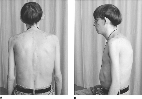
Aris RM, Renner JB, Winders AD, et al. Increased rate of fractures and severe kyphosis: sequelae of living into adulthood with cystic fibrosis. Annals of Internal Medicine. 1998;128:186-193.

Atwood HL, MacKay WA. Essentials of neurophysiology. Toronto: Decker, 1989.

Ayres SM, Kozam RL, Lukas DS. The effects of intermittent positive pressure breathing on intrathoracic pressure, pulmonary mechanics and the work of breathing. American Review of Respiratory Disease. 1963;87:370-379.

Bach JR. Airway secretion management and general pulmonary rehabilitation considerations for patients with neuromuscular ventilatory failure. Journal of Neurological Rehabilitation. 1992;6(2):75-80.

Bach JR. Mechanical insufflation-exsufflation: a comparison of peak expiratory flows with manually assisted coughing techniques. Chest. 1993;104:1553-1562.

Bach JR. Update and perspective on noninvasive respiratory muscle aids. Part 2: The expiratory aids. Chest. 1994;105:1538-1544.

Bach JR. Respiratory muscle aids for the prevention of pulmonary morbidity and mortality. Seminars in Neurology. 1995;15(1):72-83.

Bach JR. Mechanical insufflation/exsufflation: has it come of age? A commentary. European Respiratory Journal. 2003;21:385-386.

Bach JR, Alba AS. Noninvasive options for ventilatory support of the traumatic high level quadriplegic patient. Chest. 1990;98(3):613-619.

Bach JR, Alba AS, Bodofsky E, Curran FJ, Schultheiss M. Glossopharyngeal breathing and noninvasive aids in the management of post-polio respiratory insufficiency. Birth Defects. 1987;23:99-113.

Bach JR, Baird JS, Plosky D, Navado J, Weaver B. Spinal muscular atrophy type 1: management and outcomes. Pediatric Pulmonology. 2002;34:16-22.

Bach JR, Smith WH, Michaels J, et al. Airway secretion clearance by mechanical exsufflation for post-poliomyelitis ventilator assisted individuals. Archives Physical Medical Rehabilitation. 1993;74:170-177.

Bach JR, Vis N, Weaver B. Spinal muscular atrophy Type 1. A non-invasive management approach. Chest. 2000;117:1100-1105.

Bachrach LK, Loutit CW, Moss RB, Marcus R. Osteopenia in adults with cystic fibrosis. American Journal of Medicine. 1994;96:27-34.

Barry PW, Fouroux B, Pedersen S, O’Callaghan C. Nebulizers in childhood. European Respiratory Review. 2000;10(76):527-535.

Baxter GD. Therapeutic lasers: theory and practice. Edinburgh: Churchill Livingstone, 1994.

Baydur A, Gilgoff I, Prentice W, et al. Decline in respiratory function and experience with long-term assisted ventilation in advanced Duchenne’s muscular dystrophy. Chest. 1990;97:884-889.

Bazuaye EA, Stone TN, Corris PA, Gibson GJ. Variability of inspired oxygen concentration with nasal cannulas. Thorax. 1992;47:609-611.

Beck G, Barach A. Value of mechanical aids in the management of a patient with poliomyelitis. Annals of Internal Medicine. 1954;40(6):1081-1094.

Beck GJ, Scarrone LA. Physiological effects of exsufflation with negative pressure (EWNP). Chest. 1956;29:80-95.

Belman MJ. Exercise in patients with chronic obstructive pulmonary disease. Thorax. 1993;48:936-946.

Bereiter M, 2001 Glossopharyngeal breathing in motor neurone disease. Personal communication. Schweizer Paraplegiker Zentrum, Switzerland

Bersten AD. Humidification and inhalation therapy. In: Bersten AD, Soni N, editors. Oh’s intensive care manual. 5th edn. Edinburgh: Butterworth-Heinemann; 2003:321-327.

Bethune D. Neurophysiological facilitation of respiration in the unconscious adult patient. Physiotherapy Canada. 1975;27(5):241-245.

Bethune D. Facilitation of respiration in unconscious adult patients. Respiratory Technology. 1976;12(4):18-21.

Bethune D. Neurophysiological facilitation of respiration. In: Pryor JA, editor. Respiratory care. Edinburgh: Churchill Livingstone, 1991.

Bianchi AL, Pasaro R. Organization of central respiratory neurons. In: Miller AD, Bianchi AL, Bishop BP, editors. Neural control of the respiratory muscles. New York: CRC Press, 1997.

Bianchi C, Grandi M, Felisari G. Efficacy of glossopharyngeal breathing for a ventilator-dependent, high-level tetraplegic patient after cervical cord tumor resection and tracheotomy. American Journal of Physical Medicine and Rehabilitation. 2004;83(3):216-219.

Bickerman H. Exsufflation with negative pressure (EWNP); elimination of radiopaque material and foreign bodies from bronchi of anesthetized dogs. AMA Archives of Internal Medicine. 1954;93:698-704.

Bishop BP. The abdominal muscles. In: Miller AD, Bianchi A, Bishop BP, editors. Neural control of the respiratory muscles. New York: CRC Press, 1997.

Blackwell R. Asthma - Evidence & tradition. Acupuncture Association of Chartered Physiotherapy. Journal (United Kingdom). 2004;July:33-40.

Boots RJ, George N, Faoagali JL, et al. Double-heater-wire circuits and heat-and-moisture exchangers and the risk of ventilator-associated pneumonia. Critical Care Medicine. 2006;34(3):687-693.

  Page 208 

Bott J, Keilty SEJ, Noone L. Intermittent positive pressure breathing - a dying art? Physiotherapy. 1992;78:656-660.

Botton E, Saraux A, Laselve H, Jousse S, Le Goff P. Musculoskeletal manifestations in cystic fibrosis. Joint Bone Spine. 2003;70(5):327-335.

Boyling JD, Palastanga N. Grieve GP, editor. Modern manual therapy of the vertebral column, 2nd edn., Edinburgh: Churchill Livingstone, 1994.

Bradley JM, O’Neill B, 2005 Short-term ambulatory oxygen for chronic obstructive pulmonary disease. Cochrane Database of Systematic Reviews, Issue 4. Art. No.: CD004356 www.cochrane.org/reviews (accessed 5 July 2007)

Braggion C, Cappelletti LM, Cornacchia M, Zanolla L, Mastella G. Short-term effects of three physiotherapy regimens in patients hospitalized for pulmonary exacerbations of cystic fibrosis: a cross-over randomized study. Pediatric Pulmonology. 1995;19:16-22.

Bransen RD, Davis K, Campbell RS, Johnson DJ, Porembka DT. Humidification in the intensive care unit: prospective study of a new protocol utilizing heated humidification and a hygroscopic condenser humidifier. Chest. 1993;104:1800-1805.

Bray C. Thoracic mobilisation in the management of respiratory and cardiac problems. In: The forgotten thoracic spine. Australia: Manipulative Physiotherapists Association of Australia Symposium, University of Sydney; 1994.

Brazier D. Endotracheal suction technique - putting research into practice. Journal of the Association of Chartered Physiotherapists in Respiratory Care. 1999;32:13-17.

British Standards Institution. Specification for gas powered nebulisers for the delivery of drugs. BS7711 Part 3. London: British Standards Institution, 1994.

British Thoracic Society & Scottish Intercollegiate Guidelines Network, 2004 British guideline on the management of asthma www.enterpriseportal2.co.uk/filestore/bts/asthmafull.pdf (accessed 5 July 2007)

Buntain HM, Greer RM, Schluter PJ, et al. Bone mineral density in Australian children, adolescents and adults with cystic fibrosis: a controlled cross sectional study. Thorax. 2004;59:149-155.

Burge PS. Getting the best out of bronchodilator therapy. Patient Management. 1986;July:155-185.

Butler DS. Mobilisation of the nervous system. Melbourne: Churchill Livingstone, 1991.

Campbell AH, O’Connell JM, Wilson F. The effect of chest physiotherapy upon the FEV1 in chronic bronchitis. Medical Journal of Australia. 1975;1:33-35.

Campbell EJ, Baker D, Crites-Silver P. Subjective effects of humidification of oxygen for delivery by nasal cannula. Chest. 1988;93:289-293.

Carr L, Pryor JA, Hodson ME. Self-chest clapping: patients’ views and the effects on oxygen saturation. Physiotherapy. 1995;81:753-757.

Carr L, Smith RE, Pryor JA, Partridge C. Cystic fibrosis patients’ views and beliefs about chest clearance and exercise - a pilot study. Physiotherapy. 1996;82:621-626.

Casaburi R, Patessio A, Loli F, et al. Reductions in exercise lactic acidosis and ventilation as a result of exercise training in patients with obstructive pulmonary disease. American Review of Respiratory Disease. 1991;143:9-18.

Cecins NM, Jenkins SC, Pengelley J, Ryan G. The active cycle of breathing techniques - to tip or not to tip? Respiratory Medicine. 1999;93:660-665.

Cegla UH, Bautz M, Fröde G, Werner Th. Physiotherapie bei patienten mit COAD und tracheobronchialer instabilität-verleich zweier oszillierender PEP-systeme (RC-CORNET®, VRP1 DESITIN). Pneumologie. 1997;51:129-136.

Cegla UH, Jost HJ, Harten A, et al. Course of severe COPD with and without physiotherapy with the RC-Cornet®. Pneumologie. 2002;56(7):418-424.

Chang HK, Weber ME, King M. Mucus transport by high frequency non-symmetrical oscillatory flow. Journal of Applied Physiology. 1988;65:1203-1209.

Chatham K, Ionescus AA, Nixon LS, Shale DJ. A short-term comparison of two methods of sputum expectoration in cystic fibrosis. European Respiratory Journal. 2004;23:435-439.

Chatham K, Marshall C, Campbell IA, Prescott RJ. The Flutter VRPI device in post-thoracotomy patients. Physiotherapy. 1993;79:95-98.

Chatwin M, O’ Driscoll D, Corfield D, Morrell M, Simonds A. Controlled trial of intrapulmonary percussion in adults and children with stable severe neuromuscular disease. American Journal of Critical Care Medicine. 2004;169:A438.

Chatwin M, Ross E, Hart N, et al. Cough augmentation with mechanical insufflation/exsufflation in patients with neuromuscular weakness. European Respiratory Journal. 2003;21:502-508.

Chevaillier J, 2002 Autogenic drainage (AD). In: Physiotherapy in the treatment of cystic fibrosis (CF), 3rd edn. International Physiotherapy Group for Cystic Fibrosis (IPG/CF), pp 12-15 www.cfww.org/IPG-CF/index.asp (accessed 5 July 2007)

Chiappetta A, Beckerman R. High frequency chest wall oscillation in spinal muscular atrophy (SMA). RT Journal for Respiratory Care Practitioners. 1995;8(4):112-114.

Chiumello D, Pelosi P, Gattinoni L. Conditioning of inspired gases in mechanically ventilated patients. In: Vincent J-L, editor. Intensive Care Medicine. Springer, 2002. (ISBN 0387916253)

Chuter TAM, Weissman C, Starker PM, Gump FE. Effect of incentive spirometry on diaphragmatic function after surgery. Surgery. 1989;105:488-493.

Chuter TAM, Weissman C, Mathews DM, Starker PM. Diaphragmatic breathing maneuvers and movement of the diaphragm after cholecystectomy. Chest. 1990;97:1110-1114.

Clarke SW. Inhaler therapy. Quarterly Journal of Medicine. 1988;67(253):355-368.

Clarke SW. Physical defences. In Brewis RAL, Corrin B, Geddes DM, Gibson GJ, editors: Respiratory medicine, 2nd edn., London: WB Saunders, 1995. 1

Clini E, Antoni F, Vitacca M, et al. Intrapulmonary percussive ventilation in tracheostomized patients: a randomized controlled trial. Intensive Care Medicine. 2006;32:1994-2001.

Cole P. Bronchiectasis. In Brewis RAL, Corrin B, Geddes DM, Gibson GJ, editors: Respiratory medicine, 2nd edn., London: WB Saunders, 1995. 2

Collis GG, Cole CH, Le Souëf PN. Dilution of nebulised aerosols by air entrainment in children. Lancet. 1990;336:341-343.

Conway JH. The effects of humidification for patients with chronic airways disease. Physiotherapy. 1992;78:97-101.

Conway JH, Fleming JS, Perring S, Holgate ST. Humidification as an adjunct to chest physiotherapy in aiding tracheo-bronchial clearance in patients with bronchiectasis. Respiratory Medicine. 1992;86:109-114.

Coombs HC. The relation of the dorsal roots of the spinal nerves and the mesencephalon to the control of respiratory movements. American Journal of Physiology. 1918;46:459-471.

  Page 209 

Coombs HC, Pike FH. The nervous control of respiration in kittens. American Journal of Physiology. 1930;95:681-693.

Cornacchia M, Zenorini A, Perobelli S, et al. Prevalence of urinary incontinence in women with cystic fibrosis. British Journal Urology International. 2001;88:44-48.

Cunningham S, Prasad SA, Collyer L, et al. Bronchoconstriction following nebulised Colistin in cystic fibrosis. Archives of Disease in Childhood. 2001;84(5):432-433.

Currie DC, Munro C, Gaskell D, Cole PJ. Practice, problems and compliance with postural drainage: a survey of chronic sputum producers. British Journal of Diseases of the Chest. 1986;80:249-253.

Dab I, Alexander F. The mechanism of autogenic drainage studied with flow volume curves. Monographs of Paediatrics. 1979;10:50-53.

Dail CW. ‘Glossopharyngeal breathing’ by paralyzed patients. California Medicine. 1951;75:217-218.

Dail CW, Affeldt JE, Collier CR. Clinical aspects of glossopharyngeal breathing. Journal of the American Medical Association. 1955;158:445-449.

Darbee JC, Kanga JF, Ohtake PJ. Physiologic evidence for high-frequency chest wall oscillation and positive expiratory pressure breathing in hospitalized subjects with cystic fibrosis. Physical Therapy. 2005;85(12):1278-1289.

Darbee JC, Ohtake PJ, Grant BJ, Cerny FJ. Physiologic evidence for the efficacy of positive expiratory pressure as an airway clearance technique in patients with cystic fibrosis. Physical Therapy. 2004;84(6):524-537.

David A. Autogenic drainage - the German approach. In: Pryor JA, editor. Respiratory care. Edinburgh: Churchill Livingstone; 1991:65-78.

Davidson AGF, Wong LTK, Pirie GE, McIlwaine PM. Long-term comparative trial of conventional percussion and drainage physiotherapy versus autogenic drainage in cystic fibrosis. Pediatric Pulmonology (Suppl). 1992;8:298.

Dawson SV, Elliott EA. Wave-speed limitation on expiratory flow - a unifying concept. Journal of Applied Physiology. 1977;43(3):498-515.

Dean E. Effect of body position on pulmonary function. Physical Therapy. 1985;65:613-618.

Denehy L, Berney S. The use of positive pressure devices by physiotherapists. European Respiratory Journal. 2001;17:821-829.

De Troyer A. Mechanics of the chest wall muscles. In: Miller AD, Bianchi AL, Bishop BP, editors. Neural control of the respiratory muscles. New York: CRC Press, 1997.

DeVilbiss Oxygen Services. Manufacturer’s guidelines for oxygen concentrators. West Midlands DY8 4PS, England: Sunrise Medical Ltd, 2000.

Dhand R. Nebulizers that use a vibrating mesh or plate with multiple apertures to generate aerosol. Respiratory Care. 2002;47(12):1406-1416.

Djedaini K, Billiard M, Mier L, et al. Changing heat and moisture exchangers every 48 hours rather than 24 hours does not affect their efficacy and the incidence of nosocomial pneumonia. American Journal of Respiratory and Critical Care Medicine. 1995;152:1562-1569.

Dodd ME, Abbott J, Maddison J, Moorcroft AJ, Webb AK. Effect of tonicity of nebulised colistin on chest tightness and pulmonary function in adults with cystic fibrosis. Thorax. 1997;52:656-658.

Dodd ME, Hanley SP, Johnson SC, Webb AK. District nebuliser compressor service: reliability and costs. Thorax. 1995;50:82-84.

Dodd ME, Kellet F, Davis A, et al. Audit of oxygen prescribing before and after the introduction of a prescription chart. British Medical Journal. 2000;321:864-865.

Duron B, Bars P. Effect of thoracic cutaneous nerve stimulations on the activity of the intercostal muscles and motoneurons of the cat. In: Euler C, Lagercrantz A, editors. Neurobiology of the control of breathing (Nobel conference series). New York: Raven Press, 1986.

Duron B, Rose D. The intercostal muscles. In: Miller AD, Bianchi AL, Bishop BP, editors. Neural control of the respiratory muscles. New York: CRC Press, 1997.

Eaton T, Young P, Zeng I, Kolbe J. A randomized evaluation of the acute efficacy, acceptability and tolerability of flutter and active cycle of breathing with and without postural drainage in non-cystic fibrosis bronchiectasis. Chronic Respiratory Disease. 2007;4:23-40.

Electromed, 2006 SmartVest® Airway Clearance System: prescribing protocols. Electromed, Minnesota, USA. www.electromed-usa.com (accessed 5 July 2007)

Elkins MR, Jones A, van der Schans C, 2006a Positive expiratory pressure physiotherapy for airway clearance in people with cystic fibrosis. Cochrane Database of Systematic Reviews Issue 2. Art. No.: CD003147

Elkins MR, Lane T, Goldberg H, et al. Effect of airway clearance techniques on the efficacy of the sputum induction procedure. European Respiratory Journal. 2005;26:904-908.

Elkins MR, Robinson M, Rose BR, et al. National Hypertonic Saline in Cystic Fibrosis (NHSCF) Study Group. A controlled trial of long-term inhaled hypertonic saline in patients with cystic fibrosis. New England Journal of Medicine. 2006;354(3):229-240.

Eng PA, Morton J, Douglass JA, et al. Short-term efficacy of ultrasonically nebulized hypertonic saline in cystic fibrosis. Pediatric Pulmonology. 1996;21:77-83.

Enright S, Chatham K, Ionescu AA, Unnithan VB, Shale DJ. Inspiratory muscle training improves lung function and exercise capacity in adults with cystic fibrosis. Chest. 2004;126:406-411.

Enright SJ, Unnithan VB, Heward C, Withnall L, Davies DH. Effect of high-intensity inspiratory muscle training on lung volumes, diaphragm thickness, and exercise capacity in subjects who are healthy. Physical Therapy. 2006;86(3):345-354.

Euler C. Breathing behavior. In: Euler C, Lagercrantz A, editors. Neurobiology of the control of breathing (Nobel conference series). New York: Raven Press, 1986.

European Respiratory Society. European Respiratory Society nebulizer guidelines: clinical aspects. European Respiratory Review. 10(76), 2000.

European Respiratory Society Task Force. Standardised methodology of sputum induction and processing. European Respiratory Journal. 2002;20(Supplement):37.

Everard ML, Evans M, Milner AD. Is tapping jet nebulisers worthwhile? Archives of Disease in Childhood. 1994;70:538-539.

Ewart W. The treatment of bronchiectasis and of chronic bronchial affections by posture and by respiratory exercises. Lancet. 1901;2:70-72.

Ezzo J. Is acupuncture effective for the treatment of chronic pain? A systematic review. Pain. 2000;86:217-225.

Falk M, Andersen JB. Positive expiratory pressure (PEP) mask. In: Pryor JA, editor. Respiratory care. Edinburgh: Churchill Livingstone; 1991:51-63.

Falk M, Kelstrup M, Andersen JB, Kinoshita T, Falk P, Støvring S, Gøthgen I. Improving the ketchup bottle method with positive expiratory pressure, PEP, in cystic fibrosis. European Journal of Respiratory Diseases. 1984;65:423-432.

  Page 210 

Feng W, Deng WW, Huang SG, et al. Short-term efficacy of RC-Cornet in improving pulmonary function and decreasing cohesiveness of sputum in bronchiectasis patients. Chest. 1998;114(4):320S. Suppl

Festini F, Ballarin S, Codamo T, Doro R, Loganes C. Prevalence of pain in adults with cystic fibrosis. J Cyst Fibrosis Mar;. 2004;3(1):51-57.

Fink JB, Mahlmeister MJ. High-frequency oscillation of the airway and chest wall. Respiratory Care. 2002;47:797-807.

Fleck S. Detraining: its effects on endurance and strength. National Strength and Conditioning Journal. 1994;2:22-28.

Flower KA, Eden RI, Lomax L, Mann NM, Burgess J. New mechanical aid to physiotherapy in cystic fibrosis. British Medical Journal. 1979;2:630-631.

Fok J, Brown NE, Zuberbuhler P, et al. Low bone mineral density in cystic fibrosis patients. Canadian Journal of Dietetic Practice and Research. 2002;63:192-197.

Frazier DT, Xu Fadi, Lee L-Y. Respiratory-related reflexes and the cerebellum. In: Miller AD, Bianchi AL, Bishop BP, editors. Neural control of the respiratory muscles. New York: CRC Press, 1997.

Freitag L, Bremme J, Schroer M. High frequency oscillation for respiratory physiotherapy. British Journal of Anaesthesia. 1989;63:44S-46S.

Gallon A. Evaluation of chest percussion in the treatment of patients with copious sputum production. Respiratory Medicine. 1991;85:45-51.

Geddes EL, Reid DW, Crowe J, O’Brian K, Brooks D. Inspiratory muscle training in adults with chronic obstructive pulmonary disease: a systematic review. Respiratory Medicine. 2005;99:1440-1458.

Gleeson JG, Price JF. Nebuliser technique. British Journal of Diseases of the Chest. 1988;82:172-174.

Goldman M. Mechanical coupling of the diaphragm and the rib cage. In: Pengelly LD, Rebuck AS, Campbell EJM, editors. Loaded breathing. Proceedings of an international symposium, ‘The effects of mechanical loads on breathing’. Don Mills, Ontario: Longman Canada, 1974.

Gordon J. Spinal mechanisms of motor coordination. In: Kandel ER, Schwartz JH, Jessel JM, editors. Principles of neural science. Connecticut: Appleton & Lange, 1991.

Gormezano J, Branthwaite MA. Pulmonary physiotherapy with assisted ventilation. Anaesthesia. 1972;27:249-257.

Gosselink R. Breathing techniques in patients with chronic obstructive pulmonary disease (COPD). Chronic Respiratory Disease. 2004;1:163-172.

Gosselink R, Schrever K, Cops P, et al. Incentive spirometry does not enhance recovery after thoracic surgery. Critical Care Medicine. 2000;28(3):679-683.

Gosselink RA, Wagenaar RC, Rijswijk H, Sargeant AJ, Decramer ML. Diaphragmatic breathing reduces efficiency of breathing in patients with chronic obstructive pulmonary disease. American Journal of Respiratory and Critical Care Medicine. 1995;151(4):1136-1142.

Grassino A. Influence of chest wall configuration on the static and dynamic characteristics of the contracting diaphragm. In: Pengelly LD, Rebuck AS, Campbell EJM, editors. Loaded breathing. Proceedings of an international symposium, ‘The effects of mechanical loads on breathing’. Don Mills, Ontario: Longman Canada, 1974.

Gray’s Anatomy, 1973. Longman, Edinburgh

Green M, Moxham J. The respiratory muscles. Clinical Science. 1985;68:1-10.

Gregersen GG, Lucas DL. An in vivo study of the axial rotation of the human thoraco-lumbar spine. Journal of Bone and Joint Surgery. 1967;49A:247-262.

Gross D, Zidulka A, O’Brien C, et al. Peripheral mucociliary clearance with high frequency chest wall compression. Journal of Applied Physiology. 1985;58:1157-1163.

Gumery L, Lee J, Whitehouse J, Honeybourne D. The prevalence of urinary incontinence in adult cystic fibrosis males. Journal of Cystic Fibrosis. 2005;4(Suppl 1):S97.

Gunawardena KA, Patel B, Campbell IA, Macdonald JB, Smith AP. Oxygen as a driving gas for nebulisers: safe or dangerous? British Medical Journal. 1984;288:272-274.

Halvorsen TB, Anda SS, Naess AB, Levang OW. Fatal cardiac tamponade after acupuncture through congenital sternal foramen. Lancet. 1995;345:1175.

Hansen LG, Warwick W. High-frequency chest compression system to aid in clearance of mucus from the lung. Biomedical Instrumentation and Technology. 1990;24:289-294.

Hansen LG, Warwick WJ, Hansen KL. Mucus transport mechanisms in relation to the effect of high frequency chest compression (HFCC) on mucus clearance. Pediatric Pulmonology. 1994;17:113-118.

Hardy KA, Bach JR, Stoller JK, et al. A review of airway clearance: new techniques, indications, and recommendations. Respiratory Care. 1994;39(5):440-445.

Harrison DA, Breen DP, Harris ND, Gerrish SP. The performance of two intensive care humidifiers at high gas flows. Anaesthesia. 1993;48:902-905.

Hasani A, Pavia D, Agnew JE, Clarke SW. Regional lung clearance during cough and forced expiration technique (FET): effects of flow and viscoelasticity. Thorax. 1994;49:557-561.

Henderson RC, Madsen CD. Bone density in children and adolescents with cystic fibrosis. Journal of Pediatrics. 1996;128:28-34.

Henderson RC, Specter BB. Kyphosis and fractures in children and young adults with cystic fibrosis. Journal of Paediatrics. 1994;125:208-212.

Hilaire G, Monteau R. Brainstem and spinal control of respiratory muscles during breathing. In: Miller AD, Bianchi AL, Bishop BP, editors. Neural control of the respiratory muscles. New York: CRC Press, 1997.

Hill K, Eastwood PR. Respiratory muscle training: the con argument. Chronic Respiratory Disease. 2005;2(4):223-224.

Hill K, Jenkins SC, Hillman Eastwood PR. Dyspnoea in COPD: can inspiratory muscle training help? Australian Journal Physiotherapy. 2004;50:169-180.

Hill SL, Barnes PK, Hollway T, Tennant R. Fixed performance oxygen masks: an evaluation. British Medical Journal. 1984;288:1261-1263.

Hill-Rom, 2006 The Vest TM Airway Clearance System: information for physicians. Hill-Rom, Minnesota, USA. www.hill-rom.com (accessed 5 July 2007)

Hinds CJ, Watson D. Intensive care, 2nd edn., London: WB Saunders; 1996:33. 175

Hofmeyr JL, Webber BA, Hodson ME. Evaluation of positive expiratory pressure as an adjunct to chest physiotherapy in the treatment of cystic fibrosis. Thorax. 1986;41:951-954.

Holland AE, Button BM. Is there a role for airway clearance techniques in chronic obstructive pulmonary disease? Chronic Respiratory Disease. 2006;3:83-91.

Homnick DN, Anderson K, Marks JH. Comparison of the flutter device to standard chest physiotherapy in hospitalized patients with cystic fibrosis: a pilot study. Chest. 1998;114(4):993-997.

  Page 211 

Hopwood V. Acupuncture in physiotherapy. Complementary Therapies in Medicine. 1993;1:100-104.

Hsiao SF, Wu YT, Wu HD, Wang TG. Comparison of the effectivness of pressure threshold and targeted resistive muscle training in patients with chronic obstructive pulmonary disease. Journal of the Formosan Medical Association. 2003;102:204-205.

Hutchinson GR, Parker S, Pryor JA, et al. Home-use nebulizers: a potential source of Burkholderia cepacia and other colistin-resistant, gram-negative bacteria in patients with cystic fibrosis. Journal of Clinical Microbiology. 1996;34:584-587.

Institute for Work & Health, 1996 DASH Index. The American Academy of Orthopaedic Surgeons (AAOS) www.iwh.on.ca (accessed 5 July 2007)

Jacob W. Physiotherapy in the ICU. In: Oh TE, editor. Intensive care manual. 3rd edn. Sydney: Butterworths; 1990:24. Chapter 4

Janda V. Muscles and motor control in cervicogenic disorders: assessment and management physical therapy for the cervical and thoracic spine. In Grant R, editor: Clinics in physical therapy, 2nd edn., New York: Churchill Livingstone, 1994.

Jean A, Car A, Kessler JP. Brainstem organization of swallowing and its interaction with respiration. In: Miller AD, Bianchi AL, Bishop BP, editors. Neural control of the respiratory muscles. New York: CRC Press, 1997.

Jeffrey AA, Warren PM. Should we judge a mask by its cover? Thorax. 1992;47:543-546.

Jenkins SC, Soutar SA, Moxham J. The effects of posture on lung volumes in normal subjects and in patients pre- and post-coronary artery surgery. Physiotherapy. 1988;74:492-496.

Jobst KA. A critical analysis of acupuncture in pulmonary disease: efficacy and safety of acupuncture needle. Journal of Alternative and Complementary Medicine. 1995;1:54-85.

Jones PW. Interpreting thresholds for a clinically significant change in health status in asthma and COPD. European Respiratory Journal. 2002;19:398-404.

Jull G, Trott P, Potter H, et al. A randomized controlled trial of exercise and manipulative therapy for cervicogenic headache. Spine. 2002;27(17):1835-1843.

Kelleher WH, Parida RK. Glossopharyngeal breathing. British Medical Journal. 1957;2:740-743.

Kendrick AH, Smith EC, Denyer J. Nebulizers - fill volume, residual volume and matching of nebulizer to compressor. Respiratory Medicine. 1995;89:157-159.

Kennedy WL. ACSM’s Guidelines for Exercise Testing and Prescription. Media, PA: Williams & Willsins, 1995.

Kenyon JN. Modern techniques of acupuncture; Vol 1: A practical scientific guide to electro-acupuncture. Wellingborough: Thorsons, 1983.

Kenyon JN. Acupressure techniques. Canada: Healing Arts Press, 1988.

Kieselmann R, 2002 Modified AD. In: Physiotherapy in the treatment of cystic fibrosis (CF), 3rd edn. Inter-national Physiotherapy Group for Cystic Fibrosis (IPG/CF), pp 16-17 www.cfww.org/IPG-CF/index.asp (accessed 5 July 2007)

King M, Phillips DM, Gross D, et al. Enhanced tracheal mucus clearance with high frequency chest wall compression. American Review of Respiratory Disease. 1983;128:511-515.

King M, Phillips DM, Zidulka A, Chang HK. Tracheal mucus clearance in high-frequency oscillation II. Chest wall versus mouth oscillation. American Review of Respiratory Disease. 1984;130:703-706.

King M, Zidulka A, Phillips DM, et al. Tracheal mucus clearance in high frequency oscillation Effect of peak flow rate bias. European Respiratory Journal. 1990;3:6-13.

Kishi H, Mishima HK, Sakamoto I, Yamashita U. Stimulation of retinal pigment epithelial cell growth neuropeptides in vitro. Current Eye Research. 1996;15:708-713.

Kluft J, Beker L, Castagnino M, et al. A comparison of bronchial drainage treatments in cystic fibrosis. Pediatric Pulmonology. 1996;22:271-274.

Koepchen HP, Abel H-H, Klussendorf D, Lazar H. Respiratory and cardiovascular rhythmicity. In: Euler C, Lagercrantz A, editors. Neurobiology of the control of breathing (Nobel conference series). New York: Raven Press, 1986.

Koh JL, Harrison D, Palermo TM, Turner H, McGraw T. Assessment of acute and chronic pain symptoms in children with cystic fibrosis. Pediatric Pulmonology. 2005;40(4):330-335.

Komi PV, Hakkinen K. Strength and power. In: Dirix A, Knuttgen HG, Tittel K, editors. The Olympic book of sports medicine. Oxford: Blackwell; 1991:181-193.

Konstan MW, Stern RC, Doershuk CF. Efficacy of the Flutter device for airway mucus clearance in patients with cystic fibrosis. Journal of Pediatrics. 1994;124:689-693.

Kraemer W, Adams K, Cararelli E, et al. American College of Sports Medicine position stand. Progressive models in resistance training for healthy adults. Medicine and Science in Sports and Exercise. 2002;34:364-380.

Laghi F, Tobin MJ. Disorders of the respiratory muscles. American Journal of Respiratory and Critical Care Medicine. 2003;168:10-48.

Langlands J. The dynamics of cough in health and in chronic bronchitis. Thorax. 1967;22:88-96.

Lannefors L. Inhalation therapy: practical considerations for nebulisation therapy. Physical Therapy Reviews. 2006;11:21-27.

Lannefors L, Wollmer P. Mucus clearance with three chest physiotherapy regimes in cystic fibrosis: a comparison between postural drainage, PEP and physical exercise. European Respiratory Journal. 1992;5(6):748-753.

Lapin CD. Airway physiology, autogenic drainage, and active cycle of breathing. Respiratory Care. 2002;47(7):778-785.

Lawrence RM, Rosch PJ, Plowden J. Magnet therapy: the pain cure alternative. California: Prima Publishing, 1998.

Lawrence VA, Cornell JE, Smetana GW. Strategies to reduce postoperative pulmonary complications after noncardiothoracic surgery: systematic review for the American College of Physicians. Annals of Internal Medicine. 2006;144(8):596-608.

Lee D. The thorax: an integrated approach, 2nd edn., Minneapolis: Orthopaedic Physical Therapy, 2003.

Leith DE, Bradley M. Ventilatory muscle strength and endurance training. Journal of Applied Physiology. 1976;41:508-516.

Lewis RA, Fleming JS. Fractional deposition from a jet nebuliser: how it differs from a metered dose inhaler. British Journal of Diseases of the Chest. 1985;79:361-367.

Lichstein E, Chadda KD, Naik D, Gupta PK. Diagonal ear lobe crease: relevance and implications as a coronary risk factor. New England Journal of Medicine. 1974;290:615-616.

Linde D, Jobst K, Panton J. Controlled trial of acupuncture for disabling breathlessness. Lancet. 1986;2:1417-1419.

  Page 212 

Lindemann H. The value of physical therapy with VRP1 Desitin (‘Flutter’). Pneumologie. 1992;46(12):626-630.

Lock SH, Blower G, Prynne M, Wedzicha JA. Comparison of liquid and gaseous oxygen for domiciliary portable use. Thorax. 1992;47:98-100.

Lopez-Vidriero MT, Reid L. Bronchial mucus in health and disease. British Medical Bulletin. 1978;34(1):63-74.

Lotters F, van Tol B, Kwakkel G, Gosselink R. Effect of controlled inspiratory muscle training in patients with COPD: a meta-analysis. European Respiratory Journal. 2002;20:570-576.

Lund I, Lundberg T, Carleson J, et al. Are minimal, superficial or sham acupuncture procedures acceptable as inert placebo controls. Acupuncture in Medicine. 2006;24(1):13-15.

Lunteren E, Dick TE. Muscles of the upper airway and accessory respiratory muscles. In: Miller AD, Bianchi AL, Bishop BP, editors. Neural control of the respiratory muscles. New York: CRC Press, 1997.

Maa SH, Gauthier D, Turner M. Acupressure as an adjunct to a pulmonary rehabilitation program. J Cardiopulmonary Rehabilitation. 1997;17(4):268-276.

Macklem PT. Physiology of cough. Transactions of the American Broncho-Esophalogical Association. 1974:150-157.

Maddison J, Dodd M, Webb AK. Nebulised colistin causes chest tightness in adults with cystic fibrosis. Respiratory Medicine. 1994;88(2):145-147.

Main E, Prasad A, van der Schans C, 2005 Conventional chest physiotherapy compared to other airway clearance techniques for cystic fibrosis. Cochrane Database of Systematic Reviews, Issue 1. Art. No.: CD002011

Maltais F, Le Blanc P, Jobin J, Caraburi R. Peripheral muscle dysfunction in chronic obstructive pulmonary disease. Clinics in Chest Medicine. 2000;21:665-677.

Make BJ, Hill NS, Goldberg AL, et al. Mechanical ventilation beyond the intensive care unit. Report of a consensus conference of the American College of Chest Physicians. Chest. 1998;113(Suppl 5):289S-344S.

Mashanskii VF, Markov IuV, Shpunt VKh, Li SE, Mirkin AS. Topography of the gap junctions in human skin and their possible role in the non-neural transmission of information [Article in Russian]. Arkhiv Anatomii, Gistologii i Ébriologii. 1983;84(3):53-60.

Massery M. Musculoskeletal and neuromuscular interventions: a physical approach to cystic fibrosis. Journal of the Royal Society of Medicine. 2005;98(Suppl 45):55-66.

Massie RJ, Towns SJ, Bernard E, et al. The musculoskeletal complications of cystic fibrosis. Journal of Paediatrics and Child Health. 1998;34(5):467-470.

McArdle WD, Katch FI, Katch VL. Exercise physiology, energy, nutrition, and human performance, 5th edn., Williams & Wilkins, Philadelphia: Lippincott, 2001.

McCarren B, Alison JA. Physiological effects of vibration in subjects with cystic fibrosis. European Respiratory Journal. 2006;27(6):1204-1209.

McCool FD. Inspiratory muscle weakness and fatigue. RT: The Journal for Respiratory Care Practitioners. 1992;5(6):32-41.

McDonnell T, McNicholas WT, FitzGerald MX. Hypoxaemia during chest physiotherapy in patients with cystic fibrosis. Irish Journal of Medical Science. 1986;155:345-348.

McGuire S, 2006 The I-neb TM adaptive aerosol delivery (AAD®) system - a holistic approach to inhaled drug delivery including regulatory approval. Drug Delivery Report Spring/Summer 68-71

McIlwaine PM, Wong LT, Peacock D, Davidson AG. Long-term comparative trial of conventional postural drainage and percussion versus positive expiratory pressure physiotherapy in the treatment of cystic fibrosis. Journal of Pediatrics. 1997;131(4):570-574.

McIlwaine PM, Wong LT, Peacock D, Davidson AG. Long-term comparative trial of positive expiratory pressure versus oscillating positive expiratory pressure (flutter) physiotherapy in the treatment of cystic fibrosis. Journal of Pediatrics. 2001;138(6):845-850.

McKenzie DK, Frith PA, Burdon JGW, Town GI. The COPDX Plan: Australia and New Zealand Guidelines for the management of COPD. Medical Journal Australia. 2003;178(6 Suppl):S1-s40.

Mead J, Takishima T, Leith D. Stress distribution in lungs: a model of pulmonary elasticity. Journal of Applied Physiology. 1970;28:596-608.

Mead J, Turner JM, Macklem PT, Little JB. Significance of the relationship between lung recoil and maximum expiratory flow. Journal of Applied Physiology. 1967;22:95-108.

Medic-Aid Ltd. Nebulizer therapy training pack. Bognor Regis, W Sussex, UK: Medic-Aid Ltd, Heath Place, 1996.

Medical Research Council Working Party. Long-term domiciliary oxygen therapy in chronic hypoxic cor pulmonale complicating chronic bronchitis and emphysema. Lancet. 1981;1:681-686.

Menkes HA, Traystman RJ. Collateral ventilation. American Review of Respiratory Disease. 1977;116:287-309.

Milla CE, Hansen LG, Warwick W. Different frequencies should be prescribed for different high frequency chest compression machines. Biomedical Instrumentation & Technology. 2006;40:319-324.

Miller AD, Bianchi AL, Bishop BP. Overview of the neural control of the respiratory muscles. In: Miller AD, Bianchi AL, Bishop BP, editors. Neural control of the respiratory muscles. New York: CRC Press, 1997.

Miller JM, Ashton-Miller JA, DeLancey JO. A pelvic muscle precontraction can reduce cough-related urine loss in selected women with mild SUI. Journal of the American Geriatrics Society. 1998;46(7):870-874.

Miller MR. Peak expiratory flow meters. European Respiratory Society: The Buyer’s Guide. 2000;3:12-14.

Miller S, Hall DO, Clayton CB, Nelson R. Chest physiotherapy in cystic fibrosis: a comparative study of autogenic drainage and the active cycle of breathing techniques with postural drainage. Thorax. 1995;50:165-169.

Miske LJ, Hickey EM, Kolb SM, Weiner DJ, Panitch HB. Use of the mechanical in-exsufflator in pediatric patients with neuromuscular disease and impaired cough. Chest. 2004;125:1406-1412.

Moraes T, Carpenter S, Taylor L. Cystic fibrosis incontinence in children. Pediatric Pulmonology Supplement. 2002;24:315.

Morgan MDL, Silver JR, Williams SJ. The respiratory system of the spinal cord patient. In: Bloch RF, Basbaum M, editors. Management of spinal cord injuries. Baltimore: Williams and Wilkins; 1986:78-115.

Mortensen J, Falk M, Groth S, Jensen C. The effects of postural drainage and positive expiratory pressure physiotherapy on tracheobronchial clearance in cystic fibrosis. Chest. 1991;100(5):1350-1357.

Mustfa N, Aiello M, Lyall RA, et al. Cough augmentation in amyotrophic lateral sclerosis. Neurology. 2003;61:1285-1287.

  Page 213 

Nakamura S, Mikami M, Kawakami M, et al. Comparative evaluation of the flutter and the cornet in improving the cohesiveness of sputum from patients with bronchiectasis. European Respiratory Journal. 1998;12(Suppl 28):212s-213s.

National Institute for Health and Clinical Excellence, 2004 Chronic obstructive pulmonary disease - management of chronic obstructive pulmonary disease in adults in primary and secondary care. Developed by the National Collaborating Centre for Chronic Conditions. NICE clinical guideline 12. www.nice.org.uk/CG012NICEguideline (accessed 5 July 2007)

National Institute for Health and Clinical Excellence, 2006 Urinary incontinence. The management of urinary incontinence in women. Developed by the National Collaborating Centre of Women’s and Children’s Health. NICE clinical guideline 40. www.nice.org.uk/CG040NICEguideline (accessed 5 July 2007)

Nathan H. Osteophytes of the vertebral column. An anatomical study of their development according to age, race and sex with considerations as to their aetiology and significance. Journal of Bone and Joint Surgery. 1962;44:A243.

Nava S, Barbarito N, Piaggi G, De Mattia E, Cirio S. Physiological response to intrapulmonary percussive ventilation in stable COPD patients. Respiratory Medicine. 2006;100:1526-1533.

Nebuliser Project Group of the British Thoracic Society Standards of Care Committee. Current best practice for nebuliser treatment. Thorax. 1997;52(Suppl 2):S1-S106.

Nelson HP. Postural drainage of the lungs. British Medical Journal. 1934;2:251-255.

Neumeister W, Kuhlemann H, Bauer T, et al. Effect of acupuncture on quality of life, mouth occlusion pressures and lung function in COPD. Medizinische Klinik. 1999;94:106-109.

Newhouse PA, White F, Marks JH, Homnick DN. The intrapulmonary percussive ventilator and flutter device compared to standard chest physiotherapy in patients with cystic fibrosis. Clinical Pediatrics. 1998;37:427-432.

Newman SP, Pellow PGD, Clarke SW. Droplet size distributions of nebulised aerosols for inhalation therapy. Clinical Physics and Physiological Measurement. 1986;7:139-146.

Newman SP, Weisz AWB, Talaee N, Clarke SW. Improvement of drug delivery with a breath actuated pressurised aerosol for patients with poor inhaler technique. Thorax. 1991;46:712-716.

Niere K, Jerak A. Measurement of headache frequency, intensity and duration: comparison of patient report by questionnaire and headache diary. Physiotherapy Research International. 2004;9(4):149-156.

Nocturnal Oxygen Therapy Trial Group. Continuous or nocturnal oxygen therapy in hypoxemic chronic obstructive lung disease: a clinical trial. Annals of Internal Medicine. 1980;93:391-398.

Nogier PMF. Handbook to auriculotherapy. France: Maisonneuve SA, 1981.

Oberwaldner B, Evans JC, Zach MS. Forced expirations against a variable resistance: a new chest physiotherapy method in cystic fibrosis. Pediatric Pulmonology. 1986;2:358-367.

Oberwaldner B, Theissl B, Rucker A, Zach MS. Chest physiotherapy in hospitalized patients with cystic fibrosis: a study of lung function effects and sputum production. European Respiratory Journal. 1991;4:152-158.

O’Donnell DE, McGuire M, Samis L, Webb KA. The impact of exercise reconditioning on breathlessness in severe chronic airflow limitation. American Journal of Respiratory and Critical Care Medicine. 1995;152:2005-2013.

O’Driscoll BR, Kay EA, Taylor RJ, Bernstein A. Home nebulizers: can optimal therapy be predicted by laboratory studies? Respiratory Medicine. 1990;84:471-477.

Oermann CM, Sockrider MM, Giles D, et al. Comparison of high-frequency chest wall oscillation and oscillating positive expiratory pressure in the home management of cystic fibrosis: a pilot study. Pediatric Pulmonology. 2001;32:372-377.

O’Leary S, Jull G, Kim M, Vicenzino B. Cranio-cervical flexor muscle impairment at maximal, moderate, and low loads is a feature of neck pain. Manual Therapy. 2007;12(1):34-39.

Oleson T, 1998 Auriculotherapy manual. ISBN 0-9629-415-5-7, p 7

Olsen MF, Lonroth H, Bake B. Effects of breathing exercises on breathing patterns in obese and non-obese subjects. Clinical Physiology. 1999;19(3):251-257.

O’Neill B, Dodd ME. Oxygen on the move: practical considerations for physiotherapists. Physical Therapy Reviews. 2006;11:28-36.

O’Neill SO, McCarthy DS. Postural relief of dyspnoea in severe chronic airflow limitation: relationship to respiratory muscle strength. Thorax. 1983;38:595-600.

Orr A, McVean RJ, Web AK, Dodd ME. Questionnaire survey of urinary incontinence in women with cystic fibrosis. British Medical Journal. 2001;322:1521.

Overend TJ, Anderson CM, Lucy SD, et al. The effect of incentive spirometry on postoperative pulmonary complications. Chest. 2001;120:971-978.

Pakala R, Benedict CR. Effect of serotonin and thromboxane A2 on endothelial cell proliferation: effect of specific receptor antagonists. Journal of Laboratory and Clinical Medicine. 1998;131(6):527-537.

Parasa RB, Maffulli N. Musculoskeletal involvement in cystic fibrosis. Bulletin of the Hospital for Joint Diseases. 1999;58(1):37-44.

Partridge C, Pryor J, Webber B. Characteristics of the forced expiration technique. Physiotherapy. 1989;75:193-194.

Patterson JE, Bradley JM, Elborn JS. Airway clearance in bronchiectasis: a randomized crossover trial of active cycle of breathing techniques (incorporating postural drainage and vibration) versus test of incremental respiratory endurance. Chronic Respiratory Disease. 2004;1:127-130.

Patterson JE, Bradley JM, Hewitt O, et al. Airway clearance in bronchiectasis: a randomized crossover trial of active cycle of breathing techniques versus Acapella. Respiration. 2005;72(3):239-242.

Pavia D, Webber B, Agnew JE, et al. The role of intermittent positive pressure breathing (IPPB) in bronchial toilet. European Respiratory Journal. 1988;1(Suppl 2):250S.

Peiper A. Cerebral function in infancy and childhood. New York: Consultants Bureau, 1963.

Pelosi P, Solca M, Ravagnan I, et al. Effects of heat and moisture exchangers on minute ventilation, ventilatory drive, and work of breathing during pressure-support ventilation in acute respiratory failure. Critical Care Medicine. 1996;24(7):1184-1188.

Pendleton N, Cheesbrough JS, Walshaw MJ, Hind CRK. Bacterial colonisation of humidifier attachments on oxygen concentrators prescribed for long-term oxygen therapy: a district review. Thorax. 1991;46:257-258.

Perry RJ, Man CGW, Jones RL. Effects of positive end-expiratory pressure on oscillated flow rate during high frequency chest compression. Chest. 1998;113:1028-1033.

  Page 214 

Pfeiffer KA, Pivarnik JM, Womack CJ, Reeves MJ, Malina RM. Reliability and validity of the Borg and OMNI rating of perceived exertion scales in adolescent girls. Medicine and Science in Sports and Exercise. 2002;34(12):2057-2061.

Pfleger A, Theissl B, Oberwaldner B, Zach MS. Self-administered chest physiotherapy in cystic fibrosis: a comparative study of high-pressure PEP and autogenic drainage. Lung. 1992;170:323-330.

Pike SE, Machin AC, Dix KJ, Pryor JA, Hodson ME. Comparison of flutter VRP1 and forced expirations (FE) with active cycle of breathing techniques (ACBT) in subjects with cystic fibrosis. Netherlands Journal of Medicine. 1999;54:S55-56.

Practical Ear Needling Therapy. Hong Kong: Medicine & Health Publishing Co; 1980.

Prasad SA, Balfour-Lynn IM, Carr SB, Madge SL. A comparison of the prevalence of urinary incontinence in girls with cystic fibrosis, asthma, and healthy controls. Pediatric Pulmonology. 2006;41(11):1065-1068.

Prasad SA, Main E. Finding evidence to support airway clearance techniques in cystic fibrosis. Disability and Rehabilitation. 1998;20(6/7):235-246.

Pryor JA. The forced expiration technique. In: Pryor JA, editor. Respiratory care. Edinburgh: Churchill Livingstone; 1991:79-100.

Pryor JA, 2005 A comparison of five airway clearance techniques in the treatment of people with cystic fibrosis. Thesis submitted for the degree of Doctor of Philosophy, Imperial College London

Pryor JA, Tannenbaum E, Cramer D, et al. A comparison of five airway clearance techniques in the treatment of people with cystic fibrosis. Journal of Cystic Fibrosis. 2006;5(Suppl 1):S76. 347

Pryor JA, Webber BA. An evaluation of the forced expiration technique as an adjunct to postural drainage. Physiotherapy. 1979;65:304-307.

Pryor JA, Webber BA, Hodson ME, Batten JC. Evaluation of the forced expiration technique as an adjunct to postural drainage in treatment of cystic fibrosis. British Medical Journal. 1979;2:417-418.

Pryor JA, Webber BA, Hodson ME. Effect of chest physiotherapy on oxygen saturation in patients with cystic fibrosis. Thorax. 1990;45:77.

Pryor JA, Webber BA, Hodson ME, Warner JO. The Flutter VRPI as an adjunct to chest physiotherapy in cystic fibrosis. Respiratory Medicine. 1994;88:677-681.

Putt MT, Paratz JD, 1996 The effect of stretching pectoralis major and anterior deltoid muscles on the restrictive component of chronic airflow limitation. In: Proceedings of the National Physiotherapy Conference, Brisbane, Queensland. Australian Physiotherapy Association, Brisbane, Queensland

Ram FSF, Wedzicha JA, 2002 Ambulatory oxygen for chronic obstructive pulmonary disease. Cochrane Database of Systematic Reviews, Issue 1. Art. No.: CD000238

Ramirez-Sarmiento A, Orozco-Levi M, Guell R, et al. Inspiratory muscle training in patients with chronic obstructive pulmonary disease: structural adaptations and physiological outcomes. American Journal of Respiratory and Critical Care Medicine. 2002;166:1491-1797.

Rampes H, James R. Complications of acupuncture. Acupuncture Medicine. 1995;13(1):26-33.

Ravilly S, Robinson W, Suresh S, Wohl ME, Berde CB. Chronic pain in cystic fibrosis. Paediatrics. 1996;98(4 Pt 1):741-747.

Reid WD, Huang J, Bryson S. Diaphragm injury and myofibrillar structure induced by resistive loading. Journal of Applied Physiology. 1994;76:176-184.

Reid WD, Samrai B. Respiratory muscle training for patients with chronic obstructive pulmonary disease. Physical Therapy. 1995;75(11):996-1005.

Reisman JJ, Rivington-Law B, Corey M, et al. Role of conventional physiotherapy in cystic fibrosis. Journal of Pediatrics. 1988;113:632-636.

Reynolds JEF, editor. Martindale. The extra pharmacopoeia. 31st edn. London: Royal Pharmaceutical Society; 1996:1060-1063.

Richler DW, Ballanyi K, Ramirez J-M. Respiratory rhythm generation. In: Miller AD, Bianchi AL, Bishop BP, editors. Neural control of the respiratory muscles. New York: CRC Press, 1997.

Ricksten SE, Bengtsson A, Soderberg C, Thorden M, Kvist H. Effects of periodic positive airway pressure by mask on postoperative pulmonary function. Chest. 1986;89:774-781.

Ridge D. Laser acupuncture and respiratory disease. Acupuncture Today. 6(2), 2005.

Rodrigo G, Pollack C, Rodrigo C, Rowe B, Walters EH, 2001 Heliox for treatment of exacerbations of chronic obstructive pulmonary disease. Cochrane Database of Systematic Reviews 2001, Issue 1. Art. No.: CD003571

Rodrigo G, Pollack C, Rodrigo C, Rowe BH, 2006 Heliox for non-intubated acute asthma patients. Cochrane Database of Systematic Reviews, Issue 4. Art. No.: CD002884

Rood M, 1973 Unpublished lectures given at the University of Western Ontario, London, Ontario

Ross J, Gamble J, Schultz A, Lewiston N. Back pain and spinal deformity in cystic fibrosis. American Journal of Diseases in Children. 1987;141(12):1313-1316.

Royal College of Physicians. Domiciliary oxygen therapy services. In: Clinical guidelines and advice for prescribers. London: Royal College of Physicians; 1999.

Sahl W, Bilton D, Dodd M, Webb AK. Effect of exercise and physiotherapy in aiding sputum expectoration in adults with cystic fibrosis. Thorax. 1989;44:1006-1008.

Sahrmann SA. Diagnosis and treatment of movement impairment syndromes. St Louis: Mosby, 2005.

Sanderson PJ. Common bacterial pathogens and resistance to antibiotics. British Medical Journal. 1984;289:638-639.

Sancho J, Servera E, Diaz J, Marin J. Efficacy of mechanical insufflation-exsufflation in medically stable patients with amyotrophic lateral sclerosis. Chest. 2004;125:1400-1405.

Scherer TA, Barandun J, Martinez E, Wanner A, Rubin EM. Effect of high-frequency oral airway and chest wall oscillation and conventional chest physical therapy on expectoration in patients with stable cystic fibrosis. Chest. 1998;113:1019-1027.

Scherer TA, Spengler CM, Owassapian D, Imhof E, Boutellier U. Respiratory muscle endurance training in chronic obstructive lung disease. American Journal of Respiratory and Critical Care Medicine. 2000;162:1709-1714.

Schneiderman-Walker J, Pollock SL, Corey, et al. A randomized controlled trial of a 3-year home exercise program in cystic fibrosis. Journal of Pediatrics. 2000;136(3):304-310.

Schoeffel RE, Anderson SD, Altounyan REC. Bronchial hyperreactivity in response to inhalation of ultrasonically nebulised solutions of distilled water and saline. British Medical Journal. 1981;283:1285-1287.

Schöni MH. Autogenic drainage: a modern approach to physiotherapy in cystic fibrosis. Journal of the Royal Society of Medicine. 1989;82(Suppl 16):32-37.

  Page 215 

Scottish Intercollegiate Guidelines Network, 2004 Management of urinary incontinence in primary care, p 79 www.sign.ac.uk/pdf/sign (accessed 5 July 2007)

Selsby D, Jones JG. Some physiological and clinical aspects of chest physiotherapy. British Journal of Anaesthesia. 1990;64(5):621-631.

Shah P, Dhurjon L, Metcalfe T, Gibson JM. Acute angle closure glaucoma associated with nebulised ipratropium bromide and salbutamol. British Medical Journal. 1992;304:40-41.

Sharp JT, Drutz WS, Moisan T, Forster J, Machnach W. Postural relief of dyspnea in severe chronic obstructive pulmonary disease. American Review of Respiratory Disease. 1980;122:201-211.

Shneerson J. Transtracheal oxygen delivery. Thorax. 1992;47:57-59.

Sieck GC, Prakash YS. The diaphragm muscle. In: Miller AD, Bianchi AL, Bishop BP, editors. Neural control of the respiratory muscles. New York: CRC Press, 1997.

Sinha R, Bergofsky E. Prolonged alteration of lung mechanics in kyphoscoliosis by positive pressure hyperinflation. American Review Respiratory Disease. 1972;106:47-57.

Sivasothy P, Brown L, Smith IE, Shneerson JM. Effects of manually assisted cough and insufflation on cough flow in normal subjects, patients with chronic obstructive pulmonary disease (COPD), and patients with respiratory muscle weakness. Thorax. 2001;56:438-444.

Smaldone GC, Vinciguerra C, Marchese J. Detection of inhaled pentamidine in health care workers. New England Journal of Medicine. 1991;325:891-892.

Smith K, Cook D, Guyatt GH, Madhavan J, Oxman AD. Respiratory muscle training in chronic airflow limitation: a meta-analysis. American Review of Respiratory Disease. 1992;145:533-539.

Solcher J, Dechman G. Inspiratory muscle function in chronic obstructive pulmonary disease (COPD). Phy Ther Rev. 1998;3:31-39.

Stainton MC, Neff EJ. The efficacy of Seabands for the control of nausea and vomiting in pregnancy. Health Care for Women International. 1994;15(6):563-575.

Starke ID, Webber BA, Branthwaite MA. IPP Band hypercapnia in respiratory failure: the effect of different concentrations of inspired oxygen on arterial blood gas tensions. Anaesthesia. 1979;34:283-287.

Stiller K, Simionato R, Rice K, Hall B. The effect of intermittent positive pressure breathing on lung volumes in acute quadriparesis. Paraplegia. 1992;30(2):121-126.

Stites SW, Perry GV, Peddicord T, Cox G, Becker B. Effect of high frequency chest wall oscillation on the central and peripheral distribution of aerosolized diethylene triamine penta-acetic acid as compared to standard chest physiotherapy in cystic fibrosis. Chest. 2006;129:712-717.

Sukumalchantra Y, Park SS, Williams MH. The effect of intermittent positive pressure breathing (IPPB) in acute ventilatory failure. American Review of Respiratory Disease. 1965;92:885-893.

Sumi T. The segmental reflex relations of cutaneous afferent inflow to thoracic respiratory motoneurons. Journal of Neurophysiology. 1963;26:478-493.

Suri R, Wallis C, Bush A, et al. A comparative study of hypertonic saline, daily and alternate-day rhDNase in children with cystic fibrosis. Health Technology Assessment. 2002;6(34):1-60.

Sutton PP, Parker RA, Webber BA, et al. Assessment of the forced expiration technique, postural drainage and directed coughing in chest physiotherapy. European Journal of Respiratory Diseases. 1983;64:62-68.

Sykes MK, McNicol MW, Campbell EJM. Respiratory failure, 2nd edn., Oxford: Blackwell Science; 1976:153.

Thiagamoorthy S, Carter M, Merchant S, et al. Administering, monitoring and withdrawing oxygen therapy (letter). Respiratory Medicine. 2000;94:1253.

Thompson B. Asthma and your child, 5th edn., Christchurch, New Zealand: Pegasus Press, 1978.

Thompson B, Thompson HT. Forced expiration exercises in asthma and their effect on FEV1. New Zealand Journal of Physiotherapy. 1968;3:19-21.

Thoracic Society. The nomenclature of bronchopulmonary anatomy. Thorax. 1950;5:222-228.

Tilling SE, Hayes B. Heat and moisture exchangers in artificial ventilation. British Journal of Anaesthesia. 1987;59:1181-1188.

Tomashefski JF, Bruce M, Goldberg HI, Dearborn DG. Regional distribution of macroscopic lung disease in cystic fibrosis. American Review of Respiratory Disease. 1986;133:535-540.

Tomkiewicz RP, Biviji A, King M. Effects of oscillating airflow on the rheological properties and clearability of mucous gel simulants. Biorheology. 1994;31:511-520.

Toussaint M, De Win H, Steens M, Soudon P. Effect of intrapulmonary percussive ventilation on mucus clearance in Duchenne muscular dystrophy patients: a preliminary report. Respiratory Care. 2003;48:940-947.

Tucker B, Jenkins S, Cheong D, Robinson P. Effect of unilateral breathing exercises on regional lung ventilation. Nuclear Medicine Communications. 1999;20:815-821.

Tzeng AC, Bach JR. Prevention of pulmonary morbidity for patients with neuromuscular disease. Chest. 2000;118:1390-1396.

van der Schans CP. Forced expiratory manoeuvres to increase transport of bronchial mucus: a mechanistic approach. Monaldi Archives of Chest Disease. 1997;52:367-370.

van der Schans C, Prasad A, Main E, 2000 Chest physiotherapy compared to no chest physiotherapy for cystic fibrosis. Cochrane Database of Systematic Reviews, Issue 2. Art. No: CD001401

van der Schans CP, van der Mark Th W, de Vries G, et al. Effect of positive expiratory pressure breathing in patients with cystic fibrosis. Thorax. 1991;46:252-256.

Varekojis SM, Douce FH, Flucke RL, et al. A comparison of the therapeutic effectiveness of and preference for postural drainage and percussion, intrapulmonary percussive, ventilation, and high frequency chest wall compression in hospitalized cystic fibrosis patients. Respiratory Care. 2003;48(1):24-28.

Vargas F, Bui H, Boyer A, et al. Intrapulmonary percussive ventilation in acute exacerbations of COPD patients with mild respiratory acidosis: a randomized controlled trial. Critical Care. 2005;9:R382-R389.

Vater M, Hurt PG, Aitkenhead AR. Quantitative effects of respired helium and oxygen mixtures on gas flow using conventional oxygen masks. Anaesthesia. 1983;38:879-882.

Vernon H, Mior S. The Neck Disability Index: a study of reliability and validity. Journal of Manipulative and Physiological Therapeutics. 1991;14:409-415.

Vianello A, Corrado A, Arcaro G, et al. Mechanical insufflation-exsufflation improves outcomes for neuromuscular disease patients with respiratory tract infections. American Journal of Physical Medicine and Rehabilitation. 2005;84:83-88.

  Page 216 

Vibekk P. Chest mobilisation and respiratory function. In: Pryor JA, editor. Respiratory care. Edinburgh: Churchill Livingstone; 1991:103-119.

Vickers AJ. Systematic review of acupuncture anti-emesis trials. Journal of the Royal Society of Medicine. 1986;89:303-311.

Volsko TA, DiFiore JM, Chatburn RL. Performance comparison of two oscillating positive expiratory pressure devices: Acapella versus Flutter. Respiratory Care. 2003;4892:124-130.

Vora VA, Vara DD, Walton R, Morgan MDL. The assessment of nebulised bronchodilator treatment in COPD by shuttle walk test and breathing problems questionnaire. Thorax. 1995;50(Suppl 2):A29.

Wanner A. Clinical aspects of mucociliary transport. American Review of Respiratory Disease. 1977;116:73-125.

Ward RJ, Danziger F, Bonica JJ, Allen GD, Bowes J. An evaluation of postoperative respiratory maneuvers. Surgery, Gynecology and Obstetrics. 1966;123:51-54.

Warren VC. Glossopharyngeal and neck accessory muscle breathing in a young adult with C2 complete tetraplegia resulting in ventilator dependency. Physical Therapy. 2002;82(6):590-600.

Warwick WJ, Hansen LG. Vest (HFCC) bronchial drainage therapy: two year follow-up. Pediatric Pulmonology. 1991;11(3):265-271.

Warwick WJ, Wielinski CL, Hansen LG. Comparison of expectorated sputum after manual chest physical therapy and high-frequency chest compression. Biomedical Instrumentation & Technology. 2004;38:470-475.

Watson D, Trott P. Cervical headache - an investigation of natural head posture and upper cervical flexor muscle performance. Cephalgia. 1993;13:272-284.

Webb AK, Egan JJ, Dodd ME. Clinical management of cystic fibrosis patients awaiting and immediately following lung transplantation. In: Dodge JA, Brock DJH, Widdicombe JH, editors. Cystic fibrosis - current topics. Chichester: John Wiley; 1996:332. 3

Webber BA. The active cycle of breathing techniques. Cystic Fibrosis News. 1990;Aug/Sep:10-11.

Webber BA. The role of the physiotherapist in medical chest problems. Respiratory Disease in Practice. 1991;Feb/Mar:12-15.

Webber BA, Higgens JM. Glossopharyngeal (‘frog’) breathing - what, when and how? Video or DVD available: telephone. 1999;44(0):1494. 725724

Webber BA, Hofmeyr JL, Morgan MDL, Hodson ME. Effects of postural drainage, incorporating the forced expiration technique, on pulmonary function in cystic fibrosis. British Journal of Diseases of the Chest. 1986;80:353-359.

Webber BA, Parker R, Hofmeyr J, Hodson M. Evaluation of self-percussion during postural drainage using the forced expiration technique. Physiotherapy Practice. 1985;1:42-45.

Webber BA, Shenfield GM, Paterson JW. A comparison of three different techniques for giving nebulized albuterol to asthmatic patients. American Review of Respiratory Disease. 1974;109:293-295.

Wedzicha JA, Calverley PMA. All change for home oxygen services in England and Wales. Thorax. 2006;61:7-9.

Weg JG, Haas CF. Long-term oxygen therapy for COPD. Improving longevity and quality of life in hypoxemic patients Postgraduate Medicine. 1998;103(4):143-144., 147-148, 153-155

Weindler J, Kiefer RT. The efficacy of postoperative incentive spirometry is influenced by the device-specific imposed work of breathing. Chest. 2001;119:1858-1864.

Weiner P, Azgad Y, Garnam R. Inspiratory muscle training combined with general exercise training in COPD. Chest. 1992;102:1351-1356.

Weiner P, Weiner M. Inspiratory muscle training may increase peak inspiratory flow in chronic obstructive pulmonary disease. Respiration. 2006;73:151-156.

Wen AS, Woo MS, Keens TG. Safety of chest physiotherapy in asthma. American Journal of Respiratory and Critical Care Medicine. 1996;153:A77.

West JB. Respiratory physiology - the essentials, 7th edn., Baltimore: Williams and Wilkins, 2004.

Westerdahl E, Lindmark B, Almgren S-O, Tenling A. Chest physiotherapy after coronary artery bypass graft surgery - a comparison of three different deep breathing techniques. Journal of Rehabilitation Medicine. 2001;33(2):79-86.

White AA, Panjabi MM. Clinical biomechanics of the spine, 2nd edn., Philadelphia: Lippincott, 1990.

White D, Stiller K, Roney F. The prevalence and severity of symptoms of incontinence in adult cystic fibrosis patients. Physiotherapy Theory Practice. 2000;16:35-42.

White S, Sahrmann S. Physical therapy for the cervical and thoracic spine. In Grant R, editor: Clinics in physical therapy, 2nd edn., New York: Churchill Livingstone, 1994.

Whitman J, van Beusekom R, Olson S, Worm M, Indihar F. Preliminary evaluation of high frequency chest compression for secretion clearance in mechanically ventilated patients. Respiratory Care. 1993;38(10):1081-1087.

Wilson AM, Nikander K, Brown PH. Drug device matching. European Respiratory Review. 2000;10(76):558-566.

Wilson GE, Baldwin AL, Walshaw MJ. A comparison of traditional chest physiotherapy with the active cycle of breathing in patients with chronic suppurative lung disease. European Respiratory Journal. 1995;8(Suppl 19):171S.

Wilson R, Cole PJ. The effect of bacterial products on ciliary function. American Review of Respiratory Disease. 1988;138:S49-S53.

Winck JC, Goncalves MR, Lourenco C, Viana P, Almeida J, Bach JR. Effects of mechanical insufflation-exsufflation on respiratory parameters for patients with chronic airway secretion encumbrance. Chest. 2004;126:774-780.

Wolfsdorf J, Swift DL, Avery ME. Mist therapy reconsidered: an evaluation of the respiratory deposition of labelled water aerosols produced by jet and ultrasonic nebulizers. Pediatrics. 1969;43:799-808.

Wollmer P, Ursing K, Midgren B, Eriksson L. Inefficiency of chest percussion in the physical therapy of chronic bronchitis. European Journal of Respiratory Diseases. 1985;66:233-239.

Wurtemberger G, Hutter BO. Health-related quality of life, psychological adjustment and compliance to treatment in patients on domiciliary liquid oxygen. Monaldi Archives for Chest Disease. 2000;55(3):216-218.

Young P. Ambulatory and training oxygen: a review of the evidence and guidelines for prescription New Zealand. Journal of Physiotherapy. 2005;33(1):7-12.

Zach MS, Oberwaldner B. Effect of positive expiratory pressure breathing in patients with cystic fibrosis. Thorax. 1992;47:66-67.

Zack MB, Pontoppidan H, Kazemi H. The effect of lateral positions on gas exchange in pulmonary disease. American Review of Respiratory Disease. 1974;110:49-55.

  Page 217 

Zhang X. Relationship between cerebral cortex and acupuncture: inhibition of visceral pain. In: Zhang X, editor. Research on acupuncture, moxibustion and acupuncture anaesthesia. Beijing: Science Press; 1986:227. Springer-Verlag, Berlin