Parakeratosis of the ruminal epithelium does not, as far as is known, cause clinical illness but opinions on its effect on weight gain and productivity vary. There is evidence that the development of parakeratosis increases and then reduces the absorption of volatile fatty acids from the rumen and that the addition of volatile fatty acids to a calf starter increases the incidence of the condition. The abnormality has been observed most commonly in cattle and sheep fed high-concentrate rations of alfalfa pellets that have been subjected to heat treatment, and does not occur in cattle fed on rations containing normal quantities of unpelleted roughage. The incidence of the disease does not appear to be related to the feeding of antibiotics or protein concentrates.
In affected rumens the papillae are enlarged, leathery, dark in color and often adhered to form clumps. Histologically there is an increase in thickness of the cornified portion of the ruminal epithelium and a persistence of nuclei in the cornified cells. Some of the affected cells contain vacuoles. The greatest severity of lesions is present on the dorsal surface of the rumen about the level of the fluid ruminal contents. It is thought that they are caused by the lowered pH and the increased volatile fatty acid content in the rumen liquor. The fact that unprocessed, whole grain – on which animals gain weight as readily as on processed grain – does not lead to the development of the disease is probably related to the higher pH and higher concentration of acetic versus longer-chain volatile fatty acids in the ruminal liquor. The incidence of affected animals in a group may be as high as 40%.
Ruminal tympany is abnormal distension of the rumen and reticulum caused by excessive retention of the gases of fermentation, either in the form of a persistent foam mixed with the rumen contents or as free gas separated from the ingesta. Normally, gas bubbles produced in the rumen coalesce, separate from the rumen contents to form pockets of free gas above the level of the contents and finally are eliminated by eructation.
Etiology Ingestion of bloating forages or interference with eructation mechanism
Epidemiology Primary ruminal tympany (frothy bloat) is a major problem in cattle pastured on bloating forages (legumes) and in feedlot cattle fed high-level grain rations with minimal roughage. Occurs within few days after turning into bloating pasture. High morbidity and mortality possible and cost of control makes pasture bloat an economically important disease. Bloating forages most dangerous in prebloom stage and when covered with dew in the morning. Feedlot bloat common when feed contains 80% grain and is ground fine. Secondary ruminal tympany (free-gas bloat) occurs in single animals due to interference with eructation because of physical obstruction of esophagus or eructation mechanism, as in reticular adhesions
Clinical signs Cattle may be found dead on pasture. Mild to marked distension of left abdomen, which is tympanic; when severe, distends right abdomen also. Severe distress, dyspnea, protrusion of tongue. Passage of stomach tube in frothy bloat reveals froth and failure to release significant amount of gas; in secondary free-gas bloat, large quantities of gas released with ease. If severe, animal may die in few hours or less if tympany not relieved
Lesions Marked congestion and hemorrhages of tissues of cranial aspect of body (tongue, nasal sinuses, lymph nodes and proximal part of esophagus – bloat line) compared to caudal because of ruminal tympany. Distended rumen, frothy contents if examined early; later the froth dissipates
Diagnostic confirmation Excessive quantity of froth or free-gas in rumen
Differential diagnosis Primary bloat is easily recognizable and there are no other diseases of the reticulorumen that result in ruminal tympany. Secondary bloat must be differentiated from causes of failure of eructation, including esophageal obstruction, chronic reticuloperitonitis, vagus indigestion and tetanus
Treatment Remove animals from bloating pasture. In severe cases, emergency rumenotomy. In less severe cases, passage of stomach tube or trocar and cannula to release rumen gas. Antifoaming agents into rumen
Control Pasture bloat Management strategies to reduce rate of rumen fermentation. Use of grass–legume mixtures. Delay grazing each day until dew is off; feed hay before grazing. Feed forage supplements prior to grazing. Strategic use of antifoaming agents to pastured cattle. Sustained-release antifoaming agents such as monensin. Feedlot bloat Use total mixed rations containing chopped roughage and grain
Primary ruminal tympany or frothy bloat is caused by the production of a stable foam that traps the normal gases of fermentation in the rumen. The essential feature is that coalescence of the small gas bubbles is inhibited and intraruminal pressure increases because eructation cannot occur.
Leguminous or pasture bloat is due to the foaming qualities of the soluble leaf proteins in bloating legumes and other bloating forages ingested by cattle on pasture. Alfalfa hay may also cause bloat. Feedlot bloat is caused by feeding finely ground grain, which promotes frothiness of rumen contents; the cause is not clear.1 The feeding of large quantities of grain to cattle results in marked changes in the total numbers and proportions of certain ruminal protozoa and bacteria. Some species of encapsulated bacteria increase in numbers and produce a slime which may result in a stable foam.2
Feedlot bloat may also be of the free-gas type based on the observations that gas may be easily released with a stomach tube. Feedlot cattle are susceptible to esophagitis, ruminal acidosis, rumenitis, overfill and ruminal atony each of which can interfere with eructation and cause secondary ruminal tympany and free-gas bloat.
Frothiness of the ruminal contents is the vital factor in pasture bloat. The froth in the rumen contents is not a true foam but rather a dispersion of gas and particles in liquid.3 The liquid lamellae between the bubbles are wide, and fragments of chloroplast membranes are dispersed in the fluid. The stable dispersion of small feed particles is primarily responsible for the frothiness of the rumen fluid. The concentration of chloroplast membrane particles (measured as chlorophyl) is higher in frothy rumen fluid than in nonfrothy liquid.
The soluble leaf cytoplasmic proteins were once considered to be the principal foaming agents but their role is now questioned.3 It is now accepted that bloat-causing legumes are more rapidly digested by rumen microflora than non-bloat-causing forages and that rupture of leaf mesophyll cells leads to the release of chloroplast particles. These particles are readily colonized by rumen microflora and gas bubbles are trapped among the particles, which prevent coalescence of bubbles by preventing drainage of rumen fluid from the liquid lamellae between the bubbles. The higher foam production in bloat-prone cattle is attributed to slower rates of passage of the liquid phase of ruminal contents.4 The slower clearance enhances microbial activity and promotes gas production, which contributes to stable foam formation. Rapid clearance decreases microbial gas production, enhances protein bypass and reduces the probability of bloat.
In general, bloat-causing legumes are susceptible to rapid digestion by rumen microflora, while bloat-safe legumes are digested more slowly.
The condition of the rumen prior to feeding is an important factor in the immediate susceptibility of an animal to pasture bloat.3 A predisposed rumen is characterized by an excess of dispersed particulate matter with adherent microbes, which provides an active inoculum for the fermentation of incoming feedstuffs. The soluble leaf protein may contribute to the frothiness but is not the primary foaming agent. The chloroplast particles in the rumen have a slower rate of clearance from the rumen in bloating animals than in nonbloating ones. It is also known that bloating animals have larger rumen volumes than nonbloating animals. Since chloroplast particles are negatively charged, it is possible that the concentrations of ions such as sodium, potassium, calcium and magnesium in the rumen fluid prior to feeding are associated with the onset of bloat.3
The froth in feedlot bloat is associated with high-level grain diets. The viscosity of the ruminal fluid is markedly increased because of the production of insoluble slime by certain species of bacteria that proliferate to large numbers in cattle on a high-carbohydrate diet. The slime may entrap the gases of fermentation. The delay in occurrence of feedlot bloat suggests that a gradual change in the microbial population of the rumen may be an important factor in explaining the cause. The physical form of a grain ration appears to be related to grain bloat. As in frothy legume bloat, where a rapid release of leaf nutrients is important in producing bloat, it seems likely that the small particle size of ground feed could have the same effect.
Fine particulate matter can markedly increase foam stability. The feeding of ground grain of fine particle size (geometric mean particle size 388 μm) was associated with more rumen froth than the use of a coarse particle size (715 μm). The pH of the rumen contents also plays an important part in the stability of the foam (maximum stability occurs at a pH of about 6) and the composition of the diet and the activity and composition of the rumen microflora are known to influence this factor.
The rate of flow and composition of the saliva has an effect on the tendency for bloat to occur. Saliva may have a buffering effect on the pH of the rumen contents or it may influence the contents because of variation in its content of mucoproteins. The physical effects of dilution of ruminal ingesta by saliva may also be important. There is a negative correlation between the moisture content of the feed and the incidence of bloat. Feed of a low fiber and high water content depresses the volume of saliva secreted. Also, bloat-susceptible cows secrete significantly less saliva than nonsusceptible cows and there are differences in the composition of saliva that are genetically determined.3
In summary, primary frothy pasture bloat occurs when there is rapid digestion of leaf material by rumen microorganisms, leading to the release of chloroplast particles into the liquid phase of the rumen contents, which prevents the coalescence of the gas bubbles. In addition, there is a slower rate of clearance of these particles from the rumen in bloating cows, which also have larger rumen volumes. In primary frothy feedlot bloat, the fine particle size of the feed and the presence of rumen microorganisms that produce slime may be important factors.
Physical obstruction to eructation occurs in esophageal obstruction caused by a foreign body, by stenosis of the esophagus, by pressure from enlargements outside the esophagus, such as tuberculous lymphadenitis or bovine viral leukosis involvement of bronchial lymph nodes, or by obstruction of the cardia. Interference with esophageal groove function in vagus indigestion and diaphragmatic hernia may cause chronic ruminal tympany and the condition also occurs in tetanus, particularly in young animals and in poisoning with the fungus Rhizoctonia leguminicola, probably as a result of spasm of the esophageal musculature. Carcinoma, granulomatous lesions associated with Actinomyces bovis near the esophageal groove and in the reticular wall, and papillomata of the esophageal groove and reticulum are less common causes of obstructive bloat. Tetanus in cattle is usually accompanied by secondary free-gas bloat due to spasm of the esophagus and inability to eructate normally.
Interference with the nerve pathways responsible for maintenance of the eructation reflex may also occur. The receptor organs in this reflex are situated in the dorsal aspect of the reticulum and can discriminate between gas, foam and liquid. The afferent and efferent nerve fibers are contained in the vagus nerve but the location of the central coordinating mechanism has not been defined. Depression of this center or lesions of the vagus nerve can interrupt the reflex, which is essential for removal of gas from the rumen.
Normal tone and motility of the musculature of the rumen and reticulum are also necessary for eructation. In anaphylaxis, bloat occurs commonly because of ruminal atony and is relieved by the administration of epinephrine or antihistamine drugs. A sudden marked change in the pH of the rumen contents due to either acidity or alkalinity causes ruminal atony but the tympany that results is usually of a minor degree only, probably because the gas-producing activity of the microflora is greatly reduced. Hypocalcemia in milk fever of cattle is commonly associated with secondary free-gas bloat due to ruminal atony, which is reversible following treatment with calcium salts.
While most cases of feedlot bloat associated with outbreaks are of the frothy type (primary) and cannot be easily relieved with a stomach tube, sporadic cases are of the free-gas type, which suggests that they are secondary. Possible causes of the ruminal atony and failure of eructation include: esophagitis, acidosis, rumenitis and failure of rumination because of an all-grain diet. Feedlot cattle on high-level grain diets for long periods will not ruminate normally and their rumen movements are significantly reduced.
Chronic ruminal tympany occurs in calves up to 6 months of age. Persistence of an enlarged thymus, continued feeding on coarse indigestible roughage, and the passage of unpalatable milk replacer into the rumen, where it undergoes fermentation and gas production, instead of into the abomasum, have all been suggested as causes but the condition usually disappears spontaneously in time and in most cases the cause is undetermined.5 Necropsy examination of a number of cases has failed to detect any physical abnormality, although a developmental defect appears to be likely because of the age at which it occurs. Unusual postures, particularly lateral recumbency, are commonly characterized by secondary tympany. Cattle may die from secondary tympany if they become accidentally cast in dorsal recumbency in handling facilities, crowded transportation vehicles, irrigation ditches and other restrictive positions.
In some cases of vagus indigestion characterized by ruminal hyperactivity the secondary bloat may be of the frothy type because of ruminal hyperactivity.
Pasture bloat occurs in both dairy and beef cattle that graze pastures consisting of bloating forages. The incidence is highest when the pasture is lushest. Spring and autumn are the most dangerous seasons, when the pastures are lush and young and the leaves of the plants contain a high concentration of soluble proteins. Dry hot conditions and matured plants, and thus midsummer, are the forerunners of a decline in incidence. Sheep can also be affected but appear to be much less susceptible than cattle.6
Feedlot bloat occurs in feedlot cattle during the 50–100 days when cattle are fed large quantities of grain and small quantities of roughage. In some cases the use of pelleted, finely ground feed has been associated with outbreaks of feedlot bloat. High-producing dairy cows that are fed 12–22 kg of grain daily may also develop grain bloat.
Reliable current field data on the incidence of pasture bloat in cattle are not available. Canadian observations in 1975 indicated that cattle fed fresh alfalfa typically bloat on 35% of the feeding days and 10% of the total animal days. Frothiness of rumen contents, observed in fistulated cattle, occurs on about 50% of the feeding days and 25% of the animal days.3 In dairy herds in New Zealand, the average death rate due to legume pasture bloat has ranged from 0.3–1.2%. A survey of 312 dairy farms in New Zealand over a period of 2 months revealed that 87% of all farms experienced bloat, ranging from mild to severe.7 The percentage of lactating cows dying of bloat in spring of 1986 averaged 0.83%. The highest death rate of milking cows in an individual herd was 16% and in young stock 48%.7 The majority of variation among farms in bloat severity was not accounted for by any of the management, soil or pasture factors measured.
In a survey of Kansas feedlots (60 lots totalling 450 000 head of cattle) the incidence of deaths due to bloat was 0.1%; 0.2% of cattle had severe bloat and 0.6% moderate bloat. In a Colorado feedlot, during one full year, bloat was the cause of 3% of all mortalities. In the same study, bloat was among the four most common causes of sudden death or of cattle found dead without having been seen ill. Outbreaks of feedlot bloat are usually of the frothy type (primary), while the sporadic cases are of the free-gas type and secondary to lesions that cause dysfunction of eructation.
Several risk factors have an influence on the occurrence of primary bloat and possibly contribute to its causation. Dietary, weather and animal factors have received most attention.
Alfalfa (Medicago sativa), red clover (Trifolium pratense) and white clover (Trifolium repens) are the principal bloat-causing legumes.
Alfalfa has been recognized for its superior yield and quality in seeded pastures. Alfalfa is the most productive and most widely adapted forage species and is considered the ‘queen of forages’.
Sweet clover and alsike clover are also bloat-causing forages.
Bloat also occurs occasionally when cattle are grazed on cereal crops, rape, cabbages, leguminous vegetable crops, including peas and beans, and young grass pasture with a high protein content. An increasing occurrence of bloat is noted when cattle are grazed on young green cereal crops such as winter wheat, especially if it is heavily fertilized and irrigated.
Frothy bloat may also occur in cattle fed alfalfa hay, even when mixed with cereal grains and another hay. Outbreaks are commonly associated with particular lots of hay, often containing fine particles. Alfalfa hay produces a frothy bloat with a typical viscous consistency of the rumen contents but it is commonly more subacute and chronic rather than acute and peracute as in pasture bloat.
Bird’s foot trefoil (Lotus corniculatus), cicer milk vetch (Astragalus cicer), arrowleaf clover (Trifolium vesiculosum), sainfoin (Onobrychis viciifolia), and crown vetch (Coronilla varia) are the bloat-safe forages. The bloat-safe forages contain tannins that bind with soluble proteins and inhibit microbial digestion.3
Condensed tannins (CTs) or proanthocyanidins comprise polymerized flavan-3-ol-units, and those occurring in temperate forages have a relative molecular mass of 2000–4000, comprising 10–12 units condensed together.8 Tannins normally occur in plant vacuoles. CTs from L. corniculatus (bird’s foot trefoil) and Lotus pedunculatus (big trefoil) differ considerably in their chemical structures. The levels of CTs in big trefoil and bird’s foot trefoil are 45 times greater than in red clover (T. pratense) and 150 times greater than in alfalfa (Medicago saliva).8 The protein-precipitating properties of grazing CT-containing legumes has long been known to eliminate bloat. The minimum plant CT concentration needed to make forage bloat-safe has not been discovered but 5 g CT/kg dry matter (DM) or greater has been proposed. Most common legumes and grasses used in temperate agriculture have CT concentrations well below this value, and both conventional plant breeding and genetic engineering techniques are being examined to increase these levels (Table 6.9).
Table 6.9 Condensed tannin content of legumes, grasses and herbs fed to ruminants in temperate grazing systems
Forage | Total (g/kg DM) |
---|---|
Legumes | |
Big trefoil (Lotus pedunculatus) | 77 |
Bird’s foot trefoil (Lotus corniculatus) | 47 |
Sulla (Hedysanum cornonarium) | 45 |
Sainfoin (Onobrychis vicifolia) | 29 |
Red clover (Trifolium pratense) | 1.7 |
Alfalfa (Medicago sativa) | 0.5 |
Grasses | |
Perennial ryegrass (Lolium pervenne) | 1.8 |
Herbs | |
Chicory (Chicorum intybus) | 4.2 |
Sheep’s burnet (Sanguisorba minor) | 3.4 |
Source: modified from Barry & McNabb.8
The maturity of the forage is the major plant factor affecting the incidence of pasture bloat.4 Grazing very succulent pasture – immature, rapidly growing legumes in the prebloom stage – is the biggest single risk of bloat in cattle.4 The bloat potential of alfalfa varies significantly with the phenological stage of the plant. The greatest risk to cattle occurs during the vegetative stage of growth, and the risk declines during the bud stage and may be absent during the bloom stage.9 Feeding cattle freshly chopped alfalfa herbage daily at different stages of growth resulted in animal-days of bloat of 62, 10 and 0, respectively, for the vegetative, bud and bloom stages of the alfalfa.9 The leaf:stem ratio decreased from 1.2 to 0.5 and 1.5 to 0.4 in two different years as the crop matured from vegetative to bloom stage.9 The absence of bloat during bloom can be attributed to the much lower leaf:stem ratio at that stage. As most chloroplasts are within the leaves, the lower leaf:stem ratio at bloom would reduce the concentration of these fragments. A leaf:stem ratio of less than 0.5 (1:2) could be used as an indicator of a low potential for bloat in alfalfa.
The rapid rate of digestion of the immature bloating forages results in the production of a foam. In the summer months, especially under irrigated conditions when the growth rate of alfalfa is rapid, bloat occurs in cattle fed alfalfa herbage at the vegetative to prebud stages of growth. Alfalfa’s potential for causing bloat is highest when moisture conditions are optimal for vegetative growth. Under these conditions the stems become turgid and fleshy but not fibrous; the leaves are soft and easily crushed between the fingers. In autumn, the growth rate of alfalfa is slower because of lower temperatures. A rapid rate of growth of the alfalfa is a necessary condition for bloat. Field observations of the relationship between plant factors to alfalfa bloat found that the percentages of dry matter and acid detergent fiber were lower, and the concentration of chlorophyl, total nitrogen and soluble nitrogen were higher on days when bloat occurred.10
Ingestion of the more succulent parts of plants and avoidance of the more mature portions can be a precipitating factor and tympany is less likely to occur if the crop is harvested and fed than if it is grazed. Restriction of the grazing area has a similar effect, by forcing the cattle to eat the entire plants. A high incidence is recorded when pasture is wet but this is probably due to the rapid growth of the plants during heavy rainfall periods rather than to the physical wetness of the crop. Under experimental conditions the production of tympany is not influenced by the water content of clover or by wilting. Other plant factors that are known to be associated with an increased tendency to bloat are liberal administration of urea to the pasture, a high intake of glucose, calcium and magnesium, and a high nitrogen intake.
A high herbage potassium to sodium ratio can increase the risk of bloat in cattle, which may be caused by digestion rate.11 There is some indication that sodium fertilizer can affect the digestion rate of perennial ryegrass and white clover.11 Sodium fertilizer increased maximum gas output from grass and rate of production, which was associated with an increase in grass digestibility; however, in clover it had the opposite effect, thereby potentially reducing bloat in cows fed a high-legume diet.
Results from two decades of bloat research (1973–1993) at Kamloops have been reviewed.4 Every cultivar of alfalfa tested in its vegetative to early bloom stages of growth caused bloat. The dry matter disappearance over the first 6–8 hours was highest in alfalfa that is bloat-inducing.12 Lower rates of dry matter disappearance were found in sainfoin, bird’s foot trefoil and cicer milk vetch, which confirms the bloat-safe features of these alternative legume forages.4 Bloat was positively associated with the level of fraction 1 protein and total soluble protein in alfalfa, supporting the concept of a decreased probability of bloat with advancing stages of plant maturity. To maintain a high incidence of bloat at the research station, it was necessary to harvest the forage at vegetative to early bloom stages of growth. The risk of bloat was twice as great when the forage height was less than 25 cm than when it was more than 50 cm.4
The risk of bloat was reduced by waiting until the dew was off the alfalfa before allowing cattle to graze.13 This confirms the practice of many cattlemen of delaying morning grazing ‘until the dew has dried’. Bloat was observed 2–17 times more often when cattle were fed between 0700 and 0800 hours than when they were fed 4 hours later in both grazing and feedlot trials. Ruminal chlorophyl was higher before the early feeding than before the late feeding, suggesting that feeding later in the morning reduced the predisposition of cattle to bloat by increasing particle clearance from the rumen.
The risk of bloat was also reduced when cattle grazed alfalfa continuously than when grazing was interrupted and cattle were allowed to graze for only 6 hours daily. Pasture management systems that promote continuous and rapid ruminal clearance (more bypass, less gas production) are most likely to reduce the incidence of bloat.
The relationship of weather conditions to the occurrence and incidence of pasture bloat has been examined under Canadian conditions.10 Under ordinary grazing conditions, bloat occurs sporadically over large parts of the growing season. The occurrence of pasture bloat was not associated with a simple, unique weather variable.4 The effect of temperature on the incidence of bloat is complex. Bloat seems to occur when moderate daytime temperatures (20–25°C) permit optimum vegetative growth. Cool overnight temperatures in combination with moderate daytime temperatures may induce bloat in the fall. Cool temperatures delay maturation and extend the vegetative growth phase of forage crops, and optimize conditions for bloat. On a daily basis, bloat tended to be preceded immediately by nights and days that were cooler than usual. Bloat can also occur after a killing frost.10
This occurs in hand-fed cattle confined in feedlots and barns when insufficient roughage is fed or the feed is too finely ground. Two separate sets of circumstances conducive to feedlot bloat have been identified. In one, the cattle are being fed a high-level grain finishing ration in which grain comprises more than 80% of the weight of the ration. The effect of these rations on the rumen is a tendency to acidity and a shortage of rumen-stimulating roughage, which may interfere with motility and eructation. In the other situation, grain comprises 30–70% of the ration, with the same but less marked effect as above, but the roughage component is alfalfa hay with its own bloat-inducing capacity.3
Cattle vary in their susceptibility to primary ruminal tympany, especially that caused by legumes, and this individual susceptibility may be inherited. Cows can be classified according to their susceptibility to pasture bloat into high or low susceptibility and their progeny are similar.3,14 Total exchange of rumen contents between high-susceptibility and low-susceptibility animals produces a temporary exchange of susceptibilities that lasts about 24 hours. A number of inherited characteristics are related to bloat.3,14 They include ruminal structure and motility, composition of salivary proteins, rate of salivation and the greater capacity of the rumen contents of high-susceptibility animals to degrade mucoproteins that would either reduce antifoaming activity or increase foam-stabilizing activity.3,14 A salivary protein, bSP30, is correlated with susceptibility to bloat in cattle herds selected for high or low bloat susceptibility.15 One obvious application for such a protein marker for bloat would be to screen cattle to eliminate highly susceptible herds. Blood and urinary metabolites in cattle have also differed with respect to susceptibility to bloat.16
There may also be differences between animals in the rate and extent of physical breakdown of feed in the rumen and the rate of passage of solids out of the rumen.17 However, neither differences in gas production nor foam production nor the stability of the foam are important factors in distinguishing between high-susceptibility and low-susceptibility cows.18
One major physiological difference between high and low susceptibility is volume of rumen fluid.19 It is suggested that low-susceptibility cows do not bloat because they have a lower relative volume of rumen digesta than high-susceptibility cows.
Under experimental conditions the production of tympany is not influenced by the rate of intake or the total intake of dry matter. Susceptibility increases with time when a tympany-producing diet is fed for a relatively short period. However, animals accustomed over very long periods to grazing bloating pastures may be less susceptible than other animals. Accordingly the mortality rate in young cattle is much higher than in mature animals.
There may be a common biological basis for partial preference for grass and clover in sheep and cattle. Dairy heifers select between 50% and 65% white clover when given a free choice between adjacent ryegrass and white clover monocultures.20 There is also a diurnal pattern to preference, with a stronger preference for clover in the morning, with the preference for grass increasing towards evening. Providing animals with anti-bloat treatment (slow-release monensin capsules) did not have any effect on the proportion of clover selected.
Primary ruminal tympany causes heavy losses through death, severe loss of production and the strict limitations placed on the use of some high-producing pastures for grazing. For example, it is estimated that bloat costs the dairy industry in New Zealand $50 million annually. The incidence of the disease has increased markedly with the improvement of pastures by heavy applications of fertilizers and the use of high-producing leguminous pasture plants, and losses in cattle at times have reached enormous proportions.
The most obvious form of loss is sudden death. Although this is the dramatic loss, especially when a large number of cattle are unexpectedly found dead, an equivalent loss occurs as the result of reduced food intake. For example, on clover-dominant pasture (60–80% white clover) where bloat was common the weight gains of cattle grazing it were 20–30% less than normal. It has been argued that the returns achieved by good bloat prevention in pastured cattle would not compensate for the costs incurred, but the opposite view is strongly held.
Normally, gas bubbles produced in the rumen fluid coalesce, separate from the rumen contents to form pockets of free gas above the level of the contents, and are finally eliminated by eructation. Much of the gas of fermentation and acidification of bicarbonate will be eructated. A grass-fed cow can produce 100 L during the first hour of feeding. A cow maintained on a legume diet may produce 200 L per hour.21
The composition and kinetics of the gas in the rumen headspace of lactating dairy cattle grazing white clover and perennial ryegrass pastures has been determined.22 Before grazing, rumen headspace gas was composed of carbon dioxide 65%, methane 31% and nitrogen 4%; 1 hour after grazing, the headspace gas was composed of carbon dioxide 76%, methane 22% and nitrogen 2%. The composition of the headspace gas was not affected by antibloat capsules that release 250 mg/d of monensin. The headspace gas from bloated cows contained slightly less carbon dioxide and slightly more nitrogen than that from nonbloated cows.
In frothy bloat, the gas bubbles remain dispersed throughout the rumen contents, producing an abnormal increase in the volume of the ruminoreticular contents and, consequently, inhibiting eructation. The characteristic frothiness of ruminal contents is caused by inadequate coalescence of gas bubbles.
In free-gas bloat the gas bubbles coalesce and separate from the rumen fluid but the animals cannot eructate the pockets of free gas because of abnormalities of the reticulorumen or esophagus.
Most cases of naturally occurring pasture or feedlot bloat are not accompanied by ruminal atony. In the early stages there is unusually pronounced hypermotility. Most of the gas is mixed with the solid and fluid ruminal contents to form a dense, stable froth. Some free gas is present but the amount that can be removed by a stomach tube or trocar and cannula does little to relieve the distension of the rumen. In general, free-gas bloat characterized by the accumulation of free gas is due to esophageal obstruction or ruminal atony.
If the eructation reflex is functional, the experimental introduction of very large amounts of gas does not cause tympany, since eructation removes the excess. Bloat-producing forages do not produce more gas than safe feeds and the simple production of excessive gas is known not to be a precipitating factor.
Frothiness of the ruminal contents interferes with function of the cardia and inhibits the eructation reflex. Rumen movements are initially stimulated by the distension and the resulting hypermotility exacerbates the frothiness of the ruminal contents. Terminally there is a loss of muscle tone and ruminal motility.
The most distinctive aspect of bloated cattle is abdominal distension, particularly the left abdomen, due to distension of the rumen. Experimentally there is a relationship between reticulorumen volume, intraruminal pressure and the abdomen of cows fed fresh alfalfa.21 The volumes of gas in a bloated cow are large, 50–70 L, and there is an exponential increase in intraruminal pressure with increasing rumen volume, especially as the potential for further increases in the abdomen diminishes. Most severely bloated cows will attempt to urinate and defecate when intraruminal pressures exceeds 25 cmH2O but some cows can tolerate pressures in excess of 50 cmH2O. As the intraruminal pressure increases, occlusion of the vena cava occurs, causing congestion of the caudal part of the body. In addition, the pressure exerted by the distended rumen on the diaphragm is very high, which results in reduced lung capacity and death from hypoxia.
Bloat is a common cause of sudden death (or found dead) in cattle. Pastured beef cattle that die of bloat are usually found dead because they are not observed as regularly as dairy cattle. Feedlot cattle that die of bloat are commonly found dead in the morning, which may be due to their relative inactivity during the night or to the lack of observation, detection and treatment. Dairy cattle that are being milked and observed regularly will commonly begin to bloat within 1 hour after being turned into a bloat-producing pasture. There is commonly a lag period of 24–48 hours before bloating occurs in cattle that have been placed on a bloat-producing pasture for the first time. They may bloat on the first day but more commonly they bloat on the second and third days. A similar situation has been observed in pastured beef cattle, which have been on a particular pasture for several days or weeks before bloat occurs. This is always a surprise to the owner and the veterinarian, who find it difficult to explain why bloat suddenly becomes a problem on a pasture that cattle have grazed safely for some time.
In primary pasture bloat, obvious distension of the rumen occurs quickly, sometimes as soon as 15 minutes after going on to bloat-producing pasture, and the animal stops grazing. The distension is usually more obvious in the upper left paralumbar fossa but the entire abdomen is enlarged. There is discomfort and the animal may stand and lie down frequently, kick at its abdomen and even roll. Frequent defecation and urination are common. Dyspnea is marked and is accompanied by mouth breathing, protrusion of the tongue, salivation and extension of the head. The respiratory rate is increased up to 60/min. Occasionally, projectile vomiting occurs and soft feces may be expelled in a stream.
In mild bloat, the left paralumbar fossa is distended, the animal is not in distress, and 5–7 cm of skin over the left paralumbar fossa may be easily grasped and ‘tented’, which provides a measure of the degree of abdominal distension and tautness of the skin.
In moderate bloat, a more obvious distension of the abdomen is evident, the animal may appear anxious and slightly uncomfortable, and the skin over the paralumbar fossa is usually taut but some can be grasped and tented.
In severe bloat, there is prominent distension of both sides of the abdomen, the animal may breathe through its mouth and protrude the tongue. It is usually uncomfortable, anxious and may be staggering. The skin over the left flank is very tense and cannot be grasped and tented.
Ruminal contractions are usually increased in strength and frequency in the early stages and may be almost continuous, but the sounds are reduced in volume because of the frothy nature of the ingesta. Later, when the distension is extreme, contractions are decreased and may be completely absent. The low-pitched tympanic sound produced by percussion over the rumen is characteristic. Before clinical tympany occurs, there is a temporary increase in eructation, but this disappears in the acute stages. The course in ruminal tympany is short but death does not usually occur in less than 3–4 hours of the onset of clinical signs. Collapse and death almost without struggle occur quickly.
If animals are treated by trocarization or the passage of a stomach tube, only small amounts of gas are released before froth blocks the cannula or tube. In a group of affected cattle, some will be bloated and the remainder have mild to moderate distension of the abdomen. These animals are uncomfortable, graze for only short periods and their milk production is decreased. The drop in production may be caused by depression of food intake or by failure of milk letdown.
In secondary bloat, the excess gas is present as a free gas cap on top of the ruminal contents, although frothy bloat may occur in vagus indigestion with increased ruminal motility (see vagus indigestion). There is usually an increase in the frequency and strength of ruminal movements in the early stages followed by atony. Passage of a stomach tube or trocarization results in the release of large quantities of gas and subsidence of the ruminal distension. If an esophageal obstruction is present it will be detected when the stomach tube is passed.
In both severe primary and secondary bloat there is dyspnea and a marked elevation of the heart rate up to 100–120/min in the acute stages. A systolic murmur may be audible, caused probably by distortion of the base of the heart by the forward displacement of the diaphragm. This murmur has been observed in ruminal tympany associated with tetanus, diaphragmatic hernia, vagus indigestion and esophageal obstruction and disappears immediately if the bloat is relieved.
In cattle that have died from bloat within an hour previously there is protrusion and congestion of the tongue, marked congestion and hemorrhages of lymph nodes to the head and neck, epicardium and upper respiratory tract, friable kidneys and mucosal hyperemia in the small intestine. The lungs are compressed and there is congestion and hemorrhage of the cervical portion of the esophagus but the thoracic portion of the esophagus is pale and blanched. In general, congestion is marked in the front quarters and less marked or absent in the hindquarters. The rumen is distended but the contents are much less frothy than before death. A marked erythema is evident beneath the ruminal mucosa, especially in the ventral sacs. The liver is pale because of expulsion of blood from the organ. Occasionally, the rumen or diaphragm have ruptured. In animals dead for several hours there is subcutaneous emphysema, almost complete absence of froth in the rumen, and exfoliation of the cornified epithelium of the rumen with marked congestion of submucosal tissues.
The approach to treatment depends on the circumstances in which bloat occurs, whether the bloat is frothy or due to free gas, and whether or not the bloat is life-threatening.
It is often necessary to advise an owner to use some first-aid measures before the veterinarian arrives on the farm. All animals should be removed immediately from the source of the bloating pasture or feed. In severe cases in which there is gross distension, mouth-breathing with protrusion of the tongue and staggering, an emergency rumenotomy is necessary to save the life of the animal. Once the animal falls down death occurs within a few minutes and many animals have died unnecessarily because owners are unable or reluctant to do an emergency rumenotomy. Using a sharp knife, a quick incision 10–20 cm in length is made over the midpoint of the left paralumbar fossa through the skin and abdominal musculature and directly into the rumen. There will be an explosive release of rumen contents and marked relief for the animal. There is remarkably little contamination of the peritoneal cavity, and irrigation and cleaning of the incision site followed by standard surgical closure usually results in uneventful recovery with only occasional minor complications.
When presented with ruminating cattle with a distended abdomen and with marked distension of the left paralumbar fossa the most obvious diagnosis is ruminal tympany.
• Primary bloat is likely if the dietary conditions are present and the passage of a stomach tube reveals the presence of froth and the inability to release gas
• Secondary bloat is likely if the history indicates that distension of the abdomen and left flank has been present for a few days or if the bloat has been intermittent within the last several days. Passage of a stomach tube will detect esophageal obstruction or stenosis, both of which are accompanied by difficult swallowing and, in acute cases, by violent attempts at vomiting
• In secondary bloat associated with vagus indigestion, the history usually indicates that distension of the abdomen has been progressive over the last several days or few weeks with loss of weight and scant feces. In addition, the rumen is grossly enlarged and the ventral sac is commonly enlarged and distends the right lower flank
• Tetanus is manifested by limb and tail rigidity, free-gas bloat, prolapse of the third eyelid and hyperesthesia
• Carcinoma and papillomata of the esophageal groove and reticulum and actinobacillosis of the reticulum cannot usually be diagnosed antemortem without exploratory rumenotomy
• Animals found dead. One of the difficult situations encountered in veterinary practice is the postmortem diagnosis of bloat, especially in animals found dead at pasture in warm weather. Blackleg, lightning strike, anthrax and snakebite are common causes of cattle being found dead and the necropsy findings are characteristic. A diagnosis of bloat must depend on an absence of local lesions characteristic of these diseases, the presence of marked ruminal tympany in the absence of other signs of postmortem decomposition, the relative pallor of the liver and the other lesions described above
The trocar and cannula have been used for many years for the emergency release of rumen contents and gas in bloat. However, the standard-sized trocar and cannula does not have a large enough diameter to allow the very viscous stable foam in peracute frothy bloat to escape quickly enough to save an animal’s life. A larger-bore instrument (2.5 cm in diameter) is necessary and an incision with a scalpel or knife must be made through the skin before it can be inserted into the rumen. If any size of trocar and cannula fails to reduce the intraruminal pressure and the animal’s life is being compromised by the pressure, an emergency rumenotomy should be performed. If the trocar is successful in reducing the pressure, the antifoaming agent of choice can be administered through the cannula, which can be left in place until the animal has returned to normal in a few hours. Owners should be advised on the proper use of the trocar and cannula, the method of insertion and the need for a small incision in the skin, and the care of cannulas left in place for several hours or days.
A corkscrew-type trocar and cannula has been recommended for long-term insertion in cases of chronic bloat that occur in feedlot cattle and in beef calves following weaning. The etiology of these is usually uncertain; insertion of a cannula for several days or use of a rumen fistula will often yield good results.
For less severe cases, owners may be advised to tie a stick in the mouth like a bit on a horse bridle to promote the production of excessive saliva, which is alkaline and may assist in denaturation of the stable foam. Careful drenching with sodium bicarbonate (150–200 g in 1 L of water) or any nontoxic oil as described below is also satisfactory.
The passage of a stomach tube of the largest bore possible is recommended for cases in which the animal’s life is not being threatened. The use of a Frick oral speculum and passage of the tube through the oral cavity permits the passage of tubes measuring up to 2 cm in diameter, whereas this may not be possible if passed through the nasal cavity. In free-gas bloat, there is a sudden release of gas and the intraruminal pressure may return to normal. While the tube is in place, the antifoaming agent can be administered. In frothy bloat, the tube may become plugged immediately on entering the rumen. A few attempts should be made to clear the tube by blowing through the proximal end of the tube and moving it back and forth in an attempt to locate large pockets of rumen gas that can be released. However, in frothy bloat it may be impossible to reduce the pressure with the stomach tube and the antifoaming agent should be administered while the tube is in place.
If the bloat cannot be relieved but an antifoaming agent has been administered, the animal must be observed closely for the next hour to determine if the treatment has been successful or if the bloat is becoming worse, which requires an alternative treatment.
In an outbreak of feedlot bloat, the acute and peracute cases should be treated individually as necessary. There may be many ‘swellers’, which are moderate cases of bloat that will usually resolve if the cattle are coaxed to walk. After a few minutes of walking they usually begin to eructate. Shaking of experimentally reproduced foam results in loss of stability of foam and coalescence into large bubbles and the movement of walking has the same effect. If walking is effective in reducing the foam, the animals should be kept under close surveillance for several hours for evidence of continued bloating, which is unusual.
Details of the oils and synthetic surfactants used as antifoaming agents in treatment are described in the section on control because the same compounds are used in prevention. Any nontoxic oil, especially a mineral one that persists in the rumen, not being biodegradable, is effective and there are no other significant differences between them. Their effect is to reduce surface tension and foam. A dose of 250 mL is suggested for cattle but doses of up to 500 mL are commonly used. An emulsified oil or one containing a detergent such as dioctyl sodium sulfosuccinate is preferred because it mixes effectively with ruminal contents. Of the synthetic surfactants, poloxalene is the one in most general use for leguminous bloat and a dose of 25–50 g is recommended for treatment. It is not as effective for feedlot or grain bloat. Alcohol ethoxylates are a promising new group of compounds for use as bloat remedies and both poloxalene and the ethoxylates are more effective and faster than oil, which is relatively slow and better suited to prevention than treatment. All three are recommended as being satisfactory for legume hay bloat, but poloxalene is not recommended for feedlot bloat. All of them can be given by drench, stomach tube or through a ruminal cannula. The effect of all is enhanced if they are thoroughly mixed with the ruminal contents; if rumen movements are still present mixing will occur. If the rumen is static it should be kneaded through the left flank.
Alfasure (a water-soluble pluronic detergent) is effective for the treatment of alfalfa bloat when 30 mL is given intraruminally using a 6 cm 14-gauge hypodermic needle directly into the rumen through the abdominal wall in the middle of the paralumbar fossa.23 The median time of disappearance after treatment was 25 minutes; the swelling returned to normal within 52 minutes.
Following the treatment of the individual cases of bloat the major problem remaining is the decision about whether or not, or when, or under what conditions, to return the cattle to the bloat-producing pasture or to the concentrate ration in the case of feedlot cattle. The possible preventive measures are presented under control but, unless one of the reliable ones can be instituted, the cattle should not be returned until the hazardous period has passed. This is difficult on some farms because the bloat-producing pasture may be the sole source of feed.
The prevention of pasture bloat is difficult. Grazing management strategies are the principal methods used for the prevention of pasture bloat, along with controlling pasture yields and quality.24 Several different management practices have been recommended, including the prior feeding of dry, scabrous hay, particularly sudan grass, cereal hay and straw, restricting the grazing to 20 minutes at a time or until the first cow stops eating, harvesting the crop and feeding it in troughs, and strip grazing to insure that all available pasture is utilized each day. The principle of each of these strategies is to decrease the rate of rumen fermentation. These methods have value when the pasture is only moderately dangerous but may be ineffective when the bloat-producing potential is high. In these circumstances the use of simple management procedures is unreliable because the occurrence of bloat is unpredictable. In other cases, the strategies such as limited grazing are impractical. Generally, the farmer does not know if the pastures are dangerous until bloat occurs and, once effective prophylactic methods are being used, it is difficult to know when they are no longer required. The bloat-producing potential of a pasture can change dramatically almost overnight and the management strategy can be quickly nullified.
The probability of legume bloat decreases with advancing stages of plant maturity due to a decrease in the soluble protein content of the legume. Alfalfa at the vegetative stage of growth results in the highest incidence of bloat compared with the bud and bloom stages, with moderate and no bloat, respectively.25 These results indicate the potential for grazing management through selection of plant phenology (periodic phases of plant growth) as a method of bloat control. In practice, it would be essential to recognize the predominant stage of growth of the stand before turning cattle into the pasture. The leaf:stem ratio should also be considered as a factor.
Seeding cultivated pastures to grass– legume mixtures is the most effective and least costly method of minimizing pasture bloat, particularly for beef herds grazing over large areas under continuous grazing systems. In a grass–legume mixture, a legume content of 50% is suggested as the maximum bloat-safe level. However, this ratio may be impractical for large areas, especially on rolling terrain, where it is impossible to maintain a uniform 50:50 stand. If cattle have a tendency to avoid the grass and select the legume, the potential for bloat increases. Bloat can occur in mixed pastures where the proportion of legume is less than 15%, possibly because of selective grazing.
Because of the potential for causing bloat, grasses alone or nonbloating forages may be used. Sainfoin, bird’s foot trefoil, cicer milk vetch and crown vetch are useful bloat-safe legumes in regions where they are adapted. However, their yield, vigor, regrowth, winter-hardiness and persistence are well below the superior growth and production characteristics of alfalfa. Seeding grasses alone avoids the problem of bloat but the benefits of including a legume in the mixture include much greater production, higher protein and nutritional value and lower fertilization costs. A decision to use grass with or without bloat-safe legumes should be based on the economic benefits of the greater protein from alfalfa or clover compared with the possible losses from bloat.
At present, a pasture comprising equal quantities of clovers and grasses comes closest to achieving this ideal but with available pasture plants and current methods of pasture management this clover:grass ratio is not easy to maintain. Research work in this area is directed towards selecting cattle that are less susceptible to bloat. More practical are the moves being made to breed varieties of legume that are low on bloat-producing potential.
The incidence of frothy bloat can be substantially reduced if alfalfa herbage contains as little as 25% orchardgrass.26
Proanthocynanidins, also known as CTs, are phenolic plant secondary compounds widely distributed through the plant kingdom, especially in woody plants and in certain forages.27 In ruminants fed high-quality fresh forage diets (25–35 g nitrogen (N/kg DM) and 10–11 MJ of metabolizable energy (ME)/kg DM) most proteins are rapidly solubilized and release between 56% and 65% of the N concentration in the rumen during mastication; consequently large losses of nitrogen occur (250–25%) as ammonia is absorbed from the rumen. Thus, the inefficient use of nitrogen by ruminants needs research to focus on improving nitrogen retention by the animal and natural plant compounds with known ability to reduce proteolysis, such as CTs, which exert their effects by complexing with proteins.
Forages containing moderate concentrations of CTs can exert beneficial effects on protein metabolism in sheep, slowing degradation of dietary protein to ammonia by rumen microflora and increasing protein outflow from the rumen, thus increasing absorption of amino acids in the small intestine of the animal. This can result in increases in lactation, wool growth and live weight gain, without changing voluntary feed intake. Dietary CTs can also contribute to improved animal health by reducing the detrimental effects of internal parasites in sheep and the risk of bloat in cattle. In contrast, high dietary CT concentrations (6–12% DM) depress voluntary feed intake, digestive efficiency and animal productivity.27
The literature on the effect of CTs on the nutrition and health of ruminants fed fresh temperate forages has been reviewed.28 Forages containing substantial amounts of CTs are nonbloating because of the protein-precipitating properties of CTs.8,28 CTs interact with proteins in feed, saliva and microbial cells, with microbial exoenzymes and with endogenous proteins and other feed components, which alters digestive processes compared with diets free from CT.29 Tannin levels exceeding 40–50 g/kg DM in forages may reduce protein and DM digestibility of the forages by ruminants. At low to moderate levels, CTs increase the quantity of dietary protein, especially essential amino acids, flowing to the small intestine. Unlike alfalfa, legumes that contain CTs do not cause bloat. Dietary CTs may provide a means to beneficially manipulate protein digestion and/or prevent pasture bloat in ruminants.
White clover and alfalfa (lucerne) contain only trace amounts of CTs in their leaves but are used extensively in animal production because of their high nutritive value. A minimum concentration of 5 g/kg DM of CTs is necessary for a high probability of preventing bloat. The transfer of DNA coding for CT production in leaves from plant species such as lotus, sulla and sainfoin into legumes such as white clover and alfalfa that normally only express low levels of CTs in leaf tissue has been proposed. Investigations to produce alfalfa and white clover containing 5 g CT/kg DM using gene transfer technology have been conducted in Australia and Canada with the objective of producing a nonbloating alfalfa cultivar.30 Concentrations of 0.75–1.25 g CT/kg DM have been achieved but are well below the value of 5 g/kg DM estimated to reduce bloat.
The literature on the use of alternative temperate forages to improve the sustainable productivity of grazing ruminants, relative to grass-based pastures has been reviewed.30
Forages comprise a major proportion of the diet in most ruminant animal production systems. Grazed forages are used especially during the late spring, summer and early autumn in many countries, while in some regions such as Australasia and South America, ruminant animal production is based on year-round grazing of forages, with no indoor housing. Grazing systems are generally based on swards of which the major portion consists of grasses (perennial ryegrass (Lolium perenne) in the case of New Zealand), with a legume (white clover (T. repens) in the case of New Zealand) forming a minor portion (approximately 20%), mainly to fix atmospheric nitrogen and to provide a higher-quality feed. Different grasses and legumes form the grazed pastures in other countries. The grazing of alternative forages is being developed for the sustainable control of internal parasites, with reduced anthelmintic use, for increasing reproductive performance in sheep and the growth rate in young animals, and for reducing the incidence of bloat in cattle.30
It has been long accepted in ruminant nutrition that the feeding value of legumes is greater than that of grasses, owing to their more rapid particle breakdown, faster rumen fermentation, lower rumen mean retention time and consequently greater voluntary feed intake. Despite these advantages, legumes have never attained their true potential in many grazing systems because of three principal disadvantages: legumes generally grow slowly in winter, producing less feed per hectare than grasses; rumen frothy bloat in cattle is caused by rapid solubilization of protein in many legumes; and the presence in some legumes of estrogenic substances depresses reproductive performance when grazed by ewes during the breeding season. Thus the identification of legumes that could overcome these limitations would offer major advantages. The herb chicory (Chicorum intybus) and the CT-containing legumes bird’s foot trefoil (L. corniculatus) and sulla (Hedysarum coronarium) offer the most advantages.30 Chicory and sulla promoted faster growth rates in young sheep and deer in the presence of internal parasites, and showed reduced methane production. Grazing on L. corniculatus was associated with increases in reproductive rate in sheep, increases in milk production in both ewes and dairy cows and reduced methane production, effects that were mainly due to its content of CTs. The risk of frothy bloat in cattle grazing legumes is reduced when the forage contains 5 g CT/kg DM or greater.
The degree to which sulla, chicory and bird’s foot trefoil are adopted by livestock farmers will depend upon their agronomy under grazing, as well as their nutritive and feeding values. All three have no means of vegetative propagation under grazing and thus plant density declines with time. With careful management, such as not grazing during wet winter weather, stands of chicory can last 4–6 years under New Zealand conditions and is gaining acceptance by farmers. Chicory is often seeded with a legume such as red clover, which has a similar lifespan and fixes nitrogen. L. corniculatus is best suited to hot, dry summer climates and warm winter climates and stands will persist for 3–4 years under these conditions, indicating a role in future dryland grazing systems. When grown in environments that have regular summer rainfall, a stand of L. corniculatus lasts only 2 years, as a result of competition from grasses, volunteer legumes and weeds.30 Sulla is biennial, with a life of one winter and two grazing seasons; it has a specialized requirement for inoculation with Rhizobium bacteria. These factors and the lack of commercial seed supply have reduced the acceptance of sulla by livestock producers in New Zealand, despite its obviously high nutritive value and high feeding value.
Based on research initiated in western Canada in the 1970s, alfalfa cultivars have been selected with a low potential for bloating based on low initial rates of digestion.25 In field trials, a new alfalfa cultivar (AC Grazeland) reduced the incidence and severity of bloat on pasture compared with the control cultivar Beaver.31 The initial rate of digestion was 85% of unselected alfalfa, and the incidence of bloat at three locations over 3 years was significantly reduced.32 However, the new cultivars are not bloat-safe; they are bloat-reduced cultivars.24
Fertilization and grazing management may be used to maintain a 50:50 mixture of grass and alfalfa. Nitrogen fertilizer and heavy or frequent grazing promote grass growth at the expense of alfalfa. In areas where the incidence of bloat is high, the critical upper limit of alfalfa may be as low as 25–30%. In addition to seeding alfalfa to form 25–30% of the total stand, mixtures grown in sandy areas, which are more prone to drought than heavy soils, are less likely to produce bloat. Although alfalfa–grass mixtures may be seeded to produce the desired proportion of alfalfa and grass, selective grazing and variation in the terrain of the field may allow excessive intake of alfalfa, resulting in bloat. The period following mechanical harvesting or intensive grazing of alfalfa– grass mixtures may pose a potential risk of bloat, because alfalfa generally recovers faster than grass after cutting.
The ideal companion grass should have the same seasonal growth pattern and regrowth characteristics as alfalfa. Smooth bromegrass is widely grown in a mixture with alfalfa but its regrowth after grazing or cutting is lower than alfalfa. Consequently, pasture bloat may occur when an alfalfa–bromegrass mixture is used in rotational grazing systems. Sufficient time must elapse between rotations to allow regrowth of the bromegrass.
Meadowgrass has faster regrowth than smooth bromegrass. Similarly, orchardgrass and timothy have fast regrowth characteristics and are the best choices in areas where they are adapted.
Uniform and regular intake is the key to managing cattle on legume pastures. Waiting until the dew is off before placing animals on pasture is a common practice and is probably useful when animals are first exposed to a legume pasture. Before animals are placed on a legume pasture they should be fed coarse hay to satiety. This prevents them from gorging themselves and overeating the fresh and lush legume forage. Thereafter, they should stay on the pasture. Mild bloat may occur on first exposure, but the problem should disappear in a few days because animals usually adapt to legume pastures with continuous grazing. If the legume pasture continues to have a high bloat potential, the animals should be removed until the legume becomes more mature and less bloat-provoking.
The effect of feeding a forage supplement such as chopped straw combined with cane molasses and soyabean meal to dairy cattle prior to being placed on a clover pasture twice daily has been examined as a strategy to reduce the incidence of bloat.33 The energy and protein content were varied by the content of molasses and soyabean meal. A high-energy, high-protein supplement increased the incidence of bloat, and a low-energy, high-protein supplement reduced the incidence compared to grazing alone. The feeding of silage prior to grazing reduced the incidence of bloat among cows grazing both tall and short swards. The most suitable forages to feed when there is a risk of bloat are those that are slowly fermented in the rumen but are eaten in sufficient quantity to reduce periods of rapid forage intake.
Bloat is often associated with discontinuous grazing such as removal of animals from the legume pasture for a period of time, e.g. overnight. Similarly, outbreaks may occur when grazing is interrupted by adverse weather, such as storms, and by biting flies or other insect pests. These factors alter normal grazing habits, generally resulting in more intensive, shorter feeding periods that may increase the incidence of bloat.
In strip grazing the field is grazed in strips that are changed every 1–3 days. This is done by careful placement of an electric fence so that the grazing strip is moved further and further away from the entrance. In this way the animals are forced to graze a greater proportion of the entire plant, which increases the dry matter intake and proportionately decreases the intake of soluble protein, which results in a decrease in the rate of digestion in the rumen. In some situations, the most reliable methods for the prevention of bloat in dairy cows are either strip grazing of pasture sprayed daily with oil or pluronics, or twice-daily drenching with the same preparations.
The frequency of alfalfa bloat can be decreased by grazing pastures that have been swathed and wilted. Wilting swathed alfalfa for 24 hours produces changes in the protein configuration of the sulfhydryl and disulfide content of the proteins.25 Compared with feeding a fresh swath, wilting a swath for 24 or 48 hours reduces the incidence of alfalfa bloat. The reduction is greatest by 48 hours and may be eliminated after 48 hours. A reduction in moisture content during wilting may be sufficient to eliminate the risk of bloat. Alfalfa silage is virtually bloat-free because of protein degradation by proteolysis during ensiling.
The bloat-potential of alfalfa hay is unpredictable. The best indicators are leafy, immature hay with soft stems. Hay grown under cool, moist conditions is more likely to cause bloat than hay produced in hot, dry areas. Reports of bloat on damp, moldy hay are common but not documented and are unexplained. Since fine particles and leaves are especially dangerous, chopping hay can increase the incidence of bloat.
When alternative roughages are available, a coarse grass hay, cereal grain hay or straw can be substituted for a portion of the bloat-causing hay. In dairy herds, alfalfa hay can be fed in the morning and grass hay in the evening. Animals should be adjusted gradually to new lots of alfalfa hay; old and new lots should be mixed for the first 5 days of feeding.
Rations containing a 50:50 mixture of alfalfa hay and grain are most dangerous but the risk of bloat is low when grain consists of less than 35% of the mixture.
One satisfactory strategy for the prevention of pasture bloat is the administration of antifoaming agents.
Oils and fats have achieved great success for the control of pasture bloat in New Zealand and Australia.
Individual drenching is sometimes practiced but because of the time and labor involved it is most suited to short-term prophylaxis. It is popular as an effective standard practice in pastured cattle in New Zealand. The common practice is to administer the antifoaming agent (antibloat drench) at the time of milking using an automatic dose syringe that is moved up and down to reach each cow in the milking parlor. Cows become conditioned quickly and turn their heads to the operator to receive their twice-daily dose of 60–120 mL of the oil. The duration of the foam-preventing effect is short,lasting only a few hours, and increasing the dose does not significantly lengthen the period of protection.
The combined use of sodium chloride and antibloat drenching of lactating dairy cows in New Zealand may stimulate the closure of the reticular groove, causing the swallowed fluid to by-pass the reticulorumen, rendering the drenching with the antibloat solution ineffective.34 The proportion of antibloat–sodium-chloride fluid bypassed was considered to be of no practical significance to the protection from bloat in most animals. However, there may be decreased protection in 10–15% of drenched cows. Thus, cows should be drenched with these compounds at separate times, morning for one, evening for the other, or, if drenching at the same milking, drench with the antibloat solution first, followed by a separate drench with sodium chloride.
If the oil or fat is emulsified with water it can be sprayed on to a limited pasture area that provides part or all of the anticipated food requirements for the day. Backgrazing must be prevented and care is required during rainy periods when the oil is likely to be washed from the pasture. The method is ideal where strip-grazing is practiced on irrigated pasture but is ineffective when grazing is uncontrolled.
The oil can be administered at the rate of 120 g per head in concentrates fed before the cattle go on to the pasture or by addition to the drinking water to make a 2% emulsion. Oil can be added to water in all available troughs, turning off the water supply and refilling the troughs when they are emptied. However, the actual intake of the oil cannot be guaranteed. Climatic conditions also cause variations in the amount of water that is taken, with consequent variation in the oil intake. Thus it is best to make provision for a daily intake of 240–300 g of oil per head during those periods when the risk of bloating is highest. The recommended procedure is to provide an automatic watering pump that injects antifoaming agents into all the drinking water supplies in amounts that will maintain a concentration of 1% of the antifrothing agent. Hand replenishment means that the preparation must be added twice daily. Surfactants are preferred to oils because of their faster action, the smaller dose rates (5–8 mL in 10–20 mL of water) and their longer period of effectiveness (10–18 h).
Antifoaming agents can be applied with a large paint brush to the flanks of cows as they go out of the milking shed. A preparation that is palatable to cattle and encourages them to lick their flanks is preferred. This has been a popular method of controlling bloat in dairy cows in Australia, but failures are not infrequent, especially in individual cows.
Many different oils have been used and most vegetable oils, mineral oil and emulsified tallow are effective. The choice of oil to be used depends on local availability and cost. If the oils are to be used over an extended period, some consideration must be given to the effects of the oil on the animal. Continued administration of mineral oil causes restriction of carotene absorption and reduces the carotene and tocopherol content of the butter produced. Linseed oil, soya oil and whale oil have undesirable effects on the quality and flavor of the milk and butter. Peanut oil and tallow are the most satisfactory. In most areas the tympany-producing effect of pasture is short-lived and may last for only 2–3 weeks. During this time the pasture can be grazed under the protection of oil administration until the bloat-producing period is passed.
Commercially available sources of CTs, and plant extracts of Yucca schidigera (yucca) are a natural source of steroidal saponins.35 Both compounds were ineffective in preventing bloat in cattle fed fresh alfalfa herbage when used as a water-soluble feed supplement added to the drinking water or given as a top-dressing.
Poloxalene is a non-ionic surfactant (surface active agent) that has been used successfully for the prevention of leguminous bloat for 25 years.36 It is a polyoxythylene polyoxypropylene block polymer and highly effective for use in cattle grazing lush legume pasture or young cereal crops such as wheat pasture. Poloxalene moderates the ingestive behavior of cattle grazing immature alfalfa.37 In cattle the recommended daily level of poloxalene for prevention of bloat is 2 g/100 kg BW. In high-risk situations it may be advisable to administer the drug at least twice daily. Poloxalene is unpalatable and its use in drinking water was not possible until the introduction of the pluronic L64, which is suitable for mixing with drinking water and is effective. It needs to be introduced to the cattle several weeks before the bloat season commences. It is commonly used as an additive to grain mixtures, in feed pellets and in mineral blocks. The use of pluronics administered by mixing with molasses to be licked from a roller drum was popular for a short period of time for the control of bloat in pastured beef cattle but consumption was erratic and control of bloat unreliable. The alternative of mixing pluronics with the drinking water is also not dependable. Pluronic poisoning has occurred in grass- and milk-fed young calves given a pluronic-type detergent bloat drench because the attendant thought the calves were mildly bloated.38 Dyspnea, bellowing, convulsions and death in 24 hours occurred.
Alfasure, a polyoxypropylene–polyoxyethylene glycol surfactant polymer, is very effective for the prevention of bloat when used at 0.05% in drinking water of cattle fed fresh alfalfa herbage and when added as a top dressing.35 An Alfasure spray on pasture is completely effective in eliminating the occurrence of bloat in cattle grazing alfalfa at the vegetative to bud stage of growth.23
These products are known to have equal foam-reducing qualities to poloxalene and have the advantage of better palatability so that they can be administered by a voluntary intake method such as medicated blocks. Small-scale field trials show that these blocks are palatable and attractive and should be satisfactory in reducing the severity and prevalence of bloat. Not all cattle visit them voluntarily, so some cases of bloat are likely to occur. The blocks contain 10% of the alcohol ethoxylate, known as Teric, and a daily consumption of 17–19 g of it is usual. Application of Teric to the flanks of cows has not been as successful as a bloat prevention as has similar application of oils.
Alcohol ethoxylate and pluronic detergents controlled the occurrence of bloat in sheep fed freshly harvested alfalfa in confinement and in grazing studies wherein the products were added to the water supply.39 In cattle grazing early to late bud alfalfa stands, the addition of the products to the water supplies prevented the occurrence of bloat.
Rumen modifiers such as the ionophore monensin have been used to control bloat using controlled-release capsules and liquid formulations.40
Sustained-release capsules containing antifoaming agents are available for the control of pasture bloat. The capsule is administered into the rumen, where it opens, exposing an antifoaming agent, which diffuses slowly from a matrix.Monensin, a polyether ionophore antibiotic, is potentially an important agent for bloat relief in dairy cows grazing legume-based pasture.17 A monensin controlled-release intraruminal capsule is available that releases approximately 300 mg/head per day for 100 days.17 Experimental and field studies indicate that monensin can reduce the severity of bloat and increase milk production in dairy cows grazing legume pastures.17 In dairy farms in Australia, sustained-release monensin capsules were effective in reducing the incidence of clinical bloat in pasture-fed cattle.41 There was also a significant decrease in the use of pasture spraying, drinking water administration and flank-spraying of antifoaming agents on the farms using the capsules, with no compensatory rise in the use of other bloat-prevention techniques.
A controlled-release monensin capsule reduced the incidence of bloat by about 50% in experimental steers fed alfalfa at the vegetative to early bud stages of growth.42
Oral drenching with a liquid formulation of monensin is effective in reducing bloat in milking cows grazing white-clover– ryegrass or red-clover pastures.40 A daily dose of 300 mg per cow given as an oral drench in a volume of 100 mL daily provided protection for 24 hours.
Feedlot high-level grain rations should contain at least 10–15% roughage, which is cut or chopped and mixed into a complete feed. This ensures that cattle will consume a minimum amount of roughage. The roughage should be a cereal grain straw or grass hay. The use of leafy alfalfa hay may be hazardous. The roughage may be fed separately in the long form as a supplement to the grain ration but this practice is dangerous because the voluntary intake of roughage will very considerably. The more palatable the grain ration, the less total roughage will be eaten and outbreaks of feedlot bloat may occur.
Best results in feedlot bloat are achieved by the incorporation of nonbloating roughages in the grain ration at a level of at least 10%, and avoiding fine grinding of the grain. Grains for feedlot rations should be only rolled or cracked, not finely ground. If the grain is very dry, the addition of water during processing will prevent pulverization to fine particles. The use of pelleted rations for feedlot cattle cannot be recommended because a fine grind of the grain is normally necessary to process a solid pellet. When the pellet dissolves in the rumen, a fine pasty rumen content forms, which may be associated with the development of a stable foam. In addition, it is difficult to incorporate a sufficient quantity of roughage into a pellet.
The use of dietary antifoaming agents for the prevention of feedlot bloat has had variable success. The addition of tallow at the level of 3–5% of the total ration has been successful, judged empirically, but controlled trials did not reduce bloat scores.17 If animal fats are effective in preventing feedlot bloat they would be useful as a source of energy and for the control of dust in dusty feeds. Poloxalene is ineffective for the prevention of feedlot bloat.
The addition of a 4% salt to feedlot rations has been recommended when other methods are not readily available. However, feed intake and rate of body weight gain will be reduced. A high salt diet increased water intake, causes an alteration in the proportion of disrupted cells in the forage due to changes in fermentation, and increases the rate of flow of particulate material out of the rumen. Other management factors considered to be important in the prevention of feedlot bloat generally include: avoid overfeeding after a period of temporary starvation, e.g. after bad weather, machinery failure, transportation or feed handling failure, and insure that the water supply is available at all times.
Because of the high costs of bloat from deaths, lost production, treatment costs and extra labor, one possible long-term solution is to breed cattle with reduced susceptibility to bloat.14 Bloat score on a single day is heritable but the required testing procedures are expensive in labor and can put the lives of otherwise valuable animals at risk. Selection on bloat score has been achieved successfully in an experimental herd, and genetic markers and candidate genes for bloat susceptibility are now being explored.14 The ultimate aim is to assist the dairy industry to identify bloat-susceptible animals, so that they can be culled or used less frequently as parents in the national herd. Work in New Zealand suggests that the prospects are good for providing the dairy industry with a means of removing bloat-susceptible cattle. Carrier sires could be identified, using a marker, and these sires could be withheld from the teams of widely used proven sires available for commercial use. The use of noncarrier artificial insemination sires in the dairy cattle industry could minimize the bloat problem in one generation, by removing all homozygous bloat-susceptible progeny from the population.14 There has been no recent research on this aspect of bloat in cattle.
Apart from the impressive reduction in clinical and fatal cases of ruminal tympany resulting from the prophylactic use of oils, there are the added advantages of being able to utilize dangerous pasture with impunity and the reduction of subclinical bloat and its attendant lowering of food intake. Production may rise by as much as 25% in 24 hours after the use of oil. Nevertheless, these preventive methods should be considered as temporary measures only. The ultimate aim should be the development of a pasture of high net productivity where the maximum productivity is consistent with a low incidence of bloat and diarrhea.
Howarth RE, Cheng KJ, Majak W, Costerton JW. Ruminant bloat. In: Milligan LP, Grovum WL, Dobson A, editors. Control of digestion and metabolism in ruminants. Proceedings of the 6th International Symposium on Ruminant Physiology, Banff, Canada, September 10–14 1984. Englewood Cliffs, NY: Prentice-Hall; 1986:516-527.
Mathison GW. Effects of processing on the utilization of grain by cattle. Anim Feed Sci Technol. 1996;58:113-125.
Cheng KJ, McAllister TA, Popp JD, et al. A review of bloat in feedlot cattle. J Anim Sci. 1998;76:299-308.
Aerts RJ, Barry TN, McNabb WC. Polyphenols and agriculture: beneficial effects of proanthocyanidins in forages. Agric Ecosystem Environ. 1999;75:1-12.
Barry TN, McNabb WC. The implications of condensed tannins on the nutritive value of temperate forages fed to ruminants. Br J Nutr. 1999;81:263-272.
Berg BP, Majak W, McAllister TA, et al. Bloat in cattle grazing cultivars selected for a low initial rate of digestion: a review. Can J Plant Sci. 2000;80:493-502.
Coulman B, Goplen B, Majak W, et al. A review of the development of a bloat-reduced alfalfa cultivar. Can J Plant Sci. 2000;80:487-491.
McMahon LR, McAllister TA, Berg BP, et al. A review of the effects of forage condensed tannins on ruminal fermentation and bloat in grazing cattle. Can J Plant Sci. 2000;80:469-485.
Popp JD, McCaughey WP, Cohen RDH, McAllister TA, Majak W. Enhancing pasture productivity with alfalfa: a review. Can J Plant Sci. 2000;80:513-519.
Galyean ML, Rivera JD. Nutritionally related disorders affecting feedlot cattle. Can J Anim Sci. 2003;83:13-20.
Min BR, Barry TN, Attwood GT, McNabb WC. The effect of condensed tannins on the nutrition and health of ruminants fed fresh temperate forages: a review. Anim Feed Sci Technol. 2003;106:3-19.
Ramirez-Restrepo CA, Barry TN. Alternative temperate forages containing secondary compounds for improving sustainable productivity in grazing ruminants. Anim Feed Sci Technol. 2005;120:179-201.
1 Gaylean ML, Rivera JD. Am J Anim Sci. 2003;83:13.
2 Cheng KJ, et al. J Anim Sci. 1998;76:299.
3 Howarth RE, et al. Milligan LP, Grovum WL, Dobson A, editors. Control of digestion and metabolism in ruminants. Englewood Cliffs, NY: Prentice-Hall. 1986:516-527. Proceedings of the 6th International Symposium on Ruminant Physiology, Banff, Canada, September 10–14 1984.
4 Majak W, et al. J Anim Sci. 1995;73:1493.
5 Doll K. Bovine Pract. 1989;24:49.
6 Colvin HW, Backhus RC. Compend Biochem Physiol. 1988;91:635.
7 Carruthers VR, et al. Proceedings of the 4th Seminar of the Dairy Cattle Society. Wellington: NZ Veterinary Association, 1987;11.
8 Barry TN, McNabb WC. Am J Nutr. 1999;81:263.
9 Thompson DJ, et al. Can J Anim Sci. 2000;80:725.
10 Hall JW, Majak W. Am J Anim Sci. 1991;71:861.
11 Phillips CJC, et al. Vet J. 2001;161:63.
12 Hall JW, et al. Can J Anim Sci. 1994;74:451.
13 Hall JW, Majak W. Am J Anim Sci. 1995;75:271.
14 Morris CA, et al. Proc NZ Soc Anim Prod. 1991;57:1997.
15 Rajan GH, et al. Anim Genet. 1996;27:407.
16 Carruthers VR, Morris CA. Proc NZ Soc Anim Prod. 1994;54:289.
17 Lowe LB, et al. Aust Vet J. 1991;68:17.
18 Cockrem FRM, et al. Anim Prod. 1987;45:37.
19 Cockrem FRM, et al. Anim Prod. 1987;45:43.
20 Rutter SM, et al. Appl Anim Behav. 2004;85:1.
21 Waghorn GC. NZ J Agric Res. 1991;34:213.
22 Moate PJ, et al. J Agric Sci (Cambs). 1997;129:459.
23 Majak W, et al. Can J Anim Sci. 2005;85:111.
24 Popp JD, et al. Can J Plant Sci. 2000;80:513.
25 Majak W, et al. J Range Manage. 2001;54:490.
26 Majak W, et al. Can J Anim Sci. 2003;83:827.
27 Aerts RJ, et al. Agric Ecosystem Environ. 1999;75:1.
28 Min BR, et al. Anim Feed Sci Technol. 2003;106:3.
29 McMahon LR, et al. Can J Plant Sci. 2000;80:469.
30 Ramirez-Restrepo CA, Barry TN. Anim Feed Sci Technol. 2005;120:179.
31 Berg BP, et al. Can J Plant Sci. 2000;80:493.
32 Coulman B, et al. Can J Plant Sci. 2000;80:487.
33 Phillips CJC, et al. Vet Rec. 1996;139:162.
34 McLeay LM, et al. NZ Vet J. 2002;50:77.
35 Majak W, et al. Can J Anim Sci. 2004;84:155.
36 Hall JW, et al. Can Vet J. 1994;35:702.
37 Dougherty CT, et al. Grass Forage Sci. 1992;47:180.
38 Teague WR. NZ Vet J. 1986;34:104.
39 Stanford K, et al. J Dairy Sci. 2001;84:167.
40 Agnew KEW, et al. New Z Vet J. 2000;48:74.
Perforation of the wall of the reticulum by a sharp foreign body initially produces an acute local peritonitis, which may spread to cause acute diffuse peritonitis or remain localized to cause subsequent damage, including vagal indigestion and diaphragmatic hernia. The penetration of the foreign body may proceed beyond the peritoneum and cause involvement of other organs resulting in pericarditis, cardiac tamponade, pneumonia, pleurisy and mediastinitis, and hepatic, splenic or diaphragmatic abscess. These sequelae of traumatic perforation of the reticular wall are set out diagrammatically in Figure 6.4.
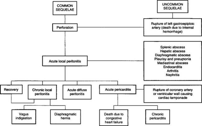
This complexity of development makes diagnosis and prognosis difficult, and the possibility that a number of syndromes may occur together further complicates the picture. All these entities except endocarditis are dealt with together here, even though many of them are diseases of other systems.
Traumatic reticuloperitonitis is caused by the penetration of the reticulum by metallic foreign objects that have been ingested in prepared feed. Baling or fencing wire that has passed through a chaff-cutter, feed chopper or forage harvester is one of the most common causes. In one series of 1400 necropsies, 59% of lesions were caused by pieces of wire, 36% by nails and 6% by miscellaneous objects. The metal objects may be in the roughage or concentrate or may originate on the farm when repairs are made to fences, yards and in the vicinity of feed troughs.
The wire from motor vehicle radial tires may be the cause.1-3 Used tires are commonly used to hold down plastic sheeting over silage piles. The wire is gradually released from the tires, which are in a state of deterioration, and is mixed with the feed supply, or the tires may be inadvertently dropped into a feed mixer wagon and become fragmented, mixing the pieces of wire throughout the ration.
Etiology Penetration of reticulum by metallic foreign objects such as nails and pieces of wire, including tire wire, that were ingested by the animal and located in the reticulum
Epidemiology Most common in adult dairy cattle fed prepared feeds
Signs Sudden anorexia and fall in milk yield, mild fever, ruminal stasis and local pain in the abdomen. Rapid recovery may occur, or the disease may persist in a chronic form or spread widely to produce an acute, diffuse peritonitis
Clinical pathology In acute local peritonitis, neutrophilia and regenerative left shift; in chronic form, leukopenia and degenerative left shift. Peritoneal fluid contains marked increase in nucleated cells and total protein. Plasma protein concentration increased. Radiography and ultrasonography of abdomen
Lesions Localized reticuloperitonitis and varying degrees of locally extensive fibrinous adhesions. Abnormal peritoneal fluid. Abscesses and adhesions possible throughout the peritoneal cavity
Diagnostic confirmation Reticuloperitonitis and metallic foreign body
• Acute local traumatic reticuloperitonitis must be differentiated from: simple indigestion, acute carbohydrate engorgement, acute intestinal obstruction, abomasal volvulus, pericarditis, acute pleuritis, perforated abomasal ulcer, postpartum septic metritis, pyelonephritis acute hepatitis, acetonemia
• Acute diffuse or generalized peritonitis must be differentiated from those diseases causing severe toxemia or acid–base imbalance, dehydration, and shock which include the following: carbohydrate engorgement, acute intestinal obstruction, advanced vagus indigestion, abomasal volvulus, perforated abomasal ulcer, and miscellaneous causes of generalized peritonitis
• Chronic traumatic reticuloperitonitis must be differentiated from early stages of vagus indigestion, hepatic abscessation, traumatic splenitis, chronic pneumonia and pleuritis, and miscellaneous causes of chronic peritonitis such as peritoneal abscesses secondary to intraperitoneal injections
Treatment Antimicrobials daily for several days, reticular magnet and immobilization in stall to promote adhesions. Rumenotomy to remove foreign body if medical treatment unsuccessful or in valuable animal
Control Prevent exposure of cattle to metallic foreign objects that can be ingested. Feed processing equipment should be equipped with magnets to remove metallic foreign bodies
In an abattoir survey of the gastrointestinal tract of 1491 slaughter cows in Denmark, foreign bodies were found in 16% of the cows. Of 286 foreign bodies, 11% were tire wires, 14% fencing wires, 5% screws, 9% nails, 37% mixed pieces of metal, 2% copper and 22% remnants of boluses containing antiparasitic drugs.3 A significant association was found between the type of foreign body and the presence of lesions, and a significant association between the cross-section of the foreign body and the presence of lesions. There was also an association between the end shape of the foreign body and the presence of lesions. Tire wire was the most common traumatizing foreign body, as 81% of all lesions were associated with tire wires.
Adult dairy cattle are most commonly affected because of their more frequent exposure but cases occur infrequently in yearlings, beef cattle, dairy bulls, sheep and goats. In the series of 1400 referred to above, 93% were in cattle over 2 years old and 87% were in dairy cattle. In the Danish abattoir survey of cows (see Etiology), foreign body lesions were present in 10% of the cows.3 Magnets, one or two, were found in only 7% of the cows. All magnets collected iron filings and fencing wire (30%), and ‘other pieces of metal’ (39%) were the predominant contents of the magnet. There were no lesions in 97% of the cows with magnets, and a significant association was found between the use of magnets and the absence of lesions.
The disease is much more common in cattle fed on prepared feeds, especially those fed inside for part of the year. It is almost unknown in cattle fed entirely on pasture. Accordingly, it is much more common in the winter months in the northern hemisphere. The incidence is low in sheep and goats.
The incidence is usually sporadic but outbreaks have occurred when sources of wire have become mixed into feed supplies, as in the case of perforation of the alimentary tract by pieces of tire wire.2 Over a period of 6 months, 30% of 170 lactating dairy cows in one herd exhibited clinical signs suggestive of hardware disease associated with the ingestion of tire wire in the feed supplies.
There are few studies of the epidemiology of traumatic reticuloperitonitis. The effects of 23 veterinary diagnoses, host characteristics and production were examined on the risks of ruminal acidosis and traumatic reticuloperitonitis.4 The lactational incidence risk for the disease in Finnish Ayrshire dairy cattle was 0.6%,4 which is similar to observations made in Holstein–Friesian cows.5 The risk of the disease in the former study increased with early metritis, nonparturient paresis, ketosis, acute and chronic mastitis, and foot and leg problems. It is unknown how metritis and mastitis could be risk factors for traumatic reticuloperitonitis. The median day of occurrence was on 113 days after calving, which makes it unlikely that calving was a risk factor. Similarly, dystocia was not found to be a risk factor.
When several or more cases occur in a cluster outbreak, the nature of the feed supply should be considered as a risk factor. The use of used tires to secure plastic sheeting over silage piles may be an important risk factor.
The disease is economically important because of the severe loss of production it causes and the high mortality rate. Many cases go unrecognized and many more make spontaneous recoveries. In industrialized countries, metallic foreign bodies may be present in the reticulum in up to 90% of normal cattle and residual traumatic lesions may be present in as many as 70% of dairy cows. Among the clinically affected animals, about 25% develop incurable complications. The other 75% can be expected to recover completely with conservative treatment or routine surgical intervention.
Lack of oral discrimination by cattle leads to the ingestion of foreign bodies that would be rejected by other species. Swallowed foreign bodies may lodge in the upper esophagus and cause obstruction or in the esophageal groove and cause vomiting, but in most instances they pass to the reticulum. Radiological examination of goats that have been fed foreign bodies experimentally indicate that they may first enter various sacs of the reticulorumen before reaching the reticulum. Many lie there without causing harm but the honeycomb-like structure of the reticulum provides many sites for fixation of the foreign body, and contractions of the reticulum are sufficient to push a sharp-pointed object through the wall.
Most perforations occur in the lower part of the cranial wall of the reticulum but some occur laterally in the direction of the spleen and medially towards the liver.
If the reticular wall is injured without penetration to the serous surface no detectable illness occurs, and the foreign body may remain fixed in the site for long periods and gradually be corroded away.A piece of wire can disappear in 6 weeks but certain nails last much longer and are unlikely to corrode away in less than 1 year. The ease with which perforation occurs has been illustrated by the artificial production of the disease. Sharpened foreign bodies were given to 10 cows in gelatin capsules. Of 20 pieces of wire and 10 nails, 25 were found in the reticulum. Of the 20 pieces of wire 18 had perforated or were embedded in the wall or plicae. Only one of the nails was embedded. Complete perforations were caused by 13 foreign bodies and incomplete by six. All cows suffered at least one perforation, showed clinical signs of acute local peritonitis and recovered after surgical removal of the foreign bodies.
Many foreign bodies may not remain embedded but are commonly found free in the reticulum if surgery is carried out about 72 hours after illness commences. This may be due to necrosis around the penetrating object and the reticular contractions moving the foreign body back into the reticulum. Objects that are deeply embedded or have kinks, barbs or large diameters tend to remain in situ and cause persistent peritonitis.
The initial reaction to perforation is one of acute local peritonitis and, in experimentally induced cases, clinical signs commence about 24 hours after penetration. The peritonitis causes ruminal atony and abdominal pain. If the foreign body moves back into the reticulum spontaneous recovery may occur.
Resolution of acute fibrinous local peritonitis is characterized by the development of fibrous adhesions, which gradually become long, stringy strands over a period of weeks and months; motility of the reticulum is restored and the animal may recover fully. Followup ultrasonographic examinations of cows with traumatic reticuloperitonitis in which rumenotomies were done found that the adhesions disappeared in most of the animals by 6 months.6
Depending on the severity of the local peritonitis, the ventral aspect of the reticulum becomes adherent to varying degrees to the abdominal floor and diaphragm. This results in decreased reticular motility. Ultrasonography of cows with traumatic reticuloperitonitis reveals that the biphasic contractions of the reticulum are slower than normal or indistinct and the number of contractions are reduced.7 Reticular abscesses are common complications and may be located between the reticulum and the ventral body wall, between the reticulum and the right thoracic wall and between the reticulum and the spleen.8 Persistent local peritonitis with or without abscesses results in reduced reticulorumen motility, inappetence to anorexia, a capricious appetite (may eat hay not concentrate), chronic ruminal tympany, persistent mild fever, abdominal pain on deep palpation, and changes in the hemogram and feces. Immobilization of the reticulum impairs the clearance function of the reticulum, which results in the passage of poorly comminuted feces characterized by an increased proportion of large particles.9
Spread of the inflammation causing generalized or diffuse peritonitis may occur in cows that calve at the time of perforation and in cattle that are forced to exercise. Immobility is a prominent clinical finding and may be a protective mechanism so that adhesions are able to form and localize the peritonitis. Animals made to walk or transported long distances frequently suffer relapses when these adhesions are broken during body movements. Generalized peritonitis results in toxemia, alimentary tract stasis, dehydration and shock.
During the initial penetration of the reticulum, the foreign body may penetrate beyond the peritoneal cavity and into the pleural or pericardial sacs. This may occur more commonly in cows in advanced pregnancy than in nonpregnant cows, because of the gravid uterus, although this is uncertain. Complications such as pericarditis occur most commonly in cows after the sixth month of pregnancy.
Details of the pathogenesis of the more common complications are presented under traumatic pericarditis, vagus indigestion, diaphragmatic hernia and traumatic abscess of the spleen and liver. Less common sequelae include rupture of the left gastroepiploic artery causing sudden death due to internal hemorrhage and the development of a diaphragmatic abscess, which infiltrates tissues to the ventral abdominal wall at the xiphoid process, rupturing to the exterior and sometimes discharging the foreign body. Hematogenous spread of infection from a diaphragmatic abscess or chronic local peritonitis is a common cause of endocarditis and its sequelae of polysynovitis and arthritis, nephritis and pulmonary abscessation. Penetration into the pleural cavity causes acute suppurative pleurisy and pneumonia. In rare cases the infection is localized to the mediastinum causing abscessation, which causes pressure on the pericardial sac and congestive heart failure. Rarely, the foreign body penetrates to the abomasum, causing abomasitis, pyloric stenosis and abomasal ulceration. Even more rarely, puncture of the reticular vein by a migrating metal wire may lead to fatal hemorrhage causing sudden death.10
Characteristically, the onset is sudden with complete anorexia and a marked drop in milk yield, usually to about a third or less of the previous milking. These changes occur within a 12-hour period and their abrupt appearance is typical. Subacute abdominal pain is common in most cases. The animal is reluctant to move and does so slowly. Walking, particularly downhill, is often accompanied by grunting. Most animals prefer to remain standing for long periods and lie down with great care; habitual recumbency is characteristic in others. Arching of the back occurs in about 50% of cases, along with the appearance of tenseness of the back and the abdominal muscles so that the animal appears gaunt or ‘tucked-up’. Defecation and urination cause pain and the acts are performed infrequently and usually with grunting. This results in constipation, scant feces and in some cases retention of urine. Rarely, acute abdominal pain with kicking at the belly and stretching occurs. In others there is recumbency and reluctance to stand.
A moderate systemic reaction is common in acute localized peritonitis. The temperature ranges from 39.5–40°C (103–104°F), rarely higher, the heart rate is about 80/min and the respiratory rate about 30/min. Temperatures above 40°C (104°F) accompanied by heart rates greater than 90/min suggest severe complications. The respirations are usually shallow and, if the pleural cavity has been penetrated, are painful and accompanied by an audible expiratory grunt.
Rumination is absent and reticulorumen movements are markedly depressed and usually absent. The rumen may appear to be full because of the presence of a free-gas bloat with moderate distension of the left paralumbar fossa. On palpation of the fossa, the ruminal gas cap is usually larger than usual and the rumen contents more doughy than normal. Deep palpation of the gas cap in the fossa may be required to feel the rumen pack below the gas cap.
Pain can be elicited by deep palpation of the abdominal wall just caudal to the xiphisternum. Palpation is done using short, sharp pushes with the closed fist or knee over an imaginary band about 20 cm wide covering the ventral third of the abdomen from the left to the right side with the cranial border of the band being the point just caudal to the xiphisternum. This area should be probed with at least six deep palpations on both sides of the abdomen while listening with a stethoscope over the trachea for evidence of a grunt. Pinching the withers to cause depression of the back and eliciting a grunt is also an effective diagnostic aid, except in large adult cows and bulls; for these the sharp elevation of a solid rail held horizontally under the abdomen is a useful method for eliciting a grunt. A positive response to any of these tests is a grunt of pain, which may be audible some distance away but is best detected by auscultation of the trachea. Rarely, a grunt may also be audible by auscultation over the trachea when infrequent reticulorumen contractions occur.
The course of acute local peritonitis is short and the findings described above are most obvious on the first day; in most cases they subside quickly and may be difficult to detect by the third day. In these cases, in addition to persistent anorexia and ruminal atony, the most constant finding is the abdominal pain, which may require deep palpation for its demonstration. In cases that recover spontaneously or respond satisfactorily to conservative treatment there may be no detectable signs of illness by the fourth day.
In chronic peritonitis the appetite and milk yield do not return to normal after prolonged therapy with antimicrobials. The body condition is usually poor, the feces are reduced in quantity and there is an increase in undigested particles. In some cases, the temperature may be within the normal range, which makes the diagnosis uncertain. A persistent slightly elevated temperature is supportive evidence of the presence of a chronic inflammatory lesion. The grunt test may be positive or negative; often it is uncertain. The gait may be slow and careful and, occasionally, grunting may occur during rumination, defecation and urination. Rumination activities are infrequent, the rumen is usually smaller than normal, chronic moderate bloat is common and there is ruminal atony or some moderate reticulorumen activity.
Reticular abscesses in cows are characterized by poor body condition, a relatively full rumen but with reduced ruminal contractions or almost complete ruminal atony, persistent mild bloat, an arched back with a tense abdomen and a grunt indicating abdominal pain, and undigested particles in the feces. Most have a clinical history of not responding to prolonged therapy with antimicrobials. These can be diagnosed with radiography and ultrasonography.
Rectal examination of cattle with acute or local traumatic reticuloperitonitis may cause a painful grunt when the animal strains during the examination. The feces are usually dry and firm and covered by a thin coating of mucus because of prolonged retention. In acute localized peritonitis the rumen may feel larger than normal and the gas cap is easily palpable. In acute and chronic generalized peritonitis, fibrinous adhesions may be palpable between the rumen and the left abdominal wall or between loops of intestine, or in the pelvic cavity.
Acute diffuse peritonitis is characterized by the appearance of profound toxemia within a day or two of the onset of local peritonitis. Alimentary tract motility is reduced, mental depression is marked and the temperature is elevated or subnormal in severe cases, especially those that occur immediately after calving. The heart rate increases to 100–120/min and a painful grunt may be elicited by deep digital palpation at almost any location over the ventral abdominal wall. This stage is usually followed by rapid collapse and peripheral circulatory failure and an absence of pain responses. Terminally, recumbency and depression are common.
There is a record of sudden death in a 20-month-old pregnant heifer in which the reticular vein was punctured by a migrating piece of metal wire, causing fatal hemorrhage into the reticulum. At necropsy, a large blood clot was present in the reticulum, the rumen contents were red brown and no reticular adhesions were present.10
There is a record of iatrogenic reticulitis that occurred as a result of the oral administration of intraruminal anthelmintic boluses, which may have lodged in the reticulum and become filled with other foreign objects ingested by the animal, resulting in a syndrome similar to acute traumatic reticuloperitonitis.11 Inappetence, reduced milk production, reduced reticulorumen motility, abdominal pain and scant feces were present. On exploratory rumenotomy the reticulum contained two cylindrical boluses filled with stones, nuts and bolts. Removal of the boluses was followed by prompt recovery.
The total and differential leukocyte counts provide useful diagnostic and prognostic data. The differential leukocyte count is usually considerably more indicative of acute peritonitis than the total count.
In acute local peritonitis a neutrophilia (mature neutrophils above 400/μL) and a left shift (immature neutrophils above 200/μL) are common. This is a regenerative left shift. Both the neutrophilia and the left shift will be increased on the first day and will last for up to 3 days, when in uncomplicated cases the count begins to return to normal. In chronic cases the levels do not return completely to normal for several days or longer periods and there is usually a moderate leukocytosis, neutrophilia and a monocytosis.
In acute diffuse peritonitis a leukopenia (total count below 4000/μL) with a greater absolute number of immature neutrophils than mature neutrophils (degenerative left shift) occurs, which suggests an unfavorable prognosis if severe. The degree of lymphopenia (lymphocyte count below 2500–3000/μL) is an indication of a stress reaction to inflammation.
There is a significant difference in total plasma protein levels between cattle with traumatic reticuloperitonitis and those with other diseases of the gastrointestinal tract that might be confused with the former.12 The mean plasma protein concentrations, measured before surgery, were 88 ± 13 g/L for traumatic reticuloperitonitis and 77 ± 12 g/L for controls. In severe diffuse peritonitis the fibrinogen levels may be increased up to 10–20 g/L.12
Cut-off points for total plasma protein (TPP) and plasma fibrinogen (PF) were determined to differentiate between traumatic reticuloperitonitis and other gastrointestinal diseases with similar clinical findings.13 There was moderate negative dependence between sensitivities of TPP and PF at the 8.82 g/dL and 766 mg/dL cut-off points, and mild negative dependence between their specificities at the 7.78 g/dL and 691 mg/dL cut-off points, respectively.13 Acceptable accuracy (98% or 86% specificity with 62% or 88% sensitivity, respectively) was obtained with serial interpretation of the tests.
Abdominocentesis and analysis of peritoneal fluid can be a valuable diagnostic aid. The best site for abdominocentesis is uncertain because the rumen occupies a large portion of the ventral abdominal wall and avoiding penetration of it is difficult. Cattle have a low volume of peritoneal fluid and failure to obtain a sample is not unusual. Empirically, the best sites are those in which, on an anatomical basis, there are recesses between the forestomachs, abomasum, diaphragm and liver. These are usually 10–12 cm caudal to the xiphisternum and 10–15 cm lateral to the midline. A blunt-ended teat cannula is recommended but with care and caution a 16-gauge 5 cm hypodermic needle may also be used. The hair of the site is clipped, the skin is prepared aseptically and a local anesthetic is applied. The skin in incised with a stab scalpel and the cannula is pushed carefully and slowly through the abdominal wall. The latter will twitch and a ‘pop’ will be felt when the peritoneum is punctured. When the cannula is in the peritoneal cavity the fluid may leak out without the aid of a vacuum. If it does not, a syringe may be used to apply a vacuum while the needle is manipulated in an attempt to locate some fluid.
If no fluid can be obtained, a trocar and cannula 80 mm long and with a 4 mm internal diameter can be used with success. The trocar and cannula are inserted into the abdomen, the trocar is removed and an 80 cm long 10 French gauge infant feeding tube is inserted into the abdomen through the cannula, leaving about 10–20 cm outside. The tube acts as a wick and within several minutes fluid can be collected into vials. At least three different sites should be attempted to obtain peritoneal fluid. Peritonitis in cattle is characterized by a marked fibrinous response and localization of a lesion, and the amount of exudative fluid available at the abdominocentesis sites may be minimal. Thus the failure to obtain fluid does not preclude the presence of peritonitis.
Laboratory evaluation of peritoneal fluid consists of determinations of total white blood cell count, differential cell count, total protein and culture for pathogens. The interpretation of the analysis of the peritoneal fluid can be unreliable because to date only a few correlations have been made between the laboratory findings and the presence or absence of peritoneal lesions. A nucleated cell count above 6000 cells/μL and total protein above 3 g/dL is consistent with the diagnosis of peritonitis in 80% of cases. Using a differential cell count, a relative neutrophil count more than 40% and a relative eosinophil count less than 10% was frequently associated with the diagnosis of peritonitis.
Metal detectors were used at one time to aid in the diagnosis of traumatic reticuloperitonitis. Ferrous metallic foreign bodies can be detected with metal detectors but the instruments are of limited use because most normal dairy cows are positive for metal over the reticular area.
Right flank laparoscopy using a flexible fiberoptic laparoscope, 14 mm diameter and 1120 mm working length, is a reliable diagnostic aid for the presence of traumatic reticuloperitonitis.
Radiological examination of the reticulum with the animal in dorsal recumbency (dorsal reticulography) is an accurate diagnostic method for the evaluation of cattle with suspected traumatic reticuloperitonitis.14 However, the lack of adequate radiographic equipment in private veterinary practices precludes its routine use. Also, the technical difficulties of positioning the animal and the increased potential for personnel exposure associated with manual restraint suggests that it may not be practical except for valuable animals that may warrant referral to a veterinary medical center.
The cranioventral abdomen of cattle can be evaluated using two cranial abdominal and one caudal thoracic radiographs. An X-ray machine with a capacity of 1000–1250 mA and 150 kV is necessary, which is usually only available in veterinary teaching hospitals. However, such techniques may be appropriate in valuable animals in which an accurate diagnosis and prognosis for surgical treatment may be desirable.15 In a consecutive series of standing lateral cranial abdominal radiographs, the sensitivity and specificity for detecting traumatic reticuloperitonitis or pericarditis was 83% and 90%, respectively.16 These values are higher than those achieved with dorsal recumbency. In standing lateral radiographs, an enlarged reticulum was associated with a final diagnosis of vagal indigestion. Alteration in reticulodiaphragmatic separation does not correlate with any specific disease process. The presence of focal perireticular gas collections and reticular foreign bodies greater than 1 cm in length unattached to a magnet were good indicators of traumatic reticuloperitonitis. Radiography is best suited for identification of radiodense foreign bodies in and outside the reticulum (these cannot be visualized ultrasonographically).14
Features found to be reliable in the diagnosis of traumatic reticuloperitonitis using lateral radiographs of the reticulum include:
• Atypically positioned foreign bodies
• Abnormal gas shadows in the region of the reticulum
• Depressions in the cranioventral margin of the reticulum.17
The reticulum is commonly markedly displaced caudally from the diaphragm or dorsally or caudodorsally from the ventral abdominal wall. Space-occupying masses of the density of soft tissue, with or without gas inclusions, gas shadows and gas– fluid interfaces in the region of the reticulum, were highly predictive of peritonitis (specificity 97%, positive predictive value 96%).
Ultrasonography is a suitable method for investigation of reticular contractions in healthy ruminants and in cattle for the diagnosis of traumatic reticuloperitonitis.18,19 The literature on the use of ultrasonography as a diagnostic aid in gastrointestinal disease in cattle has been reviewed.20 The reader is referred to an excellent atlas and textbook on the use of ultrasonography in cattle.19
The reticulum and adjacent organs of cows can be examined with ultrasonography using a 3.5 MHz linear transducer applied to the ventral midline of the thorax over the sixth and seventh intercostal spaces and from the left and right sides of the midline.21,22 It may not be possible to image the reticulum in large cows in good body condition because of the high proportion of fat in the muscle layers. In older cows, calcification of the xiphisternum may interfere with imaging. The most common reason for being unable to visualize the reticulum in sick animals is the displacement of the reticulum by a markedly distended rumen or by space-occupying lesions such as abscesses and fibrin-containing effusions. The pattern, number, amplitude and duration of the interval between contractions can be visualized.21 The contour of reticulum, the reticular contractions and the organs adjacent to the reticulum can be imaged. The biphasic reticular contractions can be visualized at the rate of 4 during a 4-minute period.21,22 During the first incomplete contraction, the reticulum contracts by a mean of about 7.2 cm and during the second contraction the reticulum disappears from the screen.
In contrast to radiography, ultrasonography provides more precise information about the contour of the reticulum and reticular motility.7,22 In cattle with traumatic reticuloperitonitis, ultrasonography can be used to identify morphological changes in the region of the cranial, ventral or caudal reticular wall.20 The caudoventral reticular wall is the most frequently affected, often in association with the craniodorsal blind sac of the rumen. The changes in the contour of the reticulum depend on the severity of the inflammatory changes.
The reticulum can be visualized in more than 90% of cows in spite of interference by the ribs and sternum. In cows with disturbed reticular motility, biphasic contractions are slower than normal, or indistinct, and the number of contractions is reduced. Fibrinous material appears as echogenic deposits, sometimes accompanied by hypoechogenic fluid. Reticular abscesses have an echogenic capsule with a hypoechogenic center. Involvement of the spleen, omasum, liver and abomasum may also be imaged. Neither magnets nor foreign bodies can be visualized by ultrasonography.22
Reticular activity is almost always affected in cattle with traumatic reticuloperitonitis. The frequency, amplitude or velocity of contractions, singly or combined, may be abnormal. The frequency can be reduced from 3 to 2, 1 or no contractions per 3 minutes. The reduction in the amplitude of contractions varies: when formation of adhesions is extensive, reticular contractions appear indistinct. Although the pattern of biphasic contraction is often maintained, the reticulum contracts only 1–3 cm. The velocity of reticular contractions may be normal but can be markedly reduced. In cattle with reticulo-omasal obstruction due to a foreign body, the frequency of reticular contractions may be increased.23
Reticular abscesses associated with traumatic reticuloperitonitis can be visualized by ultrasonography8 (Fig. 6.5). The amplitude of reticular contractions is reduced, the reticulum is displaced from the ventral body wall, and the abscesses have hypoechogenic centers and echogenic capsules.
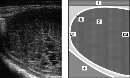
Fig. 6.5 Ultrasonogram and schematic of a reticular abscess in a cow with chronic traumatic reticuloperitonitis. The abscess is between the reticulum and the ventral abdominal wall. The ultrasonogram was obtained from the sternal region with a 5.0 MHz-linear transducer. 1 = Ventral abdominal wall; 2 = Abscess; 3 = Capsule of the abscess; 4 = Reticulum. Cr, Cranial; Cd, Caudal.
(Reproduced with kind permission of U. Braun.)
Peritoneal effusion is visible as an accumulation of fluid without an echogenic margin and restricted to the reticular area. Depending on the fibrin and cell content, the fluid may be anechoic or hypoechogenic. Fibrinous deposits are easily identified in the fluid and bands of fibrin are sometimes seen within the effusion. Occasionally, the peritoneal effusion is considerable and extends to the caudal abdomen.
The spleen, particularly its distal portion, is often affected. Fibrinous changes are frequently seen as echogenic deposits of varying thickness, often surrounded by fluid, between the spleen and reticulum or rumen. The spleen may be covered by fibrinous deposits. Occasionally, one or more splenic abscesses are visible, and the vasculature may be dilated, indicating splenitis.20
These two techniques have been compared in cows with traumatic reticuloperitonitis. The major advantages of radiography are that metallic foreign bodies can be visualized and their position determined. It has a specificity of 82%, a positive predictive value of 88% and a sensitivity of 71%.24 Abnormal gas shadows or gas–fluid interfaces observed on radiographs are highly diagnostic for the disease and have a specificity of 97% and positive predictive value of 88%. However, they are seldom seen on radiographs and their sensitivity is only 19%. The position of the reticulum is a good criterion for the diagnosis of traumatic reticuloperitonitis, with a specificity of 80% and a positive predictive value of 82%. Thick-walled changes or abscessation should be suspected when the reticulum is displaced caudodorsally from the sternum. Changes in the contour of the reticulum such as indentations are highly suggestive of inflammation, with a specificity of 95% and positive predictive value but a low sensitivity of only 34%.
The major advantage of ultrasonography is being able to visualize and assess reticular motility.19,22,24 Even in the presence of severe adhesions and abscessation, the reticulum may maintain its basic contractile rhythm, but much reduced. Abscesses have an echogenic capsule of varying width and a central cavity filled with hypoechogenic material. Purely fibrous deposits are echogenic, and fibrinous deposits containing an accumulation of fluid from inflammatory processes are echogenic interspersed with hypoechogenic accumulations of fluid (Fig. 6.6).24 Radiography and ultrasonography complement each other and the combined results can be used to decide whether an exploratory laparotomy is indicated, if the animal should be treated conservatively with antibiotics, or if it should be slaughtered for salvage.22
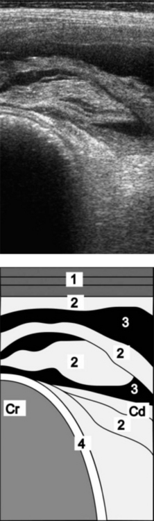
Fig. 6.6 Ultrasonogram and schematic of the reticulum in a cow with chronic traumatic reticuloperitonitis. The reticulum is covered with fibrinous deposits. The ultrasonogram was obtained from the sternal region with a 5.0 MHz-linear scanner. 1 = Lateral abdominal wall; 2 = Fibrinous deposits; 3 = Anechoic fluid; 4 = Reticulum. Cr, Cranial; Cd, Caudal.
(Reproduced with permission from U. Braun.)
Localized traumatic reticuloperitonitis is characterized by varying degrees of locally extensive fibrinous adhesions between the cranioventral aspects of the reticulum and the ventral abdominal wall and the diaphragm. Adhesions and multiple abscesses may extend to either side of the reticulum involving the spleen, omasum, liver, abomasum and ventral aspects of the rumen. Large quantities of turbid, foul-smelling peritoneal fluid may be present, containing fibrinous clots. Some cases of reticular abscesses are solitary and there are adhesions between the reticulum, diaphragm and ventral body wall, which are strictly localized. The size of the abscess varies. It may be from 5–10 cm in diameter or else a single one may be irregularly shaped and measure 30 × 10 × 10 cm, along with multiple smaller ones measuring around 3 × 3 × 3 cm.24 The foreign body can usually be found perforating the cranioventral aspect of the reticulum, although it may have fallen back into the reticulum, leaving only the perforation site and its surrounding inflammation as evidence of the site of penetration. A reticular magnet with many pieces of metallic foreign bodies stuck to it may be present in the reticulum, the mucosa of which is usually normal.
In acute diffuse peritonitis a fibrinous or suppurative inflammation may affect almost the entire peritoneal cavity with extensive fibrinous adhesions of various stages of development involving the forestomach, abomasum, small and large intestines, liver, bladder, reproductive tract and pelvic cavity. Large quantities of turbid, foul-smelling fluid containing clots of fibrin are usually present. Loops of intestine and omenta are commonly stuck together by thick layers of fibrin.
Typical acute traumatic reticuloperitonitis is characterized by a sudden onset of complete anorexia, marked drop in milk production, mild fever, ruminal atony, pain on deep palpation of the ventral abdomen, an elevated leukocyte count with a left shift in the hemogram and a peritoneal fluid sample that indicates inflammation.
However, the times at which cases of traumatic reticuloperitonitis are seen varies from day 1, when the syndrome is typical, to day 3 or 4, by which time the acuteness has subsided so much clinically that confusion with other diseases is a significant possibility. The sudden onset of anorexia and marked drop in milk production will usually be noted in lactating dairy cattle but not in dry dairy cattle or beef cattle, including mature bulls whose feed intake and behaviors are not monitored daily. In these animals the clinical findings can change in a few days and be characterized by anorexia to inappetence, normal temperature, ruminal hypotonicity or atony and no evidence of abdominal pain on deep palpation of the abdomen.
The clinician must review the history carefully, conduct a thorough clinical examination and attempt to intensify the diagnostic efforts on those abnormalities that are present.
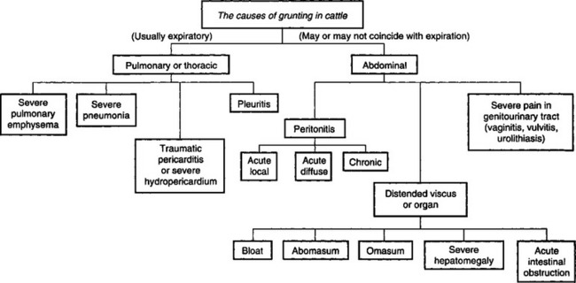
The differential diagnosis of gastrointestinal dysfunction of cattle is summarized in Table 6.2. An algorithm for the causes of grunting in cattle is shown in Fig. 6.7.
Acute local traumatic reticuloperitonitis must be differentiated from those diseases in which sudden anorexia, sudden drop in milk production, ruminal atony, abdominal pain and abnormal feces are common. They include the following:
• Simple indigestion characterized by sudden anorexia or inappetence, normal mental state, full rumen but atonic, perhaps uncomfortable if ingested large quantities of palatable feed like fresh silage, normal vital signs, abnormal feces and spontaneous recovery in 24 hours are typical
• Obstruction of reticulo-omasal orifice with a foreign body such as a roll of polyethylene twine causes intermittent inappetence, a slightly enlarged rumen with normal motility, slight reduction in the amount of feces, a decrease in milk yield for 24–48 hours followed by a return to normal and then subsequent relapses. A grunt is not present, the temperature, heart and respiratory rates are normal and the hemogram is normal. Obstruction of the reticulo-omasal orifice with foreign bodies such as rope can cause distension and hypermotility of the rumen and persistent vomiting.23 A rumenotomy must be done to make the diagnosis
• Acute carbohydrate engorgement characterized by sudden anorexia, diarrhea and dehydration, weakness, tachycardia, staggering, ruminal distension and atony, fluid-splashing sounds in the rumen with a rumen pH of less than 5 and a history of access to grain
• Acute intestinal obstruction characterized by sudden anorexia, mild abdominal pain perhaps with kicking at the abdomen and stretching, ruminal atony, mild dehydration, scant feces or complete absence of feces, straining on rectal examination, dark blood-stained feces and perhaps distended loops of intestine palpable on rectal examination
• Abomasal volvulus (following right-side dilatation) characterized by anorexia, dehydration, tachycardia, distended right abdomen, ping audible over right flank, distended viscus palpable on rectal examination. Usually in lactating dairy cows a few weeks after parturition and following the clinical findings of right-side dilatation of the abomasum that lasts several days culminating in the volvulus but may occur spontaneously in some cows with no immediate history of previous illness
• Pericarditis. Continued high fever, toxemia, anorexia, tachycardia and muffled heart sounds suggest pericarditis, which is marked by markedly elevated total leukocyte and neutrophil counts. In pericarditis, the heart sounds are muffled and the typical to and fro fluid-splashing sounds are audible. The jugular veins are engorged and other signs of congestive heart failure such as anasarca are present. Pericardiocentesis to obtain foul-smelling, turbid fluid is diagnostic
• Acute pleuritis is characterized by a fever, toxemia, anorexia, painful respirations that may be accompanied by a grunt, pain on digital palpation of intercostal spaces, ruminal atony, and abnormal and muffled lung sounds. Fluid on thoracentesis
• Perforated abomasal ulcer causes acute local peritonitis characterized by marked pain on palpation over a much larger area of the abdominal wall and in the early stages is most marked on the right-hand side. If, as is usual, the peritonitis becomes diffuse the syndrome cannot be distinguished clinically from that caused by traumatic reticuloperitonitis. Extension from a metritis to involve the peritoneum is suggested by other signs of the primary disease
• Postpartum septic metritis occurs a few days after parturition and is characterized by anorexia, fever, tachycardia, ruminal hypotonicity to atony, reduced amount of feces and foul-smelling vaginal discharge, and retained placenta may be present. Very important to examine the uterus vaginally for the presence of the placenta, which may be protruding through the cervix
• Acute local peritonitis due to penetration of the uterine wall by a catheter or of the rectal wall by a foreign body thrust sadistically into the rectum may be difficult to differentiate unless the painful area of the peritoneum can be determined. Acute local peritonitis can be differentiated from indigestion, acute ruminal impaction and acetonemia by the presence of fever, local abdominal pain and the abrupt fall in milk yield and appetite
• Pyelonephritis is occasionally accompanied by mild abdominal pain but can be distinguished by the presence of pus and blood in the urine
• Acute hepatitis or severe hepatic abscess is characterized by anorexia, fever, decreased ruminal movements, reluctance to move, a painful grunt on deep palpation over the cranial aspects of the right lower flank, icterus if obstruction of the bile ducts has occurred, and a poor response to therapy. A marked neutrophilia is typical of hepatic abscessation secondary to traumatic reticuloperitonitis
• Acetonemia. Traumatic reticuloperitonitis usually causes a secondary acetonemia when it occurs during early lactation and the presence of ketonuria should not be used as the sole basis for differentiation of the diseases. Differentiation may be extremely difficult if the peritonitis is of 3–4 days’ duration. Response to treatment may also serve as a guide. The history is often helpful; the appetite and milk yield fall abruptly in traumatic reticuloperitonitis but slowly over a period of several days and not to the same degree in acetonemia
Acute diffuse peritonitis is characterized by anorexia, fever, toxemia, tachycardia, dehydration, weakness leading to recumbency, distended abdomen, ruminal atony, spontaneous grunting or a grunt on deep palpation over the abdomen, fluid-splashing sounds and pings on auscultation and percussion or ballottement of the abdomen due to ileus, scant feces, perhaps palpable fibrinous adhesions on rectal palpation, profuse quantities of abnormal peritoneal fluid and marked changes in the hemogram. It must be differentiated from those diseases causing severe toxemia or acid–base imbalance, dehydration and shock, which include: carbohydrate engorgement, acute intestinal obstruction, advanced vagus indigestion, abomasal volvulus, perforated abomasal ulcer and miscellaneous causes of generalized peritonitis.
The clinical findings of chronic traumatic reticuloperitonitis are not typical. Each chronic case may have a different combination of clinical findings, which makes the diagnosis uncertain. The clinical findings that may be present include inappetence to anorexia, mild fever, loss of body condition, lack of rumination, ruminal hypotonicity to atony, moderate bloat, scant feces containing increased amounts of undigested feed particles, possibly a grunt on deep palpation of abdomen, and changes in the hemogram. The presence of abnormal peritoneal fluid is highly supportive. It must be differentiated from early stages of vagus indigestion, hepatic abscessation, traumatic splenitis, chronic pneumonia and pleuritis, and miscellaneous causes of chronic peritonitis such as peritoneal abscesses secondary to intraperitoneal injections.
Two methods of treatment are in general use: conservative treatment with or without the use of a magnet, and rumenotomy. Both have advantages and each case must be considered when deciding on the form of treatment to be used.
Conservative treatment comprises immobilization of the animal, administration of antimicrobials for the inflammation and the oral administration of a magnet to immobilize the foreign body. The cow is tied, stanchioned or confined in a box stall for several days. Immobilization of the animal facilitates the formation of adhesions.
Penicillin or broad-spectrum antimicrobials given parenterally daily for 3–5 days are widely used with empirical success. Because of the high probability that a mixed gastrointestinal flora is the cause of the lesion it is more rational to use a broad-spectrum antimicrobial such as the tetracyclines or trimethoprim-potentiated sulfonamides rather than penicillin, which is commonly used because of cost and a short withdrawal period in the event that the animal does not respond favorably in a few days. For lactating dairy cattle, those antimicrobials with a short milk withdrawal period are desirable. However, there are no published clinical trials to indicate the preferential value of any particular antimicrobial. The general effect appears to be good and a high rate of recovery is recorded with antimicrobials parenterally combined with immobilization provided treatment is begun in the early stages of the disease. Cows past their sixth month of pregnancy are unlikely to recover completely and commonly relapse.
Surgical removal of the foreign body through a rumenotomy incision is widely used as a primary treatment. It has the advantage of being both a diagnostic procedure in the first instance and a satisfactory treatment. The recovery rate varies, depending on when the surgery is done relative to the time of initial penetration, but is approximately the same as that obtained with the conservative treatment described above. In both instances 80–90% of animals recover compared with about 60% in untreated animals. Failure to improve is usually due to involvement of other organs or to the development of locally extensive peritonitis and reticular abscesses associated with persistent penetration of the foreign body or, uncommonly, generalized peritonitis.
Based on follow-up ultrasonography of cows that had surgery for traumatic reticuloperitonitis, the inflammatory adhesions resolved and disappeared in the majority of animals by 6 months.6 As a consequence, reticular function normalizes. In animals with severe adhesions, there is a marked disturbance of digesta passage and, in these animals, extensive abscesses are present.
Persistent penetration by the foreign body necessitates removal for optimum results but a rumenotomy is necessary to determine the extent of the lesion. Radiography and ultrasonography as described above may assist in determining the presence and location of the foreign body. A single preoperative dose of antimicrobial such as potassium penicillin G at 10 million IU given intravenously is recommended to avoid complications after a rumenotomy in cattle.
The recovery rate after surgery is likely to be much lower if only complicated cases are selected for rumenotomy and conservative treatment is given to the early mild cases. In one series the recovery rate in the cases treated conservatively was 84% and in those difficult cases treated surgically it was 47%.
Reticular abscesses may be drained through an ultrasound-guided transcutaneous incision.8
The choice of treatment is largely governed by economics and the facilities and time available for surgery. A rumenotomy, satisfactorily performed, is the best treatment but is unnecessary in many cases because of the tendency of the foreign body to return to the reticulum. A commonly used practice is to treat the animal conservatively for 3 days and if marked improvement has not occurred by that time to consider a rumenotomy. A rumenotomy is highly desirable in cows in the last 3 months of pregnancy if severe sequelae are to be avoided. Movement of the cow during the early stages of the disease is undesirable because of the risk of disrupting the adhesions that localize the infection.
Cases of chronic traumatic reticuloperitonitis are best treated by rumenotomy because of the probability that the foreign body is still embedded in the wall. Acute diffuse peritonitis is highly fatal but if detected early daily treatment with broad-spectrum antimicrobials may be effective.
All processed feed should be passed over magnets to remove metallic material before being fed to cattle. The use of synthetic string instead of wire has resulted in a major decrease in the incidence of the disease.
Small cylindrical or bar magnets, 7.5 cm long by 1.0–2.5 cm diameter with rounded ends, are used to prevent the disease but are also used in acute cases to minimize penetration of the foreign body. When given orally to normal healthy animals the magnets locate in the reticulum within a few days, where they remain indefinitely and maintain their magnetic pull. The magnets attract foreign bodies, which then do not penetrate the reticular wall as easily as when they are free. The extensive prophylactic use of these magnets in a dairy herd has reduced the incidence of the disease and its complications by 90–98%. The magnets are given to herd replacement heifers at 18 months to 2 years of age as part of a herd health program.
The effects of magnets in traumatic reticulitis was examined in the Danish study of cows at slaughter (see under Etiology).3 Two magnets tested were cylindrical cage magnets with different fields of magnetic force. Magnet I had a magnetic force of attraction of 110 mT; magnet II had a force of 210 mT. Magnets were found in only 7% of the cows. There were no lesions in 97% of the cows with magnets. Magnet II was superior to magnet I in attracting all types of foreign bodies, including tire wires. Thus the prophylactic use of magnets should be promoted to reduce the occurrence of foreign body lesions.3
It is unlikely that magnets will extract a firmly embedded foreign body from the wall of the reticulum but loosely embedded ones with long free ends may be returned to the reticulum and loose foreign bodies will be immobilized. The position of the foreign body within the reticulum greatly influences the efficacy of treatment with a magnet. A foreign body at an angle to the ventral aspect of the reticulum of more than 30° is less likely to become attached to a magnet than a foreign body situated horizontally on the ventral aspect of the reticulum.25 There have been only a few reports of physical injury to the wall of the reticulum being caused by the magnets or the foreign bodies that may be attached to them. A compass can be used to locate the presence and position of the magnet.
Braun U. Atlas und Lehrbuch der Ultraschall-diagnostik beim Rind. Berlin: Blackwell Wissenschafts-Verlag, 1997.
Braun U. Ultrasonography in gastrointestinal disease in cattle. Vet J. 2003;166:112-124.
Braun U. Ultrasound as a decision-making tool in abdominal surgery in cows. Vet Clin North Am Food Anim Pract. 2005;21:33-53.
1 Harwood D. Vet Rec. 2004;154:574.
2 Monies R. Vet Rec. 2004;154:735.
3 Cramers T, et al. Vet Rec. 2005;157:287.
4 Grohn YT, Bruss ML. J Dairy Sci. 1990;73:655.
5 Dohoo IR, et al. Prev Vet Med. 1984;2:655.
6 Herzog K, et al. Dtsch Tierarztl Wochenschr. 2004;111:57.
7 Braun U, et al. Vet Rec. 1993;133:416.
8 Braun U, et al. Vet Rec. 1998;142:184.
9 Rehage J, et al. J Am Vet Med Assoc. 1995;207:1607.
10 Hailat N, et al. Can Vet J. 1993;34:698.
11 Sheldon IM. Vet Rec. 1995;136:126.
12 Ward JL, Ducharme NG. J Am Vet Med Assoc. 1994;204:874.
13 Jafarzadeh SR, et al. Prev Vet Med. 2004;65:1.
14 Farrow CS. Vet Clin North Am Food Anim Pract. 1999;15:397.
15 Fubini SL, et al. J Am Vet Med Assoc. 1990;197:1060.
16 Partington BP, Biller DS. Vet Radiol. 1991;32:155.
17 Braun U, et al. Vet Rec. 1993;132:103.
18 Kaske M, et al. J Vet Med A. 1994;41:748.
19 Braun U. Atlas und Lehrbuch der Ultraschall-diagnostik beim Rind. Berlin: Blackwell Wissenschafts-Verlag, 1997.
20 Braun U. Vet J. 2003;166:112.
21 Braun U, Gotz M. Am J Vet Res. 1994;55:325.
22 Braun U. Vet Clin North Am Food Anim Pract. 2005;21:33-53.
23 Braun U. Vet Rec. 2002;150:580.
Etiology Reticular adhesions from traumatic reticuloperitonitis and failure of passage of ingesta from reticulorumen and abomasum resulting in accumulation in forestomach and abomasum. Abomasal emptying defect in sheep (uncertain etiology)
Epidemiology Primarily mature dairy cattle; also in mature beef cows and bulls. Also occurs in sheep as abomasal emptying defect of uncertain etiology
Signs Gradual distension of abdomen, especially left upper abdomen and bilateral aspects of ventral abdomen. Inappetence to anorexia and scant feces containing undigested long particles. Large L-shaped rumen viewed from rear. Rumen hypermotility or atony. Dehydration
Clinical pathology Hemoconcentration, metabolic alkalosis with hypochloremia and hypokalemia, increased ruminal chloride levels
Lesions Reticular adhesions. Enlarged rumen containing pasty and frothy material or fluid contents. Abomasum impacted with semi-dry ingesta
Differential diagnosis Ruminal distension with hypermotility: indigestion of late pregnancy, obstruction of the reticulo-omasal orifice. Ruminal distension with atony: chronic traumatic reticuloperitonitis. Abomasal impaction: abomasal impaction, dietary in origin. Omasal impaction: phytobezoars blocking the abomasal pylorus, abomasal ulceration without melena
Treatment Fluid and electrolyte therapy, rumen lavage, rumenotomy, drain reticular abscess, slaughter for salvage
The etiology has been controversial but has been divided into two major subcategories of complications of traumatic reticuloperitonitis: vagal nerve injury and reticular adhesions. In addition there are some other causes.
Historically, it was thought that vagus indigestion was caused by vagal nerve dysfunction due to vagal nerve injury associated with complications of traumatic reticuloperitonitis. It was hypothesized that the inflammatory and scar tissue lesions affected vagal nerve fibers supplying the forestomach and abomasum. The naturally occurring syndrome was similar to the Hoflund syndrome created by experimentally sectioning the vagus nerves and thus the term ‘vagus indigestion’ was coined.
The prevailing explanation was that dorsal vagal nerve injury resulted in achalasia of the reticulo-omasal orifice (anterior stenosis) and inhibited the passage of ingesta from the reticulorumen into the omasum and abomasum, resulting in an enlarged rumen with abnormal rumen contents. Similarly, injury of the pyloric branch of the ventral vagus nerve resulted in achalasia of the pylorus (posterior stenosis) and inhibited the flow of ingesta from the abomasum resulting in abomasal impaction. Both abnormalities resulted in scant feces containing undigested long feed particles.
However, while in many cases of vagus indigestion there are extensive adhesions between the reticulum and adjacent organs, there is little evidence of vagal nerve injury. It is also known that the syndrome can occur without any gross evidence of inflammation of the serosa of the forestomach and abomasum over which the vagus nerves are located. In the absence of gross lesions, it has been suggested that microscopic lesions of the medial reticular wall where vagal tension receptors are located may interfere with forestomach motility and esophageal groove reflexes.
New information based on clinical– pathological examination of clinical cases has questioned the long-held view that vagal nerve injury is an important cause of this syndrome.
Mechanical impairment of reticular motility and esophageal groove dysfunction as a result of reticular adhesions is probably the most important cause of the syndrome.1 An examination of 42 dairy cows with complications of traumatic reticuloperitonitis found that the primary mechanism was a disturbance in particle-separation processes in the reticulorumen attributable to mechanical inhibition of reticular motility associated with extensive inflammatory parareticular adhesions.1 Based on examination of necropsy tissue grossly and histologically, there was no evidence of vagal nerve injury. Perireticular abscesses near the reticulo-omasal orifice of cattle can cause the disease.2
Several causes unrelated to traumatic reticuloperitonitis have been recorded. Actinobacillosis of the rumen and reticulum is a less common cause. In sheep, peritonitis associated with Sarcosporidia and Cysticercus tenuicollis may be a cause. Fibropapillomas of the cardia can mechanically occlude the distal esophagus and cause interference with forestomach motility.3 Abomasal impaction in sheep has been recorded but the etiology and pathogenesis have not been determined. Disturbances similar to those that occur under natural conditions have been produced by sectioning the vagus nerve. Following surgery for right-sided abomasal displacement or volvulus, some cattle develop a vagus-indigestion-like syndrome characterized by anorexia, scant feces, ruminal distension and abdominal distension. It has been suggested that distension of the abomasum and thrombosis of its vessels may have caused injury to the ventral vagus nerve.4
Pyloric achalasia is described as part of a secondary indigestion due to septicemia and toxemia but this is not well documented. There is also ruminal distension with fluid material, abomasal reflux into the reticulorumen, dehydration, hypochloremia, hypokalemic metabolic alkalosis and uremia.
Indigestion of late pregnancy of cows is considered a type of vagus indigestion in which the rumen and abomasum are grossly distended, but the cause is uncertain.5 There is no evidence that the effects of an advanced pregnancy alone will cause a vagus-indigestion-like syndrome.
Peripheral nerve sheath tumors, such as a solitary schwannoma have been described causing a syndrome similar to vagus indigestion in a mature cow.6
A vagal indigestion-like syndrome may be a postsurgical complication of right abomasal displacement.7 Gastric wall injury, peritonitis and vagal nerve lesions may be causative factors. It occurs in 14–21% of cases, and only 12–20% of cases return to normal production. (See under Right-side displacement of the abomasum and abomasal volvulus.)
The syndrome occurs most commonly in dairy cows that have a history of traumatic reticuloperitonitis, which may have occurred several weeks or a few months previously. The disease is not restricted to dairy cows – it also occurs in beef cattle and in mature bulls.
The syndrome of vagus indigestion is characterized by disturbances in the passage of ingesta through the reticulo-omasal orifice (failure of omasal transport, anterior functional stenosis) and disturbances in the passage of ingesta through the pylorus (pyloric stenosis, posterior functional stenosis). Stenosis is a misnomer because there is no evidence of stenosis but achalasia of the sphincters may occur. The characteristic clinical findings are distension of the rumen with pasty and/or frothy contents because of increased time and maceration in the reticulorumen, alterations in reticulorumen motility, with consequences such as dehydration, an increase in undigested particles in the feces, scant feces, acid–base imbalance and secondary starvation. It is an outflow abnormality of the reticulorumen and abomasum.
Based on careful clinicopathological observations of 42 cows with complications of traumatic reticuloperitonitis including ‘vagus indigestion’, it is now proposed that the disturbances in the flow of ingesta are associated with particle-separation in the reticulorumen caused by mechanical inhibition of reticular motility associated with extensive adhesions of the reticulum.1 Experimentally impaired reticular contractions in sheep support the central role of reticular motility for the separation of particles in the forestomach, the outflow of digesta from the reticulorumen and transpyloric digesta flow.8
Normally, reticulorumen motility results in stratification of ruminal contents into three layers of ingesta in addition to the most dorsal gas pocket. The top layer, consisting of firm fibrous material of low-density particles (coarse hay), floats on the middle layer of liquid ingesta, consisting of particles of medium density; the bottom layer consists of fine particles of high density. The solid material remains in the rumen and is digested until the particle size is sufficiently small (1–4 mm in cattle) to pass through the reticulo-omasal orifice. The size of the digested plant fragments in ruminant feces can be considered an indirect measurement of forestomach function. In cows, the presence of large plant particles (> 0.5 cm) in the feces indicates inadequate rumination or abnormalities in forestomach motility.9
In normal cattle, the mean retention time of particles in the reticulorumen depends on particle size and density. The density of large feed particles is low because of their air-filled interior. During biphasic reticular contractions, most of these large, light particles are pushed caudodorsally in the rumen. Thus, large particles are retained in the reticulorumen, because outflow through the reticulo-omasal orifice occurs mainly during the maximum portion of the second reticular contraction. Feed particles with a high density (small and well digested) are moved out of the reticulorumen preferentially, because the majority of them remain in the reticulum during the biphasic contraction.
If reticular motility is inhibited, the balance of particle retention time and particle outflow in the reticulorumen is disturbed. Immobilization of the reticulum experimentally causes a decrease in feed intake, an increase in ruminal volume, a decrease of mean retention time of light plastic particles, a four-fold increase in mean retention time of heavy plastic particles, a marked increase in the amount of large particles in the feces, and an increase in abomasal volume. Such changes reflect the changes occurring in naturally occurring vagus indigestion. An increase in the amount of large particles in the feces of cows with traumatic reticuloperitonitis is indicative of inhibited clearance function of the reticulum.
Liquid consistency of the abomasal contents is important to insure physiological transpyloric flow. In cows with uncomplicated traumatic reticuloperitonitis, the process of particle separation in the reticulorumen is disturbed, which results in an increase in the amount of large particles in the feces. In uncomplicated traumatic reticuloperitonitis, the reticulorumen is not large and the abomasum is not impacted because the fluid outflow is probably adequate to flush even large particles out of the abomasum.
In cows with pyloric stenosis and an increase in the size of the abomasum, the rumen contents are homogeneous and pasty and not stratified. Thus, consistency of rumen outflow contents changes markedly. Normally, transpyloric digesta flow depends predominantly on hydrodynamic factors, especially viscosity. Even small increases in viscosity of abomasal contents may cause a marked decrease in abomasal outflow.
Disturbances of the passage of digesta in cows with traumatic reticuloperitonitis develop in three phases.
• In the first phase, reticulorumen motility is decreased because of immobilization of the reticulum caused by the inflammation, pain and fever. Immobilization of the reticulum impairs clearance function of the reticulum, resulting in poorly comminuted feces
• The second phase occurs when the adhesions are extensive enough to cause additional impairment of reticular motility. Particle distribution within the reticulorumen is changed, resulting in a loss of stratification. Although feed intake decreases, the volume of the reticulorumen increases because rumen outflow is decreased. During the second phase, comparatively small amounts of rumen outflow contents can exit the abomasum, because the dry-matter content of the material is similar to that of a clinically normal cow. During this phase, the rumen may become hypermotile because of excitation of low-threshold tension receptors as a consequence of moderate rumen distension
• The third phase is characterized by a further change in the consistency of rumen contents, resulting in a homogeneous pasty mass of relatively high viscosity. The increase in dry-matter content of the rumen outflow material inhibits transpyloric digesta flow. The abomasum enlarges, and reflux of abomasal contents may occur. It is suggested that the primary underlying process of reflux of abomasal contents in cows with posterior stenosis is a disturbance of ruminal outflow.1
In summary, the current hypothesis for the pathogenesis indicates that disturbances of the passage of ingesta consists of two phases of the same syndrome. Pyloric stenosis represents the phase with the most severe clinical consequences. The prognosis is poorer for cows with anterior stenosis than for those with uncomplicated traumatic reticuloperitonitis and is poorer for cows with posterior stenosis than for those with anterior stenosis. Only a small percentage of cows with traumatic reticuloperitonitis develop disturbances of digesta passage through the reticulo-omasal orifice and not all cows with anterior stenosis develop posterior stenosis. The extent and location may determine the course of the syndrome and how rapidly it develops. In cows with acute traumatic reticuloperitonitis the consistency of the adhesions changes from a widespread fibrous type to a stringy type after several months, and with time the reticulum may regain sufficient motility to provide its clearance function.
This is characterized by accumulation of ingesta in the reticulorumen, known also as failure of omasal transport. If the ruminal wall is atonic the ingesta accumulates without bloat occurring; if it has normal motility the ruminal wall responds to the distension by increased motility and the production of frothy bloat. Ruminal motility will be almost continuous (3–6/min) but the contractions are ineffective in propelling the ingesta into the omasum. As a result the rumen enlarges to fill the majority of the abdomen, which accounts for the gross distension of the abdomen. The dorsal sac of the rumen enlarges to the right of the midline, and the ventral sac enlarges to fill most or all of the right lower quadrant of the abdomen; this results in the ‘L-shaped’ rumen as viewed from the rear of the animal. The continuous rumen contractions also result in frothy rumen contents, which can be fatal if progressive and not relieved. Occasionally there is free gas bloat. Bradycardia occurs commonly and has been attributed to increased vagal tone of the injured nerve, causing parasympathetic slowing of the heart, but this has not been documented.
Obstruction of the reticulo-omasal orifice by foreign bodies such as polyethylene twine ingested by the animal may cause a syndrome indistinguishable from anterior functional stenosis.10
This is characterized by failure of transpyloric outflow resulting in abomasal impaction with large particles. Abomasal fluid containing hydrochloric acid may reflux into the rumen if the fluid does not move from the abomasum into the small intestines.11 This is known as the abomasal reflux syndrome. The chloride concentrations in the rumen fluid increase and there is a hypochloremia and hypokalemia. Bile acids may also reflux from the duodenum into the rumen of animals with an ileus of the small intestine.12 Associated with pyloric achalasia there is in some cases an apparent failure of the esophageal groove to permit the passage of ingesta into the rumen, this organ containing only fluid. The syndrome observed depends on the stage of the disease at which the animal is first examined.
Depending on the location and severity of the functional obstruction and distension or impaction, there will be varying degrees of dehydration and a tendency towards a metabolic hypochloremic, hypokalemic alkalosis. In pyloric stenosis with abomasal impaction there is sequestration of abomasal fluid in the abomasum and a reflux of abomasal contents into the rumen, resulting in a ruminal chloride concentration of more than 20 mmol/L. In anterior stenosis, the abomasal fluid can pass into the duodenum and neither metabolic alkalosis nor dehydration can be expected.
A vagus-indigestion-like syndrome may occur in cattle treated for right-side displacement of the abomasum or abomasal volvulus.7 Possible mechanisms include vagus nerve injury, overstretching of the abomasal wall during prolonged distension resulting in neuromuscular junction alterations and autonomic motility modification, thrombosis and abomasal wall necrosis, and peritonitis.
Three similar but separate clinical syndromes have been recognized, with some clinical findings characteristic of all three, including:
• Inappetence for several days or complete anorexia with evidence of loss of body weight
• An enlarged ‘papple’-shaped abdomen (pear-shaped on the right and apple-shaped on the left) with or without bloat. The upper left abdomen is distended and the lower half of the abdomen is distended bilaterally
• Dehydration and electrolyte imbalance with metabolic alkalosis
• Enlarged rumen palpable on rectal examination
• Scant feces with an increase in undigested particles
• Enlarged ingesta-impacted or fluid-distended abomasum palpable through right flank or on rectal examination (except cannot be easily palpated in advanced pregnancy)
The occurrence of this type is not particularly related to pregnancy or parturition. Moderate to severe bloat is common. There is evidence of loss of body weight. The animal has usually been inappetent or anorexic intermittently for the past few weeks. The abdomen is prominently distended and the rumen movements represented by the abdominal ripples are often unusually prominent and may occur at the rate of 4–6 per minute. The sounds of the rumen contractions are often reduced or almost absent in spite of hyperactivity because the rumen contents are pasty and frothy. Initially, this contradiction is misleading because the hyperactivity of the rumen tends to indicate normal reticulorumen activity. Fluid-splashing sounds may be audible on ballottement of the left and right flanks if the rumen is distended with excessive quantities of fluid. The feces are scant and pasty and contain undigested particles. The temperature is usually normal and bradycardia (44–60 beats/min) may be present. A systolic murmur that waxes and wanes with respiration, being loudest at the peak of inspiration, may be present because of the ruminal distension and tympany causing compression of the heart and distortion of the valves. The murmur disappears when the tympany is relieved.
Ruminal distension is obvious on rectal examination. The dorsal sac of the rumen is grossly distended to the right of the midline and is pushed back against the brim of the pelvis; the ventral sac is also enlarged and occupies much of the right lower quadrant of the abdomen. This may be difficult to appreciate in advanced pregnancy. Viewed from the rear the enlarged rumen is L-shaped, giving an external silhouette with the left flank distended from top to bottom and the right flank distended only in the lower half – the ‘papple’-shaped abdomen.
An important aspect of the clinical history of ‘vagus indigestion’ cases is that standard treatments for ruminal tympany and impaction usually have no effect on the course of the disease. If the acid–base imbalances can be corrected and hydration maintained and adequate nutritional status maintained until parturition occurs in these cows, the prognosis is favorable and the recovery rate is high.
This type occurs most commonly in late pregnancy and may persist after calving. The cow is clinically normal in all respects except that she is anorexic, passes only scant amounts of soft pasty feces, has a distended abdomen and will not respond to treatment with purgatives, lubricants or parasympathetic stimulants. Ruminal movements are seriously reduced or absent and there may be persistent mild bloat. Fluid-splashing sounds may also be audible on ballottement of the left and right flanks if the rumen is distended with excessive quantities of fluid. The temperature and heart rate are usually normal. There is no pain on deep palpation of the ventral abdomen. On rectal examination the primary abnormality is gross distension of the rumen, which may almost block the pelvic inlet. The animal loses weight rapidly, becoming weak and recumbent. At this stage the heart rate increases markedly. The animal dies slowly of inanition.
Most cases of abomasal impaction also occur late in pregnancy and are manifested by anorexia and a reduced volume of pasty feces. There may be no abdominal distension and no systemic reaction until the late stages, when the heart rate rises rapidly. The distended and impacted abomasum may be palpable in the lower right abdomen as a heavy, doughy viscus. On rectal examination the impacted abomasum may be palpable as a doughy viscus that pits on pressure in the right lower quadrant. If the animal is in advanced pregnancy the impacted abomasum may not be palpable through the abdominal wall or by rectal palpation but the gravid uterus may feel as if it is displaced into the pelvic cavity by the enlarged abomasum. Rumen movements are usually completely absent. As in the first type, affected animals usually become weak and recumbent and die slowly of inanition and electrolyte and acid–base imbalances. In some cases, the impacted abomasum may rupture and cause death in a few hours.
Combinations of these types may occur; in particular, distension of the rumen with atony combined with abomasal impaction is the most commonly observed syndrome.
Indigestion of late pregnancy in cattle characterized by distension and hypermotility of the rumen with distension of the abomasum has been described but is probably not due to advanced pregnancy alone.5 In late pregnancy, the abomasum is difficult to examine clinically either through the abdominal wall or by rectal examination. The presence of fluid-splashing sounds on ballottement and auscultation over the right lower flank is indirect evidence of distension of the abomasum with fluid. The distended abomasum can be palpated and evaluated by left or right side laparotomy (celiotomy).
In most cases there are no abnormalities on hematological examination although a moderate neutrophilia, a shift to the left and a relative monocytosis may suggest the presence of chronic traumatic reticuloperitonitis. Hemoconcentration is common, associated with the clinical dehydration. Total plasma protein concentrations may be increased, similar to traumatic reticuloperitonitis.
The rumen is grossly enlarged and the contents are pasty and may be frothy. The contents may have undergone some putrefaction. In some cases the rumen is grossly distended with liquid rumen contents containing floating large particles of ingesta. The reticulum and omasum are usually grossly enlarged and the reticulo-omasal orifice is commonly dilated and filled with rumen contents. The omasum may be almost twice its normal size and is firmer than normal. Sectioning of the omasum reveals rumen contents impacted between its leaves. The abomasum may be up to twice its normal size and firm on palpation. The abomasum is impacted and grossly distended with semi-dry partially digested ingesta that resembles partially dried rumen contents. Erosions and ulcers may be present in the pyloric part of the abomasum. The intestines may be relatively empty and the feces in the large intestine are pasty, containing an increased amount of undigested particles.
Lesions between the reticulum and ventral abdominal floor and the diaphragm vary considerably from thick fibrinous suppurative adhesions to multiple abscesses containing a foreign body or noninflammatory fibrous bands and strings.
The salient clinical features of vagus indigestion in cattle are inappetence for several days leading to anorexia, a gradually enlarging abdomen, especially on the left side, scant feces, failure to respond to common medical therapy, loss of body condition and varying degrees of dehydration. Obtaining an accurate history is of paramount importance. Most cases of vagus indigestion have been affected for at least several days or a few weeks. The diagnosis can be perplexing in those cases that occur in late pregnancy because the animal has usually been housed and fed with other dry cows and daily observation of feed intake and fecal output have not been made, so it is difficult to obtain an accurate and helpful history. The clinical examination should focus on the state of the rumen and the abomasum. In valuable animals a left-side exploratory laparotomy and rumenotomy will often be necessary in order to make a diagnosis. This will allow the determination of the presence of reticular adhesions, obstructions of the reticulo-omasal orifice and the state of the abomasum.
The various forms of vagus indigestion must be differentiated from diseases of the forestomach and abomasum resulting in distension and hypermotility or atony of the rumen and enlargement of the abomasum.
• Ruminal distension with hypermotility is typical of vagus indigestion and, if accompanied by anorexia, dehydration, scant and abnormal feces, and a large L-shaped rumen on rectal examination, it must be differentiated from:
• Ruminal distension with atony must be differentiated from diseases of the forestomach and abomasum in which there is failure of passage of ingesta. These include:
The prognosis in most cases in unfavorable but also unpredictable. The problem is to determine the location and extent of the lesion, which may be difficult or impossible even on exploratory laparotomy or rumenotomy.
If the rumen is grossly distended with fluid or mushy rumen contents, it can be emptied using a large-bore (25 mm inside diameter) stomach tube followed by flushing warm water into the rumen and lavaging it by gravity flow. The contents are usually well macerated and foul-smelling. Emptying the rumen not only relieves the pressure but allows for easier examination of the abdomen.
Some cases respond beneficially following fluid and balanced electrolyte therapy for 3 days combined with the oral administration of mineral oil (5–10 L) daily for 3 days or dioctyl sodium sulfosuccinate as described under the treatment of abomasal impaction of dietary origin. Other cases do not respond but there is no reliable method of knowing which ones will respond other than by attempting treatment for a few days. Valuable pregnant cows near parturition may be maintained on fluid and electrolyte therapy for several days or until near enough to term to induce parturition and hopefully obtain a live calf. Some cows will recover following parturition but the syndrome may recur in the next pregnancy. The use of hypertonic saline solution, 1.8%, is effective for the correction of experimentally induced hypochloremic metabolic alkalosis in sheep and could be of beneficial value for use in cattle.15
Rumenotomy and emptying of the rumen is usually followed by slow recovery over a period of 7–10 days when there is ruminal hypermotility. The creation of a permanent ruminal fistula to permit the escape of gas in cases where gas retention is a problem may cause dramatic improvement. Surgical correction of abomasal distension or impaction by abomasotomy is usually unsatisfactory because the motility of the abomasum does not return. Surgical drainage of perireticular abscesses into the reticulum or omasum at the site of the lesion through a rumenotomy incision has been successful in prolonging survival of affected cattle for at least 1 year.2 Reticular abscesses may be drained successfully by ultrasound-guided transcutaneous incision.16 For some cases of vagus indigestion, the most satisfactory procedure may be to recommend slaughter for salvage. In suspected cases of obstruction of the reticulo-omasal orifice by rope or twine, an exploratory rumenotomy is required to remove the foreign object.
1 Rehage J, et al. J Am Vet Med Assoc. 1995;207:1607.
2 Fubini SL, et al. J Am Vet Med Assoc. 1989;194:811.
3 Gordon PJ. Vet Rec. 1997;140:69.
4 Rebhun WC, et al. Compend Contin Educ Pract Vet. 1988;10:387.
5 Van Mere DC, et al. J Am Vet Med Assoc. 1995;206:625.
6 Bradshaw J, et al. Vet Rec. 2003;153:784.
7 Sattler N, et al. Can Vet J. 2000;41:777.
8 Kaske M, Midasch A. Am J Nutr. 1997;78:97.
9 Constable PD, et al. Compend Contin Educ Pract Vet. 1990;12:1008. 1169
10 Braun U. Vet Rec. 2002;150:580.
11 Braun U, et al. Vet Rec. 1990;126:107.
12 Braun U, et al. Vet Rec. 1989;124:373.
13 Kopcha M. J Am Vet Med Assoc. 1988;192:783.
14 Ruegg PL, et al. J Am Vet Med Assoc. 1988;193:1534.
Herniation of a portion of the reticulum through a diaphragmatic rupture causes chronic ruminal tympany, anorexia and displacement of the heart.
Most cases occur because of weakening of the diaphragm by lesions of traumatic reticuloperitonitis, but diaphragmatic rupture can occur independently of a foreign body and congenital defects of the diaphragm may be a cause in some animals. An unusually high incidence of herniation of the reticulum through the diaphragm, sometimes accompanied by the abomasum, has been recorded in buffalo in India.
The usual syndrome is similar to that of vagus indigestion in which ruminal hypermotility is present. It seems probable that there is either achalasia of the reticulo-omasal sphincter due to involvement of the vagus nerve or impairment of function of the esophageal groove caused by the fixation of the reticulum to the ventral diaphragm. The disturbance of function in the forestomachs suggests that food can get into the rumen but cannot pass from there to the abomasum. The hypermotility is thought to be due to overdistension of the rumen and to be the cause of the frothy bloat.
There is usually no interference with respiration without major herniation but displacement and compression of the heart occur commonly.
There is a capricious appetite and loss of condition for several weeks before abdominal distension due to accumulation of fluid and froth in the rumen, persistent moderate tympany of the rumen, occurs. Grinding of the teeth may occur and the feces are pasty and reduced in volume. Rumination does not occur but occasionally animals regurgitate when a stomach tube is passed.
The temperature is normal and bradycardia may be present (40–60/min). Breathing is usually normal. A systolic murmur may be present and the intensity of the heart sounds may suggest displacement of the heart, usually anteriorly or to the left. Reticular sounds are audible just posterior to the cardiac area in many normal cows and they are not significantly increased in diaphragmatic hernia.
A more severe syndrome is recorded in cases where viscera other than a portion of the reticulum is herniated. Peristaltic sounds may be audible in the thorax and there may be interference with respiration and signs of pain with each reticular contraction. Affected animals usually die from inanition in 3–4 weeks after the onset of bloat.
Laboratory examinations are of no value in diagnosis. Radiological examination after a barium meal has facilitated diagnosis.
The majority of cases are complications of traumatic reticuloperitonitis and a fistulous tract is often found in the vicinity of the diaphragmatic rupture which is usually 15–20 cm in diameter. A portion of the reticulum protrudes into the right pleural cavity to form a spherical distension usually 20–30 cm in diameter, but more extensive in some cases. The reticulum is very tightly adherent to the hernial ring which is thickened by fibrous tissue. The omasum and abomasum are relatively empty but the rumen is overfilled with frothy, porridge-like material which contains very little fiber. Less common cases are those in which part of the reticulum, the omasum and part of the abomasum are herniated.
• Other causes of chronic bloat must be considered in the differential diagnosis, especially vagus indigestion with hypermotility, which is also often accompanied by a systolic murmur. The two can only be differentiated by rumenotomy but there is the hazard that cases of diaphragmatic hernia are not relieved by the operation and tympany returns rapidly, sometimes necessitating a permanent ruminal fistula
• Passage of a stomach tube is usually necessary to determine whether or not a physical obstruction is present in the esophagus. Regurgitation is likely to occur in cases of diaphragmatic hernia and this occasionally causes blockage of the esophagus with ingesta, simulating choke
• Causes of diaphragmatic hernia other than traumatic reticuloperitonitis include violent trauma to the abdomen and straining at parturition. In both instances there is probably a primary weakness of the diaphragm. In buffalo this is thought to be an anatomical characteristic of the species, the weakness being located in the right half of the diaphragm
Most recorded attempts at surgical repair in cattle have been unsuccessful and treatment has not usually been recommended. The animals could not be left as they were, so salvage by slaughter has been the usual outcome.
The ruminal contents are frothy, and trocarization or passing a stomach tube has virtually no effect in reducing the tympany, nor have standard antifrothing agents. The tympany is usually not sufficiently severe to require emergency rumenotomy. The signs may be partly relieved by keeping the animal confined with the forequarters elevated.
Perforation of the pericardial sac by a sharp foreign body originating in the reticulum causes pericarditis with the development of toxemia and congestive heart failure. Tachycardia, fever, engorgement of the jugular veins, anasarca, hydrothorax and ascites, and abnormalities of the heart sounds are the diagnostic features of the disease.
Etiology Perforation of pericardial sac by foreign body originating from the reticulum
Epidemiology Usually mature cattle; may have had history of traumatic reticuloperitonitis
Signs Depression, toxemia, fever, inappetence to anorexia, engorged jugular veins, brisket edema, heart sounds muffled and accompanied by pericardial friction rubs and to-and-fro fluid movement sounds
Clinical pathology Marked neutrophilia. Pericardiocentesis yields foul-smelling and turbid fluid
Lesions Distension of pericardial sac, foul-smelling, grayish fluid containing fibrin. Adhesions and sinus tracts to reticulum
Diagnostic confirmation Pericardiocentesis
Differential diagnosis Common causes of congestive heart failure in cattle include endocarditis, myocardiopathy (lymphomatosis), congenital cardiac defect
Treatment Antimicrobials. Prognosis unfavorable. Euthanasia commonly recommended
Traumatic pericarditis is caused by penetration of the pericardial sac by a migrating metal foreign body from the reticulum. The incidence is greater during the last 3 months of pregnancy and at parturition than at other times. Approximately 8% of all cases of traumatic reticuloperitonitis will develop pericarditis. Most affected animals die or suffer from chronic pericarditis and do not return to completely normal health.
The penetration of the pericardial sac may occur with the initial perforation of the reticular wall. However, the animal may have had a history of traumatic reticuloperitonitis some time previously, followed by pericarditis, usually during late pregnancy or at parturition. In this case it is probable that the foreign body remains in a sinus in the reticular wall after the initial perforation and penetrates the pericardial sac at a later date. Physical penetration of the sac is not essential to the development of pericarditis, infection sometimes penetrating through the pericardium from a traumatic mediastinitis.
Introduction of a mixed bacterial infection from the reticulum causes a severe local inflammation, and persistence of the foreign body in the tissues is not essential for the further progress of the disease. The first effect of the inflammation is hyperemia of the pericardial surfaces and the production of friction sounds synchronous with the heart beats. Two mechanisms then operate to produce signs: the toxemia due to the infection and the pressure on the heart from the fluid which accumulates in the sac and produces congestive heart failure. In individual cases one or other of these two factors may be more important. Depression is characteristic of the first and edema of the second. Thus an affected animal may be severely ill for several weeks with edema developing only gradually, or extreme edema may develop within 2–3 days. The rapid development of edema usually indicates early death.
If chronic pericarditis persists there is restriction of the heart action due to adhesion of the pericardium to the heart. Congestive heart failure results in most cases but some animals may recover. An uncommon sequel after perforation of the pericardial sac by a foreign body is rupture of a coronary artery or the ventricular wall. Death usually occurs suddenly due to acute, congestive heart failure from compression of the heart by the hemopericardium, and often without premonitory illness.
Depression, anorexia, habitual recumbency and rapid weight loss are common. Diarrhea or scant feces may be present and grinding of the teeth, salivation and nasal discharge are occasionally observed. The animal stands with the back arched and the elbows abducted. Respiratory movements are more obvious, being mainly abdominal, shallow, increased in rate to 40–50/min and often accompanied by grunting. Engorgement of the jugular veins, and edema of the brisket and ventral abdominal wall are common and in severe cases there may even be edema of the conjunctiva with grape-like masses of edematous conjunctiva hanging over the eyelids. A prominent jugular venous pulse is usually visible and extends proximally up the neck.
Pyrexia (40–41°C, 104–106°F) is common in the early stages and an increase in the heart rate to 100/min and a diminution in the pulse amplitude are constant. Rumen movements are usually present but depressed. Pinching of the withers to depress the back or deep palpation of the ventral abdominal wall behind the xiphoid sternum commonly elicits a marked painful grunt. A grunt and an increased area of cardiac dullness can also be detected by percussion over the precordial area, preferably with a pleximeter and hammer.
Auscultation of the thorax reveals the diagnostic findings. In the early stages before effusion commences, the heart sounds are normal but are accompanied by a pericardial friction rub, which may wax and wane with respiratory movements. Care must be taken to differentiate this from a pleural friction rub due to inflammation of the mediastinum. In this case the rub is much louder and the heart rate will not be so high. Several days later when there is marked effusion, the heart sounds are muffled and there may be gurgling, splashing or tinkling sounds. In all cases of suspected pericarditis, careful auscultation of the entire precordium on both sides of the thorax is essential as abnormal sounds may be audible only over restricted areas. This is especially so in chronic cases.
Most affected animals die within a period of 1–2 weeks, although a small proportion persist with chronic pericarditis. The obvious clinical findings in the terminal stages are gross edema, dyspnea, severe watery diarrhea, depression, recumbency and complete anorexia. Enlargement of the liver may be detectable by palpation behind the upper part of the right costal arch in the cranial part of the right paralumbar fossa. Death is usually due to asphyxia and toxemia.
Animals which have recovered from an initial pericarditis are usually affected by the chronic form of the disease. Body condition is poor, the appetite is variable, there is no systemic reaction and the demeanor is bright. Edema of the brisket is usually not prominent but there is jugular engorgement. Auscultation reveals variable findings. The heart sounds are muffled and fluid splashing sounds may be heard over small discrete areas corresponding to the loculi of fluid in the sac, or there may be irregularity of the heart beat. The heart rate is rapid (90–100/min) and the pulse is small in amplitude. These animals remain unthrifty and are unlikely to withstand the stress of another pregnancy or lactation.
A pronounced leukocytosis with a total count of 16 000–30 000/μL accompanied by a neutrophilia and eosinopenia is usual although less dramatic changes are recorded in one series of cases.
When gross effusion is present the pericardial fluid may be sampled by centesis with a 10 cm 18-gauge needle over the site of maximum audibility of the heart sound, usually in the fourth or fifth intercostal space on the left side. In mid-stage pericarditis the fluid is usually easily obtained, and is foul-smelling and turbid, which is diagnostic for pericarditis. In chronic pericarditis only small amounts may be present and a sample may not be obtainable.
In acute cases there is gross distension of the pericardial sac with foul-smelling, grayish fluid containing flakes of fibrin, and the serous surface of the sac is covered by heavy deposits of newly formed fibrin. A cord-like, fibrous sinus tract usually connects the reticulum with the pericardium. Additional lesions of pleurisy and pneumonia are commonly present. In chronic cases the pericardial sac is grossly thickened and fused to the pericardium by strong fibrous adhesions surrounding loculi of varying size which contain pus or thin straw-colored fluid.
The typical clinical findings in pericarditis are chronic illness, toxemia, fever, congestive heart failure and muffled heart sounds. The major causes of congestive heart failure in cattle are pericarditis, endocardial disease, myocardiopathy and cor pulmonale (pulmonary hypertension due to chronic pulmonary disease). Endocarditis, lymphomatosis with cardiac involvement and congenital cardiac defects are all likely to be confused with traumatic pericarditis because of the similarity of the abnormal heart sounds.
• Endocarditis is usually associated with a suppurative process in another organ, particularly the uterus or udder, and, although the abnormal heart sounds are typical bruits rather than pericardial friction sounds, this may be difficult to determine when extensive pericardial effusion has occurred
• Lymphomatosis is usually accompanied by lesions in other organs or the presence of a marked leukocytosis and lymphocytosis
• Congenital cardiac defects may not cause clinical abnormality until the first pregnancy but can be diagnosed by the presence of loud murmurs, a pronounced cardiac thrill and an absence of toxemia
• Less common causes of abnormal heart sounds include thoracic tumors and abscesses, diaphragmatic hernia and chronic bloat, which cause distortion of the atria and atrioventricular orifices. They are associated with other diagnostic signs, particularly displacement of the heart
• In severely debilitated animals or those suffering from severe anemia a hemic murmur which fluctuates with respiration may be audible
• Occasional cases of hematogenous pericarditis are encountered, and in some cases of pasteurellosis a fibrinous pericarditis may be present, but there is usually serious involvement of other organs and the pericarditis is only secondary
The results of treatment are usually unsatisfactory but salvage of up to 50% of cases can be achieved by long-term treatment with antimicrobials. Rapid onset of generalized edema represents a poor prognosis. Drainage of the pericardial sac may temporarily relieve the edema and respiratory embarrassment but relapse usually occurs within a few days. Selected cases of traumatic pericarditis have been treated satisfactorily by pericardiotomy.
Traumatic splenitis and hepatitis occur relatively uncommonly as sequelae to traumatic reticuloperitonitis and are manifested either by continuation of the illness caused by the initial perforation or by apparent recovery followed by relapse several weeks later. The prominent clinical findings include fever (39.5–40.5°C, 103–105°F), tachycardia, gradual decrease in feed intake and milk yield but ruminal movements may be present and may be normal. Percussion of the abdomen over the site usually used to detect the pain of traumatic reticuloperitonitis gives a negative response although deep, forceful palpation may elicit a mild grunt. The diagnostic sign is pain on palpation with the thumb in the last two intercostal spaces halfway down the abdomen on the right side when there is hepatic involvement, and on the left side when the spleen is affected.
The total leukocyte count is elevated (above 12 000/μL) with a marked neutrophilia and a left shift. Rumenotomy is not usually undertaken except for diagnostic purposes. Treatment with antibacterial drugs is effective if commenced sufficiently early. Oral treatment with sulfadimidine has been effective in some cases.
Omasal impaction as a clinical entity is difficult to define and is usually diagnosed at necropsy when the omasum is enlarged and excessively firm. It seems unlikely that it could cause death and is frequently observed in animals dying of other disease. It is reputed to occur when feed is tough and fibrous, particularly alfalfa stalks and loppings from fodder trees, or under drought feeding conditions in sheep that are fed on the ground. In the latter, the impaction is due to the accumulation of soil in the omasum. Chronic recurrent bouts of indigestion occur and are manifested by decreased rumen motility, infrequent and scanty feces, refusal to eat grain and a negative ketone test. Pain may be elicited and the hard distended viscus palpated on deep pressure under the right costal arch or in the seventh to ninth intercostal spaces on the right side. It may also be palpable per rectum as a large, round, firm mass with a checkered surface to distinguish it from the smooth surface of the abomasum. Repeated dosing with mineral oil is recommended as treatment.
At necropsy, the omasum is grossly distended; patches of necrosis may be present on the leaves and peritonitis may be evident. Necrosis of the ruminal lining may also be present. Clinically the disease is manifested by complete anorexia, cessation of defecation, an empty rectum and subacute abdominal pain with disinclination to move or lie down.
Diseases of the abomasum associated with metabolic disease, lactational stress and nutritional inadequacies are common in dairy cattle. The common diseases of the abomasum are:
• Left-side displacement of the abomasum (LDA)
• Right-side displacement of the abomasum (RDA)
Their recognition is due in part to improved diagnostic techniques and increased awareness of their occurrence, but perhaps there is also an increase in their frequency because of intensified cattle production. Dairy cattle are being selected for high milk production and are being fed large quantities of grain and kept more commonly in total confinement where exercise is limited – all of which may contribute to abomasal atony, which is the precursor of abomasal displacements. A review of abomasal displacement in cattle is available.1
A number of general comments are summarized here that apply to most diseases of the abomasum.
The normal abomasum cannot usually be examined by the standard techniques of clinical examination except indirectly by auscultation and paracentesis. In LDA, the tympanitic sounds (pings) audible on auscultation and percussion between the middle to upper third of the ninth and 13th ribs and over the left paralumbar fossa are characteristic. In RDA the tympanitic sounds (pings) audible on auscultation and percussion between the lower third of the ninth and 13th ribs and extending into the right paralumbar fossa, and the fluid-splashing sounds audible on auscultation and ballottement of the right lower to middle third of the abdomen, are characteristic. An enlarged abomasum may be palpable on rectal examination deep in the right lower quadrant of the abdomen depending on the size of the animal and the size of the distended abomasum, and provided the animal is not in advanced pregnancy.
In abomasal volvulus, the clinical findings are similar to right-side displacement but much more severe. On rectal palpation a fluid-filled abomasum feels tense; an impacted abomasum pits on digital pressure. (An impacted enlarged omasum is usually situated slightly to the right of midline deep in the abdomen below the palpable kidney; it feels firm and does not pit on pressure.) In abomasal impaction, the enlarged, firm, doughy viscus can usually be palpated behind the lower aspect of the right costal arch but the gravid uterus of later pregnancy commonly makes this difficult. Following parturition the abomasum is more readily detectable by palpation through the abdominal wall or rectally.
The abomasum can be visualized by ultrasonography over the ventral midline caudal to the xiphoid process and from both left and right paramedian regions lateral to the midline site. The abomasum can be clearly differentiated from adjacent viscera because of its contents, which appear as a heterogeneous, moderately echogenic structure with echogenic stippling.2 Abomasal motility cannot be observed but the relative size of the abomasum can be detected. Ultrasonographic examination of the abomasum of neonatal lambs provides an immediate indication of whether or not the lambs have sucked and may be useful in investigations of neonatal mortality.3
Centesis of abomasal contents is a safe procedure if done carefully.4,5 Percutaneous ultrasound-guided abomasocentesis can be done to evaluate the nature and chemical composition of abomasal contents.5 The procedure is done at a site where the abomasum is large and no other viscera are located. The optimum site for abomasocentesis is 10–27 cm caudal to the xiphoid process and on the ventral midline, or up to 10 cm caudal and to the right of it. A spinal needle (0.12 × 9.0 cm) with a stylet is guided by ultrasonography through the skin and abdominal wall and into the abomasum. Abomasal fluid is assessed for color, smell and the presence of blood, and pH. Normally, the color ranges from olive green to gray, and the fluid has a sour smell. The pH varies from 1.38–4.50. Higher values occur with abomasal hemorrhage, the presence of bile or chronic abomasitis due to ostertagiasis.
In a healthy, nonpregnant cow, the abomasum is positioned below the rumen in the ventral part of the abdomen and is orientated towards the left side of the animal. During pregnancy, the enlarging uterus forces the abomasum into a more cranial position. This change is assumed to contribute to the development of an LDA, which generally occurs during the first 3 weeks after parturition.7
The anatomical position of the abomasum in cows during the last weeks of pregnancy and through the first 6 weeks after calving has been examined using ultrasonography.5,7 The uterus was always located on the ventral abdominal wall and the rumen had no contact with the ventral abdominal wall. During the last weeks of pregnancy, the abomasum was located in a small region of the left ventral side of the abdominal cavity. At parturition, the abomasum was positioned high on the left side and then descended. The abomasum was furthest from the midline immediately after calving. The position of the abomasum changed in a circadian rhythm. Eating and ruminating can influence its position. A pocket of the abomasum, the piriform sac of the fundus, was detectable on the left side and was more pronounced when the abomasum was larger. Its position was related to the interval after calving, the feed intake and the pH and osmotic pressure of the rumen fluid.7 These findings explain, in part, the high incidence of LDA in the first weeks after parturition. The pronounced lateral orientation of the abomasum predisposes the cow to development of left-side displacement.
The flow of rumen fluid into the abomasum can result in the production of carbon dioxide and methane gases, which when their absorption or the motility of the abomasum is decreased, are unable to escape from the blind pocket in the abomasum and may be a major factor in the pathogenesis of left side displacement. This may explain why 80% of displaced abomasums occur towards the left side.
The size of the blind pocket after calving may determine the development of a displacement on the left side. Cows with their abomasum in a high position would be expected to be at increased risk of a displacement. There is considerable variation between individual cows and having the abomasum in a high position was negatively associated with the animal’s feed intake after calving. Feed intake immediately after calving is negatively associated with body condition score during the dry period. High feed intakes were associated with a low position of the abomasum, which is probably a result of increased rumen filling: an enlarged rumen caused by a high feed intake forces the abomasum downwards. Thus inadequate feed intake is associated with displaced abomasum.
Diseases of the abomasum that cause stasis and accumulation of ingesta, fluid and gas in the viscus result in varying degrees of dehydration, metabolic alkalosis, hypochloremia and hypokalemia. The metabolic alkalosis and hypokalemia are often accompanied by muscular weakness and paradoxic aciduria. When these changes are severe, as in right-side dilatation, abomasal torsion and abomasal impaction, intensive fluid therapy is necessary for a favorable response. However, in spite of exhaustive efforts, because of irreversible abomasal atony the recovery rate is low.
Abomasal luminal pressure is increased in left-side displacement and in volvulus of the abomasum.8 This may be associated with the pathogenesis of ulceration in long-standing cases of LDA and with the prognosis of survival in abomasal volvulus. The luminal pressure is high in abomasal volvulus and higher in cattle that die or are sold for slaughter than in cattle that survive and are retained in the herd. Thus measurement of luminal pressure during surgery for volvulus may be of value in formulating prognosis for survival.
Abomasal hypomotility and a decreased rate of abomasal emptying are thought to be important factors in the etiology and pathogenesis of several diseases of the abomasum of adult cattle and calves. Because abomasal hypomotility has been associated with hypocalcemia, endotoxemia, acidosis and alkalosis, hyperinsulinemia and hyperglycemia, the approach in treatment of suspected abomasal hypomotility in adult cattle and calves has been the correction of acid– base and electrolyte imbalances, control of the effects of endotoxemia and elimination of Gram-negative bacterial infections.9 Neostigmine, metoclopramide or erythromycin have been used in ruminants for the treatment of abomasal hypomotility on the basis that these drugs have a prokinetic effect in other animals. Prokinetic agents have the ability to stimulate, coordinate and restore gastric, pyloric and small-intestinal motility.
Erythromycin is an effective prokinetic agent in healthy sucking milk-fed calves similar to its effects in humans, dogs and horses.9 Intramuscular administration of erythromycin at 8.8 mg/kg increased the frequency of abomasal luminal pressure waves and the mean abomasal luminal pressure and decreased the half-time of abomasal emptying by 37%.9 Metoclopramide, neostigmine and low-dose (0.88 mg/kg) erythromycin did not alter abomasal motility, mean luminal pressure or emptying rate.
Abomasal emptying rate and volume in calves has been determined using nuclear scintigraphy and acetaminophen absorption methods.10 Ultrasonography has also been used to evaluate abomasal volume, location and emptying rate in sucking calves.11
Reflux of abomasal fluid into the omasum and reticulorumen occurs when the abomasal fluid fails to move normally through the pylorus into the small intestine. This occurs most commonly in diseases of the abomasum, left-side displacement, right-side dilatation and vagus indigestion. Reflux may also occur in peritonitis, compression of the abomasum in advanced pregnancy, intussusception and toxemias. The rumen chloride levels increase from a normal of 10–25 mmol/L to 80–100 mmol/L and the buffering capacity of the rumen is decreased from 80–110 mmol/L to less than 50 mmol/L. Hypochloremic, hypokalemic metabolic alkalosis occurs. Treatment consists of removing excessive quantities of fluid from the rumen and the administration of large quantities of balanced electrolytes or simply saline intravenously. The intravenous administration of hypertonic saline solution, 1.8%, is effective for the correction of experimental hypochloremic metabolic alkalosis in sheep.12
Duodenal–abomasal reflux occurs normally in cattle and may increase during abomasal displacement; the influx is lower in LDA than in RDA. The concentration of bile acids in the abomasum is twice as high in LDA and RDA as in healthy cattle.13
A series of abomasal emptying defects in sheep were characterized by weight loss, anorexia, variable degrees of abdominal distension, increased concentrations of rumen chloride and grossly enlarged abomasa.14 No explanation for the emptying defect was found at necropsy.
The administration of apomorphine to sheep causes expulsion of acidic abomasal contents back into the preabomasal compartments without expulsion of gastric contents through the mouth – ‘internal vomiting’. In sheep, it is estimated that approximately 280 g of sodium bicarbonate given orally would be necessary to return the ruminal pH to the neutral range.
1 Geishauser T. J Vet Med A. 1995;42:229.
2 Braun U, et al. Vet Rec. 1997;140:93.
3 Scott PR, et al. Vet Rec. 1997;141:524.
4 Braun U, et al. Vet Rec. 1997;140:599.
5 Braun U. Vet J. 2003;166:112.
6 Chapman HW. Can J Vet Res. 1986;50:291.
7 Van Winden SCL, et al. Vet Rec. 2002;151:446.
8 Constable PD, et al. J Am Vet Med Assoc. 1992;201:1564.
9 Witteck T, et al. Am J Vet Res. 2005;66:545.
10 Marshall TS, et al. Am J Vet Res. 2005;66:364.
11 Witteck T, et al. Am J Vet Res. 2005;66:537.
12 Fubini SL, et al. Am J Vet Res. 1991;52:1898.