LEFT-SIDE DISPLACEMENT OF THE ABOMASUM

ETIOLOGY

The cause of LDA in cattle is multifactorial but is related primarily to feed intake before and after calving. The transition period occurring 2 weeks prepartum through 2–4 weeks postpartum is the major risk period in the etiology of LDA. The prepartum depression of feed intake and the slow postpartum increase in intake are risk factors causing decreased ruminal fill, reduced forage to concentrate ratio and increased incidence of other postpartum diseases. Excessive amounts of concentrate during the prepartum period increase the risk of left displaced abomasum, which may occur from the decreased ruminal fill caused by greater prepartum intake depression and reduced forage to concentrate ratio, decreased ruminal motility from lower ruminal fill and higher volatile fatty acid concentration, and decreased abomasal emptying.1 The feeding of high levels of concentrate to dairy cattle results in a decrease in abomasal motility and increased accumulation of abomasal gas.

Synopsis

Etiology Gaseous distension and hypomotility of abomasum possibly due to feeding high levels of concentrate to dairy cattle in late pregnancy

Epidemiology High-producing dairy cows within 6 weeks of calving. Insufficient crude fiber and roughage in ration. Concurrent disease such as hypocalcemia and ketosis may be risk factors but this is uncertain

Signs Inappetence, ketosis, decreased milk production, abdomen usually smaller than normal, reticulorumen movements not clearly audible or absent, rumen pack not easily palpable, ping over left paralumbar fossa and cranial to it. Fatty liver and abomasal ulcers are possible complications

Clinical pathology Ketonemia, ketonuria. Normal hemogram

Lesions Not usually fatal

Diagnostic confirmation Laparotomy to confirm displacement

Differential diagnosis LDA must be differentiated from those common diseases of the forestomach and abomasum that cause inappetence to anorexia, ketosis, reduced or abnormal reticulorumen motility, and abnormal sounds on percussion and auscultation of the left abdomen. They are: simple indigestion, primary ketosis, traumatic reticuloperitonitis, vagus indigestion, fat cow syndrome

Treatment Open and closed surgical techniques to replace abomasum and secure in normal position

Control Avoid negative energy balance prepartum, avoid overconditioning of cows prepartum, provide optimal feed bunk management, maximize dry matter intake in late pregnancy

EPIDEMIOLOGY

Occurrence

LDA occurs most commonly in large, high-producing adult dairy cows immediately after parturition. Approximately 90% of cases occur within 6 weeks following parturition. Occasional cases occur a few to several weeks before parturition. The disease is common in the UK and North America, where dairy cattle are fed grain for high milk production and the animals are usually housed for part of the year or kept under confinement (zero grazing, loose housing). The disease is uncommon in Australia and New Zealand, where much less concentrate is fed to dairy cattle and the animals are usually on pasture for most of the year. However, it can occur in pasture-fed dairy cattle.2 The importance of exercise in the etiology of LDA has not been explored. The incidence of LDA is higher during the winter months, which may be a reflection of either a higher frequency of calving or relative inactivity.

Calves

The disease has been recorded in calves up to 6 months of age and, rarely, in heifer calves 4 and 8 weeks of age in which abomasal ulceration perforation and peritonitis, and perforation of the abdominal wall occurred.3 It was not possible to determine if the ulceration led to the atony with subsequent displacement of the abomasum, or if the displacement facilitated the ulceration.

Lactational incidence rate

The lactational incidence risk of LDA for dairy herds in Ontario, Canada is about 2%.4 In one survey of the prevalence of disease in dairy herds, during a 3-year period, 24% of herds reported at least one case of LDA and there was a prevalence of 1.16% among the affected herds and 0.35% when all herds surveyed were considered. The mean rate of occurrence in a cow population over a period of years in Denmark was 0.62% with a range of 0.2–1.6%. In Norway 88% of the abomasal displacements are left-sided and 12% are right-sided.

Case fatality

In one series of observations, the case fatality rate was much higher (21%) in cows with LDA and diarrhea than in cows with LDA and normal feces (8%).

Risk factors

Dietary risk factors
Prepartum nutrition and management

Based on observations in dairy herds, significant associations were found between negative energy balance prepartum, as reflected by increased non-esterified fatty acid concentrations, and the occurrence of LDA.5 High body condition scores, suboptimal feed bunk management, prepartum diets containing more than 1.65 Mcal of NEl/kg of DM, winter and summer seasons, high genetic merit and low parity were significant risk factors. Cows fed these high-energy diets during the dry period may become obese, which may result in a decline in dry matter intake before calving. Calving during hot summer months also decreases dry matter intake. It is suggested that hepatic lipidosis may be an important risk factor for LDA. Herds with a high mean predicted transmitting ability (PTA) were associated with a high occurrence of LDA.

Ketosis diagnosed prior to the occurrence of LDA has been implicated as a risk factor. Ketosis is associated with low dry matter intake, which would reduce rumen fill and volume, reducing forestomach motility and, potentially, abomasal motility. A low rumen volume also offers less resistance to LDA.

High-level grain feeding

LDA is a disorder of throughput because of its relationship to diseases associated with high milk production and concentrate feeding. The practice of beginning to feed concentrates to high-producing dairy cattle during the last few weeks of the dry period in preparation for the transition to lactation after parturition (lead feeding) may be a high risk factor for LDA. Cows dried off in high body condition scores are at increased risk of LDA because of inadequate dry matter intake around the time of parturition.6

High-level grain feeding increases the flow of ruminal ingesta to the abomasum, which causes an increase in the concentration of volatile fatty acids, which can inhibit the motility of the abomasum.5-7 This inhibits the flow of digesta from the abomasum to the duodenum so that ingesta accumulates in the abomasum. The large volume of methane and carbon dioxide found in the abomasum following grain feeding may become trapped there, causing its distension and displacement. However, the role of an increase in abomasal volatile fatty acid concentration as the cause of the abomasal atony is controversial.

Dietary crude fiber

A crude fiber concentration of less than 16–17% in the diet of dairy cows was considered a significant risk factor for LDA. Some initial epidemiological studies indicated that cows affected with LDA were higher producers than their herdmates, and they were from higher-producing herds than herds without LDAs. The affected cows were also older and heavier than the average for cows examined in the survey.

The feeding of an experimental completely pelleted ration to dairy cattle resulted in an increased incidence of LDA: 17% compared to 1.6% in cows receiving loose alfalfa hay, sorghum silage and an 18% crude protein concentrate. The pelleted ration was finely ground and the short length of the dietary fiber may have been a risk factor by increasing volatile fatty acid and gas production.

In summary, feeding rations high in carbohydrates, inadequate levels of roughage and crude fiber levels below 17% during the last few weeks of pregnancy are probably important dietary risk factors.

Animal risk factors

A hospital-based case-control study of LDA and abomasal volvulus in cattle based on the medical records of 17 North American veterinary teaching hospitals over a period of 10 years compared risk factors for the two diseases.8

Breed and age of cow

LDA occurs predominantly in Holstein– Friesian, Guernsey and Jersey cows. The breed disposition for LDA has been controversial. Some studies have found higher risk of LDA occurring in Holstein– Friesian cattle and a lower risk in Brown Swiss cattle compared with the risk in Simmental–Red Holstein cross cows in Switzerland.9 In other studies, a breed disposition for displaced abomasum was found in Ayrshire, Canadienne, Guernsey, Holstein–Friesian and Jersey cattle. In-vitro studies of contractile activity of the abomasal wall of healthy cows of different breeds did not find any differences between breeds of cattle.9

The ratio of LDA to abomasal volvulus cases was 7.4 to 1. The risk for the two diseases increased with age with greatest risk at 47 years of age. Dairy cattle were at higher risk of developing LDA than beef cattle, with an odds ratio of 95. Female cattle were at a higher risk of developing LDA than male cattle, with an odds ratio of 29.

Season of the year

The odds of both diseases varied considerably throughout the year, with the lowest number of cases in the autumn. The odds of abomasal volvulus and LDA were highest in January and March, respectively. The greater incidence of the disease in the spring may also be related to the depletion of roughage supplies on farms in the Midwest region of the USA. In other regions of the world, the disease occurs throughout the year independently of the incidence of parturition.

Influence of weather

The possible effects of weather on the incidence of abomasal displacement has been examined.10 In a study over a period of 2 years, on 26 farms with a total of 6500 Holstein–Friesian lactating cows, 373 cases of abomasal displacement occurred. A change from sunny, warm and dry days to cool, overcast and humid days was associated with an increased incidence of displacement. There were no effect of either wind velocity or atmospheric pressure.

Milk production

The relationship between high milk yield or high milk yield potential and LDA has been examined in several studies and the results are inconclusive. In some observations, a higher incidence of the disease occurred in high-yielding cows. Later studies found no difference in herd milk yield between high- and low-incidence herds. Genetic correlations between LDA and production of milk and protein are very small and should be independent for selection. In some studies, dairy herds with a high mean PTA milk index were associated with a high occurrence of LDA.

Late pregnancy

Because parturition appears to be the most common precipitating factor, it has been postulated that during late pregnancy the rumen is lifted from the abdominal floor by the expanding uterus and the abomasum is pushed forward and to the left under the rumen. Following parturition, the rumen subsides, trapping the abomasum, especially if it is atonic or distended with feed, as it is likely to be if the cow is fed heavily on grain.

Proportionately fewer cases of abomasal volvulus than LDA occurred during the first 2 weeks after parturition – 28% and 57%, respectively.8 Because proportionately fewer cases of abomasal volvulus develop in the immediate postpartum period it is suggested that the rumen volume may directly influence the direction of abomasal displacement. On the basis of the findings, it is suggested that abomasal atony is a prerequisite for abomasal volvulus and LDA, and that existence of a less than full abdomen because of reduced rumen volume is a major risk and facilitates development of abomasal volvulus and LDA.8 It is suggested that normal rumen volume is an effective barrier against LDA and that the high incidence of LDA in lactating dairy cattle is the result of the additive effects of decreased rumen volume, increased abdominal void immediately after parturition and increased exposure to factors that induce abomasal atony. Additional indirect evidence for the rumen barrier hypothesis is that feeding a high-roughage diet, containing at least 17% crude fiber, immediately prior to parturition is a commonly recommended and successful strategy for minimizing the incidence of LDA.

Concurrent diseases

Cows with an LDA are more likely to have had retained placenta, ketosis, stillborn calf, metritis, twins or parturient paresis than control cows.11,12 Concurrent diseases were present in 30% of abomasal volvulus cases and 54% of LDA cases. The greater incidence of concurrent disease in LDA suggests that inappetence and anorexia results in decreased rumen volume, which would predispose to displacement. Diseases of the wall of the abomasum (secondary ulcer) and ketosis and fatty liver are common concurrent diseases in dairy cows with LDA.8

Pre-existing subclinical ketosis

Ketosis is one of the most common complications of LDA but whether or not pre-existing subclinical ketosis is a risk factor for LDA has been controversial. Some clinical studies have reported that subclinical ketosis is a risk factor for LDA.4,13 The serum concentrations of aspartate transaminase (AST), the serum and milk beta-hydroxybutyrate and milk fat to protein ratio may be used in dairy cows during the first and second weeks after parturition as tests to predict the subsequent diagnosis of LDA.13,14 AST values between 100–180 U/L, and beta-hydroxybutyrate values between 1000–1600 μmol/L were associated with increased odds ratio and likelihood ratio of LDA. When cutoff values were increased, sensitivity decreased and specificity increased. The evaluation of two milk ketone tests as predictors of LDA in dairy cows within 2 weeks of parturition (median of 6 days postpartum and 12 days prior to the diagnosis of LDA) found high specificity but low sensitivity for prediction of subsequent occurrence of LDA.5 Increased ketone body concentration in milk is claimed to be a significant risk factor for LDA. This correlates with an increased fat to protein ratio in the first milk dairy herd improvement test as a predictor of subsequent LDA.15 However, the studies that conclude that pre-existing subclinical ketosis occurs before the occurrence of LDA, and is a risk factor (cause and effect relationship), do not provide evidence that the cause of the ketosis was not a pre-existing LDA. It is possible for the LDA to develop over a period of several days to a few weeks in susceptible cows, which would affect feed intake and contribute to the pathogenesis of ketosis. In addition, the sensitivity and specificity of the clinical diagnostic techniques (auscultation and percussion) are unknown and it is plausible that some cases of LDA are not recognized in their very early stages when the fundus of the abomasum has moved only a small distance up along the left lateral abdominal wall (see Anterior displacement under Clinical findings). The studies did not describe how the diagnosis of LDA was made. Cows with LDA are also twice as likely to have another disease than cows without LDA4 and the presence of those diseases could be risk factors for ketosis.

Hypocalcemia

Hypocalcemia, which occurs commonly in mature dairy cows at the time of parturition, has been suggested as an important contributing factor in LDA. Blood calcium levels affect abomasal motility; motility is normal down to a threshold value of 1.2 mmol total calcium/L and below that level abomasal motility is absent. In a series of 510 dairy cows, those with hypocalcemia 12 hours before parturition (serum ionized calcium concentrations < 4.0 mg/dL or total serum calcium concentration < 7.9 mg/dL) had a 4.8 times greater risk of developing LDA than did normocalcemic cows. Other studies concluded that hypocalcemia is not an important risk factor for LDA.13 In cows with LDA, the ionized calcium is not significantly different from controls.

Metabolic predictors of left side displacement of abomasum

There is a predictive association of prepartum non-esterified fatty acids (NEFA) and postpartum beta-hydroxybutyrate concentrations with LDA.12 In cows with subsequent LDA, mean NEFA concentrations began to diverge from the means of cows without LDA 14 days before calving, whereas mean serum NEFA concentrations did not diverge until the day of calving. Prepartum, only NEFA concentration was associated with the risk of subsequent LDA. Between 0 and 6 days before calving, cows with NEFA concentration of 0.5 mEq/L or less were 3.6 times more likely to develop LDA after calving. Between 1 and 7 days postpartum, retained placenta, metritis and increasing serum concentration of beta-hydroxybutyrate and NEFA were associated with increased risk of subsequent LDA. Serum beta-hydroxybutyrate was a more sensitive and specific test than NEFA concentration. The odds of LDA were eight times greater in cows with serum beta-hydroxybutyrate levels of 1200 μmol/L or higher. Cows with milk beta-hydroxybutyrate concentration of 200 μmol/L or higher were 3.4 times more likely to develop LDA. Serum calcium concentration was not associated with LDA. In summary, the strategic use of metabolic tests to monitor transition dairy cows should focus on NEFA in the last week prepartum and beta-hydroxybutyrate in the first week postpartum.12

Genetic predisposition

An inherited and breed predisposition to LDA has been suggested and examined but the results are inconclusive. The data of 7416 Canadian Holstein cows were examined to estimate genetic parameters for the most common diseases of dairy cows.7 The heritability of displaced abomasum across lactations was 0.28 and the estimates between displaced abomasum and production traits were small.7 In the hospital-based study in North America, the odds of LDA in Guernsey cattle were higher than in Holstein cattle.8

Miscellaneous animal risk factors

Unusual activity, including jumping on other cows during estrus, is a common history in cases not associated with parturition. Occasional cases occur in calves and bulls but the disease occurs only rarely in beef cattle. Retained fetal membranes, metritis and mastitis occur commonly with LDA but a cause-and-effect relationship has been difficult to establish. In one retrospective study the disease was associated in terms of increased relative risk with periparturient factors such as stillbirth, twins, retained placenta, metritis, aciduria, ketonuria and low milk yield in the previous lactation.

Economic importance and effects on production and survivorship

The economic losses from the disease include lost milk production during the illness and postoperatively, and the cost of the surgery. The effects of LDA on test-day milk yields from 12 572 cows from parities 1–6 over a 2-year period were evaluated.16 From calving to 60 days after diagnosis, cows with LDA yielded on average 557 kg less milk than cows without LDA and 30% of the losses occurred before diagnosis.16 Milk loss increased with parity and productivity and milk losses were greatest in highest-yielding cows. Cows with LDA were nearly twice as likely to have another disease as were cows without LDA. Cows with LDA are more likely to be removed from the herd at any point in time after the diagnosis than their herdmates.17 Cows with LDA survived a median of 18 months, and control cows survived a median of 27 months. Low milk production is a common reason for removal of cows with an LDA and the probability of removal increased as lactation number increased.

PATHOGENESIS

In the nonpregnant cow, the abomasum occupies the ventral portion of the abdomen very nearly on the midline, with the pylorus extending to the right side caudal to the omasum. As pregnancy progresses, the enlarging uterus occupies an increasing amount of the abdominal cavity. The uterus begins to slide under the caudal aspects of the rumen, reducing rumen volume by one-third at the end of gestation. This also forces the abomasum forward and slightly to the left side of the abdomen, although the pylorus continues to extend across the abdomen to the right side. After calving, the uterus retracts caudally towards the pelvic inlet, which under normal conditions allows the abomasum to return to its normal position.18

During LDA, the pyloric end of the abomasum slides completely under the rumen to the left side of the abdomen. The relative lack of rumen fill and abomasal atony allows the abomasum to distend and move into the left side of the abdomen.

A decline in plasma concentration of calcium around the time of parturition may contribute to the abomasal atony.

Normally, the abomasum contains fluid and is located in the ventral part of the abdomen. In postpartum cows, the abomasum may shift to the left without causing any clinical signs.19 Abomasal atony and gaseous distension are considered to be the primary dysfunctions in LDA.20 The existence of abomasal atony precedes distension and displacement of the abomasum. The gas accumulated in the abomasum consists mainly of methane (70%) and carbon dioxide.7 In a normal abomasum, the gas production is equal to the clearance in an oral or aboral direction. When motility of the abomasum is inadequate, accumulation of gas occurs. The origin of the excess gas is uncertain but there is evidence that the gas in the abomasum originates from the rumen in association with increased concentrate feeding and an increase in volatile fatty acid concentrations in the abomasum. A high-grain, low-forage diet can promote the appearance of volatile fatty acids in the abomasum by reducing the depth of the ruminal mat or raft (consisting primarily of the long fibers of forages). Physical reduction of forage particle length by chopping forages too finely prior to ensiling or overzealous use of mixer wagons also can contribute to loss of rumen raft.1 The rumen raft captures grain particles so that they are fermented at the top of the ruminal fluid. The volatile fatty acids produced at the top of the ruminal fluid are generally absorbed from the rumen with little volatile fatty acid entering the abomasum. In cows with an inadequate rumen raft, grain particles fall to the ventral portion of the rumen and reticulum, where they are fermented or pass on to the abomasum. The volatile fatty acids produced in the ventral rumen can pass through the rumenoreticular orifice to enter the abomasum before the rumen can absorb them. A thick ruminal raft is generally present during the dry period, when cows are fed a high forage diet, but the depth of the raft is rapidly reduced in early lactation, especially if dry matter intake decreases. Also, when cows are fed a higher grain ration, there is less regurgitation of the cud and mastication, and less saliva produced, which affects buffering of the rumen.

The amount of effective fiber determines the consistency and depth of the rumen raft and stimulates rumen contractions.

Total mixed rations that are easily sorted by cows may affect the ratio of forage to concentrate of total feed consumed, which contributes to the development of an LDA.

The atonic gas-filled abomasum becomes displaced under the rumen and upward along the left abdominal wall, usually lateral to the spleen and the dorsal sac of the rumen. It is primarily the fundus and greater curvature of the abomasum that becomes displaced, which in turn causes displacement of the pylorus and duodenum. Based on epidemiological observations presented earlier, it is hypothesized that a reduced rumen volume in the immediate postpartum period when there is some abdominal void allows this displacement to occur. The omasum, reticulum and liver are also displaced to varying degrees. The displacement of the abomasum invariably results in rupture of the attachment of the greater omentum to the abomasum. In some cases, the LDA resolves spontaneously; such cases are known as ‘floaters’.

Insulin resistance is common in cows with an LDA.20 High insulin concentrations associated with hyperglycemia but independent of ketosis are common in cows with LDA. In-vitro studies of abomasal motility indicate that the contractions of the longitudinal muscle from the pyloric myenteric plexus of cows with an LDA or RDA are significantly reduced compared to muscle from normal cows. In cows with an LDA and high concentrations of blood glucose and insulin, the myoelectrical activity of the abomasum was reduced, but increased following surgical correction along with a decrease in the concentrations of glucose and insulin.20

Compression by the rumen of the impounded part of the abomasum causes a great decrease in the volume of the organ and interference with normal movements. There is probably some interference with the function of the esophageal groove due to slight rotation of all the stomachs in a clockwise direction, and this impedes forward passage of digesta. The obstruction of the displaced segment is incomplete and, although it contains some gas and fluid, a certain amount is still able to escape and the distension rarely becomes severe. There is no interference with blood supply to the trapped portion so that effects of the displacement are entirely those of interference with digestion and movement of the ingesta, leading to a state of chronic inanition. In occasional cases the abomasum becomes trapped anteriorly between the reticulum and diaphragm – anterior displacement of the abomasum.

A mild metabolic alkalosis with hypochloremia and hypokalemia are common, probably because of the abomasal atony, continued secretion of hydrochloric acid into the abomasum and impairment of flow into the duodenum. Affected cattle usually develop secondary ketosis which, in fat cows may be complicated by the development of the fatty liver syndrome. Endotoxemia does not occur in LDA or RDA.21

Abomasal luminal gas pressure, volume and perfusion in cows with LDA or abomasal volvulus

The luminal pressure in LDA is increased (median 8.7 mmHg; range 3.5–20.7 mmHg),22 which may contribute to the pathogenesis of abomasal ulceration. Abomasal luminal pressure and volume is higher in cattle with an abomasal volvulus than in cattle with an LDA.23 Abomasal perfusion decreases as luminal pressure increases in cattle with an abomasal volvulus or LDA.

Perforating abomasal ulceration

Perforating abomasal ulceration and acute local peritonitis with fibrinous adhesions also occur in some cases of LDA.24 Abdominal pain and pneumoperitoneum are common sequelae. The ulcers may perforate acutely and cause rapid death due to acute diffuse peritonitis. Duodenal ulceration has also been associated with LDA.

CLINICAL FINDINGS

General appearance and ketosis

Usually within a few days or a week following parturition there will be inappetence, sometimes almost complete anorexia, a marked drop in milk production and varying degrees of ketosis, based on ketonuria and other clinical findings of ketosis. It is not uncommon to diagnose an LDA that was treated for ketosis, improved for a few days and then relapsed.

On inspection of the abdomen, the left lateral abdomen appears ‘slab-sided’ because the rumen is smaller than normal and displaced medially. The temperature, heart rate and respirations are usually within normal ranges. The feces are usually reduced in volume and softer than normal but periods of profuse diarrhea may occur.

Status of reticulorumen and spontaneous abomasal sounds

Ruminal movements are commonly present but decreased in frequency and intensity, and sometimes inaudible even though there are movements of the left paralumbar fossa indicating rumen motility. In some cases, the rumen pack is palpable in the left paralumbar fossa, and the rumen contractions and sounds can be detected in the fossa as in normal cows. However, the rumen sounds may not be audible over an area anterior to the fossa where they are also audible in normal cows. The absence of normal ruminal sounds in the presence of abdominal ripples suggests the presence of an LDA.

Auscultation of an area below an imaginary line from the center of the left paralumbar fossa to just behind the left elbow reveals the presence of high-pitched tinkling sounds, which often have a progressive peristaltic character. These are abomasal sounds and may occur several times per minute or infrequently (as long as 5 min apart). They are not related in occurrence to ruminal movements and this can be ascertained by simultaneous auscultation over an area between the upper third of the ninth and 12th ribs and palpation of the left paralumbar fossa for movements of the dorsal sac of the rumen. While auscultating over the same area and ballotting the left lower abdomen just below the fossa, high-pitched fluid-splashing sounds of the LDA are commonly audible.

Pings of the left-side displacement of the abomasum

Percussion, using a flick of the finger or a plexor, and simultaneous auscultation over an area between the upper third of the ninth and 12th ribs of the abdominal wall commonly elicits the high-pitched tympanitic sounds (pings) that are characteristic of LDA. These pings may not be present if the cow has just previously been transported to a clinic for surgery but they will commonly reappear in 24–48 hours. Occasionally, careful, repeated, time-consuming examinations using percussion and simultaneous auscultation are necessary to elicit the pings.

Acute left-side displacement of the abomasum

In rare cases there is initially a sudden onset of anorexia accompanied by signs of moderate abdominal pain and abdominal distension. These are the acute cases, which are uncommon. An obvious bulge caused by the distended abomasum may develop in the anterior part of the upper left paralumbar fossa and this may extend up behind the costal arch almost to the top of the fossa. The swelling is tympanitic and gives a resonant note on percussion. In acute cases the temperature may rise to 39.5°C (103°F) and the heart rate to 100/min but in the more common subacute cases the temperature and pulse rate are normal. The appetite returns but is intermittent and selective, the animal eating only certain feeds, particularly hay. There may be transitory periods of improvement in appetite and disappearance of these sounds, especially after transport or vigorous exercise.

Concurrent perforating abomasal ulceration

Perforating abomasal ulceration occurs concurrently in some cases of LDA, resulting in localized peritonitis and pneumoperitoneum.24 Affected cattle have the ping over the left abdomen typical of an LDA, but a ping over both the right and left paralumbar fossae due to pneumoperitoneum is also common. Abdominal pain due to the local peritonitis is characterized by tensing of the abdominal wall, grunting and arching of the back on deep palpation over the abomasal area. The peritonitis is associated with a fever. The prognosis in these cases is unfavorable.

Other clinical features

Ultrasound examination

Ultrasound examination can assist in the diagnosis of abomasal displacements.25 In cattle with LDA the abomasum is seen between the left abdominal wall and the rumen. It contains fluid ingesta ventrally and a gas cap of varying size dorsally. Occasionally, the abomasal folds are seen in the ingesta. In cattle with RDA, the liver is displaced medially from the right abdominal wall by the abomasum, which has an ultrasonographic appearance similar to that described for left displacement.

Rectal examination

On rectal examination a sense of emptiness in the upper right abdomen may be appreciated. The rumen is usually smaller than expected and only rarely is the distended abomasum palpable to the left of the rumen. Occasionally, there is chronic ruminal tympany.

Secondary ketosis and fatty liver

Cows in fat body condition at parturition commonly have severe ketosis and the fatty liver syndrome secondary to LDA. The disease is not usually fatal but affected animals are usually less than satisfactory production units.

Anterior displacement of abomasum

In anterior displacement, the distended abomasum moves in a cranial direction and becomes trapped between the reticulum and the diaphragm. In one series of 161 cases of abomasal displacement, anterior displacement accounted for 12% of all cases.26 The clinical findings are similar to those described above except that the characteristic LDA pings cannot be elicited over the typical region. Normal rumen contractions can be heard in the usual position and gurgling sounds characteristic of a distended abomasum may be audible just behind and above the heart and on both sides of the thorax.26 It is necessary to auscultate over the ventral left abdominal wall, especially over an area extending from the middle of the sixth to eighth ribs, above and below an imaginary line drawn between the point of the elbow and the tuber coxae. If a rumenotomy is done the distended abomasum can be felt between the reticulum and diaphragm.

Atrial fibrillation

A paroxysmal atrial fibrillation is present in some cases, which is considered to be caused by a concurrent metabolic alkalosis. Following surgical correction the arrhythmia usually disappears.

Course of left-side displacement of the abomasum

The course of an LDA is highly variable. Undiagnosed cases usually reach a certain level of inanition and may remain at an equilibrium for several weeks or even a few months. Milk production decreases to a small volume and the animal becomes thin, with the abdomen greatly reduced in size.

Unusual cases of left-side displacement

Occasional cases occur in cows that are clinically normal in all other respects. In one case, a cow had an LDA, which was confirmed at necropsy, for 1.5 years, during which time she calved twice and ate and produced milk normally.

Left-side displacement of the abomasum in calves

In calves, the clinical findings include inappetence, reduced weight gain, recurrent distension of the left paralumbar fossa and a metabolic ping and fluid-splashing sounds on auscultation and percussion of the left flank.

CLINICAL PATHOLOGY

Hemogram

There are no marked changes in the hemogram unless there is intercurrent disease, particularly traumatic reticuloperitonitis or abomasal ulcer. A moderate to severe ketonuria is always present but the blood glucose level is within the normal range. There is usually a mild hemoconcentration evidenced by elevations of the packed cell volume (PCV), hemoglobin and total serum protein. A mild metabolic alkalosis with slight hypochloremia and hypokalemia may also be present.

Serum biochemistry

Ketosis is the most common complication of LDA and severe cases of ketosis are commonly accompanied by fatty liver. The blood levels of AST and beta-hydroxybutyrate can be measured in dairy cows during the first and second weeks after parturition as possible tests to predict the subsequent diagnosis of LDA.14 AST values between 100–180 U/L and beta-hydroxybutyrate values between 1000–1600 μmol/L were associated with increased odds ratio and likelihood ratio of LDA.

In cows with fatty liver, plasmal lipoprotein concentrations are decreased. In addition to those of lipoprotein lipids, concentrations of apolipoprotein B-100 (apo-100), the major apoprotein in very low-density lipoproteins and low-density lipoproteins, and apolipoprotein A-1 (apoA-1), the predominant protein constituent of high-density lipoprotein, also are reduced in cows with fatty liver. Decreased serum levels of apo-100 and apoA-I occur in cows with ketosis and LDA and may be used during the stages of nonlactation and early lactation to predict cows susceptible to ketosis and LDA.27 Dairy cows with LDA also have low plasma and liver α-tocopherol, and plasma vitamin E values may decrease in cows with increased liver triglyceride content.28

A mild hypocalcemia is usually present but parturient hypocalcemia is uncommon.

Cowside tests of milk and urinary ketones

Milk ketone tests can also be used as predictors of LDA in dairy cows within 2 weeks of parturition.4 In the first week of lactation, the Pink test liquid and the Ketolac test strip were highly sensitive for the detection of subclinical ketosis when used in milk.29 The Ketotest (a milk beta-hydroxybutyrate test strip) is also highly sensitive for the detection of subclinical ketosis.30

Urine ketones: the Ketostix (urine nitroprusside strip detecting acetoacetate) can be used on a regular basis to detect subclinical ketosis.30

Metabolic predictors of left side displacement of abomasum

Metabolic tests to predict the occurrence of LDA can be used strategically in the last week prepartum and the first week postpartum12 (details under Metabolic predictors of left side displacement of abomasum, in Animal risk factors, under Epidemiology, above).

Liver function

Cows with LDA may have varying degrees of fatty liver.31,32 In liver biopsy samples, more about 55% of cows with LDA, fatty degeneration may be present.31 In some cows with LDA, liver biopsies found 31% fat infiltration and in those same animals, serum AST and gamma-glutamyl transferase levels were increased.32

Abomasocentesis

Centesis of the displaced abomasum through the 10th or 11th intercostal space in the middle third of the abdominal wall may reveal the presence of fluid with no protozoa and a pH of 2. Ruminal fluid will have protozoa and a pH of between 6 and 7. Fluid is not always present in appreciable quantity in the abomasum and a negative result on puncture cannot be interpreted as eliminating the possibility of abomasal displacement.

NECROPSY FINDINGS

The disease is not usually fatal but carcasses of affected animals are sometimes observed at abattoirs. The displaced abomasum is trapped between the rumen and the ventral abdominal floor and contains variable amounts of fluid and gas. In occasional cases it is fixed in position by adhesions, which usually arise from an abomasal ulcer. Fatty liver is common in cows that died from complications of LDA within a few days of parturition or following surgery.

DIFFERENTIAL DIAGNOSIS

Left-side displacement of the abomasum occurs most commonly in cows within a few days of parturition and is characterized by gauntness, a relatively slab-sided left abdomen and secondary ketosis. The characteristic pings can usually be elicited by percussion and auscultation. The presence of secondary ketosis in a cow immediately after parturition should arouse suspicion of the disease. Primary ketosis usually occurs in high-producing cows 2–6 weeks after parturition. The response to treatment of primary ketosis is usually permanent when treated early, while the response to treatment of the ketosis due to LDA is temporary and a relapse in a few days is common.

Left-side displacement of the abomasum must be differentiated from those common diseases of the forestomach and abomasum that cause inappetence to anorexia, ketosis, reduced or abnormal reticulorumen motility, and abnormal sounds on percussion and auscultation of the left abdomen.

Common differentials

Simple indigestion is characterized by normal vital signs, inappetence to anorexia, history of change of feed, reduced milk production, a relatively full rumen with reduced frequency and intensity of contractions, the absence of pings and spontaneous recovery in 24 hours

Primary ketosis is characterized by inappetence, decline in milk production, strong ketonuria, normal vital signs, full rumen with reduced frequency and intensity of contractions, dry but normal amount of feces and response to therapy with dextrose and propylene glycol in 12–24 hours

Traumatic reticuloperitonitis in its acute form is characterized by ruminal stasis, mild fever, a grunt on deep palpation over the xiphoid sternum and a slight neutrophilia with a regenerative left shift. However, in subacute and chronic traumatic reticuloperitonitis a painful grunt may be absent, the temperature and hemogram may be normal and on auscultation and percussion the atonic rumen may be mistaken for an LDA. The tympanitic sounds of an atonic rumen occur over a larger area than with LDA and are not as high-pitched as those of LDA – they have been called ‘pungs’. An exploratory laparotomy may be necessary to distinguish between the two, although laparoscopy, ultrasonography and abdominocentesis are alternatives

Vagus indigestion is characterized by progressive abdominal distension due to a grossly distended rumen with or without an enlarged abomasum, and is more common before parturition. Dehydration is also common

Fat cow syndrome at parturition is characterized by excessive body condition, inappetence to anorexia, ketonuria, reduced to absent reticulorumen motility, but usually no pings over the rumen

TREATMENT

Surgical correction is now commonly practiced and several techniques have been devised with emphasis on avoidance of recurrence of the displacement.

Open surgical techniques

Right paramedian abomasopexy and right paralumbar fossa omentopexy are the most widely used means of correcting left displacement of the abomasum. The right paralumbar fossa omentopexy is popular because the animal is standing and the surgeon can work alone without assistance. More skill is required than for the right paramedian abomasopexy. The right paramedian abomasopexy requires less manipulation because the abomasum usually returns to its normal position when the cow is placed in dorsal recumbency. The major disadvantage is the number of people required to restrain the animal in dorsal recumbency. There is little difference in the cost of doing a right paramedian abomasopexy compared to a right paralumbar fossa omentopexy, of which there are modifications from the original description. Based on field studies there is also no difference in either the reproductive performance or incisional complications following surgery. Based on milk yield at 1 month after surgery, some results indicate a slight preference for a right paramedian abomasopexy.

Closed suture techniques

A few closed suturing abomasopexy techniques have been advocated because they are rapid and inexpensive but the complications that can occur indicate that laparotomy and omentopexy are desirable. In the blind suture technique, the precise location of insertion of the sutures is unknown. Complications include peritonitis, cellulitis, abomasal displacement or evisceration, complete forestomach obstruction and thrombophlebitis of the subcutaneous abdominal vein.

Roll-and-toggle-pin suture procedure

The roll-and-toggle-pin suture, a modification of the closed suture technique, is also available and has been compared with right paralumbar fossa pyloro-omentopexy. The roll-and-toggle technique, as with other closed repositioning and stabilization techniques, is generally less expensive and provides results comparable with the open surgical techniques.

Advantages of the closed suture technique include confirmation of suture placement in the abomasum by identification of abomasal gas, and deflation of the abomasum during correction.

Cows with LDA corrected by toggle-pin suture procedure produced less milk than control cows, and all the decrease in production occurred in the first 4 months of lactation.33 The occurrence of LDA did not affect the period from calving to conception, nor did it affect subsequent conception rate, but it was associated with an extended period between calving and first postpartum artificial insemination. A higher proportion of LDA cattle were sold or died. Death and culling were more pronounced immediately after the diagnosis of LDA and the toggle-pin suture procedure.

Survivorship following surgery to correct left-side displacement of the abomasum

In a series of 564 cases of displaced abomasum (466 LDA, 98 RDA), survival after surgery was evaluated after 10 days and 15 months.34 More LDA than RDA cows were discharged as cured (82% vs 74%). However, survival after the early postsurgical period was similar for RDA and LDA cows. In LDA cows, the factors associated with a favorable prognosis were a short duration of disease, an undisturbed general condition, good appetite, normal feces, a higher body weight, lower hematocrit, hemoglobin and erythrocyte counts, lower urea, AST and bilirubin, and higher serum sodium, potassium and chloride concentrations compared with cows with an unfavorable prognosis.34 A thorough clinical and laboratory examination with special emphasis on general physical condition, liver function and dehydration status are important in determining the prognosis of abdominal surgery in LDA.

Treatment of ketosis

Parenteral dextrose and oral propylene glycol are necessary for treatment of the ketosis and to avoid fatty liver as a complication. Postsurgical convalescence of cows with LDA is clearly related to disturbances in energy metabolism and fatty liver.35 During convalescence, in cows with no fatty liver or moderate fatty liver, the feed intake and daily milk production increases steadily. In cows with severe fatty liver feed intake remains low. This emphasizes the need for effective treatment of excessive lipomobilization, ketosis and fatty liver along with surgical correction of the LDA. All cases of LDA should be corrected as soon as possible to minimize the incidence of peritoneal adhesions and abomasal ulcers, which may perforate and cause sudden death.

Rumen transfaunation following surgery for left-side displacement of the abomasum

The administration of 10 L of rumen fluid via a stomach tube immediately after surgical correction of an LDA, and on the next day, resulted in a beneficial effect characterized by a greater feed intake, less degree of ketonuria and higher milk yield compared to control cows given water.36

CONTROL

The transition period occurring 2–3 weeks before and after calving is a major risk period in the etiology of LDA. The prepartum depression of dry matter intake and the slow postpartum increase in dry matter intake are risk factors causing lower ruminal fill, reduced forage to concentrate ratios (in nontotal mixed ration feeding systems) and increased incidence of other postpartum diseases.37 Retained fetal membranes, metritis and either clinical or subclinical ketosis and hypocalcemia are probable risk factors for LDA. Excessive amounts of concentrate, or too rapid an increase in concentrate feeding during the peripartum period, increases the risk of LDA, as higher volatile fatty acid concentration in the abomasal contents leads to decreased abomasal motility and emptying and excess gas in the abomasum. (See further details of importance of crude fiber in the rumen and its effect on the abomasum under Pathogenesis, above.)

Prepartum nutrition and management

Reduction of the incidence of LDA in a dairy herd can be achieved by optimal nutrition and management during the dry period.6,38 The following principles are important:

Avoid a negative energy balance prepartum by avoiding overconditioning and by providing optimal feed bunk management to cows in late gestation

Feed some concentrates prior to calving to insure development of ruminal papillae

Maximize dry matter intake in the immediate postpartum period

Ensure palatable feed and water available to periparturient cows at all times

Feed bunk management must ensure that cows have adequate access to fresh feed at all times to maximize dry matter intake in late pregnancy and thus improve energy balance

Energy density of prepartum diets should not exceed 1.65 Mcal of NEl/kg of DM.

Every effort should be made to minimize dietary alterations near parturition that could result in indigestion. The amount of grain and corn (maize) silage fed prepartum should be kept at a minimum, while other forages are fed ad libitum.

Several experiments have shown no response in production to feeding large quantities of grain or concentrates (lead feeding) before parturition when cows were in good condition at drying-off and were fed well following parturition. Consequently, there seems little reason to continue the practice of steaming-up cows before parturition.

Crude fiber intake

Ensuring an adequate intake of a high-fiber diet to dairy cows during the ‘far-off’ and ‘close-up’ periods in late pregnancy and the immediate ‘postfreshening’ period is of critical importance to the prevention of this disease.6 The high-fiber diet will physically expand the rumen and provide a barrier against abomasal migration.8 The basic principle is to maintain adequate ruminal filling before and after calving.38 This requires careful analysis and implementation of the dry cow feeding program.6 Readers are referred to National Research Council, 2001 (see Review literature) for details on feeding programs for dairy cattle.

The emphasis in the dry cow feeding program must be on increasing dry matter intake, increasing particle length and effective fiber content of the ration. Feeding a high-roughage diet is consistent with one of the most commonly recommended and successful management strategies for minimizing LDA during the postparturient period. This means insuring adequate fiber content of at least 17%. An adequate level of fiber will also aid in the control of subacute ruminal acidosis, which may occur when dairy cows are fed grain in the latter part of the dry period in preparation for lactation.

Monensin in controlled-release capsule prepartum

Monensin is an ionophore antibiotic that alters volatile fatty acid production in the rumen in favor of propionate, which is a major precursor for glucose in the ruminant. A monensin controlled-release capsule is available as an aid in the prevention of subclinical ketosis in lactating dairy cattle. The device delivers 335 mg of monensin per day for 95 days. A monensin controlled-release capsule has been shown to decrease the incidence of subclinical ketosis, displaced abomasum and multiple illnesses when administered to dairy cows 3 weeks before calving.39 It is likely that these effects on clinical health are mediated by improved energy balance in monensin-supplemented cows. There are improvements in energy indicators such as increased glucose and decreased beta-hydroxybutyrate after calving.

The administration of a monensin controlled-release capsule to cows 3 weeks prepartum significantly decreased NEFA and beta-hydroxybutyrate and significantly increased concentrations of serum cholesterol and urea in the week immediately precalving.39 No effect of treatment was observed for calcium, phosphorus or glucose in the precalving period. After calving, concentrations of phosphorus were lower and beta-hydroxybutyrate tended to be lower, and cholesterol and urea were higher in monensin-treated cows. There was no effect of treatment on NEFA, glucose or calcium in the first week after calving. Monensin treatment administered precalving significantly improved indicators of energy balance in both the immediate precalving and postcalving periods. The prevalence of subclinical ketosis as measured by cowside tests was lower in monensin-treated cows. These findings indicate more effective energy metabolism in monensin-treated cows as they approach calving, which is important for the prevention of retained placenta, clinical ketosis and displaced abomasum. In general, a 40% reduction in both LDA and clinical ketosis can be expected with precalving administration of monensin controlled-release capsules.40 In addition, a 25% decrease in retained placenta may occur.

Genetic selection

There is some evidence that LDA is a moderately heritable trait and that the incidence may be lowered by genetic selection.41 However, this has not been explored on a practical basis.

REVIEW LITERATURE

Geishauser T. Abomasal displacement in the bovine — a review on character, occurrence, etiology and pathogenesis. J Vet Med A. 1995;42:229-251.

Shaver RD. Nutritional risk factors in the etiology of left displaced abomasum in dairy cows: a review. J Dairy Sci. 1997;80:2449-2453.

Geishauser T, Leslie K, Duffield T. Prevention and prediction of displaced abomasum in dairy cows. Bovine Pract. 2000;34:51-55.

Geishauser T, Leslie K, Duffield T. Metabolic aspects in the etiology of displaced abomasum. Vet Clin North Am Food Anim Pract. 2000;16:255-265.

National Research Council. Nutrient requirements of dairy cattle, 7th ed. Washington, DC: National Academy Press, 2001.

Van Winden SCL, Kuiper R. Left displacement of the abomasum in dairy cattle: recent developments in epidemiological and etiological aspects. Vet Res. 2003;34:47-56.

Braun U. Ultrasonography in gastrointestinal disease in cattle. Vet J. 2003;166:112-124.

REFERENCES

1 Shaver RD. J Dairy Sci. 1997;80:2449.

2 Jubb TK, et al. Aust Vet J. 1991;68:140.

3 Mueller K, et al. Vet J. 1999;157:95.

4 Geishauser T, et al. J Dairy Sci. 1997;80:3188.

5 Van Winden SCL, et al. Vet Rec. 2004;154:501.

6 National Research Council. Nutrient requirements of dairy cattle, 7th ed., Washington, DC: National Academy Press; 2001:196-197.

7 Van Winden SCL, Kuiper R. Vet Res. 2003;34:47.

8 Constable PD, et al. Am J Vet Res. 1992;53:1184.

9 Zulaf M, et al. Am J Vet Res. 2002;63:1687.

10 Da Silva C, et al. Dtsch Tierarztl Wochenschr. 2004;111:49.

11 Rohrbach BW, et al. J Am Vet Med Assoc. 1999;214:1660.

12 LeBland SJ, et al. J Dairy Sci. 2005;88:159.

13 Geishauser T, et al. Vet Clin North Am Food Anim Pract. 2000;16:255.

14 Geishauser T, et al. Am J Vet Res. 1997;58:1216.

15 Geishauser T, et al. Can J Vet Res. 1998;62:144.

16 Detilleux JC, et al. J Dairy Sci. 1997;80:121.

17 Geishaser T, et al. J Dairy Sci. 1998;81:2346.

18 Van Winden SCL, et al. J Dairy Sci. 2003;86:1465.

19 Van Winden SCL, et al. Vet Rec. 2002;151:446.

20 Pravettoni D, et al. Am J Vet Res. 2004;65:1319.

21 Wittek T, et al. J Vet Intern Med. 2004;18:574.

22 Constable PD, et al. J Am Vet Med Assoc. 1992;201:1564.

23 Wittek T, et al. Am J Vet Res. 2004;65:597.

24 Cable CS, et al. J Am Vet Med Assoc. 1998;212:1442.

25 Braun U. Vet J. 2003;166:112.

26 Zadnik T. Vet Rec. 2003;153:24.

27 Oikawa S, et al. Am J Vet Res. 1997;58:121.

28 Mudron P, et al. J Vet Med A. 1997;44:91.

29 Geishauser T, et al. J Dairy Sci. 2000;83:296.

30 Carrier J, et al. J Dairy Sci. 2004;87:3725.

31 Komatsu Y, et al. J Vet Med A. 2002;49:482.

32 Sevinc M, et al. Rev Med Vet. 2002;153:477.

33 Raizman EA, Santos JEP. J Dairy Sci. 2002;85:1157.

34 Rohn M, et al. J Vet Med A. 2004;51:294-300.

35 Rehage J, et al. Schweiz Arch Tierheilkd. 1996;38:361.

36 Rager KD, et al. J Am Vet Med Assoc. 2004;225:915.

37 Barrett SC. Cattle Pract. 2003;11:127.

38 Stengarde LU, Pehrson BG. Am J Vet Res. 2002;63:137.

39 Duffield T, et al. J Dairy Sci. 2003;86:1171.

40 Duffield T, et al. J Dairy Sci. 2002;85:397.

41 Geishauser T, et al. Bovine Pract. 2000;34:51.

RIGHT-SIDE DISPLACEMENT OF THE ABOMASUM AND ABOMASAL VOLVULUS

Synopsis

Etiology Abomasal atony associated with high-level grain feeding. Cause in calves unknown

Epidemiology Mature dairy cows within a few weeks of calving. Abomasal volvulus usually preceded by right-side displacement of abomasum but not a necessary precursor. Occurs in calves spontaneously

Signs Inappetence to anorexia, depression, absence of rumination, scant abnormal feces, distension of right abdomen, ping over right flank, fluid-splashing sounds on ballottement of right flank, distended abomasum may be palpable rectally. Abomasal volvulus manifested by anorexia, abdominal pain, tachycardia, absence of feces, ping, fluid-splashing sounds, severe dehydration and shock, and distended and tense abomasum rectally. High case fatality rate unless surgically corrected

Clinical pathology Hypokalemia, hypochloremia, metabolic alkalosis, severe dehydration

Lesions Gross distension and/or torsion of abomasum

Diagnostic confirmation Laparotomy

Differential diagnosis Dilatation and displacement of abomasum: impaction of abomasum in vagus indigestion, abomasal ulceration with dilatation, cecal torsion, chronic or subacute traumatic reticuloperitonitis. Abomasal volvulus: intestinal obstruction, acute diffuse peritonitis. Pings in right abdomen: Right-side displacement of abomasum, abomasal volvulus, cecal dilatation, intestinal obstruction, dilatation of descending colon and rectum, pneumoperitoneum

Treatment Medical treatment if detected early. Deflation of distended abomasum. Surgical correction. Fluid and electrolyte therapy. Oral fluid and electrolyte therapy

Control Nothing reliable

ETIOLOGY

The etiology of right-side displacement of the abomasum (RDA) is not well understood but it is probably similar to LDA. Abomasal atony is thought to be the precursor of dilatation and displacement, and consequently abomasal volvulus. The cause of the abomasal atony and gaseous distension is thought to be related to the feeding of grain and the production of excessive quantities of gas and volatile fatty acids.1 The dilatation is thought to be the result of primary distension of the abomasum occurring because of either obstruction of the pylorus or primary atony of the abomasal musculature. In adult cattle with RDA, there is no obstruction of the pylorus and atony of the abomasum seems to be the more likely cause. In calves, there may be an obstruction of the pylorus resulting in dilatation.

EPIDEMIOLOGY

Occurrence and incidence

Lactating dairy cows

Dilatation, RDA and abomasal volvulus occurs primarily in adult dairy cows, usually within the period 3–6 weeks after calving.2 The disease is being recognized with increased frequency because of improvements in diagnostic techniques and perhaps because more cows are being fed intensively for milk production. Incidence data based on individual dairy herds are not available but based on cases of abomasal disease admitted to a veterinary teaching hospital the ratio of abomasal volvulus to LDA was 1 to 7.4.2

Beef cattle

Abomasal displacement and volvulus has been described in beef cattle breeds from 1 month to 6 years of age with a median age of 10 months.3 The typical case was under 1 year of age.

Calves

Abomasal volvulus occurs in young calves from a few weeks of age up to 6 months, usually without a history of previous illness, which suggests that the cause may be accidental. Abomasal bloat occurs in calves with no apparent predisposing cause.

Mature bulls and pregnant cows

Abomasal volvulus has also occurred in bulls and pregnant cows but to a much lesser degree.

Risk factors

There is little information available on the epidemiology of right-side displacement of the abomasum and abomasal volvulus. Most of the risk factors described for LDA are relevant to RDA and abomasal volvulus. The feeding of high levels of grain to high-producing dairy cows in early lactation is considered to be a major risk factor. However, there are no good reliable data to support this cause-and-effect relationship. Why the disease occurs in a small percentage of high-producing dairy cattle being fed high-level grain rations is unknown.

When this disease was originally described, the incidence appeared to be higher in Scandinavian countries than elsewhere. The risk factors in those situations were not identified but it was thought that indoor winter feeding and the shift of the acid–base balance to an alkalotic state during the winter months might be important factors. In Denmark, the ingestion of large quantities of soil particles on unwashed root crops used as feed is considered to be significant. This may be the reason for the higher incidence of the disease in the later part of the winter. However, attempts to reproduce the condition by feeding large quantities of sand have been unsuccessful. Because atony is often associated with vagus indigestion, a relationship between the two has been suspected but there are usually no lesions affecting the reticulum or vagus nerves.

A hospital-based epidemiological study of the risk factors for abomasal volvulus and FDA was performed using the medical record abstracts derived from the veterinary teaching hospitals of 17 North American veterinary schools.2 The risk for abomasal volvulus increased with increasing age, with a greater risk in dairy cows 4–7 years of age. Dairy cattle were at a much higher risk than beef cattle. Approximately 28% of cases of abomasal volvulus occurred within the first 2 weeks and 52% within 1 month following parturition. This indicates that proportionately fewer cases of abomasal volvulus than left displacement occur during the first 2 weeks following parturition. The hospital case fatality rate for abomasal volvulus and LDA was 23.5%, and 5.6%, respectively.2

It is suggested that abomasal atony is a prerequisite for the development of right-side displacement and abomasal volvulus, and that following parturition the abdominal void facilitates such development. The direction of the displacement could be influenced predominantly by the volume of the forestomach. Immediately after parturition, displacement occurs to the left because of a reduction in the size of the rumen volume. Several weeks later the dilated abomasum moves caudally and dorsally in the right abdomen because the volume of the forestomach is much larger thereby providing an effective barrier (rumen barrier).

Abomasal volvulus has also occurred following correction of LDA by casting and rolling.4

PATHOGENESIS

Dilatation and displacement phase

In RDA, abomasal atony occurs initially, resulting in the accumulation of fluid and gas in the viscus leading to gradual distension and displacement in a caudal direction on the right side (dilatation phase). During the dilatation phase, which commonly extends over several days, there is continuous secretion of hydrochloric acid, sodium chloride and potassium into the abomasum, which becomes gradually distended and does not evacuate its contents into the duodenum. This leads to dehydration and metabolic alkalosis with hypochloremia and hypokalemia. These changes are typical of a functional obstruction of the upper part of the intestinal tract and occur in experimental RDA and experimental obstruction of the duodenum in calves.

The abomasal luminal pressure in naturally occurring abomasal volvulus is increased (median 11.7 mmHg; range 4.1–32.4 mmHg).5 Increased luminal pressure in abomasal volvulus could cause mucosal injury by local vascular occlusion and affect the prognosis. Among cattle with abomasal volvulus, the abomasal luminal pressure was significantly higher in those that died or were sold following surgery (median 20.6 mmHg) than in cattle that recovered and were retained in the herd (median 11.0 mmHg). Calculation of likelihood ratios suggest that selecting cattle with a value of 16 mmHg for luminal pressure optimized the distribution of cattle into productive and nonproductive groups.

The abomasal luminal gas pressure and volume were higher in cattle with an abomasal volvulus than in cattle with an LDA.6 As luminal gas pressure increases, abomasal perfusion decreases, resulting in varying degrees of ischemia to the abomasal mucosa. In cows with an abomasal volvulus, lactate concentration in the gastroepiploic vein was greater than that in the jugular vein, whereas no difference in lactate concentrations was detected in cows with an LDA. This indicates that cattle with a large and tensely distended abomasum associated with a volvulus or LDA should have the viscus decompressed as soon as possible to minimize the potential for ischemia-induced injury to the abomasal mucosa, which may result in ulcers and perforations.

An experimental model of hypochloremic metabolic alkalosis by diversion of abomasal outflow in sheep has been described.7,8 A similar model in adult lactating dairy cows resulted in weakness and depression in 10–12 hours, dehydration, hypochloremia, hypokalemia, hypocalcemia and a milk alkalosis.9

Up to 35 L of fluid may accumulate in the dilated abomasum of a mature 450 kg cow, resulting in dehydration, which will vary from 5–12% of body weight. In uncomplicated cases, there is only slight hemoconcentration and a mild electrolyte and acid–base imbalance, with moderate distension of the abomasum. These cases are reversible with fluid therapy. In complicated cases there is severe hemoconcentration, hypovolemia and dehydration10 and marked metabolic alkalosis with a severely distended abomasum. The degree of dehydration is a reliable preoperative prognostic aid. The hypovolemia, compression of the caudal vena cava and stimulation of the sympathetic nervous system in response to distension and twisting of the abomasum results in tachycardia, which is also a reliable preoperative prognostic aid.10

In cattle with severe and prolonged abomasal volvulus, a metabolic acidosis may develop and be superimposed on the metabolic alkalosis, leading to a low base excess concentration of extracellular fluid. In the experimental disease in sheep, the metabolic acidosis observed terminally was associated with an increase in plasma lactate concentration probably due to hypovolemic shock and anaerobic metabolism.7 These severe cases require surgery and intensive fluid therapy. A paradoxic aciduria may occur in cattle affected with metabolic alkalosis associated with abomasal disease. This may be due to the excretion of acid by the kidney in response to severe potassium depletion or to the excretion of acid metabolites as a result of starvation, dehydration and impaired renal function. In the experimental model in sheep, renal net acid excretion decreases and sodium excretion increases initially, followed by increased net acid excretion and decreased sodium excretion resulting in aciduria and marked sodium conservation.8

Volvulus phase

Following the dilatation and displacement phase, the distended abomasum may twist in a clockwise or anticlockwise (viewed from the right side) direction in a vertical plane around a horizontal axis passing transversely across the body in the vicinity of the omasoabomasal orifice. The volvulus will usually be of the order of 180–270° and causes a syndrome of acute obstruction with local circulatory impairment and ischemic necrosis of the abomasum. Detailed examinations of necropsy specimens of volvulus of the abomasum indicate that the displacements can occur in a dual axial system. One system relates to displacements of the abomasum on a pendulum model, the point of suspension being situated on the visceral surface of the liver and the arms consisting of parts of the digestive tract adjacent to the abomasum. The other system comprises axes centered on the abomasum, about which this organ is able to rotate without changing its position in the abdomen. A theoretical analysis of the types of displacement of the abomasum that can occur is described.

In some cases the abomasum and omasum are greatly distended and form a loop with the cranial part of the duodenum. This loop may twist up to 360° in a counterclockwise direction as viewed from the rear or from the right side of the cow. The reticulum is drawn caudally on the right side of the rumen by its attachment to the fundus of the abomasum. The probable mode of rotation is in a sagittal plane. Abomasal volvulus with involvement of the omasum and reticulum does occur but represents only about 5% of cases. Pressure and tension damage to the ventral vagal nerve trunk and to the blood vessels are in part responsible for the poor prognosis in severe cases, even after successful surgical correction.

There is speculation that violent exercise and transportation may be contributory factors in the pathogenesis of acute abomasal volvulus, which occurs occasionally in mature cows and young calves without a history of immediate previous illness associated with the dilatation phase. The metabolic changes that occur are similar to those described above.

Postsurgical complication in right-side displacement of the abomasum or abomasal volvulus

A vagus-indigestion-like syndrome may occur in cattle treated for RDA or abomasal volvulus.11 Possible mechanisms include: vagus nerve injury, overstretching of the abomasal wall during prolonged distension resulting in neuromuscular junction alterations and autonomic motility modification, thrombosis and abomasal wall necrosis, and peritonitis.

CLINICAL FINDINGS

Dilatation and displacement phase

In right-side dilatation and displacement there is usually a history of calving within the last few weeks with inappetence and decreased milk production; the feces are reduced in amount and are abnormal. The cow may have been treated for an uncertain disorder of the digestive tract within the last several days. Anorexia is usually complete when the abomasum is distended. There is usually depression, dehydration, no interest in feed, perhaps increased thirst and sometimes muscular weakness. Affected cows will commonly sip water continuously. The temperature is usually normal, the heart rate will vary from normal to 100/min, and the respirations are usually within the normal range. The mucous membranes are usually pale and dry. The reticulorumen is atonic and the rumen contents (the rumen pack) feel excessively doughy. The distended abomasum may be detectable as a tense viscus on palpation immediately behind and below the right costal arch. Ballottement of the middle third of the right lateral abdomen immediately behind the right costal arch along with simultaneous auscultation will reveal fluid-splashing sounds suggesting a fluid-filled viscus. In many cases the dilatation continues and after 3–4 days the abdomen is visibly distended on the right side and the abomasum can be palpated on rectal examination. It may completely fill the right lower quadrant of the abdomen and feel tense and filled with fluid and gas. Percussion and simultaneous auscultation over the right middle to upper third of the abdomen commonly elicits a characteristic high-pitched ping.

Volvulus phase

Abomasal volvulus usually develops several days after the onset of dilatation of the abomasum but it is usually not possible to distinguish precisely the stages of the disease. However, in abomasal volvulus, the clinical findings are usually much more severe than during the dilatation phase. The abdomen is visibly distended, depression and weakness are marked, dehydration is obvious, the heart rate is 100–120/min and respirations are increased. Recumbency with a grossly distended abdomen and grunting may occur and represents a poor prognosis. A rectal examination is very important at this stage. In the dilatation stage the partially distended abomasum may be palpable with the tips of the fingers in the right lower quadrant of the abdomen. It may not be palpable in large cows. In the volvulus phase, the distended tense viscus is usually palpable in the right abdomen anywhere from the upper to the lower quadrant.

The feces are usually scant, soft and dark in color. The soft feces must not be mistaken for diarrhea, as is commonly done by the owner of the animal. Cattle with abomasal volvulus usually become recumbent within 24 hours after the onset of the volvulus. Death usually occurs in 48–96 hours from shock and dehydration. Rupture of the abomasum may occur and cause sudden death.

Acute abomasal volvulus (adult cattle)

In acute abomasal volvulus in adult cattle there is a sudden onset of abdominal pain with kicking at the abdomen, depression of the back and crouching. The heart rate is usually increased to 100–120/min, the temperature is subnormal and there is peripheral circulatory failure. The animal feels cool and the mucous membranes are pale, dry and cool. The abdomen is grossly distended on the right side and auscultation and percussion reveal the tympanitic sounds of a gas-filled viscus. Fluid-splashing sounds are audible on percussion. Paracentesis of the distended abomasum will usually reveal large quantities of blood-tinged fluid with a pH of 2–4. The distended abomasum can usually be palpated on rectal examination but the torsion may have moved it in a cranial direction and not uncommonly these are not as readily palpable as when only dilated. The feces are scant, soft and dark in color and become blood-stained or melenic in the ensuing 48 hours if the cow lives long enough. In some cases there is profuse watery diarrhea.

Acute abomasal volvulus (calves)

In calves with acute abomasal volvulus, there is a sudden onset of anorexia, acute abdominal pain with kicking at the belly, depression of the back, bellowing and straining. The heart rate is usually 120–160/min, the abdomen is distended and tense, and auscultation and percussion over the right abdomen reveal distinct high-pitched pings. Palpation behind the right costal arch reveals a tense viscus that is painful on even moderate palpation.

Abomasal displacement and volvulus in beef cattle

Abdominal distension, anorexia and colic are common historical findings.3 Clinically, there is abdominal distension, tachycardia and colic, and a high-pitched ping is audible on percussion over the right side of the abdomen. A distended gas-filled viscus is commonly palpable on rectal examination. The course of the disease in beef cattle appears to be more protracted than in dairy cattle.

Postsurgical complication in abomasal volvulus

The most frequent complication encountered following surgical correction of RDA and abomasal volvulus resembles vagus indigestion, which occurs in 14–21% of cases.11 The case fatality rate is high, with only 12–20% of affected animals returning to normal production. In affected cattle, there is ruminal distension, rumen hypermotility or atony, and abnormal feces (usually scant and dry).

CLINICAL PATHOLOGY

Serum biochemistry

There are varying degrees of hemoconcentration (increased PCV and total serum proteins), metabolic alkalosis, hypochloremia and hypokalemia.

The severity of volvulus can be classified, and the prognosis evaluated, according to the amount of fluid in the abomasum and the concentration of serum chloride and the heart rate:

Group 1 – abomasum distended principally with gas

Group 2 – abomasum distended with gas and fluid, and surgical reduction possible without removal of fluid

Group 3 – abomasum distended with gas and fluid, 1–29 L of fluid removed before reduction of abomasum

Group 4 – abomasum distended with gas and fluid, more than 30 L of fluid removed before reduction of torsion.

The serum chloride levels and heart rates before surgery are also valuable prognostic aids. Cows classified as group 3 or 4 or those having presurgical chloride levels equal to or below 79 mEq/L (79 mmol/L)or pulse rates of 100/min or more have a poor prognosis.

The base excess concentration of the extracellular fluid can be a useful prognostic and diagnostic indicator in cows with abomasal volvulus or right displacement of the abomasum. In one retrospective study cows with a base excess of ± 5.0 mEq/L (5.0 mmol/L) had abomasal torsion rather than displacement. The survival rate of cows with abomasal volvulus was 50% with a base excess ± 0.1 mEq/L (0.1 mmol/L), whereas it was 84% if the base excess was + 10.0 mEq/L (10.0 mmol/L).

A cross-sectional study of the serum electrolyte and mineral concentrations in dairy cows with abomasal displacement or volvulus at the time of on-farm diagnosis found lower serum calcium, phosphorus, magnesium, potassium and chloride levels and an increase in the anion gap compared to controls.12

Urinalysis

Paradoxic aciduria may also be present.

Hemogram

The total and differential leukocyte count may indicate a stress reaction in the early stages, and in the later stages of volvulus there may be leukopenia with a neutropenia and degenerative left shift due to ischemic necrosis of the abomasum and early peritonitis.

Abomasocentesis

Centesis of the distended abomasum will yield large quantities of fluid without protozoa and a pH of 2–4. The fluid may be serosanguineous when volvulus is present.

PROGNOSTIC INDICATORS

Several clinical and laboratory findings have been examined as prognostic indicators of cows affected with RDA and abomasal volvulus. In one series of 458 cows with right displacement or abomasal volvulus, a decreased temperature and tachycardia when first examined indicated a poor prognosis.13 Using multiple logistic regression of three admission variables (heart rate, base excess and plasma chloride) and five surgical variables (heart rate, base excess, diagnosis, method of decompression and appearance of abomasal mucosa), it was possible to predict the outcomes with a high degree of accuracy.14 In another series of 80 cattle with abomasal volvulus, the heart rate, hydration status, period of inappetence and serum alkaline phosphatase were the best preoperative prognostic indicators.10 An anion gap of 30 mEq/L was indicative of a poor prognosis and was more accurate than either serum chloride or base excess values.15 The surgical and postoperative findings in cattle with abomasal volvulus are good prognostic indicators of outcome.16 Cattle with omasal–abomasal volvulus have a worse prognosis than those without omasal involvement. Large abomasal fluid volume, venous thrombosis and blue or black abomasal color before decompression are all indicative of a poor prognosis.

The evaluation of the degree of circulatory insufficiency, dehydration and levels of base excess and blood lactate are also used but are less reliable. Postoperatively decreased gastrointestinal motility is an unfavorable prognostic sign.

NECROPSY FINDINGS

In abomasal dilatation the abomasum is grossly distended with fluid and some gas. The rumen may contain an excessive amount of fluid. In some cases there may be impaction of the pylorus with particles of soil or sand and there may be an accompanying pyloric ulcer. In abomasal volvulus the abomasum is grossly distended with brownish, sanguineous fluid and is twisted usually in a clockwise direction (viewed from the right side), often with displacement of the omasum, reticulum and abomasum. In complete volvulus the wall of the abomasum is grossly hemorrhagic and gangrenous and may have ruptured.

DIFFERENTIAL DIAGNOSIS

The diagnosis and differential diagnosis of right-side dilatation, displacement and volvulus of the abomasum is dependent on consideration of the presence or absence of pings in the right abdomen, the findings on rectal examination and the other clinical findings, including the history. Detecting a ping on percussion and auscultation of the right abdomen must be accompanied by a rectal examination to determine the presence and nature of a gas-filled viscus to account for the ping.

Dilatation and displacement of abomasum

The characteristic features of dilatation and right-sided displacement of the abomasum are: recent calving, a vague indigestion since calving, soft scant feces, a ping over the right abdomen and the presence of the distended tense viscus in the right lower abdomen. It must be differentiated from the following:

Impaction of the abomasum associated with vagus indigestion is characterized by an enlarged abomasum that pits on digital palpation and feels like a doughy mass behind the lower aspect of the costal arch, situated on the floor of the abdomen, whereas most cases of dilatation are situated more dorsally adjacent to the right paralumbar fossa. Pings are not present in abomasal impaction. A laparotomy may be required to distinguish between them

Subacute abomasal ulceration with moderate dilatation of the abomasum in a recently calved cow may not be distinguishable clinically from RDA. The presence of melena suggests abomasal ulcers but these may be present as secondary complications in dilatation and RDA

Cecal torsion is characterized by distension of the right flank, tympanitic sounds on auscultation and percussion, and the cecum can usually be palpated and identified tentatively, on rectal examination, as a long (60–80 cm), usually easily movable, cylindrical, tense tube (10–20 cm in diameter), with a blind sac

Fetal hydrops is characterized by bilateral distension of the lower abdomen and an enlarged gravid uterus palpable on rectal examination

Chronic or subacute traumatic reticuloperitonitis may resemble abomasal dilatation but in the former there may be a grunt on deep palpation, the feces are usually firm and dry, the abdomen is gaunt and a mild fever may be present. However, a laparotomy may be necessary to make the diagnosis. Abdominocentesis may be useful

Abomasal volvulus is characterized by abdominal distension of the right side, pings on percussion and auscultation, dehydration, weakness and shock with a heart rate up to 120/min. The distended viscus can usually be palpated in the right lower quadrant of the abdomen. It must be differentiated from the following:

Intestinal obstruction is characterized by a history of sudden onset of anorexia, abdominal pain, scant feces, which may be blood-tinged, and the affected portion of the intestines or loops of distended intestine may be palpable rectally
Acute diffuse peritonitis as a sequel to local peritonitis in a cow soon after calving may be indistinguishable from acute abomasal volvulus. There is severe toxemia, tachycardia, dehydration, abdominal distension, grunting, weakness, recumbency and rapid death. Paracentesis of the peritoneal cavity will assist in the diagnosis

Pings over the right abdomen

Diseases resulting in pings over the right abdomen include dilatation and distension of the abomasum, cecum, cranial duodenum, parts of the small intestine, descending colon and rectum and pneumoperitoneum.

The evaluation of a ping is dependent upon the size of the area and location of the sound elicited by percussion and simultaneous auscultation. The common clinical characteristics of these pings are as follows:

Dilatation and right-side displacement of the abomasum: the ping is usually audible between the ninth and 12th ribs extending from the costochondral junction of the ribs to their proximal third aspects. Rarely will the ping extend into the paralumbar fossa in right-side dilatation and displacement

Abomasal volvulus: the area of the ping is typically larger than that of the RDA and extends more cranially and caudally, often extending into the right paralumbar fossa but not completely filling the fossa. Also, the ventral border of the ping area in an abomasal volvulus is variable, often horizontal because of the level of fluid within the abomasum

Cecal dilatation: the ping is usually confined to the dorsal paralumbar fossa and caudal one or two intercostal spaces. In dilatation and torsion of the cecum the ping usually fills the paralumbar fossa and extends cranially and caudally the equivalent of two rib spaces. The ascending colon is often involved in a torsion of the cecum, which will result in an enlarged ping area extending from the paralumbar fossa. In dilatation of the ascending colon the ping may be centered over the proximal aspects of the 12th and 13th ribs

Intestinal obstruction: the presence of multiple, small areas of ping that vary in pitch and intensity is characteristic of dilatation of the jejunoileum caused by intussusception or intestinal volvulus

Dilatation of descending colon and rectum: a ping in the right caudal abdomen just ventral to the transverse processes of the vertebrae indicates dilatation of the descending colon and rectum, which is commonly heard following rectal examination

Pneumoperitoneum: pings may be audible over a wide area of the dorsal third of the abdomen bilaterally. In one study, the sensitivity and predictive values of abomasum as the source of the ping were 98% and 96% respectively; for cecum and/or ascending colon, the sensitivity and predictive values were both 87%

TREATMENT

The prognosis in right-side dilatation, displacement and volvulus is favorable if the diagnosis is made within a few days after the onset of clinical signs and before large quantities of fluid accumulate in the abomasum. Slaughter for salvage may be the best course of action for cattle of commercial value. Cows with considerable economic worth can be treated as outlined here. Not all cases require surgical correction: medical treatment is possible in mild cases.

Medical therapy for mild cases

In mild cases of dilatation and minimal displacement with a mild systemic disturbance, empirical treatment with 500 mL of 25% calcium borogluconate intravenously may yield good results. The rationale for the calcium administration is to improve abomasal motility. Affected cows are also offered good-quality hay but no grain for 3–5 days and monitored daily. Surgical correction may not be necessary if the appetite and movements of the alimentary tract return to normal in a few days. The ping in the right abdomen may gradually become smaller in 2–3 days and eventually disappear.

In mild cases of dilatation with only slight hemoconcentration and metabolic alkalosis, early treatment with fluids and electrolytes intravenously and orally will often yield good results. The fluid therapy is essential to restore motility of the gastrointestinal tract, particularly the abomasum, which is distended with fluid and must begin evacuating its contents into the duodenum for absorption of the electrolytes to occur. The cow will usually not regain her appetite until the abomasal atony has been corrected.

A combination of hyoscine–butyl bromide and dipyrone and fasting has been recommended based on field experience.17 Recovery occurred in about 77% of affected cows within 48 hours.

Deflation of distended abomasum in calves

Gas can be removed from a grossly distended (bloated) abomasum of calves as an emergency measure prior to surgical correction18 by laparotomy. The calf is placed in dorsal recumbency and the abdomen is punctured with a 16-gauge 12 cm hypodermic needle at the highest point of the distended abdomen between the umbilicus and the xiphoid. After the distension is relieved and fluid therapy is begun, the need for a laparotomy can be assessed and performed if necessary.

Surgical correction

In the more advanced cases of dilatation, displacement and volvulus, a right flank laparotomy for drainage of the distended abomasum and correction of the volvulus if present is necessary. The surgical techniques in common use have been described. Intensive fluid therapy is usually necessary preoperatively and for several days postoperatively to correct the dehydration and metabolic alkalosis and to restore normal abomasal motility. Electromyographic studies of the postoperative abomasal and duodenal motility reveal loss of motility, some retrograde motility and loss of spike activity. Cholinergics have been used to help restore motility but are not reliable. Rumen transplants to restore rumen function and appetite will provide a more effective stimulus to restore gastrointestinal tract motility.

Postsurgical complications resembling a vagus-indigestion-like syndrome have been described11 (see under Clinical findings, above).

Fluid and electrolyte therapy

The composition of the fluids and electrolyte solutions that are indicated in RDA and abomasal volvulus has been a subject of much investigation. There are varying degrees of dehydration, metabolic alkalosis, hypochloremia and hypokalemia. With the aid of a laboratory it is possible to monitor the serum biochemistry during administration of the fluids and electrolytes and to correct certain electrolyte deficits by adding (‘spiking’) the appropriate electrolytes to the fluids. Without a laboratory, the veterinarian has no choice but to use the solutions that are considered safe and judicious. Balanced electrolyte solutions containing sodium, chloride, potassium, calcium and a source of glucose will commonly suffice. A mixture of 2 L of isotonic saline (0.85%), 1 L of isotonic potassium chloride (1.1%) and 1 L of isotonic dextrose (5%) given at the rate of 4–6 L/h intravenously is also recommended and reliable. Experimentally induced hypochloremic, hypokalemic metabolic alkalosis in sheep has been corrected using 0.9 (300 mosmol/L), 3.6 (1200 mosmol/L) and 7.2% (2400 mosmol/L) of sodium chloride solutions given intravenously19 over a 2-hour period with the administered volume determined by the estimated total extracellular fluid chloride deficit. Significant difference was not found among treatments, with all solutions resulting in return of clinicopathologic variables to pre-experimental values within 12 hours. It is suggested that rapid intravenous replacement of chloride with small volumes of hypertonic saline solution is safe and effective for correction of experimentally induced hypochloremic, hypokalemic metabolic alkalosis in sheep. Clinical trials are needed to evaluate the efficacy of hypertonic saline solution (7.2%) for the correction of naturally occurring right-side displacement and volvulus of the abomasum.

Acidifying solutions

Isotonic solutions of potassium chloride and ammonium chloride (KCl 108 g, NH4Cl 80 g, H2O 20 L) will provide a source of potassium and chloride and will correct the alkalosis. This solution can be given intravenously at the rate 20 L over 4 hours to a 450 kg cow. This may be followed by the use of balanced electrolyte solutions at the rate of 100–150 mL/kg BW over a 24-hour period. However, acidifying solutions such as potassium chloride and ammonium chloride must be used carefully and ideally the serum biochemistry should be monitored every hour to insure that acidosis does not occur. The above solutions are considered safe when given as described. Normal saline is also effective and potassium solutions may not be necessary unless there is severe hypokalemia.

Oral therapy

Oral electrolyte therapy has been recommended, particularly in the postoperative period following surgical drainage of the distended abomasum. A mixture of sodium chloride (50–100 g), potassium chloride (50 g) and ammonium chloride (50–100 g) is given orally daily postoperatively along with the parenteral fluids as necessary. Treatment with potassium chloride (50 g/day) orally can be continued daily until the cow resumes her normal appetite.

CONTROL

No reliable information is available on the control of right-side dilatation, displacement and volvulus of the abomasum. Because its pathogenetic mechanism is similar to LDA it would seem rational to recommend feeding programs that are used for the control of LDA.

REVIEW LITERATURE

Geishauser T. Abomasal displacement in the bovine — a review on character, occurrence, etiology and pathogenesis. J Vet Med A. 1995;42:229-251.

REFERENCES

1 Geishauser T. J Vet Med A. 1995;42:229.

2 Constable PD, et al. Am J Vet Res. 1992;53:1184.

3 Roussel AJ, et al. J Am Vet Med Assoc. 2000;216:730.

4 St Jean G, et al. Cornell Vet. 1989;79:345.

5 Constable PD, et al. J Am Vet Med Assoc. 1992;201:1564.

6 Wittek T, et al. Am J Vet Res. 2004;65:597.

7 Smith DF, et al. Am J Vet Res. 1990;51:1715.

8 Lunn DP, et al. Am J Vet Res. 1990;51:723.

9 Ward JL, et al. Can J Vet Res. 1994;58:13.

10 Constable PD, et al. J Am Vet Med Assoc. 1991;198:2077.

11 Sattier N, et al. Can Vet J. 2000;41:777.

12 Delgardo-Lecaroz R, et al. Can Vet J. 2000;41:301.

13 Fubini SL, et al. J Am Vet Med Assoc. 1991;198:460.

14 Grohn YT, et al. Am J Vet Res. 1990;51:1895.

15 Garry FB, et al. J Am Vet Med Assoc. 1988;192:1107.

16 Constable PD, et al. J Am Vet Med Assoc. 1991;199:892.

17 Buchanan M, et al. Vet Rec. 1991;129:111.

18 Kumper H. Bovine Pract. 1995;29:80.

19 Ward JL, et al. Am J Vet Res. 1993;54:1160.

DIETARY ABOMASAL IMPACTION IN CATTLE

Dietary abomasal impaction occurs in cattle in the prairie provinces of western Canada during the cold winter months, and elsewhere with similar circumstances, when the animals are fed poor-quality roughage. The disease is most common in pregnant beef cattle which increase their feed intake during extremely cold weather in an attempt to meet the increased needs of a higher metabolic rate.1 The disease has also occurred in feedlot cattle fed a variety of mixed rations containing chopped or ground roughage (straw, hay) and cereal grains and in late pregnant dairy cows on similar feeds.

Synopsis

Etiology Ingestion of large quantities of low-quality roughage during cold weather

Epidemiology Pregnant primiparous beef cattle during cold weather consuming low-quality roughage

Signs Anorexia, scant feces, distension of abdomen, loss of body weight. Normal vital signs initially. Rumen full and atonic. Right lower flank distended and may be able to palpate abomasum through abdominal wall and rectally. Gradually become weak and recumbent

Clinical pathology Metabolic alkalosis, hypochloremia, hypokalemia

Lesions Gross enlargement of abomasum impacted with dry, rumen-like contents

Diagnostic confirmation Laparotomy

Differential diagnosis Impaction of abomasum associated with vagus indigestion, impaction of omasum, diffuse peritonitis, intestinal obstruction

Treatment Slaughter for salvage. Medical treatment with dioctyl sodium sulfosuccinate. Abomasotomy

Control Provide nutrient requirements for pregnant beef cattle during cold weather

ETIOLOGY AND EPIDEMIOLOGY

The consumption of excessive quantities of poor-quality roughage which are low in both digestible protein and energy is the primary cause.1 Impaction of the abomasum with sand can also occur in cattle if they are fed hay on sandy soils or root crops that are sandy or dirty.2 Outbreaks of impaction with sand have occurred in which up to 10% of cattle at risk were affected.

The disease occurs most commonly in young pregnant beef cows that are kept outdoors year-round, including during the cold winter months, when they are fed roughages consisting of either grass or legume hay or cereal straw, which may or may not be supplemented with some grain. In these circumstances cows commonly lose 10–15% of their total body weight from October to May and even more during very cold winters. In one retrospective study of the necropsy reports of cattle that died with abomasal impaction, 20% of the animals had lesions of traumatic reticuloperitonitis, 60% were thought to be due to the ingestion of too much poor-quality roughage without a supplement of concentrate, and 20% did not fit into either category.3

When large quantities of long roughage without sufficient grain are fed during very cold weather, the cattle cannot eat sufficient feed to satisfy energy needs, so that the roughage is then provided in a chopped form. The chopped roughage is commonly mixed with some grain in a mix mill but usually at an insufficient level to meet the energy requirements. Cattle can and do eat more of these chopped roughage–grain mixtures than of long roughage because the smaller particles pass through the forestomachs at a more rapid rate. But impaction of the abomasum, omasum and rumen may occur because of the relative indigestibility of the roughage. Outbreaks may occur affecting up to 15% of all pregnant cattle on individual farms when the ambient temperature drops to −5 to −10°C (14 to −22°F) for several days.

Omasal and abomasal impaction has occurred in a group of beef suckler cows in late gestation housed in straw yards and fed solely on pea haulum.4 The disease has also occurred in feedlot cattle fed similar rations (e.g. 80% roughage, 20% grain) in an attempt to reduce the high cost of grain feeding and to satisfy beef grading standards that put the emphasis on producing a smaller amount of fat cover. With these constraints and the increased emphasis on roughage feeding, it is possible that the incidence of abomasal impaction may increase in feedlot cattle. The feeding of almond shells to dairy replacement heifers has also resulted in abomasal impaction.5

The ingestion of gravel (stones) by dairy cattle kept in dry-lot facilities can result in complete, nonstrangulating intraluminal obstruction of the abomasum and duodenum.6 The gravel, consisting of sand and small stones, may be inadvertently mixed with the feed when it is being scraped from bunker silos. It is also possible that some cows may ingest the gravel through pica.

PATHOGENESIS

Chopped roughage and finely ground feeds pass through the forestomachs of ruminants more quickly than long roughage and perhaps in this situation the combination of low digestibility and excessive intake leads to excessive accumulation in the forestomachs and abomasum.

When large quantities of sand are ingested, the omasum, abomasum, large intestine and cecum can become impacted. The sand that accumulates in the abomasum causes abomasal atony and chronic dilatation.

Once impaction of the abomasum occurs, a state of subacute obstruction of the upper alimentary tract develops. The hydrogen and chloride ions are continually secreted into the abomasum in spite of the impaction and atony and an alkalosis with hypochloremia results. Varying degrees of dehydration occur because fluids are not moving beyond the abomasum into the duodenum for absorption. Potassium ions are also sequestered in the abomasum, resulting in a hypokalemia. Almost no ingesta or fluids move beyond the pylorus, and dehydration, alkalosis, electrolyte imbalance and progressive starvation occur. The impaction of the abomasum is usually severe enough to cause permanent abomasal atony.

CLINICAL FINDINGS

Complete anorexia, scant feces and moderate distension of the abdomen are the usual presenting complaints given by the owner. The onset is usually slow and progressive over a period of several days. Cattle that have been affected for several days have lost considerable weight and are too weak to rise. The body temperature is usually normal but may be subnormal during cold weather, which suggests that the specific dynamic action of the rumen is not sufficient to meet the energy needs of basal metabolism. The heart rate varies from normal to 90–100/min and may increase to 120/min in advanced cases where alkalosis, hypochloremia and dehydration are marked. The respiratory rate is commonly increased and an expiratory grunt due to the abdominal distension may be audible, especially in recumbent cattle. A mucoid nasal discharge usually collects on the external nares and muzzle, which is usually dry and cracking because of the failure of the animal to lick its nostrils and the effects of the dehydration.

The rumen is usually static and full of dry rumen contents, or it may contain an excessive quantity of fluid in those cattle that have been fed finely ground feed. The pH of the ruminal fluid is usually within the normal range (6.5–7.0). The rumen protozoan activity ranges from normal to a marked reduction in numbers and activity as assessed on a low-power field. The impacted abomasum is usually situated in the right lower quadrant of the abdomen on the floor of the abdominal wall. It usually extends caudally beyond the right costal arch but may or may not be easily palpable because of the gravid uterus, but an impacted omasum may also be palpable. It may be impossible, however, to distinguish between an impacted abomasum and an impacted omasum. In feedlot steers and nonpregnant heifers the impacted abomasum and omasum may be easily palpable on rectal examination. Deep palpation and strong percussion of the right flank may elicit a ‘grunt’ as is common in acute traumatic reticuloperitonitis, and this is probably due to overdistension of the abomasum and stretching of its serosa.

The course of the disease depends on the extent of the impaction when the animal is first examined and the severity of the acid–base and electrolyte imbalances. Severely affected cattle will die in 3–6 days after the onset of signs. Rupture of the abomasum has occurred in some cases and death from acute diffuse peritonitis and shock occurs precipitously in a few hours. In sand impaction, there is considerable weight loss, chronic diarrhea with sand in the feces, weakness, recumbency and death within a few weeks.

Severe impaction and distension of the rumen and the abomasum can occur in cattle given access to large quantities of finely chopped straw during the cold winter months. There is gross distension of the abdomen, anorexia, scant dry feces, and affected animals will drop large, dry, fibrous cuds. The rumen is grossly distended and usually static.

Cows fed solely on pea haulm are dull and anorexic with grossly distended abdomens and varying degrees of bloat.4 Cattle with obstruction of the abomasum and duodenum with gravel are anorexic, depressed and weak.6 The abdomen may be distended and rumen hypomotility or atony is present. The feces are scant. The obstruction cannot usually be felt on rectal examination and a right flank laparotomy is necessary to make the diagnosis. A marked hypochloremic, hypokalemic metabolic alkalosis is characteristic.

CLINICAL PATHOLOGY

A metabolic alkalosis, hypochloremia, hypokalemia, hemoconcentration and a total and differential leukocyte count within the normal range are common.

NECROPSY FINDINGS

At necropsy the abomasum is commonly grossly enlarged to up to twice normal size and impacted with dry rumen-like contents. The omasum may be similarly enlarged and impacted with the same contents as in the abomasum. The rumen is usually grossly enlarged and filled with dry ruminal contents or ruminal fluid. The intestinal tract beyond the pylorus is characteristically empty and has a dry appearance. Varying degrees of dehydration and emaciation are also present. If rupture of the abomasum occurs, lesions of acute diffuse peritonitis are present. Abomasal tears, ulcers, and necrosis of the walls of the rumen, omasum or abomasum may occur.3

TREATMENT

Salvage or treatment?

The challenge in treatment is to be able to recognize the cases that will respond to treatment and those that will not and should therefore be slaughtered immediately for salvage. Those that have a severely impacted abomasum and are weak with a marked tachycardia (100–120/min) are poor treatment risks and should be slaughtered. Rational treatment would appear to consist of correcting the metabolic alkalosis, hypochloremia, hypokalemia and dehydration and attempting to move the impacted material with lubricants and cathartics, or surgically emptying the abomasum. Balanced electrolyte solutions are infused intravenously on a continuous basis for up to 72 hours at a rate of 100–150 mL/kg BW over a 24-hour period. Some cases will respond remarkably well to this fluid therapy and begin ruminating and passing feces in 48 hours. The use of acidifying isotonic solutions of mixtures of ammonium chloride and potassium chloride at a rate of 20 L per 24-hour period for a 450 kg animal as described under the treatment for RDA is also recommended.

DIFFERENTIAL DIAGNOSIS

The clinical diagnosis of impacted abomasum depends on the nutritional history, the clinical evidence of impaction of the abomasum and the laboratory results. The disease must be differentiated from abomasal impaction as a complication of vagus indigestion, omasal impaction, diffuse peritonitis and acute intestinal obstruction due to intestinal accidents or enteroliths and lipomas.

Impaction of the abomasum as a complication of traumatic reticuloperitonitis usually occurs in late pregnancy, commonly only in one animal; a mild fever may or may not be present and there may be a grunt on deep palpation of the xiphoid. The rumen is usually enlarged and may be atonic or hypermotile. Depending on the lesion present a neutrophilia may be present, suggestive of a chronic infection. A hypochloremia is common, as in dietary impaction. In many cases it is impossible to distinguish between the two causes of impacted abomasum and a laparotomy may be necessary to explore the abdomen for evidence of peritoneal lesions. Cattle with abomasal impaction as a complication of traumatic reticuloperitonitis are usually a single incident and have usually been ill for several days, whereas those with dietary impaction have usually been ill for only a few days and more than one may be affected3

Impaction of the omasum occurs in advanced pregnancy and is characterized by anorexia, scant feces, normal rumen movements, moderate dehydration and an enlarged omasum that may be palpable per rectum or behind the right costal arch. The serum electrolytes may be within normal limits if the abomasum is normal

Diffuse peritonitis is characterized by anorexia, toxemia, dehydration, scant feces and a grunt on deep palpation and percussion. However, in peracute cases the abdominal pain may be absent. Fibrinous adhesions may be palpable on rectal examination, and paracentesis may yield some diagnostic peritoneal exudate, but a negative result cannot rule out peritonitis. The presence of a marked leukopenia and neutropenia or a neutrophilia may assist in the diagnosis, but it is often necessary to perform an exploratory laparotomy to confirm the diagnosis

Intestinal obstructions due to intestinal accidents or enteroliths result in anorexia, scant feces, dehydration and abdominal pain, and the abnormality may be palpable on rectal examination. The rumen is usually static and filled with doughy contents. Fluid and gas accumulations in the intestines anterior to the obstruction may be detectable as fluid-splashing sounds by using simultaneous auscultation and succussion of the abdomen

Dioctyl sodium sulfosuccinate is administered into the rumen by stomach tube at a dose rate of 120–180 mL of a 25% solution for a 450 kg animal repeated daily for 3–5 days. It is mixed with 10 L of warm water and 10 L of mineral oil. The amount of mineral oil can be increased to 15 L/d after the third day and for a few days until recovery is apparent. A beneficial response cannot be expected in less than 24 hours and most cattle that do respond will show improvement by the end of the third day after treatment begins. Cholinergics such as neostigmine, physostigmine and carbamylcholine have been used but appear not to alter the outcome.

Surgery

Surgical correction consists of an abomasotomy through a right paramedian approach and removal of the contents of the abomasum. The results are often unsuccessful, probably because of abomasal atony that exists and that appears to worsen following surgery.7 An alternative approach may be to do a rumenotomy, empty the rumen and infuse dioctyl sodium sulfosuccinate directly into the abomasum through the reticulo-omasal orifice in an attempt to soften and promote the evacuation of the contents of the abomasum. The placement of a nasogastric tube into the omasal groove and into the abomasum through a rumenotomy procedure is described. Mineral oil can then be pumped into the abomasum at the rate of 2 L/day for several days. Recovery should occur within 5–7 days. A rumenotomy and emptying of the rumen is necessary in the case of severe straw impaction of the rumen.

The induction of parturition using 20 mg of dexamethasone intramuscularly may be indicated in affected cattle that are within 2 weeks of term and in which the response to a few days’ treatment has been unsuccessful. Parturition may assist recovery as a result of a reduction in intra-abdominal volume. In sand impaction, affected cattle should be moved off the sandy soil and fed good hay and a grass mixture containing molasses and minerals. Severely affected cattle should be treated with large daily doses of mineral oil – at least 15 L/d.

Gravel obstruction of the abomasum and duodenum can be corrected surgically by right flank laparotomy.6

CONTROL

Provision of nutrient requirements during cold weather

Prevention of the disease is possible by providing the necessary nutrient requirements for wintering pregnant beef cattle with added allowances for cold windy weather when energy needs for maintenance are increased. When low-quality roughage is to be used for wintering pregnant beef cattle, it should be analyzed for crude protein and digestible energy. Based on the analysis, grain is usually added to the ration to meet the energy and protein requirements.1 Pregnant beef cows fed a diet of 94% barley straw for 83 days during the cold winter months may consume only 70% of their energy requirements.1 Such straw-based diets must be supplemented with protein and energy.1 During prolonged periods of cold weather, wintering pregnant beef cattle should be given additional amounts of feed to meet the increased feed requirement for maintenance, which has been estimated to be 30–40% greater during the colder months than during the warmer months. These increased requirements are due almost equally to the effects of reduced feed digestibility and the increased maintenance requirements.8

Nutrient requirements for beef cattle

The published nutrient requirements of beef cattle are guidelines for the nutrition of cattle under average conditions and higher nutrient levels than those indicated may be necessary to provide for maintenance requirements, particularly during periods of cold stress.9 Adequate amounts of fresh drinking water should be supplied at all times and the practice of forcing wintering cows to obtain their water requirements from eating snow while on low-quality roughage is extremely hazardous. The question of whether or not low-quality roughages should be chopped or ground for wintering pregnant beef cattle is controversial. The daily voluntary intake of low-quality roughage can be increased by chopping or grinding but neither processing method increases quality or digestibility; in fact digestibility is usually decreased. If increased consumption during cold weather exceeds physical capacity and the nutrient requirements are still not satisfied, impaction of the abomasum may occur. Thus during the coldest period of the winter low-quality roughages must be supplemented with concentrated sources of energy such as cereal grains.10

Avoid excessive fiber

Omasal and abomasal impaction due to the provision of excessive poor-quality roughage is preventable by supplementation with appropriate sources of energy and protein.

REFERENCES

1 Mathison GW, et al. Can J Anim Sci. 1981;61:375.

2 Hunter R. J Am Vet Med Assoc. 1975;166:1179.

3 Ashcroft RA. Can Vet J. 1983;24:375.

4 Simkins KM, Nagele MJ. Vet Rec. 1997;141:466.

5 Mitchell KJ. J Am Vet Med Assoc. 1991;198:1408.

6 Cebra CK, et al. J Am Vet Med Assoc. 1996;209:1294.

7 Blikslager AT, et al. Compend Contin Educ Pract Vet. 1993;15:1571.

8 Christopherson RJ. J Anim Sci. 1976;56:201.

9 National Research Council. Nutrient requirements of beef cattle, 7th ed. Washington, DC: National Academy of Sciences, 1996.

10 Christopherson RJ, et al. Martin J, et al, editors. Animal production in Canada. Edmonton, Alberta: University of Alberta, Faculty of Extension, 1993.

ABOMASAL IMPACTION IN SHEEP

Abomasal dilatation and impaction in sheep as a result of an emptying defect has been reported in Suffolk sheep1 and in the Dorset breed.2 Affected sheep are ewes, usually 2–6 years of age and in late gestation or recently lambed. The duration of illness varies from several days to a few months and affected animals may become emaciated. The diets fed to affected animals consisted of grain and good-quality hay. Rams have also been affected. Clinically, they are characterized by progressive weight loss, anorexia, variable degrees of distension of the right lower abdomen, palpable masses in the right lower abdomen, increased concentrations of rumen chloride and a grossly enlarged and impacted abomasum.1 Hypochloremia, hypokalemia and metabolic alkalosis are common1 and ruminal chloride levels are increased up to 38.5 mmol/L, suggesting reflux from the abomasum.2 Treatment has been ineffective and the case fatality rate may exceed 90%. At necropsy, the abomasum is grossly enlarged and commonly contains rumen-like contents, which are dry and doughy. In some cases the abomasum contains an excessive quantity of fluid.

There is a report of abomasal impaction with anorexia causing high mortality in young lambs.3 Affected lambs developed anorexia, dullness and reluctance to walk. Sudden death occurred in lambs less than 1 month of age, and progressive loss of body condition and dehydration occurred in older lambs. Affected animals did not suck their dams normally. It is suggested that the ewes had insufficient milk for the lambs, which consequently forced them to begin consuming solid feed at an early age. The impaction was associated with the presence of phytobezoars, trichophytobezoars and coagulated, rubber-like milk clots in the abomasum, commonly at the entrance to the pylorus.

Abdominal enlargement due to abomasal dilatation and impaction associated with multiple adenomata of the abomasal mucosa has been recorded in an adult ewe.4

REFERENCES

1 Smith RM, et al. Vet Rec. 1992;130:468.

2 Gabb K, et al. Vet Rec. 1992;131:127.

3 Njau BC, et al. Vet Res Commun. 1988;12:491.

4 Andrews AH. Vet Rec. 1994;134:605.

ABOMASAL PHYTOBEZOARS AND TRICHOBEZOARS

A velvety form of abomasal phytobezoar occurs in goats and sheep in the arid regions of southern Africa and causes significant economic loss.1,2 The composition of the bezoars resembles that of pappus hairs and stems of the Karoo bushes. They have a striking velvety appearance. Phytobezoars have been experimentally reproduced in goats and sheep by feeding the mature flowers or seeds and pappus hairs of Karoo bushes.2

Rumenoabomasal lesions have been reported to occur in steers 20–24 months of age with a history of inappetence and weight loss, and licking their own and other animals’ haircoat.3 Numerous hairs (0.5–1.5 cm in length) were found implanted in the abomasal mucosa, especially in the region of the torus pyloricus. Areas of hair implantation were frequently accompanied by scattered and severe abomasitis, erosions and ulcers. Thickening of the rugae and plicae of the pylorus was present. In the rumen, rumenitis and hyperkeratosis, characterized by short, reddish edematous ruminal papillae containing small numbers of trapped hairs, were present. The severity of the lesions increased with the number of hairs implanted in the mucosa.

REFERENCES

1 Bath GF, et al. J S Afr Vet Assoc. 1992;63:103.

2 Bath GF, et al. J S Afr Vet Assoc. 1992;63:108.

3 Tanimoto T. Vet Pathol. 1994;31:280.

ABOMASAL ULCERS OF CATTLE

Abomasal ulceration occurs in mature cattle and calves and may cause acute abomasal hemorrhage with indigestion, melena and sometimes perforation, resulting in a painful acute local peritonitis or acute diffuse peritonitis and rapid death, or a chronic indigestion with only minimal abomasal hemorrhage. Some calves have abomasal ulceration at necropsy or slaughter that was subclinical.

Synopsis

Etiology Cause of primary ulceration unknown. Many ulcers occur secondary to lymphoma, LDA and viral diseases

Epidemiology Mature lactating dairy cattle, hand-fed calves, nursing beef calves. Risk factors not understood. Presence of hair balls not a risk factor in calves

Signs Melena, pallor due to anemia, abdominal pain, acute local peritonitis due to perforation

Clinical pathology Melena, occult blood in feces, anemia

Lesions Ulceration of mucosa, blood in abomasum. Acute local peritonitis if perforated

Diagnostic confirmation Abomasotomy

Differential diagnosis Duodenal ulceration, acute and chronic traumatic reticuloperitonitis if ulcer perforated, acute diffuse peritonitis if perforated, right-side dilatation of abomasum

Treatment Antacids. Blood transfusions. Kaolin and pectin. Surgical excision

Control Nothing reliable

ETIOLOGY

Primary ulceration

While many different causes of primary abomasal ulceration have been suggested the cause is unknown. Possible causes that have been considered but for which there is no reliable evidence of a cause and effect relationship include:

Abomasal hyperacidity in adult cattle – but there is no direct evidence to support the hypothesis

Mechanical abrasion of the pyloric antrum due to the ingestion of coarse roughage, such as straw, or the presence of trichobezoars

Bacterial infections such as Clostridium perfringens type A or unidentified fungi

Trace mineral deficiencies such as copper deficiency

Concurrent stress as in cattle with severe inflammatory processes or in severe pain

Abomasal hyperacidity in calves nursing their dams or calves hand-fed milk or milk-replacers has also been proposed as a cause of primary ulceration1 but there is no direct evidence.

Secondary ulceration

Abomasal ulceration secondary to other diseases occurs. Examples include lymphoma of the abomasum and erosions of the abomasal mucosa in viral diseases such as bovine virus diarrhea, rinderpest and bovine malignant catarrh.

EPIDEMIOLOGY

Primary abomasal ulcers

Primary abomasal ulcers occur in lactating dairy cows, mature bulls, hand-fed calves, veal calves and sucking beef calves. The epidemiological circumstances for each of these groups are presented here.

Lactating dairy cows

Some observations have found that acute hemorrhagic abomasal ulcers occur in high-producing mature dairy cows in early lactation, while others have found that most acute bleeding ulcers occurred in cows 3–6 months after parturition. The close relationship of the disease to parturition suggests that a combination of the stress of parturition, the onset of lactation and high-level grain feeding is associated with acute ulceration in dairy cows.

However, epidemiological observations of acute hemorrhagic abomasal ulceration in cattle have found no association with the stress of calving. The incidence was highest in dairy cows during the summer months when the animals were grazing on pasture. There was also a direct association between amount of rainfall, amount of fertilizer used, and stocking rate, and the amount of milk produced by affected cows. This suggests that some factor in grass may be a risk factor in the acute disease in mature dairy cattle.

Mature high-producing dairy cows in early lactation may develop acute hemorrhagic ulceration of the abomasum following a prolonged illness such as pneumonia or after having been to a cattle show and sale. This suggests that stress may be an important contributing cause.

The prevalence of abomasal ulcers in mature cattle varies depending on the population of animals surveyed. Of cattle admitted to a veterinary teaching hospital over a 4-year period, 2.17% had confirmed abomasal ulcers. In surveys at abattoirs the prevalence may reach 6%. The case fatality rate for mature cattle with confirmed abomasal ulcers is about 50%, for those with severe blood loss or diffuse peritonitis the case fatality rate is usually 100%. Type I nonperforating abomasal ulcers were found in 21% of cows examined at the abattoir and there was no clinical evidence of the ulcers before slaughter, but 32% of the animals were anemic and 44% were hyperproteinemic, which could be expected in cattle with chronic blood loss.2

Mature bulls and feedlot cattle

Acute bleeding ulcers occur occasionally in mature dairy and beef bulls, particularly following long transportation, prolonged surgical procedures and in painful conditions such as a fractured limb or rupture of the cruciate ligaments of the stifle joint. Abomasal ulcers have also been the cause of sudden death in yearling feedlot cattle. Examination of a random sample of the abomasa of feedlot cattle revealed that erosions were present in up to 33% of the animals, depending on their origin.3 It is hypothesized that the feeding of high levels of grain in feedlot cattle may be a risk factor associated with abomasal erosions.

Hand-fed calves

Ulcers of the abomasum are common in hand-fed calves when they are weaned from milk or milk replacer and begin consuming roughage. The causes of the acute ulceration are unknown but by association it appears that some calves are susceptible when they are changing from a diet of low dry matter content (milk or milk replacer) to one of a higher dry matter content (grass, hay, grain). Most of these ulcers are subclinical and nonhemorrhagic. The incidence of abomasal ulcers in milk-fed veal calves is higher when the animals have access to roughage than when roughage is not provided.4 The type of roughage may also be a factor: pellets produced from corn silage were associated with more lesions than pellets produced from barley straw or alfalfa hay.4 Occasionally, milk-fed calves under 2 weeks of age are affected by acute hemorrhagic abomasal ulcers, which may perforate and cause rapid death.

Perforating abomasal ulcers have occurred in calves up to 6 months of age, with the majority between 6 and 12 weeks of age.5,6 Left-side displacement of the abomasum was present in 70% of the cases.

Veal calves

Abomasal ulceration is a common finding in veal calves slaughtered at 3–5 months of age. The incidence and severity of lesions are greatest in loose-housed calves with access to straw and fed milk substitute ad libitum. There was no evidence that erosions and ulcers found in the majority of veal calves affected their growth rate or welfare. No relationship was found between the presence of abomasal erosions and ulcers and the behavior of crated veal calves fed milk for 22–24 weeks.

Sucking beef calves

Well-nourished sucking beef calves, 2–4 months of age, may be affected by acute hemorrhagic and perforating abomasal ulcers while they are on summer pasture. Abomasal trichobezoars are commonly present in these calves, but whether the hair balls initiated the ulcers or developed after the ulcers is uncertain.

Abomasal ulcers and abomasal tympany occurs in range beef calves from 3–12 weeks of age in beef herds in the north central region of the USA, along the eastern slopes of the Rocky Mountains and in Alberta.7

In a retrospective study of 46 abomasotomies in young beef calves in western Canada, in affected herds the average incidence was 1.0% with a range among herds from 0.2–5.7%.8 In 80% of surgeries of the abomasum, abomasal ulcers were found, and hairballs were present in the abomasum of 76%, but this does not necessarily mean that hairballs are a causative agent (see below). Calves housed in pens or on stubble fields were nearly three times as likely to receive surgery for abomasal disease than those kept on pasture.

On-farm investigations of western Canadian beef herds that had reported abomasal ulcers in calves found that the average number of suspected and confirmed cases of fatal abomasal ulcers were 2.4 and 1.9 per farm, respectively.9 Most producers reported that the affected calves had died without exhibiting any clinical signs and that the affected calves were average or above average in growth performance. Most (85.6%) of the ulcers occurred in calves under 2 months of age. Most (93.3%) of the fatal ulcers were perforating, the remainder (6.7%) were hemorrhagic ulcers.10 The peak number of cases occurred in April and May but this seasonal incidence reflects the age structure of the calf population in Canada, where most beef calves are born during the late winter and early spring months. There was no sex predilection and no evidence of breed predisposition. There was no evidence to suggest that C. perfringens type A, Helicobacter pylori or Campylobacter spp. were involved in ulcer formation.11

The relationship between the abomasal hairballs and perforating abomasal ulcers in unweaned beef calves under 4 months of age has been examined.12 For many years it was thought that the presence of hairballs in the abomasum abraded the mucosa, initiating an ulcerogenic process, eventually culminating in a perforating ulcer. However, finding hairballs in the abomasum of nursing beef calves with perforating ulcers does not necessarily mean that the hairballs caused the ulcer. Hairballs are present in the abomasum of the same class of calves that die from other diseases unrelated to the abomasum. Calves under 1 month of age dying of an ulcer were almost four times more likely to have an abomasal hairball than were calves dying of all other diseases. But this relationship did not exist in older calves over 30 days of age, in which about 60% of all calves, regardless of the cause of death, had an abomasal hairball. The prevalence of hairballs in the young and old ulcer calves was 57.7% and 56.7%, respectively; in the old nonulcer calves it was 63.3%. The prevalence of hairballs in the young nonulcer calves was 20.1%.

Two factors may account for the lower prevalence in young nonulcer calves. First, more than half (55%) of the nonulcer calves died in the first few weeks of life, compared with only 12.5% of the ulcer calves. Thus calves in the ulcer group had more time to develop an abomasal hairball. Second, the majority (68%) of the calves died of enteritis and sepsis, making them less likely to engage in normal nursing behavior, which involves muzzling and licking the udder, resulting in the ingestion of hair. Only 57% of calves dying of perforating ulcer had a hairball, indicating that the hairballs are not necessary for an ulcer to develop. This is supported by field observations of pathologists, who report that only 25% of calves with a perforating ulcer had an abomasal hairball.12 Another argument against the hairball theory is that 89% of perforations occurred in the body of the abomasum, a region that has a poorly developed musculature and is incapable of producing strong peristaltic contractions. It is suggested that the weak frictional forces generated in this region could exert an abrasive action upon the mucosal surfaces. In summary, it is suggested that abomasal hairballs are not necessary for abomasal ulcers to develop in nursing beef calves.12

Dietary factors in calves fed milk or milk replacer

The cause of the high prevalence of abomasal ulceration in nursing beef calves is unknown. A low abomasal luminal pH due to the diet has been proposed as a possible factor. Experimentally, feeding dairy calves (17 days of age) cow’s whole milk, resulted in lower abomasal luminal pH compared to the feeding of two different milk replacers (an all milk protein or combined milk and soy protein milk replacer).1 It has been hypothesized that the sucking of cow’s whole milk results in a lower mean abomasal luminal pH and, because fasting or infrequent sucking of milk replacer results in a sustained period of low abomasal luminal pH, this may provide evidence for primary abomasal ulceration in nursing beef calves.13 This may be related to the occurrence of abomasal ulceration in nursing beef calves after a period of inclement weather, during which time the frequency of nursing may be decreased.

Captive white-tailed deer

Abomasal ulceration has been described in 32 of 200 captive white-tailed deer examined by necropsy over a period of 3.5 years.14 Ulceration was most common in the abomasal pylorus and at the abomasal–duodenal junction. All deer had intercurrent disease, including bacterial pneumonia, enterocolitis, intussusception, chronic diarrhea, capture myopathy and experimentally induced tuberculosis. The anatomical distribution of abomasal ulcers resembled that seen in veal calves.

Secondary abomasal ulcers

Abomasal ulcers occur secondary to left- and right-side abomasal displacements, abomasal impaction or volvulus, lymphomatosis and vagus indigestion, or unrelated to other diseases.

PATHOGENESIS

Any injury to the gastric mucosa allows diffusion of hydrogen ions from the lumen into the tissues of the mucosa and also permits diffusion of pepsin into the different layers of the mucosa, resulting in further damage. There may be only one large ulcer but more commonly there is evidence of numerous acute and chronic ulcers.

A classification of abomasal ulcers in cattle is as follows.

Type 1: Nonperforating ulcer

There is incomplete penetration of the abomasal wall resulting in a minimal degree of intraluminal hemorrhage, focal abomasal thickening, or local serositis. Nonbleeding chronic ulcers commonly cause a chronic gastritis.

Type 2: Ulcer causing severe blood loss

There is penetration of the wall of a major abomasal vessel, usually in the submucosa, resulting in severe intraluminal hemorrhage and anemia. In acute ulceration with erosion of a blood vessel there is acute gastric hemorrhage with reflex spasm of the pylorus and accumulation of fluid in the abomasum, resulting in distension, metabolic alkalosis, hypochloremia, hypokalemia and hemorrhagic anemia. Usually within 24 hours there is release of some of the abomasal contents into the intestine, resulting in melena. The ruminal chloride level may increase in about 40% of cows with bleeding ulcers, which suggests abomasal reflux of acid into the rumen.15

Plasma gastrin activity increases significantly in cattle with bleeding abomasal ulcers.16

Type 3: Perforating ulcer with acute, local peritonitis

There is penetration of the full thickness of the abomasal wall, resulting in leakage of abomasal contents. Resulting peritonitis is localized to the region of the perforation by adhesion of the involved portion of abomasum to adjacent viscera, omentum or the peritoneal surface. Omental bursitis and empyema may develop, with the accumulation of a large quantity of exudate and necrotic debris in the omental cavity.

Abomasal–pleural fistula associated with cranial displacement of the abomasum and abomasal ulceration has been described in a 11-month-old bull.17

Type 4: Perforating ulcer with diffuse peritonitis

There is penetration of the full thickness of the abomasal wall, resulting in leakage of abomasal contents. Resulting peritonitis is not localized to the region of the perforation; thus digesta is spread throughout the peritoneal cavity.

In nursing beef calves, about 90% of perforated abomasal ulcers occur in the body of the abomasum, with a propensity for the greater curvature.12

In some calves the ulcers are subclinical and the factors that determine how large or how deep an ulcer will become are unknown. Based on abattoir studies it is evident that abomasal ulcers will heal by scar formation.

CLINICAL FINDINGS

The clinical syndrome varies depending on whether ulceration is complicated by hemorrhage or perforation. The important clinical findings of hemorrhagic abomasal ulcers in cattle are abdominal pain, melena and pale mucous membranes. At least one of these clinical findings is present in about 70% of cattle with abomasal ulcers. The case fatality rates for cattle with types 1, 2, 3 or 4 are 25, 100, 50 and 100%, respectively. In the common clinical form of bleeding abomasal ulcers there is a sudden onset of anorexia, mild abdominal pain, tachycardia (90100/min), severely depressed milk production and melena. Acute hemorrhage may be severe enough to cause death in less than 24 hours. More commonly there is subacute blood loss over a period of a few days with the development of hemorrhagic anemia. The feces are usually scant, black and tarry. There are occasional bouts of diarrhea. Melena may be present for 4–6 days, after which time the cow usually begins to recover or lapses into a stage of chronic ulceration without evidence of hemorrhage.

Melena is almost a pathognomonic sign of an acute bleeding ulcer of the abomasum. However, the presence of normal-colored feces does not preclude the presence of chronic nonbleeding ulcers, which may be the cause of an intractable indigestion. The use of an occult blood test on the feces will aid in differentiating those that are equivocal. Abomasal ulceration secondary to lymphoma of the abomasum is characterized by chronic diarrhea and melena. The ulcer does not heal.

In some cases the abomasum is grossly distended and fluid-splashing sounds are audible on succussion similar to those in RDA. Moderate dehydration is common and affected cows commonly sip water continuously and grind their teeth frequently.15 The prognosis in chronic ulceration is poor because of the presence of several ulcers and the development of chronic abomasal atony. Some cows improve temporarily but relapse several days later and fail to recover permanently. Duodenal ulceration and abdominal abscesses have also been described.18

Perforation of ulcer

Perforation of an ulcer is usually followed by acute local peritonitis unless the abomasum is full and ruptures, when acute diffuse peritonitis and shock result in death in a few hours. With the development of local peritonitis, with or without omental adhesions, there is a chronic illness accompanied by a fluctuating fever, anorexia and intermittent diarrhea. This is common in dairy cows in the immediate postpartum period. Pain may be detectable on deep palpation of the abdomen and the distended, fluid-filled abomasum may be palpable behind the right costal arch. Periabomasal abscess formation from a perforated ulcer also occurs and is similar to local peritonitis.

In calves with a perforated abomasal ulcer, abdominal distension and abdominal pain are common.5

Perforation of an abomasal ulcer and the development of an abomasal–pleural fistula has been described in an 11-month-old bull.17 Pleuritis, pericarditis, unilateral pneumothorax and pulmonary abscessation were present.

Nursing beef calves

Calves with abomasal ulceration may have a distended gas-filled and fluid-filled abomasum that is palpable behind the right costal arch. Deep palpation may reveal abdominal pain associated with local peritonitis due to a perforated ulcer. Unless an abomasal ulcer has extended to the serosa it is unlikely that it can be detected by deep palpation. Many cases of abomasal ulcers, particularly in calves, cause no apparent illness.

CLINICAL PATHOLOGY

Melena

The dark brown to black color of the feces is usually sufficient indication of gastric hemorrhage but tests for occult blood may be necessary. Results from experiments simulating abomasal hemorrhage indicate that the transit time for blood to move from the abomasum to the rectum ranges from 7–19 hours. The available fecal occult blood tests may not detect slow abomasal hemorrhage at any one sampling. This can be overcome by testing several fecal samples over a 2–4-day period and reading multiple smears per specimen. The sensitivity of the occult blood tests increases after the fecal samples have been stored at room temperature for 2 days. The predictive value of the occult blood test may be a more reliable diagnostic indicator of abomasal disease than abdominal pain or the presence of anemia. When perforation has occurred, with acute local peritonitis, there is neutrophilia with a regenerative left shift for a few days, after which time the total leukocyte and differential count may be normal.

Hemogram

In acute gastric hemorrhage there is acute hemorrhagic anemia.

Plasma gastrin activity

Plasma gastrin concentration increases significantly in cattle with bleeding abomasal ulcers. The mean plasma gastrin concentration in healthy cattle was 103.2 pg/mL; in cattle with bleeding abomasal ulcers the mean was 1213 pg/mL.16

NECROPSY FINDINGS

Ulceration is most common along the greater curvature of the abomasum. There is a distinct preference for most of the ulcers to occur on the most ventral part of the fundic region with a few on the border between the fundic and pyloric regions. The ulcers are usually deep and well defined but may be filled with blood clot or necrotic material and often contain fungal mycelia, which may be of etiological significance in calves. The ulcers will measure from a few millimeters to 5 cm in diameter and are either round or oval with the longest dimension usually parallel to the long axis of the abomasum. In bleeding ulcers the affected artery is usually visible after the ulcer is cleaned out.

Most cases of perforation in cattle are walled off by omentum, with the formation of a large cavity 12–15 cm in diameter in the peritoneal cavity that contains degenerated blood and necrotic debris. Material from this cavity may infiltrate widely through the omental fat. Adhesions may form between the ulcer and surrounding organs or the abdominal wall (omental bursitis and omental emphysema). Multiple phytobezoars are commonly present in the abomasum of beef calves with abomasal ulcers. The mucosal changes associated with abomasal ulceration in veal calves reveal an increase in the depth of the mucosa with a loss of mucins in the region of erosions and ulcers.

Abomasal ulcers in captive white-tailed deer were characterized by focal to multifocal, sharply demarcated areas of coagulation necrosis and hemorrhage extending through the mucosa, with fibrin thrombi in mucosal blood vessels of small diameter. Visible bacteria were not associated with ulcerative lesions.14

DIFFERENTIAL DIAGNOSIS

Acute abomasal ulceration in mature cattle is characterized by abdominal pain, melena and pallor. The melena may not be evident for 18–24 hours after the onset of hemorrhage. Examination of the right abdomen may reveal a distended abomasum and a grunt on deep palpation over the abomasum, caudal to the xiphoid sternum on the right side. Tachycardia is common

Duodenal ulceration may cause melena and a syndrome indistinguishable from hemorrhagic abomasal ulceration

Chronic abomasal ulceration in mature cattle is difficult to diagnose clinically if the hemorrhage is insufficient to result in melena. The clinical findings of chronic ulceration are similar to several other diseases of the forestomach and abomasum of mature cattle. An illness of several days duration with inappetence, ruminal hypotonicity, scant feces and dehydration are common to many of those diseases. The presence of occult blood in the feces of hemorrhagic anemia suggests ulceration. The hemorrhage may be intermittent and repeated fecal tests for occult blood may be necessary. A positive result for occult blood may also be due to abomasal volvulus, intestinal obstruction or blood-sucking helminths

Abomasal ulceration with perforation and local peritonitis is indistinguishable from acute traumatic reticuloperitonitis unless hemorrhage and melena occur. However, the abdominal pain elicited on deep palpation is most intense over the right lower abdomen and lateral aspect of right lower thoracic wall

Abomasal ulceration with perforation in sucking beef calves is characterized by sudden onset of weakness, collapse, moderate abdominal distension shock and rapid death. It must be differentiated from other causes of diffuse peritonitis and intestinal obstruction

Chronic abomasal ulceration in sucking beef calves associated with hair balls and chronic abomasitis from eating sand and dirt cannot usually be diagnosed as a separate entity

TREATMENT

The conservative medical approach is usually used for the treatment of abomasal ulcers in cattle.

Blood transfusions

Blood transfusions and fluid therapy may be necessary for acute hemorrhagic ulceration. The most reliable indication for a blood transfusion is the clinical state of the animal.15 Weakness, tachycardia and dyspnea are indications for a blood transfusion. A hematocrit below 12% warrants a transfusion. In the case of severe blood loss, a dose of 20 mL/kg BW may be necessary.

Coagulants

Parenteral coagulants are used but are of doubtful value.

Antacids

The goal of antacid treatment is to create an environment that is favorable to ulcer healing. This can be done by decreasing acid secretion (oral or parenteral administration of histamine type-2 receptor antagonists [H2 antagonists] and proton pump inhibitors) or neutralizing secreted acid (oral administration of magnesium hydroxide and aluminum hyroxide).19 The elevation of the pH of the abomasal contents would abolish the proteolytic activity of pepsin and reduce the damaging effect of the acidity on the mucosa.

Histamine type-2 receptor antagonists

These compounds increase gastric pH through selective and competitive antagonism of histamine at the H2-receptor on the basolateral membrane of parietal cells, thereby reducing acid secretion. H2-receptor antagonists are characterized pharmacologically by their ability to inhibit gastric acid secretion and kinetically by their similarity in absorption, distribution and elimination.

Cimetidine and ranitidine are synthetic H2 antagonists that inhibit basal as well as pentagastrin- and cholinergic-stimulated gastric acid secretion. Both have been used extensively to treat gastric ulcers in many species, including horses, dogs and humans. Oral and parenteral administration of cimetidine and ranitidine increases abomasal pH in sheep and cattle. High doses of cimetidine (20 mg/kg BW intravenously, or 50–100 mg/kg orally) increase abomasal pH in weaned lambs for more than 2 hours. Daily oral administration of cimetidine (10 mg/kg BW for 30 d) to veal calves may facilitate healing of abomasal ulcers. Because ranitidine is three to four times more potent than cimetidine, results of studies in ruminants suggest that oral administration of cimetidine (50–100 mg/kg) and ranitidine (10–50 mg/kg) should increase abomasal pH in milk-fed calves.

Experimentally, the oral administration of cimetidine (50 or 100 mg/kg every 8 h) and ranitidine (10 or 50 mg/kg every 8 h) to normal calves fed milk-replacer caused a significant dose-dependent increase in mean 24-hour abomasal luminal pH.19 However, the effects of these agents have not been examined in calves with known abomasal ulcers.

Alkalinizing agents

Compounds such as magnesium hydroxide and aluminum hydroxide are weak bases that have a direct effect on gastric acidity by neutralizing secreted acids. Aluminum hydroxide directly absorbs pepsin, thereby decreasing the proteolytic activity of pepsin in the stomach. Both compounds bind bile acids, thereby protecting against ulceration induced by bile reflux.

Experimentally, the oral administration of commercially available preparations containing aluminum hydroxide and magnesium hydroxide to calves being fed milk-replacer resulted in a short-term increase in abomasal luminal pH.20 However, as with the synthetic H2 antagonists, the efficacy of these weak bases to aid in the treatment of calves with abomasal ulcers has not been determined.

Magnesium oxide (500–800 g/450 kg BW weight daily for 2–4 d) has been successful empirically in some cases of abomasal ulceration in mature cattle. The injection or infusion of the antacid directly into the abomasum would probably be much more effective but injections of the abomasum through the abdominal wall are not completely reliable. An abomasal cannula placed through the abdominal wall may provide a means of ensuring the infusion of antacids directly into the abomasum.

Kaolin and pectin

Large doses of liquid mixtures of kaolin and pectin (2–3 L twice daily for a mature cow) to coat the ulcer and minimize further ulcerogenesis have been suggested, and used with limited success.

Surgical excision

Surgical excision of abomasal ulcers has been attempted, with some limited success. The presence of multiple ulcers may require the radical excision of a large portion of the abomasal mucosa and hemorrhage is usually considerable. A laparotomy and exploratory abomasotomy are required to determine the presence and location of the ulcer. The diagnostic criteria for deciding to do surgery have not been described, which makes it difficult to select cases with a favorable prognosis. Valuable animals with clinical evidence of chronic ulceration or those that relapse should be considered for surgical correction. Surgical correction of perforated abomasal ulcers in calves is possible and may be successful.

PREVENTION

Recommendations for the prevention of abomasal ulceration in cattle cannot be given because the etiology is so poorly understood.

REFERENCES

1 Constable PD, et al. J Vet Intern Med. 2005;19:97.

2 Braun U, et al. J Vet Med A. 1991;38:357.

3 Jensen R, et al. Am J Vet Res. 1992;53:110.

4 Wensing T, et al. Vet Res Commun. 1986;10:1985.

5 Von Rademacher G, Lorch A. Tierarztl Umsch. 2001;56:563.

6 Lorch A, von Rademacher G. Tierarztl Umsch. 2001;56:572.

7 Mills KW, et al. J Vet Diagn Invest. 1990;2:208.

8 Katchnik R. Can Vet J. 1992;33:459.

9 Jelinski MD, et al. Agri-Practice. 1995;16:16.

10 Jelinski MD, et al. Prev Vet Med. 1996;26:9.

11 Jelinski MD, et al. Can Vet J. 1995;36:379.

12 Jelinski MD, et al. Can Vet J. 1996;37:23.

13 Ahmed AF, et al. J Dairy Sci. 2002;85:1502.

14 Palmer MV, et al. J Comp Pathol. 2001;125:224.

15 Braun U, et al. Vet Rec. 1991;129:279.

16 Ok M, et al. J Vet Med A. 2001;48:563.

17 Costa LRR, et al. Can Vet J. 2002;43:217.

18 Weaver AD. J Am Vet Med Assoc. 1989;195:1603.

19 Ahmed AF, et al. Am J Vet Res. 2001;62:1531.

20 Ahmed AF, et al. J Am Vet Med Assoc. 2002;220:74.

ABOMASAL BLOAT (DISTENSION) IN LAMBS AND CALVES

Abomasal bloat or severe distension occurs in lambs and calves fed milk-replacer diets. Feeding systems that allow lambs to drink large quantities of milk replacer at infrequent intervals predispose them to abomasal bloat. This situation can occur under ad libitum feeding when the supply of milk replacer is kept at about 15°C (59°F) or higher, and particularly if it is not available for several hours. Lambs fed warm milk replacer to appetite twice daily appear to be very susceptible to abomasal bloat. Ad libitum feeding of cold milk replacers containing few or no insoluble ingredients, and adequately refrigerated, results in little or no bloating. The pathogenesis of the abomasal tympany is thought to be associated with a sudden overfilling of the abomasum followed by the proliferation of gas-forming organisms, which release an excessive quantity of gas that cannot escape from the abomasum. The severe distension causes compression of the thoracic and abdominal viscera and blood vessels leading to them. This results in asphyxia and acute heart failure. Affected lambs and calves will become grossly distended within 1 hour after feeding and die in a few minutes after the distension of the abdomen is clinically obvious. At necropsy, the abomasum is grossly distended with gas, fluid and milk replacer, which is usually not clotted. The abomasal mucosa is hyperemic.

Abomasal bloat also occurs in Norway in lambs 15–30 days of age just prior to being turned on to pasture.1 Housing these lambs on floors with built-up litter when silage is used as a roughage is a predisposing epidemiological factor. It is postulated that affected lambs eat bedding contaminated with feces, which may result in the growth of an abnormal gas-producing microflora in the abomasum.

Abomasal bloat, hemorrhage and ulcers occur in young lambs in Norway.1 Affected lambs are 3–4 weeks of age. The major clinical findings are tympany and colic. There is severe abdominal pain, such as stretching of the hind legs, lifted tails, repeated attempts to defecate and anorexia. Untreated lambs die within a few hours but some lambs are found dead without having shown any clinical signs. Some lambs are anemic and have melena.

Affected lambs, approximately 1 week before developing abomasal bloat, had significantly lower serum iron levels than unaffected lambs.2 The administration of iron dextran to lambs during their first week of life reduced the incidence of abomasal bloat, suggesting that iron deficiency may be a predisposing factor.

At necropsy, there is abomasal tympany, abomasal hemorrhage and ulceration.1 Lambs with ulcers had a higher frequency of trichophytobezoars than the cases without ulcers or the controls. Sarcinia-like bacteria were found in sections of and smears from the abomasum in 79% of cases.3 Clostridium fallax and Clostridium sordelli were also cultured from some cases, but their causative significance is uncertain.

REFERENCES

1 Vatn S, Ulvund MJ. Vet Rec. 2000;146:35.

2 Vatn S, Torsteinbo WO. Vet Rec. 2000;146:462.

3 Vatn S, et al. J Comp Pathol. 1999;122:193.

OMENTAL BURSITIS

Inflammation of the omental bursa occurs rarely, usually in dairy cattle. The causes include perforated abomasal ulcers of the medial wall of the abomasum, penetration of the ventral wall of the blind sac of the rumen, penetration of the reticulum by a foreign body, spread of an umbilical infection to the greater omentum, extension of an abdominal abscess and localized peritonitis, with subsequent spread to the omental bursa secondary to postpartum parametritis. Inflammation of the bursa results in the accumulation of inflammatory exudate in the bursal cavity, which enlarges beyond its normal capacity. There may also be rupture of the leaves of the greater omentum, resulting in diffuse peritonitis, ileus or functional obstruction of the intestines.

Clinical findings include anorexia of several days’ duration, chronic toxemia, dehydration and abdominal distension, particularly of the right lower flank. Fluid-splashing sounds may be audible on auscultation and percussion of the right flank. On rectal examination a large, amorphous, spongy mass may be palpable anterior to the pelvic brim in the right upper quadrant of the abdomen. The peritoneal fluid may reveal evidence of a chronic suppurative inflammation. A neutrophilia and an increase in the serum fibrinogen are common. There may also be a metabolic alkalosis with hypochloremia and hypokalemia.

Treatment consists of surgical drainage and long-term therapy with antimicrobials. At necropsy there is diffuse fibrinous and necrotizing peritonitis and a large accumulation of purulent exudate in the omental bursa.

Diseases of the intestines of ruminants

CECAL DILATATION AND VOLVULUS IN CATTLE

Cecal dilatation occurs primarily in dairy cattle in the first few months of lactation. The cecum may be dilated with gas or distended with ingesta, and volvulus may occur. Clinically it is characterized by inappetence, drop in milk production, decreased amount of feces, a ping over the right upper flank and a distended, easily recognizable viscus on rectal palpation. The prognosis is usually good if the diagnosis is made early.

ETIOLOGY

The etiology is uncertain. Experimentally, a rise in the concentration of volatile fatty acids in the cecum can result in cecal atony. Dietary carbohydrates not completely fermented in the rumen are fermented in the cecum, resulting in an increase in the concentration of volatile fatty acids, a drop in pH and cecal atony. Butyric acid has the greatest depressant effect on cecal motility while acetic has the least. Inhibition of cecal motility may lead to accumulation of ingesta and gas in the organ and consequently dilatation, displacement and possible volvulus.

The concentrations of absolute and undissociated acetic, propionic, butyric, i-valerianic and n-valerianic acids, and total volatile fatty acids are significantly higher in samples collected from the cecum and proximal loop of the ascending colon of cows with cecal dilatation or dislocation compared with concentrations in control cows.1 However, the role of increased concentrations of volatile fatty acids in the etiology and pathogenesis of cecal dilatation or dislocation is uncertain.

EPIDEMIOLOGY

Dilatation and volvulus of the cecum occurs in well-fed, high-producing dairy cows 3–5 years of age during the first 12 weeks after parturition.2 The disease occurs throughout the year but most commonly during the calving season in North America and Europe. There is a record of five cases occurring in lactating dairy cows on one farm within 9 days.3 The cows were pastured day and night on grass dominated by white clover and received a 22% crude protein concentrate in the milk parlor twice daily in addition to silage. Cecal volvulus has also been described in sheep.4

Atony or hypotonicity affecting the cecum and proximal loop of the ascending colon is thought to initiate the disease, leading to dilatation and displacement, including volvulus. The feeding of grain increases the concentration of volatile fatty acids in the cecum, lowering the pH of cecal contents and inhibiting cecal motility.

PATHOGENESIS

The pathogenesis of cecal dilatation, displacement and volvulus is thought to be similar to that which occurs in dilatation and displacement of the abomasum. The combination of intestinal gas and decreased cecal motility results in accumulation of fluid and gas in the cecum followed by dilatation and displacement of the cecum into the pelvic inlet. This results in a mild indigestion, or the dilatation may be subclinical and may be detected incidentally when the cow is examined for other purposes. There may be volvulus or torsion of the cecum but the outcome is probably the same.

In cecal volvulus, the apex of the cecum is rotated cranially and the cecal body becomes distended.2,5 The viscus and the first few segments of the proximal loop of the ascending colon twist about the mesentery, causing incarceration and eventually strangulation obstruction of the affected portions of the intestine. Torsion is a condition in which the cecum is twisted on its longitudinal axis; it may occur cranial to the ileocecocolic junction, at the ileocecocolic junction, or caudal to the junction. Torsion of the cecum may occur to the left or right and in each case involves the proximal loop of the ascending colon.6 The net effect is partial or total obstruction of the intestinal tract, accumulation of gas and/or ingesta in the cecum, varying degrees of paralytic ileus, reduced amount of feces and necrosis of the cecum because of ischemia. Cecal impaction is characterized by gross distension of the viscus with dry ingesta and in a mature cow the cecum may measure 90 cm in length by 20 cm in diameter.7 The severity of the disease depends primarily on the degree of twisting of the cecum and its adjacent spiral colon, which results in ischemic necrosis. Rarely, a prolapse of the small intestine through a tear in the mesentery of the small intestine near its root may also pull the cecum cranially by the ileocecal fold and cause an anticlockwise volvulus as viewed from the right side of the animal.8

It has been postulated that hypomotility of the cecum and proximal loop of the ascending colon may be responsible for the delayed recovery from and recurrence of cecal dilatation and displacement that occur following surgical evacuation of the cecum. However, the myoelectric activity of the cecum and proximal loop of the ascending colon in cows after spontaneous dilatation and displacement of the cecum indicates that delayed recovery is not caused by hypomotility.9 The myoelectrical activity of the cecum is well coordinated with the ileum and the proximal loop of the ascending colon.10

CLINICAL FINDINGS

In cecal dilatation without volvulus, there are varying degrees of anorexia, mild abdominal discomfort, a decline of milk production over a period of a few days and a decreased amount of feces.2,5 In some cases there are no clinical signs and the dilated cecum is found coincidentally on rectal examination. In simple dilatation, the temperature, heart rate and respirations are usually within normal ranges. A distinct ping is detectable on percussion and simultaneous auscultation in the right paralumbar fossa, extending forward to the 10th intercostal space.5 Simultaneous ballottement and auscultation of the right flank may elicit fluid-splashing sounds. There may be slight distension of the upper right flank but in some cases the contour of the flank is normal.

In cecal volvulus, anorexia, ruminal stasis, reduced amount or complete absence of feces, distension of the right flank, dehydration and tachycardia are evident, depending on the severity of the volvulus and the degree of ischemic necrosis. There may be some evidence of mild abdominal pain characterized by treading of the pelvic limbs and kicking at the abdomen. The ping is centered over the right paralumbar fossa and may extend to the 10th and 12th intercostal spaces. Fluid-splashing sounds are usually audible on ballottement and auscultation of the right flank.

On rectal examination the distended cecum can usually be palpated as a long, cylindrical, movable organ measuring up to 20 cm in diameter and 90 cm in length. Palpation and identification of the blind end of the cecum directed towards the pelvic cavity is diagnostic. In simple dilatation, with minimal quantities of ingesta, the cecum is enlarged and easily compressible on rectal palpation. In cecal volvulus, the viscus is usually distended with ingesta and feels enlarged and tense on rectal palpation. The blind end of the cecum may be displaced cranially and laterally or medially, and the body of the cecum is then felt in the pelvic cavity. Varying degrees of distension of the colon and ileum may occur, depending on the degree of displacement or volvulus present. Rupture of the distended cecum may occur following rectal palpation or transportation of the animal. This is followed by shock and death within a few hours.

Ultrasonographic examination of the cecum

The cecum and proximal and spiral ansa of the colon can be visualized ultrasonographically using a 3.5 MHz linear transducer in mature cows.11 The cecum can be visualized from the middle region of the abdominal wall. It extends caudocranially, varies in diameter from 5.2–18.0 cm and is situated immediately adjacent to the abdominal wall. The lateral wall of the cecum appears as a thick, echogenic, crescent-shaped line. It can be visualized as far cranially as the 12th intercostal space. Although its junction cannot be identified, the proximal ansa of the colon is recognizable on the basis of its anatomical position and its diameter, which is smaller than that of the cecum. The spiral ansa of the colon and the descending colon are situated dorsal to the cecum and can be identified by moving the transducer horizontally along the abdominal wall to the last rib. The spiral ansa of the colon is situated ventral to the descending colon, and its walls appear as thick echogenic lines. In a contracted state, the spiral colon has the appearance of a garland.

The ultrasonographic findings in cows with dilatation, torsion and retroflexion of the cecum have been described and compared with the findings on laparotomy.12 The wall of the proximal ansa of the colon and of the dilated cecum closest to the abdominal wall is visible in all cows and appears as an echogenic semicircular line immediately adjacent to the peritoneum. The contents of the cecum and of the proximal and spiral ansa of the colon are not always visible because of gas. In some cows, the contents are hypoechogenic to echogenic in appearance. The dilated cecum can be imaged from the right abdominal wall at the level of the tuber coxae. The cecum can be imaged from the 12th, 11th and 10th intercostal spaces in some cows, and in other cows the cecum and proximal ansa of the colon are situated immediately adjacent to the right abdominal wall by the liver and/or gall bladder. The diameter of the cecum, measured at various sites, varies from 7.0–25.0 cm. Cecal dilatation can be diagnosed on the basis of the results of rectal examination in most cows but in all cows ultrasonographically. Dilatation and caudal displacement of the cecum and dilatation and craniodorsal retroflexion of the cecum can be visualized. In some cows, the direction of the retroflexed cecum cannot be determined.

CLINICAL PATHOLOGY

A mild degree of dehydration may be present and a compensated hypochloremia and hypokalemia occur.13 Hematological values are normal in most affected cattle unless there is necrosis of the cecum accompanied by peritonitis.13

DIFFERENTIAL DIAGNOSIS

Cecal dilatation and volvulus must be differentiated from right-side dilatation and volvulus of the abomasum. The ping in cecal dilatation and volvulus is usually centered in the paralumbar fossa; in abomasal dilatation and volvulus it is usually centered over the last few ribs and lower in the middle third of the right abdomen. The distended cecum is usually easily palpable rectally in the upper part of the abdomen and is readily identified as the cecum because it is movable. In dilatation and volvulus of the abomasum, the distended viscus is usually palpable in the right lower quadrant of the abdomen much further forward than a dilated cecum and not movable. In many cases, the distended abomasum can barely be touched with the tips of the fingers, while the distended cecum can be palpated easily

Intestinal obstruction of the small intestines or other parts of the large intestine are characterized by subacute abdominal pain, absence of feces, more marked systemic signs such as dehydration and tachycardia, and perhaps the presence of distended loops of intestine on rectal examination

TREATMENT

The method of treatment depends on the severity of the case and whether there is uncomplicated dilatation and displacement caudally or if volvulus is present.

Medical therapy

Mild cases of uncomplicated gaseous dilatation may be treated conservatively by feeding good-quality hay and recovery can occur in 2–4 days. The use of parasympathomimetic drugs such as neostigmine given subcutaneously every hour for 2–3 days has been recommended14 but controlled trials were not done. Xylazine is contraindicated for the abdominal pain associated with cecal dilatation because it reduces myoelectrical activity of the cecum and proximal loop of the ascending colon.15 Cisapride at 0.08 mg/kg BW shows some promise.15 Bethanechol at 0.07 mg/kg BW and neostigmine at 0.02 mg/kg BW increased the frequency of cecocolic spike activity, the duration of cecocolic spike activity and the number of cecocolic propagated spike sequences every 10 minutes.16 Bethanechol is considered superior to neostigmine because it induces more pronounced coordinated and aborally propagated spike activity.16

Surgical correction

For torsion and volvulus with the accumulation of ingesta and the possibility of necrosis of the cecum, the treatment of choice is surgical correction and the prognosis is usually good.5,14 The recurrence rate of cecal dilatation and displacement ranges from 11–13% within the first week after surgery, whereas the long-term recurrence rate is about 25%.9 In severe cases with necrosis of the cecum, partial resection or total typhlectomy may be necessary. Extensive cecal necrosis requires total typhlectomy, which can be successful and lactating dairy cows may thrive and their milk production may be excellent in the current lactation.17

REFERENCES

1 Abegg R, et al. Am J Vet Res. 1999;60:1540.

2 Braun U, et al. Vet Rec. 1989;125:265.

3 Leonard D. Vet Rec. 1996;139:576.

4 Modransky PD, et al. J Am Vet Med Assoc. 1989;194:1726.

5 Fubini SL, et al. J Am Vet Med Assoc. 1986;189:96.

6 Geishauser T, Pfander C. Dtsch Tierarztl Wochenschr. 1996;103:205.

7 Desrochers A, St-Jean G. Can Vet J. 1995;36:430.

8 Kemble T, et al. Vet Rec. 1994;134:521.

9 Stocker S, et al. Am J Vet Res. 1997;58:961.

10 Meylan M, et al. Am J Vet Res. 2002;63:78.

11 Braun U, Amrein E. Vet Rec. 2001;149:45.

12 Braun U, et al. Vet Rec. 2002;150:75.

13 Braun U, et al. Vet Rec. 1989;125:396.

14 Braun U, et al. Vet Rec. 1989;125:430.

15 Steiner A, et al. Am J Vet Res. 1995;56:623.

16 Steiner A, et al. Am J Vet Res. 1995;56:1081.

17 Green MJ, Husband JA. Vet Rec. 1996;139:233.

INTESTINAL OBSTRUCTION IN CATTLE

Intestinal obstructions in cattle include volvulus, intussusception and strangulation. The characteristic clinical findings are anorexia, abdominal pain, absence of feces, the passage of dark fecal blood and mucus, dehydration and acid–base imbalance and death if physical obstructions are untreated.

Synopsis

Etiology Physical obstruction of intestine due to intussusception, volvulus, strangulation, mesenteric torsion, luminal blockages

Epidemiology Uncommon, but do occur

Signs Abdominal pain (treading of hindlegs, stretching, kicking at abdomen), scant or absence of feces, feces may be bloodstained, rumen stasis, distension of abdomen (later stages), distended loops of intestine, progressive dehydration and toxemia leading to shock and recumbency

Clinical pathology Hypochloremic, hypokalemic, metabolic alkalosis, hemoconcentration

Lesions Intussusception, volvulus, strangulation, peritonitis

Diagnostic confirmation Laparotomy

Differential diagnosis Adult cattle: diffuse peritonitis, acute local peritonitis, abomasal ulcers, right-side displacement and volvulus of abomasum, grain overload, duodenal ileus, urethral obstruction in male ruminants. Calves under 2 months of age: abomasal dilatation – dietary in origin, abomasal volvulus, perforated abomasal ulcers, intussusception, torsion of root of mesentery, acute diffuse peritonitis, peracute to acute enteritis

Treatment Surgical correction

Control Nothing reliable

ETIOLOGY AND EPIDEMIOLOGY

The commonest causes are the intestinal accidents – volvulus, intussusception and strangulation – in which there is physical occlusion of the intestinal lumen. A functional obstruction occurs with local or general paralytic ileus – the lumen remains physically patent but there is no passage of ingesta along it.

There are three common groups of causes:

Physical obstruction of the intestinal lumen along with infarction of the affected section of intestine – intestinal accidents

Physical obstruction of the intestinal lumen – luminal blockages

Functional obstructions with no passage of intestinal contents but with the lumen still patent – paralytic ileus.

Intestinal accidents

Volvulus

Volvulus of the small intestine is rare and sporadic in cattle and occurs more commonly in dairy cattle than beef cattle.1,2 It is not more common in calves than in adults but there may be a decreased risk in cattle over 7 years of age compared to calves under 2 months of age.1

Mesenteric torsion

This is most common in calves and young cattle, e.g. coiled colon on its mesentery. As in cecal torsion, the colon may be dilated before torsion develops. A case has been described in a mature cow, which recovered following surgery.3

Intussusception

Intussusceptions are rare in cattle and most common in calves under 2 months of age.4 The high prevalence of diarrhea due to enteritis in calves suggests that enteritis may be a risk factor in this age group.

Four types of intussusception are recognized in cattle:5

The enteric type involves one segment of the small intestine, usually the distal jejunum or ileum, invaginating into another. The enteric type is most common in adults, with the distal jejunum most commonly affected due to the length and mobility of its mesenteric attachments. The high incidence of jejunojejunal intussusception in cattle has been attributed to the length and mobility of the jejunal mesenteric attachments, especially the distal third

In the ileocecocolic type, the ileum invaginates into the cecum or into the proximal colon at the cecocolic junction

The cecocolic type occurs with invagination of the cecal apex into the proximal colon

In the colonic type, invagination of the proximal colon, or sometimes the spiral colon, occurs into a more distal segment.

The latter three do not occur commonly in adult cattle, presumably because the mesenteric fat deposits and the ileocecocolic ligament stabilize the intestine. In calves, the incidence of intussusception is more uniformly distributed among the four types, presumably because of the thin, fragile nature of the mesentery, which may be more susceptible to tearing under tension and allowing increased movement of adjacent segments of intestine. A series is recorded in cows with intestinal polyposis: polyps in the mucosa dragged a section of intestine into an invagination in the next section. There is also intussusception of colon into spiral colon; and intussusception of the spiral colon has been described in an adult bull.6 One recorded intussusception has been associated with a transmural adenocarcinoma in an aged cow.

Strangulation

Strangulation may occur through a mesenteric tear or behind a persistent vitelloumbilical band, the ventral ligament of the bladder, through the lateral ligament of a bull’s bladder, or via an adhesion, especially one between the omentum and an abscess of the umbilical artery in a young animal. Rupture of the small intestinal mesentery and strangulation of the intestine has been described in adult postparturient cows.7 A persistent urachus can also cause intestinal strangulation in mature cattle. Herniation of distal jejunum into a partially everted urinary bladder of a mature cow has been reported.8 Strangulation of the duodenum by the uterus during late pregnancy in cows has been described.9 The whole of the uterus had passed through a gap between the mesoduodenum and duodenum and with increasing weight had led to strangulation of the duodenum. The mesoduodenum and both walls of the greater omentum adjacent to its caudal edge were not connected with the duodenum, probably as a result of a congenital inhibitory malformation.

Gut tie has been described in male cattle that have recently been castrated using the open method and traction of the spermatic cord.10 When the spermatic cord is pulled and broken during castration, it may recoil through the inguinal ring and become entangled around small intestine, causing a physical obstruction. It is also possible that traction of the spermatic cord may tear the peritoneal fold of the ductus deferens that attaches the ductus to the abdominal wall, permitting loops of intestine to pass through this hiatus and resulting in incarceration.

Compression stenosis

This may arise from a blood clot from an expressed corpus luteum site on an ovary, or traumatic duodenitis caused by migration of a metallic foreign body.

Cecal dilatation

This can be followed by cecal volvulus (see Cecal dilatation and volvulus, above).

Incarceration of small intestine

Incarceration by remnants of the ductus deferens is recorded.8

Luminal blockages

External pressure

External pressure by fat necrosis of mesenteries and omenta, and also lipomas may occur.

Ileal impaction in cows

Ileal impaction in Swiss Braunvieh cows in Switzerland has been described.11 The cause is uncertain but may be related to seasonal influences and winter feeding with a hay-based ration.

Fiber-balls or phytobezoars

These may be common in areas where fibrous feeds, e.g. Romulea bulbocodium or tree loppings, form a large part of the diet. The ability of R. bulbocodium to survive dry autumns and dominate the pasture insures that many fiber balls develop in the abomasum in autumn. Obstructions do not occur until the next spring when pasture is lush. The disease is common in late pregnancy or the first 2 weeks of lactation or after a period of activity such as estrus. Bezoars pass at this time from the abomasum into the first part of the duodenum, where they stick fast.

Trichobezoars (hairballs)

In cold climates a more common obstruction is by trichobezoars. Cattle confined outside have long shaggy hair coats and licking themselves and others probably leads to ingestion of the hair. Hairballs causing obstruction of the small intestine of young beef calves has been described.12

‘Rectal paralysis’

In cows near parturition, an apparent rectal paralysis leading to constipation may occur. The cause is unknown but is considered to be the result of pressure by the fetus or fetuses on pelvic nerves.

Duodenal ileus

Duodenal ileus caused by obstruction or compression of the duodenum has been described in mature cows.13 The lumen may be obstructed by phytobezoars, blood clots, or compression from or adhesion to a liver abscess.

Functional obstructions

Peritonitis and hypocalcemia are two common causes of functional obstruction in cattle.

PATHOGENESIS

Physical obstruction

Physical obstruction of the small intestines of cattle results in an absence of feces, distension of the intestine cranial to the obstruction with fluid and gas, acute abdominal pain and a hypochloremic, hypokalemic metabolic alkalosis and dehydration. The alkalosis results from small-intestinal and abomasal reflux into the rumen, with chloride and hydrogen ion sequestration in the abomasum. Ileus of the small intestines is one of the most common consequences of obstruction, resulting in distension and hypomotility cranial to the obstruction. The myoelectric activity patterns occurring during small intestinal obstruction are disorganized in the segment orad to the obstruction, characterized by rapidly migrating, prolonged, high-amplitude spikes that sometimes occur in clusters.14 This probably accounts for the intermittent abdominal pain.

Ileal impaction in Swiss Braunvieh cows in Switzerland is characterized clinically by anorexia, sudden drop in milk production and some evidence of colic, including shifting of weight from leg to leg and occasional kicking at the abdomen. The ventral aspect of abdomen was enlarged and pear-shaped, and a tense abdominal wall was present in some cows. A ping could be elicited over the right abdomen in most cows. The feces in the rectum may be reduced in amount or there may be none. On rectal palpation, dilated loops of both small and large intestine are usually palpable. On laparotomy, the impaction was situated at the ileocecal valve, and the ileum proximal to ileocecal junction was impacted with ingesta for up to 15 cm in length. The color of the serosa of the ileum and distal part of the jejunum was normal.

Volvulus and intussusception

Volvulus of the small intestine is a rotation of the entire small intestine, with or without the cecum and spiral colon, or of only the distal third of the jejunum and the proximal portion of the ileum about its mesenteric axis. The volvulus results in intestinal distension, vascular compromise, intestinal necrosis and eventually death unless surgically corrected.1

Intussusception is the invagination of one portion of the intestine into the lumen of an adjacent segment of intestine. Jejunojejunal intussusception is the most common form in cattle, although isolated cases of ileocecal, ileocecocolic, cecocolic and colocolic intussusception also occur. In most cases the intussusception is single, but doubles do occur. There are reports of cattle surviving after sloughing of an intussusceptum but these are rare and death usually occurs 5–8 days after the onset of clinical findings if surgical correction is not carried out.

In general, the effects of intestinal accidents in cattle are not as remarkable as in the horse. Neither the abdominal pain nor the cardiovascular collapse is as severe in adult cattle as in horses with similar lesions. The exception is in calves, in which the effects are more marked and more rapid. Distension of the abdomen occurs much more frequently in calves than in adult cattle.15 Involvement of large segments of intestine as in torsion of the root of the mesentery may result in metabolic acidosis because of the rapid onset of shock. Ischemic necrosis of the intestinal wall results in various degrees of severity of peritonitis and abnormal peritoneal fluid containing erythrocytes, leukocytes and increased serum proteins.

Hemorrhage into the intestinal tract at the level of the obstruction results in the passage of small quantities of dark blood, which may be almost black if the obstruction is high up in the small intestinal tract. Distension of intestines with fluid and gas cranial to the obstruction may cause some mild distension of the abdomen but primarily if the large intestine is obstructed as in torsion of the coiled colon. The longer duration of the disease and the profound depression that develops suggest that endotoxemia, as in horses, may be the lethal agent, but the course is much slower than in the horse.

The effect of myoelectric activity of the cecum and proximal loop of the ascending colon on motility of this segment of intestine in experimental obstruction of the large intestine in cattle has been examined.14 Obstruction of the colon results in prestenotic hypermotility (colic motor complex) or prolonged propulsive peristaltic waves directed toward the obstruction site. This may represent an effort of the intestine to overcome the obstruction in order to re-establish the continuity of the passage of ingesta.

Patterns of myoelectric activity in the small and large intestine of cows orad and aborad to an obstruction site have been measured.16 Myoelectric activity in the ileum immediately orad to the occlusion was characterized by abolition of the migrating myoelectric complex and a constant pattern of strong bursts of long duration. Organized cyclic activity occurred in the large intestine despite complete disruption of the small-intestinal migrating myoelectric complexes, indicating the presence of mechanisms able to initiate and regulate coordinated myoelectric patterns in the large intestine independently of the small intestine.16

Duodenal ileus

In duodenal ileus caused by obstruction of the lumen by phytobezoars or compression of the duodenum by a liver abscess associated with traumatic reticuloperitonitis in mature cows, there is abomasal and duodenal reflux into the rumen resulting in metabolic alkalosis with hypochloremia and increased ruminal chloride.13 The obstruction caused by phytobezoars and liver abscesses may occur at almost any segment of the duodenum.13 The ileus results in a marked reduction in gastrointestinal motility and distension of the forestomach and abomasum due to the accumulation of excessive quantities of fluid, which results in dehydration. Abdominal pain is associated with the distension of the duodenum. The ileus results in marked decrease in movement of ingesta and the feces are markedly reduced in quantity. Duodenal obstruction caused by malposition of the gallbladder in a heifer has been described.17

Functional obstruction

In functional obstruction, there is paralytic ileus and an increase in the transit time of ingesta and feces. The feces are scant and do not contain blood.Sequestration of fluids in the intestines may result in varying degrees of dehydration and a metabolic alkalosis with hypochloremia and hypokalemia.

CLINICAL FINDINGS

General findings

There is an initial attack of acute abdominal pain in which the animal kicks at its abdomen, treads uneasily with the hindlegs, depresses the back and may groan or bellow from pain. The pain occurs spasmodically and at short, regular intervals and may occasionally be accompanied by rolling. This stage of acute pain usually passes off within a few (8–12) hours and during this time there is anorexia and little or no feces are passed. The temperature and respiratory rates are relatively unaffected and the heart rate may be normal or elevated, depending on whether or not blood vessels are occluded. If there is infarction of a section of intestine there will be signs of endotoxic shock, including low blood pressure, very rapid heart rate, and muscle weakness and recumbency. These signs are absent in cases where the blood supply of the intestine is not compromised. For example, in cecal torsion the heart rate may be normal. In all cases, as the disease progresses and dehydration becomes serious the heart rate rises and may reach as high as 100/min just before death.

When the acute pain has subsided, the cow remains depressed, does not eat nor ruminate and passes no feces. The circulation, temperature and respirations are usually within normal limits and ruminal activity varies. In most cases there is complete ruminal stasis but, in exceptional cases, movements will continue, though they are usually greatly reduced. The rumen pack feels dry and firm on palpation through the abdominal wall.

Abdomen

The abdomen is slightly distended in all cases. Where there is distension of loops of intestine, as in ileus due to dietary error, there may be some distension of the right abdomen. Fluid-splashing sounds can be elicited by ballottement and simultaneous auscultation over the right abdomen in most cases and in a minority of cases over the left abdomen. With obstruction of the pylorus the splashing sounds can be elicited only on the right side, just behind the costal arch and approximately halfway down its length. Regurgitation of fluid ingesta through the nose is common.

Feces

The character of the feces is highly variable. In the early stages they will be normal but passed frequently and in small amounts. It may be necessary to carry out a rectal examination because the feces may not be passed from the anus. In some cases they will be hard, turd-like lumps, usually covered with mucus. Blood is often present, not as melena but as altered red blood, in the form of a thick red slurry, leaving dried flakes of it around the anus, especially in intussusception. The last fecal material is more mucoid and may consist entirely of a plug of mucus. In some cases of obstruction caused by fiber balls the fecal material is pasty, evil-smelling and yellow-gray in color.

Rectal examination

When there is intussusception or volvulus of the small intestine, the affected segment is usually felt in the lower right abdomen but the site varies with the nature of the obstruction. It is important to appreciate that not all intestinal obstructions can be palpated on rectal examination. It depends on the location of the affected segment of intestine: those in the anterior part of the abdomen are not palpable, those in the caudal part of the abdomen may be palpable. In addition, the affected segment may or may not be palpable, and the adjacent segments cranial to the obstruction may be palpable as distended segments of intestine.

In intussusception the affected segment may be palpable, usually as an oblong, sausage-shaped mass of firm consistency, but if a long length of intestine is involved a spiral develops and is palpable as such. In volvulus the intestinal loop may be small, soft and mobile. In many cases, it is possible to follow tightly stretched mesenteric bands coursing dorsoventrally in the middle part of the abdomen.1 Palpation of distended loops of intestine may cause distress, especially in the early stages, and distension of a number of loops may increase intra-abdominal pressure to the point where entry of the hand beyond the pelvis is difficult. Within a few days, the rectum is empty except for tarry mucus and exudate and insertion of the arm usually causes pain and vigorous straining. Distension of loops of intestine is not nearly as obvious as in horses with intestinal obstruction and may not occur unless the colon or cecum is involved.

Duodenal ileus

Duodenal ileus in mature cows is characterized by anorexia, depression, dehydration, abdominal pain (treading, kicking and stretching, frequent lying down and standing up), rumen distension and hypotonicity, moderate bloat in some cases, scant feces and the presence of fluid-splashing sounds on auscultation and ballottement of the right abdomen.13 Rectal examination may reveal no abnormal findings or an enlarged L-shaped rumen and distended loops of small intestine. Ultrasonography can be used to visualize the distended duodenum in the 10th to 12th intercostal spaces.18 If only one loop of intestine is visible, it indicates distension of the duodenum; when several loops of intestine are visible it indicates ileus of the jejunum or ileum. Duodenal obstruction caused by malposition of the gallbladder in cattle can be diagnosed using abdominal ultrasonography and laparotomy.17

Torsion of the coiled colon (mesenteric root torsion)

This can cause death in less than 24 hours. It is characterized by distension of the right abdomen and a number of distended loops of intestine can be palpated. When there is torsion or dilatation of the cecum, there is usually one grossly distended intestinal loop extending horizontally across the abdomen just cranial to the pelvis and caudally or medially to the rumen. It may be possible to palpate the blind end of the cecum, and in cases which have been affected for several days the organ may be so distended with fluid and gas that it can be seen through the right flank, or fluid sounds can be produced by ballottement or simultaneous percussion and auscultation. Rarely, the distended cecum may be located in the left paralumbar fossa between the rumen and the abdominal wall, in a position reminiscent of an LDA. The disease is likely to recur in the same cow in subsequent years, and a case of chronic dilatation that persisted for 10 months is recorded.

Lipomas and fat necrosis

These abnormalities are usually easily palpable as firm, lobulated masses that can be moved manually. They may encircle the rectum. An obstructing phytobezoar may be palpable on rectal examination in the right anterior abdomen. It is usually 5–15 cm in diameter and so mobile that when touched it may immediately pass out of reach. Affected cattle may remain in this state for 6–8 days but during this time there is a gradual development of a moderate, pendulous, abdominal enlargement, profound toxemia and an increase in heart rate. The animal becomes recumbent and dies at the end of 3–8 days.

CLINICAL PATHOLOGY

Clinicopathologic findings are generally nonspecific and of limited assistance in making a diagnosis or assessing prognosis preoperatively.

Serum biochemistry

Hypochloremia, hyponatremia, azotemia, and hyperglycemia are common.1

Hemogram

Hemoconcentration, a mild left shift and an inverted neutrophil-to-lymphocyte ratio are common in cases of intussusception.4

NECROPSY FINDINGS

In small-intestinal volvulus, gross changes are consistent with vascular thrombosis and intestinal necrosis.1 Serosal, omental and mesenteric hemorrhages of varying degrees of transmural necrosis are common. Intestinal contents include gas, ingesta and various amounts of blood. In both intussusception and volvulus extensive intestinal necrosis and diffuse peritonitis are common.

TREATMENT

Slaughter for salvage may be the most economical option for the disposition of animals which are of commercial value. If the diagnosis of intestinal obstruction requiring surgery can be made early in the course of the disease, the animal will usually pass premortem and postmortem inspection at a slaughter house. When diffuse peritonitis secondary to vascular thrombosis and intestinal necrosis has developed, the animal should be destroyed and disposed of accordingly.

Surgical correction

Surgical correction of physical obstructions of the intestine is the only method of treatment for animals in which survival and recovery are desirable. Right side paralumbar fossa celiotomy is the most common approach. The methods for surgical correction are presented in textbooks dealing with large-animal surgery. Survival rates for correction of volvulus of the entire small intestine have been 44%; 86% for volvulus of the distal jejunum and ileum.1 Survival rates were much higher in dairy cattle (63%) than beef cattle (22%).1 Survival rates for intussusception in cattle were about 50%.4 In ileal impaction in cattle, the postoperative outcome following laparotomy and massage of the contents of the impacted ileum into the cecum is excellent.11

Fluid therapy

Fluid and electrolyte therapy given intravenously may be necessary preoperatively and always postoperatively (see Ch. 2). Multiple electrolyte solutions or normal saline are effective even though metabolic alkalosis with hypochloremia and hypokalemia may be present.

Antimicrobials

Antimicrobials pre- and postoperatively are recommended for the control of peritonitis, which is inevitable.

DIFFERENTIAL DIAGNOSIS

Acute intestinal obstruction in mature cattle is characterized by sudden onset of anorexia, reticulorumen atony, usually moderate abdominal pain, scant feces, fluid-splashing sounds over the right abdomen, possibly distended loops of intestine on rectal palpation, and a progressively worsening course. It must be differentiated from other diseases of the forestomach and abomasum that result in scant feces, reduced reticulorumen activity, abdominal pain, and distended loops of intestine on rectal examination (see Table 6.2). Those diseases include: vagus indigestion with or without abomasal impaction, diffuse peritonitis, RDA, abomasal ulcers, duodenal ileus (see Table 6.2)

Hemorrhagic jejunitis syndrome of dairy cattle is a sporadic disease characterized by sudden anorexia and loss of milk production, moderate abdominal distension, weakness leading to recumbency, bloody to dark feces (melena), fluid-splashing sounds on ballottement over the right abdomen, tachycardia and distended firm loops of small intestine palpable on rectal examination. The case fatality rate is high. At necropsy there is severe necrohemorrhagic enteritis or jejunitis with intraluminal hemorrhage or blood clots

Cecal dilatation and volvulus is characterized by gastrointestinal atony with inappetence, possibly distension of the right abdomen, a high-pitched ping on auscultation and percussion of the right paralumbar fossa, and the cecum is easily identifiable by rectal examination

Renal and ureteric colic may simulate intestinal obstruction but occur rarely. Acute involvement of individual renal papillae in pyelonephritis in cattle is also thought to cause some of these attacks of colic

Urethral obstruction in male ruminants causes abdominal pain but there are additional signs of grunting, straining, distension of the urinary bladder and tenderness of the urethra. Defecation is not affected

Photosensitive dermatitis in cattle is also accompanied by kicking at the belly but the skin lesions are obvious and there are no other alimentary tract signs

Acute intestinal obstruction in calves under 2 months of age must be differentiated from abomasal dilatation – dietary in origin, abomasal volvulus, perforated abomasal ulcers, intussusception, torsion of the root of mesentery, acute diffuse peritonitis, peracute to acute enteritis and gastrointestinal tympany – dietary in origin. The salient features of each of these diseases is summarized in Table 6.4

Nonsteroidal anti-inflammatory drugs

NSAIDs have also been used for their anti-inflammatory and antiendotoxic effects.

REFERENCES

1 Anderson DE, et al. J Am Vet Med Assoc. 1993;203:1178.

2 Fubini SL, et al. Vet Surg. 1996;15:150.

3 Simkins KM. Vet Rec. 1998;142:48.

4 Constable PD, et al. J Am Vet Med Assoc. 1997;210:531.

5 Horne MM. Can Vet J. 1991;32:493.

6 Strand E, et al. J Am Vet Med Assoc. 1993;202:971.

7 Garber JL, Madison JB. J Am Vet Med Assoc. 1991;198:864.

8 Peter AT, et al. Can Vet J. 1989;30:830.

9 Koller U, et al. Vet J. 2001;162:33.

10 Scott PR, et al. Vet Rec. 1997;140:559.

11 Nuss K, et al. Vet J 2005;169: In press.

12 Abutarbush SM, Radostits OM. Aust Vet J. 2004;45:324.

13 Braun U, et al. Schweiz Arch Tierheilkd. 1993;135:345.

14 Steiner A, et al. J Vet Med A. 1994;41:53.

15 Naylor JM, Bailey JV. Aust Vet J. 1987;28:657.

16 Meylan M, et al. J Vet Intern Med. 2003;17:571.

17 Boerboom D, et al. J Am Vet Med Assoc. 2003;223:1475.

18 Braun U, et al. Vet Rec. 1995;137:209.

HEMORRHAGIC BOWEL SYNDROME IN CATTLE (JEJUNAL HEMORRHAGE SYNDROME)

Hemorrhagic bowel syndrome, also known as jejunal hemorrhage syndrome, is a recently recognized disease of cattle characterized clinically by a syndrome similar to obstruction of the small intestine causing abdominal distension, dehydration and shock due to necrohemorrhagic enteritis affecting primarily the small intestine. At necropsy there is segmental necrohemorrhagic enteritis of the small intestine and large intraluminal blood clots. In spite of intensive medical and surgical therapy, the prognosis is unsatisfactory and the case fatality rate is almost 100%.

ETIOLOGY

The etiology is unknown. C. perfringens type A has been isolated from the intestines of naturally occurring cases but its significance is uncertain. Because C. perfringens type A can be found in the intestinal tracts of healthy cattle and is able to proliferate quickly after death, the role of the organism in the pathogenesis of hemorrhagic jejunitis is uncertain.

EPIDEMIOLOGY

The disease occurs sporadically, primarily in mature lactating dairy cows in North America.1,2 Individual cases have occurred in beef cows.3 In Germany, the disease occurs in Simmental cattle.4

The morbidity is low but the case fatality rate is almost 100%.3

Investigations of herds with cases have failed to identify any reliable possible risk factors.2 Most cases occur in lactating dairy cows in the first 3 months of lactation. In a single dairy herd, 22 cases occurred in a period of 4 years. Affected cows ranged from 2–8 years of age and the time since parturition ranged from 9–319 days.

A mail and conference survey of dairy cattle veterinarians in Minnesota found that the disease occurred with greatest frequency in lactating dairy cows in early lactation under a wide variety of management systems, in varying herd sizes and in both free-stall and tie-stall housing systems.1 The incidence appeared to be higher in herds of more than 100 cows and in herds using total mixed rations.

As part of the National Animal Health Monitoring System’s Dairy 2002, information was collected about hemorrhagic jejunitis in dairy cattle in the USA.5 The disease was observed in 9.1% of herds within the previous 5 years and in 5.1% of herds during the preceding 12 months. Risk factors found to be associated with the disease during the preceding 12 months were large herd size, administration of bovine somatotrophin and routine use of milk urea nitrogen concentration to determine ration composition. Use of pasture as part of the lactating cow ration during the growing season was associated with decreased odds of the disease in herds with a rolling herd average milk production of 20 000 lb or less, whereas in herds with higher milk production, use of pasture was not associated with the occurrence of the disease. For individual cows with signs consistent with the disease, the third lactation was the median of the parity distribution and the medial time between parturition and the onset of clinical signs was 104 days. In summary, management practices implemented to achieve high milk production may increase the risk of developing the disease in dairy cattle. Increased consumption of high-energy diet seems to be the most plausible common pathway of all the risk factors that have been described.5

Feeding rations high in soluble carbohydrates has been suggested as a possible risk factor by providing the intestinal environment for C. perfringens type A to proliferate and produce enterotoxins, similar to the situation that may cause hemorrhagic enteritis, abomasitis and abomasal ulceration in calves.6

PATHOGENESIS

The primary lesion is an acute localized necrotizing hemorrhagic enteritis of the small intestine leading to the development of an intraluminal blood clot, which causes a physical obstruction of the intestine, and ischemia and devitalization of the wall of the affected segment of the intestine.6 The lesion is similar to hemorrhagic enterotoxemia associated with C. perfringens in young rapidly growing calves, lambs or piglets.

There is gastrointestinal stasis with accumulation of intestinal gas and fluids proximal to the obstructed intestine, resulting in distended loops of intestine, hypochloremia, hypokalemia, dehydration and varying degrees of anemia. The serum biochemistry changes are those of an obstruction of the upper small intestine and sequestration of abomasal secretions, with resultant hypokalemia and hypochloremia. The hemorrhagic enteritis is progressive, with the ischemia and necrosis extending through the intestinal wall, and within 24–48 hours there is marked fibrinous peritonitis, dehydration, continued electrolyte imbalance, marked toxemia and death.

CLINICAL FINDINGS

Common historical findings include sudden anorexia and depression, marked reduction in milk production, abdominal distension, weakness progressing to recumbency, bloody to dark-red feces or dry scant feces, dehydration and abdominal pain, including bruxism, vocalization, treading and kicking at the abdomen.6 Sudden death without prior clinical findings has been reported.6

On clinical examination there is depression, dehydration, the body temperature may be normal to slightly elevated, the heart rate is increased to 90–120 beats/min, the mucous membranes are pale and the respiratory rate is increased. The abdomen is usually distended moderately over the right side. The rumen is usually atonic. Fluid-splashing sounds are commonly audible by succussion over the right abdomen. In some cases, a ping can be elicited over the right abdomen.

On rectal examination, the feces are black–red, jelly-like and sticky, and smell like digested blood.4 On deep palpation of the right abdomen, distended loops of intestine may be palpable, some of which are firm (those loops containing the blood clot) while others may be resilient, representing loops of intestine proximal to the blood clot obstruction that contain excessive fluid and gas and in which the intestine is in a state of ileus.

The course of the disease in most cases is 2–4 days. Even with intensive fluid and electrolyte therapy, affected animals continue to worsen progressively, become weak, recumbent and die, or euthanasia is chosen.

On laparotomy, the abomasum is commonly distended with fluid. Up to 60–100 cm of small intestine may be distended and firm to touch, with a markedly dark red to purplish hemorrhagic serosal surface covered with fibrin tags. The mesenteric band may be too tense to allow exteriorization of the affected intestine. Manipulation of the affected intestine may lead to its rupture because of its thin and fragile intestinal wall due to ischemia and devitalization. The small intestine proximal to the affected segment is usually distended with fluid and gas and compressible; that distal to the affected segment is usually relatively empty.

CLINICAL PATHOLOGY

Hematology

The hemogram is variable and not diagnostic. Leukocytosis and mature neutrophilia with increased band neutrophils and increased fibrinogen concentrations are common but neutropenia with a left shift may also occur.7 The PCV and plasma protein concentrations are variable.

Serum biochemistry

Metabolic alkalosis with compensatory respiratory acidosis, hypokalemia and hypochloremia are common, which is consistent with abomasal outflow obstruction due to the obstruction caused by the clotted blood or ileus.7

NECROPSY FINDINGS

The abdomen is moderately distended as a result of marked dilatation of the small intestine, which is dark red, hemorrhagic and commonly covered by fibrinous exudate. The affected segment of intestine, especially the jejunum and ileum, may be 1 m or more in length and contains a firm blood clot, adherent to the mucosa, which is necrotic and hemorrhagic over the entire length of the affected portion.

Histologically, there is multifocal submucosal edema and neutrophil infiltration, segmental necrosis, ulceration, and mucosal and transmural hemorrhage (hematoma) of the jejunum. Frequently, the epithelium is completely sloughed and, in the area of attachment of the blood clot, the mucosa is absent.7 Extensive fibrin and neutrophil infiltration occur on the serosal surface and fibrinous peritonitis is common.

C. perfringens type A has been isolated from the intestinal contents of typical cases but its significance is unknown.

TREATMENT

No specific treatment is available. For valuable animals, intensive fluid and electrolyte therapy is indicated. Because of the possibility of clostridial infection, penicillin is indicated if treatment is attempted. Laparotomy and resection of the affected segment of the intestine and anastomosis is indicated but has been unsuccessful to date.

DIFFERENTIAL DIAGNOSIS

The disease must be differentiated from other causes of acute physical or functional obstruction of the small intestine causing distended loops of intestine, fluid-splashing sounds on ballottement of the abdomen and dehydration and electrolyte imbalances. These include intussusception, cecal dilatation and volvulus and diffuse peritonitis (causing ileus). In ileal impaction in mature cows, distended loops of intestine are palpable on rectal examination but on laparotomy the abnormalities consist of ileal impaction and distended loops of intestine which are amenable to treatment.

Diseases causing melena and dysentery include bleeding abomasal ulcers, acute salmonellosis and coccidiosis.

Transabdominal ultrasonography (Fig. 6.8) can be used to detect ileus of the small intestine and distension of loops of small intestine with homogeneous echogenic intraluminal material compatible with intraluminal hemorrhage and clot formation.2

image

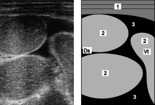
Fig. 6.8 Ultrasonogram and schematic of the abdomen in a cow with ileus due to obstruction of the jejunum with coagulated blood (hemorrhagic bowel syndrome). The jejunal loops are dilated and there is anechoic fluid (transudate) between the dilated loops. The ultrasonogram was obtained from the right abdominal wall caudal to the last rib using a 5.0 MHz-linear scanner. 1 = Lateral abdominal wall; 2 = Dilated jejunal loops; 3 = Anechoic fluid between the jejunal loops. Ds, Dorsal; Vt, Ventral.

(Reproduced with kind permission of U. Braun.)

CONTROL

No control or prevention strategies have been developed.

REFERENCES

1 Godden S, et al. Bovine Pract. 2001;35:97.

2 Dennison AC, et al. J Am Vet Med Assoc. 2002;221:686.

3 Abutarbush SM, et al. Can Vet J. 2004;45:48.

4 Von Rademacher G, et al. Tierarztl Umsch. 2002;57:399.

5 Berghans RD, et al. J Am Vet Med Assoc. 2005;226:1700.

6 Kirkpatrick MA, et al. Bovine Pract. 2001;35:104.

7 Abutarbush SM, Radostits OM. Aust Vet J. 2005;46:711.

INTESTINAL OBSTRUCTION IN SHEEP

Intestinal obstructions are not commonly observed in sheep unless a series of them causes a noticeable mortality. Some notable occurrences have been:

Heavy infestation with nodular worm (Oesophagastomum columbianum) leading to high prevalence of intussusception occlusion by adhesion

High incidence of intussusception in traveling sheep for no apparent reason

Cecal torsion (red-gut) in sheep grazing lush pastures of alfalfa or clover in New Zealand. Affected lambs survive only a few hours and up to 20% of a flock are affected. The outstanding postmortem lesion is a distended, reddened cecum and/or colon that has undergone torsion. The rumen is smaller and the large intestine larger than normal because of the high digestibility of the diet. All ages, except sucking lambs, are affected and the mortality rate may be as high as 20%. Sheep that are seen alive have a distended abdomen, show abdominal pain and have tinkling sounds on auscultation of the right flank.

TERMINAL ILEITIS OF LAMBS

This disease causes poor growth in lambs 4–6 months old. The circumstances usually suggest parasitism or coccidiosis. The terminal 50–75 cm of the ileum is thickened and resembles the classical lesion of Johne’s disease. Chronic inflammation is evident and there are some shallow ulcers in the epithelium. The terminal mesenteric lymph node is enlarged. Histopathological examination of affected ileal wall shows mucosa thickened by epithelial hyperplasia, leukocytic infiltration and connective tissue infiltration. The cause is unknown, and the course of the disease has not been identified because most affected lambs are likely to be culled for ill-thrift.