PULMONARY HEMORRHAGE

Pulmonary hemorrhage is uncommon in farm animals but does occur occasionally in cattle, and exercise-induced pulmonary hemorrhage (EIPH) occurs in 45–75% of exercised horses. Pulmonary hemorrhage also occurs in horses with pulmonary abscesses, tumors or foreign bodies. Tracheobronchoscopic, radiographic and ultrasonographic examinations are useful in identifying the site and cause of the hemorrhage.

Cattle

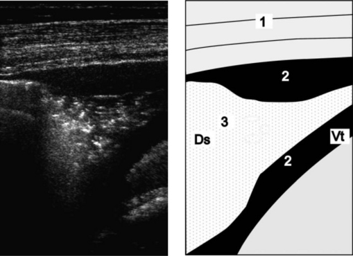
In cattle the most common cause is erosion of pulmonary vessels adjacent to lesions of embolic pneumonia associated with vena caval thrombosis and hepatic abscessation. The onset of hemorrhage may be sudden and affected animals hemorrhage profusely and die after a short course of less than 1 hour. Marked epistaxis and hemoptysis, severe dyspnea, muscular weakness and pallor of the mucous membranes are characteristic. In other cases, episodes of epistaxis and hemoptysis may occur over a period of several days or a few weeks along with a history of dyspnea.

EXERCISE-INDUCED PULMONARY HEMORRHAGE OF HORSES (EIPH, BLEEDERS)

Synopsis

Etiology

Pulmonary hemorrhage during exercise

Epidemiology

Present in most (> 80%) Thoroughbred and Standardbred racehorses, although clinical signs are less common. Occurs worldwide in any horse that performs strenuous exercise. Rarely causes death

Pathogenesis

Probably associated with rupture of pulmonary capillaries by the high pulmonary vascular pressures generated during exercise. There may be a contributory role for inflammation and obstruction of small airways, and tissue damage caused by large and rapid changes in intrathoracic pressure

Clinical signs

Epistaxis is an uncommon but very specific sign of EIPH in horses that have just exercised. Affected horses may cough or suddenly slow during a race. Endoscopic examination of the trachea and bronchi reveals blood

Clinical pathology

Presence of hemosiderin-laden macrophages in tracheal aspirates or bronchial lavage fluid

Lesions

Fibrosis and discoloration of the caudodorsal regions of the lungs. Fibrosis, accumulation of hemosiderin-laden macrophages in interstitial tissue, inflammation and bronchial artery angiogenesis. Horses dying acutely have blood-filled airways and heavy, wet lungs

Diagnostic confirmation

Demonstration of blood in the trachea or bronchi by endoscopic examination, or cytological examination of tracheal aspirates or bronchoalveolar lavage fluid

Treatment

None of demonstrated efficacy. Furosemide is used as prophylaxis

Control

There are no specific control measures, however, prevention of environmental and infectious respiratory disease may reduce the incidence of the disease

Etiology

EIPH occurs in horses during strenuous exercise.

Epidemiology

EIPH is primarily a disease of horses, although it has been reported in racing camels.1 EIPH occurs in horses worldwide and there does not appear to be any geographical distribution. It is a disorder of horses that run at high speed, such as Thoroughbred or Standardbred racehorses. The disorder is uncommon in endurance horses or draft breeds, although it does occur in horses used for these activities. As a general rule, the more intense the exercise or the higher the speed attained, the greater the proportion of horses with EIPH.

The prevalence of EIPH varies with the method used to detect it and the frequency with which horses are examined, as discussed later in this section. Epistaxis associated with exercise is almost always attributable to pulmonary hemorrhage and occurs only in a small proportion of racehorses.2-5 Epistaxis occurs in only 3% of horses that have blood detected in the trachea by endoscopic examination performed within 2 hours of racing.5 The prevalence of epistaxis in racehorses varies between 0.1 and 9.0%, with the frequency depending on the breed, age and sex of horses selected for study, the type of racing and the timing and frequency of observation of horses after racing. Epistaxis is more common in older horses.2,3 There are conflicting reports of a sex predisposition, although epistaxis may be more common in female Thoroughbreds.2,3 Epistaxis is more common after races of less than 1600 m than in longer races,2 although not all sources agree on this point.3,6 However, horses in steeplechase races, which are typically longer than 2000 m, are at greater risk of epistaxis than are horses in flat races.2,6 Epistaxis is relatively uncommon and most horses with EIPH do not have epistaxis.

There are a variety of other methods of detecting EIPH, including endoscopic examination of the airways and microscopic examination of tracheal aspirates or bronchoalveolar lavage fluid.

Almost all Thoroughbred racehorses in active training have hemosiderophages in bronchoalveolar lavage fluid, indicating that all have some degree of EIPH.7 The prevalence of EIPH decreases when diagnosis is based on endoscopic examination of horses after exercise or racing.

Exercise-induced pulmonary hemorrhage is very common in Thoroughbred racehorses, with estimates of prevalence, based on a single endoscopic examination of the trachea and bronchi, of 43–75%.6,8-10 The prevalence increases with the frequency of examination, with over 80% of horses having evidence of EIPH on at least one occasion after examination after each of three consecutive races.11 The prevalence of EIPH in Standardbred racehorses is assumed to be lower, with 26–34% of horses reported to have blood in the trachea after racing.12,13 However, these studies were based on a single examination and one12 only reported as positive those horses with blood covering more than one half the tracheobronchial tree. When examined after each of three races, 87% of Standardbred racehorses have evidence of EIPH on at least one occasion,14 suggesting that EIPH is as common in Standardbred racehorses as it is in Thoroughbred racehorses.

Exercise-induced pulmonary hemorrhage occurs in approximately 62% of racing Quarter horses, and has been observed in Quarter horses used for barrel racing.15 The disorder occurs in racing Appaloosa horses.16 Approximately 11% of polo ponies are affected with EIPH.17 The disease occurs in draft horses but is not well documented.

Age is considered a risk factor for EIPH, with the prevalence of the disorder being higher in older horses.8-10 There is no consistent association of sex with prevalence of EIPH.8-1013 Among Thoroughbred racehorses the prevalence of EIPH increases with increasing speed,10,18 being greater in Thoroughbreds after racing than after breezing (galloping). Lesions of EIPH are not detected in young Thoroughbred racehorses that have trained at speeds of less than 7 m/s.10,18

Pathogenesis

The cause of EIPH is rupture of alveolar capillary membranes with subsequent extravasation of blood into interstitial and alveolar spaces.19 The source of blood in such instances is the pulmonary circulation. Bleeding from bronchial circulation during exercise has been suggested, based on histological evidence of bronchial angiogenesis in horses that have experienced previous episodes of EIPH,20 but contribution of the bronchial circulation to EIPH has not been demonstrated. Regardless of the contribution of bronchial circulation to blood in the airways, the likely initial lesion is in capillaries associated with the pulmonary circulation. Hemorrhage into the interstitial space and alveoli, with subsequent rostral movement of blood in the airways, results in blood in the trachea and bronchi.

Rupture of alveolar capillaries occurs secondary to an exercise-induced increase in transmural pressure (pressure difference between the inside of the capillary and the alveolar lumen). If the transmural stress exceeds the tensile strength of the capillary wall, the capillary ruptures.19 The proximate cause of alveolar capillary rupture is the high transmural pressure generated by positive intracapillary pressures, which are largely attributable to capillary blood pressure, and the lower intra-alveolar pressure generated by the negative pleural pressures associated with inspiration.

During exercise, the absolute magnitudes of both pulmonary capillary pressure and alveolar pressure increase, with a consequent increase in transmural pressure. Strenuous exercise is associated with marked increases in pulmonary artery pressure in horses.22-24 Values for mean pulmonary arterial pressure at rest of 20–25 mmHg increase to more than 90 mmHg during intense exercise because of the large cardiac output achieved by exercising horses. The increases in pulmonary artery pressure, combined with an increase in left atrial pressure during exercise, probably result in an increase in pulmonary capillary pressure. Combined with the increase in pulmonary capillary pressure is a marked decrease (more negative) in pleural, and therefore alveolar, pressure during exercise. The pleural pressure of normal horses during inspiration decreases from approximately –0.7 kPa (–5.3 mmHg) at rest to as low as –8.5 kPa (64 mmHg) during strenuous exercise.25 Together, the increase in pulmonary capillary pressure and decrease (more negative) in intrapleural (alveolar) pressure contribute to a marked increase in stress in the alveolar wall. Although the alveolar wall and pulmonary capillaries of horses are stronger than those of other species, rupture may occur because the wall stress in the alveolus exceeds the mechanical strength of the capillary.26

Other theories of the pathogenesis of EIPH include: small-airway disease, upper airway obstruction, hemostatic abnormalities, changes in blood viscosity and erythrocyte shape, intrathoracic sheer forces associated with gait, and bronchial artery angiogenesis.20,27 It is likely that the pathogenesis of EIPH involves several processes, including pulmonary hypertension, lower alveolar pressure and changes in lung structure, that summate to induce stress failure of pulmonary capillaries.

Obstruction of either the upper or lower airways has been proposed as a cause of EIPH. Inspiratory airway obstruction results in more negative intrapleural, and therefore alveolar, pressures. This effect is exacerbated by exercise, with the result that alveolar transmural pressure is greater in horses with airway obstruction.28,29 The higher transmural pressure in such horses may increase the severity of EIPH, although this has not been demonstrated. Moreover, while inspiratory airway obstruction may predispose to EIPH, the prevalence of this condition is much less than that of EIPH, indicating that it is not the sole factor inducing EIPH in most horses.

Horses with moderate to severe EIPH have histological evidence of inflammation of the small airways,18,30 and there is a clear association between the presence of EIPH and inflammatory changes in bronchoalveolar or tracheal aspirate fluid.6 However, instillation of autologous blood into the airways induces a marked inflammatory response in normal horses,31 and it is therefore unclear whether inflammation alone induces or predisposes to EIPH or whether the inflammation is a result of EIPH. Theoretically, small-airway inflammation and bronchoconstriction have the potential to produce intrathoracic airway obstruction and, therefore, a more negative alveolar pressure. Given that small-airway disease is common in horses, there is the potential for an important effect of factors, such as viral infections, air pollution and allergic airway disease, to contribute to the initiation or propagation of EIPH.

The characteristic location of lesions of EIPH in the caudodorsal lung fields has led to the proposal that hemorrhage is a result of tissue damage occurring when waves of stress, generated by forelimb foot strike, are focused and amplified into the narrowing cross-sectional area of the caudal lung lobes.27 According to the theory, the locomotor impact of the forelimbs results in transmission of forces through the scapula to the body wall, from where they pass into the lungs and caudally and dorsally. As the wave of pressure passes into the narrower caudodorsal regions of the lungs it generates progressively greater shearing forces that disrupt tissue and cause EIPH. However, studies of intrapleural pressures have not demonstrated the presence of a systemic pressure wave passing through the lung and do not provide support for this hypothesis.32

Horses with EIPH have been suspected of having defects in either hemostasis or fibrinolysis. However, while exercise induces substantial changes in blood coagulation and fibrinolysis, these is no evidence that horses with EIPH have defective coagulation or increased fibrinolysis.33,34

Regardless of the cause, rupture of pulmonary capillaries and subsequent hemorrhage into airways and interstitium causes inflammation of both airways and interstitium with subsequent development of fibrosis and alteration of tissue compliance. Heterogeneity of compliance within the lungs, and particularly at the junction of normal and diseased tissue, results in the development of abnormal shear stress with subsequent tissue damage. These changes are exacerbated by inflammation and obstruction of small airways, with resulting uneven inflation of the lungs.35 The structural abnormalities, combined with pulmonary hypertension and the large intrathoracic forces associated with respiration during strenuous exercise, cause repetitive damage at the boundary of normal and diseased tissue with further hemorrhage and inflammation. The process, once started, is lifelong and continues for as long as the horse continues to perform strenuous exercise.20

Clinical findings

Poor athletic performance or epistaxis are the most common presenting complaints for horses with EIPH. While poor performance may be attributable to any of a large number of causes, epistaxis associated with exercise is almost always secondary to EIPH.

Epistaxis due to EIPH occurs during or shortly after exercise and is usually first noticed at the end of a race, particularly when the horse is returned to the paddock or winner’s circle and is allowed to lower its head. It is usually bilateral and resolves within hours of the end of the race. Epistaxis may occur on more than one occasion, especially when horses are raced or exercised at high speed soon after an initial episode.

Exercise-induced pulmonary hemorrhage and performance

Failure of racehorses to perform to the expected standard (poor performance) is often, accurately or not, attributed to EIPH. Many horses with poor performance have cytological evidence of EIPH on microscopic examination of tracheobronchial aspirates or bronchoalveolar lavage fluid or have blood evident on endoscopic examination of the tracheobronchial tree performed 30–90 minutes after strenuous exercise or racing.7,36 However, it is important to recognize that EIPH is very common in racehorses and it should be considered the cause of poor performance only after other causes have been eliminated. Severe EIPH undoubtedly results in poor performance and, on rare occasions, death of Thoroughbred racehorses.37 Thoroughbred horses with EIPH racing in Victoria, Australia have impaired performance compared to unaffected horses. Affected horses have a lower likelihood of finishing in the first three places, are less likely to be elite money earners and finish further behind the winner than do unaffected horses.5

Results of studies in Standardbred racehorses indicate either a lack of effect of EIPH on performance or an association between EIPH and superior performance. There was no relationship between presence of EIPH and finishing position in 29 Standardbred racehorses with intermittent EIPH examined on at least two occasions,14 nor in 92 Standardbred racehorses examined on one occasion.13 However, of 965 Standardbred racehorses examined after racing, those finishing first or second were 1.4 times more likely (95% confidence interval 0.9–2.2) to have evidence of EIPH on tracheobronchoscopic examination than were horses that finished in seventh or eighth position.38

Physical examination

Apart from epistaxis in a small proportion of affected horses, there are few abnormalities detectable on routine physical examination of horses with EIPH. Rectal temperature and heart and breathing rates may be elevated as a consequence of exercise in horses examined soon after exercise, but values of these variables in horses with EIPH at rest are not noticeably different from horses with no evidence of EIPH. Affected horses may swallow more frequently during recovery from exercise than do unaffected horses, probably as a result of blood in the larynx and pharynx. Coughing is common in horses recovering from strenuous exercise and after recovery from exercise; horses with EIPH are no more likely to cough than are unaffected horses. Other clinical signs related to respiratory abnormalities are uncommon in horses with EIPH. Respiratory distress is rare in horses with EIPH and, when present, indicates severe hemorrhage or other serious lung disease such as pneumonia, pneumothorax or rupture of a pulmonary abscess. Lung sounds are abnormal in a small number of EIPH-affected horses and when present are characterized by increased intensity of normal breath sounds during rebreathing examination. Tracheal rales may be present in horses with EIPH but are also heard in unaffected horses.

Tracheobronchoscopy

Observation of blood in the trachea or large bronchi of horses 30–120 minutes after racing or strenuous exercise provides a definitive diagnosis of EIPH. The amount of blood in the large airways varies from a few small specks on the airway walls to a stream of blood occupying the ventral one-third of the trachea. Blood may also be present in the larynx and nasopharynx. If there is a strong suspicion of EIPH and blood is not present on a single examination conducted soon after exercise, the examination should be repeated in 60–90 minutes. Some horses with EIPH do not have blood present in the rostral airways immediately after exercise, but do so when examined 1–2 hours later. Blood is detectable by tracheobronchoscopic examination for 1–3 days in most horses, with some horses having blood detectable for up to 7 days.

Bronchoscopic examination can be used to estimate the severity of EIPH through the use of a grading system.39,40 The interobserver repeatability of tracheobronchoscopic assessment of severity of EIPH using a 0–4 grading scale is excellent:40

Grade 0: No blood detected in the pharynx, larynx, trachea or main stem bronchi

Grade 1: Presence of one or more flecks of blood or ≤ 2 short (< quarter the length of the trachea) narrow (< 10% of the tracheal surface area) streams of blood in the trachea or main stem bronchi visible from the tracheal bifurcation

Grade 2: One long stream of blood (> half the length of the trachea) or > 2 short streams occupying less than one-third of the tracheal circumference

Grade 3: Multiple, distinct streams of blood covering more than one-third of the tracheal circumference. No blood pooling at the thoracic inlet

Grade 4. Multiple, coalescing streams of blood covering > 90% of the tracheal surface with pooling of blood at the thoracic inlet.

It is assumed that a higher score represents more severe hemorrhage, but while the repeatability of this scoring system has been established, the relationship between the amount of blood in the large airways and the actual amount of hemorrhage has not been established.

Radiography

Thoracic radiography is of limited use in detecting horses with EIPH. Radiographs may demonstrate the presence of densities in the caudodorsal lung fields of some horses but many affected horses have minimal to undetectable radiographic abnormalities.41 Examination of thoracic radiographs of horses with EIPH may be useful in ruling out the presence of another disease process, such as a pulmonary abscess, contributing to the horse’s pulmonary hemorrhage or poor athletic performance.

Prognosis

Horses that have experienced one episode of epistaxis are more likely to have a second episode. For this reason most racing jurisdictions do not permit horses with epistaxis to race for a period of weeks to months after the initial instance, with more prolonged enforced rest after a subsequent episode of epistaxis and retirement from racing after a third bout. The recurrence rate after one episode of epistaxis in Thoroughbred horses is approximately 13.5% despite affected horses not being permitted to race for 1 month after the initial episode.2 This high rate of recurrence suggests that the inciting pulmonary lesions have not healed.

Clinical pathology

Examination of airway secretions or lavage fluid

The presence of red cells or macrophages containing either effete red cells or the breakdown products of hemoglobin (hemosiderophages) in tracheal or bronchoalveolar lavage fluid provides evidence of EIPH. Detection of red cells or hemosiderophages in tracheal aspirates or bronchoalveolar lavage fluid is believed to be both sensitive and specific in the diagnosis of EIPH.7 Examination of airway fluids indicates the presence of EIPH in a greater proportion of horses than does tracheobronchoscopic examination after strenuous exercise or racing. The greater sensitivity of examination of airway fluid is probably attributable to the ability of this examination to detect the presence of small amounts of blood or its residual products and the longevity of these products in the airways. While endoscopic examination may detect blood in occasional horses up to 7 days after an episode of EIPH, cellular evidence of pulmonary hemorrhage persists for weeks after a single episode.42 Red blood cells and macrophages containing red cells are present in bronchoalveolar lavage fluid or tracheal aspirates for at least 1 week after strenuous exercise or instillation of autologous blood into airways and hemosiderophages are present for at least 21 days and possibly longer.42

Recent studies have reported on the use of red cell numbers in bronchoalveolar lavage fluid as a quantitative indicator of EIPH. However, this indicator of EIPH severity has not been validated nor demonstrated to be more reliable or repeatable than tracheobronchoscopic examination and visual scoring. Furthermore, considerable concern exists over the suitability of red cell counts in bronchoalveolar lavage fluid for assessment of severity of EIPH given that an unknown area, although presumably small, of the lung is examined by lavage and that there is a risk that this area of lung may not be representative of the lung as a whole, similar to the situation of examination of bronchoalveolar lavage fluid of horses with pneumonia. Bronchoalveolar lavage of sections of both lungs, achieved using an endoscope, may obviate some of these concerns.

Tracheal aspirates may be obtained any time after exercise by aspiration either during tracheobronchoscopic examination or through a percutaneous intratracheal needle. Aspirates obtained through an endoscope may not be sterile, depending on the collection technique. Bronchoalveolar lavage fluid can be obtained through either an endoscope wedged in the distal airway or a cuffed tube inserted blindly into a distal airway. Collection of fluid through an endoscope has the advantage of permitting examination of the distal airways and selection of the area of lung to be lavaged. However, it does require the use of an endoscope that is longer (2 m) than those readily available in most equine practices. Use of a commercial bronchoalveolar lavage catheter does not require use of an endoscope and this procedure can be readily performed in field situations.

DIFFERENTIAL DIAGNOSIS

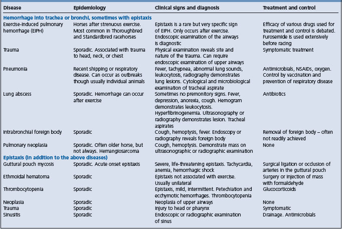
Epistaxis and hemorrhage into airways can occur as a result of a number of diseases (Table 10.5).

Table 10.5 Causes of epistaxis in horses

image

Necropsy

Exercise-induced pulmonary hemorrhage is a rare cause of death of racehorses, but among race horses that die during racing for reasons other than musculoskeletal injuries, EIPH is common.37 Necropsy examination of horses is usually incidental to examination for another cause of death. Pertinent abnormalities in horses with EIPH are restricted to the respiratory tract. Grossly, horses examined within hours of strenuous exercise, such as horses examined because of catastrophic musculoskeletal injuries incurred during racing, may have severe petechiation in the caudodorsal lung fields. Horses with chronic disease have blue/gray or blue/brown discoloration of the visceral pleural surfaces of the caudodorsal lung fields that is often sharply demarcated, especially on the diaphragmatic surface. The discoloration affects both lungs equally with 30–50% of the lung fields being discolored in severe cases. Affected areas do not collapse to the same extent as unaffected areas and, in the deflated lung, have a spleen-like consistency. On cut surface, the discolored areas of lung are predominantly contiguous with the dorsal pleural surface and extend ventrally into the lung parenchyma. Areas of affected lung may be separated by normal lung. There is proliferation of bronchial vessels, predominantly arteries and arterioles, in affected areas. Histologically, affected areas exhibit bronchiolitis, hemosiderophages in the alveolar lumen and interstitial spaces, and fibrosis of interlobular septa, pleural and around vessels and bronchioles.

Treatment

Therapy of EIPH is usually a combination of attempts to reduce the severity of subsequent hemorrhage and efforts to minimize the effect of recent hemorrhage. Treatment of EIPH is problematic for a number of reasons. Firstly, the pathogenesis of EIPH has not been determined although the available evidence supports a role for stress failure of pulmonary capillaries secondary to exercise-induced pulmonary hypertension. Secondly, there is a lack of information using large numbers of horses under field conditions that demonstrates an effect of any medication or management practice (with the exception of bedding) on EIPH. There are numerous studies of small numbers of horses (< 40) under experimental conditions but these studies often lacked the statistical power to detect treatment effects and, furthermore, the relevance of studies conducted on a treadmill to horses racing competitively is questionable. Treatments for EIPH are usually intended to address a specific aspect of the pathogenesis of the disease and will be discussed in that context.

Prevention of stress failure of the pulmonary capillaries

There is interest in reducing the pressure difference across the pulmonary capillary membrane in an effort to reduce EIPH. Theoretically, this can be achieved by reducing the pressure within the capillary or increasing (making less negative) the pressure within the intrathoracic airways and alveolus.

Reducing pulmonary capillary pressure

Furosemide administration as prophylaxis of EIPH is permitted in a number of racing jurisdictions worldwide, most notably Canada, the USA, Mexico and most of the South American countries. Within the USA and Canada, almost all Thoroughbred, Standardbred and Quarter horse racing jurisdictions permit administration of furosemide before racing.

The efficacy of furosemide in treatment of EIPH is uncertain. While field studies of large numbers of horses do not demonstrate an effect of furosemide on the prevalence of EIPH,43 studies of Thoroughbred horses running on a treadmill provide evidence that furosemide reduces the severity of EIPH.44 Under field conditions, based on tracheobronchoscopic evaluation of the severity of bleeding, furosemide has been reported to reduce or have no influence on the severity of bleeding.43,45 This apparent inconsistency may be attributable to measurement of red blood cell counts in bronchoalveolar lavage fluid of horses that have run on a treadmill not being representative of effects of furosemide under field conditions. The weight of evidence, albeit unconvincing, from field studies does not support a role for furosemide in preventing or reducing the severity of EIPH.

The mechanism by which furosemide may reduce the severity of EIPH is unknown, although it is speculated that furosemide, by attenuating the exercise-induced increase in pulmonary artery and pulmonary capillary pressure of horses, reduces the frequency or severity of pulmonary capillary rupture.

Furosemide is associated with superior performance in both Thoroughbred and Standardbred racehorses,46,47 which further complicates assessment of its efficacy in treating EIPH.

An increase in pulmonary capillary pressure secondary to altered rheostatic properties of blood during exercise has been suggested as a possible contributing factor for EIPH.48

Increasing alveolar inspiratory pressure

Airway obstruction, either intrathoracic or extrathoracic, increases airway resistance and results in a more negative intrathoracic (pleural) pressure during inspiration to maintain tidal volume and alveolar ventilation. Causes of extrathoracic airway obstruction include laryngeal hemiplegia and other abnormalities of the upper airway, whereas intrathoracic obstruction is usually a result of bronchoconstriction and inflammatory airway disease. Horses with partial extrathoracic inspiratory obstruction or bronchoconstriction and airway inflammation associated with recurrent airway obstructive disease (heaves) have pleural (and hence alveolar) pressures that are lower (more negative) than those in unaffected horses or in horses after effective treatment.

Partial inspiratory obstruction, such as produced by laryngeal hemiplegia, exacerbates the exercise-induced decrease in intrapleural pressures with a consequent increase in transmural capillary pressures.28,29 These changes may exacerbate the severity of EIPH although an association between upper airway obstructive disease and EIPH has not been demonstrated. Surgical correction of airway obstruction is expected to resolve the more negative intrapleural pressure, but its effect on EIPH is unknown.

Recently, the role of the nares in contributing to upper airway resistance, and hence lowering inspiratory intrapleural pressure during intense exercise, has attracted the attention of some investigators. Application of nasal dilator bands (Flair® strips) reduces nasal resistance by dilating the nasal valve and reduces red cell count of bronchoalveolar lavage fluid collected from horses after intense exercise on a treadmill.44 Furthermore, application of the nasal dilator strips to horses in simulated races reduces red cell count in bronchoalveolar lavage fluid of some, but not all, horses.49

The role of small-airway inflammation and bronchoconstriction in the pathogenesis of EIPH is unclear. However, horses with EIPH are often treated with drugs intended to decrease lower airway inflammation and relieve bronchoconstriction. Beta-adrenergic bronchodilatory drugs such as clenbuterol and albuterol (salbutamol) are effective in inducing bronchodilation in horses with bronchoconstriction, but their efficacy in preventing EIPH is either unknown or, in very small studies, is not evident. Corticosteroids, including dexamethasone, fluticasone and beclomethasone administered by inhalation, parenterally or enterally reduce airway inflammation and obstruction but have no demonstrated efficacy in preventing EIPH. Cromolyn sodium (sodium cromoglycate) has no efficacy in preventing EIPH.

Water vapor treatment (inhalation of water-saturated air) has been proposed as a treatment for EIPH because of its putative effect on small-airway disease. However, water vapor treatment has no effect on EIPH.

The use of bedding of low allergenic potential (shredded paper) to prevent EIPH has no apparent effect on prevalence of the condition.50 While it is suggested that preventing or minimizing small-airway disease may reduce the severity of EIPH, studies to demonstrate such an effect have not been reported. However, optimizing the air quality in barns and stables and preventing infectious respiratory disease appear sensible precautions.

Interstitial inflammation and bronchial angiogenesis

Hemorrhage into interstitial tissues induces inflammation with subsequent development of fibrosis and bronchial artery angiogenesis.30,42 The role of these changes in perpetuating EIPH in horses is unclear but is probably of some importance. Treatments to reduce inflammation and promote healing with minimal fibrosis have been proposed. Rest is an obvious recommendation and many racing jurisdictions have rules regarding enforced rest for horses with epistaxis. While the recommendation for rest is intuitive, there is no information that rest reduces the severity or incidence of EIPH in horses with prior evidence of this disorder.

Similarly, corticosteroids are often administered, either by inhalation, enterally or parenterally, in an attempt to reduce pulmonary inflammation and minimize fibrosis. Again, the efficacy of this intervention in preventing or minimizing severity of EIPH has not been documented.

Excessive bleeding
Coagulopathy and fibrinolysis

Exercise induces substantial changes in blood coagulation and fibrinolysis. However, there is no evidence that horses with EIPH have defective coagulation or increased fibrinolysis.33,34 Regardless, aminocaproic acid, a potent inhibitor of fibrin degradation, has been administered to horses to prevent EIPH. The efficacy of aminocaproic acid in preventing EIPH has not been demonstrated. Similarly, estrogens are given to horses with the expectation of improving hemostasis, although the effect of estrogens on coagulation in any species is unclear. There is no evidence that estrogens prevent EIPH in horses.

Vitamin K is administered to horses with EIPH, presumably in the expectation that it will decrease coagulation times. However, as EIPH is not associated with prolonged bleeding times, it is unlikely that this intervention will affect the prevalence or severity of EIPH.

Platelet function

Aspirin inhibits platelet aggregation in horses and increases bleeding time. Seemingly paradoxically, aspirin is sometimes administered to horses with EIPH because of concerns that increased platelet aggregation contributes to EIPH. There is no evidence that aspirin either exacerbates or prevents EIPH.

Capillary integrity

Capillary fragility increases the risk of hemorrhage in many species. Various bioflavonoids have been suggested to increase capillary integrity and prevent bleeding. However, hesperidin and citrus bioflavonoids have no efficacy in prevention of EIPH in horses. Similarly, vitamin C is administered to horses with EIPH without scientific evidence of any beneficial effect.

Summary of treatment options

Selection of therapy for horses with EIPH is problematic. Given that most horses have some degree of pulmonary hemorrhage during most bouts of intense exercise, the decision must be made not only as to the type of treatment and its timing but also which horses to treat. Moreover, the apparently progressive nature of the disease with continued work highlights the importance of early and effective prophylaxis and emphasizes the need for studies of factors such as air quality and respiratory infections in inciting the disorder.

The currently favored treatment for EIPH is administration of furosemide before intense exercise. Its use is permitted in racehorses in a number of countries. Increasingly persuasive laboratory evidence of an effect of furosemide in reducing red cell count in bronchoalveolar lavage fluid collected from horses soon after intense exercise supports the contention that furosemide is effective in reducing the severity of EIPH in race horses. However, it should be borne in mind that neither the relationship between severity of EIPH and red cell count in bronchoalveolar lavage fluid nor the efficacy of furosemide in reducing severity of EIPH in race horses in the field has been demonstrated. In fact, there is evidence that furosemide does not reduce the prevalence of EIPH and other evidence that it does not reduce the severity of EIPH under field conditions. The association between furosemide administration and superior performance in Standardbred and Thoroughbred racehorses should be borne in mind when recommending use of this drug.

Prevention and control

There are no documented preventive strategies. Rest is an obvious recommendation for horses with EIPH, but the hemorrhage is likely to recur when the horse is next strenuously exercised. The duration of rest and the optimal exercise program to return horses to racing after EIPH is unknown, although some jurisdictions require exercise no more intense than trotting for 2 months. Firm recommendations cannot be made on duration of rest because of a lack of objective information.

Although a role for lower airway disease (either infectious or allergic) in the genesis of EIPH has not been demonstrated, control of infectious diseases and minimization of noninfectious lower airway inflammation appears prudent.

Concern about the role of impact waves in the genesis of EIPH has led to discussion of ‘low-stress’ training protocols, but these have not been adequately evaluated.

REFERENCES

1 Akbar SJ, et al. Vet Rec. 1994;135:624.

2 Takahashi T, et al. J Am Vet Med Assoc. 2001;218:1462.

3 Weideman H, et al. J S Afr Vet Assoc. 2003;74:127.

4 Williams R, et al. Equine Vet J. 2001;33:478.

5 Hinchcliff KW, et al. J Am Vet Med Assoc. 2005;227:768.

6 Newton JR, Wood JLN. Equine Vet J Suppl. 2002;34:417.

7 McKane SA, et al. Aust Vet J. 1993;70:401.

8 Mason DK, et al. Snow DH, Persson SGB, Rose RJ, editors. Equine exercise physiology. Cambridge: Granta. 1983:57.

9 Pascoe J, et al. Am J Vet Res. 1981;42:703.

10 Raphel CF, Soma LR. Am J Vet Res. 1982;43:11237.

11 Sweeney CR, et al. Am J Vet Res. 1990;51:772.

12 MacNamara B, et al. J Am Vet Med Assoc. 1990;196:443.

13 Speirs VC, et al. Aust Vet J. 1982;59:38.

14 Lapointe JM, et al. Equine Vet J. 1994;26:482.

15 Hillidge CJ, et al. J Equine Vet Sci. 1984:4.

16 Hillidge CJ, et al. J Equine Vet Sci. 1986;5:351.

17 Voynick BT, Sweeney CR. J Am Vet Med Assoc. 1986;188:301.

18 Oikawa M. J Comp Pathol. 1999;121:339.

19 West JB, et al. J Appl Physiol. 1993;75:1097.

20 Pascoe JR. Proc Am Assoc Equine Pract. 1996;42:220.

21 West JB, Mathieu-Costello O. Equine Vet J. 1994;26:441.

22 Birks EK, et al. J Appl Physiol. 1997;82:1584.

23 Langsetmo I, et al. Equine Vet J. 2000;32:379.

24 Manohar M, et al. Br Vet J. 1993;149:419.

25 Art T, et al. Respir Physiol. 1990;82:279.

26 Birks EK, et al. Respir Physiol. 1994;97:235.

27 Schroter RC, et al. Equine Vet J. 1998;30:186.

28 Hackett RP, et al. Am J Vet Res. 1999;60:485.

29 Ducharme NG, et al. Equine Vet J Suppl. 1999;30:27.

30 O’Callaghan MW, et al. Equine Vet J. 1987;19:411.

31 McKane SA, Slocombe RF. Equine Vet J Suppl. 2002;34:451.

32 Jones JH, et al. Equine Vet J Suppl. 2002;34:391.

33 Bayly WM, et al. Snow DH, Persson SGB, Rose RJ, editors. Equine exercise physiology. Cambridge: Granta. 1983:64.

34 Johnstone IB, et al. Can J Vet Res. 1991;55:101.

35 Robinson NE, Derksen FJ. Proc Am Assoc Equine Pract. 1980;26:421.

36 Martin BBJr, et al. J Am Vet Med Assoc. 1999;214:673.

37 Boden LA, et al. Equine Vet J. 2005;37:269.

38 Rohrbach BW. J Am Vet Med Assoc. 1990;196:1563.

39 Pascoe JR, et al. Am J Vet Res. 1981;42:703.

40 Hinchcliff KW, et al. Am J Vet Res. 2005;66:596.

41 Pascoe JR, et al. Vet Rad. 1983;24:85.

42 McKane S, Slocombe R. Equine Vet J. 1999;30:126.

43 Birks EK, et al. Equine Vet J Suppl. 2002;34:375.

44 Geor RJ, et al. Equine Vet J. 2001;33:577.

45 Pascoe JR, et al. Am J Vet Res. 1985;46:2000.

46 Gross DK, et al. J Am Vet Med Assoc. 1999;215:670.

47 Soma LR, et al. Equine Vet J. 2000;32:334.

48 Fedde MR, Erickson HH. Equine Vet J. 1998;30:329.

49 Valdez S, et al. J Am Vet Med Assoc. 2004;224:558.

50 Mason DK, et al. Vet Rec. 1984;115:268.

PULMONARY EMPHYSEMA

Pulmonary emphysema is distension of the lung caused by overdistension of alveoli with rupture of alveolar walls with or without escape of air into the interstitial spaces. Overinflation describes the situation in which there is enlargement of airspaces without tissue destruction. Pulmonary emphysema is always secondary to some primary lesion which effectively traps an excessive amount of air in the alveoli. It is a common clinicopathological finding in many diseases of the lungs of all species and is characterized clinically by dyspnea, hyperpnea, poor exercise tolerance and forced expiration.

ETIOLOGY

Pulmonary emphysema is an important lesion only in cattle, although occasional cases occur in pigs. The bovine lung is highly susceptible to the development of emphysema from many different causes, not all of them respiratory in origin. In those of respiratory origin it is common to find pulmonary emphysema when the primary lesion in the lung causes trapping of air in alveoli or terminal bronchioles. Endotoxemia, for example, can result in diffuse alveolar damage associated with thromboangiitis resulting in pulmonary edema and emphysema. Some causes of emphysema are as follows:

Cattle

Acute interstitial pneumonia

Parasitic pneumonia with pulmonary edema in acute anaphylaxis

Perforation of the lung by foreign body as in traumatic reticuloperitonitis

Poisoning by the plants Senecio quadridentatus, rape, Zieria arborescens, Perilla frutescens and the fungus Periconia spp. are recorded as causing pulmonary emphysema in cattle

Pulmonary abscess.

Horses

Bronchiolitis due to viral infection of the respiratory tract in young horses.

All species

Secondary to bronchopneumonia

Poisoning by oleander, Bryophyllum pinnatum and moldy sweet potatoes1-3

Acute chemical injury – as in inhalation of welding fumes

Chlorine gas poisoning

Local or perifocal emphysema is also a common necropsy finding around local pulmonary lesions, especially atelectasis, often with no respiratory dysfunction. In calves and pigs the emphysema is sometimes sufficiently extensive to kill the animal.

PATHOGENESIS

Emphysema occurs because of destruction of the connective tissues of the lung, including the supporting and elastic tissue of the pulmonary parenchyma. Tissue damage resulting in emphysema in humans is caused by the action of proteases in the lung. Whether this occurs in the farm animal species is unknown but is a consideration. An initial lesion probably leads to an area of weakness from which emphysema spreads during coughing or exertion. In interstitial emphysema there is the additional factor of distension of the connective tissue with air and compression collapse of the alveoli.

The development of interstitial emphysema depends largely upon the amount of interstitial tissue that is present and is most common in cattle and pigs. Whether there is simple overdistension of alveoli or whether their walls are also ruptured is very important in prognosis and treatment. Excellent recoveries occur in simple alveolar emphysema, especially those occurring acutely at pasture. This suggests that the lesion is functional and that the alveoli are not substantially damaged.

The pathophysiological consequences of emphysema depend upon the inefficiency of evacuation of pulmonary air-space and failure of normal gaseous exchange in the lungs. The elastic recoil of the tissue is diminished, and when the thorax subsides during expiration incomplete evacuation occurs. Because of the increase in residual volume, the tidal volume must be increased to maintain normal gaseous exchange. Retention of carbon dioxide stimulates an increase in the depth of respiration but maximum respiratory effort necessitated by exercise cannot be achieved. Anoxia develops and metabolism of all body tissues is reduced. The characteristic effect of emphysema is to produce an increase in expiratory effort necessitated by the failure of normal elastic recoil.

Interference with the pulmonary circulation results from collapse of much of the alveolar wall area and a consequent diminution of the capillary bed. The decreased negative pressure in the chest and the abnormally wide respiratory excursion also cause a general restriction of the rate of blood flow into the thorax. The combined effect of these factors may be sufficient to cause failure of the right ventricle especially if there is a primary defect of the myocardium. Acidosis may also result because of the retention of carbon dioxide.

CLINICAL FINDINGS

Characteristically, diffuse pulmonary emphysema causes severe expiratory dyspnea with a grunt on expiration and loud crackling lung sounds on auscultation over the emphysematous lungs. In severe cases in cattle, the emphysema is commonly interstitial and dissection of the mediastinum and fascial planes results in subcutaneous emphysema over the withers. In severe cases in cattle, open-mouth breathing is common.

In cattle and pigs the presence of pulmonary emphysema in pulmonary disease is often not detectable clinically.

CLINICAL PATHOLOGY

There is hypoxemia and, often, hypercapnia. Compensatory polycythemia may develop. There are no characteristic hematological findings but, if there is a significant secondary bronchopneumonia, a leukocytosis and left shift may be evident. In the appropriate location, an examination of feces for lungworm larvae may be desirable. In cases suspected of having an allergic origin, swabs of nasal secretion may reveal a high proportion of eosinophils and a hematological examination may show eosinophilia.

NECROPSY FINDINGS

The lungs are distended and pale in color and may bear imprints of the ribs. In interstitial emphysema the interalveolar septae are distended with air, which may spread to beneath the pleura, to the mediastinum and under the parietal pleura. There may be evidence of congestive heart failure. On histopathological examination a bronchiolitis is present in most cases. This may be diffuse and apparently primary or originate by spread from a nearby pneumonia.

TREATMENT

The treatment of pulmonary emphysema will depend on the species affected, the cause of the emphysema and the stage of the disease.

There is no known specific treatment for the pulmonary emphysema associated with acute interstitial pneumonia in cattle, which is discussed under that heading. The emphysema secondary to the infectious pneumonias will usually resolve spontaneously if the primary lesion of the lung is treated effectively. In valuable animals, the administration of oxygen may be warranted if the hypoxia is severe and life-threatening. Antihistamines, atropine and corticosteroids have been used for the treatment of pulmonary emphysema secondary to interstitial pneumonia in cattle but their efficacy has been difficult to evaluate.

DIFFERENTIAL DIAGNOSIS

Acute emphysema in cattle is often accompanied by pulmonary edema with the presence of consolidation and crackles in the ventral parts of the lungs. It may be similar to acute pulmonary congestion and edema caused by anaphylaxis but forced expiration is not a characteristic of these latter conditions.

Acute pneumonia in cattle or horses is characterized by fever and localization of abnormal respiratory sounds, which are not as marked nor as widely distributed as those of emphysema.

Chronic pneumonia is characterized by dyspnea, chronic toxemia, crackles and wheezes and poor response to therapy.

Pneumothorax is accompanied by forced inspiration and an absence of normal breath sounds.

REFERENCES

1 Oryan A, et al. Zentralbl Vet A. 1996;43:625.

2 Reppas GP. Aust Vet J. 1995;72:425.

3 Medeiros RM, et al. Vet Hum Toxicol. 2001;43:205.

PNEUMONIA

Pneumonia is inflammation of the pulmonary parenchyma usually accompanied by inflammation of the bronchioles and often by pleuritis. It is manifested clinically by an increase in the respiratory rate, changes in the depth and character of respirations, coughing, abnormal breath sounds on auscultation and, in most bacterial pneumonias, evidence of toxemia.

ETIOLOGY

Pneumonia may be associated with viruses, bacteria, or a combination of both, fungi, metazoan parasites and physical and chemical agents. Most of the pneumonias in animals are bronchogenic (inhalation) in origin but some originate by the hematogenous route, such as pneumonia of foals and calves with septicemia. The pneumonias which occur in farm animals are grouped here according to species.

Cattle

Pneumonic pasteurellosis (shipping fever) – M. haemolytica, P. multocida with or without parainfluenza-3 virus

Histophilus somnus in feedlot cattle is not necessarily associated with the septicemic form of the disease. The role of the organism as a primary pathogen in acute bovine respiratory disease is uncertain

Enzootic pneumonia of calves – parainfluenza-3, adenovirus-1, -2 and -3, rhinovirus, bovine respiratory syncytial virus, reovirus, bovid herpesvirus-1 (the IBR virus), plus Chlamydia spp., Mycoplasma spp., Pasteurella spp., Mannheimia spp., Actinomyces pyogenes, Streptococcus spp., Bedsonia sp. and Actinobacillus actinoides

Pneumonia and arthritis in beef calves associated with Mycoplasma bovis and Mycoplasma californicum

Viral interstitial pneumonia in recently weaned beef calves associated with bovine respiratory syncytial virus; it may also occur in yearling and adult cattle

Contagious bovine pleuropneumonia – Mycoplasma mycoides

Acute and chronic interstitial pneumonia associated with d, l-tryptophane, moldy hay and other pneumotoxic agents

Atypical interstitial pneumonia associated with ryegrass staggers in calves2

Massive infestation with pig ascarid larvae

Lungworm pneumonia – Dictyocaulus viviparus

Klebsiella pneumoniae infection in calves and nursing cows with mastitis associated with this organism

Sporadically in tuberculosis associated with M. bovis

Fusobacterium necrophorus as a complication of calf diphtheria, and sporadically in feedlot cattle

There is a preliminary report of circovirus in adult cattle with pneumonia3

Mycotic pneumonia associated with Mortierella wolfii in adult cattle.4

Pigs

Enzootic pneumonia – Mycoplasma sp. with Pasteurella sp. secondarily

Pneumonic pasteurellosis – P. multocida

Pleuropneumonia – Actinobacillus pleuropneumoniae

Interstitial pneumonia – septicemic salmonellosis

Bordetella bronchiseptica, Salmonella choleraesuis

Influenza virus5

Porcine reproductive and respiratory syndrome virus5

Haemophilus parasuis5

Actinobacillus pyogenes5

Paramyxovirus causing respiratory and central nervous system disease in pigs6

Uncommonly, lungworm pneumonia

Anthrax by inhalation, causing pulmonary anthrax.

Horses

Pleuropneumonia in mature horses due to aerobic and anaerobic bacteria.7-9 The aerobic bacteria most commonly isolated are alpha-hemolytic Streptococcus spp., Pasteurella spp., Escherichia coli and Enterobacter spp. The anaerobic bacteria most frequently isolated are Bacteroides spp., Prevotella spp., Fusobacterium spp. and Clostridium spp.9,10

Newborn foals –Streptococcus spp., E. coli, Actinobacillus equuli and other agents causing septicemia in this age group

In immunodeficient foals, pneumonia associated with adenovirus or Pneumocystis jiroveci (formerly P. carinii)11

Immunosuppression following corticosteroid therapy for other diseases12

Older foals – R. equi, equine herpesvirus-1 (the EVR virus), equine influenza virus13

Bronchointerstitial pneumonia in foals 1–8 months of age – etiology uncertain14,15

Eosinophilic pneumonia secondary to parasite migration (Parascaris equorum) or Dictyocaulus arnfieldi infection

Interstitial proliferative pneumonia in foals from 6 days to 6 months of age, and the adult form in horses 2 years of age and older16

Nicoletella semolina in adult horses17

Bordetella bronchiseptica in adult horses18

Pleuropneumonia associated with pulmonary hydatidosis in a horse19

As a sequel to strangles

Influenza20

Rarely, as a sequel to equine viral arteritis or equine viral rhinopneumonitis in adult animals

Glanders and epizootic lymphangitis (Histomonas farcinicus) usually include pneumonic lesions

Paecilomyces spp. in foals21

Mycotic pneumonia associated with Emmonsia crescens (adiaspiromycosis) in adult horses22

Strenuous exercise in very cold conditions can cause damage to the airways of horses (and probably other species).23

Sheep

Pneumonic pasteurellosis (Mannheimia spp.) as acute primary pneumonia in feedlot lambs, or secondary to parainfluenza-3 or Chlamydia sp. infection

Newborn lambs – uncommonly Streptococcus zooepidemicus, Salmonella abortus-ovis

Severe pneumonia due to Mycoplasma sp. in lambs – kageda in Iceland and Switzerland

Symptomless pneumonias without secondary infection – adenovirus, respiratory syncytial virus, reovirus, Mycoplasma spp. (including M. ovipneumoniae, M. dispar)24

Corynebacterium pseudotuberculosis – sporadic cases only

Melioidosis (Pseudomonas pseudomallei)

Lungworm (Dictyocaulus filaria)

Ovine herpesvirus-225

Progressive interstitial pneumonia (maedi) and pulmonary adenomatosis (jaagsiekte)

Carbolic dip toxicity.

Goats

Pleuropneumonia associated with Mycoplasma strain F 38 or Mycoplasma capri, a devastating disease

Chronic interstitial pneumonia with cor pulmonale as a common sequel may be associated with a number of Mycoplasma spp., but M. mycoides var. mycoides appears to be the most commonly recorded

Parainfluenza type 326

Contagious ecthyma virus27

Retrovirus infection.

All species

Toxoplasmosis – rare, sporadic cases

Systemic mycoses

Aspiration pneumonia is dealt with as a separate entity

Sporadic secondary pneumonia associated with Streptococcus sp., Corynebacterium sp., Dermatophilus sp.

Interstitial pneumonia, pulmonary consolidation and fibrosis by toxins in plants – Eupatorium glandulosum in horses, Zieria arborescens (stinkwood) in cattle, Astragalus spp. in all species.

EPIDEMIOLOGY

In addition to the infectious agents which cause the pneumonia, there are risk factors which contribute to the susceptibility of the animal. Three risk factors interact in the pathogenesis of specific pneumonias:

Animal

Environmental and management

Pathogen.

These are of paramount importance in any consideration of pneumonia and the details of the epidemiology of each specific pneumonia are presented with each specific disease in this book. As examples, some of the commonly recognized risk factors include:

The weaning of beef calves in northern climates

The long transportation of beef cattle to feedlots

The collection and mixing of animals at auction marts where they might be deprived of feed and water for prolonged periods

The transportation of Thoroughbred horses farther than 500 miles and viral respiratory tract disease or exposure to horses with respiratory tract disease12,28

Housing dairy calves in poorly ventilated overcrowded barns

Marked changes in weather.

Susceptibility to pneumonia is determined by the animal’s resistance to infection by agents that cause or predispose to pneumonia. Factors that impair innate resistance or adaptive resistance (immunity) increase the animal’s susceptibility to pneumonia. For instance, shipping not only increases the risk of exposure of animals to pathogens to which they have not been exposed but also can impair innate resistance through damage to the respiratory tract by airborne irritants, dehydration, food deprivation and the effects of stress. There is a distinct trend evident since 1994 of increasing mortality from respiratory disease among cattle in feedlots,29 although the reasons have not been identified.

PATHOGENESIS

Pulmonary defense mechanisms

Under normal conditions the major airways and the lung parenchyma prevent the entry of and neutralize or remove injurious agents, so that the lung contains very few, if any, organisms beyond the large airways. Many infections of the respiratory tract originate from aerosolized particles carrying infectious agents that arise external to or within the respiratory tract. In order to induce an infection by the aerosol route, an etiological agent must be aerosolized, survive in the aerosol, be deposited at a vulnerable site in the respiratory tract of a susceptible host, and then multiply. Thus the pathogenesis of these respiratory infections is related to the deposition of particles and infectious agents within the respiratory tract.

Under normal conditions a complex of biochemical, physiological and immunological defense mechanisms protects the respiratory tract from inhaled particles that could be injurious or infectious. The major defense mechanisms of the respiratory tract include:

Aerodynamic filtration by the nasal cavities

Sneezing

Local nasal antibody

The laryngeal reflex

The cough reflex

Mucociliary transport mechanisms

Alveolar macrophages

Systemic and local antibody systems.

Most of the research on defense mechanisms has been done in man and in laboratory animals.

Respiratory mucociliary clearance

The mucociliary escalator has important functions in the lung’s physical defenses against the constant challenge of inhaled pathogens.30 By various physical mechanisms, mucus traps and subsequently transports inhaled particles to the pharynx, where they are normally swallowed. Mucus also protects the airways by absorbing inhaled chemicals and gases, by humidifying the inspired air and by keeping the underlying mucosa hydrated. Mucus contains antibodies, especially IgA, which together with lactoferrin and lysozyme provide immunological defense.

Airway secretions consist of two layers. An underlying liquid layer, known as the periciliary fluid, in which the cilia beat, originates largely from transepithelial osmosis. An overlying gel or mucus layer is composed of intertwined mucin strands. Airway mucus is secreted in small globules, which expand several hundredfold within seconds and are later drawn into strands and transported rostrally by ciliary activity.

The secretion of respiratory mucus is a protective mechanism by which inhaled particles touching the airway mucosa stimulate local mucus production, which then traps and transports the particle from the lung. Airway mucus is produced mainly by submucosal glands and goblet cells, also known as mucus-producing cells. Airway secretions also contain alveolar fluid, surfactant and alveolar cells, including macrophages, which are drawn into the mucociliary ladder by surface tension.

Airway mucus is a complex substance consisting of 95% water and a 5% combination of glycoproteins, proteoglycans, lipids, carbohydrates and minerals. Mucin is the main nonaqueous component. Effective mucociliary clearance or mucokinesis can occur over a range of mucus viscosity but very-low-viscosity mucus is poorly transported and tends to gravitate toward the alveoli, while excessively viscous mucus, which is also poorly transported, may lodge in the airways and become inspissated.

In respiratory disease mucociliary clearance is impaired through disruption of effective ciliary activity, or changes in the quantity or quality of the mucus or periciliary fluid, or all three factors. In viral pulmonary disease, ciliary activity can be disrupted because of temporary deciliation or lesions of the respiratory mucosa. The defective mucociliary clearance may also last for several weeks. In chronic obstructive pulmonary disease in the horse, metaplasia of ciliated epithelium to a nonciliated epithelium may occur in the smaller airways.

Changes in the quality of mucus are common in respiratory tract disease, especially increases in viscosity with pulmonary disease. The destruction of leukocytes and respiratory epithelial cells and the release of DNA increases the viscosity. Large increases in the glycoprotein content of mucus also occur, which affects the mucokinetic properties. Purulent respiratory secretions have reduced elasticity and together with the increased viscosity affect the mucociliary clearance. Acute inflammation also results in the production of serum proteins from the airway exudate, which alters the viscoelasticity of mucus and further reduces mucokinesis.

Yellow or green respiratory secretions are due to the enzyme myeloperoxidase, released from leukocytes in the static secretion, or to high numbers of eosinophils.

The quantity of mucus increases in most cases of respiratory disease as a result of stimulation of goblet cells and submucosal glands by inflammatory mediators. The abnormal production can also exacerbate the original pulmonary dysfunction. Tracheal mucociliary clearance can be assessed endoscopically, in vivo, by dropping dye or small markers on the tracheal mucosa and measuring their rate of transit visually or using radioactive particles detected by scintigraphy.30

Large particles in upper respiratory tract

Large aerosolized particles that are inhaled are removed by the nasal cavities and only small ones are able to get into the lung. In the upper respiratory tract, essentially 100% of particles more than 10 μm in diameter and 80% of particles of the 5 μm size are removed by gravitational settling on mucosal surfaces. Particles deposited between the posterior two-thirds of the nasal cavity and the nasopharynx and from the larynx to the terminal bronchioles land on airways lined by mucus-covered, ciliated epithelium and are removed by means of the mucociliary transport mechanism. The nasopharyngeal and tracheobronchial portions of the ciliated airways transport mucus toward the pharynx, where it can be eliminated by swallowing. The cilia beat most effectively in mucus at a certain elasticity, viscosity and chemical composition. Anything that interferes with the secretion and maintenance of normal mucus will interfere with the clearance of particles from the upper respiratory tract. The damaging effect of viruses on mucociliary clearance has been demonstrated in laboratory animals and in humans.

Mycoplasma pneumoniae infection slows tracheobronchial clearance for as long as 1 year, suggesting a possible explanation for the predisposition to bacterial pneumonia commonly observed after these infections. Viral diseases of the upper respiratory tract of farm animals are common and a similar interference in the mucociliary transport mechanism may explain the occurrence of secondary bacterial pneumonia.

Cough reflex

The cough reflex provides an important mechanism by which excess secretions and inflammatory exudates from the lungs and major airways can be removed from the airways and disposed of by expectoration or swallowing. In animals with relatively normal lungs, coughing represents a very effective means of expelling inhaled foreign bodies, or excessive or abnormal respiratory secretions, down to the level of the fourth- or fifth-generation bronchi. If the airways become deciliated, the cough reflex is the main and only mucus-clearance mechanism remaining. The cough reflex is valuable for transporting the increased secretions present in equine pulmonary disease and antitussive agents should therefore not be used in horses.

In the presence of severe tracheitis and pneumonia, coughing may result in retrograde movement of infected material to the terminal respiratory bronchioles and actually promote spread of the infection to distal parts of the lung. Any process that causes airway obstruction can predispose the lung to secondary bacterial infections. Experimental obstruction of the bronchi supplying a lobe of lung in sheep allows the development of secondary bacterial pneumonia. It has been postulated that damage to small airways following viral infections may allow the accumulation of exudate and cellular debris, which may facilitate secondary bacterial infections.

Small particles into lower respiratory tract

Particles of 1–2 μm size settle in the lungs through the action of gravity in the alveolar spaces and particles below 0–2 μm settle through diffusion of air. The alveolar macrophage plays a major role in clearing inhaled particles from the lung. Under normal conditions, bacteria that gain entry into the alveoli are cleared quickly and effectively in a matter of hours. Experimental parainfluenza-3 (PI-3) virus infection has the greatest adverse effect on the pulmonary clearance of M. haemolytica administered by intranasal aerosol on the seventh day following viral infection. The effect on pulmonary clearance is much less when the bacteria are given on the third or 11th day following the initial viral infection.

The presence of pre-existing antibody to M. haemolytica eliminates the effect of the viral infection on pulmonary clearance. Thus there is some evidence that in domestic animals lung clearance mechanism may be affected by a concurrent viral infection. This may have major implications in the control of some of the common infectious respiratory diseases of farm animals.

Species susceptibility

The anatomical and physiological features of the respiratory system of cattle may predispose them to the development of pulmonary lesions much more than other farm animal species. Cattle have a small physiological gaseous exchange capacity and greater resultant basal ventilatory activity. The small gaseous exchange capacity may predispose cattle to low bronchiolar or alveolar oxygen levels during exposure to high altitudes and during periods of active physical or metabolic activity. During these times, low oxygen tension or hypoxia may slow mucociliary and alveolar macrophage activity and decrease pulmonary clearance rates. The basal ventilatory activity is comparatively greater than other mammals, which results in the inspired air becoming progressively more contaminated with infectious, allergenic or noxious substances.

The bovine lung also has a higher degree of compartmentalization than other species. This may predispose to airway hypoxia peripheral to airways that become occluded. This results in reduced phagocytic activity and the retention or multiplication of infectious agents. In addition, because of the low numbers of alveolar macrophages in the bovine lung the pulmonary clearance mechanism may not be as effective as in other species. There is also a low level or atypical bioactivity of lysozyme in bovine respiratory mucus, which may make cattle more susceptible to infection of the respiratory tract than other species.

Development of pneumonia

The process by which pneumonia develops varies with the causative agent and its virulence and with the portal by which it is introduced into the lung.

Bacteria are introduced largely by way of the respiratory passages and cause a primary bronchiolitis that spreads to involve surrounding pulmonary parenchyma. The reaction of the lung tissue may be in the form of an acute fibrinous process as in pasteurellosis and contagious bovine pleuropneumonia, a necrotizing lesion as in infection with F. necrophorum or as a more chronic caseous or granulomatous lesion in mycobacterial or mycotic infections. Spread of the lesion through the lung occurs by extension but also by passage of infective material along bronchioles and lymphatics. Spread along the air passages is facilitated by the normal movements of the bronchiolar epithelium and by coughing. Bronchiectasis and pulmonary abscesses are complications and common causes of failure to respond to therapy. Hematogenous infection by bacteria results in a varying number of septic foci, which may enlarge to form lung abscesses. Pneumonia occurs when these abscesses rupture into air passages and spread as a secondary bronchopneumonia.

Viral infections are also introduced chiefly by inhalation and cause a primary bronchiolitis, but there is an absence of the acute inflammatory reaction that occurs in bacterial pneumonia. Spread to the alveoli causes enlargement and proliferation of the alveolar epithelial cells and the development of alveolar edema. Consolidation of the affected tissue results but again there is an absence of acute inflammation and tissue necrosis so that toxemia is not a characteristic development. Histologically the reaction is manifested by enlargement and proliferation of the alveolar epithelium, alveolar edema, thickening of the interstitial tissue and lymphocytic aggregations around the alveoli, blood vessels and bronchioles. This interstitial type of reaction is characteristic of viral pneumonias.

The pathophysiology of all pneumonias, regardless of the way in which lesions develop, is based upon interference with gaseous exchange between the alveolar air and the blood. Anoxia and hypercapnia develop, which results in polypnea, dyspnea or tachypnea. Consolidation results in louder than normal breath sounds, especially over the anteroventral aspects of the lungs, unless a pleural effusion is present to muffle the sounds. In bacterial pneumonias there is the added effect of toxins produced by the bacteria and necrotic tissue; the accumulation of inflammatory exudate in the bronchi is manifested by abnormal lung sounds such as crackles and wheezes on auscultation. Interstitial pneumonia results in consolidation of pulmonary parenchyma without involvement of the bronchi, and on auscultation loud breath sounds predominate in the early stages.

Extension of the pneumonia to the visceral surface of the pleura results in pleuritis, pleuropneumonia, pleural effusion and thoracic pain. Fibrinous pleuritis is a common complication of pneumonic pasteurellosis in cattle. Pleuritis and pleural effusion secondary to pneumonia and pulmonary abscess are commonly recognized in adult horses with the pleuropneumonia complex associated with aerobic and anaerobic bacteria.10 Anaerobic bacterial pleuropneumonia in the horse is accompanied by a putrid odor of the breath, the sputum or the pleural fluid.8 It is suggested that most anaerobic bacterial pulmonary infections in the horse are the result of aspiration of oropharyngeal contents, and are most commonly located in the right lung because of the proximity of the right main stem bronchus. Some horses with pleuropneumonia may develop acute hemorrhagic pulmonary infarction and necrotizing pneumonia.31

Restriction of gaseous exchange occurs because of the obliteration of alveolar spaces and obstruction of air passages. In the stage before blood flow through the affected part ceases, the reduction in oxygenation of the blood is made more severe by failure of part of the circulating blood to come into contact with oxygen. Cyanosis is most likely to develop at this stage and to be less pronounced when hepatization is complete and blood flow through the part ceases. An additional factor in the production of anoxia is the shallow breathing that occurs. Pleuritic pain causes reduction in the respiratory excursion of the chest wall but when no pleurisy is present the explanation of the shallow breathing probably lies in the increased sensitivity of the Hering–Breuer reflex. Retention of carbon dioxide with resulting acidosis is most likely to occur in the early stages of pneumonia because of this shallow breathing.

CLINICAL FINDINGS

Rapid, shallow breathing is the cardinal sign of early pneumonia

Dyspnea occurs in the later stages when much of the lung tissue is nonfunctional

Polypnea may be quite marked with only minor pneumonic lesions; the rapidity of the respiration is an inaccurate guide to the degree of pulmonary involvement

Coughing is another important sign, the type of cough varying with the nature of the lesion.

Bacterial bronchopneumonia is usually accompanied by a moist and painful cough. In viral interstitial pneumonia the coughing is frequent, dry and hacking, often in paroxysms. Auscultation of the thorax before and after coughing may reveal coarse crackling sounds suggestive of exudate in the airways. Cyanosis is not a common sign and occurs only when large areas of the lung are affected. A nasal discharge may or may not be present, depending upon the amount of exudate present in the bronchioles and whether or not there is accompanying inflammation of the upper respiratory tract. The odor of the breath may be informative: it may have an odor of decay when there is a large accumulation of inspissated pus present in the air passages; or it may be putrid, especially in horses affected with anaerobic bacterial pleuropneumonia.

In acute bacterial bronchopneumonia, toxemia, anorexia, depression, tachycardia and a reluctance to lie down are common. In the advanced stages, severe dyspnea with an expiratory grunt are common.

In viral interstitial pneumonia, affected animals are usually not toxemic but they may have a fever and be inappetent or anorexic. However, some cases of viral interstitial pneumonia can be diffuse and severe and cause severe respiratory distress, failure to respond to therapy and death within a few days. A severe bronchointerstitial pneumonia of foals aged 1–2 months of age has been described.14,32 The disease was characterized clinically by sudden onset of fever and increasingly severe dyspnea with respiratory distress and no response to treatment. In acute interstitial pneumonia of cattle, exemplified by the acute disease seen in mature cattle moved on to a lush pasture within the previous 10 days, some animals may be found dead. Other affected animals are severely dyspneic, anxious, commonly mouth-breathing and grunting with each expiration and, if forced to walk, may collapse and die of asphyctic respiratory failure.

Auscultation of the lungs is a valuable aid to diagnosis. The stage of development and the nature of the lesion can be determined and the area of lung tissue affected can be outlined. In the early congestive stages of bronchopneumonia and interstitial pneumonia the breath sounds are increased, especially over the anteroventral aspects of the lungs. Crackles develop in bronchopneumonia as bronchiolar exudation increases, but in uncomplicated interstitial pneumonia, clear, harsh breath sounds are audible. In viral interstitial pneumonia, wheezes may be audible due to the presence of bronchiolitis. When complete consolidation occurs in either form, loud breath sounds are the most obvious sound audible over the affected lung but crackles may be heard at the periphery of the affected area in bronchopneumonia. Consolidation also causes increased audibility of the heart sounds. When pleurisy is also present a pleuritic friction rub may be audible in the early stages, and muffling of the breath sounds over the ventral aspects of the lungs in the late exudative stages. If a pleural effusion is present, percussion of the thorax will reveal dullness of the ventral aspects and a fluid line can usually be outlined. Consolidation can be detected also by percussion of the thorax.

In chronic bronchopneumonia in cattle there is chronic toxemia, rough hair coat and a gaunt appearance. The respiratory and heart rates are above normal and there is usually a moderate persistent fever. However, the temperature may have returned to within a normal range even though the animal continues to have chronic incurable pneumonia. The depth of breathing is increased and both inspiration and expiration are prolonged. A grunt on expiration and open-mouth breathing indicate advanced pulmonary disease. A copious bilateral mucopurulent nasal discharge and a chronic moist productive cough are common. On auscultation of the lungs, loud breath sounds are usually audible over the ventral half of the lungs, and crackles and wheezes are commonly audible over the entire lung fields but are most pronounced over the ventral half.

With adequate treatment in the early stages, bacterial pneumonia usually responds favorably in 24 hours but viral pneumonia may not respond at all or may relapse after an apparent initial beneficial response. The transient response may be due to control of the secondary bacterial invaders. In some bacterial pneumonias, relapses also occur that are due either to reinfection or to persistence of the infection in necrotic foci that are inaccessible to antimicrobials. The final outcome depends on the susceptibility of the causative agent to the treatments available and the severity of the lesions when treatment is undertaken. Pleurisy is a common complication of pneumonia and rarely occurs independently of it, and is described later under that heading.

Pneumonia and pleuritis in horses are described separately (see Equine pleuropneumonia, below).

Congestive heart failure or cor pulmonale may occur in some animals which survive a chronic pneumonia for several weeks or months.

Medical imaging

Thoracic radiography and ultrasonography are now commonly performed in veterinary teaching hospitals and specialty clinics. They can provide considerable diagnostic assistance in assessing the severity of the lesion and explaining certain clinical manifestations that may be difficult to interpret. Ultrasonography is a useful diagnostic aid in cattle and horses with anaerobic bacterial pleuropneumonia and pulmonary abscessation.8,33,34 Gas echoes within pleural or abscess fluid were found to be a sensitive and specific indicator of anaerobic infection as was a putrid breath or pleural fluid.

In cattle with pleuropneumonia, ultrasonographic examination of both sides of the thorax may reveal accumulations of anechogenic and hypoechogenic fluid in the pleural space in the ventral aspect of the thorax.33,35 In cattle, pleural effusion associated with pleuritis is usually unilateral because the pleural sacs do not communicate. Bilateral pleural effusion may indicate either bilateral pulmonary disease or a noninflammatory cause such as right-sided congestive heart failure or hypoproteinemia.

CLINICAL PATHOLOGY

Respiratory secretions

The laboratory examination of the exudates and secretions of the respiratory tract is the most common diagnostic procedure performed when presented with cases of pneumonia. Nasal swabs, tracheobronchial aspirates and bronchoalveolar lavage samples can be submitted for isolation of viruses, bacteria and fungi, cytological examination and determination of antimicrobial sensitivity. Tracheobronchial aspirates are considered more reliable for the cytological examination of pulmonary secretions in horses with suspected pneumonia or pleuropneumonia.36 Bronchoalveolar lavage samples may be normal in horses affected with pneumonia or pleuropneumonia. In suspected cases of pleuropneumonia the collection and culture of pleural fluid is a valuable aid to diagnosis10 and both anaerobic and aerobic bacteria must be considered.10

Thoracocentesis

When pleural effusion is suspected, thoracocentesis can be used to obtain pleural fluid for analysis.

Hematology

Hematological examination can indicate if the infection is bacterial or viral in nature and its severity. The hematocrit will be elevated in severely toxemic animals that are not drinking water. Severe bacterial bronchopneumonia and pleuritis is characterized by marked changes in the leukon. Serum fibrinogen concentrations are markedly elevated in horses with pleuropneumonia and pleuritis.37 Some limited studies indicate that the measurement of acute-phase proteins in bovine respiratory disease may be a valuable diagnostic and prognostic aid.38

Serology

When viral interstitial pneumonia is suspected, acute and convalescent sera are recommended for viral neutralization titer evaluation. For specific diseases such as porcine pleuropneumonia, serum can be taken from a percentage of the herd and submitted for serotyping to determine which serotype is most prevalent in the herd.

Fecal samples

When lungworm pneumonia is suspected, fecal samples can be submitted for detection of the larvae.

Necropsy

In outbreaks of respiratory disease wherein the diagnosis is uncertain, necropsy of selected early cases will often assist in making a diagnosis.

NECROPSY FINDINGS

Gross lesions are usually observed in the anterior and dependent parts of the lobes; even in fatal cases where much of the lung is destroyed, the dorsal parts of the lobes may be unaffected. The gross lesions vary a great deal depending upon the type of pneumonia present. Bronchopneumonia is characterized by the presence of serofibrinous or purulent exudate in the bronchioles, and lobular congestion or hepatization.

In the more severe, fibrinous forms of pneumonia there is gelatinous exudation in the interlobular septae and an acute pleurisy, with shreds of fibrin present between the lobes.

In interstitial pneumonia the bronchioles are clean and the affected lung is sunken, dark red in color and has a granular appearance under the pleura and on the cut surface. There is often an apparent firm thickening of the interlobular septae. These differences are readily detected on histological examination.

In chronic bronchopneumonia of cattle there is consolidation, fibrosis, fibrinous pleuritis, interstitial and bullous emphysema, bronchi filled with exudate, bronchiectasis and pulmonary abscessation.

Lesions typical of the specific infections listed under etiology are described under the headings of the specific diseases.

TREATMENT

Antimicrobial therapy

In specific bacterial infections as listed above, isolation of affected animals and careful surveillance of the remainder of the group to detect cases in the early stages should accompany the administration of specific antimicrobials to affected animals. The choice of antimicrobial will depend on the tentative diagnosis, the experience with the drug in previous cases and the results of drug sensitivity tests. The common bacterial pneumonias of all species will usually recover quickly (24–72 h) if treated with an adequate dose of the drug of choice early in the course of the disease. Animals with severe pneumonia will require daily treatment for several days until recovery occurs. Those with bacterial pneumonia and toxemia must be treated early on an individual basis. Each case should be identified and carefully monitored for failure to recover, and an assessment made. Clinical field trials to evaluate different antimicrobials for the treatment of acute bovine respiratory disease occurring under natural conditions are becoming more common and more meaningful, particularly under commercial feedlot conditions.39

DIFFERENTIAL DIAGNOSIS

There are two major difficulties in the clinical diagnosis of pneumonia. The first is to decide that the animal has pneumonia; the second is to determine the nature of the pneumonia and its cause. The suspected cause will influence the prognosis, the clinical management and, more particularly in infectious pneumonias, the kind of antimicrobial therapy used.

There are two kinds of errors made in the clinical diagnosis of pneumonia. One is that the pneumonia is not detected clinically because the abnormal lung sounds are apparently not obvious. The other is to make a diagnosis of pneumonia because of the presence of dyspnea that is due to disease in some other body system.

In bacterial pneumonia the major clinical findings are polypnea in the early stages and dyspnea later, abnormal lung sounds, and fever and toxemia.

In viral interstitial pneumonia uncomplicated by secondary bacterial pneumonia, there is no toxemia. Pulmonary edema and congestion, embolism of the pulmonary artery and emphysema are often mistaken for pneumonia but can usually be differentiated by the absence of fever and toxemia, on the basis of the history and on auscultation findings.

Diseases of other body systems may cause polypnea and dyspnea. Congestive heart failure, the terminal stages of anemia, poisoning by histotoxic agents such as hydrocyanic acid, hyperthermia and acidosis are accompanied by respiratory embarrassment but not by the abnormal sounds typical of pulmonary involvement.

If pneumonia is present the next step is to determine the nature and cause of the pneumonia. All the practical laboratory aids described earlier should be used when necessary. This is of particular importance when outbreaks of pneumonia are encountered, in which case necropsy examination of selected cases is indicated. In single routine cases of pneumonia the cause is usually not determined. However, the age and class of the animal, the history and epidemiological findings and the clinical findings can usually be correlated and a presumptive etiological diagnosis made.

Pleuritis is characterized by shallow, abdominal-type respiration, by pleuritic friction sounds when effusion is minimal, a muffling of lung sounds on auscultation, the presence of dullness and a horizontal fluid line on acoustic percussion when there is sufficient pleural fluid present. Thoracocentesis reveals the presence of fluid.

In pneumothorax there is inspiratory dyspnea and on the affected side the abnormalities include:

Absence of breath sounds over the lobes but still audible sounds over the base of the lung

Increase in the absolute intensity of the heart sounds

Increased resonance on percussion.

Diseases of the upper respiratory tract such as laryngitis and tracheitis are accompanied by varying degrees of inspiratory dyspnea, which is often loud enough to be audible without a stethoscope. In less severe cases, auscultation of the mid-cervical trachea will reveal moist wheezing sounds on inspiration. These sounds are transmitted down into the lungs and are audible on auscultation of the thorax. These transmitted sounds must not be interpreted as due to pneumonia. In some cases of severe laryngitis and tracheitis the inspiratory sounds audible over the trachea and lungs are markedly reduced because of almost total obliteration of these organs. In laryngitis and tracheitis there is usually a more frequent cough than in pneumonia and the cough can be readily stimulated by squeezing the larynx or trachea. In pneumonia the abnormal lung sounds are audible on both inspiration and expiration. Examination of the larynx through the oral cavity in cattle and with the aid of a rhinolaryngoscope in the horse will usually reveal the lesions.

Antimicrobial agents in a long-acting base may be used to provide therapy over a 4–6-day period instead of the daily administration of the shorter-acting preparations. However, the blood levels from the long-acting preparations are not as high as the shorter-acting preparations and treatment with these compounds are not as effective in severely affected animals.

Selection of antimicrobials is based on the principles detailed in Chapter 4. Briefly, antimicrobials for treatment of bacterial respiratory disease should be active against the causative agent, should be able to achieve therapeutic concentrations in diseased lung and should be convenient to administer. The antimicrobials should be affordable and, if used in animals intended as human food, must be approved for use in such animals.

Antimicrobials for treatment of lung disease are preferably those that achieve therapeutic concentrations in diseased lung tissue after administration of conventional doses. This has been convincingly demonstrated for the macrolide (azithromycin, erythromycin),40 triamilide (tulathromycin)41 and fluoroquinolone (danofloxacin, enrofloxacin)42,43 antimicrobials and fluorfenicol44 in a variety of species. The beta-lactam antimicrobials (penicillin, ceftiofur) are effective in treatment of pneumonia in horses, pigs and ruminants despite having chemical properties that do not favor their accumulation in lung tissue.

Routes of administration include oral (either individually or in medicated feed or water), parenteral (subcutaneous, intramuscular, intravenous), or inhalational. Intratracheal administration of antimicrobials to animals with respiratory disease is not an effective means of achieving therapeutic drug concentrations in diseased tissue. Aerosolization and inhalation of antimicrobials has the theoretic advantage of targeting therapy to the lungs and minimizing systemic exposure to the drug. However, while administration by inhalation achieves good concentrations of drug in bronchial lining fluid, the drug does not penetrate unventilated regions of the lungs, in which case parenteral or oral administration of antimicrobials is indicated. Administration of gentamicin to horses and ceftiofur sodium to calves with pneumonia has been investigated. Aerosol administration of gentamicin to normal horses results in gentamicin concentrations in bronchial lavage fluid 12 times that achieved after intravenous administration.45 Aerosolized ceftiofur sodium (1 mg/kg) is superior to intramuscular administration in treatment of calves with M. haemolytica.46

Treatment of parasitic lung disease, such as that caused by migrating larvae or lung worms, is by administration of appropriate anthelmintics such as ivermectin, moxidectin or the benzimidazoles. Refer to the sections in this book that deal with these diseases for details of the specific treatments. Treatment of P. jiroveci pneumonia involves the administration of a sulfonamide–trimethoprim combination or dapsone (3 mg/kg orally every 24 h).47

The antimicrobials and other drugs recommended for the treatment of each specific pneumonia listed under Etiology are presented with each specific disease elsewhere in the book. The common causes for failure to respond favorably to treatment for bacterial pneumonia include:

Advanced disease when treatment was undertaken

Presence of pleuritis and pulmonary abscesses

Drug-resistant bacteria

Inadequate dosage of drug

Presence of other lesions or diseases which do not respond to antimicrobials.

There is no specific treatment for the viral pneumonias and while many of the Mycoplasma spp. are sensitive to antimicrobials in vitro, the pneumonias associated with them do not respond favorably to treatment. This may be due to the intracellular location of the Mycoplasma making them inaccessible to the drugs. Because viral and mycoplasmal pneumonias are commonly complicated by secondary bacterial infections, it is common practice to treat acute viral and mycoplasmal pneumonias with antimicrobials until recovery is apparent.

Intensive and prolonged therapy may be required for the treatment of diseases such as equine pleuropneumonia. It may include daily care and treatment in a veterinary clinic consisting of daily lavage of the pleural cavity including thoracostomy to drain pulmonary abscesses, and intensive antimicrobial therapy and monitoring for several weeks.48

Mass medication

In outbreaks of pneumonia where many animals are affected and new cases occur each day for several days, the use of mass medication of the feed and/or water supplies should be considered. Outbreaks of pneumonia in swine herds, lamb feedlots, veal calf enterprises and beef feedlots are usually ideal situations for mass medication through the feed or water. Mass medication may assist in the early treatment of subclinical pneumonia and is a labor-saving method of providing convalescent therapy to animals that have been treated individually. The major limitation of mass medication is the uncertainty that those animals that need the drug will actually get it in the amounts necessary to be effective. Total daily water intake by animals is a function of total dry matter intake and wellbeing, and the water consumption is therefore markedly reduced in toxemic animals. The provision of a reliable concentration of the drug in the water supply on a 24-hour basis is also a problem. However, with careful calculation and monitoring, mass medication can be a valuable and economical method of treating large numbers of animals. The method of calculating the amount of antimicrobials to be added to feed or water supplies is presented in Chapter 4 on antimicrobial therapy.

When outbreaks of pneumonia occur and new cases are being recognized at the rate of 5–10% per day of the total in the group, all the remaining in-contact animals may be injected with an antimicrobial in a long-acting base. This may help to treat subclinical cases before they become clinical and thus control the outbreak.

Other drugs

Nonsteroidal anti-inflammatory drugs are useful in the treatment of infectious respiratory disease of cattle and horses, and likely other species. The drugs act by inhibiting the inflammatory response induced by the infecting organism and tissue necrosis. Meloxicam (0.5 mg/kg subcutaneously, once), when administered with tetracycline, improves weight gain and reduces the size of lesions in lungs of cattle with bovine respiratory disease complex over those of animals treated with tetracycline alone.49 NSAIDs also improve the clinical signs of cattle with respiratory disease.50 Use of these drugs is routine in horses with pneumonia or pleuritis.

Corticosteroids have been used for their anti-inflammatory effect in the treatment of acute pneumonia. However, there is no clinical evidence that they are beneficial.

Bronchodilators have been investigated in the treatment of pneumonia in food animals. The beta-2 adrenergic agonists are potent and effective bronchodilators that can be administered orally, intravenously or by inhalation. These drugs also enhance mucociliary clearance of material from the lungs. Most administration is orally or by inhalation. The use of beta-2 adrenergic agonist bronchodilator drugs in food animals is not permitted in most countries because of the risk of contamination of foodstuffs intended for consumption by people. This is particularly the case with clenbuterol, a drug approved in many countries for use in horses that is administered to cattle illicitly as a growth promoter. People can be poisoned by clenbuterol in tissues of treated cattle. Theophylline has been evaluated as a bronchodilator to relieve respiratory distress in cattle with pneumonia.51 When it was given orally at a dose of 28 mg/kg BW daily for 3 days, along with antimicrobial therapy, to calves with naturally acquired respiratory disease, the respiratory rate and rectal temperature decreased. However, some calves died, presumably from the accumulation of lethal concentrations of plasma theophylline. It is recommended that the drug should not be used unless plasma levels can be monitored.

Supportive therapy and housing

Affected animals should be housed in warm, well-ventilated, draft-free accommodation and provided with ample fresh water and light, nourishing food. During convalescence premature return to work or exposure to inclement weather should be avoided. If the animal does not eat, oral or parenteral force-feeding should be instituted. If fluids are given intravenously care should be exercised over the speed with which they are administered. Injection at too rapid a rate may cause overloading of the right ventricle and death due to acute heart failure.

Supportive treatment may include the provision of oxygen, if it is available, especially in the critical stages when hypoxia is severe. In foals the oxygen can be administered through an intranasal tube passed back to the nasopharynx and delivered at the rate of about 8 L/min for several hours. Oxygen therapy is detailed in the general section on treatment of respiratory disease above.

REFERENCES

1 Hewicker-Trautwein M, et al. Vet Rec. 2002;151:699.

2 Pearson EG, et al. J Am Vet Med Assoc. 1996;209:1137.

3 Nayar GPS, et al. Can Vet J. 1999;40:277.

4 Gabor LJ. Aust Vet J. 2003;81:409.

5 Loeffen WLA, et al. Vet Rec. 1999;145:123.

6 Janke BH, et al. J Vet Diagn Invest. 2001;13:428.

7 Chaffin MK, Carter GK. Compend Contin Educ Pract Vet. 1993;15:1642.

8 Chaffin MK, et al. Compend Contin Educ Pract Vet 16. 1994;362:1585.

9 Racklyeft DJ, Love DN. Aust Vet J. 2000;78:549.

10 Sweeney CR, et al. J Am Vet Med Assoc. 1991;198:839.

11 Prescott JF. Equine Vet J. 1993;25:88.

12 Mair TS. Vet Rec. 1996;138:205.

13 Peek SF, et al. J Vet Intern Med. 2004;18:132.

14 Prescott JF, et al. Can Vet J. 1991;32:421.

15 Lakritz J, et al. J Vet Intern Med. 1993;7:277.

16 Nout Y, et al. Equine Vet J. 2002;34:542.

17 Kuhnert P, et al. J Clin Microbiol. 2004;42:5542.

18 Garcia-Cantu MC, et al. Equine Vet Educ. 2000;12:45.

19 McGorum BC, et al. Equine Vet J. 1994;26:249.

20 Gross DK, et al. J Vet Intern Med. 2004;18:718.

21 Foley JE, et al. J Vet Intern Med. 2002;16:238.

22 Pusterla N, et al. Equine Vet J. 2002;34:749.

23 Davis MS, et al. Equine Vet J Suppl. 2002;34:413.

24 Alley MR, et al. N Z Vet J. 1999;47:155.

25 Li H, et al. J Vet Diagn Invest. 2005;17:171.

26 Yener Z, et al. J Vet Med A. 2005;52:268.

27 de al Concha-Bermejillo, et al. J Vet Diagn Invest. 2003;15:423.

28 Austin SM, et al. J Am Vet Med Assoc. 1995;207:325.

29 Lonergan GH, et al. J Am Vet Med Assoc. 2001;219:1122.

30 Willoughby RA, et al. Can J Vet Res. 1991;55:315.

31 Carr E, et al. J Am Vet Med Assoc. 1997;210:1174.

32 Dunkel B, et al. Equine Vet J. 2005;37:435.

33 Flock M. Vet J. 2004;167:272.

34 Ramirez S, et al. Vet Radiol Ultrasound. 2004;45:172.

35 Braun U, et al. Vet Rec. 1997;141:12.

36 Rossier Y, et al. J Am Vet Med Assoc. 1991;198:1001.

37 Collins MB, et al. J Am Vet Med Assoc. 1994;205:1753.

38 Godson DL, et al. Vet Immunol Immunopathol. 1996;51:277.

39 Jim GK, et al. Can Vet J. 1992;33:245.

40 Davis JL, et al. J Vet Pharmacol Ther. 2002;25:99.

41 Benchaaoui HA, et al. J Vet Pharmacol Ther. 2004;27:203.

42 Terhune TN, et al. Am J Vet Res. 2005;66:342.

43 Apley MD, Upson DW. Am J Vet Res. 1993;54:937.

44 Aslan V, et al. Vet Q. 2002;24:35.

45 McKenzie HC, Murray MJ. Am J Vet Res. 2000;61:1185.

46 Sustronck B, et al. Res Vet Sci. 1995;59:267.

47 Clark-Price SC, et al. J Am Vet Med Assoc. 2004;224:407.

48 Dechant J. Can Vet J. 1997;38:499.

49 Friton GM, et al. Vet Rec. 2005;156:809.

50 Elitok B, Elitok OM. J Vet Pharmacol Ther. 2004;27:317.

51 McKenna DJ, et al. J Am Vet Med Assoc. 1989;195:603.

ASPIRATION PNEUMONIA

Aspiration or inhalation pneumonia is a common and serious disease of farm animals. Cases occur after careless drenching or passage of a stomach tube during treatment for other illness, for example administration of mineral oil to horses with colic.1 Even when care is taken these procedures are not without risk. Other causes include the feeding of calves and pigs on fluid feeds in inadequate troughing, inhalation occurring in the struggle for food. Dipping of sheep and cattle when they are weak, or keeping their heads under for too long, also results in inhalation of fluid. Vomiting in ruminants and horses may be followed by aspiration, especially in cattle with parturient paresis or during the passage of a stomach tube if the head is held high. Rupture of a pharyngeal abscess during palpation of the pharynx or passage of a nasal tube may cause sudden aspiration of infective material. Animals suffering from paralysis or obstruction of the larynx, pharynx, or esophagus may aspirate food or water when attempting to swallow. Aspiration pneumonia is the consistent lesion of crude oil poisoning in cattle and probably results from vomiting or regurgitation.2

Lipid pneumonia

Lipid pneumonia usually results from aspiration of mineral oil (liquid paraffin) administered for gastrointestinal disease. Pneumonia is sometimes the result of inadvertent administration of the oil into the trachea through a misplaced stomach tube, or inhalation following oral administration of oil. However, aspiration of oil can occur even when it is delivered into the stomach through a nasogastric tube,3 presumably because of regurgitation of oil either around the tube or after the tube has been removed. Administration of oil to sedated or severely depressed animals may increase the risk of aspiration.

Clinical signs include cough, tachypnea, tachycardia, pyrexia, respiratory distress and abnormal lung sounds. Radiographs can reveal an alveolar and interstitial pattern. Examination of tracheal aspirates reveals a neutrophilic inflammation and the presence of lipid. Lipid can be readily identified by Sudan or oil red O staining of smears of the aspirate in acute cases. Necropsy examination reveals consolidated lungs. On cut section of these areas oil can be visible. Chronic cases have tissue necrosis and severe interstitial pneumonia. Lipid droplets can be identified in affected lung tissue after oil red O staining of sections.4 The presence and nature of the lipid can be demonstrated by thin-layer chromatography and gas chromatography.4 The prognosis for recovery is poor. Treatment is supportive and includes anti-inflammatory drugs, antimicrobials, and oxygen. There is no specific treatment. Prevention includes careful insertion of nasogastric tubes, verification of their placement in the stomach and not administering mineral oil to animals with a distended stomach or ones that are heavily sedated or severely depressed.

Esophageal obstruction

Esophageal obstruction is a common and important cause of pneumonia in horses.2,5 Of 18 horses with esophageal obstruction that had thoracic radiographs performed, eight had evidence of aspiration pneumonia.5 Obstruction of the esophagus in horses, and in other species, leads to the accumulation of saliva and feed material in the esophagus oral to the obstruction. When the esophagus is full, this material accumulates in the pharynx with subsequent aspiration into the trachea resulting in contamination of the trachea and lower airways with feed material and oropharyngeal bacteria. Feed material is irritant and also causes obstruction of the smaller airways. Pulmonary defense mechanisms are weakened or overwhelmed by the contamination and infection and pneumonia result. The duration of esophageal obstruction is a good indicator of the risk of aspiration pneumonia, although the extent of contamination of the trachea with feed material is not.5 Affected horses are pyrexic, tachycardic, and toxemic. Lung sounds can include crackles and wheezes, but the only auscultatory abnormality can be decreased breath sounds in the ventral thorax. Radiography reveals a characteristic pattern of bronchopneumonia restricted, at least initially, to the cranioventral and caudoventral lung lobes in adult horses. Ultrasonography reveals comet tail lesions in the ventral lung fields and variable consolidation. Pleuritis is a not uncommon sequel to aspiration pneumonia. Examination of tracheal aspirates demonstrates neutrophilic inflammation with presence of degenerate neutrophils, bacteria that are both intracellular and extracellular, and plant material. Culture of tracheal aspirates yields one or more of a wide variety of bacteria including S. zooepidemicus, Pasteurella sp., Actinobacillus sp, E. coli, and anaerobes. Treatment involves prompt relief of the esophageal obstruction and administration of broad-spectrum antimicrobials such as a combination of penicillin, aminoglycoside, and metronidazole. The prognosis for recovery from aspiration pneumonia secondary to esophageal obstruction is guarded to fair, partly because the animal has to recover from two diseases – the pneumonia and the esophageal obstruction. Prevention of aspiration pneumonia in horses with esophageal obstruction includes prompt relief of the obstruction and administration of broad-spectrum antimicrobials.

Meconium aspiration syndrome

Aspiration of meconium during parturition is associated with severe lung disease in newborns. Passage of meconium in utero, and subsequent aspiration by the fetus, is a sign of fetal distress. It is suggested that fetal distress results in expulsion of meconium into the amniotic fluid. This is followed by aspiration of contaminated amniotic fluid. The passage of meconium-contaminated amniotic fluid into the lungs may occur prior to birth when the fetus gasps for air in an attempt to correct hypoxemia or when the calf takes its first breath and aspirates meconium from the oropharynx. Normally, fetal aspiration of amniotic fluid does not occur because the inspiratory forces are insufficient to allow amniotic fluid to reach the lungs, and the lung liquid, a locally produced viscous material present in the trachea and lungs, constantly flows up the major airways to the oropharynx. The result is that the fetus is doubly challenged in that it must deal with both the cause of the fetal distress and the pneumonia induced by aspiration of meconium. Although meconium is sterile, it induces a severe inflammatory response in the lungs.

The meconium aspiration syndrome is best described in newborn calves,6 although there are numerous reports of its experimental induction in piglets and lambs as a model of the human disease. In a series of calves under 2 weeks of age submitted to a diagnostic laboratory, 42.5% had evidence of meconium, squamous cells or keratin in the lung. Diffuse alveolitis with exudation of neutrophils, macrophages, multinucleated cells and obstruction of small airways with atelectasis were common.

Treatment of aspiration pneumonia in farm animals is not well described. Administration of antimicrobials is prudent. Anti-inflammatory drugs are indicated. Pentoxifylline is used in human neonates with meconium aspiration, but there are no reports of its use for this purpose in farm animals.

Dusty feed

Although farm animals fed on dusty feeds inhale many dust particles and bacteria, which can be readily isolated from the lung, this form of infection rarely results in the development of pneumonia. Much of the dust is filtered out in the bronchial tree and does not reach the alveoli. However, this may be of importance in the production of the primary bronchiolitis that so often precedes alveolar emphysema in horses. The inhalation of feed particles in pigs in a very poorly ventilated environment has been demonstrated to cause foreign body pneumonia. Also, a dry, dusty atmosphere can be created in a piggery by overfrequent changing of wood shavings used as bedding, and this can lead to the production of foreign body pneumonia. Liquids and droplets penetrate to the depths of the alveoli and run freely into the dependent portions, and aspiration pneumonia often results.

REFERENCES

1 Scarratt WK, et al. Equine Vet J. 1998;30:85.

2 Craig DR, et al. Vet Surg. 1989;18:432.

3 Davis JL, et al. Equine Vet Educ. 2001;13:230.

4 Bos M, et al. Equine Vet J. 2002;34:744.

5 Feige K, et al. Can Vet J. 2000;41:207.

6 Lopez A, Bildfell R. Vet Pathol. 1992;29:104.

CAUDAL VENA CAVAL THROMBOSIS (POSTERIOR VENA CAVAL THROMBOSIS) AND EMBOLIC PNEUMONIA IN CATTLE

Embolic pneumonia as a sequel to thrombosis of the posterior vena cava is a relatively common disease of cattle in Europe and the UK. The disease is rare in cattle less than 1 year old although it can occur at any age. A preponderance of affected animals are in feedlots on heavy grain diets and there are peaks of incidence at those times of the year when most cattle are on such diets. There is an obvious relationship between the occurrence of this disease and that of hepatic abscessation arising from lactic-acid-induced rumenitis on heavy grain diets.

The etiology and pathogenesis of the disease are based on the development of a thrombus in the posterior vena cava and the subsequent shedding of emboli. which lodge in the pulmonary artery causing embolism, endarteritis, multiple pulmonary abscesses, and chronic suppurative pneumonia. Pulmonary hypertension develops in the pulmonary artery, leading to the development of aneurysms, which may rupture causing massive intrapulmonary or intrabronchial hemorrhage. In most cases the thrombi in the vena cava originate from hepatic abscesses, or postdiaphragmatic abscesses. Usually there is an initial phlebitis and the subsequent thrombus extends into the thoracic part of the vessel. When the thrombus occludes the openings of the hepatic veins into the vena cava, there is congestion of the liver and hepatomegaly, ascites, and abdominal distension in some of these cases.1

The most common form of the disease is characterized by manifestations of respiratory tract disease. Commonly there is a history of the disease for a few weeks or longer but some animals are ‘found dead’ without prior recorded illness. There is usually fever and an increase in the rate and depth of respiration, coughing, epistaxis and hemoptysis, anemia with pallor, a hemic murmur, and a low packed cell volume. Respirations are painful and a mild expiratory grunt or groan may be audible with each respiration. Subcutaneous emphysema and frothing at the mouth are evident in some. Deep palpation in the intercostal spaces and over the xiphoid sternum may elicit a painful grunt. The lung sounds may be normal in the early stages but, with the development of pulmonary arterial lesions, embolic pneumonia and collapse of affected lung, widespread rhonchi are audible on auscultation. There can be ascites.1 In one series of cases the presence of anemia, hemoptysis, epistaxis, and widespread abnormal lungs sounds were characteristic features of the disease.2 There are accompanying nonspecific signs of inappetence, ruminal stasis and scant feces.

About one-third of affected cattle become progressively worse over a period of 2–18 days with moderate to severe dyspnea, and die of acute or chronic anemia or are euthanized on humane grounds. Almost half of the cases die suddenly as a result of voluminous intrabronchial hemorrhage. It is probably the only common cause in cattle of acute hemorrhage from the respiratory tract that causes the animal to literally drop dead. The remainder have a brief, acute illness of about 24 hours.

Some evidence of hepatic involvement is often present, including enlargement of the liver, ascites, and melena. Chronic cor pulmonale develops in some with attendant signs of congestive heart failure.

Radiography of the thorax of some affected animals has found an increase in lung density and markings. These are irregular, focal or diffuse, and nonspecific. More distinct opacities are present in some and are referable to embolic infarcts and larger pulmonary hemorrhages. Radiographic abnormalities in the lungs are detected in approximately one-third of cows with caudal vena cava thrombosis.2 Ultrasonography can be a useful diagnostic aid in detecting changes in the caudal vena cava.2,3 The caudal vena cava in affected cows is round to oval rather than the triangular shape in normal cattle,2 and the hepatic, splenic, and portal veins can be dilated.

There is typically anemia and leukocytosis. Neutrophilia with a regenerative left shift and hypergammaglobulinemia due to chronic infection are common. Serum gamma-glutamyl transpeptidase activity is high in about one-third of cases.2

The necropsy findings include a large, pale thrombus in the posterior vena cava between the liver and the right atrium. Occlusion of the posterior vena cava results in hepatomegaly and ascites. Hepatic abscesses of varying size and number are common and often near the wall of the thrombosed posterior vena cava.2 Pulmonary thromboembolism with multiple pulmonary abscesses, suppurative pneumonia and erosion of pulmonary arterial walls with intrapulmonary hemorrhage are also common. The lungs reveal emphysema, edema, and hemorrhage. A variety of bacteria including streptococci, E. coli, staphylococci and F. necrophorum are found in the abscesses in the liver.

Animals that die suddenly are found lying in a pool of blood and necropsy reveals large quantities of clotted blood in the bronchi and trachea.

The disease must be differentiated from verminous pneumonia, chronic aspiration pneumonia, pulmonary endarteritis due to endocarditis, and chronic atypical interstitial pneumonia. There is no treatment that is likely to have any effect on the disease and the principal task is to recognize the disease early and slaughter the animal for salvage if possible.

REFERENCE

1 Milne MH, et al. Vet Rec. 2001;148:341.

2 Braun U, et al. Vet Rec. 2002;150:209.

3 Braun U, et al. Schweiz Arch Tierheilkd. 1992;134:235.

CRANIAL VENA CAVAL THROMBOSIS

Thrombosis of the cranial vena cava occurs in cows.1 Cases in young animals are also recorded and it is suggested that they arise from navel infection. Clinical signs include cough, tachypnea, muffled heart sounds, exercise intolerance, and excessive pleural fluid. As in caudal vena caval thrombosis a number of pulmonary abscesses develop. Pulmonary hypertension is not a feature as it is in the caudal lesion. However, increased jugular vein pressure, dilatation of the jugular vein and local edema may all occur. Ultrasound examination can reveal thrombosis of the cranial vena cava extending into the right atrium.1

REFERENCE

1 Bueno AC, et al. Vet Radiol Ultrasound. 2000;41:551.

PULMONARY ABSCESS

The development of single or multiple abscesses in the lung causes a syndrome of chronic toxemia, cough, and emaciation. Suppurative bronchopneumonia may follow.

ETIOLOGY

Pulmonary abscesses may be part of a primary disease or arise secondarily to diseases in other parts of the body.

Primary diseases

R. equi pulmonary abscesses of foals1

S. zooepidemicu s and Actinobacillus sp. in adult horses.2 One-third of infectious causes of abscesses in horses are polymicrobial, and anaerobic bacteria are isolated in 20% of cases2

Sequestration of an infected focus, e.g. strangles in horses, caseous lymphadenitis in sheep

Tuberculosis

Actinomycosis rarely occurs as granulomatous pulmonary lesions

Aerogenous infections with ‘systemic’ mycoses, e.g. coccidioidomycosis, aspergillosis, histoplasmosis, cryptococcosis, and moniliasis

Helcococcus ovis in horses3

Mycoplasma bovis in cattle.4

Secondary diseases

Sequestration of an infected focus of pneumonia, e.g. bovine pleuropneumonia or pleuropneumonia in horses

Pulmonary abscesses secondary to ovine estrosis5

Emboli from endocarditis, caudal or cranial vena caval thrombosis, metritis, mastitis, omphalophlebitis

Aspiration pneumonia from milk fever in cows, drenching accident in sheep – residual abscess

Penetration by foreign body in traumatic reticuloperitonitis.

PATHOGENESIS

Pulmonary abscesses may be present in many cases of pneumonia and are not recognizable clinically. In the absence of pneumonia, pulmonary abscess is usually a chronic disease, clinical signs being produced by toxemia rather than by interference with respiration. However, when the spread is hematogenous and large numbers of small abscesses develop simultaneously, tachypnea occurs. In these animals the respiratory embarrassment cannot be explained by the reduction in vital capacity of the lung. However, in more chronic cases the abscesses may reach a tremendous size and cause respiratory difficulty by obliteration of large areas of lung tissue. In rare cases, erosion of a pulmonary vessel may occur, resulting in pulmonary hemorrhage and hemoptysis.

In many cases there is a period of chronic illness of varying degree when the necrotic focus is walled off by connective tissue. Exposure to environmental stress or other infection may result in a sudden extension from the abscess to produce a fatal, suppurative bronchopneumonia, pleurisy, or empyema.

CLINICAL FINDINGS

In typical cases there is dullness, anorexia, emaciation and a fall in milk yield in cattle. The temperature is usually moderately elevated and fluctuating. Coughing is marked. The cough is short and harsh and usually not accompanied by signs of pain. Intermittent episodes of bilateral epistaxis and hemoptysis may occur, which may terminate in fatal pulmonary hemorrhage following erosion of an adjacent large pulmonary vessel. Respiratory signs are variable depending on the size of the lesions, and although there is usually some increase in the rate and depth this may be so slight as to escape notice. When the abscesses are large (2–4 cm in diameter) careful auscultation and percussion will reveal the presence of a circumscribed area of dullness over which no breath sounds are audible. Crackles are often audible at the periphery of the lesion.

Multiple small abscesses may not be detectable on physical examination but the dyspnea is usually more pronounced. There may be a purulent nasal discharge and fetid breath but these are unusual unless bronchopneumonia has developed from extension of the abscess. Radiographic examination can be used to detect the presence of the abscess and give some information on its size and location.1

Most cases progress slowly and many affected animals have to be euthanized because of chronic ill-health; others die from bronchopneumonia or emphysema. Persistent fever, tachycardia, and polypnea are common. A rare sequel is the development of hypertrophic pulmonary osteoarthropathy.

The clinical findings of R. equi pulmonary abscessation in young foals are presented under that disease.

Solitary lung abscesses are not uncommon in adult horses. Presenting signs are usually low-grade fever and depression. Most horses with lung abscesses cough. There is excessive mucopurulent material in the trachea and examination of a tracheal aspirate reveals neutrophilic inflammation. Radiographic examination of the chest demonstrates the presence of one or more abscesses. Abscesses are in the caudal lung lobes in 60% of cases.2 Ultrasonography can be useful in detecting the abscess provided that it is confluent with the visceral pleura. The prognosis for life and for return to racing is excellent in horses that are treated appropriately.2

CLINICAL PATHOLOGY

Examination of nasal or tracheal mucus may determine the causative bacteria but the infection is usually mixed and interpretation of the bacteriological findings is difficult. Culture of tracheal aspirates yields growth of pathogenic bacteria in approximately 70% of samples from horses with lung abscesses.2 Hematological examination may give an indication of the severity of the inflammatory process but the usual leukocytosis and shift to the left may not be present when the lesion is well-encapsulated. In lung abscesses in foals and adult horses, hyperfibrinogenemia and neutrophilic leukocytosis are common.1

NECROPSY FINDINGS

An accumulation of necrotic material in a thick-walled fibrous capsule is usually present in the ventral border of a lung, surrounded by a zone of bronchopneumonia or pressure atelectasis. In sheep there is often an associated emphysema. In rare cases the abscess may be sufficiently large to virtually obliterate the lung. A well-encapsulated lesion may show evidence of recent rupture of the capsule and extension as an acute bronchopneumonia. Multiple small abscesses may be present when hematogenous spread has occurred.

DIFFERENTIAL DIAGNOSIS

The diagnosis might not be obvious when respiratory distress is minimal and especially when multiple, small abscesses are present. These cases present a syndrome of chronic toxemia which may be mistaken for splenic or hepatic abscess. Differentiation between tuberculous lesions and nonspecific infections may require the use of the tuberculin test. Focal parasitic lesions, such as hydatid cysts, may cause a similar syndrome, but are not usually accompanied by toxemia or hematological changes. Pulmonary neoplasms usually cause chronic respiratory disease, a progressive loss of weight and lack of toxemia.

Treatment

Pulmonary abscesses secondary to pneumonia in cattle and pigs are usually not responsive to therapy. The daily administration of large doses of antimicrobials for several days may be attempted but is usually not effective and slaughter for salvage or euthanasia is necessary. Treatment of pulmonary abscesses in adult horses by administration of broad-spectrum antimicrobials is usually effective.1,2 Most (> 80%) racehorses with single abscesses return to racing.2

There is a report of diagnosis of pulmonary abscess and bronchopleural fistula in a filly by thoracoscopy and partial pneumonectomy.6 The unusual feature of this case was the presence of a bronchopleural fistula that necessitated surgical correction. As noted above, almost all horses with solitary pulmonary abscesses recover with antimicrobial therapy.

REVIEW LITERATURE

Roy MF, Lavoie JP. Diagnosis and management of pulmonary abscesses in the horse. Equine Vet Educ. 2002;14:322.

REFERENCES

1 Lavoie JP, et al. Equine Vet J. 1994;26:348.

2 Ainsworth DM, et al. J Am Vet Med Assoc. 2000;216:1283.

3 Rothschild CM, et al. J Clin Microbiol. 2004;42:2224.

4 Adegboye DS, et al. J Vet Diagn Invest. 1995;7:333.

5 Dorchies P, et al. Vet Rec. 1993;133:325.

6 Sanchez LC, et al. Equine Vet Educ. 2002;14:290.

PULMONARY AND PLEURAL NEOPLASMS

Primary neoplasms of the lungs, including carcinomas and adenocarcinomas, are rare in animals and metastatic tumors also are relatively uncommon in large animals. Primary tumors reported in lungs or pleura of the farm animal species include:

Horses:

Granular cell tumors are the most common tumor arising in the pulmonary tissue of horses
Malignant melanomas in adult gray horses
Pulmonary adenocarcinoma (either primary or as metastatic disease)
Pulmonary leiomyosarcoma1
Bronchogenic carcinoma, pulmonary carcinoma, bronchogenic squamous cell carcinoma, pulmonary chondrosarcoma and bronchial myxoma are all rare tumors in lungs of horses
Mesothelioma arise from the visceral or parietal pleura

Cattle:

Pulmonary adenocarcinoma is the most commonly reported primary lung tumor in cattle.2 The ultrastructure and origin of some of these have been characterized
Lymphomatosis in young cattle may be accompanied by pulmonary localization

Sheep:

Ovine pulmonary adenocarcinoma (jaagsiekte sheep retrovirus)

Goats:

An asymptomatic, squamous-cell type tumor, thought to be a benign papilloma, has been observed in 10 of a series of 1600 adult Angora goats. The lesions were mostly in the diaphragmatic lobes, were multiple in 50% of the cases and showed no evidence of malignancy, although some had necrotic centers.

A wide variety of tumors metastasize to the lungs and these tumors can originate in almost any tissue or organ. A series of thoracic neoplasms in 38 horses included lymphosarcoma, metastatic renal cell carcinoma, primary lung carcinomas, secondary cell carcinoma from the stomach, pleural mesothelioma, and malignant melanoma.3

The etiology of the tumors is unknown in most cases, apart from those arising from viral infections. Equine granular cell tumors arise from the Swann cells of the peripheral nervous system in the lungs.4

Characteristically, primary pulmonary or pleural tumors arise in middle-aged to old animals. The prevalence of these tumors is not well documented, although they are rare in abattoir studies of horses.5 The tumors occur sporadically, with the exception of those associated with infectious agents (bovine lymphomatosis, ovine pulmonary adenocarcinoma).

The pathogenesis of pulmonary tumors includes impairment of gas exchange, either by displacement of normal lung with tumor tissue and surrounding atelectasis and necrosis, or obstruction of the large airways (e.g. granular cell tumor in horses).

CLINICAL FINDINGS

Clinical findings are those usually associated with the decrease in vital capacity of the lungs and include dyspnea that develops gradually, cough and evidence of local consolidation on percussion and auscultation. There is no fever or toxemia and a neoplasm may be mistaken for a chronic, encapsulated pulmonary abscess. Major clinical findings included weight loss, inappetence, and dyspnea and coughing. An anaplastic small-cell carcinoma of the lung of a 6-month-old calf located in the anterior thorax caused chronic bloat, anorexia, and loss of body weight.6 Some tumors, notably mesothelioma and adenocarcinoma, cause accumulation of pleural fluid.7,8 Hypertrophic pulmonary osteopathy occurs in some animals with pulmonary tumors.9

Granular cell tumors in horses present as chronic coughing and exercise intolerance in horses without signs of infectious disease.10 As the disease progresses there is increased respiratory rate and effort and weight loss, suggestive of severe heaves. However, horses are unresponsive to treatment for heaves. The disease can progress to cor pulmonale and right-sided heart failure. A bronchial mass is evident on radiographic or endoscopic examination. There are no characteristic hematologic or serum biochemical changes.

Hemangiosarcomas of the thoracic cavities of horses occur and are evident as excess pleural fluid with a high red blood cell count.11,12

Thymoma, or lymphosarcoma as a part of the disease bovine viral leukosis, is not uncommon in cattle and may resemble pulmonary neoplasm but there is usually displacement and compression of the heart, resulting in displacement of the apex beat and congestive heart failure. The presence of jugular engorgement, ventral edema, tachycardia, chronic tympany and hydropericardium may cause a mistaken diagnosis of traumatic pericarditis. Mediastinal tumor or abscess may have a similar effect. Metastasis to the bronchial lymph nodes may cause obstruction of the esophagus with dysphagia, and in cattle chronic ruminal tympany. This tumor is also common in goats, many of which show no clinical illness.

Radiographic or ultrasonographic examination is useful in demonstrating the presence of a mass in the lungs or thorax.13 Endoscopic examination is useful for detection of tumors that invade the larger airways, such as granular cell tumors of horses. Thoracoscopy and pleural biopsy can be useful in the diagnosis of lesions at the pleural surfaces.8

The nature of the tumor can sometimes be determined by examination of pleural fluid, into which some tumors shed cells, or of tumor tissue obtained by biopsy. Examination of pleural fluid for the presence of tumor cells is not very sensitive as many tumors do not shed sufficient numbers of cells to be detectable, but is quite specific in that detection of abnormal cells is diagnostic.

TREATMENT

There is no effective treatment with the exception of resection of localized tumors. Granular cell tumors in horses have been successfully treated by lung resection14 or transendoscopic electrosurgery.15

REFERENCES

1 Rossdale PD, et al. Equine Vet Educ. 2004;16:21.

2 Charan K, et al. Vet Rec. 1996;138:163.

3 Mair TS, Brown PJ. Equine Vet J. 1993;25:220.

4 Kagawa Y, et al. J Comp Pathol. 2001;124:122.

5 Cotchin E, et al. Vet Rec. 1975;97:339.

6 Piercy DWT, et al. Vet Rec. 1993;132:386.

7 Foreman JH, et al. J Am Vet Med Assoc. 1990;197:269.

8 Fry MM, et al. Equine Vet J. 2003;35:723.

9 Heinola T, et al. Vet Rec. 2001;149:307.

10 Pusterla N, et al. Vet Rec. 2004;153:530.

11 Freestone JF, et al. Aust Vet J. 1990;67:269.

12 Rossier Y, et al. J Am Vet Med Assoc. 1990;196:1639.

13 Mair TS, et al. Equine Vet Educ. 2004;16:30.

14 Facemire PR, et al. J Am Vet Med Assoc. 2000;217:152.

15 Ohnesorge B, et al. Vet Surg. 2002;31:375.

Diseases of the pleura and diaphragm

HYDROTHORAX AND HEMOTHORAX

The accumulation of edematous transudate or whole blood in the pleural cavities is manifested by respiratory embarrassment caused by collapse of the ventral parts of the lungs.

ETIOLOGY

Hydrothorax and hemothorax occur as part of a number of diseases.

Hydrothorax

As part of a general edema due to congestive heart failure or hypoproteinemia

As part of African horse sickness or bovine viral leukosis

Chylous hydrothorax, very rarely due to ruptured thoracic duct

Secondary to thoracic neoplasia

Yellow wood (Terminalia oblongata) poisoning of sheep

Dilated cardiomyopathy of Holstein–Friesian cattle.1

Hemothorax

Traumatic injury to thoracic wall, a particular case of which is rib fractures in newborn foals2

Hemangiosarcoma of pleura

Lung biopsy

Strenuous exercise by horses.3

PATHOGENESIS

Accumulation of fluid in the pleural cavities causes compression atelectasis of the ventral portions of the lungs and the degree of atelectasis governs the severity of the resulting dyspnea. Compression of the atria by fluid may cause an increase in venous pressure in the great veins, decreased cardiac return and reduced cardiac output. Extensive hemorrhage into the pleural space can cause hemorrhagic shock.

CLINICAL FINDINGS

In both diseases there is an absence of systemic signs, although acute hemorrhagic anemia may be present when extensive bleeding occurs in the pleural cavity. There is dyspnea, which usually develops gradually, and an absence of breath sounds, accompanied by dullness on percussion over the lower parts of the chest. In thin animals the intercostal spaces may be observed to bulge. If sufficient fluid is present it may cause compression of the atria and engorgement of the jugular veins, and a jugular pulse of increased amplitude may be present. The cardiac embarrassment is not usually sufficiently severe to cause congestive heart failure, although this disease may already be present.

The accumulation of pleural fluid or blood is evident on radiographic or ultrasonographic examination of the thorax. Large quantities of blood in the pleural cavity have a characteristic swirling, turbulent appearance.

CLINICAL PATHOLOGY

Thoracocentesis may yield a flow of clear serous fluid in hydrothorax, or blood in recent cases of hemothorax. The fluid is bacteriologically negative and total nucleated cell counts are low (< 5 × 10⁹/L, < 5000 × 106/dL). The pH, Pco2, and lactate and glucose concentrations of pleural fluid in animals with hydrothorax are similar to those of blood.

NECROPSY FINDINGS

In animals that die of acute hemorrhagic anemia resulting from hemothorax, the pleural cavity is filled with blood, which usually has not clotted, the clot having been broken down by the constant respiratory movement. Hydrothorax is not usually fatal but is a common accompaniment of other diseases, which are evidenced by their specific necropsy findings.

DIFFERENTIAL DIAGNOSIS

Hydrothorax and hemothorax can be differentiated from pleurisy by the absence of pain, toxemia and fever and by the sterility of an aspirated fluid sample.

TREATMENT

Treatment of the primary condition is necessary. If the dyspnea is severe, aspiration of fluid from the pleural sac causes a temporary improvement but the fluid usually reaccumulates rapidly. Parenteral coagulants and blood transfusion are rational treatments in severe hemothorax.

REFERENCES

1 Nart P, et al. Vet Rec. 2004;18:355.

2 Schambourg MA, et al. Equine Vet J. 2003;35:78.

3 Perkins G, et al. J Vet Intern Med. 1999;13:375.

PNEUMOTHORAX

Pneumothorax refers to the presence of air (or other gas) in the pleural cavity. Entry of air into the pleural cavity in sufficient quantity causes collapse of the lung and impaired respiratory gas exchange with consequent respiratory distress.

ETIOLOGY

Pneumothorax is defined as either spontaneous, traumatic, open, closed, or tension. Spontaneous cases occur without any identifiable inciting event. Open pneumothorax describes the situation in which gas enters the pleural space other than from a ruptured or lacerated lung, such as through an open wound in the chest wall. Closed pneumothorax refers to gas accumulation in the pleural space in the absence of an open chest wound. Tension pneumothorax occurs when a wound acts as a one-way valve, with air entering the pleural space during inspiration but being prevented from exiting during expiration by a valve-like action of the wound margins. The result is a rapid worsening of the pneumothorax. The pneumothorax can be unilateral or bilateral. The complete mediastinum of most cattle and horses means that in most instances the pneumothorax is unilateral, provided that the leakage of air into the pleural space occurs on only one side of the chest.

Rupture of the lung is a common cause of pneumothorax and can be either secondary to thoracic trauma, for example a penetrating wound that injures the lung, or lung disease. Most cases of pneumothorax in cattle are associated with pulmonary disease, notably bronchopneumonia and interstitial pneumonia.1 Pleuropneumonia is the most common cause of pneumothorax in horses.2 Pneumothorax in these instances results from ‘spontaneous’ rupture of weakened lung or development of bronchopleural fistula.

Trauma to thoracic wall can lead to pneumothorax when a wound penetrates the thoracic wall, including the parietal pleura. In cattle, the thoracic wall may be punctured accidentally by farm machinery being used around cattle, as for example when bales of hay are being moved among animals. Penetrating wounds of the thoracic wall are common causes in horses that impale themselves on fence posts and other solid objects.2,3 A special case of perforating lung injury occurs in newborns in which the rib is fractured during birth and the lung lacerated by the sharp edges of the fractured rib.4 Bullet and arrow wounds to the chest are not uncommon causes of pneumothorax in regions in which hunting is common.

Pneumothorax also occurs during thoracotomy, thoracoscopy or drainage of pleural or pericardial fluid. Pneumothorax can result from injury or surgery to the upper respiratory tract, presumably because of migration of air around the trachea into the mediastinum and subsequent leakage into the pleural space.1,2,5 Similarly, subcutaneous emphysema leads to pneumothorax via the mediastinum.6

PATHOGENESIS

Entry of air into the pleural cavity results in collapse of the lung. There can be partial or complete collapse of the lung. Collapse of the lung results in alveolar hypoventilation, hypoxemia, hypercapnia, cyanosis, dyspnea, anxiety, and hyperresonance on percussion of the affected thorax. Tension pneumothorax can also lead to a direct decrease in venous return to the heart by compression and collapse of the vena cava.

The degree of lung collapse varies with the amount of air that enters the cavity; small amounts are absorbed very quickly but large amounts may cause fatal anoxia.

CLINICAL FINDINGS

There is an acute onset of inspiratory dyspnea, which may terminate fatally within a few minutes if the pneumothorax is bilateral and severe. If the collapse occurs in only one pleural sac, the rib cage on the affected side collapses and shows decreased movement. There is a compensatory increase in movement and bulging of the chest wall on the unaffected side. On auscultation of the thorax, the breath sounds are markedly decreased in intensity and commonly absent. The mediastinum may bulge toward the unaffected side and may cause moderate displacement of the heart and the apex beat, with accentuation of the heart sounds and the apex beat. The heart sounds on the affected side have a metallic note and the apex beat may be absent. On percussion of the thorax on the affected side, a hyperresonance is detectable over the dorsal aspects of the thorax.

Affected animals are anxious, tachypneic and in variable degrees of respiratory distress. Because many cases of pneumothorax in cattle and horses are secondary to lung disease, particularly infectious lung disease,1,2 there are usually signs of the inciting disease, including fever, toxemia, purulent nasal discharge and cough. Pneumothorax secondary to chest wall trauma is usually readily apparent, although fractured ribs that lacerate the lung and cause pneumothorax or hemothorax can be easily missed on physical examination, especially in newborns.

Definitive diagnosis is based on demonstration of pneumothorax by radiographic or ultrasonographic examination. Radiography permits the detection of bilateral and unilateral pneumothorax and permits identification of other air leakage syndromes, including pneumomediastinum, pneumoperitoneum, and pneumopericardium.1,2 Many cattle with pneumonia and pneumothorax have radiographic evidence of emphysematous bullae.1 Ultrasonography is also useful in determining the extent of pneumothorax and the presence of consolidated lung and pleural fluid.

Complications of pneumothorax, other than respiratory distress and death, include septic pleuritis secondary to contamination of the pleural space, either secondary to trauma or from ruptured infected lung.

The prognosis depends on the underlying disease and its severity. Of 30 cattle with pneumothorax, mostly secondary to pneumonia, 18 survived, eight were euthanized and four died.1 Of 40 horses with pneumothorax, 23 survived, 12 were euthanized and five died.2 The prognosis is better for animals with traumatic pneumothorax or that secondary to surgery than for animals with pneumothorax due to pneumonia.1,2

CLINICAL PATHOLOGY

Hematological and serum biochemical values are indicative of the underlying or concurrent disease – pneumothorax causes no specific changes in these variables. Arterial blood gas analysis reveals hypoxemia and hypercapnia.

NECROPSY FINDINGS

The lung in the affected sac is collapsed. In cases where spontaneous rupture occurs there is discontinuity of the pleura, usually over an emphysematous bulla. Hemothorax may also be evident.

DIFFERENTIAL DIAGNOSIS

The clinical findings are usually diagnostic. Diaphragmatic hernia may cause similar clinical signs but is relatively rare in farm animals. In cattle, herniation is usually associated with traumatic reticulitis and is not usually manifested by respiratory distress. Large hernias with entry of liver, stomach, and intestines cause respiratory embarrassment, a tympanitic note on percussion and audible peristaltic sounds on auscultation.

TREATMENT

The treatment depends on the cause of the pneumothorax and the severity of the respiratory distress and hypoxemia. Animals should receive treatment for the underlying disease. Animals with closed pneumothorax that are not in respiratory distress or hypoxemic do not require specific treatment for the pneumothorax although the animal should be confined and prevented from exercising until the signs of pneumothorax have resolved. An open pneumothorax, due to a thoracic wound, should be surgically closed.

Emergency decompression of the pleural cavity using a needle into the pleural cavity, connected to a tubing and submerged into a flask of saline or water, creates a water-seal drainage. Thoracostomy tubes attached to Heimlich thoracic drainage valves are effective in preventing aspiration of air.3 Continuous suction, using thoracostomy (e.g. 24 French, 40 cm (16 in) Argyle trocar thoracic catheter) and a standard three-bottle water seal drainage system or commercial equivalent is preferable if there are large continuing air leaks that may be life-threatening.3,7 Reinflation of the lung can be monitored by repeated ultrasonic examination. The animal should be kept as quiet as possible and permitted no exercise. Prophylactic antimicrobial treatment is advisable to avoid the development of pleurisy.

REFERENCES

1 Slack JA, et al. J Am Vet Med Assoc. 2004;225:732.

2 Boy MG, Sweeney CR. J Am Vet Med Assoc. 2000;216:1955.

3 Laverty S, et al. Equine Vet J. 1996;28:220.

4 Nart P, et al. Vet Rec. 2004;18:355.

5 Kelly G, et al. Irish Vet J. 2003;56:153.

6 Hance SR, Robertson JT. J Am Vet Med Assoc. 1992;200:1107.

7 Peek SF, et al. J Vet Intern Med. 2003;17:119.

DIAPHRAGMATIC HERNIA

Diaphragmatic hernia is uncommon in farm animals. It occurs in cattle, especially in association with traumatic reticuloperitonitis,1,2 in which case the hernia is small and causes no respiratory distress and there may be no abnormal sounds in the thorax. Diaphragmatic hernias in horses are usually traumatic, in that there is a tear in the diaphragm, although a specific traumatic episode is not always identified. Collision with a motor vehicle can cause diaphragmatic hernia in horses. The disease is reported in a gelding after steeplechase racing and can occur in mares during or after parturition.3

CLINICAL FINDINGS

Clinical findings include chronic or recurrent ruminal tympany caused by herniation of reticulum preventing its normal function in eructation. Muffled heart sounds may be detectable on both sides of the thorax.

Occasional cases of acquired hernia not caused by foreign body perforation also occur in cattle and horses.

Some of the acquired diaphragmatic hernias in the horse are of long duration with an additional factor, such as the passage of a stomach tube or transportation, precipitating acute abdominal pain. In a case of traumatic hernia in a foal, a lack of exercise tolerance was the only clinical sign. Colic and dyspnea may occur as prominent clinical findings and usually as acute episodes.3,4 In some there is a history of recent thoracic trauma, although this can be severe months previously. Affected horses may have one or all of the following: tachypnea, painful or forced respirations. Colic can be sudden and severe but is usually preceded by intermittent episodes in the preceding days to months. The colic is a severe one, with the herniated intestine likely to become ischemic and necrotic. All the indications for exploratory laparotomy may be present except that the rectal findings are negative. Although the intestine may be incarcerated, abdominocentesis is likely to be negative but blood-stained fluid is present in the thoracic cavity. Clinical signs suggesting that the blood supply to the herniated intestine is compromised, but which are not accompanied by abnormal peritoneal fluid, suggest that the lesion is in the thorax, scrotum, or omental bursa.5

The presence of intestinal sounds in the thorax can be misleading; they are often present in the normal animal but their presence, accompanied by dyspnea and resonance on percussion, should arouse suspicion. Radiography, ultrasonography, thoracoscopy and exploratory laparotomy are the most useful diagnostic procedures.6 Radiography reveals the presence of gas- and fluid-filled intestinal contents in the thorax, apparent in cattle as oval rounded masses over the heart.7 Ultrasonography demonstrates presence of bowel in the thorax. There can be excessive pleural fluid.

Congenital hernias occur in all species and the defects are usually large, are in the dorsal tendinous part of the diaphragm and have thin edges. Because of the large size of the defect, much of the abdominal viscera, including liver, stomach and intestines, enters the thorax and dyspnea is evident at birth. In some cases the pericardial sac is incomplete and the diaphragm is rudimentary and in the form of a small fold projecting from the chest wall. Affected animals usually survive for a few hours to several weeks. In pigs a number of animals in each litter may be affected. Surgical repair has been performed in neonates, and successful surgical intervention is recorded in one horse, but the prognosis is usually poor.

The definitive treatment of acquired or traumatic hernia is surgical replacement of viscera in the abdomen and repair of the defect in the diaphragm. Repair of a diaphragmatic hernia through a standing thoracotomy in a cow has been described.7

REFERENCES

1 Newton-Clarke MJ, Rebhun WC. Cornell Vet. 1993;83:205.

2 Misk NA, Semieka MA. Vet Radiol Ultrasound. 2001;42:426.

3 Dabareiner RM, White NA. J Am Vet Med Assoc. 1999;214:1517.

4 Goehring LS, et al. Equine Vet J. 1999;31:443.

5 Ethell MT, et al. J Am Vet Med Assoc. 1999;215:321.

6 Vachon AM, Fischer AT. Equine Vet J. 1998;30:467.

7 Singh SS, et al. Vet Rec. 1996;139:240.

SYNCHRONOUS DIAPHRAGMATIC FLUTTER IN HORSES (THUMPS)

Synchronous diaphragmatic flutter in horses is caused by an abrupt and powerful contraction of the diaphragm synchronous with the heart beat. Contraction of the diaphragm occurs because of stimulation of the phrenic nerve as it passes over the atria of the heart. Thumps is often associated with electrolyte abnormalities in horses. The disease occurs commonly in horses used for strenuous exercise, and in particular horses used for endurance racing. The disease occurs in Standardbred and Thoroughbred race horses, and individual animals can be affected repeatedly. This disease also occurs sporadically in adult horses and ponies that have not exercised, and peripartum mares (lactation tetany).

The syndrome is characterized by a violent hiccough occurring synchronously with every heart beat. The lateral aspect of the thorax and cranial abdomen appear to jump or ‘thump’ regularly in affected horses. It is often unilateral, the contraction being felt very much more strongly on one side than the other. The horse is distressed because the hiccough interferes with eating, and to an extent with respiration. In some cases there are additional signs suggestive of hypocalcemia. These include muscular rigidity and fasciculation, and a high-stepping gait. There is often hypocalcemia, hemoconcentration, alkalosis and hypokalemia, hypochloremia and elevation of creatinine phosphokinase levels in affected horses. Hypocalcemia can be profound. The disease is reported as a consequence of hypocalcemia secondary to primary hypoparathyroidism in two Thoroughbred horses.1

The principles of treatment are correction of abnormalities in blood electrolyte concentration and hydration. Treatment with calcium borogluconate slowly and intravenously has been followed by rapid recovery in many cases. Some horses require administration of balanced isotonic polyionic electrolyte solutions intravenously (e.g. Ringer’s solution or 0.9% sodium chloride).

The pathogenesis is thought to be related to hyperirritability of the phrenic nerve caused by metabolic disturbances, including hypocalcemia, and the phrenic nerve being stimulated by each atrial depolarization to fire with each heart beat. The stimulation occurs because of the close physical proximity of the heart to the nerve in the horse. Dietary supplementation with calcium and other electrolytes during a ride is recommended but excessive calcium feeding beforehand may reduce the activity of calcium homeostatic mechanisms, and is to be avoided.

Regular veterinary inspection of all horses at the mandatory stops of endurance rides will reveal those animals with ‘thumps’, which should not be allowed to proceed in the event.

REFERENCE

1 Hudson NP, et al. Aust Vet J. 1999;77:504.

PLEURITIS (PLEURISY)

Pleuritis refers to inflammation of the parietal and visceral pleura. Inflammation of the pleura almost always results in accumulation of fluid in the pleural space. Pleuritis is characterized by varying degrees of toxemia, painful shallow breathing, pleural friction sounds and dull areas on acoustic percussion of the thorax because of pleural effusion. Treatment is often difficult because of the diffuse nature of the inflammation.

ETIOLOGY

Pleuritis is almost always associated with diseases of the lungs. Pneumonia can progress to pleuritis, and pleuritis can cause consolidation and infection of the lungs. Primary pleuritis is usually due to perforation of the pleural space and subsequent infection. Most commonly this occurs as a result of trauma,1 but it can occur in cattle with traumatic reticuloperitonitis and in any species after perforation of the thoracic esophagus.2

Secondary pleuritis refers to that which develops from infectious lung disease subsequent to the following conditions.

Pigs

Glasser’s disease

Pleuropneumonia associated with Actinobacillus (Haemophilus) pleuropneumoniae and Haemophilus influenzae suis

The prevalence of pneumonia and pleurisy in pigs examined at slaughter represents a significant loss in production.3

Cattle

Secondary to Mannheimia haemolytica pneumonia in cattle, especially feedlot cattle, which may be related to a high percentage of fibrotic pleural lesions found in adult cattle examined at the abattoir

Tuberculosis

Sporadic bovine encephalomyelitis

Contagious bovine pleuropneumonia

Histophilus somnus infection

Pleural lesions are common in veal calves examined at slaughter.

Sheep and goats

Pleuropneumonia associated with Mycoplasma spp., including Mycoplasma mycoides subsp. mycoides4 and Haemophilus spp.

Streptococcus dysgalactiae in ewes.5

Horses

The disease in horses is discussed separately in the next section. Rare causes of pleurisy and pleural effusion in horses include lymphosarcoma and equine infectious anemia. Mesothelioma of the pleura causing persistent dyspnea, pleural effusion and death is also recorded in the horse. Thoracic hemangiosarcoma is recorded as a cause of chylothorax in the horse.6

Other causes

Sporadic and nonspecific diseases may be accompanied by pleurisy. Examples include septicemias due to Pseudomonas aeruginosa; bacteremia with localization causing a primary septic pleural effusion. In horses, the infection is usually S. equi and the original disease is strangles. In goats, it is usually spread from a mycoplasmal pneumonia.

Perforation of the diaphragm occurs in traumatic reticuloperitonitis in cattle and goats. Spread into the pleural cavity can occur without actual penetration of the diaphragm, as it enters via the lymphatics. Abomasopleural fistula secondary to abomasal ulceration can cause pleuritis in cattle.7

Chronic pleuritis is an important cause of loss in commercial piggeries. The prevalence can be as low as 5.6% of pigs at slaughter in specific-pathogen-free piggeries and as high as 27% in conventional piggeries.8

PATHOGENESIS

Contact and movement between the parietal and visceral pleura causes pain due to stimulation of pain end organs in the pleura. Respiratory movements are restricted and the respiration is rapid and shallow. There is production of serofibrinous inflammatory exudate, which collects in the pleural cavities and causes collapse of the ventral parts of the lungs, thus reducing vital capacity and interfering with gaseous exchange. If the accumulation is sufficiently severe there may be pressure on the atria and a diminished return of blood to the heart. Clinical signs may be restricted to one side of the chest in all species with an imperforate mediastinum. Fluid is resorbed in animals that survive the acute disease and adhesions develop, restricting movement of the lungs and chest wall but interference with respiratory exchange is usually minor and disappears gradually as the adhesions stretch with continuous movement.

In all bacterial pleuritis, toxemia is common and usually severe. The toxemia may be severe when large amounts of pus accumulate.

CLINICAL FINDINGS

The clinical findings of pleuritis vary from mild to severe. depending on the species and the nature and severity of the inflammation. In peracute to acute stages of pleuropneumonia there are fever, toxemia, tachycardia, anorexia, depression, nasal discharge, coughing, exercise intolerance, breathing distress, and flared nostrils. The nasal discharge depends on the presence or absence of pneumonia. It may be absent or copious and its nature may vary from mucohemorrhagic to mucopurulent. The odor of the breath may be putrid, which is usually associated with an anaerobic lesion.

Pleural pain

Pleural pain (pleurodynia) is common and manifested as pawing, stiff forelimb gait, abducted elbows and reluctance to move or lie down. In the early stages of pleuritis, breathing is rapid and shallow, markedly abdominal and movement of the thoracic wall is restricted. The breathing movements may appear guarded, along with a catch at end-inspiration. The animal stands with its elbows abducted and is disinclined to move. The application of hand pressure on the thoracic wall and deep digital palpation of intercostal spaces usually causes pain manifested by a grunt, a spasm of the intercostal muscles or an escape maneuver.

Pleuritic friction sounds

These may be audible over the thoracic wall. They have a continuous to-and-fro character, are dry and abrasive, and do not abate with coughing. They may be difficult to identify if there is a coincident pneumonia accompanied by loud breath sounds and crackles. When the pleuritis involves the pleural surface of the pericardial sac a friction rub may be heard with each cardiac cycle and be confused with the friction sound of pericarditis. However, there is usually in addition a friction sound synchronous with respiratory movements and the pericardial rub waxes and wanes with expiration and inspiration. Pleural friction rubs are audible only during the initial stages of the disease – they are not audible when fluid accumulates in the pleural space.

Subcutaneous edema

Subcutaneous edema of the ventral body wall extending from the pectorals to the prepubic area is common in horses with severe pleuritis but is less noticeable in other species. Presumably this edema is due to blockage of lymphatics normally drained through the sternal lymph nodes.

Pleural effusion

In cattle, an inflammatory pleural effusion is often limited to one side because the pleural sacs do not communicate. Bilateral pleural effusion may indicate either a bilateral pulmonary disease process or a noninflammatory abnormality such as right-sided congestive heart failure or hypoproteinemia.

Dullness on acoustic percussion over the fluid-filled area of the thorax is characteristic of pleuritis in which there is a significant amount of pleural effusion. The dull area has a horizontal level topline, called a fluid line, which can be demarcated by acoustic percussion. As exudation causes separation of the inflamed pleural surfaces and the pleural effusion accumulates, the pain and friction sounds diminish but do not completely disappear. On auscultation there may still be pleuritic friction sounds but they are less evident and usually localized to small areas.

In the presence of a pleural effusion, both normal and abnormal lung sounds are diminished in intensity, depending on the amount of the effusion. Dyspnea may still be evident, particularly during inspiration, and a pleuritic ridge develops at the costal arch as a result of elevation of the ribs and the abdominal-type respiration. However, the degree of dyspnea is often subtle and careful clinical examination and counting of the breathing rate is necessary to detect the changes in breathing.

If the pleurisy is unilateral, movement of the affected side of the thorax is restricted as compared to the normal side. In cattle, the pleural effusion is commonly unilateral on the right side but both sides may be affected. Pain is still evident on percussion on deep palpation of the intercostal spaces and the animal still stands with its elbows abducted, is disinclined to lie down or move but is not as apprehensive as in the early stages. Toxemia is often more severe during this stage, the temperature and the heart rate are usually above normal and the appetite is poor. A cough will be present if there is a concurrent pneumonia and it is painful, short and shallow. Extension of the inflammation to the pericardium may occur. Death may occur at any time and is due to a combination of toxemia and anoxia caused by pressure atelectasis.

Recovery

Animals with pleuritis characteristically recover slowly over a period of several days or even weeks. The toxemia usually resolves first but abnormalities in the thorax remain for some time because of the presence of adhesions and variable amounts of pleural effusion in the loculi. Rupture of the adhesions during severe exertion may cause fatal hemothorax. Some impairment of respiratory function can be expected to persist and racing animals do not usually regain complete efficiency. Chronic pleurisy, as occurs in tuberculosis in cattle and in pigs, is usually subclinical, with no acute inflammation or fluid exudation occurring.

Medical imaging

Radiographic examination may reveal the presence of a fluid line and fluid displacement of the mediastinum and heart to the unaffected side and collapse of the lung. However, in cattle, pleural effusion cannot be located precisely by radiography because only laterolateral radiographs of the thorax can be taken.9 Ultrasonography is superior for the visualization of small volumes of pleural fluid that cannot be detected by auscultation and acoustic percussion of the thorax.

Ultrasonography

Ultrasonography is more reliable for the detection of pleural fluid in horses and cattle than radiography.10,11 Pleural fluid is easily detected as hypoechoic to anechoic fluid between the parietal pleural surface, diaphragm and lung (Fig. 10.2). Transudative pleural fluid appears homogeneously anechoic to hypoechoic. Exudative fluid is commonly present in horses and cattle with pleuropneumonia and often contains echogenic material.12 Serosanguineous or hemorrhagic fluid is also more echogenic than transudates. Fibrin appears as filmy and filamentous strands floating in the effusion with loose attachments to the pleural surfaces. Pockets of fluid loculated by fibrin are commonly imaged in horses with fibrinous pleuropneumonia. Adhesions appear as echogenic attachments between the parietal and visceral pleural surfaces; the adhesions restrict independent motion of the surfaces. The presence of small, bright echoes (gas echoes) swirling in pleural or abscess fluid is associated with anaerobic infection of the pleural cavity. Gas echoes are usually most abundant in the dorsal aspects of the pleural cavity. Other lung and pleural abnormalities that may be visualized include compression atelectasis, consolidation, abscesses and displacement of the lung as pleural effusion accumulates.

image

Fig. 10.2 Ultrasonogram and schematic of the thorax in a cow with pleuropneumonia due to infection with Mannheimia haemolytica. There is an accumulation of anechoic pleural effusion, which compresses the lung. The ultrasonogram was obtained from the distal region of the sixth intercostal space of the left thoracic wall with a 5.0 MHz linear scanner. 1 = Thoracic wall; 2 = Anechoic fluid; 3 = Lung. Ds, Dorsal; Vt, Ventral.

(Reproduced with kind permission of U. Braun.)

Pleuroscopy

Pleuroscopy using a rigid or flexible fiberoptic endoscope allows direct inspection of the pleural cavity. The endoscope is introduced into the pleural cavity in the 10th intercostal space just above the point of the shoulder. The lung will collapse but pneumothorax is minimized by the use of a purse string suture placed around the stab incision and blunt dissection of the fascia and muscle layers for insertion of the endoscope. The diaphragm, costosplenic angle, aorta, mediastinal structures and thoracic wall are clearly visible. By entering the thorax at different locations, the ventral lung, the pericardium and more of the diaphragm can be visualized. Lung and pleural abscesses and pleural adhesions may be visible.

Prognosis

The prognosis depends on the severity and extent of the pleuritis and the presence of pneumonia. The presence of dull areas over the ventral two-thirds of the thorax on both sides and more than about 6 L of pleural fluid in the pleural cavity of a mature horse suggests an unfavorable prognosis. If the disease is in an advanced stage when first recognized and there is extensive fibrinous inflammation, the response to treatment can be protracted and extensive long-term daily care will be necessary. Also, the common failure to culture the primary causative agent, particularly in horses, makes specific therapy difficult.

CLINICAL PATHOLOGY

Thoracocentesis (pleurocentesis)

Thoracocentesis to obtain a sample of the fluid for laboratory examination is necessary for a definitive diagnosis. The fluid is examined for its odor, color and viscosity, protein concentration and presence of blood or tumor cells, and is cultured for bacteria. It is important to determine whether the fluid is an exudate or a transudate. Pleural fluid from horses affected with anaerobic bacterial pleuropneumonia may be foul-smelling. Examination of the pleural fluid usually reveals an increase in leukocytes up to 40 000–100 000/μL and protein concentrations of up to 50 g/L (5.0 g/dL). The fluid should be cultured for both aerobic and anaerobic bacteria and Mycoplasma spp.

Hematology

In peracute bacterial pleuropneumonia in horses and cattle, leukopenia and neutropenia with toxic neutrophils are common. In acute pleuritis with severe toxemia, hemoconcentration, neutropenia with a left shift and toxic neutrophils are common. In subacute and chronic stages normal to high leukocyte counts are often present. Hyperfibrinogenemia, decreased albumin–globulin ratio and anemia are common in chronic pleuropneumonia.

NECROPSY FINDINGS

In early acute pleurisy there is marked edema, thickening and hyperemia of the pleura, with engorgement of small vessels and the presence of tags and shreds of fibrin. These can most readily be seen between the lobes of the lung. In the exudative stage the pleural cavity contains an excessive quantity of turbid fluid containing flakes and clots of fibrin. The pleura is thickened and the central parts of the lung are collapsed and dark red in color. A concurrent pneumonia is usually present and there may be an associated pericarditis. In the later healing stages, adhesions connect the parietal and visceral pleurae. Type I fibrinous adhesions appear to be associated with pneumonia while type II fibrinous proliferative adhesions are idiopathic.

DIFFERENTIAL DIAGNOSIS

The diagnosis of pleuritis is confirmed by:

The presence of inflammatory fluid in the pleural cavity

Pleural friction sounds, common in the early stages of pleuritis and loud and abrasive; they sound very close to the surface, do not fluctuate with coughing common in the early stages and may continue to be detectable throughout the effusion stage

The presence of dull areas and a horizontal fluid line on acoustic percussion of the lower aspects of the thorax, characteristic of pleuritis and the presence of pleural fluid

Thoracic pain, fever and toxemia are common.

Pneumonia occurs commonly in conjunction with pleuritis and differentiation is difficult and often unnecessary. The increased intensity of breath sounds associated with consolidation and the presence of crackles and wheezes are characteristic of pneumonia.

Pulmonary emphysema is characterized by loud crackles, expiratory dyspnea, hyperresonance of the thorax and lack of toxemia unless associated with bacterial pneumonia.

Hydrothorax and hemothorax are not usually accompanied by fever or toxemia and pain and pleuritic friction sounds are not present. Aspiration of fluid by needle puncture can be attempted if doubt exists. A pleural effusion consisting of a transudate may occur in cor pulmonale due to chronic interstitial pneumonia in cattle.

Pulmonary congestion and edema are manifested by increased vesicular murmur and ventral consolidation without hydrothorax or pleural inflammation.

TREATMENT

The principles of treatment of pleuritis are pain control, elimination of infection and prevention of complications.

Antimicrobial therapy

The primary aim of treatment is to control the infection in the pleural cavities using the systemic administration of antimicrobials, which should be selected on the basis of culture and sensitivity of pathogens from the pleural fluid. Before the antimicrobial sensitivity results are available it is recommended that broad-spectrum antimicrobials be used. Long-term therapy daily for several weeks may be necessary.

Drainage and lavage of pleural cavity

Drainage of pleural fluid removes exudate from the pleural cavity and allows the lungs to re-expand. Criteria for drainage include:

An initial poor response to treatment

Large quantities of fluid causing respiratory distress

Putrid pleural fluid

Bacteria in cells of the pleural fluid.

Clinical experience suggests that drainage improves the outcome.

Pleural fluid can be drained using intermittent thoracocentesis or indwelling chest tubes.3 Intermittent drainage is satisfactory in an animal with a small amount of fluid. Small (12–20 French) chest tubes are temporarily inserted at 2–3-day intervals to remove the fluid. Aspiration may not be easy in some cases as the drainage tube may become blocked with fibrin and respiratory movements may result in laceration of the lung. Drainage may be difficult or almost impossible in cases in which adhesion of visceral and parietal pleura are extensive and fluid is loculated.

Indwelling chest tubes may be required unilaterally or bilaterally depending on the patency of mediastinal fenestration and the degree of fluid loculation. A large bore (24–32 French) chest tube is inserted and secured to prevent it from sliding out. Unidirectional drainage through the tube is facilitated by a Heimlich valve and monitored regularly. Pleural fluid is allowed to drain or drip passively, since suction often results in obstruction of the tube with fibrin or peripheral lung tissue. Loculation of fluid may interfere with proper drainage and necessitate replacement of tubes. Complications include subcutaneous cellulitis or pneumothorax.

Pleural lavage may assist in removal of fibrin, inflammatory debris, and necrotic tissue; it can prevent loculation, dilute thick pleural fluid and facilitate drainage. One chest tube is placed dorsally and one ventrally; 5–10 L of sterile, warm isotonic saline is infused into each hemithorax by gravity flow. After infusion, the chest tube is reconnected to a unidirectional valve and the lavage fluid is allowed to drain.

Thoracotomy has been used successfully for the treatment of pericarditis and pleuritis and lung abscesses in cattle.13 Claims are made for the use of dexamethasone at 0.1 mg/kg BW to reduce the degree of pleural effusion. In acute cases of pleurisy in the horse analgesics such as phenylbutazone are valuable to relieve pain and anxiety, allowing the horse to eat and drink more normally.

Fibrinolytic therapy

Pleural adhesions are unavoidable and may become thick and extensive with the formation of loculation which traps pleural fluid, all of which prevents full recovery. However, some animals will stabilize at a certain level of chronicity, will survive for long periods and may be useful for light work or as breeding animals. Fibrinolytic agents such as streptokinase have been used in human medicine to promote the thinning of pleural fluid, provide enzymatic debridement of the pleurae, lyse adhesions and promote drainage of loculi. However, these have not been evaluated in farm animals with pleuritis.

REFERENCES

1 Collins MB, et al. J Am Vet Med Assoc. 1994;205:1753.

2 Dechant JE, et al. Equine Vet J. 1998;30:170.

3 Enoe C, et al. Prev Vet Med. 2002;54:337.

4 Bajmocy E, et al. Acta Vet Hung. 2000;48:277.

5 Scott PR. Vet Rec. 2000;146:347.

6 Brink P, et al. Equine Vet J. 1996;28:241.

7 Costa LR, et al. Can Vet J. 2002;43:217.

8 Cleveland-Nielsen A, et al. Prev Vet Med. 2002;55:121.

9 Braun U, et al. Vet Rec. 1997;141:12.

10 Reef VB, et al. J Am Vet Med Assoc. 1991;198:2112.

11 Flock M. Vet J. 2004;167:272.

12 Braun U, et al. Vet Rec. 1997;141:723.

13 Ducharme NG, et al. J Am Vet Med Assoc. 1992;200:86.

EQUINE PLEUROPNEUMONIA (PLEURITIS, PLEURISY)

ETIOLOGY

Pleuropneumonia of horses is almost always associated with bacterial infection of the lungs, pleura, and pleural fluid. The most common bacterial isolates from tracheal aspirates or pleural fluid of horses with pleuropneumonia are:

Aerobes or facultative anaerobes including: S. equi var. zooepidemicus, Pasteurella spp., Actinobacillus spp., Enterobacteriaceae (particularly E. coli, Klebsiella spp., and Enterobacter spp.), Pseudomonas spp., Staphylococcus spp. and Bordetella spp.1-3 S. zooepidemicus is isolated from over 60%, Enterobacteriaceae from approximately 40% of cases, and Pasteurella/Actinobacillus spp. from approximately one-third of cases.2,3 Corynebacterium pseudotuberculosis can cause septic pericarditis and pleuritis, although this is an uncommon disease.4 Mycoplasma felis is an unusual cause of pleuritis in horses.5,6 R. equi, usually a cause of pneumonia in foals, rarely causes pleuropneumonia in immunocompetent adult horses7

Obligate anaerobes, including Bacteroides spp. (including B. fragilis and B. tectum), Prevotella spp., Clostridium spp., Eubacterium and Fusobacterium spp.1-38 Bacteroides sp. are isolated from approximately 20%, Clostridium sp. from 10%, and Eubacterium sp. from 6% of horses with pleuropneumonia.2 Obligate anaerobes are cultured from approximately 70% of horses with severe pneumonia.8

Synopsis

Etiology

Most infections are polymicrobial combinations of S. equi var. zooepidemicus, Actinobacillus sp., Pasteurella sp., Enterobacteriaceae and anaerobic bacteria, including Bacillus fragilis. Disease due to infection by a single bacterial species occurs. Other causes are Mycoplasma felis, penetrating chest wounds and esophageal perforation

Epidemiology

Recent prolonged transport, racing, viral respiratory disease and anesthesia increase the likelihood of a horse developing pleuropneumonia. Aspiration of feed material secondary to esophageal obstruction or dysphagia also causes the disease

Pathogenesis

Overwhelming challenge of oropharyngeal bacteria or reduced pulmonary defense mechanisms allow proliferation of bacteria in small airways, alveoli, and lung parenchyma. Subsequent inflammation and further spread of infection involve the visceral pleura

Impaired drainage of pleural fluid and increased permeability of pleural capillaries cause the accumulation of excessive pleural fluid, which then becomes infected. Fibrin deposition and necrosis of lung causes formation of intrathoracic abscesses. Death is due to sepsis and respiratory failure

Clinical signs

Fever, depression, anorexia, respiratory distress, cough, nasal discharge, exercise intolerance, reduced breath sounds on thoracic auscultation and presence of pleural fluid and pneumonia on thoracic radiology and ultrasonography. Chronic disease is characterized by weight loss, increased respiratory rate, nasal discharge, and exercise intolerance

Clinical pathology

Leukocytosis, hyperfibrinogenemia, hypoalbuminemia, hyperglobulinemia. Pleural fluid leukocytosis, hyperproteinemia and presence of intra- and extracellular bacteria. Similar findings in tracheal aspirate

Diagnostic confirmation

Clinical signs, examination of pleural fluid

Treatment

Systemic administration of broad-spectrum antimicrobials for weeks to months, chronic effective drainage of the pleural space, and nursing care

Prevention

Reduce exposure of horses to risk factors including prolonged transportation and viral respiratory disease

Equine pleuropneumonia is associated with polymicrobial infections of the lungs and pleura in 50–80% of cases, although disease associated with infection with a single bacterial species occurs.1,2 Infections with a single bacterial species are usually by S. zooepidemicus, Pasteurella/Actinobacillus sp. or one of the Enterobacteriaceae, whereas almost all infections by anaerobes are polymicrobial.2 Infection by obligate anaerobic bacteria is associated with disease of more than 5–7 days’ duration.9

Pleuritis is also caused by penetrating chest wounds,3 perforated esophagus,10 and thoracic neoplasia.11 Other diseases, such as congestive heart failure, may cause pleural effusion without inflammation.

EPIDEMIOLOGY

Pleuropneumonia occurs worldwide in horses of all ages and both sexes, although most cases occur in horses more than 1 and less than 5 years of age.2 Estimates of the incidence or prevalence of the disease are not available. The case fatality rate varies between 5% and 65%, with the higher rate reported in earlier studies.12,13

Risk factors

The risk of a horse developing pleuropneumonia is increased by a factor of:

4 if the horse is a Thoroughbred racehorse

14 if the horse was transported more than 500 miles in the previous week

10 if the horse has a recent (< 2 week) history of viral respiratory tract disease or exposure to a horse with such disease

4 if the horse has raced within the previous 48 hours.14

Other suggested risk factors include general anesthesia, surgery, disorders of the upper airway, exercise-induced pulmonary hemorrhage, esophageal obstruction, and dysphagia.

PATHOGENESIS

Bacterial pleuropneumonia develops following bacterial colonization of the lungs with subsequent extension of infection to the visceral pleura and pleural space.9 Organisms initially colonizing the pulmonary parenchyma and pleural space are those normally present in the upper airway, oral cavity, and pharynx, with subsequent infection by Enterobacteriaceae and obligate anaerobic bacteria.9

Bacterial colonization and infection of the lower airway is attributable to either massive challenge or a reduction in the efficacy of normal pulmonary defense mechanisms or a combination of these factors.9 Confinement with the head elevated for 12–24 hours, such as occurs during transport of horses, decreases mucociliary transport and increases the number of bacteria and inflammatory cells in the lower respiratory tract and probably contributes to the development of lower respiratory tract disease.14,15 Transport alters the composition of pulmonary surfactant. which can impair the activity of pulmonary defense mechanisms, allowing otherwise innocuous bacterial contamination to cause disease.16,17

Overwhelming bacterial challenge may occur in dysphagic horses, horses with esophageal obstruction and race horses that inhale large quantities of track debris while racing. A single bout of exercise on a treadmill markedly increases bacterial contamination of the lower airways.18 Viral respiratory disease may decrease the efficacy of normal lung defense mechanisms.

Bacterial multiplication in pulmonary parenchyma is associated with the influx of inflammatory cells, principally neutrophils, tissue destruction and accumulation of cell debris in alveoli and airways. Infection spreads both through tissue and via airways. Extension of inflammation, and later infection, to the visceral pleura and subsequently pleural space causes accumulation of excess fluid within the pleural space. Pleural fluid accumulates because of a combination of excessive production of fluid by damaged pleural capillaries (exudation) and impaired reabsorption of pleural fluid by thoracic lymphatics.

Accumulation of parapneumonic pleural effusions has been arbitrarily divided into three stages: exudative, fibrinopurulent and organizational:19

1. The exudative stage is characterized by the accumulation of sterile, protein-rich fluid in the pleural space as a result of increased pleural capillary permeability

2. Bacterial invasion and proliferation, further accumulation of fluid, and deposition of fibrin in pleural fluid and on pleural surfaces occurs if the disease does not resolve rapidly and is referred to as the fibrinopurulent stage

3. The organizational stage is associated with continued fibrin deposition, restriction of lung expansion, and persistence of bacteria. The pleural fluid contains much cellular debris and bronchopleural fistulas may develop.

These categorizations are useful diagnostically and therapeutically.

CLINICAL SIGNS OF ACUTE DISEASE

The acute disease is characterized by the sudden onset of a combination of fever, depression, inappetence, cough, exercise intolerance, respiratory distress, and nasal discharge. The respiratory rate is usually elevated as is the heart rate.

Nasal discharge ranges from serosanguineous to mucopurulent, is usually present in both nares and is exacerbated when the horse lowers its head. The breath may be malodorous, although this is a more common finding in horses with subacute to chronic disease. Horses with pleuritis are often reluctant to cough and if they do, the cough is usually soft and gentle. Ventral edema occurs in approximately 50% of horses with pleuropneumonia.3

The horse may appear reluctant to move or may exhibit signs of chest pain, including reluctance to move, pawing and anxious expression, which may be mistaken for colic, laminitis, or rhabdomyolysis. Affected horses often stand with the elbows abducted.

Auscultation of the thorax reveals attenuation of normal breath sounds in the ventral thorax in horses with significant accumulation of pleural fluid. However, the attenuation of normal breath sounds may be mild and difficult to detect, especially in large or fat horses or in horses in which there is only slight accumulation of pleural fluid. Auscultation of the thorax with the horse’s respiratory rate and tidal volume increased by having it breathe with a large airtight bag over its nostrils may reveal crackles and wheezes in the dorsal lung fields and attenuation of the breathe sounds ventrally. There is often fluid in the trachea detectable as a tracheal rattle.

Percussion of the chest wall may reveal a clear line of demarcation below which the normal resonant sounds are muffled. This line of demarcation represents the dorsal limit of the pleural fluid. Both lung fields should be examined to identify localized areas of consolidation. Careful percussion of the thorax is a cheap and effective way of identifying the presence and extent of pleural fluid accumulation.

Ultrasonographic examination of the thorax is a very sensitive technique with which to detect accumulation of pleural fluid, determine the character of the fluid, identify localized areas of fluid accumulation or pulmonary consolidation, identify sites for thoracocentesis and monitor response to treatment.20,21 The examination is best performed using a 3.5–5.0 sector scanner. Linear probes, such as those used for routine reproductive examination, are adequate to identify fluid but do not allow good examination of all areas of the chest accessible with sector scanners. The entire thorax should be examined in a systematic fashion. The presence of and characteristics of fluid within the pleural space, presence and location of pulmonary consolidation or abscessation and potential sites for diagnostic and therapeutic thoracocentesis should be identified. For horses with long-standing disease, the area cranial to the heart should be examined for the presence of cranial thoracic masses (abscesses). This examination requires that the horse’s ipsilateral forelimb be placed well forward, usually with the aid of an assistant, to allow adequate visualization of the cranial thorax.

Excessive pleural fluid can be detected by thorough ultrasonographic examination of both hemithoraces. Pleural fluid initially accumulates ventrally in acute cases, but may become localized dorsally in chronic cases with septation of the pleural space and trapping of fluid

The pleural fluid may contain small gas echoes, an indication of infection with anaerobic bacteria and a poor prognosis,20 strands of fibrin or echogenic material consistent with cellular debris. Sterile pleural effusion, such as may be present during the earliest stages of the disease, is clear and homogeneous without fibrin strands. With increasing chronicity the amount of fibrin increases, the parietal and visceral pleura become thickened, and the pleural fluid becomes echogenic consistent with the presence of cellular debris

Regions of consolidated or atelectatic lung adjacent to the visceral pleura may be evident on ultrasonographic examination, but lung consolidation deeper in the lung is not evident

Ultrasonography is more sensitive than radiographic examination in detection of small quantities of pleural fluid.21

Radiographic examination of horses with excessive pleural fluid reveals ventral opacity that obscures the ventral diaphragmatic and cardiac silhouettes. It is not possible on radiographic examination to differentiate accumulation of pleural fluid from consolidation of the ventral lung lobes.21 Radiographic examination may be useful in demonstrating lesions, such as pulmonary abscesses or consolidation, that are not confluent with the visceral pleura and therefore not able to be detected by ultrasonographic examination.21

Collection of pleural fluid by thoracocentesis of both hemithoraces and of a tracheal aspirate is necessary to characterize the nature of the pleural fluid and determine the bacterial species present (see clinical pathology). Both tracheal aspirates and pleural fluid should be examined in any horse with pleuropneumonia as bacteria may be recovered from one sample but not the other.2 Examination of bronchiolar lavage fluid is not useful in diagnosing pleuropneumonia in horses.22

The clinical course of the acute form of the disease may be less than 10 days if effective therapy is instituted before the pleural effusion becomes infected or there is substantial deposition of fibrin in the pleural space. The prognosis for a return to previous function is good in horses that respond. However, most cases, even if appropriate therapy is instituted, progress to at least stage 2 of the disease process and the disease becomes chronic.

CLINICAL SIGNS IN CHRONIC DISEASE

The chronic disease is characterized by intermittent fever, weight loss, cough, increased respiratory rate, nasal discharge, malodorous breath, exercise intolerance, and depression. Severely affected horses may display signs of respiratory distress. Signs of thoracic pain are less than in the acute disease.

Findings on auscultation of the chest are similar to those of the acute disease in as much as there is attenuation of normal breath sounds ventrally and the presence of crackles and wheezes dorsally. There is frequently ventral edema of the thorax.

Ultrasonographic examination reveals the presence of excessive pleural fluid that is very echogenic, consistent with it containing cellular debris, and containing large amounts of fibrin. The visceral and parietal pleura are thickened and there may be evidence of lung atelectasis, consolidation, or abscessation. Septation of the pleural space by fibrin and fibrous tissue results in localized accumulation of purulent pleural fluid. Air in the pleural space may indicate the presence of one or more bronchopleural fistulae.

Radiographic examination reveals a combination of ventral opacity, pulmonary consolidation, pneumothorax, and abscessation.

Complications

Complications of pleuropneumonia include:

Development of jugular thrombophlebitis (25% of cases)

Pulmonary, mediastinal, or pleural abscesses (10–20% of cases)

Cranial thoracic mass (5–10% of cases)

Bronchopleural fistula (5%)

Pericarditis (2%)

Laminitis (1–14%).3,17-19

Development of intrathoracic abscesses is evident as chronic disease, weight loss, cough and fever, readily detected by a combination of ultrasonographic and radiographic examination.

Cranial thoracic masses are evident as an elevation in heart rate, prominent jugular pulse, spontaneous jugular thrombosis, and forelimb pointing. The signs are referable to a mass in the cranial thorax displacing the heart caudally and to the left and impairing venous return to the heart in the cranial vena cava.23 Ultrasonographic and radiographic examination reveals the presence of the mass.

Bronchopleural fistulae develop when a section of pulmonary parenchyma sloughs, leaving an open bronchiole that communicates with the pleural space. Mild pneumothorax develops. The bronchopleural fistula can be diagnosed by infusion of fluorescein dye into the pleural space and detecting its presence at the nares, or by pleuroscopic examination.24,26

Prognosis

The prognosis for life for horses able to be treated aggressively is very good (60–95%)3,13 and the prognosis for return to previous function if the horse survives is reasonable (60%).13 The prognosis for return to previous function for horses that develop chronic disease and complications is poor (31%).13

CLINICAL PATHOLOGY

Acute pleuropneumonia is characterized by leukocytosis with a mature neutrophilia, mild to moderate anemia, hyperfibrinogenemia, and hypoalbuminemia.24 There are similar findings in horses with chronic disease and hyperglobulinemia is also usually present. Severely affected horses with acute disease often have hemoconcentration and azotemia.

Pleural fluid in acute cases is usually cloudy and red to yellow. It has an increased leukocyte number (> 10 000 cells/μL, 10 × 109 cells/μL) comprised principally of degenerative neutrophils, an increased protein concentration (> 2.5 g/dL, 25 g/L) and may contain intracellular and extracellular bacteria.27 A Gram stain of the fluid should be examined. The pleural fluid should be cultured for aerobic and anaerobic bacteria. A putrid odor suggests infection by anaerobic bacteria. Sterile pleural fluid has a pH, Po2 and Pco2 and lactate, glucose and bicarbonate concentration similar to that of venous blood.28 Infected pleural fluid is acidic, hypercarbic and has an increased concentration of lactate and decreased concentrations of bicarbonate and glucose compared to venous blood.28

Tracheal aspirates have a leukocytosis comprised of degenerate neutrophils with intra- and extracellular bacteria. Cultures of tracheal aspirates more frequently yield growth than do cultures of pleural fluid (90% v 66%).2

DIAGNOSTIC CONFIRMATION

The presence of excessive pleural fluid containing bacteria and degenerate neutrophils in combination with clinical signs of respiratory disease provides confirmation of the disease.

DIFFERENTIAL DIAGNOSIS

Diseases that may cause respiratory distress and pleural effusion in horses include:

Intrathoracic neoplasia, including mesothelioma, lymphoma, and extension of gastric squamous cell carcinoma

Penetrating chest wounds

Esophageal perforation

Diaphragmatic hernia

Congestive heart failure

Hemangiosarcoma (causing hemothorax)

African horse sickness

Pulmonary hydatidosis29

Pulmonary infarction and pneumonia30

NECROPSY FINDINGS

The pneumonia involves all areas of the lungs but is most severe in the cranial and ventral regions. The pleura are thickened and have adherent fibrin tags and there is excessive pleural fluid. The pleural fluid contains strands of fibrin and is usually cloudy and serosanguineous to yellow. Histologically, there is a purulent, fibrinonecrotic pneumonia and pleuritis.

TREATMENT

Given early recognition of the disease and prompt institution of appropriate therapy the prognosis for horses with pleuropneumonia is favorable. However, the long course of the disease and the associated expense often limit therapeutic options and make the outcome a decision based on economic rather than medical grounds.

The principles of treatment are prompt, broad-spectrum antimicrobial therapy, removal of infected pleural fluid and cellular debris, including necrotic lung, relief of pain, correction of fluid and electrolyte abnormalities, relief of respiratory distress, treatment of complications, and prevention of laminitis.

Antimicrobial treatment

The prompt institution of systemic, broad-spectrum antimicrobial therapy is the single most important component of treatment of horses with pleuropneumonia. Antimicrobial therapy is almost always started before the results of bacterial culture of pleural fluid or tracheal aspirate are received and the antimicrobial sensitivity of isolated bacteria are determined. Use of antibiotics or combinations of antibiotics with a broad spectrum of antimicrobial activity is important because of the polymicrobial nature of most infections and because the wide range of Gram-positive and Gram-negative bacteria that may be associated with the disease makes prediction of the susceptibility of the causative organisms difficult. Furthermore, superinfection with bacteria, especially Enterobacteriaceae and obligate anaerobes, commonly occurs in horses with disease initially associated with a single bacterial species. Administration of drugs that are effective in the treatment of penicillin-resistant obligate anaerobes is also important.

Recommended doses for antimicrobials used in the treatment of pleuropneumonia are provided in Table 10.6. Antimicrobial therapy should be broad-spectrum to include coverage of the likely bacteria involved in the disease. It should therefore provide coverage against Streptococcus spp., Actinobacillus/Pasteurella spp., Enterobacteriaceae and anaerobes, including Bacteroides spp. A combination of penicillin G, an aminoglycoside and metronidazole provides broad-spectrum coverage and is a frequently used empirical therapy until the results of bacterial culture are known. Results of bacterial culture and subsequent antimicrobial susceptibility testing may aid selection of further antimicrobials. However, superinfection with Gram-negative and anaerobic bacteria is common and there is a sound rationale for continued use of a combination of antimicrobials providing broad-spectrum coverage throughout treatment of the disease.

Table 10.6 Antimicrobial agents and recommended doses for treatment of pleuropneumonia in horses

Drug Dose, route and interval Comments
Procaine penicillin G 22–44 000 IU/kg IM q 12 h Effective against Streptococcus sp. and most anaerobes with the exception of Bacteroides fragilis. Achieves low plasma concentrations but has prolonged duration of action. Cheap. Synergistic with aminoglycosides. Should not be used as sole treatment
Sodium or potassium penicillin G 22–44 000 IU/kg IV q 6 h Effective against Gram-positive organisms (except penicillinase-producing bacteria such as Staphylococcus spp.) and most anaerobes. Achieves high plasma concentrations. Synergistic with aminoglycosides. Expensive
Ampicillin sodium 11–22 mg/kg IV or IM q 6 h Wider spectrum than penicillin G. Achieves high plasma concentrations. Synergistic with aminoglycosides
Ceftiofur sodium 2.2 mg/kg IM or IV q 12 h Wide spectrum of action against Gram-positive and Gram-negative organisms and most anaerobes. Can be used as sole treatment, though not recommended. Clinical results sometimes disappointing
Chloramphenicol 50 mg/kg, PO q 6 h Good spectrum of action, including anaerobic bacteria. Poor oral bioavailability and disappointing clinical efficacy. Use prohibited in some countries. Potential human health hazard. Risk of diarrhea
Gentamicin sulfate 7 mg/kg, IV or IM q 24 h Active against Staphylococcus spp. and many Gram-negative organisms. Inactive against anaerobes. Poor activity against Streptococcus spp. Synergistic with penicillin
Enrofloxacin 7 mg/kg IV or PO q 24 h Active against some Gram-positive and Gram-negative bacteria. Not good or reliable activity against streptococci. Contraindicated in young animals because of risk of cartilage damage
Amikacin sulfate 21 mg/kg IV or IM q 24 h Wider spectrum of Gram-negative activity than gentamicin. Expensive
Trimethoprim–sulfonamides 15–30 mg/kg PO q 12 h Theoretical wide spectrum of action. Disappointing clinical efficacy
Rifampin 5–10 mg/kg PO q 12 h Penetrates abscesses well. Active against Gram-positive and some Gram-negative bacteria. Must be used in conjunction with another antibiotic (not an aminoglycoside)
Doxycycline 10 mg/kg PO q 12 h Broad spectrum of activity, but resistance unpredictable. Only moderate blood concentrations. Suitable for prolonged therapy but not treatment of the acute disease. Risk of diarrhea
Ticarcillin–clavulanic acid 50 mg/kg IV q 6 h Broader spectrum of Gram-negative activity than penicillin G. Expensive
Metronidazole 15–25 mg/kg PO q 6–8 h Active against anaerobes only. Used in conjunction with other antimicrobials (especially penicillin and aminoglycosides). Neurotoxicity rare

IV, intravenously; PO, orally; IM = intramuscularly; q, dose administered every ‘h’ hours.

Antimicrobial therapy will be prolonged in most cases, usually being required for at least 1 month and often several months. As the disease resolves it may be possible to change from parenteral antibiotics to orally administered antibiotics such as a combination of trimethoprim–sulfonamide, although the clinical response to this combination is sometimes disappointing, doxycycline or enrofloxacin.

The decision to discontinue antimicrobial therapy should be based on lack of fever, nasal discharge, respiratory distress or cough, lack of evidence of intrathoracic abscesses on ultrasonographic and radiographic examination of the thorax, and resolution of neutrophilia and hyperfibrinogenemia. There should be no appreciable pleural fluid on ultrasonographic examination.

Thoracic drainage

Chronic, effective drainage of the pleural cavity and intrathoracic abscesses is critical for successful treatment of horses with pleuropneumonia.31 Horses with sterile pleural fluid may require only a single drainage of pleural fluid. More severely affected horses may require intermittent drainage on each of several days, and most cases will require insertion of a tube into the pleural space to provide continuous drainage for several days to several weeks. Horses with chronic disease may benefit from a thoracotomy that provides continuous drainage and the ability to lavage the chest. Ultrasonographic examination of the chest is very useful in identifying the presence of pleural fluid, the optimal sites for drainage and the efficacy of drainage.

Intermittent thoracic drainage can be achieved by inserting a bovine teat cannula or similar blunt cannula into the pleural space. This should be done aseptically and under local anesthesia. If ultrasonographic examination is not available, the cannula should be placed in the sixth to eighth intercostal space on the right side or the seventh to ninth on the left side just above the level of the olecranon. Pleural fluid that does not contain large fibrin clots (which clog the cannula) can be drained and the cannula removed. However, the process is slow if large quantities of fluid must be removed. Intermittent drainage is indicated when the quantities of pleural fluid are small (< 5 L), relatively cell free or localized. This situation is most likely to occur in horses with acute disease.

Insertion of large plastic chest tubes (20–30 French, 6–10 mm outside diameter) facilitates rapid fluid removal, allows drainage of viscid fluid and provides continuous drainage. The chest tube should be inserted in an aseptic fashion under local anesthesia at sites indicated by ultrasonographic examination or as described above. A one-way valve should be attached to the external end of the tube to prevent aspiration of air and development of a pneumothorax. A balloon or condom with the end removed is an effective one-way valve. The chest tube is secured to the chest wall with a purse-string suture. The tube may be retained for several days to a week, but should be monitored frequently (every few hours) and cleared of fibrin clots as needed.

Complications of drainage of pleural fluid include: collapse of the animal if the fluid is removed too rapidly; pneumothorax; sudden death due to cardiac puncture or laceration of a coronary vessel; and perforation of abdominal viscera. Collapse can be prevented by administering fluids intravenously during pleural fluid drainage and by removing the fluid gradually (over a period of 30 min). Some horses develop a cellulitis around the chest tube that requires that the tube be removed.

Thoracotomy may be required in chronic cases to provide drainage of intrathoracic abscesses or chronic pleural effusion that is refractory to treatment with antimicrobials.31 Thoracotomy is an effective intervention in many horses with advanced pleuropneumonia and should not be considered an emergency or heroic procedure.

Pleural lavage

Infusion and subsequent removal of 5–10 L of warm saline or balance polyionic electrolyte solution into the affected pleural space may be beneficial in the treatment of cases with viscid fluid or fluid containing large amounts of fibrin and cell debris. The fluid can be infused through the chest tube that is used to drain the pleural space. Care should be taken not to introduce bacteria with the infusion.

Supportive therapy

Acutely or severely ill horses may be dehydrated, azotemic, and have acid–base disturbance. These horses should be treated with appropriate fluids administered intravenously.

Pleuropneumonia is a painful disease and every attempt should be made to relieve the horse’s chest pain. NSAIDs, including flunixin meglumine (1 mg/kg, orally, intramuscularly or intravenously, every 8 h) or phenylbutazone (2.2 mg/kg, orally or intravenously, every 12 h) often provide effective analgesia and presumably reduce inflammation in the pleural space.

Horses should be provided with good nursing care, including a comfortable stall, free access to palatable water, and a good diet. Affected horses will often not eat adequately and should be tempted with fresh and nutritious fodder.

Attention should be paid to the horse’s feet to detect early signs of laminitis and allow appropriate measures to be taken.

CONTROL

Prevention of pleuropneumonia involves reduction of risk factors associated with the disease. The main risk factors are other infectious respiratory disease and transportation. Every effort should be made to prevent and treat respiratory disease in athletic horses, including institution of effective vaccination programs. Horses with infectious respiratory disease should not be vigorously exercised until signs of disease have resolved.

Transportation of athletic horses is common and essential for their participation in competitive events. It cannot, therefore, be eliminated. Every effort should be made to minimize the adverse effects of transportation on airway health. Recommendations for transport of horses first made in 1917 are still relevant.32,33 Updated, these recommendations include:

Not transporting a horse unless it is healthy. Horses with fever should not be transported

Knowledgeable staff familiar with the horse should accompany it

Suitable periods of rest and acclimation should be provided before recently transported or raced horses are transported

The time during which horses are confined for transportation should be kept to a minimum. Horses should be loaded last and unloaded first in flights with mixed cargo

The route taken should be the most direct and briefest available

Horses should be permitted adequate time to rest at scheduled breaks. If possible, on long journeys horses should be unloaded and allowed exercise (walking) and access to hay and water

Horses should have frequent, preferably continuous, access to feed and water during transportation

Horses should not be exercised after arrival until they are free of fever, cough, or nasal discharge

Horses should not be restrained during transportation such that they are unable or unwilling to lower their heads

Air quality should be optimal in the vehicle used to transport the horse.

REVIEW LITERATURE

Chaffin MK, Carter GK. Equine bacterial pleuropneumonia. Part I. Epidemiology, pathophysiology, and bacterial isolates. Compend Contin Educ Pract Vet. 1993;15:1642-1650.

Chaffin MK, et al. Equine bacterial pleuropneumonia: Part II. Clinical signs and diagnostic evaluation. Compend Contin Educ Pract Vet. 1994;16:362-378.

Chaffin MK, et al. Equine bacterial pleuropneumonia: Part III. Treatment sequelae and prognosis. Compend Contin Educ Pract Vet. 1994;16:1585-1595.

Raidal SL. Equine pleuropneumonia. Br Vet J. 1995;151:233-262.

Racklyeft DJ, et al. Towards an understanding of Equine pleuropneumonia: factors relevant for control. Aust Vet J. 2000;78:334-338.

REFERENCES

1 Sweeney CR, et al. J Am Vet Med Assoc. 1985;187:721.

2 Sweeney CR, et al. J Am Vet Med Assoc. 1991;198:839.

3 Collins MB, et al. J Am Vet Med Assoc. 1994;205:1753.

4 Perkins SL, et al. J Am Vet Med Assoc. 2004;224:1133.

5 Hoffman AM, et al. Cornell Vet. 1992;82:155.

6 Morley PS, et al. Equine Vet J. 1996;28:237.

7 Vengust M, et al. Can Vet J. 2002;43:706.

8 Racklyeft DJ. Love DN Aust Vet J. 2000;78:549.

9 Raidal SL. Br Vet J. 1995;151:233-262.

10 Dechant JE, et al. Equine Vet J. 1998;30:170.

11 Mair TS, Brown PJ. Equine Vet J. 1993;25:220.

12 Raphel CF, Beech J. J Am Vet Med Assoc. 1982;181:808.

13 Seltzer KL, Byars TD. J Am Vet Med Assoc. 1996;20:1300.

14 Raidal SL, et al. Aust Vet J. 1995;72:45.

15 Raidal SL, et al. Aust Vet J. 1996;73:45.

16 Hobo S, et al. Am J Vet Res. 1997;58:531.

17 Hobo S, et al. Vet Rec. 2001;148:74.

18 Raidal SL, et al. Aust Vet J. 1997;75:293.

19 Chaffin MK, Carter GK. Compend Contin Educ Pract Vet. 1993;15:1642.

20 Reimer JM, et al. J Am Vet Med Assoc. 1989;194:278.

21 Reef VB, et al. J Am Vet Med Assoc. 1991;198:2112.

22 Rossier Y, et al. J Am Vet Med Assoc. 1991;198:1001.

23 Byars TD, et al. Equine Vet J. 1991;23:22.

24 Byars TD, Becht JL. Vet Clin North Am Equine Pract. 1991;7:63.

25 Worth LT, Reef VB. J Am Vet Med Assoc. 1998;212:248.

26 Mackey VS, Wheat JD. Equine Vet J. 1985;17:140.

27 Cowell RL, et al. Vet Clin North Am Equine Pract. 1987;3:543.

28 Brumbaugh GW, Benson PA. Am J Vet Res. 1990;51:1032.

29 McGorum BC, et al. Equine Vet J. 1994;26:249.

30 Carr EA, et al. J Am Vet Med Assoc. 1997;210:1774.

31 Schott HC, Mansmann RA. Compend Contin Educ Pract Vet. 1990;12:251.

32 Watkins-Pitchford H. Vet Rec. 1917;73:345.

33 Racklyeft DJ, et al. Aust Vet J. 2000;78:334.