Cervical or ‘jowl’ abscess of pigs is observed mainly at slaughter. Clinically there is obvious enlargement of the lymph nodes of the throat region, particularly the mandibular nodes. It is of considerable importance because of the losses due to rejection of infected carcasses at meat inspection.
The condemnation rate of pig heads at slaughter may be as high as 78–94% in some herds. However, based on annual reports from the Federal Meat and Poultry Inspection Service in the USA, the incidence of jowl abscesses in pigs declined steadily to a level in 1981 less than one-third of the peak incidence 20 years earlier.1 This may be due to changes in management of pig herds and the use of antibiotic feeding.
Most jowl abscesses in swine are associated with beta-hemolytic streptococci of Lancefield’s group E type IV,1,2 although P. multocida, E. coli, and A. pyogenes may also be present. Some additional serotypes have been isolated.3 The disease occurs primarily in postweaning and fattening swine. Piglets under 28 days of age are relatively resistant and even colostrum-deprived piglets are resistant to clinical disease following experimental infection.4
The disease has been produced by feeding or the intranasal or intrapharyngeal instillation of streptococci5 and they are thought to be the cause, infection occurring through the pharyngeal mucosa from contaminated food and water.6 In herds where cervical abscess is a problem, streptococci can commonly be isolated from the vaginas of pregnant sows and the pharynges of normal young pigs.7 The persistence of the infection in herds is thought to depend on the presence of carrier animals.8 Transmission occurs via feed and drinking water. After infection has occurred a bacteremia develops and abscesses are initiated in the cervical lymph nodes in a high proportion of pigs.9 Infrequently, abscesses occur in atypical sites other than the head and neck. Pigs that have recovered from the natural disease are immune to experimental challenge.10 A microtitration agglutination test is available to detect infections associated with type IV streptococci.11
Vaccination of pregnant sows with an autogenous or commercial bacterin containing streptococci and staphylococci is thought to be of value in protecting the litters of the vaccinated sows.12 Vaccination of young pigs with a whole-culture bacterin has provided some protection. but the use of an oral vaccine prepared from an avirulent strain of group E streptococci and sprayed into the oropharynx is highly effective as a preventive measure.13 A number of prophylactic regimens based on the feeding of antibiotics have been proposed and generally give good results. Chlortetracycline fed to young pigs at the rate of 220 g/tonne for 1 month is an example.14 Treatment of breeding pigs at the same time is likely to have a beneficial effect in reducing the severity of exposure of the young pigs to infection. A similar advantage can be gained by keeping the treated groups isolated from untreated groups of older pigs. Because piglets under 28 days of age are relatively resistant to clinical disease, the weaning and isolation from older pigs is a successful control program.
1 Wood RL. Proc US Anim Health Assoc. 1982;86:503.
2 Collier JR, Noel J. Am J Vet Res. 1971;32:1501.
3 Wessman GE, et al. Cornell Vet. 1983;73:307.
4 Wood RL, et al. Am J Vet Res. 1986;47:1722.
5 Armstrong CH. J Am Vet Med Assoc. 1971;160:655.
6 Collier JR, Noel J. Am J Vet Res. 1974;35:799.
7 Jones JET. Br Vet J. 1976;132:276.
8 Schmitz JA, Olson LD. Am J Vet Res. 1973;34:189.
9 Schmitz JA, et al. Am J Vet Res. 1972;33:449.
10 Jenkins EM, Collier JR. Am J Vet Res. 1978;39:325.
11 Armstrong CH, et al. Can J Comp Med. 1982;46:201.
12 Conner GH, et al. J Am Vet Med Assoc. 1965;147:479.
13 Collier JR, et al. J Am Vet Med Assoc. 1976;169:697.
14 Schmitz JA, Olson LD. J Am Vet Med Assoc. 1973;162:55. 58
Diseases associated with Staphylococcus species
Mastitis associated with Staphylococcus aureus is dealt with in Chapter 15, udder impetigo of cattle in the section on miscellaneous abnormalities of the udder, and staphylococcal pyoderma of horses under the heading of dermatitis. Other miscellaneous infections include:
• Tick pyemia of lambs (see below)
• Exudative epidermitis of pigs (see below)
• Staphylococcal septicemia of the newborn, especially lambs. This is a relatively common disease and may have a significant mortality rate, partly due to a high incidence of myocardial lesions. In most cases the navel appears to be the portal of infection but infection may also occur through marking wounds
• Periorbital eczema. A severe suppurative ulceration of the skin around the eyes, ears, nasal and maxillary area and the base of the horns occurs in adult sheep.1,2 Lesions may be unilateral or bilateral and manifest with hair loss and erythematous swelling, initially covered by a serosanguinous exudate and eventually by a red-brown scab. The disease is associated with S. aureus and may be predisposed by trauma from aggression where there is inadequate trough feeding space, or by grazing over corn stalks or other abrasive pastures. Lesions persist for 4–5 weeks but a shorter course is seen with local and parenteral antibiotic therapy
• A benign folliculitis of the face of sucking lambs.3,4 A more severe facial dermatitis is reported in 6-month-old lambs associated with a combined infection with S. aureus and contagious pustular dermatitis,5 which responded well to treatment with procaine penicillin
• Dermatitis of the legs of sheep with most lesions close to the coronet6
• An outbreak of skin abscessation associated with S. aureus infection following shearing7
• Staphylococcus hyicus, the causative organism of exudative epidermitis of pigs, is also an occasional cause of a scabby dermatitis, accompanied by itching and alopecia, on the skin of the neck and back of donkeys and horses8
• Impetigo with a superficial pustular dermatitis at the base of the teats, on the udder, in the intramammary sulcus and in the perineal area occurs in goats associated with either Staphylococcus intermedius, S. aureus, or Staphylococcus chromogens.9 Good response to a single dose of tilimicosin (10 mg/kg BW administered subcutaneously) is reported10
• A generalized dermatitis in young lambs with clinical similarities to exudative epidermitis in pigs but associated with Staphylococcus xylosus.11
1 Scott FMM, et al. Vet Rec. 1980;107:572.
2 Savey M, et al. Rec Med Vet. 1983;159:701.
3 Scott FMM, Scott GR. Vet Rec. 1984;114:23.
4 Parker BNJ, et al. Vet Rec. 1983;113:570.
5 Wilson DJ, et al. Vet Rec. 2002;150:45.
6 Synge BA, et al. Vet Rec. 1985;116:459.
7 Delafuente R. Small Rumin Res. 1997;26:283.
8 Devriese LA, Thelissen M. Vet Rec. 1986;118:76.
9 Andrews AH, Lamport A. Vet Rec. 1997;140:584.
Etiology Infection with Staphylococcus aureus predisposed by infection with Anaplasma (formerly Ehrlichia) phagocytophila
Epidemiology Disease of young lambs that occurs in areas that are habitats for Ixodes ricinus
Clinical and necropsy findings Septicemia and subsequent abscessation in internal organs
Diagnostic confirmation Isolation of organism
The disease has a complex causality and results from a septicemia produced by S. aureus predisposed by infection with Ehrlichia (Cytoecetes) phagocytophila, which is transmitted by Ixodes ricinus.1
Enzootic disease has been recorded only in the UK and occurs only in the hill areas that are habitats for the tick I. ricinus. The disease occurs in the spring and early summer. The annual incidence varies with the year and between farms. On average, 5% of lambs at risk are affected but on some farms the incidence may be as high as 29% in certain years.2
In enzootic areas the disease has considerable economic importance and has been stated to affect as many as 300000 lambs every year, the majority of which die or fail to be profitable.2
Experimental and epidemiological studies have established a clear relationship between infection with E. phagocytophila, the agent of tick-borne fever, and susceptibility to infection with S. aureus.1 The role of the tick is in the transmission of E. phagocytophila.1
Infection with E. phagocytophila produces a significant lymphocytopenia that develops 6 days after infection and affects all subsets of T and B lymphocytes, and also a prolonged neutropenia lasting for 2–3 weeks combined with a thrombocytopenia. Up to 70% of the neutrophils are parasitized from the onset of the parasitemia and have impaired function, and lambs with tick-borne fever have a much higher susceptibility to experimental infection with S. aureus than do noninfected lambs.2 The ticks are not thought to necessarily provide portals of entry for, nor to be the primary carriers of, the infection with S. aureus, although they are important in this respect.3 S. aureus can gain entry through a variety of sources and in affected flocks there is a high incidence of lambs carrying the same infection on their nasal mucosa.4
Lambs aged 2–10 weeks are affected. They may die quickly of septicemia or show signs of localization of infection. Clinically this is most evident by infections that localize in joints or the meninges to manifest as arthritis or meningitis but on postmortem examination abscesses can be found in any organ, including the skin, muscles, tendon sheaths, joints, viscera, and brain.
Treatment of the established disease has limited value and efforts should be directed at prevention or mitigation of early infection during the bacteremic phase.
The strategic use of long-acting antibiotics has shown success in this respect. On farms with enzootic disease, benzathine penicillin administered at 3 weeks of life has been shown to result in a marked decrease in the incidence of subsequent clinical disease.5 The use of long-acting tetracyclines has the additional advantage of protecting against infection with the agent of tick-borne fever as well as S. aureus infection; two treatments of lambs, the first between 1 and 3 weeks of age and the second between 5 and 7 weeks, has been shown to result in a significant reduction in morbidity and mortality.1 In addition, the treatment has been accompanied by increased weight gain.
Tick control by dipping lambs in an organophosphatic insecticide every 3 weeks significantly reduces the incidence of clinical disease and increases weight gain of clinically normal lambs. Separation of the lambs for dipping has not been associated with problems of mis-mothering.1
Pour-on preparations on the lambs are also successful.2 A combination of antibiotic and acaricide treatment may be the most effective and in one trial reduced losses from 10.3% to 0.6%.2 Dipping the whole flock in an acaricide in spring, whereas it will not completely eradicate tick infestation, will reduce the incidence of tick pyemia.
Pyemic infection with S. aureus is also recorded in association with tick infestation in camels in Saudi Arabia.6
The condition of exudative dermatitis is similar to staphylococcal scalded skin syndrome in humans associated with S. aureus.1
Staphylococcus hyicus is the cause of exudative epidermitis in suckling and weaned piglets.2 It also causes several other diseases sporadically in different animal species; bacteriuria in pigs, polyarthritis in pigs,3 abortion in pigs, flank biting and necrotic ear lesions,4 and pneumonia.5
In other species it has been associated with skin infections in horses, donkeys, and cattle, subclinical mastitis in cows and osteomyelitis in heifers.2
A second species, Staphylococcus chromogenes, is part of the normal skin flora of pigs,6,7 cattle, and poultry. It had been considered nonpathogenic until exudative epidermitis was associated with it in 2005.8 These strains also produced exfoliative toxin type B (ExLB), which was identified by PCR.
Etiology Staphylococcus hyicus
Epidemiology Affects suckling and weanling piglets under 6 weeks of age; peak incidence under 1 week of age. Morbidity 20–100%; case fatality 50–75%. Organism carried by sow
Signs Marked cutaneous erythema and pain, dehydration, extensive greasy exudate; peracute cases die; less severe cases may survive
Clinical pathology Bacterial culture of skin
Necropsy findings Exudative epidermitis; degenerative changes in kidney
Diagnostic confirmation Culture of organism
Differential diagnosis See Table 16.3
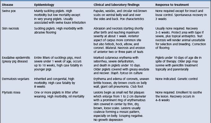
Treatment Penicillin parenterally
Control Sanitation and hygiene of pens. In outbreaks, isolate affected piglets and sows
Most cases of exudative epidermitis occur in suckling and weaned piglets under 6 weeks of age, with a peak incidence in piglets under 1 week. Occasionally groups of pigs up to 3 months of age may be affected. Within litters the incidence is high, often all piglets being affected. The morbidity will vary from 20–100% and the case fatality rate from 50–75%. The organism has been isolated from the joint fluid of lame pigs affected with arthritis.3 In 28% and 26% of studies of cases of exudative epidermitis9,10 no cases of toxigenic S. hyicus could be detected. In a recent study of 314 cases in Denmark it was shown that 20% had exfoliatum toxin A, 33% had B, 18% had C, and 22% had D in 60% of cases of exudative epidermitis investigated.
The source of the organism is unknown but the gilt or sow is probably an inapparent carrier. It can be isolated from the skin of healthy in-contact piglets and healthy sows.11 It can be frequently isolated from the vagina of prepubertal gilts and the majority of the litters from the same gilts may be colonized by the organism within 24 hours after farrowing.12 The maternal strains of S. hyicus persisted on the skin of the offspring piglets for the first 3 weeks of the piglets’ life – the critical period for outbreaks of exudative epidermitis.12 The organism has also been isolated from the atmosphere of buildings housing affected pigs. Bacteriophage typing of S. hyicus subsp. hyicus isolated from pigs with or without exudative epidermitis revealed two or more phage patterns in the isolates from each pig with the disease and a single-phage pattern in isolates from healthy pigs.13,14
Field evidence suggests that environmental stress of various kinds, including agalactia in the sow and intercurrent infection, predisposes to the disease. Lesions commonly develop first over the head, apparently in association with bite wounds, which occur when the needle teeth have not been cut or have been cut badly. Other factors include fighting following mixing of litters, excessive humidity over 70%, and following sarcoptic mange. The presence of the disease in a swine herd can account for a 35% reduction in the margin of output over feed and veterinary costs over a 2-month period.
Strains of S. hyicus can be divided into virulent and avirulent strains with regard to ability to produce exudative epidermitis in experimental piglets; both types of strain can be isolated simultaneously from diseased piglets.4 It has been shown that different types of S. hyicus expressing different types of toxin may be present in the same diseased pig.10
S. hyicus produces an exfoliative toxin that can be used to reproduce the disease.15,16 There are several toxins, including ExLA, ExLB, ExLC, ExLD, SHETa, and SHETb.17 The exfoliative toxins from isolates from different countries have recently been described.18 Strains of the organism isolated from a large number of Danish pig herds indicated different electrophoretic motility and plasma-mediated antibiotic resistance patterns.19 The antibiotic and plasmid profiles of strains isolated from pig herds may be a reflection of the use of antibiotics in those herds.20 Different types of toxin are produced.21-23
Recently the genes encoding for the exfoliative toxins SHETb, ExLA, ExLB, ExLC, and ExLD have been identified24 and sequenced.
The condition has been seen more frequently in cases of PRRS and PCV2 infections.25,26
The organism has been found as a frequent inhabitant of the skin of cattle and has been isolated from cattle with skin lesions.27,28 Naturally occurring lesions of dermatitis of the lower limbs of horses29 and similar lesions over the neck and back of donkeys30 have been recorded. Experimentally, the organism can cause lesions in horses similar to those of exudative epidermitis. A concurrent infection with Dermatophilus congolensis has also been reported.31
S. hyicus has cytotoxic activity for porcine keratinocyte cells in culture, which may indicate one of its virulence factors.31
The exfoliative toxins are actually epidermolysins32 that are active against desmoglein-1, which is a desmosomal cadherin-like molecule33 involved in cell-to-cell adhesion.34 The ExLs can cause blister formation in the porcine skin by digesting porcine desmoglein-1 in a similar way to exfoliative toxins of S. aureus.
The earliest lesion is a subcorneal pustular dermatitis involving the interfollicular epidermis. Exfoliation follows with sebaceous exudation and formation of a crust. In the well-developed case there is a thick surface crust composed of orthokeratotic and parakeratotic hyperkeratosis and neutrophilic microabscesses with numerous colonies of Gram-positive cocci.35
Although the principal lesion is an inflammatory–exudative reaction in the corium and upper layers of the epidermis, the disease is probably a systemic rather than a local one. Experimental infection of gnotobiotic pigs leads to dermatitis of the snout and ears, then the medial aspect of the thighs, the abdominal wall and the coronets. The lesions can be produced experimentally by using crude extracellular products and a partially purified exfoliative toxin.36
The morbidity varies from 10–100% and the mortality from 5–90%, with an average of 25%.35
In the peracute form, which occurs most commonly in piglets only a few days of age, there is a sudden onset of marked cutaneous erythema, with severe pain on palpation, evidenced by squealing. Anorexia, severe dehydration, and weakness are present and death occurs in 24–48 hours. The entire skin coat appears wrinkled and reddened and is covered with a greasy, gray-brown exudate that accumulates in thick clumps around the eyes, behind the ears, and over the abdominal wall. In the less acute form, seen in older pigs 3–10 weeks of age, the greasy exudate becomes thickened and brown and peels off in scabs, leaving a deep-pink-colored to normal skin surface. There is no irritation or pruritus. In the subacute form, the exudate dries into brown scales that are most prominent on the face, around the eyes, and behind the ears. In a small percentage of pigs the chronic form occurs and the course is much longer; there is thickening with wrinkling of the skin and thick scabs that crack along flexion lines, forming deep fissures. Most peracute cases die, while piglets with the less severe forms will survive if treated. Some pigs are affected with ulcerative glossitis and stomatitis.
Abortion in a sow has been attributed to the organism.37
Bacterial examination of skin swabs may reveal the presence of S. hyicus. A phage typing system can be used to determine the presence of virulent strains and to distinguish them from less virulent strains.14
Necropsy of these dehydrated, unthrifty piglets often reveals a white precipitate in the renal papillae and pelvis. Occasionally this cellular debris causes ureteral blockage. Some piglets also have a mild ulcerative glossitis and stomatitis. Microscopically, there is separation of the cells of the epidermis in the upper stratum spinosum, exfoliation of the skin, erythema, and serous exudation. The crusting dermatitis features a superficial folliculitis and a hyperkeratotic perivascular dermatitis with intracorneal pustules and prominent bacterial colonies. Degenerative changes are visible in the renal tubular epithelium.
• Bacteriology – samples of acute skin lesions (CULT). The organism forms 3–4 mm white, nonhemolytic colonies on blood agar. It is catalase- and mannitol-negative but hyaluronidase-positive
• Histology – formalin-fixed skin (multiple sites), kidney (LM).
A PCR is available but requires a pure culture and large numbers of organisms to be successful.
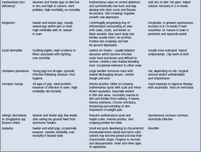
Exudative epidermitis may resemble several skin diseases of pigs of all age groups (Table 16.3). However, in exudative epidermitis there is no pruritus or fever. Careful gross examination of the lesions, particularly their distribution, the state of the hair shaft, the character of the exudate and the presence or absence of pruritus must be considered, along with skin scrapings and biopsies.
Experimentally infected piglets respond favorably to a topical application of cloxacillin 10000 IU/g of lanolin base and 1% hydrocortisone combined with parenteral cloxacillin. Treatment must be administered as soon as the lesions are visible. Procaine benzylpenicillin at a dose of 20000 IU/kg BW intramuscularly daily for 3 days is also recommended. The antimicrobial sensitivities determined in one field investigation revealed that all isolates were sensitive to novobiocin, neomycin, and cloxacillin. Novobiocin may be the antimicrobial of choice since staphylococci are universally sensitive to this antibiotic.38 However, there is no available information on the efficacy of antimicrobials for naturally occurring cases of exudative epidermitis. A study has suggested that erythromycin, sulfathiazole, and trimethoprim may be the most useful drugs, whereas penicillin and tetracyclines may not be very useful.39 Naturally occurring cases in piglets under 10 days of age respond poorly, while older pigs recover with a skin wash using a suitable disinfectant soap. The most successful treatment is antibiotics and skin washing for a period of at least 5–7 days. It is also essential to make sure that there is sufficient dietary provision of zinc, biotin, fat, selenium, and vitamin E in the diet.
The infected accommodation should be cleaned, disinfected and left vacant before another farrowing sow is placed in the pen. Strict isolation of the affected piglets and their dam is necessary to prevent spread throughout the herd. Dead piglets should be removed promptly from the premises and in-contact sows should be washed with a suitable disinfectant soap. Maternal antibodies will protect piglets in the first few weeks of life.
Autogenous vaccines have been used with varying degrees of success. It is important to use a strain that produces the exfoliative toxin, so the recent development of PCRs that identify the genes for toxin development will ensure that the right isolate is used for the autogenous vaccine. It will also facilitate the development of a commercial vaccine.
A novel approach to the control is bacterial interference. Experimentally, the precolonization of the skin of gnotobiotic piglets with an avirulent strain of S. hyicus will prevent the experimental reproduction of the disease with the virulent strain of the organism.
1 Ladhani S, et al. Clin Microbiol Rev. 1999;12:224.
2 Wegener HC, et al. Can J Vet Res. 1993;57:119.
3 Hill BD, et al. Aust Vet J. 1996;73:179.
4 Mirt D. Vet Rec. 1999;144:92.
5 Teuscher E, Higgins R. Med Vet Quebec. 1990;20:64.
6 Devriese LA, et al. Int J Syst Bacteriol. 1978;28:482.
7 Saito K, et al. J Vet Med Sci. 1996;58:711.
8 Andresen LO, et al. Vet Microbiol. 2005;105:291.
9 Andresen LO, et al. FEMS Immunol Med Microbiol. 1998;20:301.
10 Andresen LO, et al. Vet Microbiol. 1999;68:285.
11 Bara MR, et al. Aust Vet J. 1993;70:256.
12 Wegener HC, et al. Epidemiol Infect. 1992;109:433.
13 Wegener HC, et al. Res Microbiol. 1993;144:237.
14 Wegener HC. J Vet Med Series B. 1993;40:13.
15 Tanabe T, et al. Vet Microbiol. 1996;48:9.
16 Andresen LO, et al. Microb Pathog. 1997;22:113.
17 Fudaba Y, et al. Microb Pathog. 2005;39:171.
18 Andresen LO. Vet Rec. 2005;157:376.
19 Wegener HC, Schwarz S. Vet Microbiol. 1993;34:363.
20 Noble WC, Allaker RP. Vet Rec. 1992;130:466.
21 Tanabe T, et al. Infect Immun. 1993;61:2973.
22 Sato H, et al. J Bacteriol. 2000;182:4096.
23 Andresen LO, et al. FEMS Immunol Med Microbiol. 1999;23:295.
24 Ahrens P, Andresen LO. J Bacteriol. 2004;186:1833.
25 Whitaker HK, et al. J Vet Diagn Invest. 1990;2:244.
26 Wattrang E, et al. Vet Microbiol. 2002;86:281.
27 Hazirika RA, et al. Res Vet Sci. 1991;50:374.
28 Hazirika RA, et al. Indian Vet J. 1991;68:163.
29 Devriese LA, et al. Equine Vet J. 1983;15:263.
30 Devriese LA, Thelisen M. Vet Rec. 1986;118:1091.
31 Allaker RP, et al. Vet Microbiol. 1991;26:161.
32 Prevost G, et al. Curr Opin Infect Dis. 2003;16:71.
33 Amagai M, et al. Nat Med. 2000;6:1275.
34 Hankawa YNM, et al. J Clin Invest. 2002;110:53.
35 Kim J, Chae CJ. Vet J. 2004;167:104.
36 Andresen LO, et al. Microb Pathog. 1993;15:217.
37 Onet GE, Pommer JL. J Am Vet Med Assoc. 1991;199:362.
Diseases associated with Corynebacterium, Actinobaculum, and Arcanobacterium species
Arcanobacterium (formerly Corynebacterium or Actinomyces) pyogenes is an ubiquitous organism and is the primary cause of, or a secondary invader in, a wide variety of pyogenic infections in ruminants, pigs and, occasionally, horses.1 It is present in the environment, in the intestinal tract, and may be carried in the tonsils of healthy cattle.2
The organism gains access to the body through contaminated abrasions, wounds, or insect bites and the infection may remain localized in the form of a subcutaneous abscess with inflammation of the draining lymph node or it may proceed to a bacteremia with internal localization. Internal abscessation is frequently clinically silent but is important in food quality. The organism is a cause of endocarditis and commonly present in necrotizing pneumonias. It is a common isolate in omphalophlebitis and is also a cause of septic arthritis in calves.
A. pyogenes is a common isolate from iatrogenic syndromes such as frontal sinusitis following dehorning, injection site abscess, and surgical wound infections. Extensive subcutaneous abscessation and suppurative cellulitis occurs in buller feedlot cattle as a result of multiple skin abrasions.
A. pyogenes is a common isolate from intracranial and spinal abscesses, which may result from hematogenous spread, in association with parasitic migration or from invasion from surrounding structures. Outbreaks of spinal abscessation can occur in conjunction with tail-biting in pigs and ear abscessation as a result of ear-biting. A seasonal occurrence of intracranial abscessation occurs in male deer during the period following velvet shedding to the time of antler casting and is associated with wounds and fractures of the antler pedicle.3 A. pyogenes is one of the more common organisms isolated from osteomyelitis in cattle and is also the common isolate from pituitary abscesses.
A syndrome of chronic rhinitis and sinusitis with unilateral or bilateral foul-smelling nasal discharge is reported affecting dairy cattle in three herds in the UK. Contamination of feed by the discharge resulted in food refusal. There was no evidence of predisposing common respiratory viral or other bacterial infection.4
The organism is sensitive to penicillin but infections with A. pyogenes are poorly responsive to antimicrobial therapy despite in vitro sensitivity and usually require surgical drainage in combination with antibiotic therapy. Resistance to tetracyclines and tylosin is more prevalent in isolates from animals in which these antimicrobials are used for growth promotion and disease prophylaxis.5
Vaccines against the organism are commercially available in most countries but there is little evidence that they induce protective immunity against infection or the pyogenic process.6
The role of A. pyogenes in the production of mastitis in cattle is described in Chapter 15, as a secondary invader in calf and sheep pneumonia in the descriptions of the viral pneumonias of calves and sheep, and in foot abscess of sheep under that heading.
Corynebacterium pseudotuberculosis is a soil-borne organism and causes caseous lymphadenitis in sheep and goats, ulcerative lymphangitis in cattle and horses, and also external and internal abscesses in horses. The organism possesses a cytotoxic surface lipid coat that appears to facilitate intracellular survival and abscess formation and produces a phospholipase exotoxin that increases vascular permeability, has an inhibitory effect on phagocytes, and may facilitate spread of infection in the host. There are two biotypes, with the ovine and caprine isolates a different biotype from equine and cattle isolates.
The important specific diseases associated with Arcanobacterium, Corynebacterium, and Actinobaculum spp. are described later in this section. Other minor diseases include the following.
A. pyogenes is an important cause of postpartum metritis and infertility in cattle and sheep.7-10
Cows are infected soon after calving and infection is frequently accompanied by infection with the Gram-negative anaerobes Fusobacterium necrophorum and Prevotella melaninogenica (formerly Bacteroides melaninogenicus), which act synergistically, enhancing the growth and pathogenicity of each other.11 However, the inoculation of pure cultures of A. pyogenes into the nonpregnant uterus is followed by the development of endometritis with a persistent corpus luteum.12 With natural cases, infections are more common after the retention of fetal membranes. Isolation of A. pyogenes from the uterus during the postpartum period is significantly associated with endometritis, pyometritis and purulent discharge, resulting in an increase in time to first service, more services to conception, an increase in days open and an increase in cows culled because of infertility.7,9,10,13 Oxytetracycline is commonly used in therapy but minimum inhibitory concentrations (MICs) of various antibiotics for isolates from the uterus of cattle suggest that cephalosporins would be more effective.14
The organism is also a cause of abortion.9,15 The fetus is autolyzed but the organism can be isolated from all internal organs and the placenta. Abortion can occur at any stage of pregnancy and has been reproduced experimentally in early pregnancy and in mid-gestation.16 The prevalence of abortion associated with this organism is low and it accounts for fewer than 5% of abortions in cattle.9,15,16 The examination of a placentome in addition to the aborted fetus improves the accuracy of diagnosis.17 An unidentified Corynebacterium-like organism tentatively identified as Arcanobaculum pluranimalium has been isolated from the placenta and stomach contents of lambs in cases of sporadic abortion in sheep.18,19
A. pyogenes and Actinobaculum suis are occasional causes of perinatal septicemia and polyarthritis in calves, lambs, and foals.20,21
Corynebacterium pseudotuberculosis has been determined to cause a nonsuppurative arthritis and bursitis in lambs. The joints are only slightly enlarged and many animals show only mild clinical signs. Recovery occurs if the lambs are fed and confined.
A. pyogenes and P. melaninogenica have been isolated from periodontal lesions occurring in young cattle grazing new pastures sown in recently cleared forest areas in Brazil. The cause of the disease is uncertain and multifactorial but enzymes and toxins from these organisms are stated as being capable of causing primary destruction of periodontal tissue.22 Cattle are affected at an age when premolar and molar teeth erupt. Trace element deficiencies have been suspected of predisposing to the disease but are not proven to do so. One theory is that there is a large increase in Actinomycetes in soil following the clearance of the forest, that these produce antibiotics, mainly streptomycin, and that the ingestion of subinhibitory concentrations of these predisposes to adherence of bacteria to the gingival epithelium. The disease has markedly decreased in incidence in recent years as forest clearing and the occupation of virgin land for cattle raising has almost ceased.23 Cara inchada is manifest with a purulent periodontitis, halitosis, progressive loss of premolar teeth, mainly of the upper jaw, and emaciation. In risk areas attack rates and case fatality rates are high. The disease can be suppressed by including spiramycin or virginiamycin in the mineral mix fed to the cattle.
In California,24,25 Texas,26 and Colorado,27 C. pseudotuberculosis has been associated with a high regional prevalence of chronic abscessation in horses. There is no apparent breed or sex predisposition and cases occur in horses of all ages, but the majority are adult horses and there are few cases under 1 year of age. Usually, only a single horse on a farm is affected. A small proportion of farms have endemic infection, with a prevalence of disease of 5–10% and recurrent infections each year. In some years spread to naive horses results in epidemic disease. Cases can occur in all months of the year but are most common in dry months in autumn, with a peak prevalence in September, October, and November.17 There is a variation in the prevalence from year to year and, in both Texas and California, years with high prevalence of the disease have been preceded by seasons with higher than normal rainfall and conditions that promote high insect populations. Insects, such as horn flies (Habronema irritans), that produce a ventral midline dermatitis during feeding may predispose to infection, and the organism has been detected by PCR populations of H. irritans, Stomoxys calcitrans, and Musca domestica.28 Patterns of spatial and temporal clustering indicate that disease is transmitted directly or indirectly from horse to horse with an incubation period of 3–4 weeks.25 The disease occurs in a region where caseous lymphadenitis is also common in sheep. Stable hygiene, insect control and isolation of infected horses may aid in control.25
These occur in a variety of areas on the body but in the majority of cases they are in the pectoral, axillary, inguinal, or ventral midline regions.24 The abscesses may reach a diameter of 10–20 cm, with a surrounding area of edema, before they rupture 1–4 weeks later. Clinical signs include local swelling, lameness, pain on palpation, ventral edema, reluctance to move, midline dermatitis, fever and depression in the early stage, and eventually rupture of the abscesses.
Treatment of external abscesses is by hot packs to encourage opening and by surgical drainage and lavage. Ultrasound can aid in the detection of deeper abscesses. NSAIDs can be used to control swelling and pain. Recovery rates are excellent and are not improved by antimicrobial therapy.24
These occur at a variety of sites but predominantly in the liver and can occur in horses with no external abscessation. The diagnosis should be suspected in horses, in the region and the season, with a clinical history of external abscess, fever, anemia and colic, and laboratory evidence of leukocytosis with neutrophilia, anemia, hyperglobulinemia and hyperfibrinogenemia, and elevated activity of hepatic-associated enzymes. Abdominocentesis in most cases shows an elevated protein and nucleated cell count, and the organism can be cultured.24 Treatment is with antimicrobial therapy but the case fatality rate is high.
The value of a synergistic hemolysis inhibition serological test in diagnosis has been examined.24 In horses with external abscesses a range of titers was found but high titers were found in horses with internal abscessation.24
An autogenous vaccine has given protection against experimental challenge but, in field trials, there has been no difference in the incidence of infection between vaccinated and control horses.29
C. pseudotuberculosis is also recorded as a cause of pericarditis and pleuritis in a horse30 and in association with suppurative facial dermatitis following trauma.31
1 Lavoie JP, et al. Can Vet J. 1991;32:292.
2 Duncanson GR, Slater CA. Vet Rec. 1986;118:459.
3 Davidson WR, et al. J Wildl Dis. 1990;26:460.
4 Otter A, et al. Vet Rec. 2000;146:55.
5 Trinh HT, et al. Vet Microbiol. 2002;85:353.
6 Hunter P, et al. Onderstepoort J Vet Res. 1990;57:239.
7 Bonnett BN. Can J Vet Res. 1991;55:168.
8 Cohen RO, et al. J Vet Med B. 1996;43:193.
9 Yamini B, et al. Bovine Pract. 2004;38:59.
10 Tzora A, et al. Theriogenology. 2002;57:1809.
11 Smith GR. Epidemiol Infect. 1991;100:305.
12 Farin PW, et al. Theriogenology. 1989;31:979.
13 Takacs T, et al. Theriogenology. 1990;33:85.
14 Sheldon IM, et al. Vet Rec. 2004;155:383.
15 Kirkbride CA. J Vet Diagn Invest. 1993;5:64.
16 Semambo DKN, et al. Vet Rec. 1991;129:12.
17 Johnson CT, et al. Vet Rec. 1994;134:263.
18 Foster G, et al. Vet Rec. 2001;148:284.
19 Duff JP, et al. Vet Rec. 2001;148:186.
20 Hariharan H. Can Vet J. 1992;33:56.
21 Robinson JA, et al. Equine Vet J. 1993;25:214.
22 Dobereiner J. Dtsch Tierarztl Wochenschr. 1990;97:482.
23 Dobereiner J, et al. Pesque Vet Brazil. 2004;24:50.
24 Aleman M. J Am Vet Med Assoc. 1996;209:804.
25 Doherr MG, et al. Am J Vet Res. 1999;60:284.
26 Welsh RD. Equine Pract. 1990;12:7.
27 Hall K, et al. Equine Vet J. 2001;21:284.
28 Spier SJ, et al. Am J Vet Res. 2004;65:829.
29 Davies EW, Spensley MJ. J Vet Intern Med. 1992;6:137.
Etiology Corynebacterium renale
Epidemiology Organism is present in the vagina of carrier animals. Infection spread, and disease initiated, by trauma such as breeding or catheterization
Clinical findings Periodic episodes of hematuria, pyuria, colic, straining to urinate, fever, loss of condition. Palpable abnormality on rectal examination. Cystitis determined by endoscopic examination, pyelonephritis by renal biopsy and ultrasound examination
Clinical pathology Urine pH alkaline. Pyuria on microscopic examination. Elevated blood creatinine and urea concentrations
Necropsy findings Cystitis and pyelonephritis
Diagnostic confirmation Gross changes in urine, together with palpable abnormalities in the urinary tract and the presence of bacteria including C. renale in the urine
Treatment Prolonged course of penicillin. Nephrectomy
Control Avoidance of urinary catheterization. Artificial insemination
Corynebacterium renale, the specific etiological agent, has three serotypes of which types I and III appear to be the most pathogenic. Piliated forms occur and may be important in attachment to the epithelium in the reproductive and urinary tract.1,2 Corynebacterium pilosum and Corynebacterium cystitidis are commonly isolated in conjunction with C. renale but are considered part of the normal flora of the vulva.
Corynebacterium pseudotuberculosis, Arcanobacterium pyogenes, Actinobacillus equuli, Escherichia coli, and Staphylococcus aureus are sometimes found in the urinary tract of cattle and pigs affected with pyelonephritis, either alone or associated with C. renale. Of these organisms, E. coli in particular is considered a separate cause of pyelonephritis in the cow.3
C. renale has little apparent resistance to physical or chemical agents and can survive in soil for at least 56 days.3
Infection with C. renale may stimulate production of antibody that gives cross-reactions with the complement-fixing test for Johne’s disease.
The disease is widespread in Europe, North America, Australia, Africa, Japan, and Israel and probably occurs in all countries, although it seldom constitutes an important problem in any herd or area. As a rule, clinical cases are sporadic, even in herds found to harbor a significant number of carriers. Differences in disease prevalence probably reflect differences in management. One study in seven herds found an annual incidence that varied from 0.5–1.5% and in one herd was 16%.4 Subclinical infection may be more frequent than commonly recognized. Chronic cystitis and pyelonephritis (etiology unstated) have been found in 5.3% and 0.2% of cattle at slaughter.4,5
Although pyelonephritis is considered to be essentially a bovine disease, sheep are occasionally affected.
C. renale can be isolated from the urine of affected or carrier animals and in Japan has been isolated from the vagina or vaginal vestibule of approximately 6% of healthy cows.6 The incidence of cows excreting C. renale in their urine is higher in herds where the disease occurs than in herds where the disease is unknown. C. renale is present in the urinary tract of other animals, including rodents, and can be isolated from dust and the environment of infected animals.7
In cattle, infection can be transmitted by direct contact, by the use of contaminated brushes or by the careless use of catheters. A change in the policy of catheterizing cows with suspect ketosis for the collection of urine can be associated with a fall in the incidence of the disease.
There is a strong inference that in some circumstances the disease, or the infection, can be spread venereally. This is suggested by the occasional occurrence of a series of cases in a herd, usually related to the use of a particular bull,8 and the cessation of cases when artificial insemination is used. The organism can often be isolated from the prepuce, urethra, and semen of bulls that have no detectable lesions in the prepuce. C. renale can be a cause of balanoposthitis in bulls.
Cattle are seldom affected before maturity and cows appear to be much more susceptible than bulls. In cows, clinical cases are more common in early lactation. Mature cows of all ages are susceptible but in one study there was a significantly higher risk for disease in cows in their second lactation.4 An increase in clinical cases is usually found in the colder seasons of the year and heavily fed, high-producing dairy herds appear to show an increased susceptibility.
A significant decreased risk for pyelonephritis has been found for cows that have postparturient uterine disease, which may be associated with the treatment of these cows with antibiotics.
Bulls can be predisposed to pyelonephritis by obstructive urinary abnormalities.9
In Israel, the ingestion of rock rose (Cistus salvifolius) is reported to produce urinary retention and predisposes to pyelonephritis.10
The disease can be reproduced by inoculation of the organism into the urethra.11.12 It can also be reproduced by intravenous challenge, although the vulva is believed to be the natural portal of entry. Although not intentionally produced, the disease occurred in 10% of a group of cattle used to teach veterinary students the technique of urinary catheterization.
Pyelonephritis usually develops as an ascending infection involving successively the bladder, ureters, and kidneys. Trauma to the urethra, or urine stasis, may facilitate ascending infection. The destruction of renal tissue and obstruction of urinary outflow ultimately result in uremia and the death of the animal.
Piliated and nonpiliated forms of C. renale are present in infected animals but their relative importance to the pathogenesis of the disease is uncertain. Piliated forms of C. renale have a greater ability to attach to urinary tract epithelium, are more resistant to phagocytosis and are probably important to carriage of the organism and to the initial ascending infection.11 However, in the course of an infection there is a shift from piliated to nonpiliated forms, which may reflect a response to the development of antipilus antibody.13
Early signs vary considerably from case to case.14 The first sign observed may be the passage of bloodstained urine in an otherwise normal cow. In other cases, the first sign may be an attack of acute colic, manifest by swishing of the tail, treading of the feet and kicking at the abdomen, and straining to urinate, the attack passing off in a few hours. Such attacks are caused by obstruction of a ureter or renal calyx by pus or tissue debris and may be confused with acute intestinal obstruction. More often the onset is gradual with a fluctuating temperature (about 39.5°C, 103°F), capricious appetite, loss of condition and fall in milk yield over a period of weeks. Other than this, there is little systemic reaction and the diagnostic signs are associated with the urinary tract.
The most obvious sign is the presence of blood, pus, mucus, and tissue debris in the urine, particularly in the last portion voided. Urination is frequent, may occur in a dribble rather than a stream, and may be painful. Periods during which the urine is abnormal may be followed by apparent recovery with later remissions.
In the early stages, rectal examination may be negative but later there is usually detectable thickening and contraction of the bladder wall and enlargement of one or both ureters. These are not normally palpable but in chronic cases may be felt in their course from the renal pelvis of the left kidney to the bladder. The terminal portion of the ureters may also be palpated through the floor of the vagina over the neck of the bladder. The palpable left kidney may show enlargement, absence of lobulation and pain on palpation, and the right kidney may be palpable if it is significantly enlarged.14 In many cases there are no distinct clinical signs referable to the urinary tract and the history and clinical signs may be of weight loss and suspected gastrointestinal disease. In these cases examination of the urine is essential to diagnosis.
Endoscopic examination of the urethra and bladder can be diagnostic. Ultrasound examination shows a reduction in renal pelvis diameter, a reduction in renal parenchyma and the bladder wall is hyperechoic.
The course is usually several weeks or even months and the terminal signs are those of uremia.
There is proteinuria and hematuria. Urine pH is greater than 8.5 and the specific gravity has been recorded between 1.008 and 1.021.14 Microscopic examination will show pyuria. The presence of C. renale in suspected urine can be confirmed by culture, specific immunofluorescence7 or direct microscopic examination.
There may be hypoalbuminemia and hypergammaglobulinemia. Neutrophilia may be present but is not constant in all cases. There is an elevation of serum creatinine and urea, and concentrations above 1.5 mg/dL and 100 mg/dL, respectively, carry a grave prognosis.4
Renal biopsy15 and ultrasound16 can aid in diagnosis. Ultrasound may demonstrate a dilated renal collecting system containing echogenic material and can be an aid in the determination of the severity of the disease.
The kidneys are usually enlarged and the lobulation less evident than normal. The renal calyces and grossly enlarged ureters contain blood, pus, and mucus. Light-colored necrotic areas may be observed on the kidney surface. Changes visible on the cut surface include excavation of papillae, abscessation and wedge-shaped areas of necrosis that extend from the distal medulla into the cortex. The bladder and urethra are thick-walled and their mucous membranes are hemorrhagic, edematous and eroded. Histologically, the renal lesions are a confusing mixture of acute suppurative changes and various degrees of fibrosis with mononuclear cell infiltration.
Although several antibiotics appear to inhibit C. renale, penicillin remains the antibiotic of choice for treatment of pyelonephritis. Large doses (15000 IU/kg BW of procaine penicillin G) are recommended daily for at least 3 weeks.3 In early cases where little structural damage has occurred, permanent recovery can be expected following such a course of treatment.14 In general, a good prognosis is suggested by an improvement in condition, appetite and milk yield and clearing of the urine. However, in well-established cases, relapse is not uncommon and, where tissue destruction has been extensive, relief through antibiotic therapy is only temporary. In valuable animals, where ultrasound has established the diagnosis, unilateral nephrectomy may be an alternative and the surgical technique has been described.16
Cases characterized by acute colic can be differentiated from acute intestinal obstruction by the absence of a palpable obstruction and the disappearance of abdominal pain within a few hours. Chronic cases may be confused with traumatic reticulitis but may be differentiated by the urine changes present in pyelonephritis.
Sporadic cases of nonspecific cystitis can only be differentiated by culture of the urine.
Polypoid cystitis is a nonspecific result of bladder inflammation and may be a cause of dysuria and obstructive uropathy.17,18 The differential diagnosis is best made by endoscopic examination, which reveals multiple frond-like fungiform polyps and papillae on the bladder wall.
No specific control measures are usually practiced but isolation of affected animals and destruction of infected litter and bedding should reduce the population of the organism in the local environment and minimize the opportunity for transmission. Procedures such as urinary catheterization should be avoided and routine vaginal examinations should be conducted with proper hygienic precautions. Where natural breeding is practiced, some reduction in occurrence may be achieved by the introduction of artificial insemination.
1 Hayashi A, et al. Vet Microbiol. 1985;10:287. 381
2 Hayashi A, et al. Am J Vet Res. 1985;46:409.
3 Divers T. J Proc Am Assoc Bovine Pract. 1983;15:74.
4 Markusfeld O, et al. Br Vet J. 1989;145:573.
5 Herenda D, et al. Can Vet J. 1990;31:515.
6 Hiramune T, et al. Jpn J Vet Sci. 1988;50:111.
7 Osanai T, et al. Vet Microbiol. 1996;52:313.
8 Sheldon IM, et al. Vet Rec. 1995;137:100.
9 Tyler JW, et al. J Am Vet Med Assoc. 1991;198:871.
10 Yerahum I, et al. Rev Med Vet. 2002;153:627.
11 Kubota T, Yanagawa R. Am J Vet Sci. 1988;50:199.
12 Elias S, et al. Br Vet J. 1993;149:5.
13 Fukuoka T, Yanagawa R. Am J Vet Sci. 1987;49:1073.
14 Rebhun WC, et al. J Am Vet Med Assoc. 1989;194:953.
15 Scholz H, et al. Prakt Tierarzt. 1997;78:326.
16 Hayashi H, et al. J Am Vet Med Assoc. 1994;205:736.
Etiology Actinobaculum suis (Eubacterium suis, Corynebacterium suis) is the specific cause but a range of other bacteria (principally Escherichia coli) may also cause the condition
Epidemiology Infection of male pigs causes the disease in sows. Organism is in the prepuce and environment. Transmission is venereal and through dirty farrowing houses. Trauma to the urogenital tract of females predisposes to disease
Clinical findings Unexpected death in acute cases. Pain on urination, bloodstained, turbid urine accompanied by vaginal discharge. Cystitis on endoscopic examination
Clinical pathology Hematuria, pyuria, proteinuria, bacteremia. Urine pH >8.5. Demonstration of organism by culture or immunofluorescence. Azotemia, increased concentrations of urea and creatinine, hyperkalemia and hyponatremia
Necropsy findings Purulent cystitis and pyelonephritis
Diagnostic confirmation Urinalysis, endoscopic examination and demonstration of A. suis
Treatment Unrewarding unless early in the course of the disease – antimicrobials and supportive therapy. Humane destruction of cases
Control Antimicrobials by injection, in water or in feed. Insure adequate water supply for lactating sows to aid urination and improve postfarrowing hygiene
The disease is associated with a variety of agents. A variety of syndromes associated with different bacteria can be distinguished. Urinary tract infections in sows can be associated with E. coli and other bacteria (Pseudomonas aeruginosa, staphylococci, streptococci, Proteus spp., Klebsiella spp., enterococci, and A. pyogenes) and infections with most of these organisms result in catarrhal/purulent cystitis.
The specific disease is most commonly associated with Actinobaculum suis. This was formerly known as Eubacterium suis and before that as Corynebacterium suis. This organism is a Gram-positive rod that is difficult and slow to grow and requires special culture media.
All these organisms causing this problem are believed to produce the condition as a result of ascending infection. The disease is a particular problem when sows are in stalls or tethered. The condition may not be so serious when associated with all the species other than A. suis and in these cases may be seen as frequent urination, the presence of blood or pus in the urine and a progressive loss of condition.
The disease occurs in postpubertal sows that have bred.
The infection may be common but the disease is better viewed as sporadic.
The disease is probably worldwide and has been reported from Great Britain, Europe, North America, Asia, and Australia. It occurs both in outdoor and indoor units. There are no measures of prevalence, although it has been described as the most important cause of sow deaths, with up to 25% associated with urinary tract infection.1 It probably occurs more frequently than is recognized as there is a considerable subclinical infection rate. In a recent study in the USA, A. suis was isolated from 4.7% of the bladders of sows collected at random at a slaughterhouse.2
In small herds, the disease tends to occur in small outbreaks when a small number of sows become infected after being mated to a single boar. Often, the clinical outbreak may be 2–3 weeks after the use of the suspected boar. More serious outbreaks can occur in large intensive piggeries. The disease can also occur sporadically and be a normal feature of sow mortality.3
In a recent study of 1745 pregnant sows 28.3% were found to have urinary infections and A. suis was found in 20.6% of these.4 A. suis was less prevalent (13.7%) in the sows with urinary infections than in those without (23.1%). In an abattoir survey in the Netherlands the prevalence of cystitis in slaughtered sows was 11%, with a variation depending on group from 0–35%.5 In this study of 114 bladders, A. suis was not isolated but E. coli was the most commonly isolated together with S. dysgalactiae, A. pyogenes, Aerococcus viridans, and S. suis.
A. suis is a normal inhabitant of the porcine prepuce and can be isolated from the preputial diverticulum of boars of various ages.3,6,7 The prevalence of the infection in adult males may be as high as 90%3 and the organism can be isolated from the floor of the pens containing infected boars.
Infection and colonization of the preputial diverticulum may occur in pigs as early as 5 weeks of age if they are housed with older pigs. Frequently, this infection may occur from pen floor contamination because of poor hygiene. Piglets can also become infected, at an early age, from sows that have chronic cystitis and pyelonephritis, and the infection can spread rapidly to other male pigs when they are grouped at weaning.8 Although infection is common in the male pig, cystitis and pyelonephritis is extremely uncommon in males. A. suis is rarely isolated from the urogenital tract of the healthy female pig.
Clinical disease is almost entirely restricted to the female pig that has bred. Venereal transmission is believed to be the primary if not the sole method of infection of the sow.8,9
Trauma to the vagina may be an important predisposing factor allowing infection to establish and trauma at parturition with infection from the environment may also be important.
There is no doubt that, where the conditions in stall houses are bad with fecal contamination and poor drainage from the rear half of the stall, perineal and vulval contamination is much greater.
Clinical signs may occur at any age but are common at 3–4 weeks post-service. The disease is more common in sows kept in intensive confinement conditions than in those that are kept in open lots, pens, or pasture10 but it does occur in these systems if the hygiene is poor. Differences in feeding patterns and exercise that occur in the different management systems can influence the frequency and volume of water intake. This in turn has an important effect on the frequency of urination and the residual volume of urine in the bladder following micturition, which is one of the factors that may predispose to the establishment of urinary tract infection. Since many lactating sows will only stand when they are fed, usually twice a day, they will also only urinate and drink twice a day. If they cannot take in enough water from either a trough or a tap during this period at the correct flow rate, they may be suffering from an inadequate water intake.11,12 It has also been shown that crystalluria may well damage the mucosa and aid the formation of cystitis and that the crystals also support infection of the bladder, particularly where there is an insufficient water supply.13
In one of the best studies of sow mortality in the UK it was reported that the principal cause of death in up to 25% of the sows was urinary tract infection.1 If the sow mortality is below 5% then cystitis/pyelonephritis is unlikely to be an important problem in the herd.
Cystitis and pyelonephritis is of great importance as it is a major cause of death (annual death rate may be in excess of 5%)11 in sows in both Britain8 and the USA10 and even more importantly a cause of serious culling as the recommendation is to cull affected sows as they will always be a source of infection. Considerable prevention, treatment and hygiene costs will also ensue.
In healthy sows A. suis can be isolated from the vagina, but not from the bladder, for a short period after an infected service.8 The factors that allow it to establish in the urogenital tract are unknown but trauma to the vagina and urethral opening and service into the bladder are supposed factors.6,8 It has been suggested that the other organisms mentioned above may also act synergistically to damage the mucosa and facilitate colonization by A. suis.10 This may be true, as A. suis possesses pili by which it attaches to damaged bladder epithelium. Cystitis results in damage to the ureterovesicular junction and there is an ascending infection from the bladder to the kidneys.14,15 In infected sows, tortuosity of the ureters and blockage of the ureters is fairly common.8 This and the changes in the kidney may lead to chronic renal failure and even acute renal failure if the blockage is complete and bilateral.
It is worth remembering that many mildly affected sows may only show transient inappetence, and other animals are recognized because they are uremic. In the more severely affected groups the presentation is either as an acute case, usually post-service, or as a chronic one, which can occur at any time.
Most commonly sows present as acute renal failure as they cannot retain sodium and this leads to rises in plasma potassium and sudden death. Sows are suddenly severely ill, unwilling to rise, show profound depression and circulatory collapse and die within 12 hours. In one series of cases, 40% of the affected sows presented as unexpected deaths and in the remainder the mean interval between presenting signs and death was 1.6 days with the longest interval 5 days.8 In a recent study in Ireland16 the authors reported the condition mainly in fourth parity sows and above.
Where the surveillance is good the sows are observed to be depressed, anorectic, mildly febrile (normal to 39.5°C, 103°F) and sometimes show arching of the back, twitching of the tail and painful urination. There is frequent passage of bloodstained, turbid urine accompanied by vaginal discharge. Examination with a vaginal speculum will confirm the bladder as the source of the bloody discharge.
The case fatality rate is high. Sows that survive the acute disease develop chronic renal failure with weight loss and polydipsia and polyuria.9 They are usually culled for poor performance.
Endoscopic examination of the bladder in acute cases may show little other than mild inflammation but in more serious cases there are ulcerative and erosive bladder lesions.17 In sows large enough to allow rectal examination, in chronic cases it may be possible to feel the large and thickened bladder and dilated tortuous ureters. Boars are usually unaffected clinically but intermittent, hematuric episodes lasting several days have been recorded.
The condition can be seen as a sequel to any locomotory, particularly central brain or spinal condition in which there is an inability to stand to drink or micturate, e.g. organophosphorus poisoning.18
Sows’ urine is frequently turbid (83.1%)4 and usually this can be associated with the presence of crystals (96.1%).4
Urinalysis usually shows hematuria, pyuria, proteinuria, and a pronounced bacteriuria (usually in excess of 105 cfu/mL of urine). A Gram stain on a smear of the urine or pus may show the organisms. The urine is alkaline with a pH of more than 8.5 and usually approaching 9 as a result of the urease, which cleaves urea to produce ammonia. Midstream urine contains 105 cfu/mL or more.19 A number of species of bacteria may be found as suggested in the introduction but A. suis requires special culture media.3,20 It is now possible to use immunofluorescence for a more rapid and specific diagnosis.21 Examination of blood shows a pronounced azotemia, with increased concentrations of urea and creatinine and also hyperkalemia and hyponatremia. N-acetyl-B-d-glucosaminidase concentrations are elevated, indicating proximal renal tubular damage.8
A very varied pathology may be visible in these cases. In some sows there may be an extensive purulent nephritis and pyelitis with similar changes in the dilated ureters and the bladder. In others there may be severely hemorrhagic kidneys and blood in the pelvises.
In acute cases the bladder wall is swollen, edematous, and hyperemic and may be covered by a gritty, mucinous material.2 In others the bladder wall may just be thickened, inflamed and covered by an extensive thick mucus. In some of these cases the ureterovesicular valves may have been completely destroyed by necrosis.14,15 There may be minimal gross changes in the kidneys in acute cases but there may be microscopic changes of a diffuse tubular and interstitial nephritis. In the chronic case there are ulcerative and erosive lesions in the bladder wall and there may be pus in the bladder, thick-walled ureters and an obvious pyelonephritis.
• Bacteriology – kidney, bladder, and ureter for aerobic and anaerobic culture and special media for A. suis. A special urea-enriched medium21 in which dry colonies of A. suis, about 2–3 mm in diameter, grow after 2 days incubation
Diagnosis is based on high mortality, clinical findings, history of service, clinical pathology, particularly bacteriology, immunofluorescence for specific bacteria, or isolation by culture.
Early treatment with antibiotics is recommended, but if there is acute renal failure then the case fatality rate is high. Penicillin given at 15000 IU/kg daily for 3–5 days has been successful. The intramuscular injection of streptomycin at 10 mg/kg has also been used successfully.8 The isolation of other organisms may indicate the need for other broad-spectrum antibiotics. A recent outbreak of cystitis and endometritis16 associated with a falling conception rate from 88% to 75% and believed to be due to E. coli (together with staphylococci and streptococci) was successfully treated with an ammonium chloride urinary acidifier whereas the amoxicillin used previously was without effect. Enrofloxacin at the rate of 10 mg/kg BW in the feed for a period of 10 days has also proved effective, as has 2.5 mg/kg in the food for at least 20 days.
The response can be monitored by the reduction in the urine pH. Treated sows should be loose housed with access to plenty of water. The treatment should be continued for at least 2–3 weeks after the outbreak has appeared to finish clinically. Oral electrolyte therapy is also beneficial. In chronic cases the lesions are well advanced and the organisms may be contained in the calculi and then therapy is not successful and relapses may well occur. In many cases humane destruction is the best option especially as it is not possible except in very early cases to eliminate the organism. Preputial washing on a regular basis may prevent the carriage of organisms by boars especially as it has been shown that semen may be frequently contaminated by A. suis, and it is said that up to 50% of boars in some studs may be affected.
Routine prophylactic administration of antibiotics has proved of little value in the long-term control of the disease. A temporary solution has been the use of sow treatment with oxytetracycline followed by in-feed medication with 400 g/ton for 21 days.11 It is not possible to eradicate the infection from the prepuce of a boar, although daily infusions may help to reduce the infection.7 Artificial insemination with the semen treated with antibiotics is a further option.
Trauma to the vagina at mating should be reduced by boar management and the supervision of mating. There should be nonslip floors in the service areas. Animals showing distress, pain, or bleeding after mating should be treated.
The service areas should have very good hygiene with regular cleaning and disinfection after every use. The perineal region of each sow should be cleaned before mating. Farrowing accommodation and crates should also be properly cleaned and disinfected and allowed to dry before sows are introduced. The major organism (A. suis) will persist in bad floors but is susceptible to phenolic, quaternary ammonium and formalin-based products.9
Other control procedures involve the provision of an adequate water intake. This should preferably be from the mains without impurities or toxins or bacterial contamination. Loose housing and twice-a-day feeding will encourage the consumption of water. The provision of adequate numbers of drinkers for the stage of the breeding cycle, providing the necessary flow rate for each age of pig is essential (at least 1.5 L/min for gestating sows and 2.2 L/min for lactating sows). A simple check on water supply can be to check appetite: if the sows are not consuming 10 kg of food on day 18 of lactation then there is probably something wrong with water provision. Similarly, troughs must contain at least a reasonable supply of water. In the welfare codes all animals have to be provided with a fresh supply of high quality water.
Done SH, Carr JC. The urinary tract. In: Sims LD, Glastonbury JRW, editors. Pathology of the pig. Victoria, Australia: Agriculture Victoria; 1996:359-384.
Wendt, M. Urinary system disorders of pigs. Proceedings of the 15th International Pig Veterinary Society Congress, Birmingham, England 1998; vol 1:195–201.
1 Jones JET. Br Vet J. 1967;123:327.
2 Walker R, MacLachlan C. J Am Vet Med Assoc. 1989;195:1104.
3 Jones JET, Dagnall GJR. J Hyg. 1984;93:381.
4 Kjelvik, et al. In: Proceedings of the 16th International Pig Veterinary Society Congress 2000; 353.
5 Geudeke MJ. Proceedings of the 12th International Pig Veterinary Society Congress 1992; 567.
6 Pijoan C, et al. J Am Vet Med Assoc. 1983;183:428.
7 Muirhead M. Vet Rec. 1986;119:233.
8 Carr J, et al. Pig Vet J. 1991;27:122.
9 Jones JET. Vet Annu. 1984;24:139.
10 Dee SA. Vet Med. 1991;86:231.
11 Carr J, et al. In Pract. 1995;17:71.
12 Wendt M, et al. Dtsch Tierarztl Wochenschr. 1995;102:21.
13 Wendt M, et al. Dtsch Tierarztl Wochenschr. 1996;103:506.
14 Carr JC, et al. J Urol. 1992;148:1924.
15 Carr JC, et al. J Urol. 1992;149:146.
16 Dagnall GJR, Jones JET. Res Vet Sci. 1982;32:390.
17 Spillane P. Pig J. 1999;44:162.
18 Wendt M, et al. Tierarztl Prax. 1990;18:353.
19 Liebhold M, et al. Vet Rec. 1995;137:141.
20 Lingfeldt N, et al. Berl Munch Tierarztl Wochenschr. 1990;103:273.
21 Wendt M, Amsberg G. Schweiz Arch Tierheilkd. 1995;137:129.
Etiology Multifactorial. Organisms that produce urease, usually Corynebacterium renale, produce lesions only in certain circumstances of management and urinary composition
Epidemiology Disease of wether sheep and occasional disease of bulls and goats. May occur as enzootic disease in sheep on high-protein diets and following good rains
Clinical findings Pustules and scabs at preputial orifice. Extension to involve internal prepuce in severe disease with signs of urinary obstruction. Ulcers and scabs at mucocutaneous junction of vulva in ewes. Urine staining of wool predisposes to fly strike
Diagnostic confirmation Clinical
Treatment Dietary restriction, topical disinfectants, surgical opening of ventral prepuce
Control Reduction of protein intake, testosterone, hemicastrate or cryptorchid castration
The etiology is multifactorial. High urea concentrations in urine, associated with high protein in pasture, result in cytotoxic levels of ammonia when the urea is split by urease-producing organisms present in the prepuce and vagina. Estrogens in pasture, causing swelling and congestion of the prepuce, may predispose to disease. Most commonly the organism is C. renale but an outbreak of posthitis in sheep associated with Rhodococcus equi and Corynebacterium hofmannii, both of which produce urease, and not associated with C. renale, is described.1
Mycoplasma mycoides LC has also been incriminated as a cause of posthitis and vulvovaginitis in sheep.2
The disease is reported primarily from Australia, South Africa, and South America but occurs in all countries with large pastoral sheep industries.
In Australia, enzootic posthitis occurs most commonly in Merino sheep, particularly wethers over 3 years of age and young rams, but in a severe outbreak young wethers and old rams may also be affected. An ulcerative vulvitis often occurs in ewes in the same flocks in which posthitis occurs in wethers and is thought to be a venereal extension of that disease. The disease also occurs in goats.3
Posthitis is uncommon in bulls but is reported to occur at high rates and to be economically important in South America.4 There appears to be no counterpart to ovine vulvitis in cows.
The causative organism can be recovered from lesions and from the clinically normal prepuce of most sheep. It is also present in the lesions of vulvitis in ewes and posthitis in bulls and Angora goat wethers.
Flies are considered to be probable mechanical vectors, and contact with infected soil and herbage is a likely method of spread. Infection at dipping or shearing seems not to be important. Transmission to ewes appears to occur venereally from infected rams. Although the natural disease in cattle is usually benign they may act as reservoirs of infection for sheep on the same farm.
Diet and season are the major risk factors. Enzootic posthitis occurs most extensively on lush, improved pasture with a high legume content and reaches its highest incidence in the autumn in summer rainfall areas and in the spring where the major rainfall is in winter. In these circumstances it can occur in epizootic proportions in wethers. The incidence in affected flocks may be as high as 40% and in some areas the disease is so common that it is not possible to maintain bands of wethers.
Factors of lesser importance are continued wetness of the area around the prepuce due to removal of preputial hairs at shearing, a high-calcium, low-phosphorus diet and the ingestion of large quantities of alkaline water.
The high incidence in castrates and young rams is probably related to the close adherence of the preputial and penile skins, which separate in mature entire animals, and to a lesser understood influence of testosterone.
The organism is capable of hydrolyzing urea with the production of ammonia. It is believed the initial lesion in the wether (the external lesion) is caused by the cytotoxic effect of ammonia, produced from urea in the urine by the causative bacteria.5 This lesion may remain in a static condition for a long period but, when there is a high urea content of the urine associated with a high-protein diet, and continued wetting of the wool around the prepuce, the lesion proceeds to invade the interior of the prepuce, producing the ‘internal lesion’. A similar pathogenesis is postulated for vulvar lesions.
The primary lesion starts as a pustule, which breaks and forms a soft scab. Small scabs are found on the skin dorsal to the preputial orifice (external lesion) and around the external orifice on the nonhaired part of the prepuce. These may persist for long periods without the appearance of any clinical signs. The scab is adherent and tenacious. When extension to the interior of the prepuce occurs (internal lesion) there is extensive ulceration and scabbing of the preputial opening and a hard core can be palpated extending 1–2 inches into the prepuce. With pressure, a semisolid core of purulent material can be extruded from the preputial orifice. Affected sheep may show restlessness, kicking at the belly and dribbling urine as in urethral obstruction. The area is often infested by blowfly maggots. In rams, the development of pus and fibrous tissue adhesions may interfere with urination and protrusion of the penis, and cause permanent impairment of function.
Some deaths occur due to obstructive uremia, toxemia, and septicemia. During an outbreak many sheep may be affected without showing clinical signs and are detected only when they are subjected to a physical examination. Others recover spontaneously when feed conditions deteriorate.
In ewes the lesions are confined to the lips of the vulva and consist of pustules, ulcers, and scabs. These extend minimally into the vagina. Their presence may distort the vulva and the ewe may urinate onto the wool with a consequent increased susceptibility to fly strike.
In bulls, lesions are similar to the external lesions which occur in wethers but rarely there may be invasion of the interior of the prepuce. The external lesions occur at any point around the urethral orifice and may encircle it. Their severity varies from local excoriation to marked ulceration with exudation and edema. There is a tendency for the lesions to persist for several months without treatment and with highly alkaline urine.
Isolation of the causative diphtheroid bacterium may be necessary if there is doubt as to the identity of the disease.
The principal measures are restriction of the diet to reduce the urea content of the urine, removal of the wool around the prepuce or vulva to reduce the risk of fly strike, segregation of affected sheep and disinfection of the preputial area, and surgical treatment of severe cases.
Sheep can be removed on to dry pasture and their feed intake restricted to that required for subsistence only. They should be inspected at regular intervals, the wool should be shorn from around the prepuce, and affected animals should be treated individually. Weekly application of a 10% copper sulfate ointment is recommended for external lesions; when the interior of the prepuce is involved, it should be irrigated twice weekly with a 5% solution of copper sulfate. Cetrimide (20% in alcohol or water with or without 0.25% acid fuchsin) or alcohol alone (90%) are about as effective as copper sulfate preparations.
Penicillin topically or parenterally may effect a temporary response. Thiabendazole by mouth appears to have a beneficial effect on the lesions but does not eliminate them.
In severe cases the only satisfactory treatment is surgical, and surgical treatment is necessary if the prepuce is obstructed. The recommended procedure is to open the ventral sheath by inserting one blade of a pair of scissors into the external preputial orifice and cutting the prepuce back as far as the end of the urethral process; extension beyond this leads to trauma of the penis. Badly affected rams should be disposed of as they are unlikely to be of value for breeding.
Subcutaneous implantation with testosterone propionate is highly effective as a preventive but is no longer used for sheep that will be used for human food. A single implantation of 60–90 mg is effective for 3 months; although the treatments can be repeated four times a year, it is more economical to time them to coincide with periods of maximum incidence, which will vary from district to district. Three implantations in fall, winter, and spring provide an effective control program in most areas. The tablets are implanted subcutaneously, preferably at the base of the ear, using preloaded tubes to avoid undue contact to the operator.
Alternative control procedures are under investigation, in part because of public resistance to meat products exposed to synthetic hormones. One alternative is to run male lambs as cryptorchids, so-called short scrotum lambs. The testes are pushed into the inguinal canal and a rubber ring is applied to remove the scrotum. Another is to run male lambs as hemicastrates.6 The prevalence of posthitis is significantly reduced in Merino short scrotum lambs and hemicastrates.6,7 There is an increase in live weight, with no increase in fleece weight, but there are obvious masculine characteristics such as horn growth.6-8
1 Doherty ML. Vet Rec. 1985;116:372.
2 Trichard CJV, et al. Onderstepoort J Vet Res. 1992;60:29.
3 Shelton M, Livingston CW. J Am Vet Med Assoc. 1975;167:154.
4 Correa FR, et al. Cornell Vet. 1979;69:33.
5 McMillan KR, Southcott WH. Aust Vet J. 1973;49:405.
6 Foster FM, et al. Aust J Exp Agric. 1997;37:303.
7 Foster FM, et al. Wool Technol Sheep Breed. 1990;38:74.
8 Egan JP, Russell DW. Am J Exp Agric Anim Husbandry. 1981;21:268.
Etiology Corynebacterium pseudotuberculosis
Epidemiology Disease of sheep and goats. Source of infection is discharge from pulmonary or skin abscesses. Infection is through intact skin or skin wounds. Transmission in sheep occurs at shearing and dipping in sheep and in goats and sheep by direct contact
Clinical findings Abscesses in superficial lymph nodes. Respiratory or wasting disease associated with internal abscesses
Clinical pathology ELISA tests can be used to determine flock status but sensitivity and specificity is inadequate to provide reliable identification of infected individuals
Necropsy findings Abscesses in lymph nodes and internal organs
Diagnostic confirmation The clinical and necropsy features are typical. Confirmation is by bacterial culture
Treatment Surgical for superficial abscesses
Control Culling of abscessed sheep or based on serological testing, hygiene at shearing, avoidance of management risk factors, vaccination
Corynebacterium pseudotuberculosis is the specific cause of the disease. There are two proposed biotypes, ovine/caprine and equine/bovine.1 Both biotypes produce an exotoxin, phospholipidase D, which functions as a sphingomyelinase and is an immunodominant antigen.2 Variation in toxin production between strains may be related to differences in pathogenicity.3 The toxic lipid cell wall mediates resistance to killing by phagocytes and is also a virulence factor.
C. pseudotuberculosis is also the cause of ulcerative lymphangitis of cattle and horses, and contagious acne of horses, but these have been dealt with as separate diseases because they appear to have a separate pathogenesis and do not occur in association with caseous lymphadenitis.
Caseous lymphadenitis occurs in the major sheep-producing countries in the world including Australia, New Zealand, South Africa, the Middle East, North and South America, the UK, and most of northern and southern Europe. The disease did not occur in the UK and the Netherlands until the importation of infected goats in the late 1980s4,5 but subsequently spread to be an important disease in both countries.
Caseous lymphadenitis occurs in sheep and goats.
Caseous lymphadenitis increases in prevalence with age6 and reaches a peak incidence in adults. In one Australian population of unvaccinated sheep the frequency of infection at abattoir inspection was 3.4% for lambs and 54% for adult ewes7 and similar levels of prevalence are recorded in North8 and South America.9 In another large study of mature age slaughter sheep in Australia the overall prevalence of lesions was 26%, with carcass lesions in 20.4% of sheep and offal lesions in 9.5%.10 The prevalence of lesioned sheep from individual farms can be higher. The prevalence of infection in ewes culled for age in Western Australia has fallen from over 50% in the 1980s to approximately 25% in the early 2000s, which is suggested to be in part the result of the cessation of compulsory dipping for lice during this period.11 Following the introduction to British flocks in the late 1980s, outbreaks increased to reach a peak in 1998 and have decreased in occurrence since.12 An examination, by pulse field gel electrophoresis, of isolates through this period suggests that all are related to the initial introduction.13 A recent serological survey of 745 flocks showed an overall prevalence of seropositive animals of approximately 10%, with 18% of the flocks sampled having one or more positive animals.12
Prevalence rates in goats may be lower than in sheep. In domesticated goats an overall prevalence rate of 8% is recorded in the USA14 and a similar prevalence is recorded in feral goats in Australia.15 As with sheep the prevalence increases with age – the prevalence at 4 years of age is as high as 22%. The assessment of prevalence in goats based on the presence of abscess is complicated by the fact that a significant proportion of abscesses in goats may be produced by Arcanobacterium pyogenes.16
The primary habitat of C. pseudotuberculosis is in infected animals. Sources of infection are the discharges from ruptured abscessed superficial lymph nodes and the nasal and oral secretions from animals with pulmonary abscesses draining into the bronchial tree.17
The organism can survive in pus-infected soil for up to 8 months, in infected shearing sheds for approximately 4 months, and on straw, hay and other fomites for up to 2 months,18 but it is not easily isolated from the soil of infected premises.3 Low temperatures and moist conditions prolong survival time.18 Infectivity persists in sheep dips for at least 24 hours.
Infection of an animal is facilitated by the presence of skin wounds but the organism can invade through intact skin.2 Transmission is by direct contact with infective discharges or mediated by contaminated shearing equipment, contaminated shearing shed boards or holding pens, contaminated dipping or shower fluids, or dust from contaminated shearing sheds and yards.
Most studies on risk factors have been conducted in Australia and observed risk factors may not always apply to management systems in other countries.
There is a higher prevalence in older sheep, which probably reflects greater exposure to risk factors such as shearing. In the UK a disproportionate number of rams are infected and there is a significant prevalence of infection in terminal sire breeds, which are an important vector to otherwise closed flocks.12 The prevalence in rams may be related to the high stocking rate at which rams are kept for most of the year and fighting behavior with transmission through head wounds.
All breeds are susceptible but, in New Zealand, which has a mix of fine wool and meat sheep breeds, the prevalence of disease is higher in Merino and Merino-cross breeds. This may relate to greater susceptibility to skin damage at shearing because of their finer skin and the presence of neck wrinkles.19 In the UK, infection is more prevalent in terminal sire breeds.12,20
Shearing is a major risk factor in sheep and, in general, infection rates increase with the number of times sheep have been shorn. The risk for spread varies with shearings. When spread occurs, it occurs mostly within groups of sheep shorn together.21,22 Sheep may be infected by transfer of pus from abscesses discharging or cut at shearing, via shearing combs, but spread from sheep with discharging pulmonary abscesses to sheep with skin cuts is considered more important.22
Close contact of recently shorn sheep in any circumstance may facilitate transmission of the disease through the contact of infected respiratory secretions with susceptible skin. Sheep are commonly in close contact in collecting pens immediately following shearing and infected nasal and oral secretions can be deposited directly on to shearing cuts. Keeping sheep under cover for more than an hour after shearing increases the odds for spread.21
Poor hygiene in the shearing shed, allowing contamination of shearing boards and holding pens, may allow infection of sheep. Movement of infection between flocks can occur through contamination of shearing equipment, of mobile shearing sheds and infection on the clothing of shearers. Contract shearing has been shown to be a risk factor in the UK.23
Dust from contaminated yards may transmit infection to recently shorn sheep, although epidemiological studies suggest that environmental contamination is not a major risk factor for disease in Australia.21
Close contact associated with high stocking rates at pasture or in-door housing for much of the year may lead to high rates of infection. The difference in lesion distribution between sheep in the UK and Australia is believed to be due to close contact at shared feed troughs under conditions of intensive husbandry in the UK.23
The organism can persist in reused (plunge dip) or recycled (shower dip) fluids used for ectoparasite control. As few as 25 organisms/mL in the dip can produce infection.24 Sheep dipped in infected dipping fluid within a few days of having been shorn are especially susceptible to infection because of the ease of contact between the bacteria and the skin3 but spread can also occur through dips in sheep shorn 6 months previously.19,24 An experimental study, where infection-free sheep were shorn and exposed to artificially contaminated dips at 0, 2, 4, 8, and 24 weeks after shearing, showed that a larger percentage of the sheep dipped immediately after shearing seroconverted and had lymph node lesions at slaughter. However, lesions also were present at slaughter in sheep dipped 2 or more weeks after shearing and there was no significant difference in their prevalence in the groups dipped at 2–24 weeks after shearing. This supports the observation that infection can occur through intact skin, possibly in the case of dip-associated infections, influenced by loss of wool grease because of wetting agents in the dip.24 Shower dipping sheep immediately after shearing also significantly increases the odds of a high incidence of caseous lymphadenitis.19
Whereas shearing and dipping are important risk factors, disease can also be transmitted from sheep with pulmonary abscesses to nonshorn sheep by contact.17
Shearing is not a risk factor, other than with Angoras. The difference in abscess distribution in goats compared to sheep, with a predominance in the head, neck and sternum in goats, suggests that contact, fomites and trauma are important vector mechanisms. Social contact, head butting, trauma from browse and the use of common neck collars and feed troughs are probable risk factors. Pulmonary abscesses are not as prevalent in goats15 as in sheep and may be of lesser importance as a source of infection.
With both sheep and goats, contamination of soil on bedding grounds, in yards, or in shelters may result in persistence of the organism in the environment for periods significant to the transmission of the disease and can result in infection of wounds created by docking and castration and infection in the region of the sternum.
The disease can be readily produced experimentally in sheep and goats by parenteral or intradermal injection, and in both species abscesses form in regional draining lymph nodes and in internal organs.7,25,26 The disease can also be produced experimentally in sheep by exposure to contaminated dips.24 The incubation period for the development and rupture of abscesses in regional lymph nodes varies from 3 weeks to 6 months, and shedding of C. pseudotuberculosis from open abscesses averages 20 days.7,26
In the majority of young infected animals there is no overt clinical disease or impairment of health other than visible abscessation but the disease is of considerable economic importance to the sheep and goat industries. In sheep, infection has been associated with a 6.6% reduction in clean fleece weight in the first year of infection and a reduction in growth rate.25,27 Infection is a significant cause of condemnation of carcass for human consumption with condemnation rates as high as 3–5% for mutton carcasses and 0.02–0.03% for lamb carcasses.28 Condemnation rates and economic loss varies depending on country – differences in the number of abscessed lymph nodes dictating condemnation rather than carcass trimming.
In goats the hide can represent a significant proportion of the value of the carcass and blemishes produced by infection markedly reduce its value.
Clinical disease occurs in animals with the disseminated visceral form which is a cause of reproductive inefficiency, a major cause of the thin ewe syndrome, and of death and culling in older sheep in infected flocks.
Human infection is rare, produces a lymphadenitis with a long and recurrent course29 and is an occupational disease of shearers and abattoir workers with infection occurring through cuts. C. pseudotuberculosis may be present in the milk of goats from udders where the mammary lymph node is affected.30
Multiple microscopic abscesses develop in the draining lymph node by 1 day after experimental infection in the skin, and between 3 and 10 days of infection these coalesce to form typical pyogranulomas.31 The sphingomyelin-specific phospholipidase D exotoxin produced by the organism is believed to facilitate spread of infection by promoting leakage of plasma from small blood vessels at the site of infection with flooding of lymphatic spaces. Abscesses develop in 60–80% of infected sheep.25,27 The high lipid content of the bacterial cell wall gives resistance to the digestive enzymes of the phagocyte and the organism persists as a facultative intracellular parasite.3
The reduction of wool growth in the first year of infection probably results from the catabolic effects of cytokine and toxic metabolites released during the acute inflammatory and immune response to initial infection.27
Hematogenous spread of the organism results in abscess formation in many organs and these may occur in the absence of peripheral lesions. Up to 25% of affected sheep at abattoirs are recorded as having lesions only in thoracic viscera.3 This tendency for a high incidence of lesions in the lung appears to be general, but prevalence varies considerably between geographical areas. The abdominal visceral and somatic tissues are also commonly affected. Less commonly, hematogenous infection occurs in young lambs to produce septicemic disease.
There is palpable enlargement of one or more of the superficial lymph nodes. Those most commonly affected are the submaxillary, prescapular, prefemoral, supramammary and popliteal nodes. The abscesses commonly rupture and creamy to caseated pus, with no odor, is discharged. Goats have a much greater proportion of lesions in the lymph nodes draining the head, related possibly to superficial injury during browsing.15 Abscesses may subsequently develop in other lymph nodes. In the UK, clinical signs of infection in sheep are most commonly associated with the superficial lymph nodes of the head and neck.20 Both sheep and goats may also show abscess in the skin, particularly of the face, with loss of overlying hair.
In cases in which systemic involvement occurs, chronic pneumonia, pyelonephritis, ataxia, and paraplegia may be present depending on the site of infection. The debilitating disease of adult ewes commonly referred to as ‘thin ewe syndrome’ is often associated with the occurrence of internal abscesses (81% of ewes), many of which contain C. pseudotuberculosis (86%). Other bacteria, especially Moraxella spp., are also commonly present. In ewes, local spread from the supramammary lymph node to the mammary tissue is common. The resulting fall in milk yields leads to poor growth and even death of lambs and this may be a serious economic feature in badly affected flocks. Intrascrotal lesions are common in rams but do not involve the testicles or semen.
There is an increase in blood lymphocytes and neutrophils.32
C. pseudotuberculosis can be cultured from pus obtained by needle biopsy or by transtracheal wash.
Serological tests that have been used in serodiagnosis include indirect hemagglutination, hemolysis inhibition, synergic hemolysis inhibition, immunodiffusion and ELISA tests to detect antibody to cell wall antigens or to the phospholipase exoenzyme. Many of these tests have good specificity but few have high specificity and sensitivity. Some have been used to determine flock infection and have been used in eradication schemes.5 As yet, none is sufficiently reliable to confidently detect infection in individual sheep. The sensitivity of equivalent tests in goats is generally higher and they are used for official control schemes in goats in some countries. An indirect double antibody sandwich ELISA with high specificity and sensitivity at the herd level in goats and sheep is being used in herd eradication of caseous lymphadenitis in the Netherlands.33
Recently, an interferon-gamma assay that assesses cell-mediated immunity rather than humoral immunity has been developed and showed high reliability in detecting both infected and noninfected sheep and goats in a limited number of experimental animals.34,35
Caseous abscesses filled with greenish-yellow pus occur chiefly in lymph nodes and to a lesser extent in internal organs. In the early stages the pus is soft and pasty but in the later stages it is firm and dry and has a characteristic lamellated appearance. Locally extensive bronchopneumonia, with more fluid pus of a similar color, may also be present. Microscopically, nodal architecture is effaced by the abscess. As the lesion expands, the limiting fibrous wall keeps reforming, creating the ‘onion-skin’ layering noted grossly.
Suppurative lymphadenitis in lambs has also been found to be associated with infection with Pasteurella multocida, and a disease characterized by the presence of yellow-green pus in abscesses situated in close proximity to the lymph nodes of sheep is associated with a Gram-positive micrococcus. The latter disease occurs in France and Kenya and is referred to as Morel’s disease.
The organism is susceptible to antibiotics other than the aminoglycoside group but treatment is not usually attempted because the abscess is encapsulated, the organism is intracellular and response is poor. Subcutaneous abscesses can be treated with surgical drainage or extirpation.
A measure of control can be achieved by culling all animals with enlarged lymph nodes. Although this is a logical procedure it is worth noting that it is not capable of detecting early lesions, nor of detecting those animals with internal abscess but no external abscesses. Ideally, control would be by the identification and culling of infected animals using serological testing. Culling on the basis of serological tests has been used5,33,36 but the sensitivity and specificity of current tests does not make this a perfect selection method.
All docking implements, ear taggers, and shears used for the Mules operation should be dipped in strong disinfectant before each use. Similar attention should be given to the combs and cutters at shearing time. There should be good hygiene and disinfection in the shearing shed, especially of the shearing board and holding pens. Mobile shearing trailers should be cleaned and disinfected between farms. The importance of personal hygiene should be impressed on shearers and farm-specific overclothing should be provided if possible. Younger age groups should be shorn first, rams second to last and any sheep with palpable lesions last. Pus spilled on the shearing floor should be cleaned up and the area disinfected. All shearing cuts should be disinfected. There can still be a high abscess rate in flocks that practice these control procedures.16
Close contact of sheep following shearing should be avoided. All efforts must also be directed to avoid contaminating dipping fluid; one discharging abscess is capable of contaminating an entire tank of fluid. Dipping after shearing may be undesirable in badly affected flocks. The addition of an efficient bactericidal agent to the dipping fluid is worthy of consideration.
Goat housing should be free of wire or other causes of skin trauma, and communal use of equipment such as neck collars should be avoided. External parasites must be controlled. Goat herds that are free of the disease should avoid the purchase of animals from herds with a history of abscessation.
Vaccines formulated from concentrated, formalin-inactivated C. pseudotuberculosis culture supernatants containing phospholipase D have considerable efficacy and are available in many countries. Attenuated mutant vaccines also show promise.32 Vaccination does not provide complete protection against the development of abscesses but controlled field trials show a significant reduction in the number of sheep that develop abscesses and a reduction in the number of abscesses in infected sheep.8,22,25,28,37,38 Vaccinated sheep have fewer lung abscesses than unvaccinated sheep, in one study 96% fewer,22 and are less likely to spread infection from this source. Compliance with the recommended full course of the vaccine has an important influence on the efficacy of vaccination. An Australian study showed that flocks that followed the recommended protocol of two priming doses to lambs with yearly boosters to adult sheep throughout their life had an average slaughter prevalence of infection in sheep of 3%, whereas the average prevalence of lesioned sheep at slaughter from flocks that only partially followed this protocol, by administering a single dose to lambs or not giving yearly boosters to adult sheep, varied from 22–33%.39
Immunity to caseous lymphadenitis is believed to be associated with antitoxin activity10 and primarily cell-mediated,40 but colostral immunity will protect against experimental challenge at 6 weeks of life. Colostral immunity will also affect the development of immunity from vaccination and lambs in flocks with a high prevalence of caseous lymphadenitis should not be vaccinated at less than 10 weeks of age.41
Vaccination appears less successful in goats and, although it protects against experimental challenge and spread of the organism from the site of infection,42,43 there has been little protection from natural infection in field trials.38,42
All potential introductions to a flock should be examined clinically for evidence of disease. While this is not a particularly sensitive method of detection of infection, obvious clinical cases will be detected. Determining the infection status of the source flock is a safer procedure and purchase should be direct and not through markets. The ultimate method of prevention will include serological testing of individual animal introductions when adequate tests become available.
Eradication is reported in endemically infected flocks by initial culling of all sheep with clinical signs and subsequent serological testing and culling of reactors. Seropositive ewes were allowed to lamb before culling but lambs were removed at birth, isolated from the infected dams and fed cow’s colostrum and milk replacer. These procedures were coupled with rigorous disinfection of facilities, removal of bedding and topsoil from barns and pens, isolation of seronegative sheep for 6 months from previously used pastures and tracks, and hygiene at skin-damaging management procedures. Serologically positive sheep were not detected after the second screening.5
Total herd or flock eradication and replacement with infection-free animals is also possible.
1 Songer JG, et al. Am J Vet Res. 1988;49:223.
2 Muckle CA, et al. Vet Microbiol. 1992;30:47.
3 Brown CC, Olander HJ. Vet Bull. 1987;57:1.
4 Rizvi S, et al. Vet Rec. 1997;140:586.
5 Schreuder BE, et al. Vet Rec. 1994;135:174.
6 Paton MW, et al. Acta Vet Scand Suppl. 1988;84:101.
7 Batey RG. Am J Vet Res. 1986;47:482.
8 Ellis JA, et al. J Am Vet Med Assoc. 1990;196:1609.
9 Unanian MM, et al. Trop Anim Health Prod. 1985;17:57.
10 Middleton MJ, et al. Aust Vet J. 1991;68:311.
11 Paton MW, et al. Aust Vet J. 2002;80:494.
12 Baird G, et al. Vet Rec. 2004;154:505.
13 Connor KM, et al. Clin Microbiol. 2000;38:2633.
14 Ashfaq MK, Campbell SG. Vet Med Small Anim Clin. 1979;74:1161.
15 Batey RG, et al. Aust Vet J. 1986;63:33.
16 Gezon HM, et al. J Am Vet Med Assoc. 1991;198:257.
17 Ellis TM, et al. Aust Vet J. 1987;64:261.
18 Augustine JL, Renshaw HW. Am J Vet Res. 1987;47:713.
19 Bruere AN, West DM. The sheep. In Health, disease and production. Palmerston North, New Zealand: NZVA Massey University; 1993.
20 Baird G. In Pract. 2003;25:62.
21 Paton M, et al. Prev Vet Med. 1996;26:275.
22 Paton MW, et al. Aust Vet J. 1995;72:266.
23 Binns SH, et al. Vet Rec. 2002;150:263.
24 Paton MW, et al. Aust Vet J. 2002;80:494.
25 Paton MW, et al. Aust Vet J. 1988;65:117.
26 Johnson EH, et al. Small Rumin Res. 1993;12:349. 357
27 Paton MW, et al. Aust Vet J. 1994;71:47.
28 Stanford K, et al. Can J Vet Res. 1998;62:38.
29 Peel MM. Clin Infect Dis. 1997;24:185.
30 Schreuder BEC, et al. Vet Rec. 1990;127:387.
31 Pepin M, et al. Vet Microbiol. 1991;29:123.
32 Scott PR, et al. Vet Rec. 1997;141:548.
33 Derkson DP, et al. Vet Microbiol. 2000;75:167.
34 Prescott JF, et al. Vet Microbiol. 2002;88:287.
35 Menzies PI, et al. Vet Microbiol. 2004;100:129.
36 Sutherland SS, et al. Aust Vet J. 1992;69:168.
37 Eggleton DG, et al. Aust Vet J. 1991;68:317. 320, 322
38 Menzies PI, et al. Can J Vet Res. 1991;55:362.
39 Paton MW, et al. Aust Vet J. 2003;81:91.
40 Simmons CP, et al. Infect Immun. 1998;66:474.
41 Paton MW, et al. Equine Vet J. 1991;68:143.