This disease of the fleece of sheep appears confined to the Eastern Cape province of South Africa and gets its name from the region where it was first described. An unclassified Corynebacterium sp. closely resembling Corynebacterium pseudodiphtheriticum and Corynebacterium urealyticum can be isolated from the skin of affected sheep.1 This organism is rarely isolated from the skin of sheep with normal fleeces.2 The disease can be experimentally reproduced by the topical application of the organism on to the intact skin of newly shorn sheep and sheep in 5 months wool, and the organism persists in the produced lesions for at least 169 days.3
Bolo disease is a disease of medium- and medium–strong-wool Merino sheep with dense fleeces containing a high yolk content. It occurs in sheep on natural grazing. There is no sex predilection but older sheep and poor-conditioned sheep are more severely affected. It can occur in semi-arid climates and there is no apparent seasonal or climatic influence or influence of external parasites.3 The attack rate in a flock can be as high as 90% and the disease has considerable economic impact, as the wool is of inferior quality and low economic value.
Lesions occur most commonly on the sides of neck and the shoulders, and are more easily seen in unshorn sheep as well defined, dark gray to black patches and bands that vary in number and in size (20 mm to 30 cm in diameter) and are sunk below the surface of the tips of the surrounding staple. The underlying skin is red-purple in color, tender to the touch and breaks easily. There is a yellow sticky exudate on the surface of the skin and in between the wool fibers, resulting in a spiky staple. On freshly shorn sheep the affected areas are chalky white.
Histologically there is acanthosis, superficial and follicular hyperkeratosis and hyperpigmentation, and sebaceous gland hypertrophy.
Treatment regimes are not defined but high-dose parenteral penicillin as used in mycotic dermatitis might be effective.
Bolo disease can be differentiated from fleece rot and mycotic dermatitis by its clinical presentation and the epidemiological circumstances in which it occurs.
Etiology Corynebacterium pseudotuberculosis biotype 2
Epidemiology Ulcerative lymphangitis in horses is now an uncommon disease but is associated with poor stable hygiene. Disease in cattle is common in North Africa and Israel. Infection of cattle with this organism can result in subcutaneous abscesses, occurring most commonly in the summer months, and an ulcerative dermatitis of the heel of the foot, occurring most commonly in the winter months. Flies are vectors of infection
Clinical findings Pyogranulomatous abscessation at the site of infection with lymphadenitis and abscessation along drainage lymphatic tracts
Lesions Pyogranulomatous abscessation
Diagnostic confirmation Culture of C. pseudotuberculosis
Treatment Drainage of abscesses and local treatment. Antibiotics of limited value
Corynebacterium pseudotuberculosis causes the classical disease. It is a soil-borne organism that gains access to tissue through wounds or insect bites. C. pseudotuberculosis possesses a cytotoxic surface lipid coat that appears to facilitate intracellular survival and abscess formation. It also produces a phospholipase exotoxin that increases vascular permeability, has an inhibitory effect on phagocytes and may facilitate spread of infection in the host. Biotypes associated with ulcerative lymphangitis in cattle and horses are different from the biotype associated with caseous lymphadenitis in sheep and goats.
The disease was of considerable importance and widely distributed during the workhorse era but is now uncommon. The mortality rate was negligible but among the affected horses there was interference with their ability to perform. Infection occurs through abrasions on the lower limbs and is more likely when horses are crowded together in dirty, unhygienic quarters. Contact and inanimate transmission is the means of spread but passive transmission by flies is probable.1,2 As a rule only sporadic cases occur in a stable.
Infection with C. pseudotuberculosis is uncommon in most countries but has importance in North Africa and Israel.1,3-6 It is one of the most commonly diagnosed infectious diseases in cattle in Israel.
The disease may present as ulcerative lymphangitis affecting the limbs, a cutaneous form with pyogranulomatous abscess formation in areas of the body, which may also be accompanied by mastitis or internal abscessation, or a necrotic and ulcerative dermatitis on the heel of the foot accompanied by edematous swelling and lameness.5,7
In a study recording the disease in 45 herds over a 13-year period the average morbidity rate was observed to be 6.4%. The disease occurred sporadically in 26 dairy herds, with morbidity rates of up to 5%, and in an epidemic form in 19 herds, in which the morbidity rate ranged from 5–35%. Outbreaks in herds have a course of approximately 5 months. Culling and mortality rates can approach 16%.4 The disease tends not to recur in a herd following an outbreak suggesting the development of herd immunity.
Infection may spread within the herd by direct contact between infected and uninfected animals, and by mechanical transmission by houseflies or other diptera.2 Spread between herds is by introduction of infected animals.
In Israel ulcerative lymphangitis and skin abscessation is more common in mature animals but necrotic and ulcerative dermatitis on the heel of the foot occurs in heifers.4
Both outbreaks and sporadic cases of the cutaneous form occur most commonly during the summer months (March to October) in Israel, which may be related to large populations of houseflies during these months.4 In contrast the peak incidence of necrotic and ulcerative dermatitis on the heel occurs during the winter months possibly due to conditions underfoot during this season. The organism can survive for prolonged periods in contaminated environments under favorable conditions.4
In cattle, the economic losses are associated with high culling rates, a decrease in average monthly milk production and an increase in the bulk tank milk somatic cell count.5
Infection of skin wounds is followed by invasion of lymphatic vessels and the development of abscesses along their course. Generalized lymph node involvement is unusual. The organism possesses a cytotoxic surface lipid coat that appears to facilitate intracellular survival and abscess formation and produces a phospholipase exotoxin that increases vascular permeability and has an inhibitory effect on phagocytes.
Ulcerative lymphangitis: In horses the initial wound infection is followed by swelling and pain of the pastern, often sufficient to cause severe lameness. Nodules develop in the subcutaneous tissue, particularly around the fetlock. This is followed by infection of lymphatic vessels and the development of abscesses along their course. Spread to other subcutaneous sites on all parts of the body can occur.7 These may enlarge to 5–7 cm in diameter and rupture to discharge a creamy green pus. The resulting ulcer has ragged edges and a necrotic base. Lymphatics draining the area become enlarged and hard and secondary ulcers may develop along them. Lesions heal in 1–2 weeks but fresh crops may occur and cause persistence of the disease for up to 12 months.
Ulcerative lymphangitis: The lesions in cattle are similar to those in horses except that there may be draining lymph node enlargement and the ulcers discharge a gelatinous clear exudate.3
Cutaneous form. Single or multiple painful abscesses occur in the skin and subcutaneous tissue on the head, flanks, shoulders, neck, and hind legs above the stifle joint. Abscesses can reach a diameter of 15–20 cm and have a firm, fibrous tissue capsule. Abscesses ulcerate and develop draining tracts. Ruptured abscesses discharge serosanguinous exudates or blood-stained yellowish pus. The regional draining lymph nodes are enlarged but generalized lymphangitis does not occur. Recurrence occurs in a small percentage of animals following initial recovery.
Necrotic and ulcerative dermatitis on the heel of the foot. Lameness is apparent and there is edematous swelling on the distal part of the legs associated with a necrotic-ulcerative dermatitis on the heel of the foot.
The isolation of C. pseudotuberculosis from discharging lesions is necessary to confirm the diagnosis. There are no serological tests validated for infection in cattle or horses but serological tests used for serological diagnosis of caseous lymphadenitis in sheep are used for diagnosis of C. pseudotuberculosis in horses in some laboratories.
Similar lesions can be associated with infection with other pyogenic organisms including streptococci, staphylococci, and Pseudomonas aeruginosa.
Differentiation of ulcerative lymphangitis from the other diseases causing similar lesions is important because of the serious nature of such diseases as glanders and epizootic lymphangitis in horses. Restriction of the lesion to the lower limbs and absence of lymph node involvement are important features although these are shared by sporotrichosis.
C. pseudotuberculosis is sensitive to all common antimicrobial drugs with the exception of the aminoglycoside group, but systemic treatment of infected animals does not affect the recovery period.4 Local treatment of ulcers is the usual and most effective procedure but parenteral injections of penicillin or tetracycline may be necessary in severe cases. In the early stages an autogenous bacterin may have value as treatment.
Good hygiene in stables and under-foot, careful disinfection of injuries to the lower limbs usually afford adequate protection against ulcerative lymphangitis. With all manifestations, isolation of the affected animals and local treatment with antiseptics act to prevent the disease from spreading to other animals in the herd. Infected animals should not be introduced to a naive herd and there should be adequate fly control.
Contagious acne of horses is characterized by the development of pustules, particularly where the skin comes in contact with harness.
The disease is spread from animal to animal by means of contaminated grooming utensils or harness. An existing seborrhea or folliculitis due to blockage of sebaceous gland ducts by pressure from harness probably predisposes to infection. Inefficient grooming may also be a contributing cause.
Contagious acne is of limited occurrence and causes temporary inconvenience when affected horses are unable to work.
Infection of the hair follicle leads to local suppuration and the formation of pustules which rupture and contaminate surrounding skin areas. Occasional lesions penetrate deeply and develop into indolent ulcers.
The skin lesions usually develop in groups in areas which come into contact with harness. The lesions take the form of papules which develop into pustules varying in diameter from 1–2.5 cm. There is no pruritus but the lesions may be painful to touch. Rupture of the pustules leads to crust formation over an accumulation of greenish-tinged pus. Healing of lesions occurs in about 1 week but the disease may persist for 4 or more weeks if successive crops of lesions develop.
Swabs of the lesions can be taken to determine the presence of C. pseudotuberculosis.
Affected animals should be rested until all lesions are healed. Frequent washing with a mild skin disinfectant solution followed by the application of antibacterial ointments to the lesions should facilitate healing and prevent the development of further lesions. Parenteral administration of antibiotics may be advisable in severe cases.
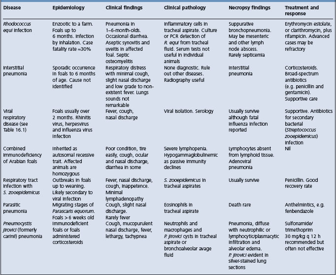
Etiology Virulent strains of Rhodococcus equi
Epidemiology Sporadic disease of 1–5-month-old foals that is endemic on some farms. Foals are infected by ingestion or inhalation during first weeks of life
Clinical signs Pneumonia, fever, respiratory distress, cough, lack of nasal discharge, failure to thrive, multiple distended joints and uveitis. Occasionally diarrhea or septic osteomyelitis
Clinical pathology Leukocytosis, hyperfibrinogenemia, inflammatory cells in tracheal aspirate
Necropsy lesions Pulmonary consolidation and abscessation. Nonseptic polyarthritis
Diagnostic confirmation Culture of R. equi from tracheal aspirate
Treatment Erythromycin estolate or microencapsulated base 25 mg/kg every 8 hours orally, in combination with rifampin 5 mg/kg every 12 hours orally. Clarithromycin (7.5 mg/kg every 12 h orally) may be substituted for erythromycin
Control Insure adequate transfer of passive immunity. Decrease stocking density. Decrease environmental contamination by virulent strains of R. equi
Rhodococcus equi is a Gram-positive, pleomorphic rod. The most important manifestation of R. equi infection is pneumonia in foals. It also causes pleuropneumonia, pneumonia, osteomyelitis, and abortion in immunocompromised and normal adult horses, abscesses that must be differentiated from tuberculosis in pigs and ruminants, and pneumonia in immunosuppressed humans.1-5 The organism is a natural inhabitant of soil, grows well at temperatures ranging from 10–40°C, and is readily isolated from the feces of herbivores and their environment. However, isolates of R. equi vary in virulence, with many isolates obtained from feces or soil not being pathogenic.6
There are a large number of virulent strains of R. equi, based on pulsed-field gel electrophoresis of chromosomal DNA.7,8 While there is evidence of clustering of strains on farms and on most farms 1 or 2 strains predominate, there is little evidence of marked regional variations in prevalence of strains of virulent R. equi.7,8 Only rarely will it be possible to link infections to a given site or region on the basis of analysis of chromosomal DNA.7
Virulence of R. equi is dependent upon the ability of the organism to enter, survive in and replicate in macrophages. Virulence is associated with the presence of highly immunogenic virulence associated proteins (Vap A, C, D, E, G, H), of which Vap A is apparently the most important, although the role of the other Vap proteins has not been determined.9,10 Vap A is a surface-expressed, lipid-modified protein that elicits an intense humoral response by foals.10 Expression of Vap A, C, D, and E is upregulated by incubation at 37°C, consistent with their role as virulence factors. Other genes probably involved in virulence are also upregulated by conditions that mimic those in vivo.11 The presence of the virulence proteins is associated with enhanced ability of virulent R. equi to survive and replicate within macrophages, whereas avirulent strains replicate poorly or not at all.12 However, the exact virulence mechanisms of R. equi are not known.13
The genes for Vaps A and C–H are present in a pathogenicity island in an 80–85 kb plasmid that is present in 98% of isolates of R. equi from foals with pneumonia.14 Most isolates from the environment, feces, pigs, cattle, and human patients with R. equi infection do not contain either of the two identified virulence plasmids.4,15,16 Virulence is associated with the presence of the plasmid, and loss of the plasmid by a strain of R. equi results in loss of virulence. There is geographical variation both within and between countries in the prevalence of each of the virulence plasmids in isolates from foals with pneumonia.15,17,18
R. equi pneumonia in foals has a worldwide distribution. Clinical disease is often sporadic but on farms where the disease is endemic annual morbidity can be as high and can vary widely from year to year.19 The median percentage of foals that developed R. equi at farms on which the disease was endemic was 6.6%, with 38% of farms having more than 10% of foals affected.20 Case fatality rates for foals on farms, as opposed to those treated at veterinary teaching hospitals, is reported as 29–42% (for 113 and 19 affected foals, respectively).20,21 The median case fatality rate for 32 farms in Texas was 25% and the case fatality rate was more than 50% for 22% of farms. The case fatality rate among foals treated at veterinary teaching hospitals is approximately 28%.22 The detection of R. equi pneumonia in one foal on a farm should prompt an examination of all other foals on that farm.
Current evidence supports the hypothesis that foals are exposed and infected within the first several days of life.23 The age at onset of clinical signs of disease associated with R. equi varies between 2 weeks and 6 months but the peak prevalence for pneumonic disease is between 1 and 3 months.24 The disease is rare in adult horses. Risk factors in foals for development of R. equi pneumonia have not been determined although a large number of factors have been examined.25 The month in which the foal was born, gestational age, dam’s parity, antimicrobial administration during the first week of life, exposure to pasture at less than 2 weeks of age, need for treatment to correct inadequate transfer of passive immunity, and size of mare/foal groups were not associated with risk of disease on farms in Texas.26
The prevalence of virulent R. equi in isolates from the environment does not appear to be greater on farms where the disease is endemic.18,27 Morbidity varies widely among geographical areas and individual farms, probably because of environmental factors that affect the number of virulent R. equi and the ease of infection. Because aerosol infection by virulent R. equi in dust is thought to be the most important route of infection of foals, factors that favor the accumulation and persistence of R. equi in soil and its ability to become aerosolized most probably increase the risk of infection. Such factors might include:24
• Hot and dry weather, favoring formation of dust
• Crowding of pastures with young horses
However, empirical demonstration of the importance of these risk factors has not been reported, with several exceptions. Soil pH, salinity and concentrations of various elements including iron, zinc, and copper are not associated with the risk of foals developing R. equi pneumonia on farms in Texas.28 These soil-associated risk factors were examined because R. equi is a normal inhabitant of the soil and of the intestine of ruminants, horses, and pigs. It is not highly resistant but it has been found to survive in moist soil for periods of longer than 12 months. The infection is considered to be soil associated and to be maintained through a soil–horse cycle.29 The number of organisms in the soil and stable areas on horse farms increases with the time that the farms have housed horses, although there is not a strong correlation between R. equi concentration in soil and prevalence of pneumonia in foals.18,30
Farms of larger size, with more resident mares, greater numbers of foals (≥15), and greater foal density per acre, and the presence of mares brought on to the farm for breeding, are all associated with greater risk of foals developing R. equi pneumonia.20,31 R. equi pneumonia does not appear to be associated with poor farm management or lack of preventative health practices such as vaccination, deworming, or administration of hyperimmune plasma.25 The practice of testing for failure of transfer of passive immunity is associated with an increased likelihood of the disease on a farm.32 However, this association probably reflects the facts that the disease is more likely on larger farms, which are more likely to perform this test, and that farms that have had the disease are more likely to institute preventive care procedures.
Most foals are exposed to infection, as demonstrated by seroconversion, but only a few develop disease. The organism colonizes the intestine of the normal foal during the first 2 months of life and has been detected in the feces as early as 5 days.33 Inhalation of the organism in dust is probably the most important route of transmission for pneumonic disease.34,35 Intestinal disease, which may be clinically inapparent, usually occurs with pulmonary disease but the source of the infection is unclear, although it may be ingestion of contaminated material or swallowing of infected respiratory secretions. Foals over 5 weeks of age have generally been resistant to experimental challenge.
Exposure of foals to R. equi is common, based on rate of seroconversion, yet the development of respiratory infection and clinical disease is much less common. The reason for this is not fully understood, although development of the disease probably depends on exposure to an infectious dose of organism and the susceptibility of the foal. Recent epidemiology studies suggest that exposure occurs within the first few days of life, before waning of maternally derived passive immunity, and that affected foals have lower CD4+:CD8+ ratios before development of the clinically apparent disease than foals that do not subsequently develop the disease.36 The differences between groups of foals was largest during the first 2 weeks of life, suggesting that foals that subsequently develop disease associated with R. equi may have impaired immune function during early life.36 In adult horses, in which the disease is rare, protective immunity is associated with both cellular and humoral immune responses characterized by enhanced immunoproliferative responses of CD4+ and CD8+ cells and presence of IgGa and IgGb antibodies to Vap A.37 Opsonizing antibody to R. equi is an important defense mechanism in experimentally infected foals and administration of R. equi hyperimmune plasma or plasma rich in anti-Vap A and C antibodies protects experimentally infected foals from developing pneumonia.38 Overall, these results suggest that foals that develop R. equi pneumonia have a T helper cell (Th)2-like response to infection, rather than a Th1-like response. Th1-like responses, which are associated with enhanced CD4+ and CD8+ responses, are believed to be important in resistance to the disease.13 Whether the switch to a Th2-like response to infection is a function of virulent R. equi or an attribute of susceptible foals has not been determined.
Experimental and clinical studies indicate that the foal is infected several weeks or months before clinical signs are observed. Virulent strains of R. equi are facultative intracellular parasites of macrophages, which they ultimately destroy. Neutrophils are bactericidal for R. equi but the organism can survive by inclusion in macrophages. Opsonization of R. equi by specific antibodies results in enhanced lysosome–phagosome fusion and greater killing of R. equi by equine macrophages and monocytes,13 whereas entry of R. equi into macrophages by nonimmune phagocytosis is not associated with enhanced killing. Its survival in the macrophage is associated with absence of phagosome– lysosome fusion.39 Nonvirulent strains do not proliferate in macrophages and monocytes. The combined action of humoral and cellular immune systems is important in preventing development of the disease after inhalation of bacteria. Without opsonization, the capacity of the pulmonary macrophage of foals to kill R. equi is impaired and the organism has been shown to be able to persist in the pulmonary macrophage of infected foals. The inability of the pulmonary macrophages to destroy R. equi leads to persistent infection in the lung and a chronic bronchopneumonia with extensive abscessation and an associated suppurative lymphadenitis.
Intestinal infection is common in foals with R. equi pneumonia,40 although clinical manifestations of the intestinal infection, such as diarrhea, are uncommon. Gastrointestinal tract infection is characterized by ulcerative lesions of the mucosa of the large intestine and cecum. In rare cases bacteremia and subsequent suppurative foci may develop in many organs, including bones and joints, liver, kidneys, and subcutis.
R. equi pneumonia of foals may present as an acute onset of inappetence, fever, depression and tachypnea or as a more chronic disease characterized by cough and failure to thrive. The former presentation is more common and usually occurs in foals less than 3–4 months of age, with younger foals being more severely affected. It is important to realize that the acute onset of the disease is preceded by a long incubation period during which clinical signs are minimal and that the development of clinical signs is associated with severe and extensive lung lesions. The foal is often in respiratory distress and is reluctant to move and to suckle. Cyanosis may be present in severe cases. Auscultation of the chest may reveal crackles and wheezes, but abnormal lung sounds are often much less apparent than the severity of the respiratory disease suggests they should be. Foals with R. equi abscesses may not have abnormal lung sounds. There is usually minimal nasal discharge.
Subcutaneous abscesses, osteomyelitis and septic arthritis may be present or develop.41 Many foals (20%) have an aseptic polyarthritis, evident as nonpainful distension of joints, at the time they develop signs of respiratory disease. Immune-mediated uveitis occurs in approximately 10% of severely affected foals.42
In older foals the disease assumes a characteristic clinical syndrome marked by development of severe lesions without clinical signs in the foal. A subacute pneumonia develops slowly with coughing, an increase in the depth of respiration with respiratory distress developing in the late stages, and characteristic loud, moist crackles or ‘rattles’ on auscultation. The foal continues to suck and the temperature is sometimes normal but the foal becomes emaciated. Severe diarrhea may follow or accompany the respiratory signs. Nasal discharge and lymph node enlargement in the throat regions are absent. Severely affected animals die in 1–2 weeks. Thoracic auscultation may not reveal any abnormalities during the preclinical stage of the disease, although careful auscultation when tidal volume of the foal is increased by exertion or use of a rebreathing bag can reveal localized wheezes and crackles or changes in the intensity of normal breath sounds (indicative of areas of lung consolidation).
Radiographic examination is a valuable aid in diagnosis and in monitoring progress in hospitalized foals.43 Affected animals show evidence of consolidation of lung tissue, lymphadenopathy, and cavitating lesions in the lungs.
Ultrasonographic examination of the chest may reveal the presence of pulmonary consolidation before clinical signs are apparent and is useful means of detecting subclinical disease.
When lesions are confined to the intestinal wall the predominant clinical sign will be diarrhea, which may be acute or chronic and intermittent.
Intra-abdominal abscesses are associated with ill-thrift, weight loss, variable abdominal distension, fever, depression and in some cases colic. Ultrasonographic examination can reveal the abscess provided that it is within the field of the ultrasound probe.
R. equi infection in Thoroughbred and Standardbred foals is associated with a reduced chance of racing as an adult compared with the overall population of foals, but affected foals that survive have a similar racing performance as adults to horses that did not have R. equi pneumonia.22,44 The morbidity and case fatality rates are provide above under Epidemiology.
Hematological evaluation usually reveals leukocytosis with neutrophilia and monocytosis, and elevation in the concentrations of acute phase proteins including plasma fibrinogen and serum amyloid A – changes characteristic, but not diagnostic, of R. equi infection.45 Monitoring of blood white cell concentration and plasma fibrinogen concentration are useful in foals from farms on which the disease is endemic. White blood cell concentrations above 13.0 × 10⁹/L (13000 cells/μL) have a sensitivity and specificity of 95% and 61% respectively for R. equi pneumonia.46 The high sensitivity means that few foals with the disease will be missed, while the moderate specificity means that a number of foals will be incorrectly suspected as having the disease. Because a high white cell count can be caused by a number of diseases other than R. equi pneumonia, foals with high white cell counts from farms on which the disease is endemic should be further examined for evidence of disease, including detailed clinical examination possibly including ultrasonographic examination, culture or PCR of tracheal aspirates, or thoracic radiography. Measurement of plasma fibrinogen concentration is less useful for detecting foals with R. equi pneumonia. Plasma fibrinogen concentrations of 400 mg/dL (0.4 g/L) have sensitivity and specificity of 91% and 51%, respectively, whereas concentrations of 600 mg/dL (0.6 g/L) have sensitivity and specificity of 38% and 96%, respectively.46 The positive and negative predictive values of the tests depends on the prevalence of the disease among the group of foals examined, being low for farms on which the disease is rare and increasing as the prevalence of the disease increases. Serial measurement of serum amyloid A concentrations is not useful for detecting foals with clinically inapparent R. equi pneumonia, nor do foals with pneumonia reliably have higher serum amyloid A concentrations than normal foals.47
Transtracheal aspirates from affected foals reveal a neutrophilic leukocytosis. Intracellular, Gram-positive pleomorphic rods characteristic of R. equi may be present in tracheal aspirates but the sensitivity of this observation has not been determined and all tracheal aspirates should be cultured.
Although numerous serological tests have been developed, including agar gel immunodiffusion, synergistic hemolysis inhibition, radial immunodiffusion and various ELISAs, none has demonstrated value in the diagnosis of the disease in individual animals. Currently available serological tests, either as single or paired samples, are not reliable in confirming or excluding the presence of R. equi pneumonia in foals.28
Culture of tracheal aspirates is the gold standard for antemortem diagnosis of the disease, although sensitivity of culture is less than that of PCR examination of tracheal aspirates. Culture of tracheal aspirates has a sensitivity of approximately 86%, based on diagnosis of R. equi pneumonia at necropsy.46 However, R. equi can be cultured from 35% of clinical normal foals in populations in which the disease is endemic.48 A PCR test for the rapid detection of R. equi in tracheal aspirates has a sensitivity of 100% and a specificity of 91% in foals with a clinical diagnosis of R. equi pneumonia.49 PCR examination of nasal swabs for presence of R. equi has a sensitivity of 15%, which is too low to be clinically useful.50 More recent quantitative real-time PCR assays permit the rapid detection and quantification of virulent (VapA-gene-positive) strains of R. equi in tracheobronchial aspirates.51 This assay detects R. equi at concentrations as low as 20cfu/mL of tracheobronchial fluid, providing a specific and highly sensitive test for the presence of this organism. A multiplex PCR test simultaneously detects R. equi and the presence of virulence factors, thereby permitting rapid differentiation of pathogenic from nonpathogenic strains of R. equi in biological samples.52
The predominant lesions are a pyogranulomatous pneumonia plus lymphadenitis of the bronchial lymph nodes.12 Grossly, the firm, raised lung nodules may reach several centimeters in diameter and be located anywhere in the lung field, especially in the cranioventral quadrant. If several nodules coalesce, the lesion may be misinterpreted as a suppurative bronchopneumonia. Histologically, organisms are easily demonstrated within the macrophages and giant cells comprising these lesions. Many cases also have ulcerative enterocolitis, with abscessation of mesenteric or cecocolic lymph nodes. Although necropsy may reveal widespread infection, many cases are subclinical.
Antemortem diagnosis is by culture of R. equi from aspirates of tracheal fluid. Currently available serological tests do not provide confirmation of disease in individual animals.
The pneumonic form of the disease may be confused with other causes of pneumonia in foals (Table 16.4). Other causes of diarrhea in this age group include parasitism due to cyathostomes, infection by Salmonella sp. and antibiotic-induced diarrhea.
The aseptic synovitis and joint effusion that frequently accompanies R. equi pneumonia should be differentiated from septic arthritis due to S. zooepidemicus, Salmonella spp., R. equi or other bacteria.
The principles of treatment are cure of R. equi infection, relief of respiratory distress and correction of associated immune-mediated diseases.
Elimination of infection requires the administration of antimicrobial agents that are both effective against the organism and able to penetrate infected macrophages to gain access to the organism. In vitro antibiotic sensitivity testing has not been demonstrated to be useful in predicting the clinical efficacy of treatment. R. equi isolates from ill foals are frequently sensitive in vitro to a variety of antibiotics, including the aminoglycosides gentamicin and neomycin, tetracycline, sulfonamides and chloramphenicol, while most are resistant to cephalosporins and penicillin.53-55 However, treatment with antibiotics other than erythromycin and rifampin is associated with a lower recovery rate. Treatment with penicillin, with or without gentamicin, chloramphenicol or tetracycline, is not effective.53 Trimethoprim–sulfadiazine combinations might be effective in some foals but are not the preferred treatment.53 Neomycin has been recommended for treatment of R. equi pneumonia56 but the risk of nephrotoxicosis, need for parenteral administration and lack of demonstration of clinical efficacy do not support its use at this time.
The treatment of R. equi pneumonia in foals is achieved by administration of macrolide antibiotics in combination with rifampin. Conventional treatment is administration of the combination of an acid-stable erythromycin (preferably estolate) at a dose of 25 mg/kg orally every 12 hours and rifampin at a dose of either 5 mg/kg every 12 hours or 10 mg/kg every 24 hours. Other esters or preparations of erythromycin are less well absorbed or have shorter elimination half-lives than the estolate ester and must be administered more frequently.32 Erythromycin ethylsuccinate does not provide optimal therapy for R. equi pneumonia in foals because of poor absorption after oral administration.55,57 The macrolide antibiotics azithromycin and clarithromycin have also been used to treat foals with R. equi pneumonia. Treatment of foals with a combination of clarithromycin (7.5 mg/kg orally every 12 h) and rifampin results in improved survival over foals treated with azithromycin (10 mg/kg orally every 24 h) and rifampin or erythromycin and rifampin in a veterinary teaching hospital.58
Therapy should be continued until the foal is clinically normal and has a normal plasma fibrinogen concentration and white blood cell count, which can require treatment for at least 1 month and often longer. Radiographic or ultrasonographic demonstration of resolution of the pulmonary consolidation and abscessation is useful in the decision to stop therapy. The case fatality rate is approximately 30% (see Epidemiology, above) even with appropriate treatment.
Side-effects of erythromycin–rifampin therapy include the development of diarrhea in some foals and their dams.59 Administration of erythromycin to foals is associated with an eightfold increase in the risk of diarrhea.50 Antibiotic therapy should be temporarily discontinued in foals that develop diarrhea.
During hot weather, some foals treated with erythromycin become hyperthermic (40–41°C, 104–105.5°F) and tachypneic and occasional deaths result from this syndrome.50,60 The basis for this hyperthermic event, which may occur in healthy foals administered erythromycin, is unknown. Affected foals should be treated urgently with antipyretics, cold water bathing and housing in a cooler environment.
The emergence of R. equi isolates resistant to rifampin during therapy of foals with R. equi pneumonia has been reported.61 However, one case had an atypical clinical course, in that the foal was 10 months of age at presentation and may have been immunosuppressed, and in the other instance the foal was treated with only rifampin. The development of resistance during monotherapy with rifampin is a recognized contraindication to the use of this drug alone.
Ancillary therapy with NSAIDs, bronchodilators, and mucolytics might be of value.53 Foals in severe respiratory distress require intranasal or intratracheal administration of oxygen.
Control measures are designed to maximize the resistance of the foal to infection and to reduce the infection pressure on the foal by decreasing contamination of the foal’s environment with virulent R. equi. Insuring adequate transfer of colostral immunoglobulins in all foals through routine monitoring of serum immunoglobulin concentrations in 1-day-old foals is an essential part of any control program. To decrease environmental contamination with virulent R. equi, efforts should be made to reduce fecal contamination of pastures and to reduce or eliminate dusty or sandy areas.24,62 These efforts should include grassing or paving of bare areas, removal and composting of fecal material on a regular basis, reduction of stocking density and reduction in the size of mare/foal bands.24
On farms with endemic disease, regular physical examination, including auscultation of foals and once-daily monitoring of rectal temperature, can permit early identification and treatment of affected foals.63 Measurement of blood white cell count, as detailed above, can be useful in early identification of affected foals. Identification of one foal affected with R. equi pneumonia on a farm should prompt an examination of all other foals on the farm.
Ultrasonographic examination of the thorax of foals may permit identification of foals with clinically inapparent pulmonary abscesses.
The administration to foals of a hyperimmune serum, obtained from mares vaccinated with an autogenous vaccine, limits the severity of disease produced by experimental challenge but has not been consistently useful in preventing or decreasing the prevalence of naturally occurring disease.19,34,64,65 There are no vaccines effective in prevention of R. equi pneumonia in foals.
Giguere S, Prescott JF. Clinical manifestations, diagnosis, treatment, and prevention of Rhodococcus equi infections in foals. Vet Microbiol. 1997;56:313-334.
Takai S. Epidemiology of Rhodococcus equi infections: a review. Vet Microbiol. 1997;56:167-176.
Cohen ND, et al. Control and prevention of Rhodococcus equi pneumonia in foals. Compend Contin Educ Pract Vet. 2000;22:1062-1069.
Meijer WG, Prescott JF. Rhodococcus equi. Vet Res. 2004;35:383-396.
1 Davis WP, et al. Vet Pathol. 1999;36:336.
2 Verdonck F, et al. Vet Rec. 2004;154:149.
3 Vengust M, et al. Can Vet J. 2002;43:706.
4 Flynn O, et al. Vet Microbiol. 2001;78:221.
5 Cornish N, Washington JA. Curr Clin Top Infect Dis. 1999;19:198.
6 Bowles PM, et al. Vet Microbiol. 1987;14:259.
7 Cohen ND, et al. Am J Vet Res. 2003;64:153.
8 Morton AC, et al. Appl Environ Microbiol. 2001;67:2167.
9 Takai S, et al. Infect Immunol. 1992;60:2995.
10 Tan C, et al. Can J Vet Res. 1995;59:51.
11 Ren J, Prescott JF. Vet Microbiol. 2003;94:167.
12 Hondalus MK, et al. Infect Immun. 1994;62:4167.
13 Meijer WG, Prescott JF. Vet Res. 2004;35:383.
14 Haites RE, et al. J Clin Microbiol. 1997;35:1642.
15 Nicholson VM, et al. J Clin Microbiol. 1997;35:738.
16 Makrai L, et al. Vet Microbiol. 2002;88:377.
17 Yuyama T, et al. J Vet Med Sci. 2002;64:715.
18 Takai S, et al. J Vet Diagn Invest. 2001;13:489.
19 Hurley JR, et al. Aust Vet J. 1995;72:418.
20 Chaffin MK, et al. J Am Vet Med Assoc. 2003;222:467.
21 Raidal SL. Aust Vet J. 1996;73:201.
22 Ainsworth DM, et al. J Am Vet Med Assoc. 1998;213:510.
23 Horowitz ML, et al. J Vet Intern Med. 2001;15:171.
24 Prescott JF, Hoffman AM. Vet Clin North Am Equine Pract. 1993;9:375.
25 Chaffin MK, et al. J Am Vet Med Assoc. 2003;222:476.
26 Chaffin MK, et al. J Am Vet Med Assoc. 2003;223:1791.
27 Martens RJ, et al. J Am Vet Med Assoc. 2000;217:220.
28 Martens RJ, et al. J Am Vet Med Assoc. 2002;221:825.
29 Takai S, et al. Vet Microbiol. 1986;12:169.
30 Prescott JF. Vet Microbiol. 1987;14:211.
31 Cohen ND, et al. J Am Vet Med Assoc. 2005;226:404.
32 Lakritz J, et al. Compend Contin Educ Pract Vet. March: 2002:256.
33 Takai S. Vet Microbiol. 1997;52:63.
34 Martens RJ, et al. Proc Am Assoc Equine Pract. 1990;35:199.
35 Martens RJ, et al. Equine Vet J. 1982;14:111.
36 Chaffin MK, et al. Vet Immunol Immunopathol. 2004;100:33.
37 Hines MT, et al. Vet Immunol Immunopathol. 2001;79:101.
38 Hooper-McGrevy KE, et al. Am J Vet Res. 2001;62:1307.
39 Brumbaugh GW, et al. Am J Vet Res. 1990;51:766.
40 Zinc MC, et al. Can Vet J. 1986;27:213.
41 Giguere S, et al. Aust Vet J. 1994;26:74.
42 Chaffin MK, Martens RJ. Proc Am Assoc Equine Pract. 1997;43:79.
43 Prescott JF, et al. Equine Vet J. 1996;28:344.
44 Ainsworth DM, et al. Am J Vet Res. 1993;54:2115.
45 Hulten C, Demmers S. Equine Vet J. 2002;34:693.
46 Giguere S, et al. J Am Vet Med Assoc. 2003;222:775.
47 Cohen ND, et al. Equine Vet J. 2005;37:212.
48 Ardans AA, et al. Proc Am Assoc Equine Pract. 1986;32:129.
49 Sellon DC, et al. J Clin Microbiol. 2001;39:1289.
50 Stratton-Phelps M, et al. J Am Vet Med Assoc. 2000;217:68.
51 Harrington JR, et al. Am J Vet Res. 2005;66:755.
52 Halbert ND, et al. Am J Vet Res. 2005;66:1380.
53 Sweeney CR, et al. Vet Microbiol. 1987;14:329.
54 Barton MD, Hughes KL. Vet Bull. 1980;50:65.
55 Jacks SS, et al. Antimicrob Agents Chemother. 2003;47:1742.
56 Barton MD. Aust Vet J. 1986;63:163.
57 Lakritz J, et al. Vet Ther. 2002;2:189.
58 Giguere S, et al. J Vet Intern Med. 2004;18:568.
59 Gustafsson A, et al. Equine Vet J. 1997;29:314.
60 Traub-Dargatz J, et al. Proc Am Assoc Equine Pract. 1996;42:243.
61 Fines M, et al. J Clin Microbiol. 2001;39:2784.
62 Giguere S, Prescott JF. Proc Am Assoc Equine Pract. 1997;43:65.
63 Higuchi T, et al. J Am Vet Med Assoc. 1998;212:976.
Diseases associated with Listeria species
There are currently six species classified within the genus Listeria1 but only Listeria monocytogenes and Listeria ivanovii (previously classified as L. monocytogenes serotype 5) are pathogenic for domestic animals. L. ivanovii is only mildly pathogenic and is an occasional cause of abortion in sheep and cattle.2-4 Aborted fetuses have suppurative bronchopneumonia and lack the multifocal hepatocellular necrosis commonly seen in abortions associated with L. monocytogenes.3 Listeria innocua is occasionally associated with encephalitis in ruminants that is clinically and pathologically similar to that associated with L. monocytogenes.5 Most, but not all, reports of both infections record that the animals were being fed silage.
Etiology Listeria monocytogenes. Ubiquitous in farm environment
Epidemiology Ruminants, particularly sheep. Prime occurrence is seasonal associated with feeding silage with high listerial growth. Also following management-induced stress. Commonly manifest with multiple cases in a group
Clinical findings Most commonly encephalitis with brainstem and cranial nerve dysfunction or abortion in last third of pregnancy. Less commonly septicemia in periparturient and neonatal sheep and goats, enteritis in weaned sheep, spinal myelitis, ophthalmitis and occasionally mastitis
Clinical pathology Culture. Pleocytosis and elevated protein in cerebrospinal fluid with encephalitis
Lesions Microabscesses in brainstem in listerial encephalitis, spinal cord in spinal myelitis, intestine in enteritis. Visceral lesions in septicemia
Diagnostic confirmation Culture and histopathology
Treatment Chlortetracycline or penicillin. Must be given early in clinical disease
L. monocytogenes is widespread in nature and has characteristics that allow its survival and growth in a wide variety of environments. There is a highly diverse range of strains some of which have the capability of causing disease in animals and humans.
Optimal growth temperatures are between 30°C and 37°C but the organism can grow and reproduce at temperatures between –0.4°C and 45°C. It can grow between pH 4.5 and 9.6 although growth at low pH is minimal at low temperatures.1 The organism is susceptible to common disinfectants.
L. monocytogenes can be divided into 16 serovars on the basis of somatic and flagellar antigens and there is considerable genetic diversity between serovars. Serovars 4b, ½a and ½b and 3 are most commonly isolated from diseased animals but there are geographical differences.1-3 Virulent strains can multiply in macrophages and monocytes and produce a hemolysin, listeriolysin O, which is believed to be a major virulence factor.3
Although the organism is widespread in nature, clinical disease in animals occurs mainly in the northern and southern latitudes and is much less common in tropical and subtropical than in temperate climates. The disease is important in North America, Europe, the UK, New Zealand, and Australia.
In the northern hemispheres listeriosis has a distinct seasonal occurrence, probably associated with seasonal feeding of silage, with the highest prevalence in the months of December through May4 but seasonal occurrence is not a feature in Australia.5
Listeriosis is primarily a disease of ruminants, particularly sheep, and the major diseases associated with L. monocytogenes are encephalitis and abortion. In ruminants it also produces syndromes of septicemia, spinal myelitis, uveitis, gastroenteritis, and mastitis. Occasional septicemic disease occurs in horses and pigs.
• Encephalitis usually occurs sporadically, affecting a single animal in a herd or flock or a few individuals over several weeks. The mean attack rate in 50 affected flocks in Britain was 2.5% with a range of 0.1–13.3%.6 More serious outbreaks can occur with attack rates as high as 35% and cases occurring over a 2-month period. The disease occurs in sheep older than 6 weeks but may be more prevalent in lambs between 6 and 12 weeks of age and ewes over 2 years of age. The case fatality is high, especially in sheep, because the short clinical course often precludes treatment
• Abortion may also occur sporadically, which is usually true in cattle, but in sheep and goats it more commonly occurs as an outbreak with an attack rate that frequently approaches 10%
• Spinal myelitis is an uncommon manifestation but is recorded as occurring in 0.8–2.5% of sheep in affected flocks and in all ages of sheep4 4 weeks following spray dipping. Spinal myelitis also occurs sporadically in cattle 12–18 months of age5
• Septicemic disease is also a less common manifestation of infection with L. monocytogenes but can occur as an outbreak with a high case fatality in newborn lambs and kids and also in periparturient ewes and does
• Ophthalmitis/uveitis occurs in both sheep and cattle, and can occur as an outbreak. Many, but not all, outbreaks have been associated with round-bale ‘self-feed’ silage in the winter period7 and eye infection is directly from the silage as the animals burrow their head into the silage to eat. L. monocytogenes is also associated with outbreaks of catarrhal conjunctivitis of cattle
• Gastroenteritis has been reported primarily by veterinary diagnostic labs in Great Britain and New Zealand as a sporadic disease affecting all ages of sheep after weaning. It occurs during the winter months most commonly in sheep fed baleage or silage. Cases occur 2 days or more after the onset of feeding.8,9 Less commonly, cases occur in sheep on root crops or on pasture where the quality of the pasture is poor and they are at high stocking densities
• Mastitis is uncommon but can occur in cattle, sheep, and goats. It results in contamination of milk with L. monocytogenes. The more common source of L. monocytogenes in raw milk is fecal contamination. In a Danish study of quarter milk samples from over 1000000 cows in 36199 herds, 0.4% of cows had listerial mastitis and 1.2% of herds had infected cows.10
The organism is common in the environment and infection is not limited to agricultural animals. L. monocytogenes has been isolated from 42 species of mammals, 22 species of birds as well as fish, crustaceans, and insects. It is truly ubiquitous in the environment and can be commonly isolated from animal feces, human feces, farm slurry, sewerage sludge, soil, farm water troughs, surface water, plants, animal feeds and the walls, floors, drains, etc. of farms and other environments.1,11,12 The ability to form biofilms may assist in its survival in the environment13 and may assist in perpetuating its presence in water troughs on infected farms.
Most feed hays, grains and formulated feeds have the potential to contain L. monocytogenes but, with most, low levels of available water restrict its multiplication.
In ruminants L. monocytogenes can be isolated from the feces and nasal secretions of healthy animals and has been isolated from the feces of cattle in 46% of 249 herds examined14 and from 82% of samples of feedstuffs.15 In temperate climates the prevalence of L. monocytogenes in the feces of ruminants appears to vary with the season, being higher in the winter period. It is also increased during periods of environmental stress and in association with the stress of lambing and transport.16-18 The presence in feces and secretions can also be influenced by the numbers of the organism in feeds fed to the animals.15,18,19 In herds where there is a high proportion of cattle excreting in feces, the organism can be isolated from dried fecal dust on walls and most farm surfaces.
L. monocytogenes is not isolated from the feces or environment in all farms and its presence in isolable numbers is largely a reflection of its presence in feed, or the presence of animals with intestinal carriage. It is apparent that in some healthy herds and flocks there may be a multitude of different strains in the silage and feed, water troughs, feces and environment in a single herd.19,20
The presence of L. monocytogenes in bulk tank milk or milk filters is used as a measure of farm infection prevalence.21 Obviously this measure is influenced by the management and environmental conditions on farms that might result in fecal contamination of the teats. Although bulk tank and milk filter infection rates provide information of possible value to measures of environmental contamination and risk for human exposure there is no evidence that this measure has any relation to risk for animal disease on the farm being studied.
L. monocytogenes is commonly present in silage, but it does not multiply to any significant extent in effectively preserved silage which is characterized by anaerobic storage, high density, a high concentration of organic acids, and a pH below 4.5. Listeria can multiply in silage above pH 5.0–5.5, the critical pH depending upon the dry matter content.10,22,23 L. monocytogenes may be present in silage which is poorly fermented but it can also occur in pockets of aerobic deterioration in otherwise good silage24 and this is the common occurrence. These areas are often indicated by mold growth and occur at the edges of the clamp and in the top few inches of the surface in plastic-covered clamps where air has circulated under the plastic. Thus the growth of L. monocytogenes is a surface problem in silage – except those that are poorly fermented – and occurs in small areas sporadically over the surface of a silage.
The risk for contamination of silage with Listeria is higher when it contains soil. Soil may be incorporated from mole-hills present in the field and it may also be incorporated in the front of the clamp during final packing. An ash content of greater than 70 mg/kg dry matter indicates soil contamination.
Big bale silage may have a higher risk for listerial infection than conventional silage because of its lower density, poorer fermentation, the greater surface area relative to clamp silage and the greater risk for mechanical damage to the plastic covering.10
Moist preserved feeds other than grass silage are at risk for listerial growth; listeriosis is recorded, for example, in association with the feeding of moist brewers’ grains, wet spoiled hay bales and silage made from commodity by-products such as orange and artichoke waste. A relatively rapid method for the quantitative assessment of the occurrence and distribution of Listeria in suspect silage is available.24
Infective material also derives from infected animals in the feces, urine, aborted fetuses and uterine discharge, and in the milk. Although immediate spread among animals in a group has been demonstrated, field observations suggest that mediate contagion by means of inanimate objects also occurs. Woody browse may be a risk factor for goats.25
With septicemic disease and abortion the organism is transmitted by ingestion of contaminated material. Lambs which develop septicemic disease may acquire infection from contamination on the ewe’s teat, from the ingestion of milk containing the organism from ewes or does with subclinical bacteremia, through the navel from the environment, and also as a congenital infection. The encephalitic form of the disease results from infection of the terminals of the trigeminal nerve consequent to abrasions of the buccal mucosa from feed or browse or from infection of tooth cavities. Spinal myelitis is believed to result from growth up spinal nerves subsequent to body area infections.
Outbreaks of encephalitis which occur in sheep after introduction to silage usually commence about 3–4 weeks later, although there is wide variation and one study of a large number of outbreaks found the median time of this period to be 44 days.6 This delay reflects the time for ascending infection.
Commonly, the serotype that is isolated from the brain of an affected animal is also present in the silage being fed. However, the recent development of methods for genetic analyses of L. monocytogenes has demonstrated that serotyping is a relatively crude tool for epidemiological studies and in many instances, although the isolate from brain may be the same serotype as that from silage, there is no relationship on genetic analysis.20,26 Possibly this reflects differences in strains at different sites in silage and the difference between the time of sampling of the silage and the time when the affected cow ate it.
Septicemic disease in sheep and goats usually occurs within 2 days of introduction to silage and abortions 6–13 days later.17,27
Despite the ubiquity of L. monocytogenes, only a small proportion of animals develop clinical disease. A number of predisposing factors have been observed, or proposed, as risk factors for disease. These include factors that cause a lowering of the host animal’s resistance and factors that increase the infection pressure of the organism. In farm animals the latter appear the most important.
Observed risk factors include:
• Sudden changes of weather to very cold and wet
• The stress of late pregnancy and parturition
• Long periods of flooding with resulting poor access to pasture.
Area outbreaks affecting several flocks can occur in sheep on poorly drained and muddy pastures following floods, but outbreaks are also described in droughts.28 Overcrowding and insanitary conditions with poor access to feed supplies may predispose housed sheep.
Breed difference in susceptibility (Angora goats and Rambouillet sheep) has been observed in some studies21,29 but not in others.4
Factors that increase the infection pressure largely involve a massive multiplication of L. monocytogenes in the feed or environment. The feeding of grass or corn silage as a major risk factor for the occurrence of listeriosis has been recognized for many decades. The increase in use of silage for feed in ruminants may be the reason for the apparent increase in the prevalence of the disease in recent years. Silage may also exert its effect by increasing the susceptibility of the host to listerial infection, although this has been disputed.30
Introduction of virulent strains to the flock may also occur via a carrier animal, and birds, such as seagulls that scavenge on sewerage areas, may carry a heavy population of the bacteria and can contaminate feed or pastures for silage. The organism persists for as long as 3 months in sheep feces and has been shown to survive for up to 11.5 months in damp soil, up to 16.5 months in cattle feces, up to 207 days on dry straw and for more than 2 years in dry soil and feces. It is resistant to temperatures of −20°C (−6°F) for 2 years and is still viable after repeated freezing and thawing.
Oral or parenteral challenge of nonpregnant sheep and goats will produce a bacteremia with minor clinical signs of pyrexia and depression in animals with no pre-existing antibody. Clinical disease is more severe in young animals and the infection clears with the development of an immune response.31-33 The challenge of animals with pre-existing antibody is not associated with clinical disease although there may be a bacteremia. Lactating animals secrete the organism in milk during the bacteremic period. Prior challenge of goats with L. ivanovii or Listeria innocua does not protect against subsequent challenge with L. monocytogenes.33
Several studies have shown that oral, conjunctival, and parenteral challenge of pregnant animals results in more severe signs of septicemia and can be followed by abortion, although this is not an invariable sequel.34 Encephalitis has not been reproduced experimentally by intravenous challenge, although meningoencephalitis may occur following this route of challenge in young lambs. Encephalitis has been reproduced experimentally by the injection of organisms into the buccal mucosa or the tooth pulp cavity, the organism traveling centripetally via the trigeminal nerve to reach the brain stem.35
In humans, listeriosis may occur as a sporadic disease or as a food-borne outbreak to produce septicemic disease, meningoencephalitis, abortion and infection in other organs. Sporadic disease may involve healthy humans of any age but the disease usually occurs in the very young or unborn, the very old and people who are otherwise immunocompromised. The case fatality is high, and overall approximately 25% of reported cases die. Active surveillance for sporadic cases indicates that approximately 2000 cases and 450 deaths occur each year in the USA to give an annual incidence of 0.8 per 100000 population.16 The incidence is increasing, possibly because of an increase in susceptible populations.
The similarity of the disease spectrum in humans and animals and the occurrence of food-borne outbreaks has led to concerns that the disease could be a zoonosis. Whereas there is a potential for zoonotic transmission it would appear that the majority of human exposures to the organism, and the risk for disease, result from contamination of foods during processing and from the particular ability of the organism to grow at refrigerator temperature.
Milk products have been incriminated in some outbreaks of disease. Numerous studies have shown that L. monocytogenes is commonly present in low numbers (usually less than 1/mL) in raw milk from some herds. In the vast majority of herds this is the result of fecal contamination during the milking process or other environmental contamination. Rarely, its presence in raw milk is from an animal with subclinical mastitis and in this case its numbers in bulk tank milk are much higher (2000–5000/mL), even when there is a single cow or goat with L. monocytogenes mastitis.36,37 In goats and sheep the presence in raw milk may also be the result of a subclinical bacteremia.
There have been concerns that the organism might survive pasteurization, especially if present in phagocytes. D-values for Listeria in milk have been determined to be in the range of 0.9 seconds at 71.1°C. The legal limit for high-temperature/short-time pasteurization in the US is 71.7°C for 15 seconds and this temperature is sufficient to inactivate numbers far beyond those present in raw milk.38 There is no evidence that the organism will survive correct pasteurization procedures.1
Bulk tank infection rates are higher in winter and spring and cross-sectional and case-control studies have shown that the risk for detecting L. monocytogenes in bulk milk is higher in those herds that used a bucket milking system rather than a pipeline system. It is also higher in herds fed component feeds, fed leftover feed, fed from plastic feed bunks and with a low frequency of feed bunk cleaning.39,40 It is lower in herds that practice premilking teat disinfection.40
Farmers or others who consume raw milk need to be aware of the risk of infection, especially if they fall within at-risk categories. There may be a particular risk with milk from goats and sheep fed silage. People associated with agriculture are also more liable to direct zoonotic transmission of listerial disease. Dermatitis with a papular and pustular rash occurs on the arms of veterinarians following the handling of infected dystocial cases and aborted fetuses. Conjunctivitis is also recorded in agricultural workers handling infected livestock.
Although L. monocytogenes rarely causes disease in pigs it is present in the tonsils and feces of some pigs at slaughter and this presence is a potential source of contamination of the carcass and the slaughterhouse environment. There is a significantly higher prevalence in the tonsils of fattening pigs than in those of sows.41 The organism can be isolated from the floors, walls, and feed in pig units. Wet feeding, poor hygiene, and a short spelling period between batches of pigs in the finishing house have been found to be risk factors for infection in pigs. Paradoxically, disinfecting the pipeline used for wet feeding was associated with a higher risk of fecal contamination than no disinfection at all.42,43
A further concern for indirect zoonotic risk of L. monocytogenes is the presence of the organism in the feces on infected farms and the potential for fecal or windborne dust spread to adjacent fields that may contain crops for human consumption.
In most animals, ingestion of the organism, with penetration of the mucosa of the intestine, leads to an inapparent infection with prolonged fecal excretion of the organism and to a subclinical bacteremia, which clears with the development of immunity. The bacteremic infection is frequently subclinical and may be accompanied by excretion of the organism in milk. Septicemic listeriosis, with or without meningitis, occurs most commonly in neonatal ruminants and in adult sheep and goats, particularly if they are pregnant and when the infection challenge is large.
The organism is a facultative intracellular pathogen that can infect cells, including intestinal cells, by directed endocytosis. It can survive and grow in macrophages and monocytes.3 Bacterial superoxide dismutase protects against the bactericidal activity of the respiratory burst of the phagocyte and listeriolysin O disrupts lysosomal membranes, allowing the organism to grow in the cytoplasm.1,3 The experimental mouse model indicates that cell-mediated immunity is important in protection against listerial infection but studies in goats suggest that the clearance of bacteremic infection and resistance to infection are also strongly associated with humoral antibody.44
In pregnant animals invasion of the placenta and fetus may occur within 24 hours of the onset of bacteremia. Edema and necrosis of the placenta leads to abortion, usually 5–10 days postinfection. Infection late in pregnancy results in stillbirths or the delivery of young that rapidly develop a fatal septicemia. Maternal metritis is constant and if the fetus is retained a fatal listerial septicemia may follow. Infection of the uterus causing abortion and intrauterine infection occurs in all mammals.
Encephalitis in ruminants occurs as an acute inflammation of the brainstem and is usually unilateral. The portal of entry is by ascending infection of the trigeminal or other cranial nerves following loss of the integrity of the buccal mucosa resulting from trauma, the shedding of deciduous or permanent teeth or from periodontitis.35 Clinical signs are characterized most strongly by an asymmetric disorder of cranial nerve function, and in particular the trigeminal, facial, vestibular, and glossopharyngeal nerves, but there is some variation in the involvement of individual cranial nerves depending upon the distribution of lesions in the brainstem. Lesions in the sensory portion of the trigeminal nucleus and the facial nucleus are common and lead to ipsilateral facial hypalgesia and paralysis; involvement of the vestibular nucleus is also common and leads to ataxia with circling and a head tilt to the affected side.45 The additional signs of dullness, head-pressing, and delirium are referable to the more general effects of inflammation of the brain developing in the agonal stages. Spread of the infection along the optic nerve may result in endophthalmitis in sheep and cattle.
Spinal myelitis possibly results from ascending infection in the sensory nerves of the skin following dermatitis from prolonged wetting of the fleece.4
L. monocytogenes is rarely found to be a cause of mastitis in cattle, despite the fact that it can be common in the dairy environment of herds that have milking practices that could be conducive to the introduction of environmental pathogens into the udder. This suggests that it is not a particularly invasive or perpetuating organism for the udder.36
When disease occurs it is usual to have an outbreak of either encephalitis or abortion. Encephalitis is the most prevalent manifestation in sheep. Septicemia in lambs may occur in conjunction with abortion but it is rare to have all three syndromes on the same farm, at least in the same temporal period. There are always exceptions to such generalities, and the occurrence of septicemia, abortion, and encephalitis in a flock of sheep is possible.27
In sheep, early signs are separation from the flock, and depression with a hunched stance. Sheep approached during this early stage show a frenetic desire to escape but are uncoordinated as they run and fall easily. The syndrome progresses rapidly with more severe depression to the point of somnolence and the development of signs of cranial nerve dysfunction. Fever – usually 40°C (104°F) but occasionally as high as 42°C (107°F) – is usual in the early stages of the disease but the temperature is usually normal when overt clinical signs are present.
Signs vary between individual sheep but incoordination, head deviation sometimes with head tilt, walking in circles, unilateral facial hypalgesia and facial paralysis are usually present. Facial hypalgesia can be detected with pressure from a hemostat and the facial paralysis is manifest with drooping of the ear, paralysis of the lips and ptosis on the same side of the face as the hypalgesia. This may be accompanied by exposure keratitis, often severe enough to cause corneal ulceration. Strabismus and nystagmus occur in some. Panophthalmitis, with pus evident in the anterior chamber of one or both eyes, is not uncommon in cattle that have been affected for a number of days. Also there is paresis of the muscles of the jaw, with poor tone or a dropped jaw, in which case prehension and mastication are slow and the animal may stand for long periods drooling saliva and with food hanging from its mouth.
The position of the head varies. In many cases there is deviation of the head to one side with the poll–nose relationship undisturbed (i.e. there is no rotation) but in others there is also head tilt. The head may be retroflexed or ventroflexed depending on the localization of the lesions and in some cases may be in a normal position. The deviation of the head cannot be corrected actively by the animal and if it is corrected passively the head returns to its previous position as soon as it is released. Progression is usually in a circle in the direction of the deviation and the circle is of small diameter. There is ataxia, often with consistent falling to one side, and an affected sheep may lean against the examiner or a fence. The affected animal becomes recumbent and is unable to rise, although often still able to move its legs. Death is due to respiratory failure.
In cattle, the clinical signs are essentially the same but the clinical course is longer.46 In adult cattle the course of the disease is usually 1–2 weeks but in sheep and calves the disease is more acute, death occurring in 2–4 days.
Outbreaks of abortion are recorded in cattle but occur more commonly in sheep and in goats. Abortion due to this organism is rare in pigs.
In cattle, abortion or stillbirth occurs sporadically and usually in the last third of pregnancy; retention of the afterbirth occurs commonly, in which case there is clinical illness and fever of up to 40.5°C (105°F). Abortion has been observed soon after the commencement of silage feeding but does not always have this association.
In sheep and goats abortions occur from the 12th week of pregnancy onwards, the afterbirth is usually retained, and there is a bloodstained vaginal discharge for several days. There may be some deaths of ewes from septicemia if the fetus is retained. In both species the rates of abortion in a group are low but may reach as high as 15%. On some farms, abortions recur each year.
This occurs as a sporadic disease in cattle and has no distinguishing clinical features from that associated with L. monocytogenes.46,47 Outbreaks in sheep are manifest with abortion and stillbirth but particularly with the birth of live infected lambs, which seldom survive long enough to walk or suck.
Acute septicemia due to L. monocytogenes is not common in adult ruminants but does occur in monogastric animals, and in newborn lambs and calves. There are no signs suggestive of nervous system involvement, the syndrome being a general one comprising depression, weakness, emaciation, pyrexia, and diarrhea in some cases, with hepatic necrosis and gastroenteritis at necropsy. The same syndrome is also seen in ewes and goats after abortion if the fetus is retained. A rather better defined but less common syndrome has been described in calves 3–7 days old. Corneal opacity is accompanied by dyspnea, nystagmus, and mild opisthotonos. Death follows in about 12 hours. At necropsy there is ophthalmitis and serofibrinous meningitis. Septicemic listeriosis is recorded in a foal.48
Infection in the udder may involve a single quarter or both quarters; it is chronic and poorly responsive to treatment. There is a high somatic cell count in milk from the affected quarter, but the milk appears normal.
There is fever, ataxia with initial knuckling of the hindlimbs progressing to hindlimb weakness and paralysis. In some cases, both in sheep and cattle, there is also paresis and paralysis of the front limbs. There is no evidence of cranial nerve involvement and affected animals are initially mentally alert, bright and continue to eat. However, there is rapid deterioration and affected animals are commonly humanely destroyed.
The organism can be cultivated from vaginal secretions for up to 2 weeks after abortion and a proportion of aborting animals also have L. monocytogenes in the milk and feces.
The cerebrospinal fluid in cases of encephalitis has increased protein and an increased number of leukocytes, most of which are mononuclear cells or lymphocytes.45,49 L. monocytogenes is not detectable by culture or PCR.50 Electrodiagnostic examinations are reported.51
Serological tests (agglutination and complement fixation tests) have been used but lack the predictive value required for diagnostic use. Ruminants commonly have antibody to Listeria and high titers are commonly encountered in normal animals in flocks and herds where there have been clinical cases. Nucleic-acid-based techniques can be used to determine the source of a strain of L. monocytogenes in an outbreak.13
Typically there are no distinctive gross changes associated with listerial encephalitis. Histological examination of central nervous system tissue is necessary to demonstrate the microabscesses that are characteristic of the disease. These are present in the brainstem in listerial encephalitis and in the cervical and/or lumbar spinal cord in outbreaks of spinal myelitis. Sampling of the forebrain will typically result in a false-negative diagnosis. Cold enrichment techniques are advisable when attempting to isolate the organism. Gram staining of paraffin-embedded tissue may permit confirmation of the diagnosis in cases for which suitable culture material is unavailable. Alternative test methods such as fluorescent antibody or immunoperoxidase tests are available in some laboratories. In one retrospective study comparing diagnostic methods, immunoperoxidase staining was superior to bacterial culture when correlated with histopathological changes.52
Visceral lesions occur as multiple foci of necrosis in the liver, spleen, and myocardium in the septicemic form and in aborted fetuses. Aborted fetuses are usually edematous and autolyzed, with very large numbers of bacteria visible microscopically in a variety of tissues. In aborting dams, there is placentitis and endometritis in addition to the lesions in the fetus.30
Sheep with enteritis show ulcerative abomasitis and some also have typhlocolitis at necropsy; histologically there are microabscesses throughout the intestine and a characteristic infiltration of degenerating neutrophils in the mucosa lamina muscularis of the abomasum.8
The intravenous injection of chlortetracycline (10 mg/kg BW per day for 5 d) is reasonably effective in meningoencephalitis of cattle but less so in sheep. Penicillin at a dosage of 44000 IU/kg BW given intramuscularly daily for 7 days, and in many cases for 10–14 days, can also be used. The recovery rate depends largely on the time that treatment is started after the onset of clinical signs. If severe clinical signs are already evident, death usually follows in spite of treatment. Usually the course of events in an outbreak is that the first case dies but subsequent cases are detected sufficiently early for treatment. Dehydration, acid–base imbalances and electrolyte disturbances must also be corrected. Cases of spinal myelitis are poorly responsive to treatment.
Treatment of listerial iritis is with systemic antibiotics in the early stages coupled with subpalpebral corticosteroid and atropine to dilate the pupil.
In vitro resistance to the tetracycline group of antimicrobials is reported.53
Control is difficult because of the ubiquitous occurrence of the organism, the lack of a simple method of determining when it is present in high numbers in the environment and a poor understanding of the risk factors other than silage. Where the risk factor is silage there may be some merit in the recommendation that a change of diet to include heavy feeding of silage should be made slowly, particularly if the silage is spoiled or if listeriosis has occurred on the premises previously. Tetracyclines can be fed in the ration of animals at risk in a feedlot. When possible, the obviously spoiled areas of silage should be separated and not fed.
Other recommendations on the feeding of silage include: avoiding making silage from fields where molehills may have contaminated the grass; avoiding soil contamination when filling the clamp; the use of additives to improve fermentation and the avoidance of silage that is obviously decayed, or with a pH of greater than 5 or an ash content of more than 70 mg/kg of dry matter.
Silage removed from the clamp should be fed as soon as possible.
Where uveitis is a problem, feeding systems that avoid eye contact with silage should be used.
A live attenuated vaccine has been shown to induce protection against intravenous challenge54 and a live attenuated vaccine in use in Norway for several years is reported to reduce the annual incidence of the disease in sheep from 4% to 1.5%.55 An economic model is available for determining whether vaccination should be practiced.56 Commercial killed vaccines are available for the control of the disease in some countries and some companies will also produce autogenous vaccines on request. The efficacy of vaccination still requires further determination; however; when economics or food availability on the farm dictate that contaminated silage must be fed, consideration might be given to vaccination as a means of providing some protection.
Gray ML, Killinger AH. Listeria monocytogenes and listeric infections. Bacteriol Rev. 1966;30:309.
Ladds PW, Dennis SM, Njoku CO. Pathology of listeric infections in animals. Vet Bull. 1974;44:67.
Scarratt WK. Ovine listeric encephalitis. Compend Contin Educ Pract Vet. 1987;9:F28-F32.
Gitter M. Veterinary aspects of listeriosis. PHLS Microb Dig. 1989;6(2):38-42.
Anon. Listeria monocytogenes. Recommendations by the national advisory committee on microbiological criteria for foods. Int J Food Microbiol. 1991;14:185-246.
Farber JM, Peterkin PI. Listeria monocytogenes, a food-borne pathogen. Microbiol Rev. 1991;55:476-511.
Low JC, Donachie W. A review of Listeria monocytogenes and Listeriosis. Vet J. 1997;153:9-29.
Fenlon DR. Listeria monocytogenes in the natural environment. In Ryser ET, Martin EH, editors: Listeria, listeriosis and food safety, 2nd edit, New York: Marcel Dekker, 1998.
1 Farber JM, Peterkin PI. Microbiol Rev. 1991;55:476.
2 Low JC, et al. Vet Rec. 1993;133:165.
4 Seaman JT, et al. Aust Vet J. 1990;67:142.
5 Schweizer G, et al. Vet Rec. 2004;154:54.
6 Wilesmith JW, Gitter M. Vet Rec. 1986;119:467.
7 Walker JK, Morgan JH. Vet Rec. 1993;132:636.
8 Clarke RG, et al. N Z Vet J. 2004;52:46.
9 Otter A, et al. Vet Rec. 2004;154:479.
10 Jensen NE, et al. Int J Food Microbiol. 1996;32:209.
11 Bosgiraud C, et al. Rev Med Vet. 1991;142:463.
12 Garcia E, et al. J Food Protect. 1996;59:950.
13 Borucki MK, et al. J Appl Environ Microbiol. 2003;69:7336.
14 Husu JR. J Vet Med B. 1990;37:276.
15 Skovgaard N, Morgan CA. Am J Food Microbiol. 1988;6:229.
16 Anon. Int J Food Microbiol. 1991;14:194.
17 Loken T, et al. Acta Vet Scand. 1982;23:380. 392
18 Fenlon DR, et al. J Appl Bacteriol. 1996;81:641.
19 Gudmundsdottir KB, et al. J Appl Microbiol. 2004;96:913.
20 Borucki MK, et al. J Food Protect. 2004;67:2496.
21 Muraoka W, et al. J Food Protect. 2003;66:1413.
22 Perry CM, Donnelly CW. J Food Microbiol. 1990;53:642.
23 Webster J. In Pract. 1984;7:186.
24 Fenlon DR. Vet Rec. 1986;118:240.
25 Johnson GC, et al. J Am Vet Med Assoc. 1996;208:1695.
26 Wesley IV, et al. J Vet Diagn Invest. 2002;14:314.
27 Low JC, Renton CP. Vet Rec. 1985;116:147.
28 Reiter R, et al. Aust Vet J. 1989;66:223.
29 Nash ML, et al. Prev Vet Med. 1995;24:147.
30 Vasquez-Boland JA, et al. Am J Vet Res. 1992;53:368.
31 Miettinen A, et al. J Clin Microbiol. 1990;28:340.
32 Low JC, Donachie G. Res Vet Sci. 1991;51:185.
33 Baetz AL, et al. Vet Microbiol. 1996;51:151.
34 Gitter M, et al. Vet Rec. 1986;118:575.
35 Barlow RM, McGorum B. Vet Rec. 1985;116:233.
36 Fedio WM, et al. Can Vet J. 1990;31:773.
37 Schoder D, et al. J Dairy Res. 2003;70:395.
38 Mackey BM, et al. Lett Appl Microbiol. 1989;9:89.
39 Hassan L, et al. Dairy Food Environ Sanit. 2002;22:326.
40 Hassan L, et al. J Dairy Sci. 2000;83:241.
41 Autio T, et al. J Food Protect. 2004;67:805.
42 Belboeil PA. Vet Res. 2003;34:737.
43 Belboeil PA. J Vet Med B. 2003;50:155.
44 Miettinen A, et al. J Clin Microbiol. 1990;28:340.
45 Scarratt WK. Compend Contin Educ Pract Vet. 1987;9:F28.
46 Gill PA, et al. Aust Vet J. 1997;75:214.
47 Alexander AV, et al. J Am Vet Med Assoc. 1992;200:711.
48 Wallace SS, Hathcock TL. J Am Vet Med Assoc. 1995;207:1325.
49 Scott PR. Br Vet J. 1993;149:165.
50 Peters M, et al. J Vet Med B. 1995;42:84.
51 Strain GM, et al. Am J Vet Res. 1990;51:1513.
52 Johnson GC, et al. J Vet Diagn Invest. 1995;7:223.
53 Vela AI, et al. Int J Antimicrob Agents. 2001;17:215.
54 Linde K, et al. Vaccine. 1995;13:923.
Diseases associated with Erysipelothrix rhusiopathiae (insidiosa)
Erysipelas of pigs is the major disease of animals associated with this bacterium but there are several other minor conditions that require mention. In pigs the condition is seen as sudden death, possibly with diamond-shaped skin lesions and also arthritis and endocarditis.
Etiology Erysipelothrix rhusiopathiae (Erysipelothrix tonsillarum is nonpathogenic)
Epidemiology Pigs worldwide. Common in unvaccinated pigs raised outdoors. High case fatality rate if not treated. Organism in environment and transmitted by carrier pigs. Important zoonosis
Clinical signs Sudden onset of acute disease, fever, anorexia, typical diamond-shaped skin lesions. Arthritis, endocarditis in chronic form
Clinical pathology Organism in blood. Hemogram and serology
Necropsy findings Skin lesions, widespread ecchymotic hemorrhages (kidney, pleura, peritoneum), venous infarction of stomach. Nonsuppurative proliferative arthritis. Vegetative endocarditis
Diagnosis Culture and isolate organism from blood in acute case and then tissues
Differential diagnosis Other septicemias of pigs:
Control Vaccination, with at most 6 month intervals until new vaccines appear
Erysipelothrix rhusiopathiae (insidiosa) is the causative bacterium and the disease can be produced in acute and subacute septicemic and chronic forms by the injection of cultures of the organism. A number of different serotypes1 have been identified, usually types 1 and 2 in pigs. Many of the serotypes have been regrouped and called Erysipelothrix tonsillarum. This is a nonpathogenic type found in the tonsil that is morphologically and biochemically similar to E. rhusiopathiae but has a very distinctive genetic profile. In many minimal-disease herds they have tended not to vaccinate and then the epizootics have occurred as a result of an increasing lack of immunity.2
Erysipelas in pigs occurs worldwide and in most countries has reached a level of incidence sufficient to cause serious economic loss due to deaths of pigs and devaluation of pig carcasses because of arthritis. An epidemic occurred in the USA in 1989/90.3 However, since total indoor confinement of swine herds and the lack of contact with contaminated soil has increased, the occurrence of the disease has decreased markedly. The exception to this would be outdoor units where no vaccination is practiced. Historically, the disease occurred most commonly in unvaccinated growing pigs over 3 months of age and adults. This is primarily because the maternal antibody is believed to last up to 3 months. The infection, usually with serotypes 1a or 2, has also been demonstrated in wild boars, so these should not be forgotten as a reservoir.4 Perhaps more importantly, these were resistant to oxytetracycline and/or dihydrostreptomycin.
The prevalence of infection with E. rhusiopathiae in carrier pigs ranges from 3–98%, with most surveys indicating that 20–40% of pigs are carriers.5 Carriers occur among vaccinated as well as unvaccinated pigs. The organism has been isolated from 10% of apparently healthy slaughter pigs.5 This explains why the usual source is other pigs.
Morbidity and case fatality rates in pigs vary considerably from area to area, largely because of variations in virulence of the particular strain of the organism involved. On individual farms or in particular areas the disease may occur as a chronic arthritis in finishing pigs, or as extensive outbreaks of the acute septicemia, or both forms may occur together. In unvaccinated pigs, the morbidity in the acute form will vary from 10–30%; the case fatality rate may be as high as 75%.
Soil contamination occurs through the feces of affected or carrier pigs.6 Other sources of infection include infected animals of other species, and birds. The clinically normal carrier pig is the most important source of infection, the tonsils being the predilection site for the organism. Young pigs in contact with carrier sows rapidly acquire the status of carriers and shedders. Since the organism can pass through the stomach without loss of viability, carrier animals may re-infect the soil continuously and this, rather than survival of the organism, appears to be the main cause of environmental contamination. The organism can survive in feces for several months. All effluent contains species of Erysipelothrix but there is no need to assume that it is E. rhusiopathiae.7 However, its persistence in soil is variable and may be governed by many factors including temperature, pH and the presence of other bacteria. It can survive for 60 months in frozen or refrigerated media, 4 months in flesh and 90 days in highly alkaline soil.
The organism can be isolated from the effluent of commercial piggeries and from the soil and pasture of effluent disposal sites for up to 2 weeks after application of the effluent containing the organism. In a survey of the occurrence of potential pathogens in slurry, the organism was found in samples from 49% of 84 cattle herds, 44% of 32 pig herds and 39% of 67 cattle and pig herds.8 Although the environment is considered to be secondary to animals as a reservoir of infection, the survival of the organism in the environment could create an infection hazard. Flies are known to transmit the disease and a lowered prevalence has been attributed to the use of insecticides.
Under natural conditions, skin abrasions and the alimentary tract mucosa are considered to be the probable portals of entry and transmission is by ingestion of contaminated feed. Occasional outbreaks occur after the use of virulent and incomplete avirulent culture as vaccines. Abortion storms in late pregnant sows with septicemic death in sucklers may be the first indication of the disease in specific-pathogen-free herds.
Spread of the infection can also occur to most other species. The organism has been recovered from sylvatic mammals in north-western Canada.9 It has been isolated from a horse affected with vegetative endocarditis.10 It has, at times, been found in fish meal but this is now used much less often in pig diets. It is possible that other species, such as cattle,11 may harbor strains that are pathogenic for swine.
There is considerable variation in the ease with which the disease can be reproduced, and in its severity. Many factors such as age, health and intercurrent disease, exposure to erysipelas and heredity govern the ease of both natural and artificial transmission. Although those factors that come under the description of stress may predispose to the condition, virulence of the strain is probably the most important factor. Smooth strains can be used successfully to produce the disease experimentally but rough strains appear to be nonpathogenic. This variation in virulence between strains of the organism has been utilized in the production of living, avirulent vaccines.
Pigs of all ages are susceptible, although adult pigs are most likely to be affected if the local strain is of relatively low virulence. Recently farrowed sows seem to be particularly susceptible. This suggests that fatigue may be a factor. Sudden diet changes have also predisposed, as have heat and cold stress. When the strain is virulent, pigs of all ages, even sucklers a few weeks old, develop the disease. Almost entire litters under 2 weeks of age may be affected.12 Piglets from an immune sow may get sufficient antibodies in the colostrum to give them immunity for some weeks. It is likely that the animals are immune to the strains that are normally found in their particular environment. It is possible that it is the arrival of new serotypes through new pig arrivals or the turning over of previously contaminated land together with an increase in stress that are the main factors. It is thought that between 20–50% of pigs may carry the organism in the tonsil by slaughter. It is known that E. rhusiopathiae from bovine tonsils is pathogenic for mice and pigs and possibly pathogenic for other animals and humans.13
At least 32 serotypes are known to exist5 and many strains;14 however, 15 probably commonly affect pigs.15 Serotypes 1 and 2 are the most common types isolated from swine affected with clinical erysipelas and are generally believed to be the only serotypes that cause the acute disease. The other serotypes are relatively uncommon and none of them has yet been a cause of acute epidemics, but some have been isolated from lesions of chronic erysipelas. Serotypes 1a, 3, 5, 6, 8, 11, 21, and type N have been isolated from pigs with chronic erysipelas, mainly arthritis and lymphadenitis.16 In the USA, 19 of the 22 serotypes have been found and the most frequent are serotypes 1, 2, 5, 6, and 21.17
Not all serotypes isolated from pigs are virulent. In a survey in Japan, the organism was found in 10% of the tonsils of healthy slaughter pigs: 54% were serotype 7, 32% serotype 2, 9.5% serotype 6 and 1.6% each of serotypes 11, 12, and 16.5 All serotype 2 isolates were highly virulent for pigs while the other serotypes were only weakly virulent. Members of the other nonvirulent or weakly virulent group, mainly serotype 7 strains, are considered to be resident in porcine tonsils. Serotypes 1 (subtypes 1a and 1b), 2, 5, 6, and 4 have been found in Puerto Rico.13 Serotypes 1a or 2 were found most commonly in pigs in Australia, less commonly in sheep and infrequently in other animals.18 Serotypes 1a and 1b accounted for 79% of the isolates from diseased pigs.18 A cluster of avirulent strains of serotype 7 from the tonsils of pigs has been identified as E. tonsillarum.19 The genetic diversity of Australian field isolates of E. rhusiopathiae and E. tonsillarum indicates widespread diversity. Those recovered from sheep or birds were more diverse than those isolated from pigs, and isolates of serovar 1 were more diverse than those of serovar 2. The diversity indicated that serotyping of E. rhusiopathiae is unreliable as an epidemiological tool.20
The relatedness of the isolates tested by DNA fingerprinting suggests that E. tonsillarum contains the former serotypes 3, 7, 10, 14, 20, 22, and 23, with serotypes 13 and 18 intermediate between this species and E. rhusiopathiae. E. rhusiopathiae represents serotypes 1, 2, 4, 5, 6, 8, 9, 11, 12, 15, 16, 17, 19, 21, and N. Some serotypes shown to be E. tonsillarum on serological grounds have been found to be E. rhusiopathiae on enzyme analysis. The serotype antigens of E. rhusiopathiae (insidiosa) are immunologically distinct, and commercial bacterins prepared from the common serotypes will not provide protection against other pathogenic serotypes. This may be an explanation for the epidemics that may occur in vaccinated pigs. The 64–66 kDa protein appears to be most immunogenic.21 Also, a variety of serotypes may be recovered from pigs affected with the septicemic and arthritic forms of the disease.
The organism is resistant to most environmental influences, and to heat (15 min at 60°C) and can survive in animal tissues at 40° C and frozen tissues and is not readily destroyed by chemical disinfection, including 0.2% phenol and by drying agents.
Because of humans’ susceptibility, swine erysipelas has some public health significance.22 Veterinarians in particular are exposed to infection when vaccinating with virulent culture. It commonly contaminates pig products23 and therefore is quite a common infection in abattoir workers or butchers, or those employed in similar trades. It usually produces a swollen finger and is known as erysipeloid. In this context, there have been recent advances in slide agglutination and latex agglutination tests for rapid diagnosis,24 which have good correlation with each other and subsequent culture. Now a PCR identifying four species has been described, principally for use in the abattoir.25 Recently, a case of endocarditis and presumptive osteomyelitis has been described, so care is needed.26
The invasion of the susceptible pig by E. rhusiopathiae can occur under particular circumstances, i.e. if weather conditions are hot and humid or in particular fields or buildings. Experimentally, it is often easier to infect the pig through scarified wounds than through intravenous infusions.
No specific virulence factors have been found but there is the presence in the pathogenic serotypes of an ability to produce a capsule that resists phagocytosis.27 Some others may produce a neuraminidase, which may cleave the mucopolysaccharides in cell walls and cause vascular damage leading to hemorrhage and thrombosis. Invasion of the bloodstream occurs in all infected animals in the first instance. Septicemia results within 1–7 days. The subsequent development of either an acute septicemia or a bacteremia with localization in organs and joints is dependent on undetermined factors. Virulence of the particular strain may be important and this may depend upon the number of recent pig passages experienced. Coagulase activity is a possible virulence factor.28 Concurrent viral infection, especially hog cholera, may increase susceptibility of the host.
Localization in the chronic form is commonly in the skin and joints, and on other heart valves, with probable subsequent bacteremic episodes, and it may start from as early as 4 days after initial infection, although the cartilage lesions may be delayed until about 8 months and they can then continue to progress for at least 2 years. Selective adherence of some strains of E. rhusiopathiae (insidiosa) to heart valves may be a factor in the pathogenesis of endocarditis.29 In joints, the initial lesion is an increase in synovial fluid and hyperemia of the synovial membrane, followed in several weeks by the proliferation of synovial villi (really a synovitis), thickening of the joint capsule and enlargement of the local lymph nodes. Diskospondylitis also occurs in association with chronic polyarthritis due to erysipelas.30 Amyloidosis may occur in pigs with chronic erysipelas polyarthritis.31 The heart lesions may begin with early inflammatory changes associated with emboli.
There has been some controversy over whether the arthrodial lesions result from primary infection or whether they result from hypersensitivity to the Erysipelothrix or other antigen. Current opinion suggests that the former is the case but that the lesions are enhanced by immunological mechanisms to persistent antigen at the site. There are increased levels of immunoglobulins IgG and IgM in the synovial fluids of pigs with polyarthritis due to E. rhusiopathiae (insidiosa)32 and the levels are considered only partly due to serum and increased permeability.
Experimentally the disease can be produced by oral dosing, by intradermal, intravenous, and intra-articular injection, and by application to scarified skin, conjunctiva, and nasal mucosa. The arthritic form of the disease can be reproduced by multiple intravenous inoculations of E. rhusiopathiae.1
The microscopic lesions include vasculitis in capillaries and venules in many sites, including glomeruli, pulmonary capillaries, and the skin. Sometimes, it is possible to see emboli of bacteria without specific stains to demonstrate bacteria.
Quite often the disease is seen for the first time in pigs approaching market weight.
The animal is found dead or is dull, depressed and with a temperature of 42°C (106–109°F), usually in finishing pigs – it is uncommon in sows.
After an incubation period of 1–7 days there is a sudden onset of high fever (up to 42°C (108°F)), which is followed some time later by severe prostration, complete anorexia, thirst and occasional vomiting. Initially, affected pigs may be quite active and continue to eat even though their temperatures are high. However, generally in an outbreak one is initially presented with one or two dead or severely affected pigs showing marked red to purple discoloration of the skin of the jowl and ventral surface (may even be whole-body cyanosis), with others in the group showing high fever, reluctance to rise and some incoordination while walking. Dyspnea is a common feature. A conjunctivitis with ocular discharge may be present. Acute nonfatal erysipelas in sows was described recently in the USA.33 In this outbreak the sows were pyrexic, lethargic, lame, and had multiple erythematous plaques.
Skin lesions are almost pathognomonic but may not always be apparent. These may take the form of the classical diamond-shaped, red, urticarial plaques about 2.5–5 cm square or a more diffuse edematous eruption with the same appearance. In the early stages the lesions are often palpable before they are visible. The lesions are most common on the belly, inside the thighs and on the throat, neck, and ears, and usually appear about 24 hours after the initial signs of illness. Sometimes they can be felt rather than seen. After a course of 2–4 days the pig recovers or dies, with diarrhea, dyspnea, and cyanosis evident terminally. The mortality rate may reach 75% but wide variation occurs. Pregnant animals may abort and it is thought that this is due to the fever but it may be that there is a direct fetal action, as congenital infections and isolations of the organism from the fetus have occurred.
The so-called ‘skin’ form is usually the acute form with more prominent skin localization but less severe signs of septicemia and with a low mortality. The skin lesions disappear in about 10 days without residual effects. In the more serious cases the plaques spread and coalesce, often over the back, to form a continuous, deep purple area extending over a greater part of the skin surface. The affected skin becomes black and hard, and the edges curl up and separate from an underlying, raw surface. The dry skin may hang on for a considerable time and rattle while the pig walks, or it may slough off.
Signs are vague and indistinct except for the joint lesions characteristic of this form of the disease. There may be alopecia, sloughing of the tail and tips of the ears, and a dermatitis in the form of hyperkeratosis of the skin of the back, shoulders, and legs; growth may be retarded. Joint lesions are commonest in the elbow, hip, hock, stifle, and knee joints and cause lameness and stiffness. The joints are obviously enlarged and are usually hot and painful at first but in 2–3 weeks are quite firm and without heat. This is especially the case when the arthritis has been present for some time, allowing healing and ankylosis to develop. Paraplegia may occur when intervertebral joints are involved or when there is gross distortion of limb joints.
A subclinical form of synovitis may occur which affects feed intake and results in a reduced rate of growth.1
Endocarditis also occurs as a chronic form of the disease with or without arthritis. Suggestive clinical signs are often absent, the animals dying suddenly without previous illness, especially at times of exertion such as mating, or movement between pens. In others there is progressive emaciation and inability to perform exercise. With forced exercise dyspnea, cyanosis and even sudden death may occur. The cardiac impulse is usually markedly increased, the heart rate is faster, and a loud murmur is audible on auscultation if the valves are badly damaged.
In Switzerland, they suspect34 chronic swine erysipelas in herds where there is vegetative endocarditis, arthritis and the culture of E. rhusiopathiae from vulval discharges. These signs are also accompanied by poor fertility and increased prevalence of abortions, stillbirths and small litter size. The same authors describe the use of vaccine to control an outbreak of purulent periparturient vulval discharge34 in which E. rhusiopathiae was the only organism isolated. In one of their studies anterior vaginal samples from 64 sows all yielded E. rhusiopathiae.35
In the acute form, examination of blood smears may reveal the presence of the bacteria, particularly in the leukocytes, but blood culture is likely to be more successful as a method of diagnosis. Repeated examinations in the chronic forms of the disease may by chance give a positive result during a bacteremic phase. final identification of the organism necessitates mouse or pigeon inoculation tests, and protection tests in these animals using anti-erysipelas serum.
In the early stages of the acute form there is first a leukocytosis followed by a leukopenia and a monocytosis. The leukopenia is of moderate degree (40% reduction in total leukocyte count at most) compared with that occurring in hog cholera. The monocytosis is quite marked, varying from a fivefold to a tenfold increase (2.5–4.5% normal levels rise to 25%).
The efficiency of agglutination tests for E. rhusiopathiae (insidiosa) is not clear. They appear to be satisfactory for herd diagnosis but not sufficiently accurate for identification of individual affected pigs, particularly clinically normal carrier animals. A more accurate complement fixation test is available but an enzyme immunoassay test is much quicker, easier and more economical to perform.36 An ELISA test has been used to measure the serological response of experimentally induced erysipelas arthritis in pigs1 and 65 kDa antigen from the organism is being evaluated as an antigen agent.37 Complement fixation tests may be more reliable.
In the hyperacute cases all that may be seen is a congested carcass with discoloration of the skin. The degree of skin discoloration may provide a clue to prognosis, in that it is said that if the skin lesions are pink to light purple then resolution will often occur within 4–7 days but the dark angry black/purple lesions have a grave prognosis.
Classic ‘diamond skin’ lesions may be present. However, the diffuse, purplish discoloration of the belly and cyanosis of the extremities common to other septicemic diseases of pigs is a more reliable finding. Internally, petechial and ecchymotic hemorrhage occurs, mainly on the pleura and peritoneum and beneath the renal capsule but also on the heart, liver, and spleen. Venous infarction of the stomach is accompanied by swollen, hemorrhagic mesenteric lymph nodes and there is congestion of the lungs and liver. Infarcts may be present in the spleen and kidney and the former much enlarged. Histological changes in all tissues are those of toxemia and thrombosis. Large numbers of intravascular organisms are often visible. There are no specific histological changes.
A nonsuppurative proliferative arthritis involving limb and intervertebral joints is characteristic. A synovitis, with a serous or serofibrinous amber-colored intra-articular effusion occurs first and degenerative changes in the subendochondral bone, cartilage and ligaments follow. When the synovial changes predominate, the joint capsule and villi are thickened. There are enlarged, dark red pedunculations or patches of vascular granulation tissue, which spread as a pannus on to the articular surface. When bony changes predominate, the articular cartilage is detached from the underlying bone, causing abnormal mobility of the joint. Ulceration of the articular cartilage may also be present. Local lymph node enlargement is usual. With time, the joint lesions often repair by fibrosis and ankylosis sufficiently to permit use of the limb.
Endocardial lesions, when present, are large, friable vegetations on the valves, often of sufficient size to block the valvular orifice. Occasionally, endocarditis may be the only lesion seen but this is a rare occurrence. Erysipelas is often said to rank below S. suis as a cause of endocarditis in growing pigs but E. rhusiopathiae (insidiosa) was the most frequent isolate from cases of endocarditis seen in slaughtered pigs.31 Infarcts occur in the kidney and these may also yield pure cultures of the organism. Chronic joint lesions are often sterile but bacteriological culture should nevertheless be attempted. The probability of positive isolation increases with the number of joints sampled, and isolations are more frequent from the smaller, distal joints.
Enrichment techniques and the use of selective media will also increase the frequency of isolation of E. rhusiopathiae (insidiosa).1,38
• Bacteriology – culture swabs from joints; synovial membranes in culture media, heart valve masses, spleen, kidney, and bone marrow, particularly from a long bone. Smears of heart blood are particularly useful in the first 1–2 days of the acute diseases. The organism is a slender, facultatively anaerobic, Gram-positive rod that produces a 1 mm gray colony after 24 hours incubation on blood agar. It may be observed singly, in short chains or as a palisade. Different morphological types (rough and smooth colonies) exist and the rough are considered less virulent. Bacteriological examination of subacute cases is less successful and chronic cases not successful. Florescent techniques have been developed to show antigen in joints. New PCR techniques have also been used39
• Histology – formalin-fixed synovial membranes, heart valve masses, spleen, kidney, skin lesions (LM).
Note: the zoonotic potential of this organism when handling carcass and submitting specimens.
Clinical signs, isolation of the agent from multiple sites including blood in the acute stages; but diagnosis from joints in chronic stages is more difficult.
Erysipelas in pigs is not ordinarily difficult to diagnose because of the characteristic clinical and necropsy findings. In the occasional situation where anthrax may have occurred in the past it is worth testing a smear of edema fluid taken by a needle from the jowl or ear region for this pathogen. The acute disease may be confused with the other septicemias affecting pigs, but pigs with erysipelas usually show the characteristic skin lesions and are less depressed than pigs with hog cholera or salmonellosis.
• Septicemic salmonellosis is characterized by gross blue-purplish discoloration of the skin, especially the ears, some evidence of enteritis, and polypnea and dyspnea
• Hog cholera is characterized by large numbers of pigs affected quickly, weakness, fever, muscle tremors, skin discoloration and rapid death; convulsions are also common
• Streptococcal septicemia and arthritis are almost entirely confined to suckling pigs in the first few weeks of life as is septicemia associated with Actinobacillus suis
• Streptococcal endocarditis has a similar age distribution to erysipelas endocarditis and bacteriological examination is necessary to differentiate them.
The chronic disease characterized by joint disease occurs in pigs of all ages but less commonly in adults and must be differentiated from the following conditions:
• Glasser’s disease in pigs is accompanied by a severe painful dyspnea. At necropsy there is serositis and meningitis
• Mycoplasma arthritis generally affects pigs less than 10 weeks of age and produces a polyserositis as well as polyarthritis. However, Mycoplasma hyosynoviae can produce simple polyarthritis in growing pigs. In general the periarticular, synovial, and cartilaginous changes are less severe in these infections when compared to erysipelas; however, cultural differentiation is frequently necessary
• Rickets and chronic zinc poisoning produce lameness in pigs but they occur under special circumstances, are not associated with fever, and rickets is accompanied by abnormalities of posture and gait that are not seen in erysipelas
• Foot rot of pigs is easily differentiated by the swelling of the hoof and the development of discharging sinuses at the coronet
• Leg weakness. In recent years there has been a marked increase in chronic osteoarthritis and various forms of ‘leg weakness’ in growing swine, probably related to the increased growth rate resulting from modern feeding and management practices.
Penicillin and anti-erysipelas serum comprise the standard treatment, often administered together by dissolving the penicillin in the serum. Penicillin alone is usually adequate when the strain has only mild virulence. Standard dose rates give a good response in the field but experimental studies suggest that 50000 IU/kg BW of procaine penicillin intramuscularly for 3 days are required for complete chemotherapeutic effect. Most animals are significantly improved within 2 days. Oxytetracycline is also useful.40 Chronic cases do not respond well to either treatment because of the structural damage that occurs to the joints and the inaccessibility of the organism in the endocardial lesions. Most strains are resistant to apramycin, neomycin, streptomycin, and spectinomycin and also sulfonamides and polymyxins.
Successful control depends on good hygiene, biosecurity (other pigs and other species), reduction of stress, an effective 6-monthly vaccination policy, preferably two doses, for all animals including boars over 3 months of age, as well as rapid diagnosis, quarantine, and treatment.
Eradication is virtually impossible because of the ubiquitous nature of the organism and its resistance to adverse environmental conditions. Complete removal of all pigs and leaving the pens unstocked is seldom satisfactory. Eradication by slaughter of reactors to the agglutination test is not recommended because of the uncertain status of the test.
General hygienic precautions should be adopted. Clinically affected animals should be disposed of quickly and all introductions should be isolated and examined for signs of arthritis and endocarditis. This procedure will not prevent the introduction of clinically normal carrier animals. All animals dying of the disease should be properly incinerated to avoid contamination of the environment. Although thorough cleaning of the premises and the use of very strong disinfectant solutions is advisable, these measures are unlikely to be completely effective. The organism is susceptible to all the usual disinfectants, particularly caustic soda and hypochlorites. Whenever practicable, contaminated feedlots or paddocks should be cultivated.
Specific-pathogen-free piggeries established on virgin soil may remain clinically free of erysipelas for several years. However, because of the high risk of introduction of the organism, it is advisable to vaccinate routinely.
Because of the difficulty of eradication, biological prophylactic methods are in common use. Immunizing agents available include hyperimmune serum and vaccines.
The parenteral administration of 5–20 mL of serum, the amount depending on age, will protect in-contact pigs for a minimum of 1–2 weeks, possibly up to 6 weeks, during an outbreak. Suckling pigs in herds where the disease is endemic should receive 10 mL during the first week of life and at monthly intervals until they are actively vaccinated, which can be done as early as 6 weeks provided the sows have not been vaccinated. Repeated administration of the serum may cause anaphylaxis because of its equine origin. For this reason, it has been withdrawn from sale in many countries.
There is no fully satisfactory vaccine available for erysipelas because of the strain variation41 and short duration of immunity but the vaccines have reduced the occurrence of clinical disease.42 Regular administration at 6-monthly intervals overcomes this to some extent but there is always the possibility of a new strain appearing. Vaccines containing serotypes 2 and 10 protect against both Erysipelothrix species.43 Serum-simultaneous vaccination has been largely replaced by the use of bacterins, for which lysate and absorbate preparations are available, or by the use of attenuated or avirulent live-culture vaccines, which are administered orally or by injection. The use of live-culture vaccines is prohibited in many countries because of the risk of variation in virulence of the strains used and the possibility of spreading infection.
None of these vaccines gives lifelong protection from a single vaccination, and the actual duration of protection achieved following vaccination varies considerably. You should not assume that protection lasts longer than 6 months. The recent identification of the region responsible for protective immunity44 should improve these vaccines in future. Most of the commercially available vaccines are formalin-treated whole cultures with an adjuvant.
There is considerable difficulty in the experimental evaluation of the efficacy of erysipelas vaccines. Strain differences in immunogenicity and variation in host response to vaccination due to innate and acquired factors influence this evaluation, as does variation in virulence of the challenge strain and the method of challenge. A recent experiment has shown that an antigen of serotype 1a will elicit a protective response to a challenge with serotypes 1a and 2b.45,46 Similar factors are involved in the variations seen in field response to the use of these vaccines. Cross-protection of mice and pigs given a live-organism vaccine against 10 serovars of E. rhusiopathiae (insidiosa) has been demonstrated.47 The use of culture filtrate from a broth culture of an attenuated strain of the organism has been evaluated to produce cross-protective antibody.48
Vaccination will reduce the incidence of polyarthritis due to erysipelas, but not mild cases of arthritis. Passively acquired maternal immunity may significantly affect the immune response to vaccination in the young piglet. Also, the immunity engendered by standard vaccines is not uniformly effective against all strains. Under certain conditions, some unusual serotypes have the potential for causing disease in animals vaccinated with vaccines containing the common serotypes. This possibility cannot be ignored and must be considered when vaccination failures occur. Nevertheless, these vaccines are valuable immunizing agents in field situations.
Following a single vaccination at 6–10 weeks of age, significant protection is provided to market age. However, a second ‘booster’ vaccination given 2–4 weeks later is advisable. In herds where sows are routinely vaccinated prior to farrowing, a persisting maternal passive immunity may require that piglet vaccination be delayed until 10–12 weeks of age for effective active immunity.
Replacement gilts and adults should also be vaccinated. Bacterins are effective, and field evidence suggests that vaccination provides immunity for approximately 6 months. Sows should be vaccinated twice yearly, preferably 3–6 weeks before farrowing, as this will also provide significant protection against the septicemic form in young sucklers. If possible, a closed herd should be maintained. Abortion may occur sporadically following the use of live vaccines.49
Vaccination is subcutaneous in the skin behind the ear, or the axilla and the flank. Reactions at the site of injection are not uncommon. Swelling with subsequent nodule formation and occasional abscessation may occur following the injection of bacterins, and modified live vaccines may produce hemorrhage in the skin at the injection site. Granulomatous lesions may occur following the use of oil-based vaccines. There is little evidence that vaccination increases the incidence of arthritis. It has been suggested by a very limited study50 that maternal antibody does not appear to interfere with the vaccination. These vaccines were also used in pigs with PRRS and found to be safe and effective. 51 In those cases where the vaccine has not worked it may be that the correct serotype was not in the vaccine or the administration and storage instructions were not followed.52
1 Eamens GJ, Nicholls PJ. Aust Vet J. 1989;66:212.
2 Shimoji Y. ; Microbes Infect. 2000;2:965.
3 Thomson JU. In: Proceedings of the George A. Young Swine Conference, 1990:83.
4 Yamamoto K, et al. Res Vet Sci. 1999;67:301.
5 Takahashi T, et al. J Clin Microbiol. 1987;25:536.
6 Fidalgo SG, Riley TV. Meth Mol Biol. 2004;268:199.
7 Rafiee M, et al. In: Proceedings of the 16th International Pig Veterinary Society Congress, 2000:783.
8 Norrung V, et al. Acta Vet Scand. 1987;28:9.
9 Langford EV, Dorward WJ. Aust Vet J. 1977;18:101.
10 McCormick BS, et al. Aust Vet J. 1985;62:392.
11 Sawada T, et al. In: Proceedings of the 17th International Pig Veterinary Society Congress, 2002, vol 2:47.
12 Bastianello SS, Spencer BT. J S Afr Vet Assoc. 1984;55:195.
13 Hassanen R, et al. Vet Microbiol. 2003;91:231.
14 Imada Y, et al. J Clin Microbiol. 2004;42:2121.
15 Ahrne S, et al. Int J Syst Bacteriol. 1995;45:382.
16 Takahashi T, et al. Jpn J Vet Sci. 1985;47:1.
17 Wood RL, et al. Am J Vet Res. 1981;42:1248.
18 Eamens GJ, et al. Aust Vet J. 1988;65:249.
19 Takahashi T, et al. Res Vet Sci. 1989;47:275.
20 Chooromoney KN, et al. J Clin Microbiol. 1994;32:371.
21 Timoney JF, Groschup MM. Vet Microbiol. 1993;37:381.
22 Brooke CJ, Riley TV. J Med Microbiol. 1999;48:789.
23 Molen G, et al. J Appl Bacteriol. 1989;67:347.
24 Kushima S, et al. Jpn J Vet Med Assoc. 2002;55:35.
25 Takeshi K, et al. J Clin Microbiol. 1999;37:4093.
26 Romney M, et al. Can J Infect Dis. 2001;12:254.
27 Yamazaki Y, et al. Zentralbl Veterinärmed B. 1999;46:47.
28 Tesh MJ, Wood RL. J Clin Microbiol. 1988;26:1058.
29 Bratberg AM. Acta Vet Scand. 1981;22:39.
30 Doige CE. Can J Comp Med. 1980;44:121.
31 Pedersen KB, et al. Acta Pathol Microbiol Immunol Scand. 1984;92:237.
32 Timoney JF, Yarkoni U. Vet Microbiol. 1976;1:467.
33 Amass SF, Scholz DA. J Am Vet Med Assoc. 1998;212:708.
34 Gertenbach W, Bilkei G. Swine Health Prod. 2002;10:205.
35 Hoffmann CW, Bilkei G. Reprod Domest Anim. 2002;37:119.
36 Kirchoff H, et al. Vet Microbiol. 1985;10:549.
37 Chin JC, et al. Vet Microbiol. 1992;31:169.
38 Blackall PJ, Summers PM. Am J Agric Anim Sci. 1978;35:1.
39 Makino SI, et al. J Clin Microbiol. 1994;32:1526.
40 Fitzsimmons M. Proc Annu Minn Swine Conf. 1990:1990.
41 Opriennnig T, et al. J Vet Diagn Invest. 2004;2004:101.
42 Haesebrouck F, et al. Vet Microbiol. 2004;100:255.
43 Enoe C, Norrung V. In: Proceedings of the 12th International Pig Veterinary Society Congress, 1992:345.
44 Shimoji Y, et al. Infect Immun. 1999;67:1646.
45 Imada Y, et al. Infect Immun. 1999;67:4376.
46 Yamazaki Y, et al. J Vet Med Series B. 1999;46:47.
47 Sawada T, Takahashi T. Am J Vet Res. 1987;48:81.
48 Sawada T, Takahashi T. Am J Vet Res. 1987;48:239.
49 Henry S. J Am Vet Med Assoc. 1979;45:453.
50 Kitajima T, et al. J Vet Med Sci. 1998;60:9.
51 Sakano T, et al. J Vet Med Sci. 1997;59:977.
52 Riising H-J, et al. In: Proceedings of the 13th International Pig Veterinary Society Congress, 1994:228.
Diseases associated with Bacillus species
Epidemiology Global occurrence and often occurs as outbreaks. Spores survive in soil for many years and disease is enzootic in certain areas. Pastoral outbreaks associated with periods of climatic extremes. Outbreaks also associated with infected feedstuffs
Clinical findings Ruminants and horses – peracute disease characterized by fever, septicemia and sudden death. This may be accompanied by subcutaneous edematous swellings in horses. More prolonged disease with cellulitis of the neck and throat occurs in swine
Clinical pathology Because of risk, hematology and blood chemistry are not performed. Demonstration of organism in blood or subcutaneous fluid
Necropsy findings Carcass not opened if anthrax suspected; the diagnosis is made from the examination of aspirated carcass blood. Exudation of tarry blood from the body orifices of the cadaver, failure of the blood to clot, absence of rigor mortis and the presence of splenomegaly
Diagnostic confirmation Identification of organism in blood or tissues by polychrome methylene blue stain of smear or by monoclonal antibody-fluorescent conjugates. Culture, Ascoli test
Bacillus anthracis is the specific cause of the disease, and pathogenic strains have plasmid-encoded virulent factors: a poly-D-glutamic capsule, which aids in resistance to phagocytosis and is encoded by virulence plasmid pX02, and a tripartite toxin comprised of edema (factor I) lethal (factor II) and protective antigen (factor III) encoded by plasmid pX01.1,2 Both plasmids are required for full virulence and avirulent strains occur.3 There is little diversity among isolates of B. anthracis but genotyping can be used for epidemiological studies.4 The organism forms spores that persist in the environment for decades.
The disease probably originated in sub-Saharan Africa4,5 and has spread to have a worldwide distribution, although the area prevalence varies with the soil, the climate and the efforts put into suppressing its occurrence. It is often restricted to particular areas, the so-called ‘anthrax belts’, where it is enzootic.
Currently, the characteristic epidemiology of anthrax in developed countries is the occurrence of multicentric foci of infection. Many sudden deaths occur without observed illness, in areas that have recently had appropriate climatic conditions and in which the disease has occurred as long ago as 30 years previously.
In tropical and subtropical climates with high annual rainfalls, the infection persists in the soil, so that frequent, serious outbreaks of anthrax are commonly encountered. In some African countries the disease occurs every summer and reaches a devastating occurrence rate in years with a heavy rainfall. Wild fauna – including hippos, cape buffalo, and elephants – die in large numbers. It is probable that predators act as inert carriers of the infection.5,6
In temperate, cool climates only sporadic outbreaks derive from the soil-borne infection. Accidental ingestion of contaminated bone meal or pasture contaminated by tannery effluent are more common sources. In this circumstance outbreaks are few and the number of animals affected is small. The development of an effective livestock vaccine coupled with the use of penicillin and the implementation of quarantine regulations has caused a marked decline in the occurrence of anthrax in most countries compared to its historical incidence.5
B. anthracis can infect animals directly from the soil or from fodder grown on infected soil, from contaminated bone meal or protein concentrates, or from infected excreta, blood, or other discharges from infected animals. The initial source is often from old anthrax graves where the soil has been disturbed.5,7
Spread of the organism within an area may be accomplished by streams, insects, dogs, feral pigs, and other carnivores, and by fecal contamination from infected animals and birds. Avian scavengers such as gulls, vultures, and ravens can carry spores over considerable distances and the feces of carrion-eating birds can contaminate waterholes. Infected wildlife are also a source for domestic animals on common grazing land.5
Introduction of infection into a new area is usually through contaminated animal products such as bone meal, fertilizers, hides, hair and wool, or by contaminated concentrates or forage. In recent times as many as 50% of consignments of bone meal imported into the UK have been shown to be contaminated with the anthrax bacillus. Outbreaks in pigs can usually be traced to the ingestion of infected bone meal or carcasses. Water can be contaminated by the effluent from tanneries, from infected carcasses and by flooding and the deposition of anthrax-infected soil.
An outbreak of anthrax has been recorded following the injection of infected blood for the purpose of immunization against anaplasmosis. There have been a number of reports of the occurrence of anthrax after vaccination, probably as a result of inadequately attenuated spores. Wound infection occurs occasionally.
Infection gains entrance to the body by ingestion, inhalation, or through the skin. While the exact mode of infection is often in doubt, it is generally considered that most animals are infected by the ingestion of contaminated food or water. It is true that experimental transmission by this means has not always been successful. Injury to the mucous membrane of the digestive tract will facilitate infection but there is little doubt that infection can take place without such injury. The increased incidence of the disease on sparse pasture is probably due both to the ingestion of contaminated soil and to injury to the oral mucosa facilitating invasion by the organism.
Inhalation infection is thought to be of minor importance in animals, although the possibility of infection through contaminated dust must always be considered. ‘Woolsorter’s disease’ in humans is due to the inhalation of anthrax spores by workers in the wool and hair industries, but even in these industries cutaneous anthrax is much more common.
Biting flies, mosquitoes, ticks, and other insects have often been found to harbor anthrax organisms and the ability of some to transmit the infection has been demonstrated experimentally.5,8,9 However, there is little evidence that they are important in the spread of naturally occurring disease, with the exception of tabanid flies.5 The transmission is mechanical only8,9 and a local inflammatory reaction is evident at the site of the bite. The tendency, in infected districts, for the heaviest incidence to occur in the late summer and autumn may be due to the increase in the fly population at that time but an effect of higher temperature on vegetative proliferation of B. anthracis in the soil is more likely.
The disease occurs in all vertebrates but is most common in cattle and sheep and less frequent in goats and horses. Humans occupy an intermediate position between this group and the relatively resistant pigs, dogs, and cats. In farm animals, the disease is almost invariably fatal, except in pigs, and even in this species the case fatality rate is high.
Algerian sheep are said to be resistant and, within all species, certain individuals seem to possess sufficient immunity to resist natural exposure. Whether or not this immunity has a genetic basis has not been determined. The most interesting example of natural resistance is the dwarf pig, in which it is impossible to establish the disease. Spores remain in tissues ungerminated and there is complete clearance from all organs by 48 hours. The ability to prevent spore germination appears to be inherited in this species.
Outbreaks originating from a soil-borne infection always occur after a major climate change, for example heavy rain after a prolonged drought, or dry summer months after prolonged rain, and always in warm weather when the environmental temperature is over 15°C (60°F). The hypothesis that these climatic conditions lead to sporulation and vegetative proliferation with the production of incubator areas for anthrax in the soil appears improbable, but spores have a high buoyant density and in wet soils could become concentrated and remain suspended in standing water with further concentration on the soil surface as the water evaporates.10 This relationship to climate has made it possible to predict ‘anthrax years’.
Other risk factors in the environment include close grazing of tough, scratchy feed in dry times, which results in abrasions of the oral mucosa, and confined grazing on heavily contaminated areas around water holes. Some genotypes appear to persist better in calcium-rich soils and organic soils and poorly drained soils have risk in endemic areas4,5,7,11
When material containing anthrax bacilli is exposed to the air, spores are formed that protract the infectivity of the environment for very long periods. The spores are resistant to most external influences including the salting of hides, normal environmental temperatures and standard disinfectants. Anthrax bacilli have remained viable in soil stored for 60 years in a rubber-stoppered bottle, and field observations indicate a similar duration of viability in exposed soil, particularly in the presence of organic matter, in an undrained alkaline soil and in a warm climate. Acid soils reduce the survival of B. anthracis.
In most developed countries vaccination of susceptible animals in enzootic areas has reduced the prevalence of the disease to negligible proportions on a national basis, but heavy losses may still occur in individual herds. Loss occurs due to mortality but also from withholding of milk in infected dairy herds and for a period following vaccination.
Anthrax has been an important cause of fatal human illness in most parts of the world, but in developed countries it is no longer a significant cause of human or livestock wastage because of appropriate control measures. However, it still holds an important position because of its potential as a zoonosis and it is still an important zoonosis in developing countries. It is a major concern as an agent of bioterrorism and is listed as a category A agent by the US Centers for Disease Control and Prevention.12
An account of an outbreak in a piggery in the UK should be compulsory reading for veterinary students as an example of the responsibilities of veterinarians in a modern public-health-conscious and litigation-minded community.13,14 In developing countries anthrax can still be a major cause of livestock losses and a serious cause of mortality amongst humans who eat meat from infected animals and develop the alimentary form of this disease, or who handle infected carcasses.15
Cutaneous anthrax has occurred in veterinarians following postmortem examination of anthrax carcasses. The areas at particular risk for infection are the forearm above the glove line and the neck. Infection begins as a pruritic papule or vesicle that enlarges and erodes in 1–2 days leaving a necrotic ulcer with subsequent formation of a central black eschar.
Upon ingestion of the spores, infection may occur through the intact mucous membrane, through defects in the epithelium around erupting teeth, or through scratches from tough, fibrous food materials. The organisms are resistant to phagocytosis, in part due to the presence of the poly-d-glutamic acid capsule, and proliferate in regional draining lymph nodes, subsequently passing via the lymphatic vessels into the bloodstream; septicemia, with massive invasion of all body tissues, follows. B. anthracis produces a lethal toxin that causes edema and tissue damage, death resulting from shock and acute renal failure and terminal anoxia.
In pigs, localization occurs in the lymph nodes of the throat after invasion through the upper part of the digestive tract. Local lesions usually eventually lead to a fatal septicemia.
The incubation period after field infection is not easy to determine but is probably 1–2 weeks.
Only two forms of the disease occur in these species, the peracute and the acute.
The peracute form of the disease is most common at the beginning of an outbreak. The animals are usually found dead without premonitory signs, the course being probably only 1–2 hours, but fever, muscle tremor, dyspnea, and congestion of the mucosae may be observed. The animal soon collapses, and dies after terminal convulsions. After death, discharges of blood from the nostrils, mouth, anus, and vulva are common.
The acute form runs a course of about 48 hours. Severe depression and listlessness are usually observed first, although they are sometimes preceded by a short period of excitement. The body temperature is high, up to 42°C (107°F), the respiration rapid and deep, the mucosae congested and hemorrhagic, and the heart rate much increased. No food is taken and ruminal stasis is evident. Pregnant cows may abort. In milking cows the yield is very much reduced and the milk may be bloodstained or deep yellow in color. Alimentary tract involvement is usual and is characterized by diarrhea and dysentery. Local edema of the tongue and edematous lesions in the region of the throat, sternum, perineum, and flanks may occur.
In pigs anthrax may be acute or subacute. There is fever, with dullness and anorexia, and a characteristic inflammatory edema of the throat and face. The swellings are hot but not painful and may cause obstruction to swallowing and respiration. Bloodstained froth may be present at the mouth when pharyngeal involvement occurs. Petechial hemorrhages are present in the skin, and when localization occurs in the intestinal wall there is dysentery, often without edema of the throat. A pulmonary form of the disease has been observed in baby pigs that inhaled infected dust. Lobar pneumonia and exudative pleurisy were characteristic. Death usually occurs after a course of 12–36 hours, although individual cases may linger for several days.
Anthrax in the horse is always acute but varies in its manifestations with the mode of infection. When infection is by ingestion there is septicemia with enteritis and colic. When infection is by insect transmission, hot, painful, edematous, subcutaneous swellings appear about the throat, lower neck, floor of the thorax and abdomen, prepuce, and mammary gland. There is high fever and severe depression and there may be dyspnea due to swelling of the throat or colic due to intestinal irritation. The course is usually 48–96 hours.
Hematology and blood chemistry examinations are not conducted because of the risk for human exposure. In the living animal the organism may be detected in a stained smear of peripheral blood. The reference standard for diagnosis is the detection, by microscopic examination, of a clearly defined metachromatic capsule on square-ended bacilli (often in chains) in a blood smear stained with aged polychrome methylene blue. The blood should be carefully collected in a syringe to avoid contamination of the environment. When local edema is evident smears may be made from aspirated edema fluid or from lymph nodes that drain that area. For a more certain diagnosis, especially in the early stages when bacilli may not be present in the bloodstream in great numbers, blood culture or the injection of syringe-collected blood into guinea-pigs is satisfactory.
Fluorescent antibody techniques are available for use on blood smears and tissue sections. Monoclonal antibodies are also used to provide specific identification of anthrax organisms.16
As the carcass decomposes and the vegetative forms of B. anthracis die, diagnosis by smear is more difficult and an immunochromatographic test for protective antigen has been developed that has high specificity and does not give positive results in recently vaccinated cattle.17 In cases where antibiotic therapy has been used, the identification from blood smears or culture may be difficult and animal passage may be necessary. Isolation of anthrax bacilli from infected soil is difficult, but a real-time quantitative PCR with reported high sensitivity has been described.18
There is a striking absence of rigor mortis and the carcass undergoes gaseous decomposition, quickly assuming the characteristic ‘sawhorse’ posture. All natural orifices usually exude dark, tarry blood that does not clot. If there is a good reason to suspect the existence of anthrax the carcass should not be opened. If a necropsy is carried out, the failure of the blood to clot, widespread ecchymoses, bloodstained serous fluid in the body cavities, severe enteritis and splenomegaly are strong indications of the presence of anthrax. The enlarged spleen is soft, with a consistency likened to ‘blackberry jam’. Subcutaneous swellings containing gelatinous material, and enlargement of the local lymph nodes are features of the disease in horses and pigs. Lesions are most frequently seen in the soft tissues of the neck and pharynx in these species.
To confirm the diagnosis on an unopened carcass, peripheral blood or local edema fluid should be collected by needle puncture. Since the blood clots poorly, jugular venipuncture may permit sample collection. Smears prepared from these fluids should be stained with polychrome methylene blue and examined (see Clinical pathology, above). These fluid samples can also be used for bacteriological culture if smear results are equivocal. The smears should be prepared and interpreted by an experienced and qualified microbiologist.
If decomposition of a carcass is advanced, a small quantity of blood may be collected from the fresh surface of an amputated tail or ear. A portion of spleen is the specimen of choice for bacteriological culture if the carcass has been opened. An immunofluorescence test is available but cross-reactivity with other Bacillus spp. makes its use impractical. An immunochromatographic test that has high specificity for protective antigen has been developed for use in decomposed carcasses.17
The Ascoli test can be used to demonstrate antigen in severely decayed tissue samples and a nested PCR technique has been used to demonstrate antigen in environmental samples;19 PCR methods can also be used to confirm the identity of bacterial isolates. If other detection methods fail, experimental animal inoculation can be attempted. Immunohistochemical detection of the bacilli in skin biopsies of cutaneous anthrax in humans has been described.20 This technique may be useful in retrospective analyses of suspect cases if suitable fresh tissue samples were not collected.
Anthrax is a reportable disease in many countries, requiring the involvement of government regulatory agencies when the disease is suspected or when the diagnosis is confirmed. Representatives of these agencies can often facilitate sample collection and transportation to an appropriate laboratory. If anthrax is suspected, then shipping diagnostic samples via the mail or courier systems is strongly discouraged. Instead, samples should be appropriately packed, labeled and transported directly to the laboratory by one of the staff members of the veterinary clinic. In some countries it may be illegal to send material such as anthrax through the mail system.
• Bacteriology – unopened carcass: blood or edema fluid in sealed, leakproof container; opened carcass: above samples plus spleen (local lymph nodes in horses, pigs) in sealed, leakproof containers (direct smear, CULT, bioassay)
• Histology – formalin-fixed spleen/local lymph nodes if carcass has been opened (LM).
Note the zoonotic potential of this organism when handling carcass and submitting specimens.
Severely ill animals are unlikely to recover but in the early stages, particularly when fever is detected before other signs are evident, recovery can be anticipated if the correct treatment is provided. Penicillin (20000 IU/kg BW twice daily) has had considerable vogue, but streptomycin (8–10 g/d in two doses intramuscularly for cattle) is much more effective. Oxytetracycline (5 mg/kg BW per day) parenterally has also proved superior to penicillin in the treatment of clinical cases after vaccination in cattle and sheep. Antiserum, if available, should also be administered for at least 5 days in doses of 100–250 mL daily but it is expensive.
In vitro studies show all isolates to be susceptible to ampicillin, streptomycin, erythromycin, tetracycline, methicillin, and netilmicin.21 It is desirable to prolong treatment to at least 5 days to avoid a recrudescence of the disease.
The control of meat- and milk-producing animals in infected herds in such a way as to avoid any risk to the human population is a special aspect of the control of anthrax. It is necessary at the same time to avoid unnecessary waste and the imposition of unnecessarily harsh prohibitions on the farmer. When an outbreak occurs, the placing of the farm in quarantine, the destruction of discharges and cadavers, and the vaccination of survivors, are part of the animal disease control program and indirectly reduce human exposure. Prohibition of movement of milk and meat from the farm during the quarantine period should prevent entry of the infection into the human food chain. Vaccination of animals, although the vaccine is a live one, does not present a hazard to humans, although there is a withholding period for meat and milk after its use.
Disposal of infected material is most important and hygiene is the biggest single factor in the prevention of spread of the disease. Infected carcasses should not be opened but immediately burned in situ or buried, together with bedding and soil contaminated by discharges. If this can not be done immediately, a liberal application of 5% formaldehyde on the carcass and its immediate surroundings will discourage scavengers. Burning is the preferred method of disposal. Approximately one cord of wood is required to effectively incinerate the carcass of a mature cow. Bags of charcoal briquettes have also been used.5
Burial should be at least 2 m deep with an ample supply of quicklime added. All suspected cases and in-contact animals must be segregated until cases cease and for 2 weeks thereafter the affected farm must be kept in quarantine to prevent the movement of livestock. The administration of hyperimmune serum to in-contact animals may prevent further losses during the quarantine period, but prophylactic administration of a single dose of long-acting tetracycline or penicillin is a much commoner tactic.
Disinfection of premises, hides, bone meal, fertilizer, wool, and hair requires special care. When disinfection can be carried out immediately, before spore formation can occur, ordinary disinfectants or heat (60°C (140°F) for a few minutes) are sufficient to kill vegetative forms. This is satisfactory when the necropsy room or abattoir floor is contaminated. When spore formation occurs (i.e. within a few hours of exposure to the air), disinfection is almost impossible by ordinary means. Strong disinfectants such as 5% Lysol require to be in contact with spores for at least 2 days. Strong solutions of formalin or sodium hydroxide (5–10%) are probably most effective. Peracetic acid (3% solution) is an effective sporicide and, if applied to the soil in appropriate amounts (8 L/m2), is an effective sterilant. Infected clothing should be sterilized by soaking in 10% formaldehyde. Shoes may present a difficulty and sterilization is most efficiently achieved by placing them in a plastic bag and introducing ethylene oxide. Contaminated materials should be damp and left in contact with the gas for 18 hours. Hides, wool, and mohair are sterilized commercially by gamma-irradiation, usually from a radioactive cobalt source. Special care must be taken to avoid human contact with infected material and, if such contact does occur, the contaminated skin must be thoroughly disinfected. The source of the infection must be traced and steps taken to prevent further spread of the disease. Control of the disease in a feral animal population presents major problems.
Immunization of animals as a control measure is extensively used and many types of vaccine are available. Those vaccines that consist of living attenuated strains of the organism with low virulence but capable of forming spores have been most successful. The sporulation character has the advantage of keeping the living vaccine viable over long periods. These vaccines have the disadvantage that the various animal species show varying susceptibility to the vaccine, and anthrax may result in some cases from vaccination. This has been largely overcome by preparing vaccines of differing degrees of virulence for use in different species and in varying circumstances. Another method of overcoming the virulence is the use of saponin or saturated saline solution in the vehicle to delay absorption. This is the basis of the carbozoo vaccine.22
The Stern avirulent spore vaccine has overcome the risk of causing anthrax by vaccination and produces a strong immunity that lasts for at least 26 months in sheep and 1 year in cattle. It is the vaccine used in most countries. Although only one dose was originally thought to be necessary, with cases ceasing about 8 days after vaccination, it now appears that two vaccinations are necessary in some situations.7,23
A febrile reaction does occur after vaccination; the milk yield of dairy cows will be depressed and pregnant sows will probably abort. The injection of penicillin, and probably other antibiotics, at this time should be avoided as it may interfere with the development of immunity.
When the disease occurs for the first time in a previously clean area, all in-contact animals should either be treated with hyperimmune serum or be vaccinated. The measures used to control outbreaks and the choice of a vaccine depend largely on local legislation and experience. Ring vaccination has been used to contain outbreaks of the disease7,24 and in enzootic areas annual revaccination of all stock is necessary. Surface contamination of a pasture (as opposed to deep soil contamination) can persist for 3 years and cattle grazing these pastures should be revaccinated annually for this period.5 In endemic areas cattle are routinely vaccinated yearly.
Milk from vaccinated cows is usually discarded for 72 hours after the injection in case the organisms in the vaccine should be excreted in the milk. Ordinarily the organisms of the Stern vaccine do not appear in the milk nor can they be isolated from the blood for 10 and 7 days respectively after vaccination. Vaccinated animals are usually withheld from slaughter for 45 days.
Deaths due to anthrax have occurred in 3-month-old llamas after vaccination with a Stern vaccine25 and may occur in goats. Older crias and adults were unaffected. It was assumed that the dose of vaccine was excessive for such young animals. In these species two vaccinations 1 month apart with the first dose one-quarter of the standard dose can be used.5
1 Moayeri M, Leppla SH. Curr Opin Microbiol. 2004;7:19.
2 Bhatnagar R, Batra S. Crit Rev Microbiol. 2001;27:167.
3 Radnedge L, et al. Appl Environ Microbiol. 2003;69:2755.
4 Smith KL, et al. J Clin Microbiol. 2000;38:3780.
5 Hugh-Jones ME, de Vos V. Rev Sci Tech. 2002;21:359.
6 Turnbull PCB. Vet Rec. 1991;128:399.
7 Turner AJ, et al. J Appl Microbiol. 1999;87:294.
8 Turell MJ, Knudson GB. J Immunol. 1987;55:1859.
9 Braak LEO, De Vos V. Onderstepoort J Vet Res. 1990;57:141.
10 Dragon DC, Rennie RP. Aust Vet J. 1995;36:295.
11 Parkinson R, et al. Can Vet J. 2003;44:315.
12 Davis RG. J Am Vet Med Assoc. 2004;224:1084.
13 Edginton AB. Vet Rec. 1990;127:321.
14 Williams DR, et al. Vet Rec. 1992;131:363.
15 Lamarque D, et al. Med Trop. 1989;49:245.
16 Ezzell JW, et al. J Clin Microbiol. 1990;26:223.
17 Muller JD, et al. Aust Vet J. 2004;82:220.
18 Ryu-ChunSun, et al. Microbiol Immunol. 2003;47:693.
19 Beyer W, et al. Microbiol Res. 1995;150:179.
20 Shieh WJ, et al. Am J Pathol. 2003;163:1901.
21 Odendaal MW, et al. Onderstepoort J Vet Res. 1991;58:17.
22 Shlyakhov E, et al. Rev Sci Tech. 1996;15:853.
23 Salmon DD, Ferrier GR. Vet Rec. 1992;130:140.