Etiology Sarcocystis neurona, a protozoa. Neospora spp. is an uncommon cause.
Epidemiology Sporadic disease occasionally occurring as localized epidemics. Endemic throughout most of the Americas. Disease is infectious but not contagious. The definitive host is the opossum (Didelphis spp.).
Clinical signs Variable, but commonly asymmetric spinal ataxia, focal, neurogenic muscle atrophy, and/or cranial nerve dysfunction.
Clinical pathology No characteristic changes in blood or cerebrospinal fluid. Western blot of blood or CSF has high sensitivity but low specificity.
Diagnostic confirmation Histologic demonstration of S. neurona in nervous tissue.
Lesions Non-suppurative myelo-encephalitis with schizonts and merozoites in neurons, glial cells and leukocytes.
Treatment Antiprotozoal agents, including ponazurial, nitazoxanide, or a combination of a sulfonamide and pyrimethamine.
Control Prevent exposure to S. neurona by minimizing fecal contamination by opossums of feed. A vaccine is available but not recommended.
The cause is Sarcocystis neurona, an ampicomplexan protozoan.1-3 Isolates of S. neurona can vary in their antigenic composition, such that variable responses are observed on immunoblot tests,4 but the importance of this variation in terms of pathogenicity of the organism is unknown. Neospora spp., including N. hughesi, cause myeloencephalitis rarely in horses.5,6
Equine protozoal myeloencephalitis (EPM) occurs in horses and ponies in Canada, United States, Central America, and Brazil. Reports of neurological disease in horses with antibodies to S. neurona in France have yet to be confirmed, but might represent cases of EPM in native horses outside of the Americas.7 The disease is reported in other countries in only horses imported from the Americas.8 Distribution of the disease appears to correlate with the range of the definitive host, Didelphis virginiana in North America, or the related species D. marsupialis and D. albiventris in South America.9 The disease has not been reported in donkeys and mules. Neurological disease associated with S. neurona has been reported in armadillos, sea otters, harbor seals, skunks, raccoons, zebra, lynxes, and cats.9
The disease usually occurs sporadically in endemic areas, although epidemics on individual farms are reported.10 The incidence of EPM is estimated to be 14 new cases per 10 000 horses per year.11 The case fatality rate is approximately 7%, although up to 14% of horses are sold or given away because they are affected by EPM.11 Approximately 40% of horses recover completely and another 37% improve but do not recover from the disease.11 Another study reports that only 55% of horses with EPM examined at a referral hospital were alive a minimum of 3 years after diagnosis and treatment.12
Seroepidemiological studies, based on detection by Western immunoblot test of multiple antibodies to S. neurona in serum,13 indicates that 45–60% of horses in the United States are exposed to the agent but do not develop disease.14-17
Vaccination with a product containing killed S. neurona induces a detectable antibody response in both serum and, in approximately 50% of horses, in the cerebrospinal fluid.18
Risk factors for development of EPM include season of the year, with the highest incidence of new cases being in summer and fall11,19; age; use; protection of feed; and presence of opossums on the farm.19 The disease occurs in horses from 2 months to 19 years of age.20 Horses <1 year of age are at lower risk of developing disease than are horse 1–4 years of age.19 Older horses are less likely to develop the disease.19 Protection of feed from contamination by opossum feces is associated with a decreased risk of disease, whereas presence of opossums on the premises was associated with an increased risk of disease.19 Horses used primarily for racing and showing are at increased risk of developing EPM with an annual incidence of 38 new cases per 10 000 horses for horses used for racing compared to an incidence of 6 cases per 10 000 horses for horses used for pleasure or farm work.11 Horses used for showing or competition have the highest annual incidence of 51 cases per 10 000 horses per year.11 The presence of previous illness is a risk factor for development of EPM.19 Transportation for 55 hours increases the susceptibility to EPM of horses experimentally infected with S. neurona.21
S. neurona is believed to have the two-host life cycle (predator–prey) typical of other Sarcocystis and Toxoplasma spp. The definitive host is the opossum, D. virginiana, and intermediate hosts include raccoons, cats, skunks, sea otter, and armadillo. The domestic cat, 9-banded armadillo, raccoon, and skunk can be infected by ingestion of sporocysts and develop sarcocysts in muscle which when fed to opossums induce shedding of sporocysts,22-26 thereby confirming the potential for these species to serve as intermediate hosts. Cats living on farms at which EPM has been diagnosed in horses have a higher rate of seroprevalence (40%) than do cats living in a city (10%),27 providing evidence for a role of cats in the epidemiology of the disease. However, others have detected a lower prevalence of seropositivity (5%) to S. neurona among cats in Texas and conclude that cats are not likely to play an important role in the epidemiology of EPM.28 At least in those areas where raccoons are present they are probably the most important intermediate host.
The definitive host is infected by ingestion of sarcocysts of S. neurona encysted in muscle of the intermediate host. The intermediate host is infected by ingestion of sporocysts derived from rupture oocysts passed in the feces of the definitive host. Sporocysts can remain infective in the environment for months, but are probably, based on behavior of other Sarcocystis spp. oocysts, killed by drying, high humidity, or freezing and thawing.29 Birds and insects also serve as transport hosts. Sporocysts ingested by the intermediate host undergo schizogony and ultimately form infective sarcocysts in muscle. Recently, S. neurona sarcocysts were detected in muscle of a 4-month-old filly, suggesting that horses might serve as intermediate hosts of the organism.30 This finding needs to be confirmed as the conventional wisdom is that in horses, S. neurona does not complete schizogony and remains as uninfective merozoites in neural tissue.31 S. neurona sarcocysts do not occur in the muscle of horses and horses are therefore not infective to other animals.
There is no evidence of transplacental infection of foals.32
The definitive and intermediate hosts of N. hughesi have not been determined. Dogs are the definitive host of the closely related N. caninum.
Details of the pathogenesis of EPM are unknown. It is assumed that after infection, probably by ingestion, sporocysts excyst and release sporozoites which penetrate the gastrointestinal tract and enter endothelial cells. Subsequently, meronts (schizonts) develop which on maturation rupture and release merozoites. Schizonts are present in cells of the central nervous system, including neurons, glial cells, and intrathecal macrophages. Schizonts multiply in the infected cells, as evidenced by the presence of merozoites. Infection induces a non-suppurative inflammation, characterized by accumulations of lymphocytes, neutrophils, eosinophils, and gitter cells. Infection of neurons, and the associated inflammatory reaction, disrupt normal nervous function and contribute to the clinical signs of weakness, muscle atrophy, and deficits in proprioception.
Mechanisms permitting infection and proliferation of the organism have not been well defined. Horses with EPM have lesser cell-mediated immunity than do asymptomatic horses,33 and the decrease in cell mediated immunity appears to be due to S. neurona suppressing immune responses to parasite-derived antigens.34,35 However, foals with severe combined immunodeficiency administered S. neurona do not develop neurologic disease, despite prolonged parasitemia and infection of visceral organs by the organism, whereas immunocompetent horses do not have prolonged parasitemia but do develop neurologic disease.36,37
The incubation period after experimental infection of young horses ranges between 28 and 42 days,3 but is not known for the spontaneous disease. The clinical findings of EPM in horses are protean, and in endemic areas EPM should be considered as a diagnosis in any horse with clinical signs referable to the nervous system. S. neurona can infect any area of the brain and spinal cord, and may affect more than one site in an individual horse. Clinical signs of EPM range from barely perceptible changes in gait or behavior to recumbency, muscle atrophy, or seizures. The onset of signs may be insidious and gradual, or acute and rapidly progressive. Affected horses do not have increased temperature or heart rate, unless complications of the nervous disease occur.
Spinal ataxia, evident as weakness, hypometria or hypermetria, and defects in proprioception are common manifestations of EPM. Multifocal spinal or cervical disease causes all four limbs to be affected, while lesions caudal to the cervical intumescence cause signs in the rear limbs only. Signs of spinal ataxia range from subtle changes in gait which are difficult to differentiate from obscure lameness due to musculoskeletal disease, through obvious spinal ataxia evident as truncal sway, toe dragging, and circumduction of feet, to spontaneous falling and recumbency. Asymmetry of clinical signs, in which one limb is affected more than the contralateral limb, is highly suggestive of EPM, as cervical stenotic myelopathy and equine degenerative myelopathy usually cause symmetrical ataxia.
Lesions in the sacral cord cause signs of cauda equina syndrome, including tail paresis and urinary and fecal incontinence.
Lesions affecting spinal cord gray matter cause focal, asymmetric muscle atrophy, absent reflexes, or focal areas of sweating. Muscles frequently affected include the quadriceps, biceps femoris, epaxial muscles, and the supraspinatus/infraspinatus group. Equine protozoal myeloencephalitis can present as a brachial plexus injury evident as radial nerve paralysis.
Cranial nerve disease is a common manifestation of EPM. Common syndromes include:
• Vestibular disease (CN VIII), evident as circling, nystagmus, head tilt, and falling toward the affected side
• Unilateral facial nerve paralysis (CN VII), evident as ear droop, lack of palpebral or corneal reflex and menace on the affected side, and displacement of the upper lip and nares away from the side of the lesion
• Dysphagia (CN IX, X, XII) and persistent dorsal displacement of the soft palate
• Masseter atrophy and weakness (CN V)
• Hypalgesia (Lack of sensation) of the nostrils and skin of the face (CN V).
EPM may also manifest as changes in personality and behavior, headshaking, and seizures.
There are no characteristic changes in the hemogram or serum biochemical variables. Diagnosis has focused on the demonstration of antibodies to S. neurona in serum or CSF by western blot, indirect fluorescence testing, or IgM capture ELISA.13,38-40
Interpretation of the results of western blot analysis of CSF for IgG antibodies to S. neurona is problematic because of the potential for blood contamination of the sample during collection,41 and the high sensitivity but low specificity of the test.13 Blood contamination of the sample is problematic in horses that are seropositive for antibodies to S. neurona and in which it is desired to know if antibodies are present in cerebrospinal fluid. Contamination of cerebrospinal fluid with blood can introduce antibodies from serum into the otherwise antibody free cerebrospinal fluid thereby causing a ‘false’ positive test.41 Contamination of cerebrospinal fluid with small quantities of blood with high concentrations of antibodies to S. neurona might not be detectable using red blood cell counts, albumin quotient, or immunoglobulin index, but could yield a positive result on western blot testing.41
The western blot test for detection of antibodies to S. neurona is sensitive (87%) but has poor specificity (44–60%).13 The utility of the test therefore depends on the pretest probability that the horse has EPM. The high sensitivity of the test means that the positive predictive value of a positive test in a horse with unequivocal signs of neurologic disease consistent with EPM, and from an area in which the disease is endemic, is very good.42 However, the positive predictive value of a positive test is very poor in horses with vague, or no, signs of nervous disease, or in horses from areas in which the disease is not endemic.42 The negative predictive value is good in either instance in that horses that test negative for the presence of antibodies are unlikely to have EPM.42 A negative result on a western blot analysis of serum or CSF, therefore, virtually eliminates the disease from the potential diagnoses, whereas a positive test in a horse with a high pretest probability of the disease contributes little to confirming the diagnosis. A positive test in a horse with a low probability of having the disease, such as presale testing of a clinically normal horse, does not mean the horse has the disease and is virtually useless in any assessment of the horse.
Foals of seropositive mares acquire antibodies, but not infection, by ingestion of colostrum from the dam.29 These antibodies can be detected in both serum and cerebrospinal fluid of foals.29,43 The mean time for foals to become seronegative for antibodies to S. neurona is 4.2 months.29 Detection of antibodies to S. neurona in serum or cerebrospinal fluid of foals less than 4–6 months of age, even those with neurologic disease, should be interpreted with caution as the antibodies are likely derived from the dam.
An indirect fluorescent antibody test reliably detects antibodies to S. neurona in serum and cerebrospinal fluid of infected horses.33,44 This test has the advantages of providing quantitative results, cheaper to perform, and is more accurate than immunoblots in the detection of antibodies. An IgM capture ELISA detects the presence of IgM antibodies to a S. neurona -specific antigen in serum and cerebrospinal fluid of naturally and experimentally infected horses, thereby providing evidence of recent infection.39 Demonstration of recent infection in a horse with signs of neurologic disease increases the probability that the horse has EPM.
Examination of other variables in CSF is of limited use in the diagnosis of EPM, and measurement of creatine kinase activity in CSF has no diagnostic usefulness.45 The use of the albumin quotient or IgG index to detect blood contamination of cerebrospinal fluid, or the intrathecal production of IgG is unreliable and not useful in the diagnosis of EPM.46
Lesions are limited to the spinal cord and brain, with the exception of neurogenic muscle atrophy. Gross lesions of hemorrhage and malacia may be visible in the central nervous system tissue. The lesions are asymmetrical, but may be more frequently encountered in the cervical and lumbar intumescences of the spinal cord. Histological examination reveals multifocal necrosis of the nervous tissue with an accompanying infiltration of macrophages, lymphocytes, neutrophils, and occasional eosinophils. This reaction is predominately non-suppurative and usually includes a degree of perivascular cuffing. Schizonts or free merozoites may be evident in tissues but are difficult to locate without immunohistochemical stains. The sensitivity of screening for the parasite in hematoxylin and eosin-stained sections of nervous tissue from cases with histologic changes suggestive of EPM was only 20%. The sensitivity improved to 51% when immunohistochemical staining of the tissue was employed.32 The same interpretative problems encountered when testing antemortem CSF samples apply when the fluid is collected at postmortem. Isolation in cell culture systems is possible but rarely attempted in diagnostic laboratories. PCR tests for these apicomplexan parasites can yield false negatives due to the random distribution of the parasite within CNS tissue.
• Histology – fixed spinal cord (several levels, including cervical and lumbar intumescences) and half of brain, including the entire brain stem, CN VII in some cases (LM, IHC, PCR).
The clinical diagnosis of EPM should be based on the detection of unequivocal neurological abnormalities consistent with EPM and the detection of antibodies to S. neurona in an uncontaminated sample of cerebrospinal fluid or blood.46 A favorable response to treatment specific for EPM increases the likelihood that the horse has EPM. A definitive diagnosis can only be achieved by necropsy.46
• Spinal ataxia: see Table 35.1.
• Cauda equina syndrome: EPM should be differentiated from polyneuritis equi; equine herpesvirus-1 myelopathy; and injection of long-acting anesthetics or alcohol around sacral nerve roots.
• Peripheral nerve lesions: Other causes of focal muscle atrophy, such as brachial plexus injury, damage to the supraspinatus nerve, or disuse atrophy can be differentiated from EPM on history and clinical signs.
• Cranial nerve disease: Signs of vestibular disease, facial or trigeminal nerve dysfunction and dysphagia associated with EPM should be differentiated from:
Specific treatment of EPM involves the administration of antiprotozoal drugs including ponazuril, diclazuril, nitazoxanide, or the combination of pyrimethamine and sulfadiazine. Ponazuril, an active metabolite of toltrazuril, is usually administered at a dosage of 5 mg/kg body weight orally once daily for 28 days. At this dosage, and at 10 mg/kg orally once daily for 28 days, administration of the drug results in resolution of clinical signs in approximately 60% of horses with EPM.47 The initial dosage is 5 mg/kg q 24 h which is continued for 28 days if signs of improvement are evident after 14 days. If signs of improvement are not seen after 14 days, the dosage is increased to 10 mg/kg orally q 24 for 14 days.48 Few adverse effects are noted, even at 30 mg/kg orally once daily for 28 days.49
Nitazoxanide is administered at 25 mg/kg bodyweight orally for the first 5 days of treatment and then at 50 mg/kg orally for days 6–28 of treatment. Adverse effects noted include fever, anorexia, diarrhea, and worsening of clinical signs of neurologic disease.50 Nitazoxanide is apparently effective in the treatment of EPM, based on a study of seven horses with the disease, but there are no reports of treatment of large numbers of horses.50
Administration of the combination of sulfadiazine (or similar drug, 20 mg/kg, PO) and pyrimethamine (1–2 mg/kg, PO) every 24 hours given 1 hour before feeding is effective in approximately 60–70% of cases.9 This treatment is continued for at least 90 days if complete resolution of clinical abnormalities occurs, or longer if the signs of EPM do not resolve. Side-effects of the administration of a combination of a sulfonamide and pyrimethamine include enterocolitis, anemia, and abortion.10 Folic acid is often added to the diet of horses being treated for EPM, but this cannot be recommended because of its lack of efficacy in preventing anemia in treated horses,10,51 and its ability to cause severe congenital abnormalities in foals born to treated mares52 and anemia and leucopenia in adult horses.51 Orally administered synthetic folates interfere with normal folate metabolism in horses being administered anti-folate drugs resulting, paradoxically, in folate deficiency.51 Adequate intake of folates in antiprotozoal-treated horses can be assured by feeding a diet containing good quality green foliage.
The decision to stop treatment in horses that do not completely recover is difficult. Some authorities recommend resampling CSF and continuing treatment until antibodies to S. neurona are no longer detectable. However, given that normal horses often have antibodies in their CSF, and that some treated horses never lose their positive western blot test, the decision to stop treatment should not be based entirely on this variable.
Some horses have a transient worsening of clinical signs in the first week of treatment. This is presumed to be due to the effect of the antiprotozoal agent causing death of protozoa with subsequent inflammation and further impairment of neurologic function. Relapse of the disease occurs in some horses when administration of antiprotozoal medication is stopped.
Supportive treatment of affected horses includes anti-inflammatory drugs (flunixin meglumine, 1 mg/kg IV, every 8–12 h; dimethyl sulfoxide, 1 g/kg as a 10% solution in isotonic saline IV, every 24 h for 3 days) and nutritional support for horses that cannot eat. Flunixin meglumine is often administered twice daily for the first 3–5 days of treatment with ponazuril or nitazoxanide, purportedly to reduce the inflammatory effects of death of protozoa in the central nervous system.
A vaccine composed of killed S. neurona is available in the United States. However, the efficacy of this vaccine has not been demonstrated. Furthermore, because the vaccine induces detectable antibody in serum and cerebrospinal fluid of vaccinated horses, there is concern that it can impair the diagnosis of EPM.18 Given the lack of demonstrated efficacy and potential for interference with diagnosis of EPM, use of the vaccine currently available in the United States (2005) is not recommended.
Sporocysts of S. neurona are resistant to usual concentrations of many of the conventional disinfectants including sodium hypochlorite (bleach), 2% chlorhexidine, 1% betadine, 5% benzyl chlorophenol, 13% phenol, 6% benzyl ammonium chloride, and 10% formalin.53 The organism is killed by heating to 55°C for 15 min or 60°C for one minute.53
Because protection of feed from contamination by opossums has been demonstrated to reduce the risk of horses developing EPM19 it is prudent to employ measures to reduce the exposure of animals and feed to opossum feces, and possibly feces of birds that might act as transport hosts.
There is interest in pharmacologic means of preventing infection of horses by S. neurona. Pyrantel pamoate has some efficacy against S. neurona in vitro but daily administration (2.6 mg/kg body weight in feed) does not prevent S. neurona infection of horses.54
1 Dubey JP, et al. J Parasitol. 1991;77:212.
2 Fenger CK, et al. J Parasitol. 1994;80:966.
3 Fenger CK, et al. Vet Parasitol. 1997;68:199.
4 Marsh AE, et al. Vet Parasitol. 2001;95:143.
5 Marsh AE, et al. J Am Vet Med Assoc. 1996;209:1907.
6 Daft BM, et al. Equine Vet J. 1996;28:240.
7 Pitel PH, et al. Int J Parasitol. 2002;32:481.
8 Katamyama Y, et al. J Vet Med Sci. 2003;65:757.
9 Dubey JP, et al. Vet Parasitol. 2001;95:89.
10 Fenger CK, et al. J Am Vet Med Assoc. 1997;210:923.
11 NAHMS. USDA: APHIS. Fort Collins, CO. 2001; #N312.0401. Available at NAHMSweb@aphis.usda.gov
12 Saville WJ, et al. J Am Vet Med Assoc. 2000;217:1181.
13 Daft BM, et al. J Am Vet Med Assoc. 2002;221:1007.
14 Saville WJ, et al. J Am Vet Med Assoc. 1997;210:519.
15 Bentz BG, et al. J Am Vet Med Assoc. 1997;210:517.
16 Blythe LL, et al. J Am Vet Med Assoc. 1997;210:525.
17 Rossano MG, et al. Prev Vet Med. 2001;48:113.
18 Witonsky S, et al. J Vet Int Med. 2004;18:98.
19 Saville WJ, et al. J Am Vet Med Assoc. 2000;217:1174.
20 Fayer R, et al. J Vet Int Med. 1990;4:54.
21 Saville WJA, et al. Vet Parasitol. 2001;95:211.
22 Dubey JP, et al. J Parasitol. 2000;86:1276.
23 Cheadle MA, et al. Int J Parasitol. 2001;31:330.
24 Tanhauser SM, et al. Int J Parasitol. 2001;31:325.
25 Cheadle MA, et al. Int J Parasitol. 2001;31:843.
26 Dubey JP, et al. Vet Parasitol. 2001;100:117.
27 Stanek JF, et al. Vet Parasitol. 2003;117:239.
28 Rossano MG, et al. J Am Vet Med Assoc. 2002;221:511.
29 Grimsley Cook A, et al. Vet Parasitol. 2001;95:187.
30 Mullaney T, et al. Vet Parasitol. 2005;133:27.
31 Bowman DD, et al. Cornell Vet. 1992;82:41.
32 Duarte PC, et al. J Parasitol. 2004;90:1345.
33 Tornquist SJ, et al. Equine Vet J. 2001;33:726.
34 Spencer JA, et al. J Parasitol. 2004;90:428.
35 Spencer JA, et al. Clin Diag Lab Immunol. 2005;12:644.
36 Sellon DC, et al. Clin Diag Lab Immunol. 2004;11:1134.
37 Rossano MG, et al. Vet Parasitol. 2005;127:3.
38 Furr M, et al. J Vet Int Med. 2002;16:618.
39 Murphy J, et al. J Vet Int Med. 2006;20:322.
40 Duarte PC, et al. J Parasitol. 2004;90:379.
41 Miller MM, et al. J Am Vet Med Assoc. 1999;215:67.
42 Cohen ND, MacKay RJ. Comp Cont Educ Pract Vet. 1997;19:1176.
43 Grimsley Cook A, et al. J Am Vet Med Assoc. 2002;220:208.
44 Duarte PC, et al. J Vet Diag Invest. 2003;15:8.
45 Jackson C, et al. J Vet Int Med. 1996;10:246.
46 Furr M, et al. J Vet Int Med. 2002;16:618.
47 Furr M, et al. Vet Therapeut. 2001;2:215.
48 Lech PJ. Comp Cont Educ Pract Vet. 2002; June:484.
49 Kennedy T, et al. Vet Therapeut. 2001;2:223.
50 Vatistas N, et al. Equine Pract. 1999;21:18.
51 Piercy RJ, et al. Equine Vet J. 2002;34:311.
52 Toribio R, et al. J Am Vet Med Assoc. 1998;212:697.
Theilerioses are those tick-borne protozoan diseases associated with Theileria spp. in cattle, sheep, and goats as well as in wild and captive ungulates. The genus Theileria belongs to the Amplicomplexa group which includes Babesia, Toxoplasma, Neospora, Plasmodium, among others. The life cycle of Theileria spp. involves cyclical development in ticks to form sporozoites which, on being injected with tick saliva into the mammalian host, develop into schizonts in leukocytes and then piroplasms (merozoites) in erythrocytes. The diseases in ruminants are characterized by fever and lymphoproliferative disorders and are associated with varying degrees of leukopenia and/or anemia.
Theileria spp. are found throughout the world and their nomenclature and classification, though still controversial, are being gradually elucidated through molecular characterization. The important pathogens of cattle are restricted to certain geographical regions after which the diseases are named1-3 (Table 26.6). East coast fever (ECF) associated with T. parva and tropical theileriosis (or Mediterranean coast fever) associated with T. annulata are the most important and are dealt with separately below. Japanese bovine theileriosis associated with T. sergenti is probably next in importance.
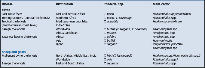
T. orientalis is responsible for oriental theileriosis, a milder disease than ECF or bovine tropical theileriosis, and hence also called benign theileriosis. It has been proposed that the species found worldwide and responsible for benign theileriosis be named T. buffeli (replacing T. orientalis in Asia and T. sergenti in Japan1) but genetic differences have been shown between T. buffeli and T. sergenti.4,5 Furthermore, T. sergenti causes a more severe disease in southeastern Asia, also called Japanese bovine theileriosis. Both T. buffeli and T. sergenti are transmitted by Haemophysalis ticks which occur in Europe, the Mediterranean basin, Asia, and Australia. Although this tick does not occur in the United States of America, T. buffeli was diagnosed in a herd of beef cattle in Missouri and was associated with severe clinical illness and death (by euthanasia) in a pregnant cow.6
In general, benign theileriosis is characterized by moderate to severe anemia in heavily parasitized cattle and moderate enlargement of lymph nodes. More severe clinical signs and economic losses have been reported from eastern Asia. The pathogenesis of the anemia is not clear but a hemolytic factor has been reported in the serum of acutely affected cattle.7 In addition, it has been shown that oxidative bursts of macrophages in experimentally infected cattle can damage red blood cells and that this may contribute to the anemia in Japanese bovine theileriosis.8 European breeds are more susceptible than zebu breeds. Even for the more pathogenic T. sergenti, native and crossbred cattle in Korea were found to be more resistant to infection than Holsteins.9 Furthermore, transplacental (vertical) transmission of T. sergenti from pregnant cows to calves has been reported.10 Transmission to the calves was confirmed by parasitological, serological, and polymerase chain reaction (PCR) assays. These are also the methods generally recommended for diagnosis.5 Calves used for the production of live vaccines against babesiosis and anaplasmosis should be free of benign theileriosis. In Australia, concurrent treatment with primaquine phosphate (six doses at 2 mg/kg) and halofuginone lactate (two doses at 1 mg/kg) was effective for this purpose.11
T. mutans, confined to Africa and the Caribbean Islands, causes a usually innocuous disease, but it may be manifested by fever, anorexia, and anemia. Another species, T. velifera, is associated with very mild theileriosis in tropical Africa. Both are transmitted by Amblyomma ticks. T. taurotragi of the eland antelope is generally non-pathogenic to cattle, but is one of the causes of cerebral theileriosis (turning sickness) in southern Africa (cerebral theileriosis can also be associated with T. parva). Parasitized lymphoblasts accumulate in cerebral, spinal, and meningeal arteries, with resultant thrombosis and infarction of affected organs. T. taurotragi is transmitted by Rhipicephalus spp.
The important pathogen of sheep and goats is T. hirci (synonym T. lestoquardi), the cause of malignant ovine theileriosis. The disease is enzootic from North Africa through the Middle East to India and China, approximately the same geographical region as bovine tropical theileriosis. Malignant theileriosis in sheep and goats is similar to bovine tropical theileriosis due to T. annulata. Like the latter, it is also transmitted by Hyalomma spp. but in China, the main vector is Haemophysalis spp. The disease can be acute, subacute, or chronic, depending on the resistance of the sheep or goats, and is seasonal, depending on availability of ticks. The acute disease is characterized by fever and very high mortality in 3–6 days.12,13 Anemia, jaundice, and enlargement of lymph nodes are characteristic, and both piroplasms and schizonts can be demonstrated in smears of blood and tissues, respectively. In subacute and chronic cases, signs are generally less marked except for anemia and emaciation. An indirect fluorescent antibody test is available. Parvaquone and buparvaquone may be used to treat early cases. Benign ovine theileriosis is caused either by T. ovis or by T. separata in Africa. Piroplasms are found in blood but there are no overt clinical signs.
In general, the pathogenesis of various forms of theileriosis is dependent on the production of schizonts in lymphocytes and piroplasms in erythrocytes. Thus, T. parva, T. annulata, and T. hirci produce numerous schizonts and piroplasms and are very pathogenic; T. mutans, T. buffeli, and T. ovis rarely produce schizonts but may cause varying degrees of anemia when piroplasms are many in red blood cells; and with T. velifera and T. separata, no schizonts have been described, the parasitemia is usually scanty and the infection is mild or subclinical.
1 Uilenberg NP, et al. Trop Anim Health Prod. 1996;28:81.
2 Coetzer JAW, Tustin RC, editors. Infectious diseases of livestock, 2nd edn., vol 1. Cape Town: Oxford University Press, 2004;448-501.
3 Brown CGD. Theileriosis. In: Sewell MMH, Brocklesby DW, editors. Handbook on animal diseases in the tropics. 4th edn. London: Bailliére Tindall; 1990:183-199.
4 Chansiri K, et al. Vet Parasitol. 1998;79:143.
5 OIE. Manual of diagnostic tests and vaccines for terrestrial animals. http://www.oie.int/eng/normes/mmanual/A_00062.htm, 2004. chapter 2.3.11. 5th edn.
6 Stockman SL, et al. Vet Pathol. 2000;37:11.
7 Hagiwara K, et al. Parasitol Res. 1995;81:470.
8 Shiono H, et al. Parasitol Res. 2003;89:228.
9 Kim GH, et al. J Protozool Res. 1999;9:103.
10 Baek BK, et al. Can J Vet Res. 2003;67:278.
11 Stewart NP, et al. Trop Anim Health Prod. 1990;22:109.
Etiology Theileria parva, an apicomplex protozoon. Vector is Rhipicephalus appendiculatus and, rarely, R. zambeziensis.
Epidemiology Endemic disease of cattle in east and central Africa; high mortality and great economic importance.
Pathogenesis Tick inoculation of sporozoites → lymphocytes in local lymph node → schizogony → lymphoid proliferation → parasitemia → more lymphoid proliferation → merozoites → erythrocytes → piroplasms → ticks. Damage mainly by schizonts.
Clinical signs Fever, enlarged superficial lymph nodes, dyspnea, wasting, and terminal diarrhea.
Clinical pathology Schizonts in lymphoblasts, piroplasms in erythrocytes, serology.
Lesions Massive pulmonary edema, hydrothorax, hydropericardium, emaciation, hemorrhages, lymphadenopathy, and widespread proliferation of lymphoblastoid cells.
• Trypanosomosis/babesiosis/anaplasmosis
• Malignant catarrhal fever/bovine virus diarrhea/Rinderpest.
Treatment Limited success with halofuginone, parvoquone, and tetracyclines.
Control Integrated approach involving resistant animal breeds, strategic application of acaricides, and vaccination by infection-and-treatment methods.
Theileria parva is an apicomplex protozoan parasite of the class Sporozoasida and order Piroplasmida. There has been considerable naming and renaming of T. parva and the associated diseases in Africa.1 ‘Classic’ East Coast Fever (ECF) occurs in East Africa and is associated with T. parva transmitted from cattle to cattle by the brown ear tick, Rhipicephalus appendiculatus. ECF also occurs either as Corridor disease in eastern and southern Africa or as January disease in central Africa. Corridor disease is transmitted from buffalo to cattle by either R. appendiculatus or R. zambeziensis and the agent responsible used to be called T. parva lawrencei. Close contact between buffalo, cattle, and ticks is essential. The disease is more acute than classical ECF, but after serial passage in cattle, it is indistinguishable from classical ECF.2 January disease occurs mainly between January and March and the agent was named T. parva bovis. The disease is also more acute than classical ECF, death sometimes occurring within 4 days. These three clinical diseases are otherwise indistinguishable from one another, hence the causative agents are currently referred to simply as T. parva.
ECF affects mainly cattle but also buffalo, and occurs in 13 countries in eastern, central, and southern Africa.3 Its occurrence is related to the distribution of the vector tick which has been recorded from large areas extending from southern Sudan in the north to western Zambia and eastern Zaire in the west, and to Mozambique and Zimbabwe in the south.4,5 The disease is prevalent throughout the wetter areas favoring the development of the tick, but is absent from the wet highlands in the horn of Africa.6 It has been eradicated from southern Africa up to the Zambezi River. The endemic scenerios range from a stable situation with high prevalence of herd infection but low fatality rates (endemic stability), to a low prevalence/high fatality scenerio (endemic instability).7 Endemic stability develops in indigenous zebu cattle exposed to constant tick challenge as in wetter areas whereas endemic instability is seen with commercial production systems utilizing imported breeds or crossbreeds and in areas with a unimodal rainfall pattern that restricts tick activity.8 Epidemics occur when there is a breakdown in tick control especially during the rainy season or when susceptible animals are introduced into an endemic area.
All susceptible cattle in endemic areas are at the risk of contracting ECF unless they are vaccinated or the tick population is under stringent control. The morbidity and case fatality rates are very high, approaching 90–100% in recently introduced exotic (Bos taurus) breeds and in previously unexposed or naive indigenous cattle.9 However, indigenous zebu cattle (Bos indicus) and African buffalo in endemic areas have a strong resistance to the disease and calfhood mortality is around 5%.9
The vector of ECF is Rhipicephalus appendiculatus and in the field, the disease occurs only where this tick is found, except for Corridor disease which may be transmitted by R. zambeziensis. Other species of Rhipicephalus and Hyalomma spp. can transmit ECF experimentally, but they are not significant. Developmental stages of the parasite occur in the tick and they pass trans-stadially through the stages of larva, nymph and adult, but there is no transovarian transmission. Consequently, larvae or nymphs become infected and transmit infection as nymphs or adults. Adults are more efficient vectors than nymphs. Mechanical transmission is of no significance. The epidemiology of the disease is thus largely dependent on the distribution and habitat of the tick and its ability to complete development to the adult stage, usually during the rainy season. Ticks may live for 1–2 years, but they lose their infection within 11 months.
The most important risk factors relate to the presence of the brown ear tick in a given area and the level of tick burden per animal, even though it takes only one tick to establish an infection that could be fatal. At low infestation rates, an average of five ticks per head (two to three per ear) will sustain endemicity; one to four per head will invite epidemicity; while an average of less than one can allow sporadic outbreaks.10 In addition, there is evidence that R. appendiculatus populations that originate from eastern Africa tend to become more highly infected with T. parva than those that originate from southern Africa, and consequently the disease they transmit is more virulent.11
The infection rate in ticks in endemic areas is usually low (1–2%) even though the immunity conferred on recovered or vaccinated animals is no longer thought to be sterile. However, soon after ECF becomes established in susceptible herds, infection rates in ticks become much higher.
Young animals are less susceptible, and indigenous breeds and buffaloes are less clinically affected than exotic breeds, but buffaloes are the carriers of Corridor disease. Other wild Bovidae may help to sustain the population of the tick vector but are not carriers of T. parva. Asiatic or water buffalo is fully susceptible.
In eastern Africa, R. appendiculatus normally occurs in grass-covered savannah and savannah woodlands, but is usually absent from extensive heavily wooded forest habitats.5 Areas that are too high, too cold, or too dry will not allow the tick to undergo more than one life cycle in a year, thereby reducing the period of transmission of theilerial parasites by the nymphs or adults. For example, the disease is most prevalent in eastern Africa where adult and immature stages of the tick occur simultaneously on cattle, leading to rapid and continuous transmission.11 In southern Africa, by contrast, there is a seasonal life cycle for the tick so that there is little overlap between the activity periods of adults (January to March) and immature stages, thereby reducing the frequency of disease transmission.
Cattle recovering from ECF have a solid immunity to homologous challenge, but the immunity is not sterile. In endemic areas, premunity is established early and this provides lifelong protection if reinfection continues and the cattle are not moved to a different location where they may be exposed to a different strain of the parasite. Indigenous cattle are able to limit explosive multiplication of schizonts during the acute phase.12 Nutritional or climatic stress may seriously reduce the animal’s premunity, even among resistant breeds. While antibody responses to the sporozoite may play some part in protection, immunity is mediated mainly by cellular mechanisms involving cell-mediated cytotoxic T-cell (CTL) responses against surface antigens of macroschizont-infected cells.13 The CTL response is parasite-specific and genetically restricted (major histocompatibility complex or MHC antigens) and the protection can be transferred between immune and naïve calves in the CD8+ T-cell fraction emanating from a responding lymph node.7
ECF can easily be reproduced by feeding infected ticks on susceptible cattle or by inoculating cattle with infected tick material, sporozoites or macroschizont-infected tissue culture cells. This is used as a method of immunization. When working with ticks or tick materials, care should be taken to avoid the risk of contracting other tick-borne diseases.
ECF has a major impact on cattle production in eastern, central, and southern Africa. It is estimated that in 1989, ECF killed 1.1 million head of cattle and caused US$168 million in losses.14 Serious losses occur in exotic and indigenous cattle, mainly from reduced production of milk and meat due to morbidity and mortality, as well as from the heavy costs incurred in implementing effective tick control. T. parva does not infect human beings.
Sporozoites of T. parva are injected into the bovine host by the tick in its saliva. Ticks must feed for 2–4 days before sporozoites in their salivary glands will mature and become infective to cattle. One tick can transmit sufficient sporozoites to cause a fatal infection in a susceptible animal. The sporozoites then enter lymphocytes and develop into schizonts in the lymph node draining the area of attachment of the tick, usually the parotid node. Infected lymphocytes are transformed to lymphoblasts which continue to divide synchronously with the schizonts so that each daughter cell is also infected. Eventually, infected lymphoblasts are disseminated throughout the lymphoid system and in nonlymphoid organs where they continue to proliferate. Later, some schizonts differentiate into merozoites, are released from the lymphoblasts and invade erythrocytes where they are referred to as piroplasms and are infective to ticks. Piroplasms ingested by ticks undergo several developmental stages and eventually form sporozoites in salivary glands, thus completing the cycle.
The dominating pathological lesion is generalized lymphoid proliferation resulting from uncontrolled proliferation of T-lymphocytes containing schizonts. This is followed later by necrosis of infected lymphoblasts induced by cytotoxic T-lymphocytes. The severe lymphocytolysis often leads to immunosuppression. Terminally, the animal develops severe pulmonary edema, probably due to release of vasoactive substances from lymphocytes disintegrating in the lungs. Erythrocytic indices are usually unchanged, but there may be terminal anemia in January disease.
The basic syndrome associated with T. parva infection lasts for a few weeks. The incubation period is 1–3 weeks, depending on the virulence of the strain and the size of the infecting dose. Experimentally, the first clinical sign is enlargement of lymph nodes in the area draining the site of tick attachment (i.e. 8–16 days after attachment). One or 2 days later, there is fever, depression, anorexia, and a drop in milk in dairy animals. In later stages, there may be nasal and ocular discharges, dyspnea, generalized lymph node enlargement, and splenomegaly. In severe cases, diarrhea occurs, sometimes with dysentery, but usually only late in the course of the disease. Emaciation, weakness, and recumbency lead to death from asphyxia in 7–10 days. Terminally, there is often a frothy nasal discharge. Occasional cases of brain involvement occur and are characterized by circling, hence ‘turning sickness’ or cerebral theileriosis.
In southern Africa, cerebral theileriosis is associated with an aberrant form of T. taurotragi originating from the eland. There are localized nervous signs and convulsions, tremor, profuse salivation, and head pressing. Infection with the strain of T. parva (formerly T. parva lawrencei) responsible for Corridor disease causes a similar acute syndrome, with the additional lesion of keratitis and accompanying blepharospasm. ECF in Zimbabwe (formerly attributed to T. parva bovis) is generally slightly less virulent but is still frequently fatal.
The parasites are evident as schizonts, sometimes in circulating lymphocytes, but mainly in biopsy smears of enlarged lymph nodes stained with Giemsa. Piroplasms are also easily visible in erythrocytes from day 16 after tick attachment and they increase in number until death. Over 30% of the red cells may be infected, but the level of intra-erythrocytic piroplasms is not correlated with the severity of the disease. T. parva piroplasms are difficult to differentiate from other piroplasms, hence the necessity to find schizonts. Blood counts will reveal a panleukopenia and thrombocytopenia with little or no anemia. The protozoa can be grown on a tissue culture of lymphoblastoid cells.
A range of serological tests is available, including indirect immunofluorescent antibody test (IFAT), complement fixation test, indirect hemagglutination test, and enzyme-linked immunosorbent assay (ELISA). The ELISA test is increasingly being used for seroepidemiological studies14 and the polymerase chain reaction (PCR) technology is available but IFAT is the most widely used test.3
The most striking lesion is massive pulmonary edema, hyperemia and emphysema, along with hydrothorax and hydropericardium. Copious froth is present in the airways. The carcass is emaciated and hemorrhages are evident in a variety of tissues and organs. There is enlargement of the liver, lymph nodes and spleen, and ulceration of abomasum and intestines. Small lymphoid nodules (the so-called pseudo-infarcts) are present in liver, kidney, and alimentary track. In protracted cases, animals may have small, exhausted lymphoid organs.
Microscopic lesions are characterized by proliferating lymphoblastoid cells and varying amounts of necrosis in lymphoid organs, lungs, liver, kidneys, the gastrointestinal tract and other tissues, somewhat similar to a multicentric lymphoid tumor. Some lymphoblasts contain schizonts, which are better seen in impression smears stained with Giemsa stain. In cerebral theileriosis, infected lymphoblasts sequester in cerebral blood vessels and cause infarction.
Specimens to submit for pathology should include lymph nodes, lungs, kidneys, liver, and any other organ with gross lesions.
The fever, depression, and lymphadenopathy of ECF can be confused with such diseases as theileriosis due to T. annulata, trypanosomosis, heartwater, malignant catarrhal fever, bovine virus diarrhea, and rinderpest. The lympoid hyperplasia may also simulate lymphoma. A knowledge of the disease history, coupled with hematological and lymph node smear examinations are usually adequate to make a definitive diagnosis.
Once an animal is manifesting clinical signs of ECF, treatment is generally considered to be either unsatisfactory or too expensive.15 Tetracyclines were the recommended treatment for many years, but they have only moderate efficacy, especially if the disease has been present for a few days. Two recently introduced drugs, halofuginone lactate and parvaquone, have had a much higher success rate, but recovered animals may become carriers unless the correct dose is used. Halofuginone lactate is an effective oral treatment for the acute syndrome at two doses, 1.2 mg/kg BW. Parvaquone (10 mg/kg BW, two doses 48 h apart) or the related buparvaquone (2.5 mg/kg BW, two doses 48 h apart) given IM is effective in most cases. In field trials, buparvaquone gives results comparable to those of parvaquone16 and cure rates are maximized by accurate diagnosis and prompt treatment of both ECF and intercurrent infections.17 Cure rates are even higher if the animals are also treated for pulmonary edema with dexamethasone18 or the diuretic, frusemide.19,20
Until recently, the main method of control of ECF was to break the transmission cycle between cattle and ticks. This was achieved through widespread and strict application of acaricides at 3-, 5-, or 7-day intervals throughout the year (intensive dipping), adherence to legislation on cattle movements and quarantine, and good livestock and pasture management. With the ever rising costs of acaricides, their effect on the environment, the development of acaricide resistance, and frequent political problems in the affected regions, this strategy to control ECF and other tick-borne diseases in Africa has been revised.21-23 Furthermore, it has been observed that indigenous cattle, constituting the majority of the herds in some of the affected countries, may lose their endemic stability with intensive dipping24 and the process is not cost-effective. An integrated approach is now advocated involving the use of genetically resistant breeds, a judicious and selective application of acaricides at 3-week intervals (strategic dipping) or when there are at least 100 ticks per animal (tactical dipping), and the use of vaccines. It was reported that monthly applications of deltamethrin-based pour-on insecticide significantly reduced the incidence of ECF and other hemoparasitic diseases in smallholder dairy farms in Kenya.25
The technique used for vaccination is immunotherapy or ‘infection-and-treatment method’. Initially, cryopreserved suspensions of T. parva sporozoites from ground-up infected ticks were injected into the patient. Now, sporozoites from cell culture are used. The infection they cause is controlled with long-acting oxytetracycline (20 mg/kg BW IM), or preferably parvaquone given at the same time, so that premunity is established. It is preferably to use a cocktail of different stocks of parasites. Vaccination, coupled with strategic dipping only when ticks are abundant, is usually successful and economically attractive,23 provided local stocks of Theileria are included. Large-scale field trials of these vaccines are being carried out throughout East and Central Africa. Initial reports indicate that calves in high risk areas should be vaccinated at 1–2 months of age26; that immunization campaigns are more efficient when concentrated in the period of low adult tick activity27; and that immunization is of no benefit in herds under intensive tick control but is of high value when combined with strategic tick control.28 Strategic control plus immunization can markedly reduce the risk of clinical ECF but immunized animals are carriers and all stages of R. appendiculatus can transmit infection from them to naïve animals.29
Limited studies have indicated that cattle could be successfully immunized without concurrent tetracycline therapy by using low pathogenicity isolates as vaccines, for example, T. parva (Boleni),30 or low infectivity sporozoite stabilates stored at –196°C for over 6 months.31 Because of the high cost of tetracyclines, this procedure would reduce the cost of vaccination by more than three-fold in the first year of field application. Furthermore, the T. parva (Boleni) isolate was reported to induce protection against a wide spectrum of Theileria stocks.
It needs to be stated that immunity is engendered so far only with live parasites that can establish an infection, but can also produce carriers. Hence, the risk inherent in the widespread use of such vaccines across national boundaries warrants further consideration. On the other hand, this process may accelerate progress to endemicity.32 The possibility of immunizing cattle with recombinant surface molecules from either the sporozoite (the p67 antigen) or the schizont, or a mixture of several antigens derived from both stages, is still being investigated.33,34 Such a recombinant vaccine would avoid the breakdowns that occur with any immunotherapeutic technique and if the right antigens are found for the vaccine, the immunity engendered is likely to be broad, robust, and not parasite stock-specific.33,35,36
Brown CGD. Theileriosis. In: Sewell MMH, Brocklesby DW, editors. Handbook on animal diseases in the tropics. 4th edn. London: Bailliére Tindall; 1990:183-199.
Lawrence JA, Perry BD, Williamson SM. East coast fever. Coetzer JAW, Tustin RC, editors. Infectious diseases of livestock, 2nd edn., vol 1. Cape Town: Oxford University Press, 2004;448-467.
Losos GJ. Theileriosis. In: Infectious tropical diseases of domestic animals. London: Longman; 1986:98-181.
Norval RAI, Perry BD, Young AS. The epidemiology of theileriosis in Africa. San Diego: Academic Press, 1992;481.
1 Perry BD, Young AS. Vet Rec. 1993;133:613.
2 Potgieter FT, et al. J S Afr Vet Assoc. 1988;59:155.
3 OIE. Manual of diagnostic tests and vaccines for terrestrial animals. http://www.oie.int/eng/normes/mmanual/A_00062.htm, 2004. chapter 2.3.11. 5th edn.
4 Lessard P, et al. Vet Rec. 1990;126:255.
5 Penny D, et al. Parasitol Today. 1990;6:1.
6 Norval RAI, et al. Prev Vet Med. 1991;10:163.
7 McKeever DJ. Res Vet Sci. 2001;70:77.
8 Billiouw M, et al. Trop Med International Health. 1999;4:A28.
9 Dolan TT. Parasitol Today. 1987;3:4.
10 Yeoman GH. Vet Rec. 1991;129:414.
11 Norval RAI, et al. Parasitology. 1991;102:347.
12 Paling RW, et al. Trop Anim Health Prod. 1991;23:203.
13 Morrison WI, et al. Morrison WI, editor. The ruminant immune system in health and disease. Cambridge: Cambridge University Press. 1986:555.
14 Anonymous. ILRAD Reports. 1991;9:1.
15 Young AS, et al. Parasitology. 1988;96:403.
16 Dolan TT, et al. Vet Rec. 1992;130:536.
17 Muraguri GR, et al. Vet Parasit. 1999;87:25.
18 Matovelo JA, et al. Trop Anim Hlth Prod. 2000;32:353.
19 Mbwambo HA, et al. Vet Parasit. 2002;108:195.
20 Musoke RA, et al. Trop Anim Hlth Prod. 2004;36:233.
21 Mukhebi AW. Vet Rec. 1995;137:17.
22 Pegram RG, et al. Trop Anim Health Prod. 1996;28:99.
23 Kariuki DP, et al. Trop Anim Health Prod. 1995;27:15.
24 Soldan AW, et al. Vet Rec. 1991;129:179.
25 Muraguri GR, et al. Insect Science and its Application. 2003;23:69.
26 Maloo SH, et al. Prev Vet Med. 2001;52:31.
27 Billiouw M, et al. Vet Parasit. 2002;107:51.
28 Minjauw B, et al. Prev Vet Med. 1999;38:35.
29 Marcotty T, et al. Vet Parasit. 2002;110:45.
30 Kanhai GK, et al. Trop Anim Health Prod. 1997;29:92.
31 Mbassa GK, et al. Vet Parasit. 1998;77:41.
32 Minjauw B, et al. Prev Vet Med. 1998;35:101.
33 Musoke A, et al. Proc Natl Acad Sci USA. 1992;89:514.
34 Bishop R, et al. Vaccine. 2003;21:1205.
Etiology Theileria annulata, an apicomplex protozoon. Vectors are Hyalomma ticks.
Epidemiology Endemic disease of cattle in Mediterranean basin and parts of Asia.
Pathogenesis As in ECF, but damage is by both schizonts and piroplasms.
Clinical signs Inapparent in local stock; fever, lymphadenopathy, wasting, anemia, and jaundice in exotics.
Clinical pathology Schizonts in macrophages and lymphocytes especially in liver smears; piroplasms in erythrocytes.
Theileria annulata is a member of the apicomplex group, like T. parva the cause of east coast fever. It is highly virulent for European dairy cattle. Infection in local zebu cattle is often subclinical.
The disease occurs from Morocco and Portugal in the west through the Mediterranean basin and the Middle East to India and China in the east. T. annulata affects cattle and is transmitted trans-stadially by the three-host tick Hyalomma anatolicum in central-western Asia and north-eastern Africa, and by the two-host tick H. detritum in the Mediterranean basin. The extent of its distribution may overlap with that of T. parva in Sudan and Eritrea and with T. sergenti in the Far East. In endemic areas, virtually all adult animals are infected, but case fatality is about 10–20% and is confined mainly to calves. Exotic animals recently introduced may have 20–90% mortality. The disease occurs when there is much tick activity, mainly in summer and the rainy seasons, and in crossbred animals. A single tick can cause fatal infection since its salivary glands usually contain numerous sporozoites. An outbreak occurred recently in a Scottish dairy farm and was believed to have been due to mechanical transmission from experimentally infected calves on a research institute associated with the farm. In the absence of natural vectors, that outbreak was quickly controlled.1
The normal state is that of endemic stability. This balance is disturbed when exotic animals are introduced and heavier losses occur. Recovered animals show a solid, long-lasting immunity, but they remain as carriers. Buffaloes are believed to be the natural hosts and may also act as carriers whereas yaks are highly susceptible. As with T. parva, immunity is mainly cell-mediated but is poor in calves. Experimental reproduction is by feeding infected ticks on cattle or by needle inoculation of sporozoites in macerated ticks, schizonts in lymphocytes, or merozoites in erythrocytes. Humans are not affected.
The life cycle of T. annulata is cattle-tick-cattle as for T. parva but unlike T. parva, the sporozoites of T. annulata invade and form schizonts mostly in macrophages/monocytes that express major histocompatibility (MHC) class II antigens, rather than in lymphocytes.4 Schizont infected cells multiply in the draining lymph nodes and disseminate rapidly throughout the lymphoid tissues and in non-lymphoid organs including the liver, kidney, lung, abomasum, and brain. Later, schizonts differentiate into merozoites and invade erythrocytes (as piroplasms). The pathogenesis therefore involves proliferation of lymphocytes and macrophages induced by schizonts and anemia with icterus induced mostly by the piroplasms. The lymphoproliferation is controlled by suppressor macrophages as a protective mechanism leading to recovery.5 Over 90% of erythrocytes may be parasitized, each by one or more merozoites. Immunosuppression may occur in the acute phase of lymphoproliferation, but is generally less marked than in ECF,3 probably because leukocyte numbers return to normal soon after the acute phase.
In a stable endemic situation, there may be only mild or no clinical disease in local zebu cattle.4 Clinical signs are acute and severe in exotic cattle and less severe in crossbreeds, and are similar to those in ECF. However, the course is longer in tropical theileriosis and may last for weeks before death. Clinical signs include marked fever, swelling of superficial lymph nodes, inappetence, tachycardia, dyspnea, pale mucous membranes and icterus. Others are diarrhea, weight loss, convulsions, torticollis and other nervous signs.6,7 In chronic cases, there may be small subcutaneous nodules from which schizonts can be demonstrated in smears.8
As with ECF, examination of smears of blood and lymph node biopsy will reveal piroplasms in erythrocytes and schizonts in lymphocytes. Schizonts of T. annulata tend to be more common in the liver than in lymph node smears, but are otherwise indistinguishable from those of T. parva. Furthermore, the piroplasms are predominantly round and oval, as opposed to T. parva which has comma- and rod-shaped piroplasms. Anemia is a significant feature of tropical theileriosis, unlike in ECF, and is associated with bilirubinemia, hemoglobinuria, and bilirubinuria. The anemia results from destruction of erythrocytes containing piroplasms but other factors may include autoimmune hemolysis and poor bone marrow response. Reduction in white cell and platelet counts is less severe than in ECF, but animals dying from the disease show persistent and severe lymphocytopenia involving mainly T-lymphocytes.6
The most commonly used serological diagnostic technique is the indirect fluorescent antibody test. For surveys, an indirect enzyme-linked immunosorbent assay (ELISA) test using a recombinant T. annulata surface protein has been described.9 The ELISA tests provide higher sensitivity and specificity than IFAT.10 Carriers can be detected by the polymerase chain reaction (PCR),11 a test that can also be used to detect infected ticks.
Apart from pallor of mucous membranes and yellowish discoloration of tissues, the postmortem lesions in animals dying from tropical theileriosis are similar to those of ECF. Liver, spleen and lymph nodes should be submitted for laboratory examination to detect schizonts whereas merozoites are detected in blood smears.
Buparvaquone is the most effective agent available, and the recommended dose is 2.5 mg/kg BW.12 In calves, supportive treatment for anemia is indicated. Halofuginone at 1.2 mg/kg is also effective but tetracycline at 20 mg/kg is less so.
Indigenous cattle live with the disease and do not require any intensive tick control or treatment. For valuable exotic stock or their crossbreeds, vaccination and strategic tick control are recommended. Vaccines can be made from either the sporozoite or the schizont. The sporozoite vaccine is based on the infection-and-treatment method using schizont-infected cell lines and simultaneous tetracycline treatment as for T. parva. It has been suggested that the most economical way to control theileriosis in India is to vaccinate calves and to reserve buparvaquone for treating clinical cases.13 The schizont vaccine was formerly blood containing a mild strain of the parasite. The newer vaccines are prepared from live schizonts grown in lymphoid cell culture and attenuated by prolonged passage. They cause virtually no adverse reactions and vaccinated cattle show good resistance to the disease for at least 3.5 years. Therefore, it is necessary to revaccinate, preferably with a different cell line vaccine, if tick population is too low to establish endemic stability. The risk for spread of the vaccine strains in the field is very low.14
Brown CGD. Theileriosis. In: Sewell MMH, Brocklesby DW, editors. Handbook on animal diseases in the tropics. 4th edn. London: Bailliére Tindall; 1990:183-199.
Pipano E, Shkap V. Theileria annulata theileriosis. Coetzer JA, Tustin RC, editors. Infectious diseases of livestock, 2nd edn., vol 1. Cape Town: Oxford University Press, 2004;486-487.
1 Williamson S, et al. State Vet J. 2003;12:1.
2 Hashemi-Fesharki R. Parasitol Today. 1988;4:36.
3 Hall FR. Parasitol Today. 1988;4:257.
4 Forsyth LMG, et al. J Comp Pathol. 1999;120:39.
5 Preston PM, et al. Parasit Res. 2002;88:522.
6 Preston PM, et al. Res Vet Sci. 1992;53:230.
7 Omer OH, et al. J Vet Prev Med. 2003;50:200.
8 Manickam R, et al. Ind Vet J. 1984;61:13.
9 Bakheit MA, et al. Parasit Res. 2004;92:299.
10 OIE. Manual of diagnostic tests and vaccines for terrestrial animals. http://www.oie.int/eng/normes/mmanual/A_00062.htm, 2004. chapter 2.3.11. 5th edn.
11 d’Oliviera C, et al. J Clin Microbiol. 1995;33:2665.
12 Sharma NN, Mishra AK. Trop Anim Health Prod. 1990;22:63.
Diseases associated with trypanosomes (tryponosomoses)
Trypanosomes are flagellated protozoan parasites that live in the blood and other body fluids of vertebrate hosts. With the help of the flagellum, they swim in body fluids, boring their way between cells. They generally possess a kinetoplast and undergo cyclical development in an arthropod vector. Their biological adaptations, morphology, and pathogenicity are fascinating and are being extensively studied. Each of the parasites causes a disease (now termed trypanosomosis rather than trypanosomiasis) in animals and humans as summarized in Table 26.7. Trypanosomoses of veterinary importance are discussed below.
Trypanosoma vivax, T. congolense, T. brucei brucei and T. simiae are the four main species responsible for African trypanosomoses affecting virtually all domestic mammals. T. vivax and T. congolense are the main pathogens of cattle. The four species are members of the Salivaria group of trypanosomes and are transmitted cyclically via the mouthparts of tsetse flies, hence the name salivarian trypanosomes. T. vivax is usually numerous in bovine blood, and can be identified by its very fast movement in wet films. In stained smears, it is a long, slender, monomorphic parasite with a rounded posterior end, a terminal kinetoplast, and a long free flagellum, but no prominent undulating membrane. T. congolense is smaller, sluggish in wet films, and often adheres to red blood cells by the anterior end. In stained smears, it is a short parasite with a marginal kinetoplast, no free flagellum, and no prominent undulating membrane. T. brucei is large like T. vivax, but its rapid movement is in confined areas of the wet film. In stained smears, it is pleomorphic and may occur as long and slender forms, intermediate forms, or short and stumpy forms. The slender and intermediate forms have a long free flagellum, pointed posterior end, subterminal kinetoplast and a prominent undulating membrane, whereas the stumpy forms resemble T. congolense, but are bigger and have a prominent undulating membrane. T. simiae is morphologically indistinguishable from T. congolense, but the organism will be swarming in the blood of pigs.
Etiology Trypanosoma congolense, T. vivax, T. brucei and T. simiae, all salivarian trypanosomes. Parasites undergo cyclical development in tsetse flies (Glossina spp.), the vector, but they can also be transmitted mechanically by other biting flies.
Epidemiology Endemic disease of all mammals in tropical Africa, also Central and South America; of greatest economic importance in cattle.
Pathogenesis Fly inoculation of metacyclic trypanosomes → chancre → trypomastigotes → intermittent parasitemia → anemia with or without tissue invasion → other clinical signs and immunosuppression.
Clinical signs Fever, apathy, pale mucous membranes, swollen lymph nodes, progressive emaciation, cachexia and death. May be acute, subacute or, often, chronic.
Clinical pathology Progressive anemia, parasite detection in blood by various methods.
Lesions Not definitive but include palor, emaciation, enlargement of liver, spleen and lymph nodes.
The epidemiology of African trypanosomosis is determined mainly by the ecology of the tsetse fly which is found only in tropical Africa. T. vivax is also transmitted mechanically by biting flies and occurs also in Central and South America. Affected countries include Bolivia, Brazil, Colombia, French Guiana, Guyana, Peru, Suriname, and Venezuela where it affects mainly cattle and sheep.1 Whereas T. congolense and T. vivax are responsible for severe disease in cattle, sheep and goats, T. brucei brucei usually causes a subclinical infection in cattle, but a severe disease in sheep, goats, horses and, occasionally, pigs. T. simiae causes a very acute and highly fatal disease in exotic pigs. It is not pathogenic to cattle, sheep, or goats.
Infection rates in cattle in endemic areas vary considerably and could be over 60%. However, as a result of various control methods, the prevalence is decreasing in many African countries, particularly in West Africa, and was as low as 10% in Mali in the 1980s.2 In a 1989–1991 survey involving approximately 20 000 cattle, 3000 sheep, and 3000 goats in Nigeria, the prevalence was 4.3% in cattle, 1.6% in sheep, and 1.0% in goats, based on parasite detection.3 Higher prevalence rates are obtained if diagnosis is based on serology, but this does not reflect current infection. T. vivax and T. congolense are the species most frequently encountered in ruminants, but with serology T. brucei is also frequently reported. Mixed infections with two or three species are common. Pigs and horses are less frequently affected than ruminants, perhaps because they are less exposed to tsetse flies than cattle that normally graze over long distances. In Central and South America, T. vivax infections appear to be spreading to new areas where they cause periodic outbreaks of serious disease in cattle.
Morbidity rates during outbreaks are variable and may reach 70% in cattle infected with T. vivax and up to 100% in pigs infected with T. simiae. It is usually much lower in sheep, goats, and horses since these are not often the preferred hosts for tsetse or are less exposed to tsetse challenges. Sheep and goats are more vigorous than cattle in defending themselves against successful feeding by tsetse flies. Case fatality also depends on parasite species, host, and its level of resistance. T. simiae is invariably fatal in exotic pigs. Some strains of T. vivax in East Africa cause similar heavy mortalities in exotic dairy cows, and infected horses are likely to die if left untreated. However, most infections in cattle in endemic areas run a chronic course and are not invariably fatal, but the animal may remain unproductive and unthrifty.
African trypanosomes can be transmitted by 23 species of tsetse (Glossina) found only in sub-Saharan Africa between latitudes 14°N and 29°S, excluding areas of high altitude, extreme drought or cold temperatures where tsetse cannot survive. Tsetse species can be grouped according to their preferred habitats as savannah species, riverine species, and forest species. The savannah species (including G. morsitans, G. austeni, G. pallidipes, G. swynnertoni, and G. longipalpis) pose the greatest threat to livestock because they inhabit grasslands where cattle are traditionally reared, they can easily adapt to other ecological niches, they feed primarily on cattle and pigs, and they are efficient vectors of trypanosomes. They are also the main vectors of Rhodesian sleeping sickness associated with T. brucei rhodesiense in humans (Table 26.7). The riverine species (G. palpalis, G. tachinoides, and G. fuscipes) are important as vectors of bovine and porcine trypanosomosis, as well as of Gambian sleeping sickness due to T. brucei gambiense. On the other hand, the 13 or so forest species (including G. fusca, G. brevipalpis, and G. longipennis) are not frequently incriminated vectors of trypanosomes even though their preferred food hosts are ruminants and suids.
The life cycle of trypanosomes in tsetse involves cyclical development for a varying length of time, depending on species and ambient temperatures. T. vivax completes its developmental cycle in the proboscis and pharynx and can be transmitted (as metacyclic trypanosomes) within a week of the initial infective feed. The cycle of T. congolense involves the midgut and proboscis and is completed in about 2 weeks. That of T. brucei is more complex: it takes 3 or more weeks and involves the midgut and salivary glands. Once infected, flies remain so for life (1–2 months). It follows that for any fly, its vectorial capacity and efficiency are highest for T. vivax and least for T. brucei.
After trypanosomes have been introduced into a herd, transmission is possible even in the absence of Glossina. Biting flies such as Tabanidae, Stomoxyinae, and Hippoboscidae are capable of mechanically transmitting trypanosomes in their mouth parts if they feed on more than one host within a short interval. This is how T. vivax is spread in areas outside the tsetse belt in Africa, as well as in Central and South America. Mechanical transmission can also occur through the needle during inoculations and in carnivores feeding on infected carcasses. Intrauterine infections occasionally occur.4
Reservoirs of infection are found in many wild animals, in trypanotolerant animals, and in chronically infected animals. Tsetse caught in and around game reserves tend to have relatively high infection rates and the relative abundance of wildlife in East Africa as compared to West Africa may explain, at least in part, why the prevalence of the disease appears to be declining more rapidly in the west.
The effect of infection varies with the host in that most wild animals, and some domestic ones, establish a balance with the parasite and remain as clinically normal carriers for long periods. Specifically, some breeds of cattle indigenous to Africa can tolerate light to moderate challenge with tsetse flies by limiting the multiplication of trypanosomes in their blood and by apparently warding off the infection, especially T. vivax.5 The phenomenon is called trypanotolerance, it is both genetic and environmental in origin, and the level of tolerance varies. Thus, the indigenous taurine breeds, such as the N’Dama, Baoule and Muturu, are more tolerant than the West African zebu,6 and amongst East African zebu cattle, the Orma Boran and Maasai zebu have superior tolerance when compared with Galana Boran and Friesian breeds.5,7 Crossbreeds of indigenous taurine and zebu animals are also more tolerant than purebred zebu. However, due to the uncertain genetic makeup of animals within these so-called breeds and crossbreeds, the level of trypanotolerance may also vary with individual animals within a given category and it can be overcome by heavy tsetse challenge, malnutrition, or other stress factors. Trypanotolerance also occurs in some indigenous breeds of small ruminants, notably the West African Dwarf sheep and goats and the East African goats, whereas the Toggenburg, British Alpine, Saanen, and Anglo-Nubian breeds of goats are fully susceptible.
The density of tsetse population in the area, and the level of their contact with the host, will determine the level of infection. This is further influenced by the vectorial capacity of the fly and the availability of its preferred host, which may not be livestock. Trekking of cattle through tsetse-infested vegetation is a risk nomadic farmers face from time to time and the risk is even greater where cattle routes converge, for example, at major bridges or watering holes. Agricultural and industrial developments generally lead to a lowering of tsetse density by destroying its habitat, whereas the establishment of game or forest reserves provides large numbers of preferred hosts or a suitable habitat for tsetse, respectively. Herds located near such reserves are therefore at a higher risk. So also are tourists visiting game parks.
In cattle, T. vivax generally produces a higher level of parasitemia than other species. And since its life cycle in the tsetse is also shorter, T. vivax is more readily transmitted than the others when animals are newly introduced into a tsetse infested area. Higher parasitemias also facilitate mechanical transmission. On the other hand, T. brucei is rarely detectable by direct examination of cattle blood, even though infection can be confirmed through other diagnostic methods. Furthermore, some animals carry infection without showing clinical signs, especially if they are trypanotolerant like the Muturu in Nigeria,8 or if infected with non-pathogenic genetic types, for example, T. congolense kilifi type in cattle.9
Animals recovering from infection with one strain/serodeme or species of trypanosome are not immune to infection with another strain/serodeme or species. This is due to the ability of trypanosomes to readily change their surface coat antigens through a process called antigenic variation. The glycoprotein surface coat is continuously shed and replaced by mechanisms not fully understood, but probably induced by antibodies. During each peak parasitemia, a mixture of variable antigenic types of parasites is present, but the dominant antigens determine the specific antibody response. These antibodies kill off the dominant population, leaving others with different dominant antigens; these multiply, and the process continues in cycles until the animal dies or the immune mechanisms catch up with the parasite and the animal recovers. This phenomenon is also responsible for the successive waves of parasitemia in infected animals. Following repeated episodes of infection and recovery (with or without treatment) in an endemic area, animals will encounter a variety of antigenic types and therefore become less susceptible to strains/serodemes in that area.
Infected animals are more susceptible to secondary infections by other microorganisms, particularly bacteria. The mechanisms involved are not fully understood, but may vary with the species of animals. In ruminants, the state of immunosuppression is abrogated once the trypanosomes are eliminated by chemotherapy.
Infection can be easily reproduced by inoculation of infected blood or other body fluid into a susceptible host. Infected flies can also be fed on the host to transmit the disease. Several laboratory animal models are available and lots of studies are done with mice and rats.
Tsetse flies infest 10 million square kilometers of Africa involving 37 countries. Hence, nagana is today the most important disease of livestock in the continent. The added risk of human infections has greatly affected social, economic, and agricultural development of rural communities. Since nagana is a wasting disease, affected animals are chronically unproductive in terms of milk, meat, manure, and traction, and the mortality rate can be high. The disease in Africa costs livestock producers and consumers an estimated US$1340 million each year.10 The anticipated losses due to T. vivax in South America exceed $160 million.1 Furthermore, the disease may impact on various immunization campaigns in endemic areas due to the fact that it can cause immunosuppression.
The animal pathogens do not infect humans, but animals can serve as reservoirs of T. brucei rhodesiense and T. brucei gambiense, the causes of human sleeping sickness, which are morphologically indistinguishable from T. brucei brucei. Human infections result from tsetse bites, generally in game parks, forest reserves, and along streams or other rural setting. The incidence has fallen greatly since the early part of the 20th century, but outbreaks still occur from time to time, especially when civil unrests force people into tsetse-infested areas. Up to 100 000 cases are reported per year and T. brucei gambiense sleeping sickness has re-emerged as a major public health problem in Central Africa, especially in the Democratic Republic of the Congo, Angola, and Southern Sudan, where civil wars have hampered control efforts.11
A rash (chancre) develops at the site of tsetse bite and this is soon followed by fever, persistent headache, and swelling of lymph nodes, spleen, and liver. Weakness and signs of cardiac involvement may be noticed early in the Rhodesian form and the disease is rapidly fatal. The Gambian form is more chronic, lasting for 3 or more years during which the patient gradually wastes away or dies from secondary infection. The parasite invades the cerebrospinal fluid leading to progressive non-suppurative meningoencephalitis which causes the patient to fall asleep often, hence the name sleeping sickness.
There are none because the vector requires strict environmental conditions to survive. People working with T. b. gambiense and T. b. rhodesiense should take precautions to avoid accidental inoculation of themselves or their co-workers with infected material in syringes or tsetse flies.
Nagana in most species is a progressive, but not always fatal disease and the main features are anemia, tissue damage, and immunosuppression. Metacyclic trypanosomes are inoculated intradermally as the fly feeds. They multiply at this site provoking a local skin reaction (chancre), which is most pronounced in a fully susceptible host and may be slight or absent with some strains or species of trypanosomes. Within the chancre, metacyclic parasites change to trypomastigote form, enter the bloodstream directly or through the lymphatics, and initiate characteristic intermittent parasitemias. Their behavior thereafter depends largely on the species of trypanosome transmitted and the host. T. vivax usually multiplies rapidly in the blood of cattle, sheep and goats, and is evenly dispersed throughout the cardiovascular system, whereas T. congolense tends to be aggregated in small blood vessels and capillaries of the heart, brain, and skeletal muscle, and rarely causes heavy parasitemias in ruminants. Both species exert their effect mainly by causing severe anemia and mild to moderate organ damage. The anemia has a complex pathogenesis involving mainly increased erythrophagocytosis, some hemolysis, and dyshemopoiesis. Very acute infections with T. vivax in cattle or T. simiae in pigs result in fulminating parasitemias and disseminated intravascular coagulation with hemorrhages. Such syndromes resemble a septicemia, and anemia may not be severe.
T. brucei and, rarely, T. vivax, have the added capability of escaping from capillaries into interstitial tissues and serous cavities where they continue to multiply. Such infections result in more severe organ damage in horses, sheep, and goats, in addition to anemia. The cerebrospinal fluid is often invaded by T. brucei alone12,13 or mixed with other species, or as a relapse after an apparently successful treatment.12,14
Animals chronically infected with any pathogenic trypanosome may develop concurrent and even fatal bacterial, viral, and other protozoan infections as a result of immunosuppression. Pregnant animals may abort, and transplacental fetal infections occasionally occur. Trypanotolerant animals control parasitemias better and have less severe anemia and organ damage. They usually recover from the disease, but may act as carriers.
There are no pathognomonic signs that would help in pinpointing a diagnosis. The general clinical picture is as follows but there are many variations determined by the level of tsetse challenge, the species and strain of the trypanosome, and the breed and management of the host. Acute episodes last for a few days to a few weeks from which the animal dies or lapses into a subacute to chronic stage, or the illness may be chronic from the beginning. Chronic cases may run a steady course, may be interrupted by periodic incidents of severe illness, or undergo spontaneous recovery.
The basic clinical syndrome appears after an incubation period of 8–20 days. There is fever, which is likely to be intermittent and to last for a long period. Affected animals are dull, anorexic, apathetic, have a watery ocular discharge, and lose condition. Superficial lymph nodes become visibly swollen, mucous membranes are pale, diarrhea occasionally occurs, and some animals have edema of the throat and underline. Estrus cycles become irregular, pregnant animals may abort, and semen quality progressively deteriorates.15,16 The animal becomes very emaciated and cachectic and dies within 2–4 months or longer. Thin, rough-coated, anemic, lethargic cattle with generalized lymph node enlargement are said to have a ‘fly-struck’ appearance.17
In general, T. congolense is more pathogenic to cattle in eastern and southern Africa, whereas T. vivax produces a more serious disease in West Africa. However, severe outbreaks of T. vivax involving exotic dairy animals in East Africa occur; affected animals show mucosal petechiation, rhinorrhagia, dysentery, and death after an illness of only a few weeks. Mixed infections are common and are usually more severe. Furthermore, intercurrent bacterial, viral, or other parasitic infections may mask or complicate the basic clinical syndrome. Immune response to bacterial, and some viral, vaccines is also depressed but is restored if trypanocidal therapy is given at the time of vaccination.18
Clinical findings peculiar to the individual trypanosome are as follows:
• T. vivax affects all agricultural species except pigs. Acute and chronic outbreaks occur, anemia is severe, and fever is usually associated with high parasitemia. A chronic form of the disease is more usual in East Africa, but an acute hemorrhagic form can occur with exotic cattle. T. vivax is less commonly seen in trypanotolerant cattle breeds
• T. congolense affects all species, usually with an acute disease lasting 4–6 weeks, but some chronic cases occur, especially in West Africa. Anemia and emaciation are severe
• T. brucei affects all species with a subacute to chronic disease in which subcutaneous edema and keratoconjunctivitis may be marked. Nervous signs are manifested in horses, pigs, and small ruminants by ataxia, circling, head pressing, and paralysis.19,20 Cattle are usually asymptomatic, with few exceptions21
• T. simiae affects exotic pigs with a fulminating infection in which the animal dies within hours or a few days of first appearing ill. There is fever, stiff gait, dyspnea, and cutaneous hyperemia, but no significant anemia.
The anemia results in a progressive drop in packed cell volume, a non-specific but useful indicator in endemic areas. The classic method of confirming diagnosis is to demonstrate parasites in a wet blood film, and in a thin or thick blood smear stained with Giemsa. This is easiest in the early stages of the disease when parasitemic peaks correspond with fever. As the disease progresses, parasitemias become infrequent and the intervals between peaks grow longer, even though the animal is still sick. To increase the accuracy of parasitological diagnosis, it is now routine to concentrate the parasites in the buffy coat layer of a microhematocrit capillary tube. The buffy layer is then examined directly at low power (Woo’s method) or in a wet preparation with a dark ground/phase contrast microscope (Murray’s method). Both tests are simple, sensitive, and applicable to field use on individual animals as well as in herds. Blood should be examined fresh but may be refrigerated for up to 24 hours, beyond which most parasites will die and disappear from the sample.22
In chronic cases, blood can also be inoculated into experimental animals, usually rodents, but this is cumbersome and is accurate for only T. brucei, and possibly T. congolense, but not T. vivax.
Another alternative is a series of standard serological tests to detect antitrypanosome antibodies in sera or other body fluids. The three tests used most often are the indirect immunofluorescent antibody test (IFAT), the capillary agglutination test (CAT), and the ELISA. These tests have the disadvantage that they indicate past as well as present infections, are difficult to standardize for different laboratories, and are not species-specific. The ELISA technique was modified to detect circulating trypanosome antigens (antigen-ELISA) using monoclonal antibodies that would distinguish between T. vivax, T. congolense, and T. brucei, and would detect only current or very recent infections.23 Extensive field trials in Africa and the Caribbean have shown that the test is not sufficiently sensitive or specific and that it needs to be combined with other parasitological methods to provide reliable results.24
The polymerase chain reaction (PCR) technique can be used to detect trypanosome DNA in tsetse tissues and in blood.25 The test is sensitive and species specific and can be combined with ELISA.26 Under experimental conditions, it can also be used to monitor therapy, especially relapses, since treated animals should become PCR negative 1–2 days after treatment.27 It is also possible to do retrospective epidemiological survey using serum banks.28 Dried blood spots on filter papers are also a useful source of DNA for the detection of T. congolense and T. brucei by PCR.29 But the test is expensive and can only be done in specialized laboratories.
The postmortem lesions are, like the clinical findings, not definitive. The carcass is marked by anemia, emaciation, anasarca, and enlargement of the liver, spleen, and lymph nodes. Body fat stores are depleted or show marked serous atrophy, especially around the heart and in bone marrow. There may be corneal opacity and testicular degeneration. In acute cases, there will be a general congestion of the viscera and extensive hemorrhages in all tissues. Chronic cases show cachexia, often complicated with secondary bacterial or other parasitic diseases.
Microscopic lesions are also not specific, except in very acute infections in which clumps of trypanosomes mixed with fibrin thrombi are found in blood vessels. Lymphoid organs are usually hyperplastic and may show varying degrees of erythrophagocytosis or hemosiderosis. Bone marrow macrophages may engulf red cells as well as other immature and mature cells.30 The interstitial tissues of various parenchymatous organs may contain a lymphoplasmacytic infiltrate. This tends to be most marked with T. brucei in which the parasites often localize extravascularly in the interstitial tissue. A severe non-suppurative meningoencephalitis or myocarditis may result. Degenerative changes may also be present in the liver, testis, and pituitary gland.
Smears from tissues, usually the cut surface of a lymph node or heart muscle, are examined for trypanosomes. With T. brucei, smears of body fluids, including the cerebrospinal fluid may contain many parasites even when they are undetectable in blood. Trypanosomes die and disintegrate soon after the host dies and will not be seen in smears if postmortem examination is delayed for even a few hours. The following organs should be taken for histopathology: lymph nodes, spleen, liver, heart, kidney, brain, and any other organ showing gross lesions. The immediate cause of death is often a combination of trypanosomosis and a concurrent bacterial or parasitic infection.
Diagnosis is based on detecting parasites in blood. Since parasitemias fluctuate, multiple samples from a herd or repeated sampling of a suspected case may be required before a specific diagnosis can be made. Furthermore, infected animals may be suffering from a concurrent disease. Emaciation and anemia can also be associated with:
Acute trypanosomosis may be confused with hemorrhagic septicemia and anthrax. Laboratory examination of blood, feces, and other tissues is required to confirm diagnosis.
The number of trypanocidal drugs available for treating and preventing infections in endemic areas is limited, and about 6 million doses are administered yearly in Africa.31 The drugs have been in the market for over 30 years, their range of therapeutic safety is small, many of them cause severe local reactions especially in horses, and some may be fatal in high doses. Furthermore, as drugs are not always available and they are expensive, underdosing is common. These, plus the fact that some drugs are used both prophylactically and therapeutically, have led to cases of drug resistance.
Ideally, each country or region establishes a group of sanative drugs which are to be used only as a break in a course of one of the more common drugs. The sanative drug should provide moderate prophylaxis and avoid the development of resistance to the prime drug. These measures have not been well-executed in many countries and this may explain the increasing reports of multiple resistance to curative, sanative, and prophylactic drugs.32,33 Nevertheless, the prevalence of drug-resistant trypanosomes in the field still remains unclear as there are no suitable methods for the detection of resistant field isolates.34 For example, drug resistance has been reported to be very low in eastern Zambia35 but widespread in Burkina Faso36 and Ethiopia.37
Strains are regarded as resistant when they fail to respond to the drug or when they relapse some time after an apparent cure. Relapses are more likely to occur if the commencement of treatment is delayed or the dose rate is inadequate. However, in field situations, there is hardly any regular monitoring of drug efficacy and animals may be reinfected with the same or other species of trypanosomes soon after an otherwise effective cure.
The common drugs in use against trypanosomes are set out below. The specific dose rates vary with animal species, the specific trypanosome, and the specific purpose (curative, prophylactic, or sanative).31,38
• Diminazene aceturate (Berenil) is used widely against T. vivax and T. congolense as a curative and sanative drug at 3.5–7 mg/kg BW. It is well-tolerated by ruminants and it is one of the two recommended drugs for bovine trypanosomosis. It is not well-tolerated by horses
• Homidium bromide (ethidium) and homidium chloride (novidium) are also widely used against T. congolense and T. vivax as curative and sanative drugs at 1 mg/kg BW
• Isometamidium (Samorin or Trypamidium) is the other preferred drug against T. vivax and T. congolense in ruminants. It is used as a curative and prophylactic drug at 0.25–1 mg/kg BW. At much higher doses (12.5–35 mg/kg BW) it can be used prophylactically against T. simiae in pigs but not without the risk of death from acute cardiovascular collapse39
• Pyrithidium bromide (prothridium) is less widely used against T. congolense and T. vivax as prophylaxis at 2 mg/kg BW
• Quinapyramine sulfate (Antrycide) is no longer used extensively in cattle, but it is the preferred curative drug (at 5 mg/kg BW) against T. brucei in horses. Quinapyramine sulfate and chloride (Antrycide prosalt) is used prophylactically at 7.4 mg/kg BW
• Suramin (naganol) may also be used against T. brucei as a curative and prophylactic drug at 10 mg/kg BW in horses and camels
• Antrycide–Suramin complex is the only other drug against T. simiae in pigs and it is used prophylactically at 40 mg/kg BW.
The control of trypanosomosis in enzootic countries involves control of tsetse fly population, prophylactic treatment, good husbandry of animals at risk, and use of trypanotolerant animals. There is no vaccine against the disease and, in spite of intensive research, none appears likely in the near future because of the ability of trypanosomes to readily change their glycoprotein surface coat through a process called antigenic variation.
Control of tsetse has been successfully attempted in some African countries, but reinvasion is frequent if the land is not properly utilized. The earliest methods involved bush clearing and elimination of game animals on which tsetse feed. These methods were effective in eradicating or controlling tsetse in some parts of the continent, especially in southern Africa, but they resulted in destroying valuable plant and animal resources, and also led to soil erosion. More recent methods involved the use of insecticides, especially DDT and endosulfan, applied strategically in the form of ground and aerial spraying over large expanses of land. As tsetse are sensitive to insecticides and no resistance has developed, considerable successes were achieved in some countries. However, spraying insecticides is costly and harmful to the environment. These harmful effects are considerably reduced if the insecticides, for example, synthetic pyrethroids, are applied directly on the animal in the form of spray or pour-on formulation. The latter offers great promise40 and also reduces tick infestation in treated animals.
Other effective methods involve targets impregnated with insecticides and traps that attract and catch tsetse. These are simple and cheap and can be constructed and maintained by local communities.41 Furthermore, they do not pollute the environment and are suitable for both small- and large-scale farming. They have been used to reduce tsetse fly population by over 97% within 7 months in a community in the Congo42 and to greatly reduce the incidence of trypanosomosis in another community in south-western Kenya over a 12-year period.43 Another method is the sterile male technique. Since the female tsetse only mates once in a lifetime, this technique is theoretically able to eradicate a targeted tsetse species in areas where other methods have been used to reduce its density. But it is expensive. Finally, it should be stated that development of the land for agriculture, industries, highways, etc. will effectively destroy the habitat for tsetse flies. This has occurred significantly in Nigeria where there were rapid economic activities and expanding human population in the 1970s and 1980s.44
Attempts at trypanosomosis control have also been directed to prophylactic dosing with chemicals such as suramin, prothridium, and isometamidium (Samorin). Prophylaxis is used along with other methods in areas where there is a heavy tsetse challenge. The prophylactic effect is supplemented by the development of antibodies, and the total period of protection may be as long as 5 months. However, it is customary to give four or five treatments per year. The productivity response to this pattern of treatment is good if general husbandry is also adequate. The downside of this approach is that it has reportedly led to drug resistance in many countries.
Trypanotolerant animals are being used to establish ranches in areas where tsetse challenge is not too heavy, but they have not been readily accepted in some countries, supposedly because they are smaller in size and they produce less milk than other indigenous breeds and crosses with exotic breeds.
For effective control of trypanosomosis in Africa and in Central and South America, an integrated approach will mostly likely produce the desired results in each region. In the absence of a vaccine, control methods must combine reduced exposure to the vectors (large scale tsetse trapping and pour-on applications) with strategic treatment of exposed animals (chemotherapy and chemoprophylaxis) along with use of trypanotolerant animals when feasible.
Anosa VO. Haematological and biochemical changes in human and animal trypanosomiasis. Rev Elev Med Vet Pays Trop. 1988;41:65-78. 151–164
Connor RJ, Van den Bossche P. African animal trypanosomoses. Coetzer JAW, Tustin RC, editors. Infectious diseases of livestock, 2nd edn., vol 1. Cape Town: Oxford University Press, 2004;251-296.
Desquesnes M. Livestock trypanosomoses and their vectors in Latin America. Paris: OIE (World Organization for Animal Health), 2004.
Hunter AG, Luckins AG. Sewell MMH, Brocklesby DW, editors. Handbook on animal diseases in the tropics, 4th edn. London: Bailliére Tindall. 1990:204-226.
Ikede BO. African trypanosomes In: Honigberg BM. Mechanisms of pathogenicity among Protozoa. Insect Sci Applic. 1986;7:363-378.
Jordan AM. Trypanosomiasis control and African rural development. London: Longman, 1988.
Losos G. Trypanosomiases. In: Infectious tropical diseases of domestic animals. London: Longman; 1986:182-318.
Stephen LE. Trypanosomiasis: A veterinary perspective. Oxford: Pergamon Press, 1986.
1 Davila AMR, et al. Ann NY Acad Sci. 2000;916:199.
2 Awan MAQ, et al. Bull Anim Health Prod Afr. 1988;36:330.
3 Anosa VO, et al. Control of animal trypanosomiasis in Nigeria. Progress Report by VO Anosa, 1993.
4 Ikede BO, et al. Acta Trop. 1988;45:5.
5 Njogu AR, et al. Vet Rec. 1985;117:632.
6 Akol GWO, et al. Vet Immunol Immunopathol. 1986;11:361.
7 Mwangi EK, et al. Ann NY Acad Sci. 1998;849:195.
8 Uza DV, et al. Bull Anim Hlth Prod Afr. 1998;46:125.
9 Bengaly Z, et al. Vet Parasit. 2002;108:1.
10 Kristjanson PM, et al. Agric Systems. 1999;59:79.
11 Aksoy S, et al. Adv Parasit. 2003;53:1.
12 Moulton JE. Vet Pathol. 1986;23:21.
13 Whitelaw DD, et al. Parasitology. 1985;90:255.
14 Masake RA, et al. Acta Trop. 1984;41:237.
15 Akpavie SO, et al. Res Vet Sci. 1987;42:1.
16 Sekoni VO, et al. Anim Reprod Sci. 1991;24:249.
17 Connor RJ, Van den Bossche P. Coetzer JAW, Tustin RC, editors. Infectious diseases of livestock, 2nd edn., vol 1. Cape Town: Oxford University Press, 2004;251.
18 Mwangi DM, et al. Trop Anim Health Prod. 1990;22:95.
19 Agu WE, Bajeh ZT. Trop Vet. 1986;4:25.
20 Onah DN, Uzoukwu M. Trop Anim Health Prod. 1991;23:39.
21 Wellde BT, et al. Ann Trop Med Parasit. 1989;83:151.
22 Ekwuruke JO, et al. Acta Trop. 1985;42:273.
23 Nantulya VM, et al. Parasite Immunol. 1987;9:421.
24 Mattioli RC, Faye JA. Acta Trop. 1996;62:71.
25 Penchenier L, et al. Parasite. 1996;3:387.
26 Masake RA, et al. Vet Parasit. 2001;96:101.
27 Bengaly Z, et al. Vet Parasit. 2002;108:1.
28 Desquesnes M. Acta Trop. 1997;63:139.
29 Katakura K, et al. Parasit Res. 1997;83:241.
30 Anosa VO, et al. Vet Pathol. 1992;29:33.
31 Finelle P. FAO Anim Prod Health Paper. 1983;37:1.
32 Ainanshe OA, et al. Trop Anim Health Prod. 1992;24:65.
33 Joshua RA. Rev Elev Med Vet Pays Trop. 1988;41:359.
34 Chitambo H, et al. Res Vet Sci. 1992;52:243.
35 Sinyangwe L, et al. Vet Parasit. 2004;119:125.
36 McDermott J, et al. Acta Tropica. 2003;86:93.
37 Assefa E, Abebe G. Vet Parasit. 2001;99:261.
38 Barry JD, Turner CMR. Parasitol Today. 1991;7:207.
39 Kinabo LDB, et al. Res Vet Sci. 1991;50:6.
40 Lohr K-F, et al. Trop Med Parasitol. 1991;42:131.
41 Dransfield RD, et al. Parasitol Today. 1991;7:287.
42 Noireau F, et al. Rev Elev Med Vet Pays Trop. 1990;43:93.
43 Brightwell R, et al. Bull Entomol Res. 1997;87:349.
44 Ikede BO, et al. Proc Intern Sci Counc Trypanosomiasis Res Control, 19th Meeting, Lome, Togo, publ no 114, 1987; p. 289.
Etiology Trypanosoma evansi (synonym T. equinum), morphologically similar to T. brucei. Parasites are transmitted mechanically by biting flies, mainly tabanids, and in Latin America, by vampire bats.
Epidemiology Endemic disease of mainly horses and camels in the tropics and subtropics, seasonality is related to fly population.
Pathogenesis Fly inoculation → parasite multiplication in blood and body fluids → clinical signs.
Clinical signs Fever, progressive emaciation, anemia, subcutaneous edema, nervous signs, death. May be acute, subacute, or chronic.
Clinical pathology Progressive anemia, parasite detection in blood by various methods, serology.
Lesions Not definitive but include palor, emaciation and jaundice.
Trypanosoma evansi, the first pathogenic trypanosome to be identified in 1880 in India, belongs to the brucei group (subgenus Trypanozoon) but is not capable of cyclical development in tsetse Glossina spp. In blood smears, T. evansi is morphologically indistinguishable from T. brucei, but at the molecular level, the structure of the kinetoplast DNA of T. evansi is different.1 T. equinum in South America is now accepted as a dyskinetoplastic variant of T. evansi rather than a separate species.
Surra has a wide distribution in areas of Africa north of the tsetse belt and in the Middle East, Asia, and Central and South America. The disease in South America is called ‘mal de caderas’ or ‘murrina’. In some countries, the incidence of surra increases significantly during the rainy season when there are large biting fly populations, the so-called ‘surra season’. The disease affects mainly camels and horses, but buffaloes and cattle are also affected. In South America, surra has not been reported in New World camelids, even though they are susceptible.2
Infection rates of 20% are not uncommon in camels living in the northeastern parts of Africa, and they can be as high as 70%.3 The case fatality in horses and camels is nearly 100% if untreated, but is much lower in cattle and buffalo where the disease tends to run a chronic course.
Several hematophagous flies can transmit T. evansi mechanically, but the most important is the horse fly (Tabanus spp.), followed by the stable fly (Stomoxys spp.). Transmission is enhanced when horses or camels congregate or are closely herded and when they have high numbers of parasites in their blood. In South America, the vampire bat also transmits the disease in its saliva. The process can be mechanical as for flies but also biological in that parasitemia occurs in the bats which may die from the infection or recover and serve as carriers. Therefore, vampire bats are simultaneously hosts, reservoirs, and vectors of T. evansi.4 Carnivores can also be infected perorally when they feed on an infected carcass.
Indigenous cattle, buffalo, and several species of wildlife may act as reservoirs of infection for horses and camels. Immune mechanisms are related to antigenic variation of the parasite and the production of antibodies by the host, as in T. brucei. The disease can be reproduced experimentally by blood inoculation. Humans are not susceptible.
Surra is one of the most important diseases of camels. Camel raising in Africa and buffalo production in Asia are severely affected by the disease. As in nagana, losses are due to reduced productivity, mortality, and cost of treatment. In Indonesia, surra is ranked as the third most important livestock disease, with losses in 1984 estimated at more than US$20 million.5
Trypanosomes are inoculated into the host from the contaminated mouthparts of biting insects or the saliva of vampire bats. Parasite multiplication in the blood and body fluids causes inflammatory changes and anemia just like T. brucei.6 The parasite frequently localizes extravascularly in tissues including the central nervous system where it is less exposed to chemotherapeutic agents.
The main clinical findings are intermittent fever, progressive anemia, edema of dependent parts of the body, dullness, listlessness, loss of body condition despite a good appetite, nasal and ocular discharge, and terminal nervous signs, including paraplegia, paralysis, delirium, and convulsion.1-4 Surra is invariably fatal in camels and horses, death occurring within a few days or a few months, but camels may exhibit chronic signs for years. These signs include a reduction in milk yield and capacity for work, and a high abortion rate in pregnant females.7 Cattle and buffalo in endemic areas usually have mild infections which may be exacerbated by stress from adverse climatic conditions, work, or intercurrent disease. Signs may include irregular estrus, abortion, and stillbirth in cows, and poor semen quality in bulls. Outbreaks of a more severe disease in indigenous zebu cattle have been reported in Thailand, characterized by nervous signs and high mortality rate; the signs included circling, excitation, jumping, aggressive behavior, lateral recumbency, convulsion, and finally death.8
As with T. brucei, parasite detection including the buffy coat method is easier in the acute phase. In the chronic phase, repeated sampling for some days may be required. In addition, suspected blood samples may be inoculated into rats or mice both of which are highly susceptible. A number of non-specific serological tests can also be used but are more reliable in areas where other forms of trypanosomes do not exist. These include mercuric chloride, formol gel, or stilbamidine test for increased serum protein levels. Specific antibody detection tests are also available, but await large-scale evaluation and standardization.9 They include direct card agglutination test (CAAT) for antibodies, the latex agglutination test (Suratex) for circulating antigens, the indirect fluorescent antibody test (IFAT), and the enzyme-linked immunosorbent assay (ELISA). In a study in Kenya, CAAT and Suratex were reported to be more sensitive than parasitological methods in revealing the true extent of surra in camel herds,10 contrary to an earlier study from Chad.11 Techniques involving the polymerase chain reaction (PCR) have been described; they are more sensitive and specific, but too expensive for routine use.4,12
The carcass is emaciated and pale and may be icteric but, as in T. brucei infections, there are no pathognomonic gross and microscopic lesions. However, a lymphoplasmacytic infiltrate of various organs, including the brain and spinal cord, is characteristic. Parasites are detectable in body fluids if the carcass is fresh. Tissues for laboratory examination should include blood, brain, lymph nodes, spleen, and liver.
Drugs used for treating nagana could be used for surra, but the outcome is less favorable owing to their low trypanocidal activity against T. evansi and their specific toxicity for camels and horses. Furthermore, the drugs are not able to cross the blood–brain barrier to reach parasites in the cerebrospinal fluid and nervous tissue. As a result, drug resistance readily occurs, especially if the accurate dose is not given.
Quinapyramine sulfate (quintrycide) is used curatively for camels, and diminazene aceturate (Berenil) for horses and camels. On the other hand, quinapyramine prosalt (trypacide), suramin (naganol) and isometamedium chloride (samorin or trypamidium) are used both curatively and prophylactically. The new drug is a water-soluble arsenical, RM110 or Cymerlasan (melarsomine), given SC at 0.3–0.6 mg/kg BW. It is as effective as berenil in infected camels.7,13
Unlike in nagana, control measures are aimed primarily at the host rather than the vector, which is abundant. The measures include detection and treatment of infected animals, prophylactic treatment of susceptible animals, and their protection from biting flies and bats, where possible. As in nagana, there is no vaccine.
Desquesnes M. Livestock Trypanosomoses and their vectors in Latin America. Paris: OIE (World Organization for Animal Health), 2004.
Hunter AG, Luckins AG. Sewell MMH, Brocklesby DW, editors. Handbook on animal diseases in the tropics, 4th edn. London: Bailliére Tindall. 1990:204-226.
Stephen LE. Trypanosomiasis: A veterinary perspective. Oxford: Pergamon Press, 1986.
1 Donelson JE, et al. J Protozool Res. 1998;8:204.
2 Kinne J, et al. J Camel Pract Res. 2001;8:93.
3 Zelleke D, et al. Trop Anim Health Prod. 1989;21:223.
4 Desquesnes M. Livestock trypanosomoses and their vectors in Latin America. Paris: OIE (World Organization for Animal Health), 2004.
5 ILRI (International Livestock Research Institute). ILRI 1995. Nairobi, Kenya: ILRI, 1996;4.
6 Ikede BO, et al. Trop Vet. 1983;1:151.
7 Schillinger D, Rottcher D. Wld Anim Rev. 1986;60:26.
8 Tuntasuva D, et al. Vet Parasitol. 1997;73:357.
9 OIE. Manual of diagnostic tests and vaccines for terrestrial animals. http://www.oie.int/eng/normes/mmanual/A_00093.htm, 2004. chapter 2.5.15. 5th edn.
10 Ngaira JM, et al. Vet Parasit. 2003;114:131.
11 Delafosse A, Doutoum AA. Rev Elev Med Vet Pays Trop. 2000;53:249.
Etiology Trypanosoma equiperdum.
Clinical signs Primary genital signs, secondary cutaneous signs and tertiary nervous signs.
Epidemiology Venereal disease of horses, mules, and donkeys, endemic in southern and northern Africa, Asia, and possibly South and Central America.
Pathogenesis Transmission during coitus → infection of genital mucosa → parasitemia → localization in skin and nervous system → edema and nervous signs.
Clinical pathology Serology; rarely, parasite detection in edema fluid and blood.
Lesions Edematous swelling and later, depigmentation of external genitalia, emaciation, anemia and subcutaneous edema.
Trypanosoma equiperdum belongs to the brucei group, subgenus Trypanozoon, but occurs only as long, slender, and monomorphic forms and is morphologically indistinguishable from T. evansi. It is the only pathogenic trypanosome that does not require an arthropod vector for its transmission and it resides more in extravascular tissue fluid than in blood.
Dourine is endemic in northern and southern Africa and Asia, and possibly South and Central America. However, it has not been reported in Latin America for over 15 years, possibly due to very strict international regulations which tend to discourage official notification of the disease.1 Dourine has been eradicated from North America and there are no recent reports of outbreaks in Europe, probably because of strict control measures. It is rare in sub-Saharan Africa, other than Ethiopia and Namibia. All Equidae are susceptible and natural infection is known to occur only in horses, mules and donkeys.
The prevalence of dourine has declined generally since the horse is no longer that important militarily, economically, and agriculturally, and because of strict control measures in many countries. Recent reports, based on clinical signs and serology, indicate a prevalence in some herds to be as high as 7% in Mongolia,2 28% in Ethiopia,3 and 29 % in Namibia.4 In South and Central America and in Europe, the disease occurs sporadically. Case mortality rate varies; in Europe it could be as high as 50–75%, but is much less elsewhere although many animals may have to be destroyed as a means of control.
Natural transmission occurs only by coitus, but infection can also be acquired through intact oral, nasal and conjunctival mucosae in foals at birth. The source of infection may be an infected stallion or mare actively discharging trypanosomes from the urethra or vagina, or an uninfected male acting as a physical carrier after serving an infected mare. The trypanosomes inhabit the urethra and vagina, but disappear periodically so that only a proportion of potentially infective matings result in infection. Invasion occurs through intact mucosa, no abrasion being necessary.
T. equiperdum is incapable of surviving outside the host and, like other trypanosomes, it dies quickly in cadavers. Some animals may be clinically normal but act as carriers of the infection for many years. Since the disease does not require an arthropod vector for its transmission, and in view of the extensive movement of horses across continents that now takes place, the risk of infection, though small, is present in every country, as with any other venereal disease. Thoroughbred horses are more susceptible than indigenous horses, and donkeys tend to show more chronic signs.
Infected animals produce antibodies to successive antigenic variants, as in T. brucei. Recovered animals often become carriers. Blood from infected horses is rarely infective to other horses, and the disease is not easily transmitted to ruminants under experimental conditions. Humans are not affected.
T. equiperdum shows a remarkable tropism for the mucosa of genital organs, the subcutaneous tissues, and the peripheral and central nervous systems. Trypanosomes deposited during coitus penetrate the intact genital mucosa, multiply locally in the extracellular tissue space, and produce an edematous swelling which may later undergo fibrosis. Subsequent systemic invasion occurs and localization in other tissues causes vascular injury and edema, manifested clinically by subcutaneous edema. Invasion of the peripheral nervous system and the spinal cord leads to incoordination and paralysis.
The severity of the clinical syndrome varies depending on the strain of the trypanosome and the general health of the horse population. The disease in Africa and Asia is much more chronic than in South America or Europe and may persist for many years, often without clinical signs, although these may develop when the animals’ resistance is lowered by other disease or malnutrition.
The incubation period varies between 1 and 4 weeks, but could extend to more than 3 months in some animals. Initial signs may not be recognized until the breeding season. The ensuing disease will manifest genital signs in the primary stage, cutaneous signs in the secondary stage, and nervous signs in the tertiary stage.5
In the stallion, the initial signs are swelling and edema of the penis, scrotum, prepuce, and surrounding skin, extending as far forward as the chest. Paraphimosis may occur and inguinal lymph nodes are swollen. There is a moderate mucopurulent urethral discharge. In mares, the edema commences in the vulva and is accompanied by a profuse fluid discharge, hyperemia, and sometimes ulceration of the vaginal mucosa. The edema spreads to the perineum, udder, and abdominal floor. In Europe, the disease is more severe, genital tract involvement often being accompanied by sexual excitement and more severe swelling.
In the secondary stage, cutaneous urticaria-like plaques, 2–5 cm in diameter, develop on the body and neck and disappear within a few hours up to a few days. These so-called ‘silver dollar spots’ are pathognomonic for dourine but are not always present, and are uncommon in endemic areas. Succeeding crops of plaques may result in persistence of the cutaneous involvement for several weeks.
The tertiary stage is characterized by progressive anemia, emaciation, weakness, and nervous signs appearing at a variable time after genital involvement. Stiffness and weakness of the limbs are evident and incoordination develops, progressing terminally to ataxia and paralysis. Marked atrophy of the hindquarters is common and in all animals there is loss of condition, in some to the point where extreme emaciation necessitates destruction. Lack of coordination of the hind legs, swelling of the external genitalia, and emaciation were the most common clinical signs in horses suspected to have dourine in Ethiopia.3
Trypanosome detection is difficult, but should be attempted in edema fluid, subcutaneous plaques, and vaginal or urethral washings or blood in early stages. Inoculation of blood into laboratory rodents is not as helpful as with other members of the brucei group. An efficient complement fixation test (CFT) is available and was the basis for a successful eradication program in Canada, but there are discrepancies in some of the CFT results.6 Other serological tests that can be used include the indirect fluorescent antibody test (IFAT), the capillary agglutination test for trypanosomes (CATT), and the enzyme linked immunosorbent assay (ELISA), but the CFT remains the most reliable.5,7 Serological tests do not distinguish between members of the brucei group and hence they are of limited value in areas where T. brucei or T. evansi is endemic, even when monoclonal antibodies are used. The polymerase chain reaction (PCR) has been used to detect trypanosome DNA and is an indication of an active infection, unlike the CFT which detects past and current infections. But the PCR test cannot yet distinguish T. equiperdum from T. evansi. Furthermore, it has not been possible to isolate new strains of T. equiperdum from clinical cases that have appeared in various parts of the world since 1982.6,8 Therefore, new internationally recognized tests for the diagnosis of dourine are needed urgently.
Emaciation, anemia, and subcutaneous edema are always present, and edema of the external genitalia may be evident or the external genitalia may have healed, leaving the characteristic depigmented scars of permanent leukodermic patches.5 Lymph nodes are enlarged and there is softening of the spinal cord in the lumbosacral region.
Histological lesions consist of lymphoplasmacytic infiltration in the spinal nerves, ganglia, and meninges of the lumbar and sacral regions, and in affected skin and mucosa. Trypanosomes can be found in sections of the skin and genital mucosa during the primary and secondary phases of the infection. Affected lymph nodes show non-specific lymphoid hyperplasia.
The full clinical syndrome is diagnostic, when present, since no other disease has the clinical and epizootiological characteristics of dourine. However, when the full clinical picture is not developed, other diseases like nagana, surra, coital exanthema, equine infectious anemia, and purulent endometritis should be considered. Recent reports of the disease have been based on clinical signs, serology and detection of trypanosome DNA, but not on parasitological detection.
Many trypanocidal drugs have been used in the treatment of dourine, but results are variable, chronic cases in particular being unresponsive to treatment. Berenil (diminazene) at 7 mg/kg BW as a 5% solution injected IM, with a second injection of half the dose 24 hours later, or suramin (10 mg/kg IV for two to three treatments at weekly intervals), or quinapyramine sulfate (3–5 mg/kg in divided doses injected SC) have been tried. The main drawback is that treated animals may remain inapparent carriers.
In dourine-free countries, an embargo should be placed on the importation of horses from countries where the disease is endemic, unless the animals have been properly tested and found negative. Eradication on an area or herd basis is by the application of the CFT, along with strict control of breeding and movement of horses. Positive reactors are disposed of, and two negative tests not less than a month apart can be accepted as evidence that the disease is no longer present. Castration or neutering of infected animals is not adequate because mating can still occur.
Barrowman P, et al. Dourine. Coetzer JAW, Thomson GR, Tustin RC, editors. Infectious diseases of livestock with special reference to southern Africa, vol 1. Cape Town: Oxford University Press. 1994:206-212.
Hunter AG, Luckins AG. Sewell MMH, Brocklesby DW, editors. Handbook on animal diseases in the tropics, 4th edn. London: Bailliére Tindall. 1990:204-226.
Luckins AG, et al. Dourine. Coetzer JAW, Tustin RC, editors. Infectious diseases of livestock, 2nd edn., vol 1. Cape Town: Oxford University Press, 2004;297-304.
OIE. Manual of diagnostic tests and vaccines for terrestrial animals. http://www.oie.int/eng/normes/mmanual/A_00080.htm, 2004. chapter 2.5.2. 5th edn.
Stephen LE. Trypanosomiasis: A veterinary perspective. Oxford: Pergamon Press, 1986.
1 Desquesnes M. Livestock trypanosomoses and their vectors in Latin America. Paris: OIE (World Organization for Animal Health), 2004.
2 Clausen PH, et al. Vet Parasit. 2003;115:9.
3 Clausen PH, et al. Tokai J Exp Clinical Med. 1998;23:303.
4 Kumba FF, et al. Onderstepoort J Vet Res. 2002;69:295.
5 Hagebock JM. Foreign Anim Dis Report. 1991;19:9.
6 Zablotskij VT, et al. Rev Sci Techn OIE. 2003;22:1087.
7 Williamson CC, et al. Onderstepoort J Vet Res. 1988;55:117.
8 OIE. Manual of diagnostic tests and vaccines for terrestrial animals. http://www.oie.int/eng/normes/mmanual/A_00080.htm, 2004. chapter 2.5.2. 5th edn.