Etiology A multifactorial disorder of energy metabolism. Negative energy to hypoglycemia and ketonemia (the accumulation in blood of acetoacetate, β-hydroxybutyrate and their decarboxylation products acetone and isopropanol).
Epidemiology The disease in sheep is associated with a falling plane of nutrition, principally in the last month of pregnancy, in ewes bearing twins and triplets but can be induced by other stress at this time.
Clinical findings Sheep have encephalopathy with blindness, muscle tremor, convulsions, metabolic acidosis and a clinical course of 2–8 days, usually terminating fatally unless treated early.
Clinical pathology Hypoglycemia, ketonemia, ketonuria.
Necropsy findings None specific. Twin lambs and fatty liver.
Diagnostic confirmation Ketonemia, ketonuria or elevated ketones in milk. Elevated β-hydroxybutyrate (BHBA) in aqueous humor of dead sheep.
Treatment Parenteral glucose with corticosteroid and oral glucose precursors such as propylene glycol, occasionally insulin, or oral glucose and electrolyte therapy. Caesarean section or induction of parturition in sheep. Case fatality high.
Control Correction of energy imbalance. Flock biochemical monitoring coupled with condition scoring.
Hypoglycemia and hyperketonemia are the primary metabolic disturbances in pregnancy toxemia. The precipitating cause is the energy demand of the conceptus in the latter part of pregnancy but there is a great deal of variation between sheep flocks in incidence of the naturally occurring disease under conditions which appear to be conducive to its development. The most important etiological factor in pregnancy toxemia is a decline in the plane of nutrition during the last 4 to 6 weeks of pregnancy. This is the period when fetal growth is rapid and the demands for energy markedly increased, particularly in ewes that are carrying twins or triplets. The disease also occurs in goats during late pregnancy with the same initiating causes.
The following classification of pregnancy toxemia is according to cause as the determination of the management cause is critical to control and prevention. These are further described below.
This is the most common manifestation and results in most flocks from a combination of a fall in the plane of nutrition during the latter half of pregnancy often coupled with a short period of food deprivation in conjunction with a management procedure in late pregnancy such as crutching, shearing, change of environment, or drenching. In pastoral sheep, the fall in the plane of nutrition can result from factors such as inadequate pasture management and improper stocking densities. In pastoral flocks, the occurrence is more common in early-lambing flocks where there is no provision for added feed in years where there is a long winter. In some outbreaks the ewes have been moved on to better pasture during late pregnancy to prevent the occurrence of ketosis but it occurs because the ewes are unaccustomed to the type of feed and do not eat well. With sheep housed in late pregnancy, the provision of poor quality hay may predispose pregnancy toxemia. A change in feed type and the feeding of moldy feed or feed contaminated with manure can also lead to decreased intake, especially with goats. Competition for inadequate trough space can also be important. Goats exhibit greater dominant/submissive characteristics than sheep and this can result in lower food intake in submissive goats in groups that are hand fed.
In all management systems, failure to identify and separate ewes bearing twins and triplets and to feed them separately from ewes bearing singles and a general failure to increase the nutritional plane of pregnant sheep in the last 6 weeks of pregnancy are predisposing factors.
This occurs without a stress induction in ewes that are very well fed and are in an overfat condition in late pregnancy. Fat ewes will experience a voluntary fall in food intake in late pregnancy, due to the reduction of the rumen volume by the pressure of intra-abdominal fat and the developing fetus. This can occur especially if there is reliance at this time on high water content feeds such as silage or root crops. Traditionally, a lack of exercise is also believed to predispose this type of pregnancy toxemia. Commonly there is concurrent hypocalcemia.
This occurs in ewes that are excessively thin. It is relatively uncommon but occurs in extensive grazing systems where there is prolonged drought and no alternative feed supply and can be seen in any production system where there is mismanagement.
This usually occurs as a sporadic disease as the result of the effect of an intercurrent disease such as foot rot or foot abscess, which affects food intake. Heavy worm infestation, e.g. with Haemonchus contortus, would add a similar drain on glucose metabolism and increase the chances of development of the disease.
This is the least common cause of the disease, one where stress is the initiator. Examples are the close shepherding or housing of late-pregnant sheep of breeds not used to being housed, the transport of late pregnant sheep and outbreaks that occur following a period of flock attack by dogs.
Pregnancy toxemia occurs in ewes in the last 6 weeks of pregnancy with the peak incidence in the last 2 weeks of pregnancy. It occurs primarily in ewes carrying triplet or twin lambs.
Pregnancy toxemia occurs wherever sheep are raised but it is primarily a disease of sheep raised in intensive farming systems, either grazing or housed during the winter. In part, this is because the breeds of sheep used in intensive farming are more likely to bear twins or triplets. In contrast, sheep breeds in extensive grazing systems commonly bear single lambs and significant outbreaks of pregnancy toxemia are uncommon except where there is drought or poor pasture management. The attack rate in a flock varies with the nature and severity of the nutritional deprivation and the proportion of the flock at risk. It can be very high in starvation pregnancy toxemia, whereas fat ewe pregnancy toxemia is generally of sporadic occurrence. In outbreaks that follow management procedures or other stressors, clinical disease is not manifest until 48 h afterwards and new cases will develop over several days. Intercurrent disease in late pregnant ewes, such as foot rot or foot abscess, may predispose pregnancy toxemia.
The natural incidence in intensively farmed sheep is approximately 2% of pregnant ewes but where there are severe management deficiencies of the disease, the disease may affect the majority of late pregnant ewes. In a study of sheep diseases in Canada, 19% of flocks were reported to have the disease.1 The case fatality is high unless treatment is initiated early in the clinical course. Even with early treatment case fatality can be high.2,3
Hypoglycemia and ketosis can be experimentally produced in pregnant sheep by under-nourishment but the resultant syndrome has biochemical and clinical differences to spontaneously occurring pregnancy toxemia. For example, loss of appetite is an early sign in spontaneous occurring disease whereas starved experimental animals, even though hypoglycemic and ketotic, will eat feed when offered and there is debate that hypoglycemia is the primary precipitating cause of the clinical signs in the naturally occurring disease.3-5
There is a great deal of variation between sheep in the ease with which the hypoglycemia and ketosis can be produced experimentally, and in the variation in incidence of the naturally occurring disease in conditions which appear to be conducive to its development.
It is probable that the difference between sheep depends upon the metabolic efficiency of the liver
The disease occurs only in ewes in the last 6 weeks of pregnancy, usually during the last month, with the peak incidence in the last 2 weeks of pregnancy. It occurs primarily in ewes carrying triplet or twin lambs, although ewes bearing a single, large lamb may also be affected.
The disease is uncommon in maiden ewes because of their low fecundity and increases in prevalence up to parity three.
Breed differences largely reflect differences in fecundity and differences in management systems. Thus, the disease is more common in British lowland breeds and their crosses than the Merino. British hill-breeds are traditionally believed more resistant to the development of pregnancy toxemia in the face of nutritional deprivation of the ewe but resistance is achieved at the expense of lamb birth weight and has the penalty of higher neonatal mortality. There are however differences in the susceptibility of individual sheep that appear to be related to differences in rates of hepatic gluconeogenesis.6
The economic effect of the disease is considerable. Without treatment, the case-fatality rate can approach 100% and in individual flocks, the disease can reach a level of incidence sufficient to be classed as an outbreak. Treated ewes that recover may have dystocia and die during parturition or develop retained placenta and metritis. Flocks that experience pregnancy toxemia also have a significantly higher than normal mortality in neonatal lambs and often a severe decrease in wool quality. Flocks that experience pregnancy toxemia are predisposed to the subsequent occurrence of hypomagnesemia in the lactating period.
Pregnancy toxemia results from inadequate energy intake in late pregnancy in ewes with more than one fetus. Approximately 60% of fetal growth takes place in the last 6 weeks of pregnancy. Ewes that are predisposed to the disease have an ineffective gluconeogenic response to the continued, preferential demands for glucose by the growing fetuses resulting in hypoglycemia, lipid mobilization and the accumulation of ketone bodies and cortisol. The reason for this predisposition is not known. The subsequent disease and metabolic changes are associated with excessive lipid mobilization.7-9 Elevated concentrations of β-hydroxybutyrate further suppress endogenous glucose production and exaggerates the development of ketosis and the negative feedback of hyperketonemia on glucose production can result in a vicious circle.10,11
The disease manifests with an encephalopathy, believed to be a hypoglycemic encephalopathy resulting from hypoglycemia in the early stages of the disease.12-14 The encephalopathy and the disease are frequently not reversible unless treated in the early stages. The onset of clinical signs is always preceded by hypoglycemia and hyperketonemia, although the onset of signs is not related to minimum blood glucose or maximum ketone levels and hypoglycemia may not be the initial precipitating cause of the syndrome.3-5 In affected ewes, there is an abnormally high level of cortisol in plasma and it has been suggested that adrenal steroid diabetes contributes to the pathogenesis.5
The increase of plasma concentrations of non-esterified fatty acids results in a depression of cellular and humoral immune responses in the experimentally produced disease15 but the clinical significance of this to naturally occurring disease is not clear. Renal dysfunction is also apparent in the terminal stages of ovine ketosis, and contributes to the development of clinical signs and the fatal outcome.
Those ewes which are carrying only one lamb and have been well fed prior to a short period of undernutrition may develop a subacute syndrome both clinically and biochemically.4,9
The earliest signs of ovine ketosis are separation from the group, failure to come up for feeding in pastoral animals or standing near the trough with the group of sheep but not eating, in housed animals, altered mental state and apparent blindness, which is manifested by an alert bearing but a disinclination to move.
The ewe will stand still when approached by attendants or dogs and will turn and face them but make no attempt to escape. It is easily captured but more difficult to restrain than normal sheep. If it is forced to move, it blunders into objects and when an obstacle is encountered, presses against it with its head. Many affected ewes stand in water troughs all day and lap the water. Constipation is usual, the feces are dry and scanty and there is grinding of the teeth.
In later stages, marked drowsiness develops and episodes of more severe nervous signs occur but they may be infrequent and are easily missed. In these episodes, tremors of the muscles of the head cause twitching of the lips, champing of the jaws and salivation, and these are accompanied by a cog-wheel type of clonic contraction of the cervical muscles causing dorsiflexion or lateral deviation of the head, followed by circling. The muscle tremor usually spreads to involve the whole body and the ewe falls with tonic– clonic convulsions. The ewe lies quietly after each convulsion and rises normally afterwards but is still blind.
In the periods between convulsions there is marked drowsiness which may be accompanied by head pressing, the assumption of abnormal postures including unusual positions of the limbs and elevation of the chin – the ‘stargazing’ posture – and incoordination and falling when attempting to walk. A smell of ketones may be detectable on the breath of the ewe.
Affected ewes usually become recumbent in 3–4 days and remain in a state of profound depression or coma for a further 3–4 days, although the clinical course is shorter in fat ewes with pregnancy toxemia. Terminally there may be a fetid diarrhea.
Fetal death occurs commonly and is followed by transient recovery of the ewe, but the toxemia caused by the decomposing fetus soon causes a relapse.
Affected ewes commonly have difficulty in lambing. Recovery may ensue if the ewe lambs or the lambs are removed by caesarean section in the early stages of the disease. In an affected flock, the disease usually takes the form of a prolonged outbreak; a few ewes become affected each day over a period of several weeks. Recovered ewes may subsequently show a wool break.
Hypoglycemia, ketonemia, and ketonuria are characteristic of the disease. The initial changes are similar to ketosis in cattle but the sequel is not. Hypoglycemia can be used as a diagnostic aid in the early stages of the disease but is of limited value later in the course as by the time that sheep become recumbent, blood glucose levels may be normal or grossly elevated. This may be the result of fetal death which has been shown to remove the suppressing effect of the fetus on hepatic neoglucogenesis.6
Ketonemia and ketonuria are constant and serum β-hydroxybutyrate concentrations are in excess of 3000 μmol/L.13 Sheep develop a severe metabolic acidosis, renal failure with a terminal uremia, and become dehydrated. Liver function tests show liver dysfunction.16 Elevation of plasma cortisol occurs in pregnancy toxemia and concentrations above 10 ng/mL are indicative of pregnancy toxemia,17 but pregnancy toxemia and clinical hypocalcemia can both cause sufficient stress to promote such an elevation.
Pregnancy toxemia in ewes is almost always fatal without treatment intervention. At necropsy, there is severe fatty degeneration of the liver and there is usually evidence of constipation, but some have fetid light coloured diarrheic feces. A large single but more commonly twin or greater number of fetuses are present. Fetuses may have died before the ewe and show autolysis.
Histopathologically there is also a poorly defined renal lesion and there may be evidence of neuronal necrosis.18 The lambs may be dead and in varying stages of decomposition. Hepatic glycogen levels are usually very low. Concentrations of β-hydroxybutyrate in the aqueous humor or the CSF >2500 or 500 μmol/L respectively, are supportive of a diagnosis of pregnancy toxemia.13
Pregnancy toxemia is usually suspected in late pregnant ewes which show nervous signs and die within 2–7 days and there may be a history of exertion, stress or sudden deprivation of food. Hypocalcemia can occur under similar circumstances but:
1. The onset is within 12 h of the stress
2. A considerable proportion of the flock will be affected at the same time
3. The disease is manifest with myasthenia
4. It has a much shorter course of 12–24 h
5. Affected animals respond well to treatment with solutions of calcium salts.
Sheep treated very early in the course of the disease generally respond favorably, but response to therapy is poor once sheep have become recumbent and the IV administration of 50% dextrose at this time may hasten death. Therapy requires the correction of fluid, electrolyte, and acid–base disturbances in addition to replacement therapy with glucose.
Ideally, individual sheep should be examined biochemically and the corrective therapy based accordingly, with fluids, electrolytes and glucose (dextrose) given over a prolonged period of time. One recommendation for glucose therapy is the administration of 5–7 g of glucose IV 6–8 times a day in conjunction with 20–40 units of zinc protamine insulin given IM every other day for 3 days.7 In many sheep-raising areas, intensive laboratory monitoring and therapy is not possible because of access, expense, or the number of sheep involved in an outbreak. In the absence of biochemical monitoring, therapy with glucose should be accompanied by the IV injection of isotonic sodium bicarbonate or lactated Ringer’s solution and the administration of further fluids by a stomach tube.
Standard doses of corticosteroids have little therapeutic effect in sheep and therefore treatment with these drugs is not recommended although they are commonly used. Very large doses are effective in ewes still able to stand but the success probably rests in the removal of the glucose drain by the induction of premature parturition. Treatment with recombinant bovine somatotrophin (0.15 mg/kg body weight) in conjunction with dextrose and electrolytes may result in a shorter duration of treatment, improve ewe survival and result in a greater viability of lambs born4,19,20 but reported results are not impressive.
Traditionally, propylene glycol or glycerine (110 g/d) given by mouth is used to support parenteral glucose therapy. Less intensive therapy includes the use of propylene glycol or glycerine alone which has given excellent results for some workers but poor results for others.
Success is reported with the oral drenching, every 4–8 h, of 160 mL of a solution containing 45 g glucose, 8.5 g sodium chloride, 6.17 g glycine and electrolytes, which is available commercially as a concentrated oral rehydration solution for calves with diarrhea.21 This therapy is now commonly used in the UK.22 Drenching of non-pregnant sheep with this solution is followed by higher blood concentrations of glucose than those achieved following drenching with glycerol or propylene glycol. Reported recovery rates in pregnancy toxemia are 90% in early cases and 55% in advanced cases.21 Vasopressin has been used to induce closure of the esophageal groove in conjunction with the oral administration of glucose. Treatment with insulin in addition to treatment with oral glucose precursors and electrolytes showed a significantly higher survival rate (87%) compared with treatment with oral glucose precursors and electrolytes alone.3
Caesarean section can be used as an alternate to replacement therapy. Provided ewes are in the early stages of the disease, removal of the lambs by cesarean section is probably the therapy that has the greatest success rate. The demand for glucose by the lambs is immediately removed and both the ewe and the lambs have a high chance of survival providing the caesarean section is conducted before there is irreversible brain damage in the ewe and providing the lambs are close to term. If the ewe is in the recumbent stage then her chance of survival is low. Cesarian section can still offer the chance for survival of the lambs but also less viable at this stage and may be dead. Ultrasound to determine if the lambs are alive, aids in the decision for caesarean section.
Induction of parturition is a further option but should only be used if the ewe is in the early stage of the disease as lambs will be delivered no earlier than 36 h after therapy, and often later. If the ewe is judged unlikely to survive this period, caesarean section is a better option. Induction with corticosteroids has been effected with dexamethasone 21-isonicotinate or the sodium phosphate form at a dose rate of 16–25 mg per ewe but dexamethasone trimethylacetate appears to be ineffective. Lambs will be born 48–72 h after injection.
Induction of parturition in normal sheep is reported with 10 mg of betamethasone or 2.5 mg of flumethasone23 but there are no reports of their efficacy in sheep with pregnancy toxemia.
When clinical cases occur, the rest of the flock should be examined daily for any evidence of ketosis and affected animals treated immediately with propylene glycol or glycerol or oral glucose/glycine/electrolyte solutions. Supplementary feeding of the flock should be commenced immediately, with particular attention given to an increase in carbohydrate intake. Cereal grain starting at 0.5 lb/head per day and increasing to 2 lb/head per day (0.25–1 kg/head per day) for large frame breeds is recommended.
Ensure that the plane of nutrition is rising in the second half of pregnancy, even if it means restricting the diet in the early stages. Ewes that are in condition score 2.5–3.0 on a 1.0–5.0 scale at 90 days of gestation and are in an ideal situation to respond to increased feeding in the latter part of gestation.22,24 If necessary, ewes with higher condition scores at the end of the first month of pregnancy can be fed to lose 0.5 condition score during the period to the third month of pregnancy without any significant effect on the ewe or lamb size or viability. Many small farm sheep producers have sheep in too high a condition score early in pregnancy.
The last 2 months are particularly important in the prevention of pregnancy toxemia as 70% of the lamb’s birth weight is gained during the last 6 weeks of pregnancy. During this period, the provision of cereal grain or a concentrate containing 10% protein at the rate of 0.25 kg/d, increasing to 1 kg/d in the last 2 weeks, has provided good protection. During this period, the ewe should gain an increase of body weight of 10% for ewes with single lambs and 18% in ewes carrying twins. For the flock this represents a flock body condition score that maintains or gains to 3.0–3.5 during this period. Higher body condition scores can result in higher birth weight of lambs but other than in stud flocks these are not economic and the standard commercial flock runs the risk of fat ewe pregnancy toxemia with higher targeted body condition scores.
At the beginning of the fourth month of pregnancy, the flock can be conditioned scored and divided into three groups; those with acceptable condition scores, those with sub-optimal condition scores, and those that are fat and the groups fed accordingly. Response should be evaluated by condition scoring at 2 weekly intervals through the fourth and fifth month of pregnancy. Maiden ewes should be fed as a separate group in order to provide for the requirement for growth in addition to the requirement for pregnancy. Attention should also be given to broken-mouthed ewes to ensure that they are maintaining an adequate body condition.
There are managemental difficulties in any nutritional program for sheep because of the way they are husbanded. Ideally, sheep should be divided into a number of sub-flocks and fed depending on whether there are one, two or three, or no fetuses present. Ultrasound offers a method for this selection.
When feeding sheep, account needs to be taken of those ewes (and does) who are timid and for this, or other reasons, slow feeders. If supplementary feeding is practiced in a confined space, with insufficient trough space for all the flock to eat at one time, and if the feed fed is in small amounts and highly edible, a proportion of ewes will get little or no feed.
Before embarking on a nutritional support program, it is advisable to estimate cost effectiveness. In sheep breeds with low twinning rates that are well managed, it is often more profitable to do nothing and to let the disease occur in the very occasional sheep and treat it accordingly.
Sudden changes in type of feed should be avoided and extra feed provided during bad weather. Shelter sheds should be available, and in purely pastoral areas, lambing should not be planned before the pasture is well grown. A high incidence is often encountered in small, well-fed flocks where the ewes get insufficient exercise. In such circumstances the ewes should be walked 30 min daily and, if pasture is available, only concentrate should be fed so that they will be encouraged to forage for themselves.
Flock monitoring for latent pregnancy toxemia during the last 6 weeks of pregnancy can be conducted using serum β-hydroxybutyrate as an indicator with concentrations of 800 μmol/L indicating adequate energy intake, 800–1600 μmol/L inadequate energy intake and levels greater than 1600 μmol/L indicating severe undernourishment. Pooled samples have been used to reduce the cost of analysis.9 Serum glucose and β-hydroxybutyrate concentrations have been found to vary significantly between flocks within the normal range.
1 Dohoo IR, et al. Can J Comp Med. 1985;49:239.
2 Scott P. In Practice. 1995;17:266.
3 Henze P, et al. J Vet Med A. 1998;45:255.
4 Andrews AH, et al. Small Rum Res. 1996;23:191.
5 Sigurdsson H. Acta Vet Scand. 1988;29:407.
6 Wastney ME, et al. Aust J Biol Sci. 1983;36:271.
7 Martenuik JV, et al. Vet Clin North Am Food Anim Pract. 1988;4:307.
8 Rook JS, Herdt TH. Vet Clin North Am Food Anim Pract. 2000;16:293.
9 Van Saun RJ. J Am Vet Med Assoc. 2000;217:1536.
10 Schlumbohm C, Harmeyer J. J Dairy Sci. 2004;87:350.
11 Schlumbohm C, Harmeyer J. J Dairy Sci. 2003;86:1953.
12 Jeffrey M, Higgins R. J Vet Rec. 1992:301.
13 Scott PR, et al. Br Vet J. 1995;151:587-589.
14 Sargison ND, et al. Br Vet J. 1994;150:271.
15 Lacetera N, et al. Am J Vet Res. 2002;63:958.
16 West HJ. Br J Nutr. 1996;75:593.
17 Ford EJR. Br Vet J. 1990;146:539.
18 Jeffrey M, Higgins RJ. Vet Rec. 1992:301.
19 Scott PR, et al. Vet J. 1998;155:197.
20 Andrews AH, Wilkinson J. Large Anim Pract. 1998;19:31.
21 Burwell JF, et al. Vet Rec. 1986;118:208.
22 Andrews A. In Practice. 1997;19:306.
Fatty liver or hepatic lipidosis is a major metabolic disease of dairy cows in early lactation and is associated with decreased health status and reproductive performance.
Etiology Mobilization of excessive body fat to liver during periods of negative energy balance at time of parturition or in early lactation of dairy cows and late pregnancy of beef cows.
Epidemiology High-producing dairy cows overfed during dry period may develop fatty liver syndrome just before or after calving precipitated by any factor or disease which interferes with feed intake. Occurs in well-conditioned beef cattle in late pregnancy when energy intake suddenly decreased. Moderate and subclinical degrees of fatty infiltration may adversely affect reproductive performance of dairy cows.
Signs Inappetence to anorexia, ruminal atony, lethargic, inactive, ketonuria, fat body condition, weakness and recumbency if worsens. Recover if continue to eat and appetite improves.
Clinical pathology Increase in serum hepatic enzyme levels, increase in ketone bodies; increased fat in liver biopsy.
Necropsy findings Fatty infiltration of liver.
Diagnostic confirmation Liver biopsy.
Treatment Fluid and electrolyte therapy including glucose IV. Propylene glycol orally. Provision of palatable feed.
Control Avoid overfeeding during late lactation and dry period. Avoid situations which reduce feed intake at time of parturition.
Fatty liver is caused by the mobilization of excessive quantities of fat from body depots to the liver. It develops when the hepatic uptake of lipids exceeds the oxidation and secretion of lipids by the liver. Excess lipids are stored as triacylglycerol in the liver and are associated with decreased metabolic functions of the liver. It occurs because of a sudden demand of energy in the immediate postpartum period in well-conditioned lactating dairy cows. It also occurs because of a sudden deprivation of feed in fat pregnant beef cattle, and is especially severe in those bearing twins. The disease is an exaggeration of what is a common occurrence in high-producing dairy cows which are in a state of negative energy balance in early lactation.1 A substantial drop in voluntary dry matter intake (VMDI) is initiated in late pregnancy and continues into early lactation.2 This decrease has traditionally been interpreted as caused by physical constraints which role may be overemphasized. The decline in intake coincides with changes in reproduction status, fat mass and metabolic changes in support of lactation and metabolic signals may have an equally important role in intake regulation. These signals include nutrients, metabolites, reproductive hormones, stress hormones, leptin, insulin, gut peptides, cytokines, and neuropeptides. Body fat, especially subcutaneous is mobilized and deposited primarily in liver but also muscle and kidney. Whether or not the cow is truly fat at parturition may not be important in determining the degree of fat mobilization, but the degree of negative energy balance in early lactation is critical.
Fatty infiltration of the liver is common in high-producing dairy cattle from a few weeks before and after parturition and is associated with several periparturient diseases, and an increase in the calving-to-conception interval. In dairy cows, fatty liver occurs primarily in the first 4 weeks after calving when up to 50% of all cows have some accumulation of triacylglycerol in the liver. A severe form of fatty infiltration of the liver immediately before or after parturition is known as the fatty liver or fat cow syndrome or pregnancy toxemia of cattle which can be highly fatal. In beef cattle, the disease occurs most commonly in late pregnancy when the nutrient intake is decreased in cattle which were previously well fed and in good body condition. The prevalence of fatty liver (more than 50 mg triacylglycerol/1 g wet liver tissue) in a sample of commercial dairy herds was 54.1%.3
In a field study, the percentage of cattle dying or being culled because of disease was affected by the amount of hepatic triglyceride: 15%, 31%, and 42% for cattle with mild, moderate, and severe hepatic lipidosis, respectively.1 Outbreaks of the disease have occurred in dairy herds in which up to 25% of all cows were affected with a case-fatality rate of 90%.
Cattle have been classified into three groups on the basis of liver fat content determined histologically 1 week after parturition.1 Less than 20% lipid corresponds to less than 50 mg/g liver by weight, 20– 40% lipid, 50–100 mg/g liver, and greater than 40% represents more than 100 mg/g liver.1 These concentrations correspond to mild, moderate, and severe cases of fatty infiltration. Cows with less than 20% lipid in the liver at 1 week after calving are considered normal, and those with more than 20%, are considered to have a fatty liver. About 30% of high-yielding dairy cows in the UK are considered to have a fatty liver 1 week after calving. Clinical evidence of hepatic disease may not occur consistently until liver lipid concentrations are in the range of 35–45% or more.
Fatty infiltration of the liver is part of a generalized fat mobilization syndrome which occurs in early lactation, particularly in high-yielding dairy cows, as milk production outstrips appetite and body reserves are used to meet the energy deficit.1
Diseases which occur commonly in early lactation predispose to fatty liver include ketosis, left-side displacement of the abomasum, mastitis, retained fetal membranes, milk fever, and the downer cow syndrome. Any disease of early lactation which affects appetite and voluntary intake can contribute to fatty liver.
The deficit occurs because dietary intake cannot meet the energy requirements for the high yield. Peak yields of milk are reached 4–7 weeks after calving, but the highest levels of voluntary feed intake are not reached until 8–10 weeks after calving. As a result of the energy deficit, the cow mobilizes body reserves for milk production and may lose a large amount of body weight.
In about 30% of high-producing cows, the infiltration is severe and is associated with reversible but significant effects on liver structure and function. In some populations of cows, the incidence of fatty liver is much lower and insignificant.1
The body condition score at calving can have a direct effect on the health, milk yield, and fertility of cows. It represents the cumulative effects of the dry period, the body condition score at drying off and the loss of body condition during the dry period. The risk of retained placenta may be greater for cows underconditioned at drying, whereas cows that lost more body condition during the dry period may be more affected by both retained placenta and metritis; the two effects are independent of each other. The risk of ketosis is increased in cows overconditioned at calving, which may be due to a long dry period. Cows calving in a higher body condition score produced more milk, fat, and protein in the first 90 days of lactation and the effect was most pronounced on milk fat content. Cows with a higher body condition score at calving were less prone to anestrus but did not conceive more successfully to first service. A reduction of 6 open days in primiparous cows was estimated for each additional unit of body condition score at calving. Multiparous cows which lost more body condition during the dry period are more prone to inactive ovaries and are more likely to be open 150 days after calving in the next lactation.
Dairy cows with abnormally long, dry periods also have a tendency to become obese and develop the fatty liver syndrome of parturition. The feeding of dairy cows in large groups as in loose housing systems has been associated with an increase in the incidence of the disease. The disease has occurred in pregnant heifers within 31 days after being turned on to grass.4
The disease can occur in non-lactating dairy cows by the imposition of a partial starvation diet in late pregnancy in an attempt to reduce the body weight of cows which are considered to be too fat. Changing the diet of pregnant beef cows from silage to straw in an attempt to reduce their body weight and the incidence of dystocia has resulted in outbreaks of the disease.
In beef cattle in North America, the severe form of the disease, pregnancy toxemia, is seen most commonly in the last 6 weeks of pregnancy in cows which are fat and pregnant with twins. The affected cows are usually well fed until late pregnancy when an unexpected shortage of feed occurs, or the cows are too fat and cannot consume sufficient low-energy feed to meet the demands of pregnancy. Under usual circumstances, the disease in beef cattle occurs sporadically: the morbidity is about 1% but the mortality is usually 100%.
Pregnancy toxemia of cattle has occurred in pregnant beef cattle in Australia and the UK. First-calf heifers were more commonly affected than older cows and most were in late pregnancy (7–9 months) or had just recently calved. Cows pregnant with twins are particularly susceptible.
Cows generally mobilize body lipid reserves in early lactation and regain these reserves during subsequent pregnancy. Lipid mobilized from body reserves makes a substantial contribution to the energetic cost of milk production in early lactation. It is usually assumed that this mobilization of body energy reserves is entirely a response to a deficit in feed energy intake relative to milk energy output. This implies that increasing the energy content of the feed being offered would decrease body energy mobilization in early lactation. A number of studies indicate that this is not always the case.5 A recent proposal indicates that both mobilization of body reserves in early lactation, and the subsequent gain in body reserves during pregnancy, are to a large extent genetically driven. Thus, body energy mobilization is not a response but rather a natural component of the reproductive cycle.5 Ignoring this preprogrammed body mobilization has important consequences for the prediction of energy requirements and the intake necessary to meet these requirements. Most methods used to predict energy requirements are based on estimates of milk production and maintenance. They do not explicitly allow for any genetically driven body energy metabolism Prediction of the cow’s energy requirements can be substantially improved, particularly in early lactation, by incorporating genetically driven body energy mobilization.
Genetically driven body lipid change is defined as that which would occur in cows kept in an environment that was in no way constraining. It then follows that environmentally driven body lipid change is defined as that which occurs in response to an environment that is constraining. A simple method to predict the genetically driven pattern of body lipid change through pregnancy and lactation in dairy cattle has been described.5 The rationale and evidence for genetically driven body lipid change have their basis in evolutionary considerations and in the hemorrhagic changes in lipid metabolism through the reproductive cycle.6 The inputs required to predict body lipid changes are body lipid mass at calving (kg) and the date of conception (days in milk). Body lipid mass can be derived from body condition score and live weight.
Based on consideration of the cyclic nature of reproductive priorities and strategies for dealing with environmental constraints, a conceptual framework describing the relationship between body lipid reserves and the reproductive cycle in dairy cattle has been developed.7 Female reproductive performance is affected by mobilization of body energy reserves. In dairy cattle, an increase rate of body energy mobilization is associated with an increase in the duration of post-calving anestrus. Also, very thin cows show a delay in post-calving return to estrus and conception rates are affected by these factors.
In North America, the introduction of the system of challenge feeding of dairy cows was associated with an increased incidence of the disease. The overall effect of the system is to provide excess energy in the diet during late pregnancy or during the dry period generally. The diets fed may contain a high percentage of the cereal grains, corn ensilage, or brewer’s grains. In this system, high-energy rations are fed beginning a few weeks before parturition. The total daily amount of feed is increased by regular increments to reach a high level at parturition and peak levels to coincide with the peak in the lactation curve several weeks after parturition. This resulted in some excessively fat cows at the time of parturition, when energy demands are high. The disease has also occurred in dairy cows which were fed excessive amounts of high-energy rations throughout the dry period. In dairy herds, the fatty liver syndrome has also been associated with an increase in the incidence of milk fever, ketosis, and left-sided displacement of the abomasum, all of which are much more difficult to treat successfully because of the fatty liver.
Overfeeding during the dry period predisposes cows to accumulate fat in adipose tissue during the prepartum period.8 Before parturition, adipose tissue from overfed cows has higher rates of esterification than the adipose tissue of cows fed a restricted energy intake. In the fatty liver of these overfed cows, the rate of gluconeogenesis is not optimal, which results prolongation of lipolysis, particularly during the first few weeks after parturition.9 The increased lipolysis after parturition leads to a major increase in the hepatic triacylglycerol concentration and to a shift in hepatic fatty acid composition.10 Unrestricted feed intake during the dry period impairs postpartum oxidation and synthesis of fatty acids in the liver of dairy cows.11
In Australia, only beef cattle have been involved in pregnancy toxemia; the fat and the obese are most commonly affected. The disease occurred most notably when there was a shift to autumn calving (February to April) when feed supplies were low because of low, late summer rainfall. The cows were in good to fat body condition because of lush pastures in the spring and early summer, but by autumn when the calving season approached, the feed supplies were low and the nutritive value of the pasture inadequate. The lack of feed combined with the expensive nature of supplementary feeding resulted in an inadequate level of nutrition during late pregnancy. Similarly, the control of internal parasitism, especially ostertagiasis, is not intensively practiced. The morbidity is usually from 1 to 3% but may be as high as 10% and the disease is usually fatal.
Fatty liver is associated with a negative energy balance which is essentially universal in dairy cow in the first few weeks of lactation.12 Most cows adapt to the negative energy balance through an intricate mechanism of metabolic adaptation. Fatty liver develops because of failure of these adaptive mechanisms. Under normal physiological conditions, the total amount of fat increases in the liver beginning a few weeks before calving, rises to an average of about 20% (of wet weight basis) 1 week after calving and declines slowly to the normal level of less than 5% by 26 weeks after calving. However, the levels vary from almost none to 70% among cows 1 week after calving. Fat mobilization begins about 2–3 weeks before calving and is probably induced by a changing hormonal environment prior to calving rather than an energy deficit. After calving, there is a larger increase in fat accumulation. The changes in the liver in dairy cows are functional and reversible and related to the metabolic demands of late pregnancy and early lactation. In experimentally induced fatty liver in periparturient dairy cows the capacity for hepatic glucogenesis before parturition is much lower than in cows without fatty liver.13 The low glucogenic capacity leads successively to low blood glucose concentrations, low insulin levels and high rates of mobilization of fatty acid, causing severe hepatic lipidosis. In subclinical fatty liver in cows, lower plasma glucose and higher mean plasma NEFA concentrations were closely related to the amount of triglyceride in the liver, and cows with increasing levels of triglyceride in the liver are less capable of maintaining concentrations of glucose and NEFAs within a small margin.14 Also insulin loses its regulatory control on the glucose concentration and the accumulation of triglyceride in the liver of the early lactating cow.
Fatty liver develops when the uptake of lipids exceeds the oxidation and secretion of lipids by the liver. Excess lipids are stored as triacylglycerol in the liver and are associated with decreased metabolic functions of the liver.1 Non-esterified fatty acids (NEFAs) incorporated into the liver and secreted as very low-density lipoproteins, or, alternatively, are oxidized in mitochondria and peroxisomes. In cattle, the major site for fatty acid synthesis is adipose tissue, not the liver. The ability to secrete hepatic triglycerides as very low-density lipoproteins is very low compared with non-ruminant animals.15 When the amount of incorporated NEFA oxidation exceeds the amount secreted as triglycerides, by and oxidized in the liver, triglycerides accumulate in the liver and fatty liver develops. During the peripartum period, plasma concentrations of steroid hormones are considerably altered to adapt to the transition from the pregnant, non-lactating state to the non-pregnant, lactating state. The hormonal alteration, particularly of estradiol (E2) and glucocorticoids, is thought to be an additional factor for fatty liver development.
Lipoproteins consist of lipids and apoproteins. Lipoprotein lipid concentrations are quickly altered by conditions such as time of feeding, whereas apoprotein concentrations are relatively stable, thereby providing apoprotein concentrations and enzyme activity as diagnostic markers for fatty liver and related diseases. Apolipoproteins (apo) include apoB-100, apoA-I, apoC-III, lecithin:cholesterol acyltransferase, haptoglobin and serum amyloid A (apoAA). Haptoglobins and apoAA, usually categorized as acute phase proteins, are intimately related to the lipoprotein metabolism
The apolipoproteins are decreased in are decreased in cows with fatty liver, ketosis, retained placenta, milk fever, and the downer cow syndrome.15
The gradual increase in plasma non-esterified fatty acids (NEFAs) during the final prepartum days may explain the gradual depression in dry matter intake and a contributing factor to triglyceride accumulation in the liver.1,12 During this period there is also an elevated level of plasma glucose and a lowered plasma β-hydroxybutyrate (BHBA) concentration. The serum levels of lecithin:cholesterol acyltransferase activity in spontaneous cases of fatty liver in cows are also decreased, which may be associated with reproductive performance because cholesteryl esters are utilized for the synthesis of steroid hormones.
The heavy demands for energy in the high-producing dairy cow immediately after parturition, or in the pregnant beef cow which may be bearing twins, result in an increased rate of mobilization of fat from body reserves, usually SC fat, to the blood which transports it to body tissues, particularly liver but also muscle and kidney. Any decrease in energy intake caused by a shortage of feed or an inability of the cow to consume an adequate amount of feed during the critical periods of late pregnancy or early lactation would result in the mobilization of an excessive amount of free fatty acids. This results in increased hepatic lipogenesis with accumulation of lipid in enlarged hepatocytes, depletion of liver glycogen and inadequate transport of lipoprotein from the liver.1 Most of the lipid infiltration of the liver in dairy cows after calving is in the form of triacylglycerols because of the increased uptake of NEFAs and a simultaneous increase in diacylglycerol acyltransferase; the activity of this enzyme is activated by fatty acids.
Ruminants may be prone to fatty liver because their hepatic tissue has limited capacity to export very low density lipoprotein.1 Also, a prepartum surge of estrogen may contribute to the development of fatty liver in ruminants by increased fatty acid esterification along with limited export of triglyceride. The serum concentrations of triacylglycerol-rich lipoproteins are reduced in cattle with naturally occurring hepatic lipidosis.
During fat mobilization, there is a concurrent loss of body condition and SC adipose tissue. The degree of mobilization will be dependent on the fatness of the cow and extent of the energy deficit. Fat and thin cows respond differently to the metabolic demands of early lactation. Fat cows appear less able to utilize mobilized fatty acids and as a result accumulate esterified fat in tissues. This can adversely influence susceptibility to disease and the response of the cow to that disease imposes further metabolic demands, particularly on muscle and protein metabolism.
Both SC fat and skeletal muscle mass are decreased after calving and fat cows lose 2.5 times more muscle fiber area than thin cows. Thus, the loss of body condition is due to total tissue mobilization (protein and fat) rather than fat alone. There appears to be a higher rate of protein mobilization in fat cows than in thin cows.
The severity of fatty liver has been arbitrarily classified into severe, moderate and mild, based on the amount of triglyceride present in the hepatocytes.1 Fatty infiltration of muscle also occurs and appears to be correlated with the degree of hepatic lipidosis; this condition may also be related to the weakness and recumbency seen in severe cases of cows with fatty liver syndrome. In severe hepatic lipidosis, the accumulation of triglyceride in the cytoplasm is accompanied by disturbances in hepatic structure and function which may result in hypoglycemia and ketonemia; these signs are manifested as anorexia and depression and there may be clinical evidence of nervous signs. Some severe cases appear to develop hepatic failure, do not respond to therapy, and become weak and recumbent and die. Terminally there is a marked hyperglycemia. leukopenia has been observed in dairy cows with more than 20% liver fat in the second week after calving. This may be related to the increased incidence of postparturient diseases such as mastitis and endometritis observed in cows with subclinical fatty liver. In cows with fatty liver, there is decreased functional capacity of the polymorphonuclear cells.16 However, this is not necessarily a cause-and-effect relationship. The case-fatality rate in severe cases may reach 50% or more.
Cows which are not fat initially do not develop fatty liver syndrome. Pregnant beef cows in thin body condition on pasture can become extremely emaciated and eventually recumbent and die of starvation, but they do not develop pregnancy toxemia.
The pathogenesis of the relationship between reduced reproductive performance and mild or moderately severe fatty liver in dairy cows within the first 2 weeks after calving is unclear.
In dairy cattle, fat cow syndrome occurs usually within the first few days following parturition and is commonly precipitated by any condition which interferes with the animal’s appetite temporarily, such as:
Affected cows are usually excessively fat with body condition scores of 5/5 or higher. Excessive quantities of SC fat are palpable over the flanks, the shoulder areas and around the tailhead. The affected cow usually does not respond to treatment for some of these diseases and becomes anorexic. The temperature, heart rate, and respiration are within normal ranges. Rumen contractions are weak or absent and the feces are usually scant. Periods of prolonged recumbency are common and affected cows may have difficulty in standing when they are coaxed to stand. A severe ketosis which does not respond to the usual treatment may occur. There is marked ketonuria. Affected cows will not eat and gradually become weaker, totally recumbent and die in 7–10 days. Some cattle exhibit nervous signs consisting of a staring gaze, holding the head high and muscular tremors of the head and neck. Terminally there is coma and tachycardia.
In cattle with moderately severe fatty liver, the clinical findings are much less severe and most will recover within several days if they continue to eat even small amounts of hay.
In fat beef cattle shortly before calving, affected cows are aggressive, restless, excited, and uncoordinated with a stumbling gait, and sometimes have difficulty in rising and they fall easily. The feces are scant and firm and there is tachycardia. When the disease occurs 2 months before calving, the cows are depressed for 10–14 days and do not eat. Eventually they become sternally recumbent. The respirations are rapid, there may be an expiratory grunt, and the nasal discharge is clear but there may be flaking of the epithelium of the muzzle. The feces are usually scant but terminally, there is often a fetid yellow diarrhea. The disease is highly fatal; the course is 10–14 days and terminally there may be coma with cows dying quietly.
In dairy cattle, there is a relationship between the occurrence of a subclinical fatty liver within the first few weeks after parturition and inferior reproductive performance due to a delay in the onset of normal estrus cycles and a reduction in the conception rate which results in an increase in the average days between calving and conception.17 There may be differences in reproductive performance between cows with mild and moderate fatty livers early after calving.5,6 However, an examination of the postpartum hormone profiles of cows with fatty liver did not reveal the pathogenetic mechanism of the reduced fertility. The fat cow syndrome may also be associated with an increased incidence of parturient paresis and unresponsive treatment for ketosis in early lactation.
The biochemical changes associated with fatty liver syndrome in cows have been described based on blood and liver samples taken from cows during abdominal surgery, transcutaneous liver biopsy, or at the abattoir immediately after slaughter.
The serum biochemical abnormalities will depend on the severity of the fatty liver. There is a significant trend toward increasing values with increasing amounts of liver fat, although there may be considerable overlap in the distribution of individual test values in a population of animals with suspected fatty liver.18
The serum biochemical abnormalities and liver tissue of 59 anorectic, ketotic, lactating Holstein heifers and cows with suspected varying degrees of fatty liver were studied retrospectively.18 Only 50% of the animals required treatment for ketosis, and only 50% had serum biochemical evidence of liver diseases, as determined by the presence of a test value of two-fold or greater than the upper limit of the reference range for at least two of the four serum tests: gamma-glutamyl transferase (GGT), aspartate aminotransferase (AST), and sorbitol dehydrogenase (SDH) activities and bile concentrations. Although cattle with severe fatty liver had significantly higher serum bilirubin concentrations and AST and SDH than cattle with less severe fatty liver, the specificity of abnormally high SDH or bilirubin concentrations for severe fatty liver was only 8%.18 Abnormally high serum AST was 83% sensitive and 62% specific for severe fatty liver. Serum glucose and total carbon dioxide concentrations were significantly lower in cattle with severe fatty liver than in those with mild or moderate fatty liver. Low serum glucose or total carbon dioxide concentrations were rare in cattle without severe fatty liver. Thus the use of a single biochemical or histopathologic criterion to define severity of disease or degree of liver compromise in anorectic, ketotic cows results in misidentification of many animals.18 It cannot be assumed that the same degree of fatty liver is of identical importance in cattle whose fatty liver developed under different circumstances or over a different time period.
Several cowside milk ketone tests are available for the detection of subclinical ketosis in postpartum dairy cows.19 The Pink test liquid and the Ketolac test strip are highly sensitive for subclinical ketosis when used with milk. The routine testing of cows postpartum for subclinical ketosis would provide a screening test for cows in the early stages of fatty liver.
A liver biopsy can be used to determine the severity of the fatty liver and the concentration of triglyceride and is the most reliable method of accurately estimating the degree of fatty infiltration of the liver.
The triglyceride concentration of liver in normal cows ranges from 10% to 15% on a wet weight (WW) basis.1 Estimation of the lipid content of bovine liver samples obtained by biopsy may be made by biochemical or histological methods. Both methods provide reasonable estimates of liver fat content over a wide range of values. The lipid content of bovine liver is highly correlated with its specific gravity and the submersion of needle biopsy specimens into water, and copper sulfate solutions with specific gravities of 1.025 and 1.055 can be used as a test to estimate lipid content.20 For routine clinical diagnosis, three solutions of specific gravities of 1, 1.025, and 1.055 can be used. Liver samples which float in all three solutions contain greater than 34% lipid, those that sink in water but float in 1.025 and 1.055 specific gravity solutions contain less than 34% but greater than 25% lipid, whereas those that float only in 1.055 specific gravity solutions contain less than 25% but greater than 13% lipid. Samples which sink in all three solutions contain less than 13% lipid. Some limited evidence indicates that cows with liver lipid concentrations above 34% are severely affected and can be expected to have clinical manifestations of hepatic insufficiency. Those with liver lipid levels between 34% and 25% are moderately affected and might have some clinical evidence of hepatic insufficiency. Those between 25% and 13% are mildly affected, which is the range of most postpartum dairy cows without any evidence of disease. Liver lipid concentrations below 13% are inconsequential.20
Ultrasonography of the liver has been used to evaluate fatty infiltration in dairy cattle21-23 and has the highest sensitivity, specificity, accuracy, and positive and negative predictive values. In the normal cow, the hepatic ultrasonogram consists of numerous weak echoes distributed homogeneously over the entire area of the liver. The echo beam gradually attenuates as it passes through the normal liver tissue. The portal and hepatic veins can be seen within the normal echotexture, and the parenchymal edges are normally visible.24 In the fatty liver, there is a diffuse nature and echogenicity are roughly proportional to the volume of fat vacuoles and the amount of triglyceride in the liver.
In severe fatal cases, the liver is grossly enlarged, pale yellow, friable, and greasy. Mild and moderate cases are usually not fatal unless accompanied by another fatal disease such as peracute mastitis. The degree of fatty infiltration in these is much less obvious. The histological changes include the occurrence of fatty cysts or lipogranulomas, enlarged hepatocytes, compression of hepatic sinusoids, a decreased volume of rough endoplasmic reticulum and evidence of mitochondrial damage.1 The latter two changes are reflected in reduced albumin levels and increased activities of liver enzymes in the blood. The proportion of the various fatty acids in the liver are altered considerably. Palmitic and oleic acid proportions are higher in fatty liver cows than in normal cows, while stearic acid is lower.25
In dairy cows, fatty liver must be differentiated from those diseases which occur commonly immediately following parturition. Left-sided displacement of the abomasum results in a secondary ketosis, inappetence, and pings over the left abdomen.
Retained placenta and metritis may be accompanied by fever, inappetence to anorexia, ruminal atony and a foul-smelling vaginal discharge. A degree of fatty liver may occur in these cows, making it indistinguishable from the effects of the retained placenta and metritis.
Primary ketosis may occur immediately after parturition or within several days rather than at the most common time, at 6–8 weeks of lactation. Inappetence, ruminal hypotonicity, marked ketonuria and a good response to glucose and propylene glycol are characteristic.
In beef cattle, pregnancy toxemia before parturition must be differentiated from abomasal impaction, vagus indigestion and chronic peritonitis.
The prognosis for severe fatty liver is unfavorable and there is no specific therapy. In general, cows with the severe fat cow syndrome which are totally anorexic for 3 days or more usually die in spite of intensive therapy. The prognosis for cases with nervous signs is very poor. Liberal quantities of highly palatable good quality hay and an ample supply of water should be provided. Those which continue to eat in increasing daily amounts will recover with supportive therapy and palatable feeds. Several different therapeutic approaches have been tried based on empirical experience.
Fluid and electrolyte therapy.
Intensive therapy directed at correcting the effects of the ketosis and the fatty liver is required. The recommended treatment includes continuous IV infusion of 5% glucose and multiple electrolyte solutions, and the intraruminal administration of rumen juice (5–10 L) from normal cows in an attempt to stimulate the appetite of affected cows. Water and multiple electrolytes (10–30 L) can be administered intraruminally.
The subcutaneous injection of 15 mg/d of glucagon for 14 days beginning at day 8 post partum decreases liver triglyceride concentrations in cows older than 3.5 years.26,27 Glucagon, containing 29 amino acids, is a pancreatic hormone which improves carbohydrate status of cows by stimulating hepatic gluconeogenesis, glycogenolysis, amino acid uptake and ureagenesis. The effect of glucagon on lipid metabolism is both direct and indirect because it directly increases lipolysis in adipose tissue but indirectly decreases lipolysis by increasing concentrations of plasma glucose and insulin. Intravenous infusions of glucagon are not practical for on-farm use.
Prednisolone at 200 mg IM daily for days decreased liver triglyceride concentrations.1
given orally at 1 L/day promotes gluconeogenesis and is used for the treatment of ketosis.
Insulin as zinc protamine at 200–300 SC twice daily promotes the peripheral utilization of glucose.
When outbreaks of fat cow syndrome occur in pregnant beef cattle, all remaining cows should be sorted into groups according to body conditions and fed accordingly. Excessively fat cows should be fed the best quality hay which is available with a supplement. Fat cows should be exercised by feeding them on the ground and forcing them to walk.
Control and prevention of fatty liver in cattle will depend on decreasing or eliminating most of the potential risk factors for the disease.1 The early recognition and treatment of diseases which affect the voluntary dietary intake in late pregnancy and immediately after parturition is necessary to minimize the mobilization of body fat stores to meet the overall energetic requirements of the cow during the period of negative energy balance, and to maintain or increase hepatic glucogenesis. Diseases such as ketosis, displaced abomasum, retained placenta, acute mastitis, milk fever, and the downer cow syndrome must be treated as early as possible to avoid varying degrees of hepatic lipidosis.
The literature on dry matter intake and energy balance in the transition period of the dairy cow has been reviewed.28
The transition from late gestation to early lactation in the dairy cow is a critical period in the lactation–gestation cycle. During this period, feed intake is at the lowest level in the production cycle. In addition to the drop in feed intake there is a concurrent transition from late gestation to lactation with huge increases in energy demands. This leads to a negative energy balance which can result in ketosis or fatty liver. Voluntary dry matter intake may decrease 25% and 52% during the final 14 days of gestation for first and second parity animals and aged (third and fourth or greater) cows.28 A negative energy balance can occur before parturition, and is more likely to occur in heifers than cows because heifers have a lower DMI and the additional need for energy requirement for growth. The fall in DMI is the usual cause of a negative energy balance rather than an increase in energy requirements for fetal growth.
The primary goal of nutritional management strategies of dairy cows during the transition period should be to support the metabolic adaptations which occur. The hallmark of the transition period of dairy cattle is the dramatic change in nutrient demands that necessitate exquisite coordination of metabolism to meet requirements for energy, amino acids, and calcium by the mammary gland after calving. Estimates of the demand for glucose, amino acids, fatty acids, and net energy by the gravid uterus at 250 days of gestation and the lactating mammary gland at 4 days post partum indicate approximately a tripling of demand for glucose, a doubling of demand for amino acids, and approximately a five-fold increase in demand for fatty acids during this period. In addition, the requirement for calcium increases approximately four-fold on the day of parturition.7 The literature on the integration of metabolism and intake regulation in periparturient animals has been reviewed.2
The primary homeorhetic adaptation of glucose metabolism to lactation is the concurrent increase in hepatic gluconeogenesis and decrease in oxidation of glucose by peripheral tissues to direct glucose to the mammary gland for lactose synthesis.7 The major substrates for hepatic gluconeogenesis are propionate from ruminal fermentation, lactate from Cori cycling, amino acids from protein catabolism or net portal-drained visceral absorption and glycerol released during lipolysis in adipose tissue.
The primary homeorhetic adaptation of lipid metabolism to lactation is the mobilization of body fat stores to meet the overall energetic requirements of the cow during a period of negative energy balance in early lactation. Body fat is mobilized into the blood stream in the form of NEFA which are used to make upwards of 40% of milk fat during the first days of lactation. Skeletal muscle uses some NEFA for fuel, particularly as it decreases its reliance on glucose as a fuel during early lactation. Given that NEFA concentrations increase in response to increased energy needs accompanied by inadequate feed intake, DMI and plasma NEFA concentrations usually are inversely related. The liver takes up NEFA in proportion to their supply but the liver typically does not have sufficient capacity to completely dispose of NEFA through export into blood or catabolism for energy. Therefore, cows are predisposed to accumulate NEFA as triglycerides within liver when large amounts of NEFA are released from adipose tissue into the circulation.7
The primary goal of nutritional management strategies of dairy cows during the transition period should be to support the metabolic adaptations just described. Industry-standard nutritional management of dairy cows during the dry period consists of a two-group nutritional scheme. The NRC Nutrient Requirements of Dairy Cattle, 7th ed., 200129 recommends that a diet containing approximately 1.25 Mcal/kg of NEL be fed from dry off until approximately 21 days before calving, and that a diet containing 1.54–1.62 Mcal/kg of NEL be fed during the last 3 weeks before calving. The primary rationale for feeding a lower energy diet during the early part of the dry period is to minimize body condition score gain during the dry period. During the last 3–4 weeks prepartum, a diet higher in energy and protein concentration than current NRC recommendations should be fed so that adequate nutrient intake occurs within the limits of the reduced voluntary dry matter intake.30 Supplying excessive energy to dairy cows during the early dry period may have detrimental carryover effects during the subsequent early lactation. Managing cows to achieve a BCS of approximately 3.0 at drying off rather than the traditional 3.5 is now recommended.
Carbohydrate formulation of the prepartum diet.
Feeding diets containing higher proportions of non-fiber carbohydrate (NCF) promotes ruminal microbial adaptation to NFC levels typical of diets fed during lactation and provide increased amounts of propionate to support hepatic gluconeogenesis and microbial protein (providing the diet contains sufficient ruminally degradable protein) to support protein requirements for maintenance, pregnancy, and mammogenesis.
Direct supplementation with glucogenic precursors.
Propylene glycol is a glucogenic precursor which has been used as an oral drench in the treatment of ketosis. Decreased concentrations of plasma NEFA and BHBA follow oral administration of propylene glycol. The administration of an oral drench of propylene glycol for 2 days beginning at calving decreased concentrations of NEFA in plasma and increased milk yield during early lactation. However, in general, the lack of consistent production responses does not support a recommendation for routine use.7 Propionate supplements added to the diet to supply substrate for hepatic gluconeogenesis have also been used but with inconsistent results.
Glycerol given orally is an effective treatment for lactational ketosis in dairy cattle. Feeding glycerol to dairy cows from 14 days prepartum to 21 days in milk did not have the glucogenic effect attributed to it when given orally as a drench to individual cows.31
Monensin provided in controlled release capsules (CRC) administered 2–4 weeks prepartum has been shown to decrease the incidence of ‘energy associated diseases’ subclinical ketosis and left-side displaced abomasum by 40%, and a 25% reduction in retained placenta.32 The capsule delivers 335 mg/d of monensin for 95 days. The common mechanism for reduction of the incidences of these ‘energy associated diseases’ is likely to be improved energy metabolism during the transition period. The net effect of monensin within the rumen is to increase ruminal propionate production at the expense of ruminal acetate and methane production so that propionate supply is increased and the overall energetic efficiency of ruminal fermentation is increased.
Added fat in transition diets.
It has been proposed that dietary fat may partially decrease concentrations of NEFA and prevent the occurrence of ketosis. Dietary long-chain fatty acids are absorbed into the lymphatic system and do not pass first through the liver. The fat can provide energy for peripheral tissues and the mammary gland and the increased energy availability would in turn decrease mobilization of body fat and decrease NEFA concentrations. However, available evidence indicates that added fat fed to cows during the prepartum period does not decrease plasma NEFA concentrations.7
Effects of specific fatty acids on NEFA supply.
A substantial amount of research has examined the metabolic roles of individual fatty acids in transition cow nutrition and metabolism. Feeding trans-10, cis-12 conjugated linoleic acid or trans-octadecanoic acid experimentally may decrease the negative energy balance but the ultimate metabolic effects in transition cows are as yet uncertain.
Because of the large economic losses associated with pregnancy toxemia in cattle, every economic effort must be made to prevent the disease. The principal method of control is to prevent pregnant cattle from becoming fat during the last trimester of pregnancy, particularly during the dry period in dairy cattle. During pregnancy, mature cattle should receive sufficient feed to meet the needs for maintenance and pregnancy and the total daily nutrient intake must increase throughout the last trimester to meet the needs of the fetus. However, this increase is usually difficult to control without some cows getting fat and others losing weight. Sorting cows into groups on the basis of size and condition and feeding accordingly is recommended. Metabolic profiles may be used as a means of assessing energy status and correspondingly the likelihood of occurrence or otherwise of acetonemia or pregnancy toxemia. Both blood glucose and BHBA levels can be used.
Body condition scoring of dairy cows at strategic times can be used to monitor the nutritional status of the herd and minimize the incidence and severity of fatty liver syndrome. The scoring should be done throughout the production cycle as part of a herd health program. Scoring done at calving, at 21–40 days, and 90–110 days postpartum can be used to monitor the nutritional status of the herd. Scoring done at 100–60 days before drying off provides an opportunity for management to make appropriate adjustments in the feeding program so that optimal body condition goals are achieved. The optimum body condition score of a cow at calving which will result in the most economical amount of milk has not yet been determined. On a scale of 5, the suggested optimum score at calving has ranged from 3 to 4. The optimum score will probably depend on the characteristics of the individual herd which include type of cow, type of feedstuffs available, season of the year, environmental temperature, and the people doing the actual body condition scoring.
Grummer RR, Mashek DG, Hayirli A. Dry matter intake and energy balance in the transition period. Vet Clin North Am Food Anim Pract. 2004;20:447-470.
Bobe G, Young JW, Beitz DC. Invited review: Pathology, etiology, prevention, and treatment of fatty liver in dairy cows. J Dairy Sci. 2004;87:3105-3124.
Friggens NC, Ingvartsen KL, Emmans GC. Prediction of body lipid changes in pregnancy and lactation. J Dairy Sci. 2004;87:988-1000.
Overton TR, Waldron MR. Nutritional management of transition dairy cows: Strategies to optimize metabolic health. J Dairy Sci. 2004;87:E105-E119.
Ingvartsen KL, Andersen JB. Integration of metabolism and intake regulation: A review focusing on periparturient animals. J Dairy Sci. 2000;83:1573-1597.
Friggens NC. Body lipid reserves and the reproductive cycle: towards a better understanding Livestock Prod Sci. 2003;83:219-236.
Herdt TH. Ruminant adaptation to negative energy balance influences on the etiology of ketosis and fatty liver. Vet Clin North Am Food Anim Pract. 2000;16:215-230.
Katoh N. Relevance of apolipoproteins in the development of fatty liver and fatty-liver related peripartum diseases in dairy cows. J Vet Med Sci. 2002;64:293-307.
Gerloff BJ. Dry cow management for the prevention of ketosis and fatty liver in dairy cows. Metabolic Disorders of Ruminants. Vet Clin North Am Food Anim Pract. 2000;16:283-292.
Hippen AR. Glucagon as a potential therapy for ketosis and fatty liver. Metabolic Disorders of Ruminants. Vet Clin North Am Food Anim Pract. 2000;16:267-282.
1 Bobe G, et al. J Dairy Sci. 2004;87:3105.
2 Ingvartsen KL, Andersen JB. J Dairy Sci. 2000;83:1573.
3 Jorritsma H, Jorritsma R. Tijdschr Diergeneeskd. 2003;128:374.
4 Jorritsma R, et al. Livestock Prod Sci. 2001;68:53.
5 Friggens NC, et al. J Dairy Sci. 2004;87:988.
6 Friggens NC. Livestock Prod Sci. 2003;83:219.
7 Overton TR, Waldron MR. J Dairy Sci. 2004;87:E105-E119.
8 Rukkwamsuk T, et al. J Dairy Sci. 1999;82:1164.
9 Rukkwamsuk T, et al. J Dairy Sci. 1999;82:500.
10 Rukkwamsuk T, et al. J Dairy Sci. 1999;82:280.
11 Murondoti A, et al. J Dairy Sci. 2004;87:672.
12 Herdt TH. Vet Clin North Am Food Anim Pract. 2000;16:215.
13 Murondoti A, et al. J Dairy Res. 2004;71:129.
14 Jorritsma R, et al. Vet Rec. 2004;155:151.
15 Katoh N. J Vet Med Sci. 2002;64:293.
16 Zerbe H, et al. Theriogenol. 2000;54:771.
17 Jorritsma R, et al. Theriogenol. 2000;54:1065.
18 Cebra K, et al. J Vet Intern Med. 1997;11:231.
19 Geishauser T, et al. J Dairy Sci. 2000;83:296.
20 Herdt TH, et al. J Am Vet Med Assoc. 1983;182:953.
21 Braun U, et al. Compen Cont Educ Vet Pract. 1996;18:1255.
22 Mohamed T, et al. J Vet Med Sci. 2004;66:341.
23 Acorda JA, et al. Vet Radiol Ultrasound. 1994;35(120):400.
24 Braun U. Am J Vet Res. 1990;51:1522.
25 Sato H, et al. J Vet Med Sci. 2004;66:85.
26 Bobe G, et al. J Dairy Sci. 2003;86:3138.
27 Hippen AR. Vet Clin North Am Food Anim Pract. 2000;16:267.
28 Grummer RR, et al. Vet Clin North Am Food Anim Pract. 2004;20:447.
29 National Research Council. Nutrient Requirements of Dairy Cattle, 7th revised ed., Washington DC: National Academy Press; 2001:13-27.
30 Gerloff BJ. Vet Clin North Am Food Anim Pract. 2000;16:283.
Etiology Deranged energy metabolism secondary to inadequate caloric intake.
Epidemiology Pregnant or lactating middle-aged, overweight ponies, donkeys, and American Miniature horses. Worldwide. Sporadic.
Clinical signs Depression, anorexia, weight loss, ventral edema, muscle fasciculation, mania, recumbency.
Clinical pathology. Hyperlipidemia (triglyceride >500 mg/dL, 5 mmol/L).
Necropsy findings. Widespread lipidosis, swollen liver, hepatic rupture.
Treatment. Enteral or parenteral feeding. Treat underlying disease.
Control. Maintain optimal body condition. Prevent disease and nutritional stress including changes in diet and prolonged transportation.
Clinical pathology Hyperlipidemia (triglyceride >500 mg/dL, 5 mmol/L).
Necropsy findings Widespread lipidosis, swollen liver, hepatic rupture.
Treatment Enteral or parenteral feeding. Treat underlying disease.
Control Maintain optimal body condition. Prevent disease and nutritional stress including changes in diet and prolonged transportation.
The disease hyperlipemia is associated with hyperlipidemia (an abnormal concentration of lipids in blood). The disease is due to a derangement in fat metabolism secondary to nutritional stress.1
The disease occurs worldwide. Although its occurrence is sporadic, multiple cases can occur on a farm. The annual incidence of the disease in ponies in southeastern Australia is 5%,2 but less than 2% in donkeys in the UK.3 The case-fatality rate is 40–80%.2 Incidence varies with season and locality; the disease in ponies in Europe occurs most commonly during late gestation (January–March), while in southern Australia, the disease is more common in ponies during early lactation (November–January).
The disease does not occur in full sized horses and is recognized in Shetland and other ponies, donkeys and miniature donkeys, and American Miniature Horses.4-6 The disease is most common in females (90% of cases), uncommon in pony stallions and geldings, and rare in foals.1,2,5,7 Most affected ponies are more than 4 years old and the peak incidence occurs in 9-year-olds.2,8 In donkeys, the incidence of disease increases with age.3
Pregnancy and lactation increase the risk of the disease in ponies1,2 but not in donkeys.3 The disease in Miniature Horses is always associated with underlying disease, such as colic which is apparently an important risk factor.4,5 Underlying disease is identified in 50% of cases in ponies, however many cases occur in pregnant or lactating mares without evidence of other disease.2,8 Overweight ponies and donkeys are at increased risk, and the onset of disease is often preceded by some sort of stress, typically transport, lactation, food deprivation, or a combination of these factors.2,3 Characteristically the disease occurs in fat, middle-aged, pregnant, or lactating ponies that experience a decrease in feed intake. However, the disease is not restricted to this demographic and thin ponies can develop the disease.
Hypertriglyceridemia is detected in horses with evidence of systemic inflammatory response syndrome (severe illness associated with decreased feed intake). There is no opacity of the plasma or serum and the hypertriglyceridemia has not been demonstrated to worsen the outcome of the underlying disease.9
The combination of the innate insulin resistance of ponies and a nutritional stressor, such as disease, pregnancy, lactation, or food deprivation, results in excessive mobilization of fatty acids from adipose tissue at a rate that exceeds the gluconeogenic and ketogenic capacity of the liver. Adipocytes of ponies, in response to norepinephrine, release fatty acids at a rate 6.5 times greater than those of horses,10 possibly providing at least a partial explanation for the difference in likelihood of differing breeds developing the disease. Lipolysis is mediated by β2-adrenergic receptors in ponies and horses.11 The induction of excessive fat mobilization in ponies is likely associated with the well characterized insulin resistance of this breed, especially in obese individuals.11,12 There is no difference between ponies and horses in the extent to which lipolysis is inhibited by insulin.11 The effect of insulin resistance on glucose uptake from the blood might be exacerbated in sick ponies by the increase in serum cortisol concentrations associated with stress or disease.12
Equids have little propensity to produce ketones and so the excess fatty acids are re-esterified in the liver to triglycerides and released into the circulation as very low-density lipoproteins (VLDL). The fundamental defect in the disease is in the regulation of free fatty acid release from fat stores due to a defect in control of hormone-sensitive lipase, the enzyme responsible for hydrolysis of triglycerides to free fatty acids and glycerol in adipose tissue. Unchecked activity of this enzyme results in mobilization of fatty acids in hyperlipemic ponies that is 40 times the rate in normal ponies. There is no dysfunction of lipoprotein lipase, the enzyme mediating uptake of plasma free fatty acids by extra-hepatic tissues, and its activity can be 300% of that of unaffected ponies.1
Hyperlipidemia causes widespread lipidosis and organ dysfunction.1 Hepatic lipidosis compromises liver function resulting in accumulation of toxic metabolites and derangement in coagulation.
The clinical course varies between 3 and 22 days but is generally 6–8 days. The unchecked disease progresses from mild depression and inappetence, through profound depression, weakness and jaundice, to convulsions or acute death in 4–7 days. Depression, weight loss, and inappetence are the initial signs in 90% of cases.2 Approximately 50% of cases have fasciculation of muscles of the limb, trunk, or neck. Ventral edema unrelated to parturition occurs in approximately 50% of cases. Inappetence progresses to anorexia and the depression which is followed by somnolence and hepatic coma. Compulsive walking or mania develop in 30% of cases. Signs of mild colic, including flank watching, stretching and rolling, are evident in 60% of cases. The incidence of jaundice is variable. Many animals show a willingness to drink but are unable to draw water into the mouth and swallow. Others continually lap at water. The temperature is normal or moderately elevated and heart rate and respiratory rates are increased above normal. Diarrhea is an almost constant feature in the terminal stages.
Visual examination of the plasma or serum phase of a blood sample collected from an affected animal reveals cloudy, milky, mildly opalescent plasma.
There is usually leukocytosis with neutrophilia. Hyperlipidemia is a consistent feature of the disease. Serum triglyceride concentrations will be at least 5 mmol/L (500 mg/dL) and may be 20 times that of normal. Serum cholesterol and free fatty acid concentrations are also increased, although less so than triglycerides. The plasma triglyceride concentration is of minimal prognostic use in ponies, but most American Miniature horses with triglyceride concentrations above 1200 mg/dL (12 mmol/L) die.5,8,13
Plasma glucose concentration is usually low. Ketonemia and ketonuria do not occur. Biochemical evidence of liver disease is characteristic of the advanced disease. Serum activity of GGT may be elevated before clinical signs of disease are apparent. Serum creatinine and urea nitrogen concentrations increase as renal function declines. Blood clotting time increases. Metabolic acidosis develops terminally. Hematological and biochemical variables may also be affected by any underlying disease.
Diagnostic confirmation of hyperlipemia is achieved by demonstration of hyperlipidemia (plasma triglyceride concentrations above 5 mmol/L (500 mg/dL)) in a horse with appropriate clinical signs.
Extensive fatty change is present in most internal organs, but especially in the liver, which is yellow to orange, swollen, and friable. Liver rupture with intra-abdominal hemorrhage may be present. Tissue pallor due to lipid accumulation is also prominent in the kidney, heart, skeletal muscle and adrenal cortex. Serosal hemorrhages of the viscera reflect disseminated intravascular coagulation. The necropsy should also include an examination for lesions which might predispose the animal to hyperlipidemia, such as pancreatic damage or laminitis. Histologically, widespread microvascular thrombosis as well as intracellular lipid in various tissues are evident.
The principles of treatment are:
• Treatment of the underlying or inciting disease
• Restoration and maintenance of a positive energy balance
• Correction of any defects in hydration, acid–base and electrolyte status
Every effort should be made to determine if there is an underlying disease, and it should be treated aggressively. Parasitism is a common inciting disease, as are equine Cushing’s disease and neoplasia (lymphosarcoma, gastric squamous cell carcinoma) in older ponies.
The negative energy balance must be corrected. A mature, non-pregnant, and non-lactating 200 kg (440 lb) pony has energy requirements (digestible energy intake) of 9.3 Mcal/d (38 mJ/d) whereas a lactating pony has energy requirements of 13.7 Mcal/d (57.2 mJ/d).14 Affected animals should be encouraged to eat and must be supplemented either orally or intravenously if they will not eat a sufficient quantity. Supplements, either oral or intravenous, are unlikely to meet all the animal’s energy requirements, but normalization and stabilization of blood glucose concentrations, and the apparent consequent changes in hormonal milieu, inhibit lipolysis and enhance clearance of triglycerides from plasma and hepatic and renal tissues.
Oral supplementation using commercial equine or human enteral nutrition preparations has been successful for treatment of the disease in American Miniature horses and donkeys.4 If these products are not available, a home made gruel consisting of alfalfa pellets and cottage cheese can be used.4 These preparations are administered every 6 h through a nasogastric tube. Alternatively, glucose can be given orally (1 g/kg, as 5% solution every 6 h, about 5 L to a 250 kg pony) or intravenously (5% solution, 100 mL/kg per day as a continuous intravenous infusion). As noted above, this dose of glucose will not meet the energy needs of the pony but might be sufficient, along with treatment of the underlying disease and supportive care, to restore normal fat metabolism. Provision of parenteral nutrition is feasible and apparently effective, but expensive and technically demanding thereby restricting its use to veterinary hospitals.
Mares in late pregnancy should be aborted and lactating mares should have the foal removed.
Dehydration and abnormalities in electrolyte and acid–base status should be corrected by oral or IV administration of isotonic fluids (lactated Ringer’s solution) and, if necessary, sodium bicarbonate.
Encephalopathy associated with liver failure should be treated with oral neomycin (20 mg/kg, every 6 h) or lactulose (1 mL/kg, every 6 h).
Hyperlipidemia should be reduced by minimizing free fatty acid production by adipose tissue and enhancing triglyceride removal from plasma. Free fatty acid production is minimized by insuring adequate energy intake and normal plasma glucose concentrations. Use of insulin and heparin has been recommended for reduction of plasma free fatty acids concentration. However, the efficacy of these treatments is not clear and the emphasis should be placed on provision of adequate energy intake rather than administration of these hormones. Insulin (protamine zinc insulin) is administered at 0.1 to 0.3 IU/kg SC every 12–24 h. Blood glucose concentrations should be monitored and the insulin dose may need to be adjusted. Heparin (40–100 IU/kg SC every 6–12 h) can be given to increase lipoprotein lipase activity and promote the clearance of triglycerides from plasma. It should be noted that lipoprotein lipase activity is not deficient in affected ponies8,10 and therefore the administration of heparin to ponies with hyperlipemia is not recommended. Severely affected ponies may have an increase in clotting time that could be exacerbated by heparin.
Corticosteroids and adrenocorticotropic hormone are contraindicated in treatment of this disease.
A mature, non-pregnant and non-lactating 200 kg (440 lb) pony has energy requirements of 9.3 Mcal/d (38 mJ/d) whereas a lactating pony has energy requirements of 13.7 Mcal/d (57.2 mJ/d) and every effort should be made to meet these requirements.14 This might require dietary supplementation during periods of nutritional stress, such as drought, late pregnancy, peak lactation, or transportation. Ponies should be maintained in optimal body condition, and nutritional stress avoided. A parasite and disease control program should be instituted. Transport of pregnant and lactating ponies should be avoided.
1 Watson TDG, Love S. Comp Cont Educ Pract Vet. 1994;16:89.
2 Jeffcott LB, Field JR. Aust Vet J. 1984;62:140.
3 Reid SW, Mohammed HO. J Am Vet Med Assoc. 1996;209:1449.
4 Moore BR, et al. J Vet Intern Med. 1994;8:376.
5 Mogg TD, Palmer JE. J. Am Vet Med Assoc. 1995;207:604.
6 Schotman AJH, Wagenaar G. Zentralbl Veterinarmed. 1969;A16:1.
7 Hughes KJ, et al. Aust Vet J. 2002;80:350.
8 Watson TDG, et al. Vet Rec. 1992;131:48.
9 Dunkel B, McKenzie HC. Equine Vet J. 2003;35:590.
10 Breidenbach A, et al. J Vet Med A. 1999;46:39.
11 Carrington EF, et al. Comp Biochem Physiol A. 2003;136:311.
12 Jeffcott LB, Field JR. Vet Rec. 1985;116:461.
13 Hughes KJ, et al. Aust Vet J. 2003;81:260.
14 Ott EA. Nutrient requirements of horses. Washington, DC: National Research Council, 1989;5.
Generalized fat necrosis and steatitis occur rarely in foals.1,2 The cause is unknown. There are widespread inflammatory changes in adipose tissue which are characterized clinically by firm plaque-like swellings under the skin. Panniculitis, an unusual form of steatitis limited to the subcutaneous tissues has been reported in an aged pony mare.1 Generalized steatitis has also been reported in an adult pony mare.2 The affected animal had lost weight in spite of having a normal appetite. Soft swellings 2–4 cm in diameter may be present under the skin of the abdominal wall and over the back. Hard plaque-like swellings may also be present over the upper cervical area. Polypnea and dyspnea may be present and ventral abdominal edema and fever may be present. Biopsy of some of the SC swelling reveals histopathological evidence of fat necrosis with mineralization. At necropsy, the SC fat is hard, dry, and yellow-white with areas of necrosis forming abscess-like lesions up to 3 cm deep and 10 cm in diameter. The fat lining the abdominal wall may contain firm yellow-white and red tissue nodules up to 3 cm in diameter. Generalized steatitis with fat necrosis (‘yellow fat disease’) has been recognized in many species at various ages and is thought to be related to a dietary deficiency of vitamin E and selenium along with an intake of polyunsaturated fatty acids.
Etiology Insufficient milk ingestion by newborn piglets in their first few days of life or piglets affected with any disease which interferes with milk intake or that have enteropathy and are unable to digest milk.
Epidemiology Most common in piglets under 3 days old. Sows have insufficient milk. Morbidity 30–70%; mortality 100%. Occurs in twin or triplet lambs exposed to hypothermia. Occurs rarely in calves with diarrhea. Occurs in premature foals.
Signs Incoordination, shivering, dull, plaintive squeal, cold periphery, pale skin, weak, recumbent, terminal convulsions, and death.
Clinical pathology Hypoglycemia.
Necropsy findings No gross lesions. Stomach may be empty.
Diagnostic confirmation Response to treatment with glucose.
Treatment IV or IP glucose solutions.
Control Insure adequate colostrum and milk intake for newborn piglets, lambs, and calves.
An inadequate intake of milk is the primary cause of hypoglycemia in piglets. This may be due to failure of the sow’s milk supply or to failure of the piglets to suck. Failure to suck may be due to such diseases as coliform septicemia, TGE, streptococcal infections, myoclonia congenita, and hemolytic disease of the newborn.1 Piglets under 4 days old rapidly develop hypoglycemia under fasting conditions; older pigs do not.2
In piglets affected with transmissible gastroenteritis (TGE), there is decreased digestion of lactose, reduced absorption of glucose following the severe and diffuse intestinal villous atrophy and, combined with the low-energy reserves of the newborn piglet, severe hypoglycemia can occur.3 Hypoglycemia may occur in newborn calves with acute severe diarrhea and when they are deprived of milk or a source of carbohydrates for more than a few days.
Hypoglycemia occurs in twin or triplet lambs which become hypothermic after 12 h of age.4
Newborn pigs encounter several challenges to their survival during the initial hours of life. One is the inherent problem of glucose homeostasis with the first day of life being the most critical period. Liver glycogen is rapidly depleted postnatally (12–24 h) for the maintenance of blood glucose. Little insulation against heat loss is provided by the sparse hair coat and the 1–2% total body fat at birth. There is only a small amount of carcass fat and no brown fat, and consequently the piglet is dependent almost exclusively on carbohydrate metabolism for subsistence. Therefore, maintenance of the physiologically critical energy metabolite, glucose, depends on the ability of the neonatal pig to compete with its littermates for regular nourishment from its dam.
Neonatal hypoglycemia in piglets occurs primarily during the first 3 days after birth. The disease has been recorded mainly from North America and the UK. Most affected piglets die if left untreated; the morbidity is usually 30–70% and may be as high as 100% in individual litters. Apart from deaths due to hypoglycemia, many piglets are too weak to avoid the sow and are killed by overlaying. Piglets which fail to ingest sufficient colostrum or milk because of a failure of the sow’s milk supply or because of an inability of the piglet to suck normally are the most common primary circumstances. A secondary determinant occurs when piglets affected with an enteritis, such as transmissible gastroenteritis, are unable to properly digest the lactose in milk and absorb sufficient glucose.
Hypoglycemia occurs in twin and triplet lambs which may be immature or undersized and are subjected to cold exposure and hypothermia.4 About 50% of the total lipid present in the newborn lambs is in the adipose tissue in the form of brown fat which is used by the lambs for non-shivering thermogenesis during the first 24 h following birth.5 However, the lipid content of newborn lambs can vary from 1.5 to 4.5% of birth weight and small lambs have low levels. Neonatal viability of lambs decreases as birth weight decreases, which may be related to their low lipid content in relation to body size.5 Additional factors include mismothering and complete absence of the ewe in lambs only a few days of age.
Hypoglycemia in calves has been recorded as a concurrent disease with diarrhea.6,7 The hypoglycemia may be secondary to the interference with absorption and digestion caused by the diarrhea. The signs are characteristic but the hypoglycemia does not respond to glucose therapy as quickly, if at all, as in other species.6 However, hypoglycemia in diarrheic calves is not considered to be a significant problem if affected calves receive a supply of milk or milk replacer during the convalescent period.
Hypoglycemia occurs in foals which are born prematurely and unable to suck the mare, those with septicemias, and those exposed to hypothermia.
The piglet is born with liver glycogen levels which may be as high as 200 mg/g WW, while muscle glycogen may reach 120 mg/g WW. The blood glucose level at birth is low at 30–60 mg/dL (1.66–3.33 mmol/L) and increases rapidly after feeding on colostrum to 95 mg/dL (5.25 mmol/L).8 Satisfactory gluconeogenesis does not develop in piglets until the 7th day after birth, and during this period glycogen stores are likely to be rapidly exhausted if the intake of milk is restricted. The blood glucose level is then extremely unstable and dependent entirely upon dietary sources. The first week of life is thus the danger period.1 Deprivation of food after this produces only loss of weight and has no effect on blood glucose levels. This particular susceptibility to hypoglycemia in the early postnatal period seems to be characteristic of the pig and may play a major role in causing losses in piglets by contributing to the effects of various infectious and non-infectious agents.
Signs appear first when blood glucose levels fall to about 50 mg/dL (2.775 mmol/L), although further depression to levels as low as 7 mg/dL (0.388 mmol/L) has been observed. Even in such extreme cases, complete recovery is possible after the administration of glucose.1 The hypoglycemic comatose state induced in piglets by fasting occurs as blood glucose values fall below 40 mg/dL (2.2 mmol/L).3 Experimental hypoglycemia produced by the injection of insulin causes a clinical syndrome similar to that of the naturally occurring disease.
In piglets with TGE, the blood glucose levels decreased from a normal of 119 mg/dL (6.6 mmol/L) to 36 mg/dL (2.0 mmol/L).9 This hypoglycemia coincides with the onset of lethargy followed by a comatose state in a few hours.
The disease is most characteristic in piglets under a few days of age. Incoordination is apparent first and the piglet has progressive difficulty in maintaining balance until recumbency becomes permanent. There is shivering, dullness, and anorexia, and often a typical weak squeal. A characteristic feature is the subnormal rectal temperature and the cold, clammy skin which also evidences marked pallor and ruffling of the hair. The pallor is related to the failing circulation. The heart rate becomes increasingly feeble and slow and may fall as low as 80/min. In many cases, there are few additional signs but convulsions are recorded as a common occurrence by some observers.1 These vary from aimless movements of the head and forelimbs to severe tetanic convulsions. In the latter, there are violent galloping movements, particularly with the hindlegs, opisthotonos, and champing of the jaws. Tortuous movements and rigidity of the neck and trunk also occur. Terminally, coma develops and death follows 24–36 h after the onset of signs. The clinical findings are similar in other species with weakness, incoordination, hypothermia, eventual recumbency, and coma being characteristic. The nervous signs are most common in the piglet and not seen in the other species.
Blood glucose levels of less than 50 mg/dL (2.8 mmol/L) in piglets are considered to indicate clinical hypoglycemia. The hypoglycemic comatose state induced in piglets by fasting occurs as blood glucose values fall below 40 mg/dL (2.2 mmol/L).3 Significant rises in blood non-protein nitrogen and urea nitrogen are often observed but appear to be related to catabolism rather than to renal dysfunction.10
In calves with acute severe diarrhea, the blood glucose may fall to below 40 mg/dL (2.2 mmol/L) in 30–50% of cases.7
There are no visible lesions. Absence of curd in the stomach is good contributory evidence of lack of intake of milk but in many cases, it will be obvious that some milk was consumed. Hepatic glycogen levels are usually negligible.
Piglets with primary hypoglycemia should be given glucose (15 mL of 20% solution) IP, repeated every 4–6 h until the animal will suck a foster dam or drink an artificial diet. Protection from cold is important and an environmental temperature of 27–32°C (80–90°F) will improve the survival rate of piglets.10 The combined use of oral fluid therapy and the IP administration of 5% dextrose at a rate of 25 mL/kg body weight to piglets affected with hypoglycemia associated with TGE did not correct the hypoglycemia.9 A newborn piglet weighing 1250 g requires 170 kcal (711 kJ) per day when maintained at 30°C (88°F); 30 mL of a 5% dextrose solution would provide approximately 1.5 g of glucose, which would yield only 5.6 kcal (23 kJ) per dose. It would be difficult to provide the energy requirements by parenteral administration of 5% dextrose because the amount of fluid injected per day should not exceed 8% of their body weight.9
Unless blood glucose levels are estimated, the predominantly nervous signs may lead to an error in diagnosis. However, hypoglycemia and a good response to treatment with glucose may occur when the hypoglycemia is secondary to another disease. A definite diagnosis of neonatal hypoglycemia must depend on elimination of other diseases as primary causes.
Coliform septicemia and enterotoxigenic colibacillosis are characterized by weakness, recumbency, collapse and dehydration. Viral encephalomyelitis and pseudorabies cause an almost identical clinical picture but are not restricted in occurrence to pigs less than 1 week old.
Bacterial meningoencephalitis, including streptococcal septicemia and listeriosis, may also affect pigs of this age. Necropsy examination should make definition of viral and bacterial infections a relatively easy task.
Watery mouth disease is characterized by weakness, drooling from the mouth, hypothermia, distended fluid abdomen and hypoglycemia.
Premature foals born several days before term, are weak and unable to stand and suck, and are hypoglycemic.
Septicemias occur in foals born at term and are characterized by depression, failure to suck, inactivity, fever, dehydration, petechiation, and death in several hours if not treated intensively.
Hypoglycemia and hypothermic lambs can be resuscitated by an IP injection of a 20% solution of glucose at a rate of 10 mL/kg body weight followed by rewarming the air at 40°C (104°F).11
Avoidance of the causative factors described earlier constitutes prevention. Piglets should be carefully observed during the first week of life for early signs of any disease and treatment instituted promptly. Maintenance of a stable environmental temperature at 32°C (90°F) may delay the onset of the disease, or in marginal circumstances prevent its occurrence.
Lambs require between 180 and 210 mL colostrum/kg body weight during the first 18 h after birth in order to provide sufficient energy for heat production.12 The administration of colostrum at a rate of 30 mL/kg body weight within a few minutes after birth, directly into the stomach using a catheter and syringe, is recommended to boost the energy supply of the small lamb.5 Ewes which are well fed during late pregnancy produce more colostrum than their lambs need, those with singletons have enough for a second lamb, but in most underfed ewes, the lamb requirements for colostrum exceed the ewe’s production. Colostrum can be readily obtained by milking those ewes with excess production. The effects of feeding ewe colostrum, cow colostrum, or ewe milk replacer, on plasma glucose in newborn lambs have been compared.13 Both ewe and cow colostrum resulted in a two-fold increase in plasma glucose within 1–3 h; the milk replacer caused marked hyperglycemia.
1 Goodwin RFW. Br Vet J. 1955;111:301.
2 Goodwin RFW. Vet Rec. 1957;69:1290.
3 Drolet R, et al. Can J Comp Med. 1984;48:282.
4 Eales FA, et al. Vet Rec. 1982;110:118.
5 Robinson JJ. Livestock Prod Sci. 1981;8:273.
6 Tennant B, et al. Cornell Vet. 1968;58:136.
7 Lewis LD, et al. Am J Vet Rec. 1975;36:413.
8 Mersmann HJ. J Anim Sci. 1974;38:1022.
9 Drolet R, et al. Can J Comp Med. 1985;49:357.
10 Morrill CC. Am J Vet Res. 1952;13:164. 171, 322, 325, 327
11 Eales FA, et al. Vet Rec. 1982;110:121.
Etiology Dietary phosphorus deficiency. Feeding cruciferous plants. May occur in copper deficiency area. Ingestion of cold water.
Epidemiology High-producing dairy cows, 2–4 weeks after calving. Feeding cruciferous crops. Copper deficient area.
Signs Hemoglobinuria, inappetence, reduced milk production, pallor of mucous membranes, tachycardia, dyspnea, icterus late stages. Death may occur. Recovery takes several days.
Clinical pathology Low serum inorganic phosphorus, low packed cell volume (PCV), dark red urine.
Necropsy findings Icterus, hepatomegaly, red urine in bladder.
Diagnostic confirmation Low serum phosphorus, low PCV, hemoglobinuria.
Differential diagnosis (See Table 20.3).
Treatment Whole blood transfusion. Sodium acid phosphate IV. Dicalcium phosphate orally.
Control Ensure adequate intake of dietary phosphorus and copper.
In North America, diets low in phosphorus or unsupplemented with phosphorus are usually associated with the disease in dairy cattle. In New Zealand, one form of the disease may be related to copper and selenium nutrition.
The feeding of cruciferous plants has been associated with the disease1 but many cases occur unassociated with such diets and their role as a cause is uncertain. The current hypothesis is that ingested hemolytic agents, some of them identified, for example in rape, some of them not, cause erythrocyte lysis in some circumstances.
Only adult cows develop the typical hemolytic syndrome, usually in the period 2–4 weeks after calving. High-producing dairy cows in their third to sixth lactations are most commonly affected. The disease does not occur commonly in beef cattle. Phosphorus-deficient soils and drought conditions are considered predisposing causes, and the disease is often a problem on particular farms. In areas of severe phosphorus deficiency, the disease may occur at pasture, but in Europe and North America, it is more common during prolonged periods of housing.
Although this disease has been observed in many countries, its relatively low incidence makes it a minor disease. The case-fatality rate may be as high as 50% but only one or two animals in a herd are affected at a time.
Experimental production of the disease in one cow has been reported after feeding a low phosphorus diet for three successive pregnancies.2 However, other signs of phosphorus deficiency occurred 18 months before hemoglobinuria developed, and the case responded well to supplementary feeding with bone meal. A prolonged hypophosphatemia is thus considered to be a predisposing cause. For example, in a group of animals in which the disease occurs, the dry cows and yearlings may have normal serum inorganic phosphorus levels, milking cows are in the low-normal range, and cows which have calved within the preceding 2 months have low levels.
In New Zealand, two distinct forms have been observed.1 In one situation, young cattle at about 2 years of age are affected with subclinical anemia of the Heinz-body type and hypophosphatemia is not a feature. In the other, the North American type of the disease is also seen in which older mature high-producing cows are affected and hypophosphatemia is common in the affected animals and in healthy herd mates.1 In New Zealand, copper deficiency is considered an important etiological factor because copper supplementation reduces the incidence of the disease in herds in marginally copper-deficient areas.3 The particular circumstances in which the erythrocytes of a cow become more sensitive than normal to these hemolysins include hypophosphatemia and hypocupremia, and in New Zealand possibly in selenium deficiency.1 However, no abnormality in copper status is present in most cases of postparturient hemoglobinuria in other countries. Low levels of copper in the blood and liver of cows with the Heinz-body anemia and in the pasture grazed are also observed. The low copper status appears to be related to the application of molybdenum and lime.
The ingestion of cold water or exposure to extremely cold weather may precipitate an episode of hemoglobinuria.4 A similar condition accompanied by hypophosphatemia has been observed in late pregnancy in Egyptian buffalo5 and in the postparturient period in Indian buffalo.6
Cases may also occur when cows graze rape, turnips, or other cruciferous plants or when large quantities of beet pulp are fed. These diets are normally low in phosphorus, beet pulp (0.10% dry matter) and turnips (0.22% dry matter).
There is an association with hypophosphatemia and a low dietary intake of phosphorus, and it is presumed that the drain of lactation causes further depletion of phosphorus reserves. The dependence of mammalian red blood cells on glucose metabolism for the main source of energy for viable function and structure makes them highly vulnerable to factors inhibitory to the glycolytic pathways. Hypophosphatemia results in a decrease in red blood cell glycolysis and adenosine triphosphate (ATP) synthesis. Subnormal concentrations of ATP predispose red blood cells to altered function and structure, a loss of normal deformability, and an increase in fragility and hemolysis with resultant hemoglobinemia and hemoglobinuria.7,8 The changes in the red blood cells are irreversible and the lifespan of the cells is probably diminished because they are unable to regain their previous structure and function. Copper and selenium may be important because they are commonly deficient in feedstuffs. Both copper and selenium may also provide some protection against the effects of orally acquired hemolytic agents in cruciferous plants.2,9 The clinical findings are those of acute hemolytic anemia and in fatal cases, death is due to anemic anoxia.
Hemoglobinuria, inappetence, and weakness develop suddenly and there is a severe depression of the milk yield, although in some less acute cases, the cow continues to eat and milk normally for 24 h after discoloration of the urine is evident. Dehydration develops quickly, the mucous membranes are pallid, and the cardiac impulse and jugular pulse are much augmented. A moderate temperature rise (40°C; 103.5°F) often occurs. The feces are usually dry and firm. Dyspnea may be obvious and tachycardia is common. Jaundice may be apparent in the late stages. Pica may be observed in the other animals in the group. The course of the acute disease extends from 3 to 5 days; the cow becomes weak and staggery and finally recumbent. Gangrene and sloughing of the tip of the tail or the digits has been observed occasionally. Death may occur within a few days. In non-fatal cases, convalescence requires about 3 weeks and recovering animals often show pica. Ketosis commonly occurs coincidentally.
In a herd where the disease occurs, there may be additional signs of phosphorus deficiency, although when the deficiency is marginal the general condition of the herd may be excellent. A similar acute syndrome to that described earlier, and less severe cases of anemia, may occur sporadically in animals on lush spring pasture.
In marginal phosphorus-deficient areas, normal non-lactating animals in an affected herd may have serum inorganic phosphorus levels within the normal range. Lactating cows in an affected herd may have moderately low levels of 2–3 mg/dL (0.65–0.97 mmol/L) and affected animals extremely low levels of 0.4–1.5 mg/dL (0.13–0.48 mmol/L). Erythrocyte counts and hemoglobin levels are also greatly reduced. Heinz bodies may be present in erythrocytes in the New Zealand disease.10 The urine is dark red-brown to black in color and usually moderately turbid. No red cells are present in the urine. A low copper status of the blood and liver of affected cows and the pasture grazed is also recorded.11
The blood is thin and icterus is widespread throughout the body. The liver is swollen, and fatty infiltration and degeneration are evident. Discolored urine is present in the bladder.
Postparturient hemoglobinuria is characterized by an acute hemolytic anemia in cows calved within the preceding 4 weeks. Other causes of acute hemolytic anemia are not confined to the post-calving period. Laboratory examination is usually necessary to confirm the diagnosis and to eliminate hematuria as a cause of the discoloration of the urine. The differential diagnosis of red urine in cattle is summarized in Table 20.3.
A transfusion of whole blood is indicated in severe cases. A delay of 12 h often seems to lead to an irreversible state. A minimum of 5 L of blood to a 450 kg cow is recommended. This will usually suffice for up to 48 h by which time an additional transfusion may be necessary if the cow is weak and the mucous membranes pale. Following successful blood transfusions, fluid therapy is recommended as both supportive therapy and to minimize the danger of hemoglobinuric nephrosis. The administration of phosphorus to acutely ill animals should include the IV administration of 60 g of sodium acid phosphate in 300 mL of distilled water and a similar dose SC, followed by further SC injections at 12-hourly intervals on three occasions and similar daily doses by mouth. Oral dosing with bone meal (120 g twice daily) or dicalcium phosphate or a suitable source of calcium and phosphorus daily for 5 days is recommended followed by inclusion in the ration. Hematinics during convalescence are recommended. Ketosis is a common complication of the disease and additional treatment for it may be required.
An adequate intake of phosphorus according to the requirements for maintenance and milk production should be insured, particularly in early lactation. A decrease in the incidence of the disease is reported after copper supplementation of cattle in a copper-deficient area.3
1 Ellison RS, et al. N Z Vet J. 1986;34:7.
2 MacWilliams PS, et al. Can Vet J. 1982;23:309.
3 Smith B, et al. N Z Vet J. 1975;23(73):109.
4 Penny RH. C Vet Rec. 1956;68:238.
5 Awad FI, El-Latif KA. Vet Rec. 1963;75(11):298.
6 Kurundkar VD, et al. Ind J Anim Sci. 1981;51:35.
7 Ogawa E, et al. Am J Vet Res. 1987;48:1300.
8 Wang XL, et al. Res Vet Sci. 1985;39:333.
9 Wang XL, et al. Res Vet Sci. 1985;39:373.
Rhabdomyolysis occurs in horses as a manifestation of a variety of diseases and usually, but not always, in response to exercise.1 Metabolic diseases including polysaccharide storage myopathy, mitochondrial myopathy, glycogen branching enzyme deficiency in foals, or vitamin E/selenium deficiency in foals and rarely adults, and malignant hyperthermia in halothane-anesthetized horses of a particular genotype all have rhabdomyolysis as a prominent feature of their presentation. Rhabdomyolysis also occurs sporadically in response to unaccustomed exercise by horses of any breed or usage, or as a recurrent disease in Thoroughbred and Standardbred race horses. A familial syndrome of recurrent exertional rhabdomyolysis is recognized in Thoroughbred race horses. A syndrome of idiopathic rhabdomyolysis occurs in horses at pasture in Europe. There is a specific syndrome involving primarily muscles of mastication.
Acute exertional rhabdomyolysis is a rapidly evolving disease of horses manifest as signs of acute muscle damage. The sporadic disease occurs on a single occasion whereas the recurrent disease occurs repeatedly in susceptible horses. Clinical manifestations of the disease during acute episodes are identical regardless of the cause or nature (sporadic versus recurrent) of the disease.
The disease discussed here is that of sporadic acute exertional rhabdomyolysis that occurs as a single event in a horse and does not have a tendency to recur. The recurrent disease is discussed elsewhere.
The etiology of most cases of sporadic acute exertional rhabdomyolysis is unknown although suggested causes include: hypothyroidism, sodium or potassium deficiency, viral infection, high carbohydrate diets, and abnormalities in metabolic function. The most common cause is performing exercise of unaccustomed intensity or duration, which can result in metabolic exhaustion and hyperthermia. However, the disease is not always associated with severe exertion or hyperthermia, and it can occur with as little exercise as slow draft work or turn out to pasture after stabling. An important contributing factor is a prolonged period (days to weeks) of rest in a horse previously accustomed to regular exercise. The disease occurs in young horses as a result of vitamin E/selenium deficiency although this is an uncommon cause in adult horses.1
Rhabdomyolysis not associated with exercise occurs during general anesthesia maintained by inhalation of halothane in horses of a specific genotype or in horses at pasture in Europe. Rhabdomyolysis also occurs in horses with Streptococcus equi infection (strangles).
Recurrent exertional rhabdomyolysis is a recognized syndrome in Thoroughbred horses and is dealt with separately.
It is likely that most cases of sporadic exertional rhabdomyolysis are a result of a combination of predisposing factors with the disease precipitated by a bout of exercise. The difficulty in detecting the presence of predisposing factors contributes to the sporadic nature of the disease.
The sporadic disease is almost always associated with exercise that is either enforced, as with horses in training or competition, or spontaneous, as with young horses turned out to pasture after a prolonged period of stabling. Clinical signs occur in horses within minutes to hours of the cessation of exercise, although signs can be apparent in horses during prolonged exercise. The epidemiology of the sporadic disease has not been well defined, in contrast to that of recurrent exertional rhabdomyolysis.
Interpretation of reports of prevalence and risk factors for exertional rhabdomyolysis is difficult because studies to date have mostly not differentiated between the recurrent exertional rhabdomyolysis of Thoroughbreds, polysaccharide storage myopathy of Quarterhorses and related breeds, and the sporadic disease in other breeds. The incidence or 1 year period prevalence of exertional rhabdomyolysis is: 1.5% in ponies in Australia; 4.9% in Thoroughbred racehorses in the USA, Australia, and Great Britain; 6.1% in National Hunt Thoroughbreds in Great Britain; and up to 13.5% in polo ponies in the USA and Great Britain.2-5 Polo, racing, rodeo, Western, and show jumping are all associated with a high period prevalence (>5% per annum) of exertional rhabdomyolysis.2
Risk factors for exertional rhabdomyolysis include exercise, breed and use, and sex. Overall, horses that exercise are approximately 10 times more likely to develop the disease than are sedentary horses, and among breed/use groups, polo horses are approximately 3 times more likely to develop the disease than are horses used for racing.2 Horses used for racing are more likely to have episodes of the disease than are horses used for pleasure riding or ‘other’ uses,2 although racing and breed (Thoroughbred or Standardbred) are confounding factors. Female race horses are three times more likely to have episodes of exertional rhabdomyolysis than are male (intact or castrated) race horses,2,6 and young, female Thoroughbreds are at greatest risk.2,3,6 Among National Hunt horses in Great Britain, females are 24 times as likely to have an episode of the disease as are males.5 Female polo ponies are not more likely to develop the disease.4 Thoroughbred racehorses and polo ponies, but not National Hunt horses, with a nervous or ‘flighty’ temperament are more likely to experience episodes of the disease.2,4-6 Other apparent risk factors include a rest day before hard exercise,2,4 feeding >4.5 kg of grain per day,2 lameness,2 playing polo at a level for which the horse is not fit,4 and playing early in the season.4
The disease occurs repeatedly in 74% of affected Thoroughbred race horses in Great Britain6 and in 20% of affected polo ponies.4
The disease is of considerable economic impact because of its frequent occurrence in athletic horses, recurrent nature, and need to rest affected horses. On average, affected Thoroughbred race horses cannot train for 6 days after an episode, and approximately two-thirds of affected horses are unable to race because of the disease.3,6 Polo ponies lose an average of 7 days of training after an episode of exertional rhabdomyolysis.4 The effect of the loss of training days for each episode is magnified because of the recurrent nature of the disease in a large proportion of affected horses. Approximately 6% of the wastage of Thoroughbred race horses in Australia is attributable to exertional rhabdomyolysis.7
The disease is due to dysfunction and death of myocytes with subsequent release of cellular constituents, including the enzymes creatine kinase, aspartate aminotransferase and carbonic anhydrase, and myoglobin. The proximate cause of myocyte death is uncertain, but is not related to accumulation of lactic acid,8 as previously supposed. Proposed mechanisms include oxidant injury to cells as a result of increased oxidant formation during exercise or inadequate antioxidant activity.9,10 Apart from horses deficient in vitamin E and/or selenium, which are rare, there is no indication that oxidant injury is a common cause of rhabdomyolysis in horses.9
Cell death is likely linked to abnormal accumulation of calcium in intracellular fluids secondary to deranged energy and/or membrane function.11 Necrosis of myocytes caused pain and inflammation in the muscle, with infiltration of inflammatory cells. Healing and regeneration of myocytes occurs over a period of weeks in the absence of further episodes of myonecrosis.
Release of cellular constituents results in electrolyte abnormalities, primarily a hypochloremic metabolic alkalosis, a systemic inflammatory response, and pigmenturia. Severely affected horses can have a metabolic acidosis. Myoglobin, and possibly other cell constituents, are nephrotoxic and acute renal failure can develop as a result of myoglobinuric nephrosis. Pain and loss of muscle function cause a stilted, short stepping gait.
The clinical findings are variable and range from poor performance to recumbency and death. Signs can be mild and resolve spontaneously within 24 h or be severe and progressive.
The most common presentation is of a horse that does not perform to expectation and displays a stiff or short stepping gait that may be mistaken for lower leg lameness. The horse may be reluctant to move when placed in its stall, be apprehensive and anorexic, paw, and frequently shift its weight. More severely affected horses can be unable to continue to exercise, have hard and painful muscles (usually gluteal muscles), sweat excessively, tremble or have widespread muscle fasciculations, be apprehensive, refuse to walk, and have elevated heart and respiratory rates. Affected horses may be hyperthermic, especially soon after exercise. Signs consistent with abdominal pain are present in many severely affected horses. Deep red urine (myoglobinuria) occurs but is not a consistent finding. Severely affected horses may be recumbent.
Mildly or inapparently affected horses have moderate increases in serum creatine kinase (CK) (20 000–50 000 IU/L), aspartate aminotransferase (AST), and lactate dehydrogenase (LDH) activity. Severely affected horses have large increases in CK (>100 000 IU/L) and other muscle-derived enzymes. Serum CK and AST activities peak approximately 5–6 and 24 h after exercise, respectively12,13 and in the absence of further muscle damage serum AST might not return to normal levels for 7–10 days. The half-life of CK activity in serum is approximately 12 h and in the absence of continuing muscle damage serum CK declines rapidly.13 The persistence of increased AST activity, compared with CK, is useful in identifying affected horses days or weeks after the episode.12
Serum myoglobin concentrations increase markedly during exercise in affected horses, and decline within 24–48 h.12 Serum carbonic anhydrase III activity is increased in horses with exertional rhabdomyolysis.14
Severely affected horses are often hyponatremic (<130 mEq/L), hyperkalemic (>5.5 mEq/L), hypochloremic (<90 mEq/L), azotemic (increased serum urea nitrogen and creatinine concentrations), and acidotic or alkalotic. Hemoconcentration (hematocrit >50%, 0.5 L/L) and increased serum total protein concentration (>80 g/L) indicative of dehydration are common. Serum bicarbonate concentration can be falsely markedly elevated in animals with severe rhabdomyolysis because of cellular constituents released from damaged muscle that interfere with the analytical method when automated clinical chemistry analyzers are used.15 Myoglobinuria is detectable either grossly or on chemical analysis and should be differentiated from hemoglobinuria or hematuria. Measurement of urinary excretion of electrolytes, although popular in the past, is of no use in diagnosing, treating, or preventing exertional rhabdomyolysis.
Muscle biopsy during the acute or convalescent stages reveals myonecrosis of Type II (fast twitch, oxidative) fibers, mild myositis, and fibrosis.
Horses dying of exertional rhabdomyolysis have widespread degeneration of striated muscle, principally the muscles of exertion, but often involving the diaphragm and heart. Affected muscles tend to be dark and swollen, but may have a pale, streaked appearance. The kidneys are swollen and have dark brown medullary streaks. Dark brown urine is present in the bladder. Histologic examination reveals widespread necrosis and hyaline degeneration of predominantly Type II (fast twitch, oxidative) fibers. In horses with recurrent disease, there may be evidence of myofiber regeneration. Myoglobinuric nephrosis is present in severely affected horses.
Biochemical confirmation of muscle damage by demonstration of increased serum CK or AST activity, in conjunction with appropriate clinical signs, provides the diagnosis.
• Ear tick (Otobius megnini) induced muscle cramping16
• Polysaccharide storage myopathy of Quarter horses
• Ionophore intoxication (monensin, lasalocid, salinomycin, narasin, maduramicin)17
• Equine lower motor neurone disease (acute form)
• Cassia occidentalis toxicosis
The treatment chosen depends on the severity of the disease. The general principles are rest, correction of dehydration and electrolyte abnormalities, prevention of complications including nephrosis and laminitis, and provision of analgesia.18
Mildly affected horses (heart rate <60 bpm, normal rectal temperature and respiratory rate, no dehydration) may be treated with rest and phenylbutazone (2.2 mg/kg, orally or IV every 12 h for 2–4 d). Horses should be given mild exercise with incremental increases in workload as soon as they no longer have signs of muscle pain. Access to water should be unrestricted.
Severely affected horses (heart rate >60 bpm, rectal temperature >39°C (102°F), 8–10% dehydrated, reluctant or unable to walk) should not be exercised, including walking back to their stable, unless it is unavoidable. Isotonic, polyionic fluids, such as lactated Ringer’s solution, should be administered IV to severely affected horses to correct any hypovolemia and to insure a mild diuresis to prevent myoglobinuric nephropathy. Less severely affected horses can be treated by administration of fluids by nasogastric intubation (4–6 L every 2–3 h). Although it has been recommended that urine should be alkalinized by administration of mannitol and sodium bicarbonate (1.3% solution IV, or 50–100 g of sodium bicarbonate orally every 12 h) to minimize the nephrotoxicity of myoglobin, this therapy is not effective in humans at risk of myoglobinuric nephrosis.19 Affected horses should not be given diuretics (e.g. furosemide).
Phenylbutazone (2.2–4.4 mg/kg, IV or orally, every 12–24 h), flunixin meglumine (1 mg/kg IV every 8 h) or ketoprofen (2.2 mg/kg IV every 12 h) should be given to provide analgesia. Mild sedation (acetylpromazine 0.02–0.04 mg/kg IM, or xylazine, 0.1 mg/kg IM, both with butorphanol, 0.01 to 0.02 mg/kg) may decrease muscle pain and anxiety. Tranquilizers with vasodilatory activity, such as acetylpromazine (acepromazine), should only be given to horses that are well hydrated. Muscle relaxants, such as methocarbamol, are often used but have no demonstrated efficacy.
Recumbent horses should be deeply bedded and repositioned by rolling every 2–4 h. Severely affected horses should not be forced to stand.
Prevention of the sporadic, idiopathic disease centers on insuring that horses are fed a balanced ration with adequate levels of vitamin E, selenium and electrolytes, and have a regular and consistent program of exercise. Despite lack of clear evidence for a widespread role for vitamin E or selenium deficiency in exertional rhabdomyolysis, horses are often supplemented with 1 IU/kg vitamin E and 2.5 μg/kg selenium daily in the feed. Care should be taken not to induce selenium toxicosis.
Sodium bicarbonate (up to 0.5 to 1.0 g/kg body weight daily in the ration) and other electrolytes are often added to the feed of affected horses, but their efficacy is not documented. Phenytoin has proven useful in the treatment of recurrent rhabdomyolysis. It is administered at a dose rate of 6–8 mg/kg, orally, every 12 h, and the dose adjusted depending on the degree of sedation produced (a reduced dose should be used if the horse becomes sedated) or lack of effect on serum CK or AST activity. Phenytoin can be administered to horses for months. Dimethylglycine, dantrolene, altrenogest, and progesterone are all used on occasion in horses with recurrent rhabdomyolysis, but again without demonstrated efficacy.
The feeding of high fat, low soluble carbohydrate diets is useful in the prevention of recurrent exertional rhabdomyolysis in Thoroughbred horses and polysaccharide storage myopathy in Quarter horses. The usefulness of this practice in preventing sporadic, idiopathic exertional rhabdomyolysis has not been demonstrated.
1 Perkins G, et al. J Vet Intern Med. 1998;12:173.
2 Cole FL, et al. Vet Rec. 2004;155:625.
3 MacLeay JM, et al. Am J Vet Res. 1999;60:1562.
4 McGowan CM, et al. Vet Rec. 2002;150:535.
5 Upjohn MM, et al. Vet Rec. 2005;156:763.
6 McGowan CM, et al. Vet Rec. 2002;151:623.
7 Bailey CJ, et al. Vet Rec. 1999;145:487.
8 Valberg S, et al. Equine Vet J. 1993;25:17.
9 Chiaradia E, et al. Comp Biochem Physiol B. 1998;119:833.
10 Sacheck JM, Blumberg JB. Nutrition. 2001;17:809.
11 Piercy RJ, Rivero JL. Equine sports medicine and surgery. London: Elsevier, 2004;77.
12 Valberg S, et al. Equine Vet J. 1993;25:11.
13 Toutain PL, et al. J Vet Pharmacol Ther. 1995;18:226.
14 Nishita T, et al. Am J Vet Res. 1995;56:162.
15 Collins ND, et al. Vet Clin Pathol. 1998;27:85.
16 Madigan JE, et al. J Am Vet Assoc. 1995;207:74.
17 Blomme EAG, et al. Equine Vet Educ. 1999;11:153.
Degeneration of the masseter muscles causes dysphagia and trismus in adult horses.1,2 The disease is associated with abnormally low serum or blood concentrations of vitamin E or selenium in some affected horses.1 Muscles of locomotion and cardiac muscle can be affected in addition to disease of the masseter muscle. Clinical signs include dysphagia, trismus, weight loss, gait abnormalities, atrophy of the masseter muscle, teeth grinding or quidding of feed, and unexpected death. Horses with extensive involvement of other muscles can have myoglobinuria.1 Signs of dysphagia and trismus are related to dysfunction of the masseter muscle. Gait abnormalities are related to disease in muscles of locomotion and unexpected death is probably due to the cardiac lesions. Serum activity of creatine kinase and aspartate aminotransferase is elevated in acute cases.1 Necropsy examination reveals diffuse swelling, muscle pallor, and white streaking of masseter muscle in acutely affected animals.1 Lesions are also detected in muscles of locomotion and myocardium in some horses.1 Chronic cases have atrophy of affected muscle. Histological changes include swelling, fragmentation and loss of striations of myocytes in acute cases and degenerating fibers replaced by fibrosis in chronic cases.1 Treatment is symptomatic and affected horses can require enteral or parenteral delivery of nutrients. Vitamin E and selenium status should be determined and administered if indicated. Prevention should focus on ensuring that horses in geographic regions in which vitamin E or selenium are deficient in feeds are supplemented with these micronutrients.
A syndrome of acute myoglobinuria occurring in horses at pasture is reported from Great Britain and Europe,1-4 and is suspected to occur in North America and Australia. Occurrence of the disease is sporadic but usually affects more than one animal in a band of horses. Localized outbreaks involving large numbers of horses are reported.1,4 The case fatality rate is usually very high, approaching 100%.1,4 The disease occurs more commonly in the autumn or winter although cases have been reported from most months of the year. There does not appear to be a breed or sex predilection to development of the disease. Younger horses might be at greater risk of the disease but this could simply reflect the age distribution of horses at pasture in areas in which the disease occurs. Atypical myopathy occurs almost exclusively in horses at pasture and is not associated with enforced exercise.
Clinical signs are those characteristic of acute rhabdomyolysis and include an abrupt onset of stiffness and reluctance to move. Progression to lateral recumbency is rapid, occurring within hours of the initial onset of signs. Recumbency is often the first indication of this disease observed in horses at pasture.3 Horses forced to stand have tremors and difficulty walking. Lumbar and gluteal muscles can be firm. Affected horses are tachycardic and tachypneic. Respiratory distress, presumably secondary to degeneration of intercostal muscle and diaphragm, is common in recumbent horses in the terminal stages of the disease. There is discolored urine (pigmenturia). Affected horses die within 24–72 h of onset of clinical signs. Serum biochemical abnormalities include massively increased serum activities of creatine kinase, lactate dehydrogenase, and aspartate aminotransferase.2-4 Serum concentrations of troponin T, a marker of myocardial damage, are above normal in most affected horses.4 Serum concentrations of vitamin E and/or selenium and red cell activity of glutathione peroxidase are not consistently abnormally low.2-4
Necropsy examination does not reliably reveal gross evidence of muscle disease although there can be swelling, edema and localized hemorrhage into muscles. There are hemorrhagic or pale areas in the ventricular myocardium of some horses.2 Histologic examination reveals the presence of widespread degeneration of myocytes, without inflammation, in muscles of locomotion and respiration.2,3 Within a muscle group some fibers are severely affected while other neighboring fibers are apparently normal.3 The ventricular myocardium has lesions of muscle degeneration in some horses.2,4 Myoglobinuric nephrosis is a consistent finding in horses that die spontaneously or are euthanized in the terminal stages of the disease.
Definitive diagnosis is based on the presence of clinical signs of muscle disease, large elevations in serum activity of muscle derived enzymes and necropsy examination.
Treatment is supportive and largely ineffectual and should follow the guidelines provided under acute rhabdomyolysis. There are no documented effective control measures although ensuring normal vitamin E and selenium status is advisable.
Votion DM, et al. Atypical myopathy. In: IVIS Reviews in veterinary medicine, 2004. Available at: www.ivis.org. (accessed August, 2005).
The secretion of a normal volume of milk but with its milk fat reduced, often to less than 50% of normal, is a significant cause of wastage in high-producing cows. It occurs most commonly in cows on low-fiber diets, for example, lush, irrigated pasture or grain rations that are ground very finely or fed as pellets.1 It is assumed that a decreased formation of acetate in the rumen is the cause of a depletion of fatty acid precursors and the fall in butterfat. Treatment is achieved by administration of sodium bicarbonate or magnesium oxide, which increase fiber digestibility and hence the propionate: acetate ratio.2 Magnesium oxide also increases the activity of lipoprotein lipase in the mammary gland and increases uptake of triglycerides by the mammary gland from the plasma.3
A slowly progressive disease of older horses caused by non-malignant hypertrophy and hyperplasia of melanotropes of the pars intermedia of the pituitary gland and characterized by polyuria, polydipsia, hirsutism, and laminitis.
The disease appears to be attributable to degeneration of the periventricular hypophyseal dopaminergic neurons with subsequent development of a non-malignant functional tumor comprised of melanotropes of the pars intermedia of the pituitary gland.1
Cushing’s syndrome caused by adrenocortical tumors is exceedingly rare in horses.2
The disease occurs worldwide in all breeds of horses and ponies. It is sporadic, non-infectious, and non-contagious. The prevalence of the disease is approximately 0.1%.3 The case-fatality rate is high (approaching 100%) after a prolonged course because of the advanced age of most affected horses, the severity of the disease and the cost of palliative treatment.
There is no apparent sex or breed predisposition, although the disease might be more common in ponies. Affected animals are usually at least 7 years old, and the average age at presentation is 20 years.
There is a loss of inhibitory effect of dopamine with subsequent hypertrophy and hyperplasia of melanotropes of the pars intermedia of the pituitary gland which results in unchecked secretion of pro-opiomelanocortin and compression of the neurohypophysis, hypothalamus and optic chiasma. Production of pro-opiomelanocortin by melanotropes is not under the negative feedback control of glucocorticoids and as a result affected horses produce large quantities of pro-opiomelanocortin, melanocyte stimulating hormone, β-endorphin, and smaller but still excessive quantities of adrenocorticotropic hormone (ACTH).4,5 Production of ACTH results in loss of the normal circadian rhythm in serum cortisol concentration and hyperadrenocorticism, and secondary diabetes mellitus. The space-occupying effects of the tumor can cause blindness because of compression of the optic chiasma, and diabetes insipidus, because of neurohypophyseal dysfunction.
Affected horses exhibit one or more of hirsutism, hyperhidrosis, polyuria, polydipsia, polyphagia, and a docile demeanor. There is often central obesity, characterized by excessive fat deposition in the crest of the neck and in the supraorbital fossae. Rarely, affected horses are blind or have seizures. Hirsutism, the presence of a long, often curly hair coat that is not shed during the warmer months, is a relatively consistent and specific finding in affected horses. Laminitis is a frequent finding in horses with equine pars intermedia dysfunction, and as many as 70% of horses with idiopathic laminitis (i.e. not clearly associated with an inducing disease such as colic or diarrhea) have evidence of pars intermedia dysfunction.6 Affected horses are often infertile and heal poorly.
There is often mild neutrophilia and lymphopenia. Serum biochemical analysis may demonstrate hyperglycemia and an increase in alkaline phosphatase activity. Resting serum cortisol concentrations of affected and normal horses are similar and not useful in diagnosis. Glucosuria is often present.
Antemortem diagnosis is achieved on the basis of clinical signs and one of several diagnostic tests. It is important that testing be based on the presence of clinical signs compatible with the disease in order to minimize the frequency of false positive diagnoses. Laboratory tests for the disease are not infallible and the results of these tests should be viewed only in the context of the horse’s clinical signs. Further complicating diagnosis of equine pars intermedia dysfunction is the slow and progressive onset of the disorder. It is therefore likely that attempting a definitive dichotomous answer (disease present or disease absent) based on laboratory testing is unreasonable– some mildly affected horses will test normal while some apparently healthy horses with histologically normal pituitary glands will test positive.7
Laboratory tests used to diagnose pars intermedia dysfunction include measurement of serum or plasma cortisol, ACTH, glucose, or insulin concentrations, the ACTH stimulation test, the thyrotropin-releasing hormone stimulation test, measurement of urinary and salivary corticoid concentrations and combinations of these tests.8 The most widely accepted laboratory tests are the overnight dexamethasone suppression test and measurement of serum ACTH concentration.8 Other tests have been suggested, but either their sensitivity and specificity have not been determined, or they involve measurement of multiple variables or of hormones for which assays are not readily commercially available.9,10 Measurement of basal serum insulin concentration is not a useful diagnostic test for equine pars intermedia dysfunction.11 Measurement of urine or salivary cortisol concentrations has been suggested as a means of diagnosing equine pars intermedia dysfunction, but neither has been validated in a sufficient number of horses to permit assessment of their clinical utility.12,13
Diagnosis with a high degree of accuracy is achieved by the overnight dexamethasone suppression test.4 After collection of a serum sample for measurement of cortisol, dexamethasone (40 μg/kg IM) is administered at about 5 p.m. A second blood sample is collected 15 h later, with the option to collect a third sample 19 h after dexamethasone administration. Normal horses will have a serum cortisol concentration of less than 1 μg/dL (28 nmol/L) in the second and third blood samples, whereas affected horses will not show a significant reduction in serum cortisol concentration from that of the initial sample. The sensitivity and specificity of this test are apparently high with both reported to be approximately 100%.8 However, recent studies of healthy horses demonstrate that there is considerable seasonal variation in the dexamethasone suppression test, with all of 39 healthy aged ponies and horses having normal tests in January (winter) but 10 of the same 39 (26%) having abnormal tests in September (autumn).7 These results suggest that these diagnostic tests should be interpreted with caution when conducted in the autumn.
Measurement of serum or plasma adrenocorticotropin (ACTH) concentration has been proposed as an accurate laboratory indicator of equine pars intermedia dysfunction.14 The upper ACTH concentration from normal horses is 35–55 pg/mL.14 However, there is considerable seasonal variation in aged normal horses, with all of 39 horses having plasma ACTH concentration <35 pg/mL in January (winter), and May (late spring) but in only three of the same 39 horses in September (autumn).7 These results demonstrate the need for caution when assessing the diagnostic importance of plasma ACTH concentrations in aged horses.
The combined dexamethasone suppression/TRH stimulation test has reported sensitivity and specificity of 88% and 79%, respectively.15 The test is performed by administering 40 μg/kg of dexamethasone phosphate (or similar dexamethasone salt) intravenously between 8 a.m. and 10 a.m. Cortisol concentration in serum is then measured 3 h later and thyroid releasing hormone (TRH, 1 mg) administered intravenously. Serum cortisol concentration is measured 30 min after TRH administration. Serum cortisol concentrations of heathy horses 30 min after TRH administration are unchanged from those at the time of TRH administration, while serum cortisol concentrations in horses with equine pars intermedia dysfunction increase by >66% of the baseline value.15
The pituitary gland is usually enlarged due to the increased numbers of melanocorte cells comprising an adenoma of the pars intermedia.18 The adrenal cortices are usually of normal width but may be thickened in some cases. With the appropriate clinical history, the observation of a well-defined nodule within the pituitary gland is usually sufficient for confirmation of the diagnosis, but histology and immunohistochemical testing of the mass can be performed. There is only fair (kappa = 34%) agreement among pathologists for histologic diagnosis of the disease.19
Treatment is palliative. There is no effective treatment of the pars intermedia adenoma and the aim of treatment is to reduce secretion of the products of the melanotropes through the use of dopamine agonists or serotonin antagonists. Treatment must be continued for the life of the horse.
The treatment of choice is administration of pergolide, a dopamine agonist, at 1.7–5.5 μg/kg orally every 24 h. The recommended starting dose is 3.0 μg/kg once daily for 2 months, at which time clinical and laboratory (plasma ACTH concentration, dexamethasone suppression test) signs of the disease should be evaluated. This treatment is superior to cyproheptadine in terms of reducing plasma ACTH concentrations and improving clinical signs of disease16 although this is not a uniformly reported observation.17 Cyproheptadine, a serotonin antagonist, is administered at 0.25 mg/kg orally every 24 h for 1 month. If an acceptable response is achieved then this dose is continued, if not, then the dose is increased to 0.25 mg/kg every 12 h.
Symptomatic treatment should include clipping of the hair coat in spring, treatment of laminitis and wounds, and prevention of injuries and infection.
1 McFarlane D, et al. J. neuro-endocrinol. 2005;17:73.
2 van der Kolk JH, et al. Equine Vet J. 2001;33:110.
3 Evans DR. Proc Am Assoc Equine Pract. 1972;18:417.
4 Dybdal NO, et al. J Am Vet Med Assoc. 1994;204:627.
5 Orth DN, et al. Endocrinology. 1982;110:1430.
6 Donaldson MT, et al. J Am Vet Med Assoc. 2004;224:1123.
7 Donaldson MT, et al. J Vet Intern Med. 2005;19:217.
8 Toribia RE. J Vet Intern Med. 2005;19:145.
9 van der Kolk JH, et al. Domest Anim Endocrinol. 1995;12:35.
10 Eiler H, et al. J Am Vet Med Assoc. 1997;211:79.
11 Reeves HJ, et al. Vet Rec. 2001;149:449.
12 Chandler KJ, Dixon RM. Vet Rec. 2002;150:773.
13 van der Kolk JH, et al. Equine Vet J. 2001;33:211.
14 Couetil L, et al. J Vet Intern Med. 1996;10:1.
15 Andrews FM, et al. Proc Am Assoc Equine Pract. 2004;50:338.
16 Donaldson MT, et al. J Vet Intern Med. 2002;16:742.
17 Perkins GA, et al. Equine Vet J. 2002;34:679.
Disorders of thyroid function due to abnormalities in the thyroid gland, pituitary gland or hypothalamus are uncommon in the domestic species and are best documented for the horse. Thyroid disorders secondary to excessive or inadequate intake of iodine, or selenium deficiency are discussed under those headings.
Disorders of thyroid function result in hypothyroidism or hyperthyroidism. Hypothyroidism can result from diseases of the thyroid gland (primary hypothyroidism), pituitary gland (secondary hypothyroidism due to reduced secretion of thyroid stimulating hormone), or hypothalamus (tertiary hypothyroidism, decreased thyrotropin [thyroid releasing hormone] secretion). Autoimmune thyroiditis has not been described in horses. Lymphocytic thyroiditis occurs in goats.1 Consumption of propylthiouracil (4 mg/kg body weight orally once daily for 4–6 weeks) induces hypothyroidism in adult horses.2,3 Administration of trimethoprim-sulfadiazine (30 mg/kg orally q.24 h for 8 weeks), which can induce hypothyroidism in humans and dogs, does not impair thyroid function of most horses.4
Hereditary congenital hypothyroidism secondary to defects in thyroglobulin production occurs in sheep, goats, and Afrikander cattle.5 The disease is inherited as an autosomal recessive trait.5 The cause of congenital hypothyroidism in foals is uncertain, although ingestion of nitrates by the pregnant dam is strongly suspected.6 Partial thyroidectomy of equine fetuses results in birth of foals with clinical and pathological characteristics similar to the spontaneous disease.7
Hyperthyroidism in horses is attributable to functional adenocarcinoma or adenoma of the thyroid gland8,9 but most thyroid tumors, are not functional.10,11
The frequency with which hypothyroidism occurs in adult horses is unknown. It is relatively common practice to administer thyroid hormone or iodinated casein to fat horses, those with laminitis, rhabdomyolysis, or anhidrosis, or to enhance fertility, but documentation of abnormal thyroid function in these animals is rare. None of 79 clinically normal brood mares had an abnormal response to thyroid stimulating hormone administration,12 indicating that hypothyroidism is uncommon. Importantly, horses with non-thyroid related illness often have low concentrations of thyroid hormones in blood without evidence of thyroid dysfunction– this is referred to as the euthyroid sick or non-thyroidal illness syndrome and is not indicative of thyroid disease.13
Abnormalities of the thyroid gland were detected in 12% of 1972 goats examined in India.1 Of thyroid glands examined from 1000 goats in India, 2.4% had colloid goiter, 39% parenchymatous goiter, 1.8% lymphocytic thyroiditis, and 2.1% were fibrotic.1
Congenital hypothyroidism in foals occurs in western Canada and the western and northern USA. One survey of necropsy records of almost 3000 equine fetuses and neonatal foals in western Canada found that 2.7% had histologic evidence of thyroid and musculoskeletal abnormalities consistent with congenital hypothyroidism.14 Congenital hypothyroidism occurs in Dutch goats, Merino sheep, and Afrikander cattle.5,15 Hypothyroidism is reported in an East Friesian ram.16
Hyperthyroidism is a sporadic disease of older horses for which other risk factors are not identified.9,17
Thyroid tumors are common in older horses with ∼50% having adenomas evident on histologic examination of the thyroid gland.11 The clinical course of such tumors is benign, although their size can be quite impressive. Thyroid adenocarcinoma is much less common but has a malignant course.10,11
Clinical characteristics of hypothyroidism in adult horses are poorly defined, largely because of the difficulty of confirming the diagnosis and the pharmacological effect of exogenous thyroid hormones. Clinical abnormalities anecdotally attributed to hypothyroidism include exercise intolerance, infertility, weight gain, maldistribution of body fat, agalactia, anhidrosis, and laminitis, among others. Definitive association of these clinical syndromes with abnormalities of thyroid function is lacking.
Thyroidectomy of horses causes a reduction in resting heart rate and body temperature, docility, decreased food intake, increased cold sensitivity, dull hair coat, and delayed shedding of hair.18,19 Blood and plasma volumes of horses increased after removal of the thyroid glands.19 Effects of thyroidectomy were reversed by administration of thyroxine, with the exception of blood and plasma volume which did not return to euthyroid values.19 Thyroidectomized horses did not become obese or develop laminitis.
Induced hypothyroidism in goats is evident as a loss of body weight, facial edema, weakness, profound depression, and loss of libido.20
Congenitally hypothyroid foals have a prolonged gestation but are born with a short silky hair coat, soft pliable ears, difficulty in standing, lax joints, and poorly ossified bones. The foals are referred to as dysmature. Characteristic musculoskeletal abnormalities include inferior (mandibular) prognathism, flexural deformities, ruptured common and lateral extensor tendons, and poorly ossified cuboidal bones.14
Horses with hyperthyroidism are tachycardic, cachexia, and have hyperactive behavior.9,17 There is usually detectable enlargement of the thyroid gland.
Thyroid adenomas are evident as unilateral, non-painful, enlargement of the thyroid gland of older (>15 years) horses. Thyroid adenocarcinoma presents as metastatic disease with both local and distant spread. Some affected horses have signs of hyperthyroidism, although this is unusual.
Hematologic abnormalities in hypothyroid horses are not well documented. Induced-hypothyroidism in horses causes increases in serum concentrations of very low density lipoprotein, triglycerides, and cholesterol, and decreased concentrations of non-esterified fatty acids.21 Induced hypothyroidism in goats caused hypoglycemia, hypercholesterolemia, and anemia.20 Hypothyroidism in a ram caused hypercholesterolemia.16
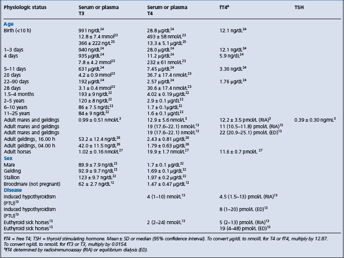
Assays are available for measurement of serum concentrations of T3, T4, free T4 (radioimmunoassay or equilibrium dialysis), and TSH.13 Values of each of these analytes varies depending on the method of analysis, physiologic status of the animal, and administration of other compounds (Table 29.8). Serum concentrations of thyroid hormones is high at birth and declines with age.22-25 There are statistically significant diurnal variations in serum concentrations of T3 and T4 in adult horses with lowest concentrations observed during the early morning hours, likely coincident with the time at which metabolic rate is lowest (Table 29.8).26 Feed restriction for 3–5 days lowers serum concentrations of T3, T4, and fT4 in horses by 24–42%.27 Administration of phenylbutazone decreases concentrations of fT4 (measured by equilibrium dialysis) and T4 by 4 days of treatment, and can persist for up to 10 days after discontinuation of phenylbutazone.28 The decrease in T4 is suggested to be attributable to displacement of T4 from protein binding sites by phenylbutazone, but this does not explain the decrease in fT4. The clinical significance of phenylbutazone-induced decreases in thyroid hormones is uncertain, but should be considered when assessing thyroid function in horses.
Table 29.8 Serum or plasma concentrations of thyroid hormones and thyroid stimulating hormone (TSH) in foals and horses
Because of the number of analytical and physiological factors that affect serum thyroid hormone concentrations, values considered normal vary considerably, as illustrated by the finding that 44 of 79 clinically normal non-pregnant broodmares had serum T4 concentrations below the reference range, although responses to TRH were normal.12 This example illustrates the need to determine reference ranges based on the methodology used and with well defined definition of the physiological state of the animals being tested.
Diagnosis of hypothyroidism is aided by demonstration of inappropriate responses of the thyroid gland to administration of TSH or TRH, although the use of these tests depends on determining the increase in serum T3 and/or T4 that is expected in normal horses and in horses with thyroid disease. Of 79 clinically normal mares, all had some increase in T3 and 77 had an increase in T4 2 hours after IV administration of 1 mg of TRH intravenously.12 The mean increase in serum T3 concentration was 4.5 times that of resting values (from 0.62 ng/mL to 2.44 ng/mL), whereas serum T4 concentration increased to a mean of 2.1 times that of resting value (from 14.7 ng/mL to 28.6 ng/mL).12 While responses to administration of TSH are reported, responses, other than complete lack of response, indicative of abnormal thyroid function have not been determined and the utility of the test has been questioned.27
The TSH response test involves administration of 5 IU of TSH intravenously. Blood samples are collected before, 30 min and 2 and 4 h after administration.29 Serum concentrations of T3 and T4 in healthy horses double after administration of TSH. An alternative involves administration of 5 IU intramuscularly and collection of blood before and 3 and 6 h after a TSH administration.30 TSH is currently unavailable.
The TRH response test requires administration of 0.5–1 mg of TRH intravenously. Serum concentrations of T3 and T4 at 2 and 4 h are double those before TRH administration in horses with normal thyroid function.31
Measurement of fT4 in serum is useful for assessment of thyroid function.3 fT4 concentrations can be normal in horses with low concentrations of T3 and T4, and in this situation are likely indicative of normal thyroid function.
Measurement of serum concentrations of TSH is useful in determining thyroid responsiveness to endogenous TSH. Elevated TSH concentrations in horses with low serum concentrations of T3, T4, or fT4 is indicative of thyroid dysfunction.13,32
Diagnosis of hypothyroidism in horses should be based on the presence of compatible clinical signs, low serum concentrations of thyroid hormones (T3, T4, fT4); elevated concentrations of thyroid stimulating hormone, lack of an increase in serum concentrations of thyroid hormones in response to administration of thyroid releasing hormone (TRH), and increased TSH concentration in serum in response to TRH administration. Diagnosis of hypothyroidism should not be based solely on clinical signs, or on the measurement of resting (unstimulated) serum T3 or T4 concentrations. At a minimum, appropriate clinical signs and documentation of an abnormal response to stimulation testing (TSH or TRH) are essential for diagnosis of hypothyroidism in horses. Measurements of fT4 concentrations determined by equilibrium dialysis are useful in determining thyroid function in sick horses in which T3 and T4 concentrations are low as fT4 concentrations will be normal in horses without thyroid disease.13
Foals with congenital hypothyroidism have abnormally low concentrations of T3 and T4, and less than expected increases in serum concentrations of these hormones in response to TSH administration.33
Horses with hyperthyroidism have markedly elevated concentrations of T3 and T4.9,17 Concentrations of T4 do not decline in response to administration of T3.17 T3 (2.5 mg) is administered intramuscularly twice daily for 3 days and serum concentrations of T3 and T4 measured. T4 concentrations in serum of healthy horses decline by approximately 80% whereas those of horses with hyperthyroidism do not decline.
Findings on necropsy examination of hypothyroid horses have not been reported. Foals with congenital hypothyroidism have histologic evidence of thyroid hyperplasia, but no gross signs of goiter.
Treatment of confirmed hypothyroidism in horses is achieved by administration of levo-thyroxine (20 μg/kg PO q24 h).34 Serum T3 concentrations peak in 1 h and then decline while concentrations of T4 peak in 2 h and persist for 24 h.34 The clinical status of the horse should be monitored during treatment and serum concentrations of T3 and T4 measured every several months. Iodinated casein, which is no longer readily available in the USA, is administered at 5 g/450 kg body weight orally once daily. Administration of thyroxine or iodinated casein for treatment of low serum thyroid hormone concentrations in horses with non-thyroidal illness syndrome (euthyroid sick syndrome) should be done judiciously.
A response to thyroxine administration is not necessarily confirmation of hypothyroidism as thyroxine can have marked effects in horses with normal thyroid function. Administration of thyroxine (up to 96 mg/470 kg horse, orally once daily) increases serum concentrations of T4 and, to a lesser extent, fT4, and decreases concentrations of TSH.35 The increases in T4 are associated with a loss of body weight, decreases in serum concentrations of triglycerides, cholesterol, and very low density lipoproteins, and an increase in whole body insulin sensitivity.35,36 Thyroxine should be administered with caution to horses with normal thyroid function.
There are no recognized control measures for hypothyroidism in adult horses. Minimizing intake of nitrates by pregnant mares appears warranted, but definitive proof of the efficacy of this practice is lacking. Pregnant mares should not be fed fodder or supplements that interfere with thyroid function.
The inherited disorder in sheep, cattle, and goats can be prevented by selective breeding.
1 Ramakrishna C, et al. Ind Vet Med J. 1992;16:37.
2 Johnson PJ, et al. Equine Vet J. 2003;35:296.
3 Breuhaus BA. J Vet Intern Med. 2002;16:109.
4 Rothschild CM, et al. J Vet Intern Med. 2004;18:370.
5 Veenboer GJM, et al. Endocrinol. 1993;132:377.
6 Allen AL, et al. Can Vet J. 1996;37:349.
7 Allen AL, et al. Equine Vet J. 1998;30:53.
8 Alberts MK, et al. J Am Vet Med Assoc. 2000;217:1051.
9 Ramirez S, et al. J Vet Intern Med. 1998;12:475.
10 Hovda LR, et al. J Am Vet Med Assoc. 1990;197:1187.
11 Dalefield RR, Palmer DN. J Comp Pathol. 1994;110:57.
12 Meredith TB, Dobrinski I. J Am Vet Med Assoc. 2004;224:892.
13 Breuhaus BA, et al. J Vet Intern Med. 2006;20:371.
14 Allen AL, et al. Can Vet J. 1994;35:31.
15 Dolling CE, Good BF. J Endocrinol. 1976;71:179.
16 Sipos W, et al. J Vet Med A. 2004;51:90.
17 Alberts MK, et al. J Am Vet Med Assoc. 2000;217:1051.
18 Lowe JE, et al. Cornell Vet. 1974;64:276.
19 Vischer CM, et al. Am J Vet Res. 1999;60:14.
20 Ramakrishna C, et al. Ind Vet J. 1995;71:1107.
21 Frank N, et al. Am J Vet Res. 1999;60:730.
22 Chen CL, et al. Am J Vet Res. 1981;42:1415.
23 Murray MJ, Luba NK. Equine Vet J. 1993;25:237.
24 Irvine CHG, Evans MJ. J Reprod Fertil Suppl. 1975;23:709.
25 Shaftoe S, et al. J Equine Vet Sci. 1988;8:310.
26 Duckett WM, et al. Equine Vet J. 1989;21:123.
27 Messer NT, et al. Am J Vet Res. 1995;56:116.
28 Ramirez S, et al. J Vet Intern Med. 1997;11:371.
29 Held JP, Oliver JW. J Am Vet Med Assoc. 1984;184:326.
30 Morris DD, Garcia M. Am J Vet Res. 1983;44:503.
31 Lothrop CD, Nolan HL. Am J Vet Res. 1986;47:942.
32 Rothschild CM, et al. J Vet Intern Med. 2004;18:370.
33 McLaughlin BG, et al. Can Vet J. 1986;27:264.
34 Chen CL, et al. J Equine Vet Sci. 1984;4:5.