Tracheal stenosis, also known as ‘honker cattle’, occurs in feedlot cattle.1 The etiology is unknown. It is characterized by extensive edema and hemorrhage of the dorsal wall of the trachea, resulting in coughing (honking), dyspnea, and respiratory stertor. Complete occlusion of the trachea may occur. Affected animals may be found dead without any premonitory signs.
In tracheal stenosis of feedlot cattle, there is marked submucosal hemorrhage dorsal and ventral to the tracheal is muscle resulting in ventral displacement of the mucosa and partial to complete occlusion of the tracheal lumen. Diffuse hemorrhage in the peritracheal connective tissue and surrounding muscles of the neck is common in animals dying of asphyxia. Histologically there is hyperemia and hyperplastic tracheal mucosa with focal erosions, squamous metaplasia, and loss of cilia. In acute cases the mucosa is markedly thickened because of hemorrhage and edema. Culture reveals a mixed bacterial flora.
In its most severe form, this is a disease characterized by insidious progression of pulmonary inflammation and fibrosis. Less severe forms, associated with transient viral or bacterial infections of the lungs, resolve when the infection is cleared from the lungs.
There are many potential agents but few have been conclusively implicated as being associated with the disease. In most instances of the disease no causal association is identified.
Interstitial pneumonia is a common finding in horses associated with various infectious agents (Hendra virus infection, Rhodococcus equi in foals, Aspergillus sp., Cryptococcus sp. and Hisptoplasma sp., Pnemoncystis carinii,1 Parascaris equorum, and Dictyocaulus arnfiedli). Intoxication with perilla ketone, derived from Perilla frutescens, causes acute restrictive lung disease of horses.2,3 Similary, ingestion of Eupratorium sp. in Australia and Hawaii causes interstitial pneumonia in horses.4 Inhalation of silica causes a similar disease in horses in California.5 Inhalation or ingestion of agricultural chemical or environmental toxins (e.g. paraquat) has the potential to cause interstitial pneumonia in other species, but this has not been demonstrated in horses.
Hypersensitivity reactions may cause severe respiratory disease in horses. Incriminating allergens include fungi (unspecified) and chicken dust.6,7
Interstitial pneumonia has also been reported subsequent to administration of an immunostimulant containing mycobacterial cell wall extract.8
The disease occurs in adult horses without apparent breed, sex, or age predisposition. In cases in which the cause is infectious, the epidemiology of the disease is characteristic of that of the causal organism.
The initial insult causes injury to parenchymal cells and an acute alveolitis.9 Alveolitis results from damage to epithelial and endothelial cells by toxic, metabolic (free radicals), or infectious agents. This is followed by a phase of cellular proliferation of type 2 pneumocytes and fibroblasts with connective tissue deposition.3 At this time there is an influx of inflammatory cells, the exact type depending to some extent on the cause of the disease. Infiltration of neutrophils, lymphocytes, and macrophages is common. Continued injury to the lung results in development of severe interstitial fibrosis and destruction of gas exchange units.
Interstitial pneumonia results in altered pulmonary function including reduced compliance, impaired pulmonary gas exchange, and a reduction in total and vital lung capacity. The work of breathing is increased.10
Horses with idiopathic interstitial pneumonia have various combinations of: weight loss, recurrent cough, depression, anorexia, fever, or respiratory distress. Signs of respiratory disease are not readily apparent at initial examination in all affected horses. As the disease progresses respiratory distress develops in most, but not all, cases. The usual history of is a gradual onset of increased respiratory effort, although some horses have a sudden onset of respiratory distress.11 Heart and respiratory rates may be elevated. Pyrexia is not a constant finding. There may be a nasal discharge. Thoracic auscultation may reveal only increased intensity of normal breath sounds or the presence of occasional crackles and wheezes. Typically, there is tachypnea with an increased respiratory effort.
Thoracic radiography reveals pulmonary disease, usually apparent as severe, diffuse interstitial disease. The interstitial opacity may be diffuse or nodular with multiple well defined opacities against an overall background of increased interstitial density. Ultrasonographic examination may reveal the presence of multiple nodules in the lung parenchyma confluent with the pleural surface. There is no excess pleural fluid.
Intradermal skin testing may be useful to identify the inciting allergen in cases of allergic interstitial pneumonia.7
The prognosis is poor in most cases of the idiopathic disease. Should recovery occur, it does so within approximately 2 weeks of diagnosis.
Hematologic examination usually reveals a neutrophilic leukocytosis.11,12 Mild anemia and hyperfibrinogenemia are common. Examination of a tracheal aspirate reveals neutrophil inflammation. In cases of verminous pneumonia, there may be an increased proportion of eosinophils in tracheal aspirate. Broncho-alveolar lavage fluid can reveal a similar neutrophilic leukocytosis or may be normal.13 Serologic testing for antibodies to fungi or other inciting agents may be useful.9
Definitive diagnosis is provided by examination of tissue obtained by lung biopsy.
The lungs do not deflate as anticipated and there may be indentations from the ribs on the surface of the lungs. Multiple pale, yellowish-white nodules 3–4 cm in diameter may be apparent on the surface of the lung and throughout the lung parenchyma. There is histologic evidence of severe, fibrosing alveolitis with minimal airway involvement. The acute phase of the disease is evident as extensive alveolar edema and hemorrhage.14
Treatment should be directed toward any cause of the disease that is identified such as administration of anthelmintics. In cases of idiopathic disease, treatment is frequently unrewarding as by the time the animal is examined the disease is often advanced. Treatment includes anti-inflammatory drugs including dexamethasone or prednisolone, antimicrobials, and supportive care. Immunosuppressive drugs, such as vincristine, are not effective. Bronchodilating drugs, such as clenbuterol, may be considered, but bronchoconstriction is not a prominent component of the disease.
1 Franklin RP, et al. J Vet Intern Med. 2002;16:607.
2 Breeze RG, et al. Equine Vet J. 1984;16:180.
3 Schmidbauer SM, et al. J Comp Pathol. 2004;131:186.
4 O’Sullivan B. Aust Vet J. 1979;55:19.
5 Berry CR, et al. J Vet Int Med. 1991;5:248.
6 Gerber H. Equine Vet J. 1973;5:26.
7 Mansmann RA, et al. J Am Vet Med Assoc. 1975;166:673.
8 Viel L, Kenney D. Proceed 12th Veterinary Respiratory Symposium 1994.
9 Bruce EH. Comp Cont Educ Pract Vet. 1995;17:1145.
10 Derksen FJ, et al. J Am Vet Med Assoc. 1982;181:887.
11 Kelly DF, et al. Equine Vet J. 1995;27:76.
12 Winder C, et al. Equine Vet J. 1988;20:298.
This is a disease of foals less than 7 months of age, and usually <2 months of age, characterized by a rapid onset of respiratory distress. The etiology is unclear in many cases, but suggested causes or agents associated with the disease include equine influenza virus infection,1 R. equi, equine herpes virus II, equine arteritis virus,2 or Pneumocystis carinii.3-6 The disease is likely a result of severe pulmonary injury by any of a number of infectious or toxic agents. The respiratory distress results from loss of pulmonary function because of necrosis of the epithelium of alveoli and terminal bronchioles.
Foals typically present with an acute onset (<4 days) of respiratory distress, pyrexia and tachycardia. Foals are depressed and reluctant to eat. There is a pronounced respiratory effort with a marked abdominal component in most affected foals. Crackles, wheezes, and increased bronchial breath sounds are auscultable in most foals. Radiographic examination reveals a bronchinterstitial pattern which is always diffuse, although in some foals there is also a focal interstitial pattern. The prognosis is guarded, with approximately 50% of affected foals dying of the disease.
There is a neutrophilic leukocytosis and hyperfibrinogenemia in most cases. Arterial hypoxemia is present in severely affected foals. Tracheal aspirate demonstrates neutrophilic inflammation. Culture of the tracheal aspirate yields Rhodococcus equi, Strep. zooepidemicus, Actinobacillus sp., and other organisms of questionable significance. Serology might demonstrate evidence of infection by equine influenza virus or equine herpes virus II.1,3,4 Viral isolation can identify equine influenza virus.1
Necropsy examination reveals the presence of diffusely reddened, wet and firm lungs that fail to collapse.3,4 The predominant histologic lesion is necrosis of the epithelium of terminal bronchioles and alveoli.
Principles of treatment are correction of hypoxemia, reduction of inflammation and removal of inciting causes. Severely affected foals may require nasal insufflation of oxygen to ameliorate or correct hypoxemia. Administration of corticosteroids has been associated with improved survival.3 Broad spectrum antibiotics are administered to treat concurrent bacterial infections and prevent secondary infection.
A sporadic disease of foals less than 10 months of age characterized by respiratory distress of several weeks duration is caused by chronic interstitial pneumonia.1 The etiology is unknown, but the disease likely represents a common final response to injury caused by any one of a number of infectious or toxic agents (see ‘interstitial pneumonia of horses’ and ‘acute bronchointerstitial pneumonia of foals’).
Affected foals are bright and alert and have markedly increased respiratory effort. The respiratory rate is elevated and there is a prominent abdominal component to respiratory effort. Fever is low grade and intermittent. Thoracic auscultation reveals increased intensity of normal breath sounds and the presence of wheezes and crackles in most affected foals. Ultrasonographic examination of the thorax reveals extensive ‘comet tail’ signs in most cases. Radiography demonstrates the presence of moderate to severe interstitial pneumonia which in some cases can include focal opacities suggestive of alveolar disease. The prognosis with appropriate treatment is excellent.
Affected foals have neutrophilic leukocytosis and hyperfibrinogenemia. Serological examination for antibodies to common respiratory viruses is unrewarding. Culture of tracheal aspirates does not consistently yield growth of known pathogens. Lung biopsy is not warranted because the characteristic changes on radiographic examination, combined with the clinical signs, are diagnostic for the disease. The risk of adverse events associated with lung biopsy outweighs any diagnostic utility given the good prognosis for complete recovery from the disease.
Treatment consists of administration of corticosteroids such as dexametha-sone phosphate at an initial dosage of 0.1–0.25 mg/kg intravenously for 3–5 days followed by a declining dose administered orally over 2–3 weeks. Prednisolone can be substituted for dexamethasone. Broad spectrum antibiotics (combination of penicillin and aminoglycoside, trimethoprim-sulfonamide, or doxycycline) should be administered for 1–2 weeks.
There are no recognized control measures, although control of infectious respiratory disease in the herd is prudent.
Diseases characterized by nervous system involvement
Etiology Several different causes including thiamin inadequacy, sulfate toxicity.
Epidemiology Sporadic disease in young well-nourished ruminants on high level grain diets and not synthesizing sufficient thiamin. Ingestion of preformed thiaminase in certain plants or production by ruminal microbes may also cause destruction of thiamin. May also occur in cattle and sheep of all ages ingesting excess amount of sulfates in feed and water.
Signs Sudden blindness, ataxia, staggering, head-pressing, tremors of head and neck, ear twitching, champing fits, clonic-tonic convulsions, recumbency, opisthotonos, rumen contractions normal initially, pupils usually normal and responsive, nystagmus, death may occur in 24–48 hours. Hydrogen sulfide odor of ruminal gas in sulfate toxicity.
Clinical pathology Erythrocyte transketolase activity decreased and thiamin pyrophosphate effect increased but both measurements difficult to interpret; blood thiamin levels decreased but not reliable.
Lesions Diffuse cerebral edema, flattened dorsal gyri, coning of cerebellum, multifocal to linear areas of fluorescence in gray and white matter borders of cortical gyri and sulci.
Diagnostic confirmation Fluorescence of gray and white matter of cortical gyri and sulci of brain.
Treatment Thiamin hydrochloride parenterally.
Control Thiamin supplementation of diet. Avoid excess feeding or access to sulfate in feed and water supplies.
Historically, PEM was considered to be caused by a thiamin inadequacy. It is now known that there are several different causes of the disease.
The evidence that a thiamin inadequacy can be associated with the disease includes the following1:
• Affected animals respond to the parenteral administration of thiamin if given within a few hours after the onset of clinical signs
• Affected animals have biochemical findings consistent with thiamin diphosphate (TDP) inadequacy
• The clinical signs and pathological lesions can be reproduced in sheep and cattle by the administration of large daily doses of pyrimidine containing structural analogs of thiamin, principally amprolium, given orally or intraperitoneally.1
PEM occurs sporadically in young cattle, sheep, and goats, and other ruminants. In North America, UK, Australia, and New Zealand, the disease has been most common in cattle and sheep which are being fed concentrate rations under intensified conditions such as in feedlots. An inadequate amount of roughage can result in a net decrease in the synthesis of thiamin.
The disease occurs most commonly in well-nourished thrifty cattle 6–18 months of age (peak incidence 9–12 months of age) which have been in the feedlot for several weeks. Feedlot lambs may also be affected only after being on feed for several weeks. The disease also occurs in goats and in antelope and whitetail deer.4 The disease may affect goats from 2 months to 3 years of age and is commonly associated with milk-replacer diets in kids or concentrate feeding in older goats. The disease occurs only rarely in adult cattle which may be a reflection of the greater quantities of roughage they usually consume. However, there are recent reports of the disease occurring in adult cows on pasture with access to drinking water containing excessive concentrations of sulfates.2
In Cuba, the disease occurred in feedlot cattle fed primarily on molasses with a minimal quantity of roughage, and was reproduced by feeding a molasses-urea and roughage diet by gradual removal of the roughage without the use of thiamin analogs.
Accurate morbidity and case fatality data are not available, but outbreaks can occur suddenly in which up to 25% of groups of feeder cattle may be affected, with case fatality rates from 25 to 50%. Case fatality rates are higher in young cattle (6–9 months) than in the older age group (12–18 months) and mortality increases if treatment with thiamin is delayed for more than a few hours after the onset of signs. In feedlot lambs, it has been suggested that approximately 19% of all deaths are due to polioencephalomalacia.
When PEM was first described in 1956, and for about 30 years, it was considered to be a thiamin deficiency conditioned by dietary factors such as high-level grain feeding and inadequate roughage. PEM occurred most commonly in well nourished young cattle from 6 to 12 months of age which were being fed high-level grain rations. The scientific investigations centered on the effects of dietary factors such as grain diets, and the presence of thiaminases in certain diets on thiamin metabolism in the rumen. In recent years, it has become clear that the disease is not etiologically specific because many different dietary factors have been associated with the occurrence of the disease, and in some instances the thiamin status of the affected animals is within the normal range. Notable examples are the recent observations linking dietary sulfate with the occurrence of the disease.
While there has been general agreement that thiamin inadequacy is associated with the cause of PEM, the possible mechanisms by which this occurs are uncertain.5 Thiamin inadequacy in ruminants could, theoretically, occur in any of the following situations where inadequate net microbial synthesis of thiamin in the rumen may occur:
• Concentrate-fed animals receiving inadequate roughage
• Impaired absorption and/or phosphorylation of thiamin
• The presence of a thiamin inhibitor in the tissues of the host
• Lack of sufficient or appropriate apoenzyme or coenzyme-apoenzyme binding for thiamin dependent systems
• Increased metabolic demands for thiamin in the absence of increased supply
• Increased rate of excretion of thiamin resulting in its net loss from the body.
Thiamin can be destroyed by thiaminases of which significant amounts can be found in the rumen contents and feces of cattle and sheep affected with naturally occurring polioencephalomalacia.
In cattle under farm conditions, using transketolase activity as a measurement of thiamin status, up to 23% of cattle under 2 years of age and 5% over 2 years may be in a thiamin-low state. Newly weaned beef calves on a hay diet are not subject to a thiamin deficiency but a low and variable proportion of young cattle on barley-based feedlot diets (1.7%) may have some evidence of thiamin deficiency based on a thiamin pyrophosphate activity effect in excess of 15%.2 The supplementation of the diet of feedlot steers on an all-concentrate barley-based diet with thiamin at 1.9 mg/kg DM resulted in an increase in average daily gain and final carcass weights. Thus some animals may be marginally deficient in thiamin which may be associated with decreased performance in cattle fed all-concentrate diets. However, thiamin supplementation of cattle on all-concentrate diets does not consistently result in improved animal performance. The experimental disease can be produced in young lambs fed a thiamin-free milk diet and it may be unnecessary to postulate that thiamin analogs produced in the rumen are essential components of the etiology.
A major factor contributing to PEM in cattle and sheep is a progressive state of thiamin deficiency caused by the destruction of thiamin by bacterial thiaminases in the rumen and intestines. Certain species of thiaminase-producing bacteria have been found in the rumen and intestines of animals with PEM. Bacillus thiaminolyticus and Clostridium sporogenes produce thiaminase type I and Bacillus aneurinolyticus produces thiaminase type II. While there is good circumstantial evidence that the thiaminases from these bacteria are the real source of thiaminases associated with the disease, it is not entirely certain. The experimental oral inoculation of large numbers of thiaminase type I producing Clostridium sporogenes into lambs did not result in the disease.
Certain species of fungi from moldy feed are also thiaminase producers but the evidence that they destroy thiamin and are associated with PEM is contradictory and uncertain.
The factors which promote the colonization and growth of thiaminase-producing bacteria in the rumen are unknown. Attempts to establish the organism in the rumen of healthy calves or lambs have been unsuccessful. Thiaminases have also been found in the rumen contents and feces of normal animals which may suggest the existence of a subclinical state of thiamin deficiency.5 Poor growth of unweaned and weaned lambs can be associated with a thiaminase-induced subclinical thiamin deficiency. Weekly testing of young lambs over a period of 10 weeks revealed that 90% of unthrifty lambs were excreting high levels of thiaminase in their feces; low levels of thiaminase activity were present in 20% of clinically normal animals, and there were significant differences in the mean erythrocyte transketolase activity of the unthrifty animals excreting thiaminase compared to the thiaminase-free normal animals.5
Field and laboratory investigations have supported an association between inferior growth rate of weaner sheep in Australia and a thiaminase-induced thiamin deficiency.5 Thiaminase activity has been detected in the feces of lambs at 2–5 days of age, with the levels increasing for 10 days and then declining over the next 3–4 weeks.5 Decreased erythrocyte transketolase activity indicated a thiamin insufficiency in lambs with high thiaminase activity and mean growth rates were 17% less than lambs with low thiaminase activity. The oral supplementation with thiamin at 2–3 weeks of age was the most appropriate prevention and treatment for subclinical thiamin deficiency.5
The parenteral or oral administration of thiamin to normal calves raised under farm conditions resulted in a marked reduction in the percentage thiamin pyrophosphate effect which is an indirect measurement of thiamin inadequacy. Goats with PEM were found to have elevated ruminal and fecal thiaminase activities, low erythrocyte transketolase activity, elevated thiamin pyrophosphate effect, low liver and brain thiamin levels, and elevated plasma glucose levels compared with goats not affected with the disease. With the increased interest in goat farming, some breeders attempt to improve body condition of breeding stock for sale or show by feeding grain or concentrate, which creates a situation similar to feedlot rearing of sheep and cattle which is conducive to the establishment of thiaminases in the rumen and the occurrence of polioencephalomalacia.
High levels of thiaminase type I are present in the rhizomes of bracken fern (Pteridium aquilinum) and horsetail (Equisetum arvense). The feeding of the bracken fern rhizomes (Pteridium esculentum) to sheep will cause acute thiamin deficiency and lesions similar to those of polioencephalomalacia but neither of these plants is normally involved in the natural disease. The disease has occurred in sheep grazing the Nardoo fern, Marsilea drummondii, in flood-prone or low-lying wet areas in Australia. The fern contains a high level of thiaminase type I activity.
Amaranthus blitoides (prostrate pigweed) may contain high levels of thiaminase and be associated with polioencephalomalacia in sheep.6
Polioencephalomalacia has been associated with diets high in sulfur, particularly in the form of sulfate. A high concentrate of sulfates in the diet of cattle has been associated with episodes of the disease in 6–18-month-old cattle. Inorganic sulfate salts in the form of gypsum (calcium sulfate) added to feedlot rations to control the total daily intake of the diet may cause PEM.7 Seasonal outbreaks have occurred in feedlot beef cattle between 15 and 30 days after introduction to a high-sulfur diet and the risk may increase when water is an important source of dietary sulfur, and during hot weather, when the ambient temperatures exceeded 32°C.8
Initial outbreaks may follow the use of a new well of water containing more sulfate than water used previously from another well, increasing from a monthly incidence of 0.07% to 0.88%. Growing cattle consume 2.4 times more water when the temperature is 32°C than at 4°C and consequently total ingestion of sulfur by consumption of high sulfate water increases during hot weather. The feed contained 2.4 g of SO4/kg DM with a total sulfur content of 0.20%. Samples of drinking water contained between 2.2 and 2.8 g of SO4/L. During hot weather daily sulfur ingestion from feed and water combined was estimated to be 64 g/animal corresponding to total dietary sulfur of approximately 0.67% of DM. Daily SO4 ingestion was approximately 160 g/animal.8 The ruminal sulfide levels were much higher 3 weeks after entering the feedlot, when the incidence of the disease was greatest, than 2 months after entering the feedlot when the risk of the disease was low.
In western Canada, there is an association between PEM and high levels of sodium sulfate in water, and range cows are usually affected when certain waters become concentrated with this salt during the summer months.2 Water containing high levels of magnesium sulfate, often called ‘gyp water’, is common in the western plains and intermountain areas of the United States and Canada.9 Ideally, water for livestock consumption should contain less than 500 ppm sulfate, and 1000 ppm is considered the maximum safe level in water for cattle exposed to moderate dietary sulfur levels or high environmental temperatures. A level of 2000 ppm of sulfate in drinking water is the taste discrimination threshold for cattle. Performance of feedlot cattle is reduced when offered water with sulfate levels of 2000 ppm or higher. The National Research Council states that the requirement of sulfur in feed to be 1500 to 2000 ppm for both growing and adult beef cattle; 4000 ppm is considered the maximum tolerated dose.9 Ruminant diets normally contain between 1500 to 2000 ppm (0.15–0.20% sulfur).
Based on National Research Council guidelines, 30 g of sulfur is the calculated maximum tolerated dose of sulfur for a 650 lb (294 kg) steer consuming 16.25 (7.39 kg; 2.5% BW) of feed daily. If the ambient temperature reaches 32°C, a 650 lb steer can drink 14.5 gallons (53.9 L) of water daily, Consumption of 14.5 gallons of water containing 3000 ppm sulfate results in a daily intake of 55 g sulfur. A feed intake of 2.5% BW would also consume 22.2 g of sulfur from feed containing 3000 ppm sulfur for a total daily intake of 77.2 g of sulfur from both feed and water which is 2.5 times the maximum tolerated dose.
In some surveys, water supplies in western Canada contained contained 8447 ppm of total dissolved solids and 5203 ppm of sulfate. A survey of the sulfate concentrations in water on farms, found that high levels of sulfate can have a detrimental effect on the thiamin status of the cattle on those farms.10 Cattle exposed to sulfate concentrations >1000 ppm had blood thiamin levels lower than those drinking water with low levels <200 ppm. This raises the possibility that a subpopulation of cattle under such circumstances could be marginally deficient in thiamin.
The total dietary intake of sulfur by cattle must be considered when investigating sulfur as a cause of PEM. In a study of one farm, water from a 6.1 m well containing 3875 mg/L of total dissolved solids with 3285 mg/L of sodium sulfate was associated with PEM in heifers 6 months of age.11 However, the water contributed about 20% of the total sulfur content in the diet of the heifers, and 60% of the dietary sulfur intake was supplied by the hay and 20% by the grain supplement. The hay contained 0.4% total sulfur which is at the maximum tolerable level for cattle and at the upper limit for hay.11 The hay consisted of variable amounts of kochia (Kochia scorpia) and Canada thistle (Cirsium arvense). Kochia scorpia (summer cypress or Mexican fireweed), is high in sulfur content and has been associated with the disease in range cattle.
PEM has occurred in pastured cattle usually 5–10 days after change from a poor to a good pasture, and may occur in range cattle grazing on dry, short, grama grass pasture.12
The levels of sulfate in water which have affected feed intake in cattle have varied from 2800 to 3340 mg sulfate per/L while other studies found no reduction in feed intake with levels up to 7000 mg/L.11 It appears that the different effects of sulfur toxicity for similar sulfur contents in saline water are attributed to the total sulfur intake. Outbreaks of the disease may occur in adult cattle on pasture drinking water containing 7200 ppm of sodium sulfate.2 Thus established guidelines for saline drinking water are not applicable when cattle are fed feeds grown in saline areas.
A combination of excessive intake of sulfur and a low dietary intake of trace minerals, especially copper, may affect the thiamin status of a cattle herd and contribute to PEM.13 Sulfur adversely affects both thiamin and copper status in sheep.14 A nutritionally related PEM has also been reproduced in calves fed a semipurified, low-roughage diet of variable copper and molybdenum concentrations and it was not related to copper deficiency.15 The disease has occurred in cattle in New Zealand fed chou moellier (Brassica oleracea) which contained sulfur concentrations of 8500 mg/kg DM.16 The morbidity was 25% and mortality 46% despite rapid conventional therapy.
Ammonium sulfate used as a urinary acidifier in the rations of cattle and sheep has been associated with outbreaks of PEM.3 Morbidity rates ranged from 16 to 48% and mortality rates from 0 to 8%. Affected animals did not respond to treatment with thiamin.
Outbreaks have occurred in sheep exposed to an alfalfa field previously sprayed with 35% suspension of elemental sulfur.17 The disease can be induced experimentally in lambs by the administration of sodium hydrosulfide into the esophagus18 and has occurred in lambs 3–4 weeks after being fed a concentrate ration containing 0.43% sulfur.19 Feeding experimental diets containing inorganic sulfur to young lambs was associated with PEM and supplementation of those diets with thiamin decreased the severity of the lesions.20 Rumen microbes are able to reduce sulfate to sulfides which may be directly toxic to the nervous system. Feeding calves (115–180 kg) a semipurified diet high in readily fermentable carbohydrate, without long fiber, and with added sodium sulfate for a total sulfur content of 0.36% resulted in PEM within 21 days of the introduction of the experimental diet.21 An odor of hydrogen sulfide was frequently detected upon passage of a stomach tube into the rumen of all calves during the experiment. The total thiamin concentrations in affected and control calves remained within normal limits.
The dietary content of copper, zinc, iron, and molybdenum may also have important modifying influences on sulfur toxicosis. Molybdenum and copper can combine with sulfur to form insoluble copper-thiomolybdate. Copper, zinc, and iron form insoluble salts with sulfide, and their expected effect would be to decrease the bioavailability of sulfide in the rumen. Conversely, low, but not necessarily deficient, dietary contents of these divalent metals could be prerequisites for excess absorption of sulfide to occur. PEM is not associated with copper deficiency but copper and sulfur metabolism are interdependent. An excess of dietary sulfur may result in depression of serum copper, or alternatively, low serum copper may potentiate the actions of toxic levels of sulfur. Chronic copper poisoning in a lamb has been associated with PEM.22 It is suggested that the copper toxicity may have caused decreased hepatic function resulting in increased plasma concentration of sulfur containing amino acids which may have predisposed to sulfur toxicity encephalomalacia.
Molasses toxicity occurs in Cuba in cattle fed on a liquid molasses-urea feeding system with limited forage. The clinical and necropsy findings are identical to polioencephalomalacia. However, molasses toxicity is not thiamin responsive and can be reversed by feeding forage. Molasses has high inorganic sulfur content and the thiamin concentrations in the brain and liver with PEM which were fed molasses and urea did not differ from those in normal cattle.
Deprivation of feed and water.
In some outbreaks there is a history of deprivation of feed and water for 24–28 hours, because of either a managerial error or frozen water supplies. In other cases, a rapid change in diet appears to precipitate an outbreak. Some outbreaks are associated with a temporary deprivation of water for 24–36 hours, followed by sudden access to water and an excessive supply of salt, a situation analogous to salt poisoning in pigs, but these require more documentation to insure that they indeed are not salt poisoning.
In sheep flocks, a drastic change in management, such as occurs at shearing time, will precipitate outbreaks in which only the yearlings are affected. Changing the diet of sheep from hay to corn silage resulted in a decrease in thiamin concentrations in ruminal fluid to about 25% of control values on hay. The cause of the drop in thiamin concentrations is unknown.
Phalaris aquatica ‘polioencephalomalacia-like’ sudden death in sheep and cattle.
The Mediterranean perennial grass, Phalaris aquatica (formerly Phalaris tuberosa) can cause sudden death in sheep and cattle throughout southern Australia.23 The nervous form of disease is similar clinically to polioencephalomalacia but atypical because of the very rapid onset and the absence of either neuronal necrosis or malacia in cerebral cortical sections from affected animals. The available evidence suggests that this form of phalaris sudden death is more likely to involve a peracute form of ammonia toxicity than a peracute form of polioencephalomalacia.
High levels of thiaminases are formed in the rumen, which destroy thiamin that is naturally synthesized.1 The circumstances in the diet or in the rumen which allow for the development of high levels of thiaminases are unknown but may be related to the nature of the ruminal microflora in young cattle and sheep fed concentrate rations which results in the development of ruminal acidosis. These rations may also allow for the development and growth of thiaminase-producing bacteria which, combined with a smaller net synthesis of thiamin in the rumens of concentrate-fed ruminants, could explain the higher incidence in feedlot animals. Experimentally PEM has been produced in lambs by continuous intraruminal infusion of a highly fermentable diet. Animals changed very rapidly to high concentrate rations develop increased ruminal thiaminase levels.
The possibility that intraruminal thiaminases may also create thiamin analogs capable of acting as thiamin antimetabolites and accentuating the disease has been studied but the results are inconclusive. The presence of naturally occurring second substrates (cosubstrates) in the rumen could produce, by the thiaminase type I reaction, a potent thiamin antimetabolite capable of accentuating the condition. In vitro studies have shown that thiaminase only caused rapid destruction of thiamin when a second substrate was added, and a large number of drugs commonly used as anthelmintics or tranquilizers may be active as second substrates. Many compounds found in the rumen of cattle are potential cosubstrates.
Amprolium has been used extensively to produce the lesions in the brains of cattle and sheep that are indistinguishable from the naturally occurring disease.1 However, since amprolium has been found in the brain tissue, the experimental disease should perhaps be known as ‘amprolium poisoning encephalopathy’. The administration of other antagonists such as oxythiamin and pyrithiamin does not produce the disease. This suggests that polioencephalomalacia is a particular form of thiamin deficiency in which the supply of thiamin is reduced by the action of intraruminal thiaminase. Thus, the thiamin status of the animal will be dependent on dietary thiamin intake, thiamin synthesis, the presence of thiaminase in the rumen and the effects of possible antimetabolites. Subclinical states of thiamin deficiency probably exist in apparently normal cattle and sheep being fed diets which are conducive to the disease. This suggests that in outbreaks of the disease the unaffected animals of the group should be considered as potential new cases and perhaps treated prophylactically.
Thiamin is an essential component of several enzymes involved in intermediary metabolism and a state of deficiency results in increased blood concentration of pyruvate, a reduction in the lactate to pyruvate ratio and depression of erythrocyte transketolase. These abnormalities affect carbohydrate metabolism in general, but in view of the specific requirements of the cerebral cortex for oxidative metabolism of glucose, it is possible that a thiamin inadequacy could have a direct metabolic effect on neurons.1 The brain of the calf has a greater dependence on the pentose pathway for glucose metabolism, in which pathway the transketolase enzyme is a rate-limiting enzyme. Ultrastructural examination of the brain of sheep with the natural disease reveals that the first change which occurs is an edema of the intracellular compartment, principally involving the astrocytes and satellite cells. This is followed by neuronal degeneration which is considered secondary. It has been suggested that the edema may be due to a reduction in ATP production following a defect of carbohydrate metabolism in the astrocyte. There are three basic lesions which are not uniform: compact necrosis, edema necrosis, and edema alone. This may suggest that a uniform etiology such as thiamin deficiency cannot be fully supported.
In the cerebral cortex of affected animals, autofluorescent spots are observed under ultraviolet 365 nm illumination and are a useful diagnostic aid. The distribution of autofluorescence corresponds to that of mitochondria in cerebrocortical neurocytes in affected calves, suggesting that metabolic impairment occurs and the autofluorescent substance is produced in the mitochondria.24 Mitochondrial swelling and disorganization of cristae are also observable in brain tissue, but are not specific to polioencephalomalacia.
Diets high in sulfur result in hydrogen sulfide production in the rumen and anaerobic bacteria from rumen samples of cattle fed high-carbohydrate, short fiber diets with added sulfate will generate hydrogen sulfide in rumen fluid broth medium.18,25 Rumen microflora adapt to higher dietary sulfate content over a period of 10–12 days before they are capable of generating potentially toxic concentrations of sulfide.26 In experimental sulfate diets which induce PEM, the rumen pH decreases during the transition to the experimental diet and acidic conditions in the rumen favor increased rumen gas cap concentrations of hydrogen sulfide. With a change of pH from 6.8 to 5.2, the percent hydrogen sulfide in the rumen gas cap increased from 47 to 97%.7
If ruminants inhale 60% of eructated gases, inhalation of hydrogen sulfide could be a route of systemic sulfide absorption, in addition to gastrointestinal absorption.21 Sulfide inhibits cellular respiration leading to hypoxia which may be sufficient to create neuronal necrosis in polioencephalomalacia. The nervous system lesions of sulfur toxicosis are indistinguishable from lesions in the naturally occurring disease.
Acute cerebral edema and laminar necrosis occur and the clinical signs are usually referable to increased intracranial pressure from the edema, and the widespread focal necrosis. Recovery can occur with early treatment which suggests that the lesions are reversible up to a certain point. ECGs of buffalo calves with amprolium-induced PEM found decreased frequency patterns, occasional spindles and decreased voltage patterns during the onset of clinical signs. In the comatose stage, there was little evidence of electrical activity. EEGs of animals treated with thiamin hydrochloride found normal awake patterns.27
Animals may be found dead without premonitory signs especially in beef cattle on pasture.2 The clinical findings are variable but characteristically, there is a sudden onset of blindness, walking aimlessly, ataxia, muscle tremors, particularly of the head with ear twitching, champing of the jaws and frothy salivation, head-pressing, and the animal is difficult to handle or move. Dysphagia may be present when one attempts to force feed hay by hand. Grinding of the teeth is common. Initially, the involuntary movements may occur in episodes, and convulsions may occur, but within several hours they become continuous. The animal usually then becomes recumbent, and there is marked opisthotonos, nystagmus, clonic-tonic convulsions, particularly when the animal is handled or moved, and tetany of the forelimbs is common. The temperature is usually normal but elevated if there has been excessive muscular activity. The heart rate may be normal, subnormal, or increased and is probably not a reliable diagnostic aid.
Rumen movements remain normal for a few days, which is an important distinguishing feature from lead poisoning in which the rumen is static.
The menace reflex is always absent in the acute stage and its slow return to normal following treatment is a good prognostic sign. The palpebral eye-preservation reflex is usually normal. The pupils are usually of normal size and responsive to light. In severe cases the pupils may be constricted. Dorsal strabismus due to stretching of the trochlear nerve is common. Nystagmus is common and may be vertical or horizontal. Optic disc edema is present in some cases but is not a constant finding.
Calves 6–9 months of age may die in 24–48 hours, while older cattle up to 18 months of age may survive for several days. Recovery is more common in the older age group.
In less severe cases, affected animals are blind, head-press into walls and fences, and remain standing for several hours or a few days. In outbreaks, some cattle will be sternally recumbent; others remain standing with obvious blindness, while others are anorexic, mildly depressed, and have only partial impairment of eyesight. Those with some eyesight will commonly return to almost normal. Some survivors are permanently blind to varying degrees but may begin to eat and drink if provided with assistance. Some cases will recover following treatment and may grow and develop normally.3
Evidence of recovery within a few hours following treatment with thamin indicates that the disease is associated with thiamin inadequacy. A failure of response indicates the possibility of sulfur toxicity polioencephalomalacia.
Sheep usually begin to wander aimlessly, sometimes in circles, or stand motionless and are blind, but within a few hours they become recumbent with opisthotonos, extension of the limbs, hyperesthesia, nystagmus, and periodic tonic-clonic convulsions. Hoggets affected at shearing time may show blindness and head-pressing but, if fed and watered, usually recover within a few days. Occasional animals show unilateral localizing signs, including circling and spasmodic deviation of the head. In goats, early signs may include excitability and elevation of the head. Blindness, extreme opisthotonos, and severe extensor rigidity and nystagmus are common.
In sulfur-induced PEM in sheep introduced to a diet containing 0.43% sulfur, clinical signs occurred 15–32 days later and consisted of depression, central blindness, and head-pressing, but no hyperesthesis, nystagmus, or opisthotonos were observed.11 In sulfur toxicity in lambs with PEM, the rumen contents may have a strong odor of hydrogen sulfide (rotten egg smell).17
There are some reports from Australia of unthriftiness in unweaned and weaned lambs being associated with thiamin deficiency due to the presence of thiaminases in the alimentary tract.5 In affected flocks the incidence of illthrift in lambs is much higher than the usual incidence and other causes of unthriftiness were ruled out. Affected lambs lose weight, may have chronic diarrhea, and become emaciated and die from starvation. In some flocks, clinical signs of PEM may occur in a small percentage of animals. The disease occurs most commonly in early July which is the coldest part of the year in Australia for lambs which are born in May and June. In affected lambs the fecal thiaminase levels are high and the blood transketolase level activity is increased above normal. Treatment of affected lambs with thiamin resulted in an increase in growth rate.5
The biochemical changes occurring in cattle and sheep with the thiamin-deficiency PEM have not been well-defined diagnostically based on thoroughly investigated naturally occurring clinical cases. However, some estimates are available including the changes which occur in the experimental disease. Interpretation of the values may also be unreliable if the animals have been treated prior to death.
The erythrocyte transketolase activity is decreased and the thiamin pyrophosphate (TPP) effect is increased. The erythrocyte transketolase activities in normal sheep will range from 40 to 60 iμ/mL red blood cells. A TPP effect of 30–50% is commonly found in normal healthy cattle and sheep and an increase to above 70–80% occurs in animals with polioencephalomalacia.
The thiamin concentrations of blood of animals with polioencephalomalacia have varied widely and may be difficult to interpret because of the possibility of thiamin analogs inducing deficiency even when blood thiamin levels are normal. However, this would not apply when blood thiamin levels are below normal. A normal reference range of 75–185 nmol/L is suggested for both cattle and sheep, and levels below 50 nmol/L are considered indicative of deficiency.28 In normal goats, the mean thiamin content of blood was 108 nmol/L, with a range of 72–178 nmol/L.12 In goats with polioencephalomalacia, blood thiamin levels were less than 66 nmol/L with a mean of 29 nmol/L. Levels as low as 1.8–3.6 μg/dL (6–12 nmol/L) have been found in suspected cases of polioencephalomalacia. The thiamin concentrations of liver, heart, and brain of cattle and sheep with polioencephalomalacia are decreased. The levels of blood pyruvate and lactate are also increased and thiamin pyrophosphate-dependent enzymes such as pyruvate kinase are decreased.3 The thiaminase activity of the feces is increased.5
The hemogram is usually normal; the total and differential leukocyte counts may indicate a mild stress reaction, a finding which may be useful in differentiation from encephalopathies due to bacterial infections.
Cerebrospinal fluid pressure taken at the cysterna magna is increased from a normal range of 120–160 mm saline to levels of 200–350 mm. The level of protein in the CSF may be normal to slightly or extremely elevated. A range from 15 to 540 mg/dL with a mean value of 90 mg/dL in affected cattle is recorded. There may also be a slight to severe pleocytosis in the CSF in which monocytes or phagocytes predominate.
Changes in rumen gas cap H2S concentrations are larger than changes in rumen fluid H2S concentrations and estimation of rumen gas H2S concentration may be a practical method of detecting pathological increases in ruminal hydrogen sulfide gas.29,30 A simple, rapid, minimally invasive method may be useful for estimating the H2S concentration of ruminal gas under field conditions. A sterile 8.9 cm 18 g needle with stillete is introduced into the gas cap of the rumen by way of the left paralumbar fossa. The needle is then connected to calibrated H2S detector tube. In cattle, with sulfate-induced polioencephalomalacia, increases in ruminal gas H2S may be as high as 100 times more than control animals.29
The effects of high dietary sulfur on brain function have been examined using evoked potentials techniques.31 Altered nerve conduction pathways occur in sheep fed high sulfur diets without supplemental thiamin compared to animals which have received thiamin.31 The visual evoked potentials are abnormal in ruminants with thiamin-responsive polioencephalomalacia.32
Diffuse cerebral edema with compression and yellow discoloration of the dorsal cortical gyri is evident and the cerebellum is pushed back into the foramen magnum with distortion of its posterior aspect.
In recovered animals, there is macroscopic decortication about the motor area and over the occipital lobes. The lesion can be identified grossly using ultraviolet illumination which results in a fluorescence that indicates necrosis of brain and engulfment of necrotic tissue by lipophages. In general, there is a good correlation between the presence of characteristic fluorescence and the biochemical changes in cases of polioencephalomalacia. A small percentage of false negatives may occur.
Histologically the lesions are widespread but most common in the cerebral cortex. There is bilateral laminar necrosis and necrosis of deeper cerebral areas. The necrosis is most prominent in the dorsal occipital and parietal cortex, but bilateral areas of necrosis are also seen less frequently in the thalamus, lateral geniculate bodies, basal ganglia, and mesencephalic nuclei. Lesions of the cerebellum are also present. The severity and distribution of the lesions probably depend on the interrelationships between clinical severity, age of affected animal, and length of illness before death.
Subnormal levels of thiamin are detectable in the liver and brain of calves with the natural disease and low levels are also found in the experimental disease.21 In the molasses-induced disease in Cuba, the tissue thiamin levels were within the normal range.
In some cases of sulfur-associated polioencephalomalacia, the rumen contents have a strong odor of hydrogen sulfide – the rotten egg smell.
None of the biochemical tests described under clinical pathology is practical. The diagnosis must be made on the basis of clinical findings and the readily available simple tests which rule out other diseases that resemble polioencephalomalacia. A careful consideration of the epidemiological history often assists in the diagnosis.
The differential clinical diagnosis for cattle is summarized in Table 32.3. Polioencephalomalacia in cattle occurs primarily in young growing animals 6–9 months of age on concentrate rations and is characterized clinically by a sudden onset of blindness, muscular tremors of the head and neck, head-pressing, nystagmus, and opisthotonos. The disease also occurs in mature beef cattle on pasture containing a high level of sulfate in their water and feed.
In cattle the disease must be differentiated from:
• Acute lead poisoning which is most common in calves after spring turnout but occurs in adult cattle too and is characterized by central blindness, tremors, convulsions, uncontrollable activity with bellowing, champing fits, hyperexcitability, rumen stasis, and death in several hours. Early treatment may be successful
• Subacute lead poisoning characterized by blindness, stupor, head pressing, rumen stasis, weak palpebral reflexes, and no response to therapy
• Hypovitaminosis-A characterized by a history of a vitamin A deficient diet and nyctalopia, peripheral blindness, dilated and fixed pupils, optic disk edema, and transient convulsions followed by recovery
• Histophilus meningoencephalitis characterized by sudden onset of ataxia, recumbency, fever, depression with eyes closed, lesions of the fundus, marked changes in hemogram, enlarged joints, and death in several hours if not treated eary.
In sheep polioencephalomalacia must be differentiated from:
• Enterotoxemia (pulpy kidney disease) due to Cl. perfringens type D in unvaccinated sheep, especially feedlot lambs, in which the clinical findings are almost identical; it occurs under the same management conditions as polioencephalomalacia. Enterotoxemia in lambs usually develops within several days after being placed on a grain ration, whereas polioencephalomalacia occurs after several weeks of grain feeding. Glycosuria in pulpy kidney disease may assist the diagnosis but a necropsy is usually more informative
• Focal symmetrical encephalomalacia also resembles polioencephalomalacia but is sporadic, usually involves only a few animals and will not respond to treatment.
The treatment of choice for thiamin deficiency PEM is thiamin hydrochloride at 10 mg/kg BW IV initially and followed by similar doses every 3 hours for a total of 5 treatments. When treatment is given within a few hours of the onset of signs, a beneficial response within 1–6 hours is common and complete clinical recovery can occur in 24 hours. Goats and sheep will commonly respond within 1–2 hours. For those which take longer to recover, the eyesight and mental awareness will gradually improve in a few days and the animal will usually begin to eat and drink by the third day after treatment. Rumen transplants of rumen juice from roughage-fed cattle may improve appetite and rumen function in those responding slowly. In sheep, following treatment with thiamin the blood transketolase activity begins to return to normal in 2–4 hours and is considered normal 24 hours after treatment.
Some cattle improve to a subnormal level within a few days and fail to continue to improve. These are usually affected with diffuse cortical and subcortical necrosis and will usually not improve further in spite of continued treatment. Those which return to a clinically normal state will usually do so by 48 hours or sooner after initial treatment. Those which are still clinically subnormal and anorexic by the end of the third day will usually remain at that level and should be slaughtered for salvage.
Treatment is ineffective in advanced cases, but unless an accurate history is available on the length of the illness, it is usually difficult to predict the outcome until 6–12 hours following treatment. Thus, it is usual practice to treat most cases with thiamin at least twice and monitor the response. If there is no beneficial response in 6–8 hours, emergency slaughter for salvage should be considered.
The oral administration of thiamin or thiamin derivatives is indicated when thiaminases are thought to be in the alimentary tract. Thiamin hydrochloride at a rate of 1 g for goats and lambs and 5 g for calves, in a drench, is recommended. However, because the action of thiaminase type I on thiamin may result in the production of thiamin analogs which may act as inhibitors of thiamin metabolism, the use of thiamin derivatives which are resistant to thiaminases, lipid soluble and absorbed from the intestine are being explored as therapeutic and prophylactic agents. Thiamin propyldisulfide can depress the thiaminase activities in the ruminal fluid of sheep with polioencephalomalacia within 2 hours after oral administration. The blood pyruvate levels and transketolase activities are also restored to normal and treated animals recovered clinically.
In outbreaks, the in-contact unaffected animals on the same diet as the affected animals may be on the brink of clinical disease. The diet should be changed to one containing at least 50% roughage or 1.5 kg of roughage per 100 kg BW. Thiamin may be added to the ration at the rate of 50 mg/kg of feed for 2–3 weeks as a preventive against clinical disease, followed by a level of 20–30 mg/kg of feed (cattle and sheep) if the animals remain on a diet that may predispose them to the disease.
There is no specific treatment for PEM caused by sulfate toxicity. The use of thiamin hydrochloride in doses given above is recommended.7
A rational approach to the control of PEM associated with thiamin inadequacy is to supplement the rations of concentrate-fed cattle and sheep with thiamin on a continuous basis. The daily requirements for protection have not been determined using controlled feeding trials but a rate of 3 mg/kg DM of feed for cattle and sheep has been recommended. This level may not be protective in all situations and response trials may be necessary to determine protective levels for different situations. Levels up to 20–30 mg/kg of feed may be necessary for protection. Most natural feedstuffs for ruminants contain thiamin at about 2 mg/kg DM which when combined with the thiamin synthesized in the rumen will meet the requirements. However, the presence of thiaminases in the rumen will necessitate dietary supplementation with thiamin, but the optimal amount that will provide protection under practical conditions is uncertain.
The IM injection of 500 mg thiamin 3 times weekly into 6-month-old calves raised under practical farm conditions will steadily reduce the percentage thiamin pyrophosphate effect to zero in about 6 weeks. The daily oral administration of 100 mg thiamin to young calves fed initially on milk substitutes and then on concentrates and hay results in a decrease in percentage pyrophosphate effect.
For animals which are fed diets associated with thiamin inadequacy, it is recommended that thiamin be added to the diet at the rate of 5–10 mg/kg DM.12 Cattle and sheep on concentrate-fed rations must also receive supplements containing all necessary vitamins and minerals, especially cobalt, a deficiency of which may be associated with some outbreaks of the disease.
The minimum amount of roughage which should be fed to feedlot cattle and sheep in order to prevent the disease and still maintain them on high levels of concentrates is unknown. A level of 1.5 kg of roughage per 100 kg BW has been recommended but this may not be economical for the feedlot whose profits are dependent on rapid growth in grain-fed cattle. Supplementation of the diet with thiamin appears to be the only alternative.
The prevention of the disease in sheep which are being moved long distances or gathered together for shearing and other management practices will depend on insuring an ample supply of roughage and water and avoiding drastic changes in management.
The prevention of the disease associated with a high sulfur intake in the feed and water supplies will depend on analysis of the feed and water for sulfate and making appropriate adjustments in the sources of feed and water in order to decrease the intake of sulfur to safe levels.
1 Loew FM. World Rev Nutr Diet. 1975;20:168.
2 Hamlen H, et al. Can Vet J. 1993;34:153.
3 Jeffrey M, et al. Vet. Rec. 1994;134:343.
4 Smits JEG, Wobeser G. Aust Vet J. 1990;31:300.
5 Thomas KW, Griffiths FR. Aust Vet J. 1987;64:207.
6 Ramos JJ, et al. Can Vet J. 2005;46:59.
7 Niles GA, et al. Bovine Pract. 2002;36:93.
8 McAllister MM, et al. J Am Vet Med Assoc. 1997;211:1275.
9 Niles GA, et al. Bovine Pract. 2002;36:101.
10 Olkowski AA, et al. Can J Anim Sci. 1991;71:825.
11 Beke GJ, Hironaka R. Sci Total Environ. 1991;101:281.
12 Rammell CG, Hill JH. Aust Vet J. 1988;36:99.
13 Gooneratne SR, et al. Can Vet J. 1989;30:139.
14 Olkowski AA, et al. Res Vet Sci. 1990;48:82.
15 Sager RL, et al. Am J Vet Res. 1990;51:1969.
16 Hill FI, Ebbett PC. Aust Vet J. 1997;45:37.
17 Bulgin MS, et al. J Am Vet Med Assoc. 1996;208:1996.
18 McAllister MM, et al. J Comp Pathol. 1992;106:267.
19 Low JC, et al. Vet Rec. 1996;138:327.
20 Rousseaux CG, et al. J Vet Med A. 1991;38:229.
21 Gould DH, et al. Am J Vet Res. 1991;52:1164.
22 Sargison ND, et al. Vet Rec. 1994;135:556.
23 Bourke CA, et al. Aust Vet J. 2005;83:168.
24 Shibahara T, et al. Aust Vet J. 1999;77:329.
25 Cummings BA, et al. Vet Pathol. 1995;56:1384.
26 Cummings BA, et al. Am J Vet Res. 1995;56:1390.
27 Sundar NS, et al. Agri Pract. 1996;17:34.
28 Hill JH, et al. NZ Vet J. 1988;36:49.
29 Gould DH, et al. J Vet Diagn Invest. 1997;9:72.
30 Gould DH. J Anim Sci. 1998;76:309.
This is a syndrome of foals less than 36 hours of age characterized by a spectrum of changes in mentation ranging from failure to suckle, abnormal behavior, seizures, through coma in otherwise apparently healthy foals.
A number of diseases cause the clinical signs consistent with this syndrome. These include antenatal, natal or postnatal hypoxia, congential and metabolic anomalies, placental abnormalities, intracranial hemorrhage, prematurity, and thoracic trauma.1 Of these causes, hypoxia before, during or soon after birth is considered the most common cause of neonatal encephalopathy, although hypoxia is rarely documented. It is important to realize the neonatal encecphalopathy is one of many manifestations of hypoxia of the fetus or neonate, the other manifestations including gastrointestinal and renal damage.2,3
The disease is sporadic and occurs worldwide with an annual incidence in foals of approximately 1%.4 Foals of either sex and of any breed born to mares of any age or reproductive history can be affected. The case fatality rate is very low for appropriately treated foals without other systemic illness.
It is speculated that hypoxia resulting from intracranial vascular accidents,5 asphyxia at birth or placental insufficiency before birth damages the central nervous system. Neurological abnormalities and a failure to nurse, result in a failure of the transfer of maternal immunoglobulins, which predisposes the foal to septicemia and hypoglycemia.
Foals that are abnormal at birth can display a range of behavioral abnormalities, from lack of suckle reflex to convulsions with extensor rigidity. The placenta of affected foals is often abnormal or there is a history of prolonged parturition. Affected foals either do not develop or lose the suck reflex, and have no affinity for the mare and are unable to locate the udder or teat. Aimless wandering and a characteristic ‘barking’ vocalization are sometimes present. Recumbent foals struggle wildly and in an uncoordinated fashion to stand. Convulsing foals usually display opisthotonos with extensor rigidity. Other signs of convulsive activity include facial twitching and grimacing, nystagmus, rapid blinking, sucking, chewing, and drooling.1 Between episodes foals are usually depressed or somnolent. Affected foals display little or no interest in the mare. Convulsing foals are tachypneic, tachycardic (>180 bpm), and hyperthermic (>39°C, 102°F) during and immediately after convulsions. It is important to recognize that the severity of clinical signs varies from very mild (foals are often described by owners as being a bit slow or dimwitted) through to grand mal seizures.
Foals that are normal at birth may develop signs by 24 hours of age. The signs are similar to those described above, with the exception that the foals are initially able to ambulate. It is important to realize that healthy newborn foals lack a mence reflex, have a hypermetric gait and intention tremor, and become flaccid when restrained.
Affected foals can take days to weeks to recover completely. Blind foals that do not have ocular lesions can take as long as 4–6 weeks to regain vision.
Ancillary testing is not usually indicated unless the foal fails to respond after approximately 7 days. At that time, CT or MRI examination of the brain might be indicated to detect congenital anomalies such as hydrocephalus. Examination of cerebrospinal fluid should be performed in any foal with signs of CNS dysfunction in the presence of fever or other signs of sepsis.
There are no hematological or serum biochemical abnormalities characteristic of the disease:
Definitive diagnosis of the disease is difficult and is based on exclusion of other diseases that can cause similar signs and, at necropsy, demonstration of intracranial lesions consistent with the disease.
Gross changes are typically limited to diffuse pulmonary congestion with a variable degree of atelectasis. In cases in which dystocia has been a contributing factor, fractured ribs, and foci of subcutaneous edema and hemorrhage are sometimes noted. Occasionally, macroscopic cerebral hemorrhages are visible. Histologicially, the key findings are hemorrhagic foci within the brain and areas of ischemic necrosis in the cerebral cortex.6 Meconium and other components of aspirated amniotic fluid accompanied by atelectasis and a mild inflammatory response may be present within the lung. In less affected foals the brain lesions are restricted to hemorrhage, cerebral swelling, and edema. Many affected foals have evidence of intracranial vascular accidents.7 Affected foals that are euthanased often have no detectable lesions in the brain.
Formalin-fixed brain, including cerebral cortex, cerebellum and brain stem, and lung for light microscopic examination.
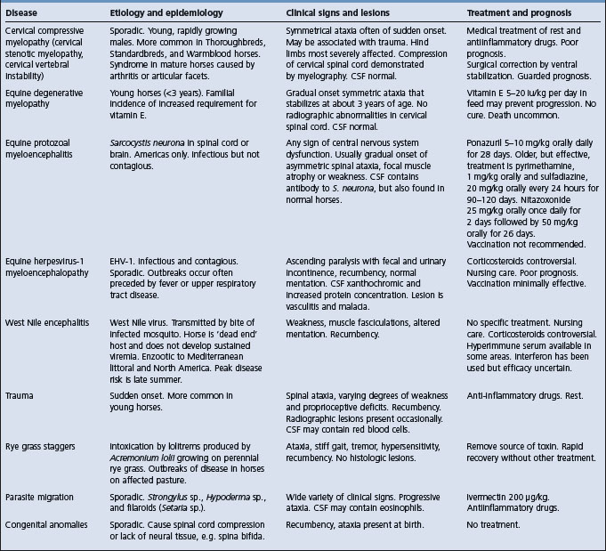
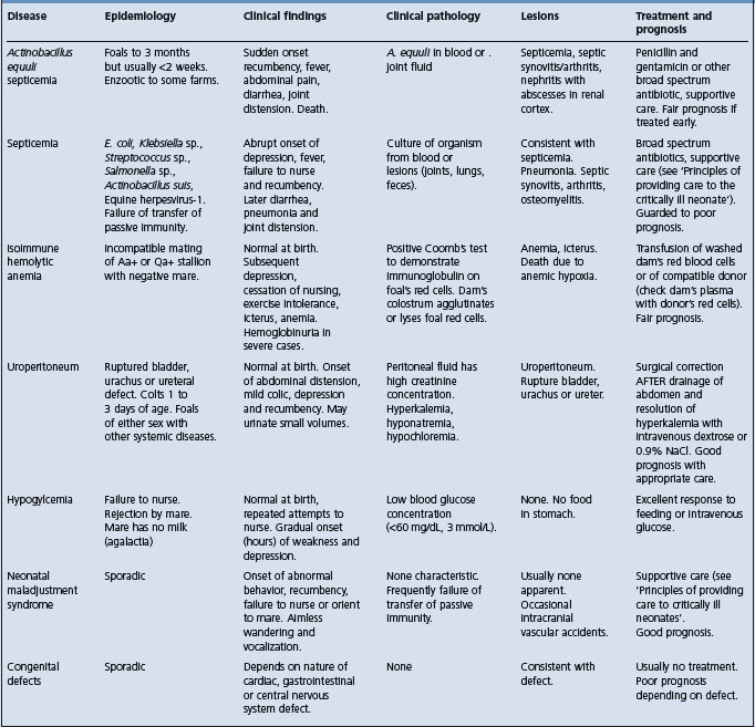
The disease must be differentiated from other diseases that cause neurological or behavioral abnormalities in foals including: sepsis; renal, hepatic, or gastrointestinal disease, which can occur secondary to fetal hypoxia; hydrocephalus; hypoglycemia; meningitis; neonatal isoimmune hemolytic anemia; and prematurity, dysmaturity, or immaturity.
The principles of treatment are:
• Treatment of cerebral edema and hemorrhage
• Correction of failure of transfer of passive immunity
• Nutritional support and general nursing care. The management of affected foals is mainly supportive and is time consuming and labor intensive.
Provision of nutritional support, treatment of failure of passive transfer of maternal immunoglobulins, and nursing care is dealt with in detail in the section ‘Principles of care of the critically ill neonate’.
For other than emergency treatment of seizures, in which diazepam (0.1–0.4 mg/kg, intravenously, as required) or midazolam (0.05–0.1 mg/kg IV, as required) are useful, phenobarbital (phenobarbitone), phenytoin and primidone are the drugs of choice for long-term control of seizure activity. Phenobarbital is administered initially at a dose of 1–3 mg/kg intravenously in 30 mL of isotonic saline infused over 15–30 minutes. Maintenance therapy is a similar dose intravenously or orally, every 8 hours, and the dose adjusted to provide control of seizures while minimizing the degree of sedation. Because of the long elimination half life of phenobarbitol in foals (∼200 hours) and the transient nature of the disease, once seizure control is achieved administration of phenobarbitol can be discontinued. Drug concentrations will be at or above the target concentration (5–30 μg/mL) for several days after the final dose. Phenytoin (5–10 mg/kg intravenously or orally initially, then 1–5 mg/kg every 4 hours) or primidone (20–40 mg/kg orally every 12–24 hours, to effect) are also used to control convulsions.
Definitive demonstration of the presence of cerebral edema or intracranial hemorrhage is impossible without sophisticated imaging devices, such as magnetic resonance imaging or computed tomography (CT).8 However, treatment is often initiated on the basis of clinical signs. None of the treatments have demonstrated efficacy, and some are controversial. Dimethyl sulfoxide (DMSO) is given intravenously at 0.5–1 mg/kg once or twice daily for 3 days as a 10% solution. Mannitol (0.25 g/kg, intravenously as a 20% solution) may be effective in treating cerebral edema but is contraindicated if intracranial hemorrhage is present. Glucocorticoids (dexamethasone, 0.2–1 mg/kg or prednisone, 1–2 mg/kg) might reduce intracranial inflammation and swelling. They might be contraindicated in foals with sepsis.
Magnesium sulfate (0.05 mg/kg per hour for one hour, then 0.025 mg/kg/h IV for up to 48 hours) is often administered to foals with suspected hypoxic encephalopathy in an attempt to minimize neuronal damage. There is no objective evidence of its efficacy in foals.
Foals with respiratory depression can be administered caffeine (10 mg/kg orally once and then 3.0 mg/kg orally q 24 hours).3 Adverse effects include agitation, hyperesthesia, tachycardia, and convulsions.
Good nursing care is critical in affected foals, and a concerted and persistent effort should be made to encourage the foal to nurse the mare. Encouraging the foal to nurse can be frustrating for the handler and mare, but should be done regularly, about every 4 hours, and preferably when the foal is hungry. Affected foals often begin to nurse quite suddenly.
Affected foals can require up to 4–6 weeks to recover completely, although most do so within 1 week of birth, and hasty decisions regarding euthanasia should not be made without recognition of the sometimes long time required for complete recovery.
1 Hess-Dudan F, Rossdale PD. Equine Vet Educ. 1996;8:24.
2 Rossdale PD. Proc Am Assoc Equine Pract. 2004;50:75.
3 Vaala WE. Proc Am Assoc Equine Pract. 1999;45:247.
4 Rossdale PD. Equine Vet J. 1972;4:117.
5 Rossdale PD, et al. Vet Rec. 1976;99:111.
6 Palmer AC, Rossdale PD. Equine Vet Sci. 1976;20:267.
Tremors in pigs are not a common clinical sign. Recently, generalised tremors have been described in grower pigs1 but until this report the condition has only been described as a neonatal condition. The condition has been known for a long time in Britain.
This group of at least four etiologically distinct congenital diseases of the nervous system has a similar clinical and pathological identity. In particular they all have congenital tremor (myoclonia congenita) and these rhythmic tremors are most evident while the piglet is standing, are reduced when it lies down, and disappear when it is asleep. It is therefore not encephalitis but an increased sensitivity of the spinal cord reflexes.
Although the tremor is present at birth it may not be observed until the piglets are 2 or 3 days old and beginning to move about actively. The tremor varies from a very rapid twitch affecting only the head to a slow tremor causing the pig to ‘dance’; there may be such a severe tremor that the body and head tremble violently and rock from side to side. There is no muscular weakness and the piglets get up and scamper about, but there may be ataxia and dysmetria so that the gait may be badly affected. Mildly affected piglets are not greatly incapacitated and can survive and eventually recover in 2–8 weeks; the tremor fades gradually, and near the end of the course it may be evident only during exercise. Badly affected piglets may be unable to get to the teat to suck and may die of starvation. Many are fatally crushed or trampled by the sow.
For convenience, entities within the syndrome have been classified into two types.
Type A has morphological lesions and there are 5 types AI, AII, AIII, and AIV for which the causes are known.
Type AI (myoclonia congenita) results from transplacental infection of the fetus by particular strains of swine fever virus.2-4 In litters affected by swine fever virus usually about 40% of the litter is affected. The characteristic lesions are cerebellar hypoplasia and cerebrospinal dysmyelinogenesis. When the disease caused by the unidentified viral infection of the fetus first appears in a piggery all litters are affected. Most pigs in each litter have the disease, but after a period of several months the disease disappears, apparently because of herd immunity. The principal sign as in the swine fever induced disease is myoclonia, and most piglets recover. Spinal dysmyelinogenesis is the characteristic lesion.
Type AII (myoclonia congenital) is caused by transplacental infection with an agent presumed to be a virus and it is believed to be the most common type of congenital tremor.
Type AIII (congenital cerebrospinal hypomyelinogenesis) is a hereditary form so far found only in Landrace and known as Landrace trembles.5 The inherited sex-linked disease occurs in the progeny of Landrace or Landrace-cross sows. Up to 11% of Landrace piglets may be affected compared to only 0.3–0.8% in the Yorkshire, Hampshire, and Duroc breeds. The mode of transmission is via a monogenic sex-linked recessive character, and half of all male pigs born are affected. Histologically, there is cerebrospinal myelinogenesis. Most affected pigs die. It is essentially a deficiency of oligodendrocytes.
Type AIV is also a genetically determined condition due to an autosomal recessive condition found in Saddlebacks. It is seen in about 25% of the progeny, and is fatal in most of them. Cerebrospinal dysmyelinogenesis is the characteristic lesion. It is characterised by a deficiency of myelin throughout the CNS.6 A type distinct from this has also been reported in Saddleback × Large White sows.7
Type AV occurs naturally in Scandinavia8 and is characterized by cerebellar hypoplasia and has been experimentally produced following dosing of pregnant sows between 45 and 75 days of gestation with trichlorfon.9
Type B is a form of congenital tremor not yet adequately characterized and without morphological lesions and is of unknown etiology. The differentiation of these diseases is now possible on the basis of histopathological and neurochemical findings and relating these to the epidemiological data. In piglets born from sows inoculated with the Weybridge congenital tremor strain of the swine fever virus in early pregnancy, the severity of the clinical signs was related to the degree of spinal myelin deficiency. Semiautomated planimetry can be used to determine the cross-sectional areas of spinal gray and white matter of affected piglets. The spinal cord cross-sectional area is significantly reduced in piglets affected with Type AI congenital tremor. The reduction is similar to Type AII. The neurochemical findings relate to the presence of particular fatty acid profiles in the cholesterol esters in the lipids of the spinal cord.
Control of the disease depends on its cause. If it is inherited the recommended procedure is to keep none of the affected recovered pigs for breeding. If the cause is one of the maternal infections of early pregnancy it is recommended practice to deliberately expose empty females to affected piglets so that they become immunized before pregnancy. Failure to identify the cause may necessitate adopting both procedures.
The disease appears to occur sporadically but when it does occur it may assume great importance. As many as 15% of litters born in a piggery may be affected, and of these 72% of the piglets may be affected and the case fatality rate amongst these may be as high as 50%. There is a feeling amongst veterinarians that the condition has increased recently but this may just be an increased awareness. If there is an increase, the specific cause of the increased prevalence has not been identified but it is possibly the unidentified virus referred to above: the virus of congenital tremor Type AII. There is also the possibility that the observed concurrent increase in prevalence of both splayleg and congenital tremor may indicate that they are possibly caused by the same virus. In this context, there is conjecture, that the PCV2 virus which is extremely small and similar in size to the particles that were described in the early electron micrographs of the spinal cord of affected piglets may be responsible. As yet there is no confirmation that splayleg and congenital tremor are PCV2 associated disorders.10
1 Desrosiers R, et al. Swine Hlth Prod. 1998;6:174.
2 Done JT, Harding JDJ. Proc Roy Soc Med. 1966;59:1083.
3 Done JT, Harding JDJ. Exc Med Int Cong Ser. 1969;173:112.
4 Harding JDJ, et al. Vet Rec. 1966;79:388.
5 Harding JDJ, et al. Vet Rec. 1973;79:527.
6 Blakemore WF, Harding JDJ. Res Vet Sci. 1974;17:248.
7 Kidd ARM, et al. Brit Vet J. 1986;142:275.
8 Kronevi T, et al. Vet Rec. 1975;96:403.
This is a disease of sheep, mainly Merino wethers, in western Queensland, characterized by stiffness and shortness of steps when forced to walk for about a kilometer. The sheep stops walking and adopts an arched back posture. At this time the body temperature is significantly elevated and the level of lymphocytes in the blood is lowered. The greatest incidence is in summer after rain and when the sheep are in full wool, and the hyperthermia and ataxia disappear when the sheep are shorn. Environmental temperatures at the time of the greatest prevalence of the disease are high, commonly 40°C. This occurs in spite of the presence of a characteristic degeneration of spinal cord tracts.1 The obvious explanation of the phenomenon is that it is a form of heat stroke and that the nervous system lesions are incidental.
Epidemiology Sporadic or endemic disease of young horses. Young, rapidly growing male horses are most commonly affected. Separate presentation in middle-aged horses in which it is sporadic.
Clinical signs Spinal ataxia evident as truncal sway, ataxia and paresis usually more severe in the hind limbs. Radiographic evidence of narrow spinal canal.
Lesions Malacia and Wallerian degeneration in the cervical spinal cord.
Differential diagnosis Equine degenerative myelopathy, equine protozoal myeloencephalitis, trauma, equine infectious anemia, cerebrospinal nematodiasis, West Nile encephalomyelitis, equine herpesvirus-1 myelopathy, aorto-iliac thrombosis, congenital vertebral malformation, diskospondylitis, and rye-grass staggers.
Diagnostic confirmation Radiography. Positive contrast myelography. Necropsy.
Treatment Anti-inflammatory drugs. Surgical fusion of vertebrae.
The cause of neurologic disease is compression of the cervical spinal cord, hence the term compressive myelopathy. The compression may be static, that is the compression is present constantly with the neck in a neutral position, or dynamic and only present intermittently when the neck is either flexed or extended. The second situation is often referred to as cervical vertebral instability. The etiology of equine cervical stenotic myelopathy (CSM) in most cases is not known. Several basic syndromes of compressive myelopathy, based on age of occurrence, are recognized:
• Equine stenotic myelopathy (ESM) in immature horses (<3 years of age, depending on breed) that is often associated with developmental joint disease in the axial and appendicular skeleton. The fundamental underlying defect appears to be a narrow diameter of the cervical vertebral canal.
• Cervical vertebral instability (CVI), a disease of horses less than 1 year of age, is often associated with malformations of one or more of the cervical vertebrae.1
• Compressive myelopathy in mature horses, >4 years (usually >7 years) of age, associated with osteoarthritis of the articular facets of the caudal cervical vertebrae, with subsequent impingement of the vertebral canal by bony and soft tissue proliferative lesions.
• Miscellaneous causes of cervical cord compression by neoplasia (melanoma, sarcoma, lymphoma),2-4 trauma (cervical vertebral fractures), arachnoid or synovial cysts1 or, rarely, discospondylitis.5
An alternative categorization is based on the nature of the bony lesion and not on the cause of compression of the spinal cord. Type 1 cervical vertebral malformation occur in horses <2 years of age that have vertebral changes that likely began in the first few months of life, and include malformations causing stenosis of the vertebral canal, malformations at the articulations of the vertebrae including osteochondrosis, and enlarged physeal growth regions.6 Type II cervical vertebral malformations tend to occur in older horses with severe osteoarthritic lesions of the vertebral articulations.6
The disease in mature horses occurs sporadically throughout the world.
The disease in young horses is sometimes endemic on farms or studs, and in particular lines of horses. There is a suggestion of a familial tendency for the disease, although this has not been well documented.
The morbidity rate may be as high as 25% of each foal crop on individual Thoroughbred farms, although the overall frequency of the disease in the general horse population is much lower.7
The disease in young horses is commonly recognized in Thoroughbred, Standardbred, Warmblood, and Quarter horses, but other breeds can be affected. Ponies are rarely, if ever, affected. The disease occurs in horses less than 4 years of age, with most cases occurring in 1–3-year-old horses. Males are more commonly affected than are females. Horses with cervical stenotic myelopathy have a narrower spinal canal than do unaffected animals and this condition, with degenerative joint disease of the articular facets and thickening of the ligamentum flavum, contributes to the greater likelihood that the horse will have spinal cord compression.7-9
It is suspected that predisposition to the disease is heritable, but this has not been demonstrated by appropriate studies. The stenotic myelopathy is believed to be the result of a combination of fast growth rate and over nutrition, and dietary restriction may reduce the incidence of the disease.7 A relationship of ESM to developmental bone disease (osteochondrosis) and dietary copper has been suggested, but is unproven.
The disease in mature horses tends to be in horses used for athletic endeavours, and is uncommon in brood mares or retired animals.
The disease is attributable to injury to the spinal cord as a result of compression by either soft tissue (joint capsule, intervertebral ligaments, or, rarely, intervertebral disk material) or cartilage and bone.
Constant or intermittent pressure on the spinal cord causes necrosis of white matter and neurons at the site of compression, and degeneration of fibers of ascending tracts cranial to the site of compression, and of descending tracts caudal to the compression.10 The ascending tracts are those associated with general proprioception whereas the descending tracts are upper motor neurons. These tracts are located superficially in the dorsolateral aspect of the cervical spinal cord and damage to them results in signs of ataxia and weakness. Tracts from the caudal limbs are more superficial, and therefore more easily injured, than ar tracts associated with the cranial limbs. Consequently, clinical signs are usually more severe in the hind limbs. The spinal cord lesions are usually, but not always, bilaterally symmetrical, as are the clinical signs.10 Proprioceptive pathways are disrupted, causing the signs of ataxia (incoordination) typical of the disease. Clinical signs vary depending on the site of the lesion (see below).
The onset of clinical signs is sometimes acute in both ESM and CVI in young horses and there may be a history of trauma, such as falling. However, the onset of clinical signs in ESM in both young and mature horses is usually gradual and insidious and in mildly affected horses the nervous disease may be mistaken for lameness of musculoskeletal origin. Affected horses are bright and alert and have a normal appetite. There may be evidence of pain on manipulation of the neck, especially in mature horses, or on firm pressure over the lateral facets.
The severity of clinical signs varies from barely detectable to recumbency. There are no defects of cranial nerves, with the exception of the cervicofacial reflex.
Mildly affected horses may have deficits that are difficult to detect and only apparent under saddle or at high speed. The owner may complain of poor performance of a race horse or dressage animal, of an animal that frequently changes leads or that is poorly gaited. Careful examination may reveal excessive circumduction of the hind feet, stumbling, and pacing when the head is elevated.
Moderately affected animals have truncal sway, the body of the horse and hind quarters swaying laterally when the horse is walked in a straight line, and excessive circumduction of the hind feet. Having the horse move in a very tight circle about the examiner often causes the circumduction to become worse in the outside hind leg and the horse to place one foot on top of the other. Affected horses will sometimes pace when walked in a straight line with the head elevated. Blindfolding the horse does not exacerbate the signs. Affected horses will stumble when walked over low objects, such as a curb, and will knuckle at the fetlocks and stumble when walked down a steep hill.
Severely affected horses often fall easily when moved or are unable to stand. The horses are bright and alert, but anxious, and display marked truncal sway and ataxia. When standing they will often have their legs in markedly abnormal positions.
Mature horses with disease secondary to arthritis of the articular facets can have hypalgesia of the skin overlying those regions and atrophy of the cervical musculature.
Horses with lesions in the cervical spinal cord cranial to C6–C7 have signs in both fore and hind limbs. The hindlimbs are more severely affected and the signs are usually, but not always,10 bilaterally symmetrical. Lesions of the cervical intumescence (C6 to T2) may cause signs that are more severe in the forelimbs than in the hindlimbs. Lesions at this site may also cause signs typical of brachial plexus injury. Focal muscle atrophy is not characteristic of ESM or CVI and there are never signs of cranial nerve, cerebral, or cerebellar disease.
After initial progression the clinical signs usually stabilize or partially resolve. However, complete spontaneous recovery is very unusual. Death is unusual unless it is by misadventure, although many affected animals are killed for humane reasons.
The ‘slap test’, in which the response of the arytenoid cartilages to a slap on the thorax is examined through an endoscope, has poorer sensitivity and specificity for detecting spinal cord disease than does a routine neurological examination.11
Acupuncture has no proven value in the diagnosis of ESM or CVI and should not be used for this purpose.
Radiographic examination of cervical vertebral column of affected horses reveals narrowing of the spinal canal.7,8 This has diagnostic utility, e.g. a ratio of spinal canal to vertebral body diameter of less than 50% for C4 is associated with a 28-fold increase in the probability of ESM.8 Other measures of spinal canal diameter are useful in the detection of stenosis and compressive myelopathy.12-14 Other radiographic signs consistent with ESM or CVI include:
• Encroachment of the caudal vertebral physis dorsally into the spinal canal (‘ski jump lesion’)
• Extension of the arch of the vertebra over the cranial physis of the next vertebra
• Sclerosis of the spinal canal
However, these signs are also common in normal horses and have poor predictive value.1,8,12,13
Myelography has been considered to provide the definitive ante mortem confirmation of spinal cord compression.4 However, the sensitivity of this technique, using a 50% reduction in the width of the dorsal dye column as a cut-off for diagnosis of the disease is 53% (95% confidence interval 34–72%, n = 22) and the specificity is 89% (95% confidence interval of 84–93%, n = 228).16 Others have found similar values for sensitivity and specificity.12 These results indicate that a positive finding on myelography is highly suggestive of the disease, but that a negative finding does not eliminate the possibility of the disease. The false-positive rate is increased to 12–27% for compression at mid-cervical sites during neck flexion.16 Myelography is superior in diagnosing compressive lesions at C6–C7 than at more proximal sites.16 Occasionally the compression is lateral rather than dorso-ventral and is not readily apparent on routine myelography.
Hematological and serum biochemical values are usually within reference ranges in affected horses. Cerebrospinal fluid from affected horses may have increased protein concentration, but this finding is neither characteristic nor specific for ESM or CVI. However, other causes of spinal ataxia may cause characteristic changes in the cerebrospinal fluid. Measurement of creatine kinase activity in CSF has no diagnostic value in horses.17
Gross examination reveals degeneration of the articular facets in many affected horses.
Impingement of soft tissues, especially the ligamentum flavum and joint structures, or cartilage and osteophytes into the spinal canal may be apparent. The spinal canal may be narrow. The spinal cord may be indented and soft at the site or sites of compression. Histologically, there is nerve fiber swelling, widespread degeneration of myelin, and astrocytic gliosis. Cranial to the compressive lesion, Wallerian degeneration is evident in the dorsal and lateral funiculi, while caudal to the compression these changes are most evident in the ventral and central lateral funiculi.10 Slight atrophy of cervical muscles is sometimes evident. There is histological evidence of stretching and tearing of the ligamentum flavum and joint capsule at affected joints especially C6 or C7.
Equine degenerative myelopathy, equine protozoal myeloencephalitis, trauma, equine infectious anemia, cerebrospinal nematodiasis (Hypoderma spp., Setatia sp., Halicephalobus deletrix), equine herpesvirus-1 myelopathy, aorto-iliac thrombosis, West Nile encephalomyelitis, congenital vertebral malformation (especially in Arabian foals), discospondylitis, tumors involving the spinal canal (melanoma, lymphoreticular neoplasia), and rye-grass staggers. See Table 36.3.
Medical treatment of the acute disease consists of rest and administration of anti-inflammatory drugs (dexamethasone 0.05–0.25 mg/kg, IV or IM every 24 hours; flunixine meglumine 1 mg/kg, IV every 8–12 hours; phenylbutazone 2.2–4.4 mg/kg, orally every 12–24 hours; and/or dimethyl sulfoxide, 1 g/kg as a 10% solution in isotonic saline, IV every 24 hours for 3 treatments).
Treatment of arthritis of the facets of mature horses can be achieved by injection of the articular facet joints with corticosteroids (40 mg methylprednisolone acetate).18 Injection of the joint is facilitated by ultrasonographic guidance.18 Injection of the joints with anti-inflammatory drugs is assumed to result in reduction in inflammation and soft tissue swelling with consequent reduced compression of the cervical spinal cord.
A ‘paced growth’ program of slowed growth achieved by nutritional restriction of young horses (foals and weanlings) has been suggested as conservative treatment for immature horses with compressive myelopathy or at high risk of developing the disease.19
Surgical fusion of cervical vertebrae is useful in the treatment of mildly to moderately affected horses,20,21 although because of issues of safety of future riders there are concerns by some authorities about the advisability of this treatment.
1 Allison N, Moeller RB. J Vet Diagn Invest. 2000;12:279.
2 Van Biervliet J, et al. J Vet Int Med. 2004;18:248.
3 Patterson-Kane JC, et al. J Comp Pathol. 2001;125:204.
4 Adolf JE, et al. Comp Cont Educ Pract Vet. 2001;23:194.
5 Furr MO, et al. J Am Vet Med Assoc. 1991;198:2095.
6 Mayhew GJ. Proc Am Assoc Equine Pract. 1999;45:67.
7 Mayhew IG, et al. Equine Vet J. 1993;25:435.
8 Moore BM, et al. Am J Vet Res. 1994;55:5.
9 Moore BM, et al. Equine Vet J. 1992;24:197.
10 Yovich JV, et al. Aust Vet J. 1991;68:326. 334
11 Newton-Clarke MJ, et al. Equine Vet J. 1994;26:358.
12 Hudson NPH, Mayhew IG. Equine Vet Educ. 2005;17:34.
13 Tomizawa N, et al. J Vet Med Sci. 1994;56:227.
14 Tomizawa N, et al. J Vet Med Sci. 1994;56:1119.
15 Rantanen NW, et al. Comp Cont Educ Pract Vet. 1981;3:S161.
16 Van Biervliet J, et al. Equine Vet J. 2004;36:14.
17 Jackson C, et al. J Vet Int Med. 1996;10:246.
18 Mattoon JS, et al. J Vet Radiol Ultrasound. 2004;45:238.
19 Donawick WJ, et al. Proc Am Assoc Equine Pract. 1989;35:525.
Stringhalt is an involuntary, exaggerated flexion of the hock during walking. It can affect one or both hindlimbs. Classic stringhalt occurs sporadically, is usually unilateral, and is usually irreversible without surgical intervention. Stringhalt can also occur secondarily to injury to the dorsal metatarsus.1
A clinically identical disease, Australian stringhalt, occurs in outbreaks in Australia, New Zealand, California, Japan, and Chile.2,3 The outbreaks tend to occur in late summer or autumn and are related to drought conditions or overgrazing of pasture with consequent ingestion of plants that would otherwise not be eaten.4 Outbreaks in Australia, California, and Virginia are related to the ingestion of Hypochoeris radicata (flatweed, cats ear).4-8 Other plants suspected to play a role in the etiology include Taraxacum officinale (dandelion), Arctotheca calendula (capeweed) or Malva parviflora (mallow) but good evidence of the role of any of these plants is lacking.
The abnormal movement is only elicited when the horse begins to move forward. The characteristic movement occurs in mildly affected horses when they are backed or turned. Most cases are manifested by a flexion of the hock that can be violent enough for the horse to kick itself in the abdomen. The hoof is held in this position for a moment and then stamped hard on the ground. If both hind legs are affected progress is very slow and difficult and the horses often use a ‘bunny hopping’ gait. In the most severe cases the horse is unable to rise without assistance. The horse’s general health is unaffected although it may be difficult for it to graze. Some cases have other signs of neurologic disease such as stiffness of the forelimbs or respiratory distress due to laryngeal paralysis. Many affected horses have unilateral (usually left) laryngeal hemiplegia evident on endoscopic examination of the larynx. Electromyographic examination reveals markedly abnormal activity including prolonged insertion activity, fibrillation potentials, and positive waves at rest and enhanced EMG activity in the right lateral digital extensor muscle on muscle contraction consistent with denervation. The changes are most severe in the long digital extensor muscle.6 Most horses recover without treatment, although complete recovery might not occur for over 1 year.2,5
There are no characteristic abnormalities in a complete blood count or serum biochemical profile. Pathological findings are restricted to a peripheral neuropathy in the tibial, superficial peroneal and medial plantar nerves and in the left and right recurrent laryngeal nerves.9,10 Lesions in affected muscles are consistent with denervation atrophy and fiber type grouping.
The signs of the disease are characteristic. Differential diagnosis of the disease involving one leg is ossifying myopathy of the semimembranosus and semitendinosus muscles. Lead toxicosis can induce similar signs in horses.
Treatment with phenytoin (15 mg/kg orally daily for 14 days) effected some improvement but the signs recurred within 1 or 2 days after treatment was discontinued.11 Myotenectomy of the lateral digital extensor muscle and tendon is reported to provide immediate relief in affected horses, even in those horses with severe bilateral disease.12 However, recovery is spontaneous in most cases and there might only be a need for surgery in the most severely affected horses. Control involves the prevention of overgrazing of pastures, particularly during droughts.
1 Crabill MR, et al. J Am Vet Med Assoc. 1994;15:205. 867
2 Araya O, et al. Vet Rec. 1998;142:462.
3 Takahashi T, et al. J Equine Sci. 2002;13:93.
4 Huntington PJ, et al. Equine Vet J. 1989;21:266.
5 Pemberton DH, Caple IW. Vet Ann. 1980;20:167.
6 Galey FD, et al. Vet Human Toxicol. 1991;33:176.
7 Gay CC, et al. Equine Vet J. 1993;25:456.
8 Gardner SY, et al. Equine Vet Educ. 2005;17:118.
9 Huntington PJ, et al. Aust Adv Vet Sci. 1987:38.
10 Slocombe RF, et al. Equine Vet J. 1992;24:174.
Epidemiology Seasonal occurrence involving only adult female sheep that are lactating, or in some cases, pregnant. Spontaneous recovery following cessation of lactation.
Clinical findings Bilateral forelimb locomotor disorder
Lesions Axonal degeneration of the radial nerve followed by regeneration.
This condition is recorded in New Zealand and the United Kingdom and is manifest with acute onset of bilateral radial nerve paralysis followed by spontaneous recovery.
It occurs only in adult ewes with an onset in late pregnancy or early lactation.1-5 Spontaneous recovery occurs following cessation of lactation1-3 but also occasionally while ewes are still nursing lambs.4 Average annual cumulative incidence varies between flocks but is less that 1%.5
In the areas of northern England and southern Scotland the condition is significantly more common in upland and lowland flocks than in those hill grazing. Stocking density is higher in affected flocks than that in non-affected flocks. Onset occurs while on pasture between March and June with a separate smaller peak in October. This seasonal occurrence could be a reflection the parturition status of flocks or an effect of seasonal influences.5
Clinical signs can be attributed to the generalized polyneuropathy affecting principally the radial nerves. Subsequent to the axonal degeneration a remyelination of the radial nerve occurs, explaining the clinical recovery. Bilateral compression of the radial nerves appears to be the cause but there is no indication of how such an injury can occur.3
The name comes from the gait exhibited by affected ewes which are unable to move their forefeet except in a synchronized bounding action. There is bilateral forelimb paresis, a palpable loss of muscle bulk in the forelimbs, and some cases also have proprioceptive deficits. The hind limbs are normal. Affected ewes lie down more frequently and may graze on their knees but continue to eat and effectively suckle their lambs.
There are no consistent abnormalities in haematology, blood biochemistry, or trace element analysis of affected sheep.4
There is axonal degeneration of the myelinated fibers of the radial nerve fibers and regeneration in recovering cases.1-3 Ventral root gangliopathy and neuronal degeneration within the spinal cord is reported1-3 but may not be evident in all cases.4
Foot rot or foot abscess involving the front feet can have the same grazing behaviour but there is no problem in differentiation when the limbs and feet are examined.
Hypocalcemia in sheep occurs in late pregnancy or during lactation and in the developing stages there is incoordination and muscle weakness. However there is rapid progression to complete muscular paresis and a dramatic response to treatment.
Equine motor neuron disease is a recently recognized neurodegenerative disease of horses in the US and Canada, with a small number of cases being recorded in Europe and South America. The disease affects horses of all breeds, with Quarter horses most commonly affected, and the incidence of the disease increases with age.1 Horses older than 2 years of age are affected. The disease is associated with stabling and lack of access to pasture, and the risk of the disease increases with decreasing serum vitamin E concentration.2,3 The etiology of the disease is unknown but is suspected to be due to oxidative injury to neurons subsequent to vitamin E deficiency.3 The clinical signs are attributable to degeneration of motor neurons in the ventral horns of the spinal cord, with subsequent peripheral nerve degeneration and widespread neurogenic muscle atrophy.
The onset of clinical signs is usually gradual but in a small proportion of affected horses the first sign is an acute onset of profound muscle weakness. Chronically affected horses have weight loss in spite of a normal or increased appetite, pronounced trembling and fasciculation of antigravity muscles, increased recumbency, and a short-strided gait; they often assume a posture with all feet under the body and a low head carriage, and frequently shift weight – all signs attributable to muscle weakness.4 The tail head is elevated in a large proportion of severely affected horses, likely a result of atrophy of the sacrocaudalis dorsalis medialis muscle. Retinal examination often reveals accumulation of lipofuscin-like pigment in the tapetal fundus.5 Electromyography, under either general or regional anesthesia, is a useful diagnostic aid.6,7 Characteristic findings include spontaneous fibrillation potentials and trains of positive sharp waves. The prognosis is poor and most affected horses do not return to normal function and are destroyed, although the disease stabilizes in some cases that can then live for a number of years after diagnosis.
There is often a mild increase in serum creatine kinase activity. Horses with equine motor neuron disease have abnormal oral and intravenous glucose tolerance tests characterized by peak glucose concentrations that are lower than expected.8 The lower peak plasma glucose concentration is attributable to a 3x greater rate of glucose metabolism (removal from blood) in affected horses compared to normal horses.9 There is also evidence that horses with equine motor neuron disease are more sensitive to insulin than are normal horses.9 Affected horses often have serum vitamin E concentrations that are below the reference range (<1.0–2.0 μg/dL, <1.0–2.0 μmol/L).3 Horses with equine motor neuron disease have higher spinal cord copper concentrations than do normal horses, but the diagnostic or clinical significance of this observation is unclear.10 Examination of cerebrospinal fluid is not useful in arriving at a diagnosis.
Examination of muscle from horses with equine motor neuron disease reveals a coordinated shift from characteristics of slow muscle to those of fast twitch muscle including contractile and metabolic functions of muscle.11 There is a lower percentage of myosin heavy chain type 1 fibers, higher percentages of hybrid IIAX and IIX fibers, atrophy of all fibers, and reduced oxidative capacity, increased glycolytic capacity, and diminished intramuscular glycogen concentrations, among other changes, in affected horses compared to normal horses.11
Diagnostic confirmation can be achieved by examination of a biopsy of the sacrocaudalis dorsalis medialis muscle or the spinal accessory nerve.7,12 The sacrocaudalis dorsalis medialis muscle is preferred because that muscle is predominantly composed of type 1 fibers and is severely affected by the disease.
Necropsy examination reveals moderate to severe diffuse muscle atrophy. Predominant histologic findings at necropsy examination include degeneration of neurons in ventral horns at all levels of the spinal cord.13 Muscle atrophy is evident as angular fibers, with predominantly type 1 fibers, or a combination of type 1 and type 2 fibers, affected.10,11 There is accumulation of lipofuschin in the fundus and in capillary endothelium of the nervous tissue.
Treatment consists of administration of vitamin E, although the efficacy of this treatment has not been determined. Administration of lyophilized, water soluble d-α-tocopherol is apparently superior to administration of the dl-α-tocopherol acetate in increasing concentrations of vitamin E in blood of horses. The usual dose is 4 iu of d-α-tocopherol per kg body weight orally once daily. Control measures should insure that horses have adequate access to pasture or are supplemented with good quality forage and/or vitamin E.
1 de la Rua-Domenech R, et al. Neuroepidemiology. 1995;14:54.
2 de la Rua-Domenech R, et al. J Am Vet Med Assoc. 1997;211:1261.
3 de la Rua-Domenech R, et al. Vet J. 1997;154:203.
4 Divers TJ, et al. Equine Vet J. 1994;26:409.
5 Riis RC, et al. Equine Vet J. 1999;31:99.
6 Podell M, et al. Prog Vet Neurol. 1995;6:128.
7 Kyles KWJ, et al. Vet Rec. 2001;148:536.
8 Benders NA, et al. Am J Vet Res. 2005;66:93.
9 van der Kolk JH, et al. Am J Vet Res. 2005;66:271.
10 Polack EW, et al. Am J Vet Res. 1999;61:609.
11 Palencia P, et al. Acta Neuropath. 2005;109:272.
The crushed tail head syndrome is a recently recognized neuromusculoskeletal disease of dairy cattle which has occurred in the US and UK.1-3 It occurs most commonly in housed lactating dairy cattle in mid-lactation and that have calved 60 days or more previously. The onset is sudden and characterized clinically by hindlimb weakness, prolonged recumbency in alleyways, but still able to stand. The hindlimbs are drawn forward under the abdomen. Walking is awkward because of hindlimb weakness or pain and affected animals walk with a still rolling gait. Knuckling of the fetlocks and hock joints are characteristic. There is flaccid paralysis of the tail and defecation and urination are not usually affected. During urination and defecation the tail is not lifted and becomes covered with feces and wet with urine. The anal reflex is commonly diminished. There may be some evidence of traumatic injury to the sacral area of the vertebral column with either visible abnormal alignment of the sacral area on movement by the animal or if the animal is rocked from side to side. In other cases, there is no external evidence of trauma or misalignment of the sacrum. No other clinical abnormalities have been observed; appetite is normal, vital signs are normal, and rumen function and the feces are normal in amount and character.
The cause is unknown. In some cases, estrus activity was observed a few days before the onset of signs which suggests that affected animals were mounted by others in the herd causing injury to the tail head and sacrum. It is suggested that traumatic injury to the sacral nerves is the cause of the paresis.
Most cases respond spontaneously and recover within a few weeks. Some reports describe the successful use of long-acting corticosteroids1 but there insufficient clinical information available to make any useful recommendation.
Polyneuritis equi (formerly cauda equina neuritis) is a demyelinating, inflammatory disease of peripheral nerves of adult horses. The etiology of the disease is unknown although infectious (adenovirus, equine herpesvirus type 1), immune (autoimmune disease), and toxic etiologies have been suggested, without conclusive substantiation. Adenovirus was isolate from 2 of 3 horses with the disease, but this observation has not been repeated and it appears unlikely at this time that adenovirus is the cause of polyneuritis equi.1 Equine herpesvirus-1 is not consistently isolated from affected horses.2
The disease occurs in adult horses in Europe and North America but has not been reported from the Southern Hemisphere. The disease is usually sporadic with single animals on a farm or in a stable affected. However, outbreaks of the disease can affect multiple horses from the same farm over a number of years.
The pathogenesis of the disease involves nonsuppurative inflammation of the extradural nerves and demyelination of peripheral nerves. Initial inflammation of the nerves causes hyperesthesia which is followed by loss of sensation as nerves are demyelinated. Both motor and sensory nerves are affected, with subsequent weakness, paresis, muscle atrophy, urinary and fecal retention and incontinence, and gait abnormalities.
The acute disease is evident as abrupt onset of hyperesthesia of the perineum and tail head, and perhaps the face, evident as avoidance of touching, and chewing or rubbing of the tail. The hyperesthesia progresses to hypalgesia or anesthesia of the affected regions.
The disease usually has a more insidious onset with loss of sensation and function occurring over days to weeks. The most common presentation is that of cauda equina syndrome with bilaterally symmetrical signs of posterior weakness, tail paralysis, fecal and urinary incontinence and retention, and atrophy of the gluteal muscles. Tail tone is decreased or absent and the tail is easily raised by the examiner. The anus is usually atonic and dilated. There are signs of urinary incontinence with urine scalding of the escutcheon and hind legs. Rectal examination reveals fecal retention and a distended bladder that is readily expressed. Male horses can have prolapse of the penis with maintained sensation in the prepuce – a finding consistent with the separate innervation of these anatomic regions. Affected horses can also have ataxia of the hind limbs, but this is always combined with signs of cauda equina disease.
Signs of cranial nerve dysfunction occur as part of the disease, but not in all cases. Cranial nerve dysfunction can be symmetrical, but is usually asymmetric. Nerves prominently involved in the genesis of clinical signs are the trigeminal (cranial nerve V), facial (CN VII), and hypoglossal nerve (CN XII), although all cranial nerves can be affected to some extent. Involvement of the cranial nerves is evident as facial paralysis (CN VII), weakness of the tongue (CN XII), and loss of sensation in the skin of the face (CN V). There can be loss of movement of the pinnae (CN VII) and head tilt (CN VIII). Laryngeal paralysis can be present (CN X). The buccal branches of CN VII can be enlarged and palpable over the masseter muscles ventral to the facial crest.
Electromyography is consistent with denervation with prolonged insertion potentials, positive sharp waves, and fibrillation.
Not all clinical signs occur in all horses and, depending on the stage and severity of the disease, some animals can have loss of sensation as the only abnormality, especially during the early stages of the disease.
The disease is inexorably progressive, the prognosis for life is hopeless, and the course of the disease is usually less than 3 months.3
Clinical pathologic abnormalities are not diagnostic. There is sometimes a mild neutrophilic leukocytosis and hypergammaglobulinemia. Serum vitamin E concentrations are usually normal. Analysis of cerebrospinal fluid demonstrates mild mononuclear pleocytosis and increased protein concentrations, but these changes are not diagnostic of the disease. Horses with polyneuritis equi have antibodies to P2 myelin protein in serum, but the diagnostic value of this test has not been determined.4
Necropsy findings are definitive for the disease. Gross findings include thickening of the epidural nerve roots that is most severe in the cauda equina. The bladder and rectum can be distended. There can be evidence of fecal and urine scalding and self-trauma of the perineum. There can be thickening of the facial nerves. Microscopic changes are characterized by a granulomatous inflammation of the extradural nerves although radiculoganglioneuritis and myelitis can also occur. There is loss of axons with demyelination and signs of remyelination. The inflammatory cells are initially lymphocytes, plasma cells, and macrophages. As the inflammation becomes more severe or chronic there is extensive proliferation of fibroblasts and fibrocytes in addition to infiltration of lymphocytes and macrophages.2 There is axonal degeneration with proliferation of the perineurium. The chronic inflammatory changes result in loss of peripheral neural architecture. Lesions are present in many regions of the spinal cord, but are most severe in the sacral division and cauda equina.2 Lysosomal accumulations are present in the semilunar, geniculate, and sympathetic chains and granulomatous lesions in the celiaco-mesenteric ganglion.5 Lesions of the cranial nerves similarly involve infiltration with lymphocytes and histiocytes, and the inflammation can extend to the terminal branches of the nerves.3
The diagnosis of polyneuritis equi is based on the presence of clinical signs of the disease, rule out of other diseases causing similar clinical signs, and necropsy examination. Diseases with manifestations similar to polyneuritis equi include:
• Equine herpesvirus-1 myeloencephalopathy
• Migrating parasites (Table 36.3)
• Sorghum–Sudan grass neuropathy
• Equine protozoal myeloencephalitis
• Rye grass staggers (Acremonium lolii)
• Trauma to the sacral vertebral column
• Abscess or neoplasia involving the sacral or caudal lumbar vertebral column
• Intentional alcohol sclerosis of tail head nerves in Quarter horses.
There is no definitive treatment for polyneuritis equi. Administration of anti-inflammatory agents, including corticosteroids, appears to be without sustained benefit. Supportive care includes evacuation of the rectum and bladder and maintenance of hydration and provision of adequate nutrition. Feeding a diet that softens feces, or administration of fecal softeners or lubricants can be beneficial. Bethanacol (0.05 to 0.1 mg/kg q8–12 hours, orally) might increase bladder tone. Topical administration of petroleum jelly or similar products can protect the skin of the perineum and escutcheon from fecal and urine scalding.
Headshaking by horses is a perplexing and troubling syndrome for which there is often no readily identifiable cause or treatment. The disorder is characterized by repeated, sudden shaking or tossing of the head.
The etiology is complex and often unclear and conditions associated with head shaking include:1,2
• Ophthalmic disease (uveitis)
• Trombicularis autumnalis (chiggers) infestation of the muzzle
• Guttural pouch disease (mycosis)
• Osteitis of the petrous temporal bone
• Dental disease (wolf teeth, ulceration, periodontal disease, periapical abscess)
• Photic head shaking (optic-trigeminal summation)
• Ethmoidal disease including hematoma
• Excessive neck flexion by rider
• Equine protozoal myeloencephalitis
• Ill-fitting tack including bit and bridle
• Obstructive airway disease (heaves, laryngeal hemiplegia, epiglottic cysts, etc.).
Most cases of the disease are idiopathic despite intensive investigation of affected horses.1 Photic head shaking is a common cause of the disease.3 Most cases have some seasonal distribution, though the reason for this is undetermined. Trigeminal neuralgia is considered an important cause of the disease.3
The epidemiology of the disease is not well defined. The syndrome occurs in horses throughout the world.4 The syndrome is sporadic, usually affects only one horse on a farm, and does not occur as outbreaks. The syndrome has a seasonal occurrence in approximately 60% of horses with the majority first demonstrating head shaking, or being most affected, during spring and summer.4,5 Head shaking is worst on sunny days and less severe on cloudy days, in approximately 60% of horses.5 Seventy five percent and 80% of affected horses have less severe signs at night or when ridden indoors, respectively.5
Affected horses are usually mature adults with onset of head shaking at 7–9 years of age in over one-half of cases,4,5 although signs can occur in horses as young as 1 year. The disease is reported twice as often in geldings as in mares.4,5 There is an apparent predisposition to the disease in Thoroughbred horses4 but this is not consistently reported.5 Most affected horses are used for general riding, although this might represent an age effect because the syndrome tends to occur in older horses which are not used for racing.5 There is no apparent association of temperament and risk of head shaking.
The pathogenesis of headshaking depends on the cause, but is assumed to involve the trigeminal nerve in most cases because of its role in sensory function of the nose and nasal mucosa.2 Headshaking is related to exposure to bright light in some animals, a condition referred to as photic or optic-trigeminal summation because of its similarity to a syndrome in people. Trigeminal neuralgia is believed to cause acute, sharp, and intense pain in the face. Although this cannot be definitively diagnosed in horses, its presence is inferred from the horse’s behavior and response to analgesia of the infraorbital or posterior ethmoidal nerves.
The clinical signs of head shaking are unmistakable. Movements of the head are sudden and apparently spontaneous and involve lateral, dorsal, ventral, or rotatory movement of the nose usually during exercise. Horses rarely have the behavior only at rest, with most being affected both at rest and during exercise and about 10% exhibiting signs only during exercise.4,5 The action often resembles that of a horse trying to dislodge something from its nose. Approximately 90% of horses have vertical movement of the head (as if flipping the nose).5 The headshaking can be so severe as to cause lateral, dorsal, or ventral flexion of the neck to the level of the caudal cervical vertebrae, although more commonly on the rostral one-third of the neck is involved, if it is involved at all. Some horses rub their nose on objects, the ground, or their front limbs, sometimes during exercise. Affected horses often snort or sneeze. There can be twitching of the facial muscles and flipping of the upper lip. The movements are sudden and at times appear to catch the horse by surprise. The frequency and/or severity of movements are usually increased during exercise. Severely affected horses can stumble and fall if headshaking occurs during exercise, rendering the horse unsafe to ride.
A grading system to classify the severity of signs is1:
1. Intermittent and mild clinical signs. Facial muscle twitching. Rideable.
2. Moderate clinical signs. Definable conditions under which head shaking occurs. Rideable with some difficulty.
3. Rideable to unpleasant to do so. Difficult to control.
This system might be useful for assessing response to therapy and concisely describing the severity of the signs.
Ancillary testing involves radiography of the skull; endoscopic examination of both nostrils and ethmoidal regions, nasopharynx, larynx, and guttural pouches; otoscopic examination of the external auditory canal and tympanic membrane (difficult to achieve in a conscious horse, a small endoscope is necessary); desensitization of the infraorbital and posterior ethmoidal nerves; biopsy of the nasal mucosa (in horses with suspected rhinitis); radiographic examination of the neck; and therapeutic trials including application of contact lenses or masks, or administration of medications (see ‘Treatment’).
There are no characteristic findings on necropsy, apart from those of any underlying disease. Evidence of lesions in the trigeminal nerve is lacking.
The disease must be differentiated from the stereotypic weaving that occurs during stabling and not during exercise.6
The principles of treatment include relief of specific underlying diseases, removal of management or environmental conditions that cause head shaking, and administration of medications.
If underlying conditions are detected, such as ear mites, dental disease, and others listed under ‘Etiology’, then these conditions should be treated effectively. Effective treatment of these conditions will alleviate head shaking, if in fact the condition was the cause of the disease. However, most horses with head shaking have seasonal or photic disease and treatment is more difficult. A survey of owners of 254 horses with head shaking revealed that only 129 horses had been treated by a veterinarian and, of those, only 6% had complete resolution of head shaking, whereas 72% had no response to treatment.7 Other treatments used were on the advice of lay ‘back specialists’, homeopathy, alternative therapies or face or head masks. Success rates for these interventions varied between 6 and 27%, with the most success obtained by use of a nose net (27%).7 Nose nets provided better control of signs than did face or eye masks. These figures on the success of treatment illustrate the refractory, and therefore frustrating, nature of the disease.
Fitting of nose masks alleviates or lessens head shaking in some horses.7,8 The design of the nose mask does not appear to be important at least in regard to whether it covers the entire rostral face or just the nostrils.8 The nose masks were most effective for treatment of up-and-down head shaking, but not for side-to-side or rubbing behavior.8
Blue tinted contact lenses have been suggested for use in horses with photic head shaking.9 Others have not found this intervention useful.4
Sclerosis of the infraorbital or posterior ethmoidal nerves is performed in those horses that have reduced or eliminated head shaking after injection of local anesthetic into the infraorbital foramen or around the posterior ethmoidal nerve.1 Sclerosis is achieved by injection of 5 mL of 10% phenol in oil.1 Care must be taken to ensure that the phenol is deposited only around the nerve. The procedure should be done under general anesthesia.
Cyproheptadine (0.3 mg/kg, orally q12 hourly) improved head shaking in 43 of 61 horses, based on owner reported efficacy.4 Responses were usually observed within 1 week of the start of therapy. Others have not replicated this success1 but found that the combination of carbamazepine (4 mg/kg orally q6–8 hours) and cyproheptadine improved clinical signs in 7 horses within 3 to 4 days of starting treatment.1
Acupuncture and chiropractic manipulation appear to be minimally effective.4,7
Prevention of exposure to bright light is an obvious recommendation, but not practical for most horse owners.
1 Newton SA, et al. Equine Vet J. 2000;32:208.
2 Newton SA. Equine Vet Educ. 2005;17:83.
3 Madigan JE, et al. Equine Vet J. 1995;27:306.
4 Madigan JE, Bell SA. J Am Vet Med Assoc. 2001;219:334.
5 Mills DS, et al. Vet Rec. 2002;150:236.
6 Cooper JJ, et al. Appl Animal Behav Sci. 2000;69:67.
7 Mills DS, et al. Vet Rec. 2002;150:311.
This condition is reported as the most common diagnosis of neurological disease in lambs under 7 days of age by VLA diagnostic laboratories in the north of England and is defined by its pathology.1 Affected flocks have single or multiple cases with up to 10% morbidity in lambs of a flock. Cases have all come from ewes carrying multiple fetuses. There is variation in the clinical manifestations between sibling lambs. The most severely affected may be stillborn. Less severely affected lambs are born weak, are small, maybe unable to rise or show ataxia and head tremor. Some lambs survive but may have residual signs of cerebellar dysfunction. The common lesion is superficial cerebrocortical neuronal necrosis. A significant proportion also has necrosis of the Purkinje cells in the cerebellum and leucoencephalopathy of the thalamus and brainstem. It is possible that this syndrome reflects hypoglycemia consequent to negative energy balance in late pregnancy.
Diseases characterized by involvement of the musculoskeletal system
Hyena disease of cattle occurs worldwide and is characterized clinically by a lateral body appearance similar to the hyena.1,2 The cause is unknown but excessive intake of vitamins A and D may be contributing factors.
In some naturally occurring cases in yearlings from one large dairy herd approximately 1% of calves were affected annually. Affected calves had received vitamins A and D3 immediately after birth, and from birth to weaning received the same vitamins from fresh milk, whole corn, a customized feed mix, and a milk supplement.2 The mean daily intake of vitamin A from birth to 6 weeks of age was approximately 80 000 iu and 6300 iu of vitamin D3, and progressively less vitamin A and D3 was fed from 6 weeks until weaning at 3 months. The National Research Council recommendations for daily vitamin intake are 2100 iu of vitamin A at birth, increasing to 6360 iu at 2 months and 330 iu of D3 increasing to 990 iu over the same period.2 Experimentally, the IM injection of vitamins A and D (2 000 000 iu and 300 000 iu, respectively) on the first day after birth followed by 30 000 iu/kg BW added to the milk replacer daily results in gross lesions in the proximal tibial growth plates in 3 weeks.3 Excessive vitamin A and vitamin D3 administration to young calves can cause hyena disease by suppression of the activity of differentiation and proliferation in chondrocytes and osteoblasts.4,5 The administration of excessive amounts of vitamins A, D3, and E to Holstein sucking calves can cause hyena disease characterized clinically by severe emaciation, generalized alopecia, dome-like cranial deformation, and high mortality.6
There is premature closure of the growth plates of the long bones resulting in a marked dissimilarity in growth and development between the forequarters and the hindquarters, the latter being comparatively underdeveloped. This gives the animal the classic contours of the hyena and this resemblance is heightened by a crest of thick, stiff bristles along the back in the midline. An aggressive attitude also develops. Affected calves are normal at birth and only develop the abnormality at 5–6 months of age. There is no apparent abnormality of sex hormones. The femur and tibia are shorter in affected than in normal animals. There are accompanying difficulties of locomotion with a tendency to fall sideways, and to frequently adopt a position of lateral recumbency. The gait is described as ‘bunny-hopping’.
German Simmental, Charolais, Black Pied, German Holstein-Friesian, and German Red Pied cattle have been involved. Genetic analysis appears to indicate that the disease is inherited as a simple recessive with incomplete penetrance but this is obviously not so in some herds.
The lesion is a chondrodystrophy affecting particularly the long bones and the lumbar vertebrae. Gross examination and radiography of the longitudinal slabs of the humeri, tibias, and femurs reveal focal to almost complete closure of the physes and physes subjected to compression are affected more than those subjected to tension.2