Adderley U, Smith R. Topical agents and dressings for fungating wounds, Cochrane Database Syst Rev. 2007;(2). doi: 10.1002/14651858. CD003948. CD003948.pub2.
Alexander S. An intense and unforgettable experience: the lived experience of malignant wounds from the perspectives of patients, caregivers and nurses. Int Wound J. 2010;7:456–465.
Araujo T. Managing the patient with venous ulcers. Ann Int Med. 2003;138:326.
Australian Bureau of Statistics (ABS). 3222.0 Population projections, Australia, Table B9. Canberra: ABS, 2008.
Australian Centre for Diabetes Strategies (Prince of Wales Hospital, Sydney) for the Diabetes Australia Guideline Development Consortium. Detection and prevention of foot problems in type 2 diabetes, part 6. Canberra: National Health and Medical Research Council, 2005. Online Available at www.nhmrc.gov.au/_files_nhmrc/publications/attachments/di12.pdf?q=publications/synopses/_files/di12.pdf 8 Jun 2012.
Australian Institute of Health and Welfare (AIHW). Australia’s health 2008, Cat. no. AUS 99. Canberra: AIHW, 2008.
Australian Wound Management Association (AWMA). Clinical practice guidelines for the prediction and prevention of pressure ulcers. Perth: Cambridge Publishing, 2001.
Australian Wound Management Association (AWMA). Bacterial impact on wound healing: from contamination to infection. Perth: AWMA, 2011. Online Available at www.awma.com.au/publications/2011_bacterial_impact_position_1.5.pdf 20 Nov 2011.
Australian Wound Management Association. Pan Pacific clinical practice guideline for the prevention and management of pressure injury. Osborne Park, WA: Cambridge Media, 2012;12. Online. Available at www.awma.com.au/publications/2012_AWMA_Pan_Pacific_Guidelines.pdf 4 Jul 2012.
Ayello E, Cuddigan J. Debridement: controlling the necrotic/cellular burden. Wound Repair Regen. 2004;17:66–78.
Banwell P, Teot L. Topical negative pressure: the evolution of a novel wound therapy. J Wound Care. 2003;12:22–28.
Beldon P. What you need to know about skin grafts and donor site wounds. Wound Essentials. 2007;2:149–155.
Bergqvist D, Lindholm C, Nelzen O. Chronic leg ulcers: the impact of venous disease. J Vasc Surg. 1999;29:752–755.
Carville K, Smith J. A report on the effectiveness of comprehensive wound assessment and documentation in the community. Primary Intention. 2004;12:41–44. 46–8.
Carville K. Wound care manual, ed 5. Osborne Park, WA: Silver Chain Foundation, 2007.
Celik S. Surgical wound infections in the intensive care unit: the nurse’s role. J Wound Ostomy Cont Nurs. 2007;34:499–504.
European Wound Management Association (EWMA). Position document: hard-to-heal wounds—a holistic approach. London: MEP Ltd, 2008.
Fernandez R, Griffiths R. Water for wound cleansing. Cochrane Database Syst Rev. 15(2), 2012. CD003861
Flanagan M. Wound management. New York: Churchill Livingstone, 1997.
Franz M, Robson M, Steed D, et al. Guidelines to aid healing of acute wounds by decreasing impediments of healing. Wound Repair Regen. 2008;16:723–748.
Gauglitz G, Korting H, Pavicic T, et al. Hypertrophic scarring and keloids: pathomechanisms and current and emerging treatment strategies. Molec Med. 2011;17:113–125. Online. Available at http://molmed.org/pdfstore/09_153_Gauglitz.pdf 20 Apr 2011.
Gethin G, Cowman S, Kolbach DN. Debridement for venous leg ulcers. Cochrane Database Syst Rev. (7):2010. CD008599. DOI: 10.1002/14651858.CD008599.
Goldman R. Hyperbaric oxygen therapy for wound healing and limb salvage: a systematic review. Phys Med Rehab. 2009;1(5):471–489.
Grey J, Enoch S, Harding K. Wound assessment. Br Med J. 2006;332:285–288.
Griggs K. Evidence summary: chronic wound management. JBI COnNECT. http://www.jbiconnect.org/connect/docs/jbi/cis/connect_gu_view_summary.php?SID=6977, 2008.
Harding K. Chronic wounds and their management and prevention is a significant public health issue. Wounds Int. 2010;7:125–126.
Harvey C. Wound healing. Orthopaed Nurs. 2005;24:143–157.
Hess C. The art of skin and wound care documentation. Adv Skin Wound Care. 2005;18:43–53.
Hopf H, Ueno C, Aslam R. Guidelines for the treatment of arterial insufficiency ulcers. Wound Repair Regen. 2006;14:693–710.
Joanna Briggs Institute (JBI). Solutions, techniques and pressure in wound cleansing. Best practice 10. Adelaide: JBI, 2006. Online Available at http://connect.jbiconnectplus.org/ViewSourceFile.aspx?0=4341 8 Jun 2012.
Joanna Briggs Institute (JBI). Burn wound management: primary care facility; evidence summary. Adelaide: JBI, 2010.
Joanna Briggs Institute (JBI). Cavity wounds: evidence summary. Adelaide: JBI, 2011.
Kaur S, Pawar M, Banerjee N, et al. Evaluation of the efficacy of hyperbaric oxygen therapy in the management of chronic nonhealing ulcer and role of periwound transcutaneous oximetry as a predictor of wound healing response: a randomised prospective controlled trial. J Anaesth Clin Pharm. 2012;28(1):70–75.
Kirshen C, Woo K, Ayello E, et al. Debridement: a vital component of wound bed preparation. Adv Skin Wound Care. 2006;19:506–517.
Kranke P, Bennett MH, Debus SE, et al. Hyperbaric oxygen therapy for chronic wounds, Cochrane Database Syst Rev. 2004;(1). doi: 10.1002/14651858.CD004123.pub2. CD004123.
Krishnamoorthy L, Morris H, Harding K. A dynamic regulator: the role of growth factors in tissue repair. J Wound Care. 2001;10:99–101.
Lawton S. Addressing the skin-care needs of the older person. Br J Commun Nurs. 2007;12:203–210.
Lewellyn M, Jones J, Hopkins A, et al. Challenging wounds. In: Harding K, Harker J, eds. Essential wound management for day-to-day practice. London: Medical Education Partnership, 2002.
Lindholm C, Bjellerup M, Christensen O. A demographic survey of leg and foot ulcer patients in a defined population. Acta Dermatol Venereol. 1992;72:227–230.
Lo S, Hu W, Hayter M, et al. Experiences of living with a malignant fungating wound: a qualitative study. J Clin Nurs. 2008;17(20):2699–2708.
Lo S, Hayter M, Hu W, et al. Symptom burden and quality of life in patients with malignant fungating wounds. J Adv Nurs. 2012;68(6):1312–1321.
McCulloch J, Kloth L, Feedar J. Wound healing: alternatives in management, ed 2. Philadelphia: FA Davis, 1995.
Moffatt C, Morrison M, Pina E. Wound bed preparation for venous leg ulcers. In: European Wound Management Association (EWMA) position document: wound bed preparation in practice. London: Medical Education Partnership; 2008:12–15.
Mulligan S, Scott L, Prentice J, et al. WoundsWest wound prevalence survey 2009: state-wide report overview. Perth: Ambulatory Care Services, Department of Health, 2009. Online Available at www.health.wa.gov.au/woundswest/docs/WWWPS_09_state_overview.pdf 8 Jun 2012.
Naylor W. Part 1: symptom control in the management of fungating wounds. World Wide Wounds. Online. Available at www.worldwidewounds.com/2002/march/Naylor/Symptom-Control-Fungating-Wounds.html, 2002. 20 Apr 2011.
Queensland Health. Pressure ulcer prevention and management resource guidelines 2009. Brisbane: Queensland Health Patient Safety Centre, 2009.
Ratliff C. Under pressure: get current on best practices for wound care. Nurs Crit Care. 2006;1:37–43.
Regan P. The impact of cancer and its treatment on wound healing. Wounds UK. 2007;3:87–95.
Royal College of Nursing (RCN). Clinical practice guidelines: the nursing management of patients with venous leg ulcers: recommendations. London: Royal College of Nursing, 1998.
Schultz G, Sibbald G, Falanga V, et al. Wound bed preparation: a systematic approach to wound management. Wound Repair Regen. 2003;11:1–28.
Shores J. Skin substitutes and alternatives: a review. Adv Skin Wound Care. 2007;20:493–508.
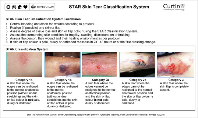
Silver Chain Nursing Association, Curtin University of Technology (School of Nursing and Midwifery). (revised 2010) STAR (Skin Tear Audit Research) skin tear classification system. Online. Available at www.silverchain.org.au/assets/files/STAR-Skin-Tear-tool-04022010.pdf, 2007. 20 Apr 2011.
Terrill P. Management of skin grafts and donor sites. Woundcare Network. 2003;11:1–5.
Traversa B, Sussman G. The role of growth factors, cytokines and proteases in wound management. Primary Intention. 2001;9:161–167.
Whitney A, Phillips L, Aslam R. Guidelines for the treatment of pressure ulcers. Wound Repair Regen. 2006;14:663–679.
Williams D, Enoch S, Miller D, et al. Effect of sharp debridement using curette on recalcitrant nonhealing venous leg ulcers: a concurrently controlled, prospective cohort study. Wound Repair Regen. 2005;13:131–137.
World Union of Wound Healing Societies (WUWHS). Principles of best practice: minimising pain at wound-dressing-related procedures. A consensus document. London: Medical Education Partnerships, 2004.
World Union of Wound Healing Societies (WUWHS). Principles of best practice: wound exudate and the role of dressings. A consensus document. London: Medical Education Partnerships, 2007.
Xue Y. Evidence summary: wound dressings. JBI COnNECT. Available at: http://www.jbiconnect.org/connect/docs/jbi/cis/connect-gen-user-view.php?IID=723&qu=1&p=1&e=1&r=1&o=1, 2008.
Young T, Fowler A. Nursing management of skin grafts and donor sites. Br J Nurs. 1998;7:324–334.
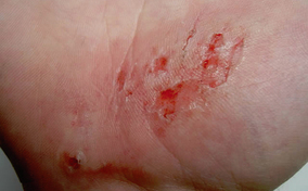
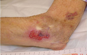 Skin tear
 Decreased tissue turgor
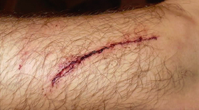
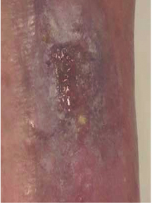
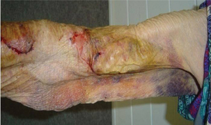 Loss of subcutaneous tissue
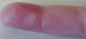


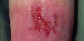
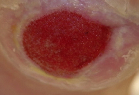
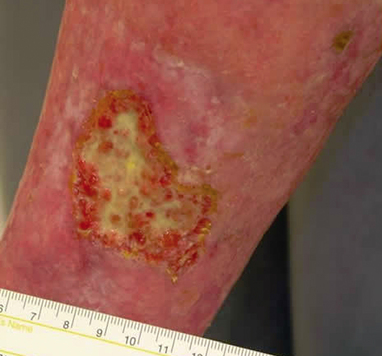
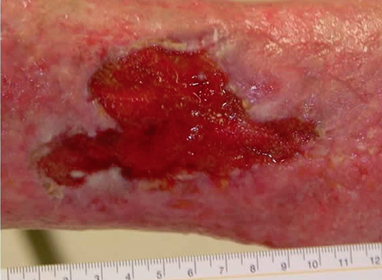
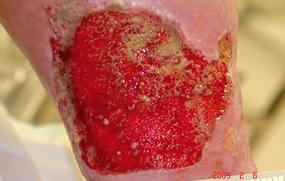 Bumpy granulation tissue
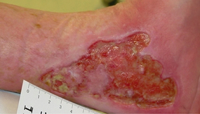 Contraction of wound edges and islands of new epithelial tissue
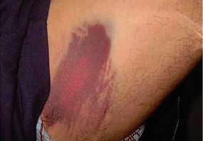
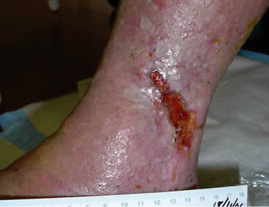

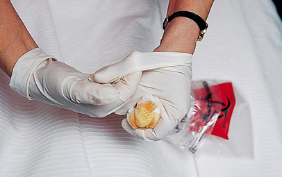 Step 8 Remove gloves over contaminated dressing