Chapter 41 Pain management
Mastery of content will enable you to:
• Define pain and other key terms.
• Review key pain models, including the biopsychosocial model of pain.
• Describe the key neural mechanisms of pain.
• Describe some key psychosocial contributors of pain.
• Perform a nursing assessment of a client experiencing pain.
• Apply a rational and evidence-based approach to pain management.
• Explain the various pharmacological approaches to treating pain.
• Describe interventions for preventing and treating opioid-related side effects.
• Describe the use of non-pharmacological pain interventions.
• Describe advanced pain techniques and the nursing responsibilities when they are used.
Regardless of the setting in which they work, nurses are responsible for the assessment and management of clients with pain. Pain is a key consideration in all patient care, and nurses play a critical role in pain management. Indeed, throughout history nurses have made important contributions to our understanding and management of pain through research and clinical practice. This chapter aims to provide an introduction to some key concepts in pain management and to encourage you to reflect on some of your own assumptions about pain.
Effective pain management requires knowledgeable and skilled practitioners. Nurses are uniquely positioned to improve patient outcomes through effective pain assessment and management, and yet there is ample evidence over several decades of poor pain assessment and under-treatment of patients in pain (Pasero and McCaffery, 2011). Despite the rapidly evolving scientific knowledge base for pain management, the data suggests that patients in pain remain poorly managed. Why might this be so?
Nelson (2007) argues that part of the problem may be that nurses view scientific and technical knowledge of pain management as less important than the human and relational skills that they bring to the client encounter. Yet in failing to engage with the science of pain, nurses risk becoming a key contributor to poorly managed pain and its sequelae, including chronic pain. Moreover, when nurses do not possess a scientific knowledge base of pain assessment and management, they are unable to participate fully as members of the interprofessional team, which is so important to collaborative pain management.
As a neurosurgical nurse I’ve got plenty to do, including helping patients with some of the things some doctors don’t do well—one of which is effectively managing patients’ pain. Let me give you an example. I recently took care of a patient whose surgeon insisted that he didn’t need much nursing care. The patient had been operated on for a herniated disc and had only been out of surgery for 1 day. Interestingly, the patient himself was a senior doctor, a gastroenterologist who’d come to our hospital and unit because of the spine surgeon’s reputation.
As soon as I began to take care of him, the patient-doctor asked me to tell him about the normal course of postoperative recovery for this kind of operation. I told him we usually discharged patients 2 days after their surgery and that we gave them a prescription for pain medication. He was very concerned about pain meds because he’d been in unbearable pain before his surgery and took pain pills daily. After the operation he continued to take these pills, but was still complaining about his pain. Plus, he had a lot of pain that we hadn’t expected from the wound site.
When his well-respected surgeon came in to do a quick exam, the patient told him that he was in pain. Although this surgeon is very competent and self-confident, pain management is not one of his strengths. He just doesn’t understand that pain is an exquisitely personal experience and that each patient experiences it differently.
So what was the doctor’s response to this particular patient? He insisted the patient was somehow ‘imagining’ his pain, not really having it. The surgeon told the patient that he’d checked on his surgery, all was well, and he should no longer be in pain. So even though I spoke with him about the patient’s pain, he didn’t come up with a different pain management strategy.
Not surprisingly, when it came time for his discharge two days later, the patient was still in so much pain that he couldn’t leave the hospital. When the surgeon and a medical student made their rounds that day, the patient again asked them about his pain. Once again, they insisted that the patient’s pain could not be as severe as he said it was.
By now, I’d been with the patient over several days. I watched how he walked and knew how uncomfortable he was. I knew I had to do something to manage his pain and recognised that ordinary pain-killers just wouldn’t work.
The first thing I did was talk to the patient. In a careful tone, I said: ‘Look, Dr X, your surgeon is very knowledgeable and competent. Most of the time patients don’t have a lot of pain. But I know from experience that the kind of inflammatory pain you’re having doesn’t resolve within the first day or two. What you need is some cortisone prescribed in a single dose in the morning for a few days. I have seen this work in other cases with worse pain than yours.’
The patient-doctor thought about this. He thanked me for the suggestion and asked me to talk to the surgeon and suggest that he order the medication. I waited until the surgeon was out of the operating room and talked with him. He’d become so frustrated with the patient that he quickly agreed. He ordered the medication, and I gave the cortisone to the patient. Pretty soon the patient was comfortable and appeared not only relaxed but serene. He approached me in the hall and said, ‘I want to thank you so much for your patience, your availability and most of all your competence and professional attitude you showed me these past days. If it hadn’t been for you, I would still be in pain. I would have been suffering the same pain that made my life intolerable and led me to have this surgery in the first place. I want to thank you for dealing with this and all the needs I’ve had since I came to the hospital.’
Nurses do these kinds of things every day. Isn’t it about time we advertised them?
From Gordon S editor 2010 When chicken soup isn’t enough: stories of nurses standing up for themselves, their patients, and their profession. New York, Cornell University Press.
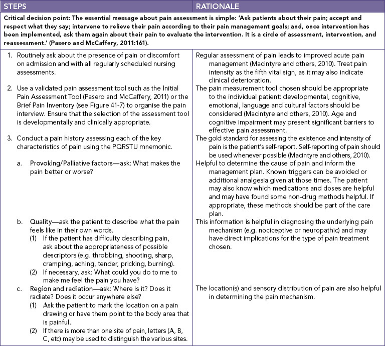
Read the following clinical example and think about how the registered nurse (RN) draws on her knowledge and experience to advocate for the client and effectively manage his pain.
The most oft-cited definition of pain in the literature and accepted by the International Association for the Study of Pain (IASP; Merskey, 1986:51) is:
Pain is an unpleasant sensory and emotional experience associated with actual or potential tissue damage or described in terms of such damage.
This definition underscores the inherent subjectivity of pain and acknowledges the importance of emotional as well as sensory factors in the pain experience. Moreover, it highlights that although pain is usually considered as a warning signal of actual or potential tissue damage, pain can occur in the absence of tissue damage, even though the experience may be described as if the damage has occurred.
The most widely accepted and clinically useful definition of pain, however, was developed by a leading nursing expert on pain, Margo McCaffery (1968:95):
• CRITICAL THINKING
Take a minute to think about McCaffery’s clinical definition of pain. Do you agree with this definition? Who is the authority about a client’s pain? Whose pain is it?
In evolutionary terms, acute pain can be understood as an important biological protective mechanism to warn the body of injury or disease. It directs immediate attention to the situation, promotes reflexive withdrawal and fosters other actions that prevent further damage and enhance healing. Acute pain usually stops long before healing has occurred, which may take days or a few weeks.
Acute pain seriously threatens a client’s recovery and should be one of the priorities in the client’s care. Acute postoperative pain delays healing and leads to systemic complications (see Table 41-1). Rehabilitation may be delayed and hospitalisation may be prolonged if acute pain is not controlled. Moreover, unrelieved acute pain can lead to the development of chronic pain problems.
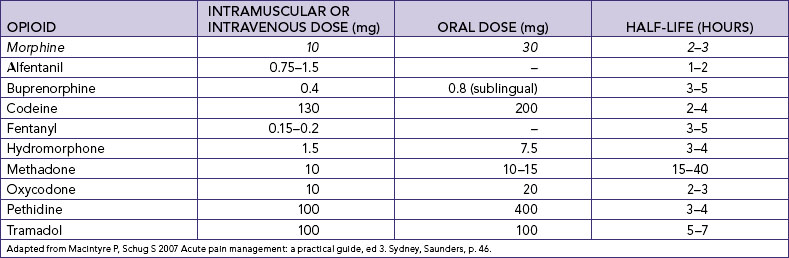
TABLE 41-1 THE HARMFUL EFFECTS OF UNRELIEVED PAIN
DOMAINS AFFECTED | SPECIFIC RESPONSES TO PAIN |
---|---|
Endocrine | ↑ Adrenocorticotrophic hormone (ACTH), ↑ cortisol, ↑ antidiuretic hormone (ADH), ↑ adrenaline, ↑ noradrenaline, ↑ growth hormone (GH), ↑ catecholamines, ↑ renin, ↑ angiotensin II, ↑ aldosterone, ↑ glucagon, ↑ interleukin-1, ↓ insulin, ↓ testosterone |
Metabolic | Gluconeogenesis, hepatic glycogenolysis, hyperglycaemia, glucose intolerance, insulin resistance, muscle protein catabolism, ↑ lipolysis |
Cardiovascular | ↑ Heart rate, ↑ cardiac workload, ↑ peripheral vascular resistance, ↑ systemic vascular resistance, hypertension, ↑ coronary vascular resistance, ↑ myocardial oxygen consumption, hypercoagulation, deep-vein thrombosis |
Respiratory | ↓ Flows and volumes, atelectasis, shunting, hypoxaemia, ↓ cough, sputum retention, infection |
Genitourinary | ↓ Urinary output, urinary retention, fluid overload, hypokalaemia |
Gastrointestinal | ↓ Gastric and bowel motility |
Musculoskeletal | Muscle spasm, impaired muscle function, fatigue, immobility |
Cognitive | Reduction in cognitive function, mental confusion |
Immune | Depression of immune response |
Developmental | ↑ Behavioural and physiological responses to pain, altered temperaments, higher somatisation, infant distress behaviour; possible altered development of the pain system, ↑ vulnerability to stress disorders, addictive behaviour and anxiety states |
Future pain | Debilitating chronic pain syndromes: postmastectomy pain, post-thoracotomy pain, phantom pain, post-herpetic neuralgia |
Quality of life | Sleeplessness, anxiety, fear, hopelessness, ↑ thoughts of suicide |
↓ = decreased; ↑ = increased.
From Pasero C, McCaffery M 2011 Pain assessment and pharmacologic management. St Louis, Mosby, p. 11. Data from Cousins M 1994 Acute postoperative pain. In Wall PD, Melzack R, editors, Textbook of pain, ed 3. New York, Churchill Livingstone; Kehlet H 1998 Modification of responses to surgery by neural blockade. In Cousins MJ, Bridenbaugh PO, editors, Neural blockade. Philadelphia, Lippincott-Raven; Mcintyre PE, Ready LB 1996 Acute pain management: a practical guide. Philadelphia, Saunders.
• CRITICAL THINKING
A 50-year-old man is under your care on a surgical ward after a fall that resulted in multiple rib fractures. He has an intercostal catheter to correct a pneumothorax. His respirations are shallow and he cannot cough effectively. Think about further assessment issues you might consider, given that he now has a temperature of 39°C. What complications could poorly controlled acute pain cause in this situation? How could this be prevented?
In contrast to acute pain, chronic pain persists constantly or intermittently past the normal time of healing and serves no biological purpose. The terms chronic, chronic non-malignant and chronic benign pain are all found in the literature. Examples of chronic non-malignant pain include arthritis, low back pain, myofascial pain, headache and peripheral neuropathy.
Chronic pain is a major cause of physical and psychosocial disability, leading to problems such as loss of employment, inability to perform daily activities, mood disorders and social isolation from family and friends. The broader economic impact of chronic pain on the Australian community is also significant, costing approximately $34.3 billion annually (Access Economics, 2007).
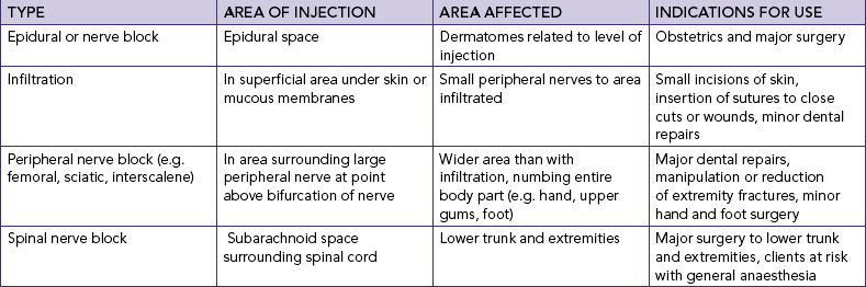
Although pain is often classified by its duration, another common way to categorise pain problems is by their underlying mechanisms. Nociceptive (physiological) pain is sustained by ongoing activation of the sensory system that subserves the perception of noxious stimuli. It implies the existence of damage to somatic or visceral tissues sufficient to activate the nociceptive system.
Neuropathic (pathophysiological) pain is sustained by injury or dysfunction of the peripheral or central nervous system (CNS). Neuropathic pain problems are common in diseases affecting the nervous system, such as diabetes mellitus, herpes zoster and multiple sclerosis. It may also result from surgery and/or trauma to nervous tissue.
Philosophical debate concerning the nature of pain has existed for thousands of years. Historically, pain has been dichotomised as either purely a psychological or a sensory phenomenon. For example, Aristotle (384–322 BC) viewed pain as an emotion, and the Stoic philosophers taught that pain could be overcome by ‘rational repudiation’ through logic and reasoning (Turk and Holzman, 1986). In contrast, pain has been understood as a specific sensation, having a direct relationship to the degree of noxious sensory stimuli. The latter has been the dominant conceptualisation of pain and has informed clinical practice over the last several hundred years.
The basic ideas of this sensory–physiological view of pain are most often attributed to the work of 17th-century philosopher René Descartes (1596–1650). Descartes attempted to explain pain in purely mechanistic terms using the scientific knowledge base of the time. Cartesian physiology held that nerves were hollow tubes that functioned with the flow of ‘animal spirits’ and contained fine filaments or threads that reached from the peripheries to the brain (Benini and DeLeo, 1999). Descartes argued in 1664 that the experience of the pain pathway was analogous to two ends of a direct communication line, ‘just as by pulling at one end of a rope [peripheral nerves] one makes to strike at the same instant a bell which hangs at the other end [brain]’ (cited in Melzack and Wall, 1996:150). To elaborate on this idea, he offered the analogy of a foot coming into contact with fire (see Figure 41-1). He proposed that a flame sets particles in the foot into activity and the motion is transmitted up the leg and back and into the head where pain becomes conscious (through the pineal gland) and elicits a reflexive response (Melzack and Wall, 1996). Thus, Descartes’ conception of pain was that of a straight-through channel from the skin to the brain. It assumed a direct and invariant relationship between stimulus intensity and perceived pain.
FIGURE 41-1 Descartes’ 1664 model of pain.
From Melzack R, Wall PD 1996 The challenge of pain, ed 2. London, Penguin Books, p. 150.
The legacy of biomedical reductionism inherited from Cartesian theory has had a profound impact on the way pain has been conceptualised and treated over at least the past three centuries. Modern pain theorists (19th and early 20th centuries) embraced this new paradigm to the extent that the neurophysiological basis for pain perception became the dominant model across disciplines and was often taught as fact rather than theory (Melzack and Wall, 1996).
The first of these theories to reach significant popularity was the specificity theory of pain as advocated by Müller (1842) and von Frey (1894). Specificity theory hypothesises that pain is a primary sensation like olfaction or taste. It assumes that there is a specific pain modality that can be stimulated only by painful stimuli, and therefore that these impulses are always felt specifically as pain and only pain. Essentially, it postulates that ‘pain receptors’ (designated as free nerve endings) generate pain impulses that are carried by specific ‘pain fibres’ (e.g. A-delta and C fibres) in peripheral nerves to the spinal cord. These impulses synapse in the spinal cord and travel via a ‘pain pathway’ (e.g. the spinothalamic tract) to a ‘pain centre’ in the brain (e.g. the thalamus or sensory cortex) where they are perceived as pain. Thus, pain is perceived as having specific central, as well as peripheral, mechanisms similar to other bodily senses. Such a model is clearly similar to the conceptual nervous system described by Descartes centuries earlier.
Based on extensive laboratory evidence, the basic assumptions of specificity theory are now viewed as wholly oversimplified. In addition to a lack of clear scientific support, specificity theory has also been unable to account for the wide variability of individuals’ subjective experiences and responses to pain and its treatment. As Turk and Flor (1999:19) have cogently argued, ‘pain in the absence of pathology, pathology in the absence of pain, individual differences in response to identical treatments, failure of neurosurgical procedures and potent analgesic agents to consistently eliminate pain, and the low association between impairment and disability fail to conform to a model of pain that presumes a direct transmission from the periphery to central nervous system structures.’
• CRITICAL THINKING
It is easy to assume that pain severity is closely tied to the amount of tissue damage based on our personal experiences of pain, but can you think of exceptions to this idea? Can you think of any examples of injury without pain, or vice versa? Why might two clients having the same surgery report different pain severity postoperatively?
In response to the inadequacies of specificity theory, several other conceptualisations of pain were proposed. Together these are grouped as variants of the pattern theory of pain, originating from the work of Goldscheider (1894). The basic premise of these models is that the transmission of nerve impulse patterns (spatial and temporal) which are produced and coded by receptors are the primary determinants of pain. Thus, there was a rejection of the earlier emphasis on identifying physiological structures in favour of defining sensory patterns or the balance of sensory input as the cause of pain. Peripheral pattern theorists (e.g. Sinclair, 1955; Weddell, 1955) suggested that, for example, all nerve fibre endings are similar (apart from those that innervate hair cells) and that pain occurs when particular spatial or temporal patterns of impulses are generated in afferent nerves by intense stimulation of non-specific receptors. None of these models, however, provided an adequate general theory of pain. Psychological aspects, when considered, were dismissed as reactions to pain.
Based on a critique of existing models, Melzack and Wall (1965) formulated their revolutionary gate-control theory of pain. The original proposition was that pain is not simply the net result of defined peripheral input. Rather, Melzack and Wall drew on converging lines of evidence to establish that pain perception is a dynamic, multidimensional experience involving psychophysiological mechanisms. According to gate-control theory, ‘the experience of pain is an ongoing sequence of activities, largely reflexive in nature at the outset, but modifiable even in the earliest stages by a variety of excitatory and inhibitory influences, as well as the integration of ascending and descending CNS activity’ (Turk and Monarch, 2002:4). Thus, from the gate-control perspective, pain perception is considered a complex interactive product of afferent sensory activity and central (psychological) processes. It was this radical shift in thinking about pain perception that is perhaps the most important contribution of the gate-control theory.
The fundamental and most original feature of the gate-control theory is the proposed neural gating system, postulated to exist in the dorsal horns of the spinal cord which modulates the transmission of nociceptive information from the periphery to the CNS on the basis of the diameters of active peripheral fibres, as well as the dynamic action of brain processes (see Figure 41-2). Specifically, the gate-control theory is based on the following hypotheses (Melzack and Wall, 1965, 1982):
• The transmission of nerve impulses from afferent fibres to spinal cord transmission cells (coined ‘T cells’) is modulated by a ‘spinal gating mechanism’ in the substantia gelatinosa of the dorsal horn.
• The spinal gating mechanism is influenced by the relative amount of activity in large-diameter (A-beta) and small-diameter (A-delta and C) fibres, so that activity in large fibres tends to inhibit transmission (i.e. ‘closes the gate’) whereas small-fibre activity tends to facilitate transmission (i.e. ‘opens the gate’).
• The spinal gating mechanism is also influenced by nerve impulses that descend from the brain.
• A specialised system of large-diameter, rapidly conducting fibres (the ‘central control trigger’) activates selective cognitive processes (e.g. meaning of pain) that then influence, by way of descending fibres, the modulating properties of the spinal gating mechanism. Thus, the central control system could modulate the spinal gate and thereby selectively alter certain afferent sensory patterns before they reach the brain.
• When the output of the spinal cord T cells exceeds a critical level, it activates the ‘action system’ or those areas of the brain that are responsible for the behavioural responses to pain and the perceptual experience of pain itself.
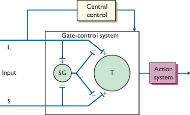
FIGURE 41-2 Schematic model of the gate-control theory.
L = large-diameter fibre; S = small-diameter fibre; SG = spinal gating mechanism; T = spinal cord transmission cell.; Redrawn from Melzack R, Wall PD 1965 Pain mechanisms: a new theory. Science 150:971.
• CRITICAL THINKING
Take a minute to think about the ideas proposed by the gate-control theory. How might it explain why rubbing an injured area on the body helps to reduce pain, or how a sportsperson can continue a game after serious injury yet report no pain?
Shortly after first publication, the theory was expanded upon by Melzack and Casey (1968) to describe the neural systems beyond the gate. The extended theory differentiated three systems related to the processing of nociceptive information, all of which are argued to contribute to the subjective experience of pain. Melzack and Casey suggested that the neospinothalamic projecting system serves to process the sensory–discriminative dimension of pain, such as location, intensity and nature of the stimulus, whereas impulses that course through the paleospinothalamic tract and paramedial ascending system activate reticular and limbic structures that provide the motivational–affective response to pain, such as aversive drive and unpleasant affect. The neocortical or higher CNS processes were predicted to further influence the sensory and motivational systems through cognitive–evaluative appraisal of input in terms of attention, meaning, past experience, and so on. Because of the continuous interactive nature of these parallel systems, considerable potential for shaping the pain experience is implied (Turk and Monarch, 2002).
The neuroanatomical and physiological assumptions of the gate-control theory have since been challenged, and it has been suggested that the theory is incomplete (e.g. see Sufka and Price, 2002). While additional mechanisms have been suggested to revise the theory in order to account for chronic pain (Melzack and Wall, 1996), Melzack (1996, 1999) himself points out that the gate-control theory still fails to explain certain phenomena such as phantom-limb pain. Melzack’s most recent work has been to shift the focus of pain theory from the peripheries and spinal mechanisms to understand how the brain creates the experience of pain in the proposed neuromatrix theory of pain.
One of the most useful contemporary models for nursing practice which brings together much of what we know about pain is the biopsychosocial model of pain. The biopsychosocial model highlights the multidimensional nature of pain, attending to the multiple factors that both contribute to and are affected by pain. Although the gate-control theory provided an integrative model of pain perception, it focused primarily on the neurophysiology of pain. The biopsychosocial model, in contrast, builds on this model by also emphasising the influence of psychosocial and behavioural components of pain (Turk and Monarch, 2003).
The conceptual view of the biopsychosocial model of pain is presented in Figure 41-3. This nested circles model demonstrates the interdependent relationships among processes that culminate in the person’s perception of pain and overt pain behaviours. Biological factors may initiate and maintain nociceptive input, psychological factors influence the appraisal and perception of pain, and social factors shape the person’s behavioural responses.
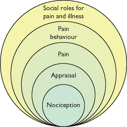
FIGURE 41-3 Biopsychosocial model of pain.
From Turk DC, Burwinkle TM 2006 Coping with chronic pain. In Carr A, McNulty M, editors, The handbook of adult clinical psychology: an evidence-based practice approach. London, Routledge, p. 647.
Following this biopsychosocial model, this chapter now discusses some of the key physical and psychosocial contributors to the pain experience.
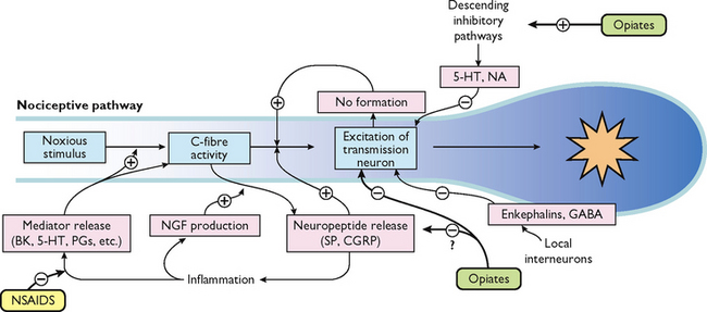
What is presented here is a brief overview of the normal neural mechanisms involved in the pain experience, called nociception. Between the stimulus of tissue injury and the subjective experience of pain is a series of complex electrical and chemical events. To understand this, it is useful to conceptualise four distinct physiological processes involved: transduction, transmission, perception and modulation (see Figure 41-4).
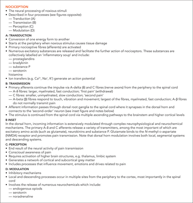
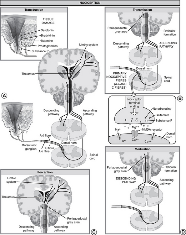
FIGURE 41-4 Nociception: ‘normal’ pain transmission.
From Pasero C, McCaffery M, editors, 2011 Pain assessment and pharmacologic management. St. Louis, Mosby, p. 5.
Transduction refers to the processes by which noxious stimuli activate nociceptors, which are primary afferent neurons that exist throughout the body (skin, subcutaneous tissue and visceral or somatic structures). Nociceptors have free nerve endings which respond selectively to mechanical (e.g. incision, tumour growth), thermal (e.g. burn, frostbite) or chemical (e.g. toxins, chemotherapy) stimuli, which are converted into a neuronal action potential.
Tissue damage and inflammation also cause the release of chemical mediators from injured cells, inflammatory cells (e.g. mast cells, lymphocytes) and primary afferent neurons which lower the excitability threshold of nociceptors and sensitise them to further sensory input. A partial list of these chemicals includes bradykinin, prostaglandins, serotonin, histamine and substance P, known collectively as the ‘inflammatory soup’. These chemicals stimulate or sensitise the nociceptors to send impulses to the spinal cord.
Transmission of action potentials generated from effective transduction occurs along two types of nociceptor fibres towards the CNS. A-delta fibres are thinly myelinated, large-diameter, fast-conducting fibres (5–30 metres/second), which transmit well-localised sensations of sharp pain. C fibres are unmyelinated, small-diameter, slow-conducting fibres (0.5–2 metres/second), which transmit pain that is poorly localised, dull and aching. Other types of sensory fibres such as A-beta fibres do not usually transmit nociceptive information, but respond to touch, movement and vibration.
For example, after spraining an ankle a person initially feels a sharp, localised pain, which is a result of A-delta fibre transmission. Within a few seconds, the pain becomes more diffuse and widespread, until the whole foot aches because of C-fibre activation. The C fibres remain exposed to the chemicals released when cells are damaged.
Transmission continues along the primary afferent fibres until they synapse in the dorsal horn of the spinal cord. Within the dorsal horn, neurotransmitters (such as glutamate, neurokinins and substance P) are released, causing a synaptic transmission from the primary afferent neurons to second-order neurons in the spinal cord which project to the brainstem and thalamus via multiple ascending pathways. Of these pathways, the spinothalamic and spinoreticular tracts are particularly important. These second-order fibres cross the midline and ascend in the anterolateral quadrant of the spinal cord.
Perception is the point at which a person is consciously aware of pain. It requires the activation of higher brain structures, presumably including the cortex. It involves both awareness and related cognitions and the occurrence of emotions and drives associated with pain (Pasero and Portenoy, 2011).
The mechanisms of pain perception remain poorly understood. From the thalamus, third-order neurons transmit impulses to various areas of the brain. However, the exact location in the brain where pain becomes a conscious experience remains unclear. Studies using functional magnetic resonance imaging (fMRI) have consistently demonstrated involvement of the thalamus, primary and secondary somatosensory cortex (S1, S2), insula, forebrain and anterior cingulate cortex (ACC) during painful stimulation. Data suggests that these areas process different aspects of pain. For example, while the perception of sensory–discriminative aspects (e.g. intensity, quality, location and pattern) is associated with the S1 and S2 cortices, the ACC and insula seem to be associated with the affective–motivational (e.g. fear, anxiety) processing of pain (Apkarian and others, 2005).
Modulation (inhibition or facilitation) of afferent input can also occur anywhere along nociceptive pathways, particularly in the spinal cord as suggested by the gate-control theory discussed earlier. When activated, descending efferent pathways originating in the brain release a number of chemicals capable of altering the processing of nociceptive input. Of the descending inhibition systems, the periaqueductal grey (PAG) region is particularly important; this projects to the dorsal horn of the spinal cord.
These descending pathways release substances such as endogenous opioids (endorphins and encephalin), serotonin and noradrenaline which can inhibit the transmission of noxious stimuli and produce analgesia. For example, when released in the spinal dorsal horn these substances can bind to receptors to prevent the release of neurotransmitters from primary afferent nociceptors such as substance P to second-order neurons, thus blocking transmission of the impulse.
• CRITICAL THINKING
The discussion so far has been about the normal physiological processes involved in nociceptive pain, but what mechanisms can explain the development of pathophysiological pain problems such as chronic inflammatory and neuropathic pain? A key development in our understanding of pain mechanisms is the concept of neuroplasticity of the nervous system, which refers to the ability of neurons throughout the peripheral and central nervous systems to change their structure and function because of nociceptive input. Explore what the terms peripheral sensitisation and central sensitisation mean and how they might contribute to the pathophysiology of acute and chronic pain problems.
Tracing the physiological processes involved in pain perception from peripheral receptor to cortex, it is easy to assume a simple and reproducible relationship among stimulus intensity, nociceptive transmission cell activity and the perceived intensity of pain. However, despite its intuitive appeal, a physiological understanding of pain is still incomplete. As discussed above, evidence clearly demonstrates there is no one-to-one relationship between tissue injury and the experience of pain.
It is useful here to make the distinction between nociception and pain. Nociception is a physiological phenomenon that involves activation of sensory transduction in nerves by thermal, mechanical or chemical energy impinging on specialised nerve endings. The nerves involved convey information about tissue damage to the CNS, but such sensations are not yet considered pain until they are subjected to higher-order psychological and cognitive processing that involves appraisals. Hence, nociception is a sensory process with nociceptive stimuli capable of producing pain. Pain, however, is a subjective, perceptual experience that results from the nociceptive input and is modulated on a number of different levels in the CNS (Turk and Monarch, 2002). Thus, nociception may be necessary for pain to occur but it is not sufficient to account for pain as a clinical phenomenon, which is always a perceptual experience.
The discussion now considers the important role of some key psychosocial factors in shaping the pain experience.
Psychological, environmental and behavioural factors play a central role in the experience of pain. Together they shape the way an individual makes sense of and gives meaning to pain. They also influence the way in which the individual will cope with pain and the extent to which it will interfere with the roles and responsibilities of daily life. They are therefore important considerations in the assessment of acute and chronic pain and holistic planning for client care.
• CRITICAL THINKING
Consider the following: ‘Here is perhaps the most difficult thought to accept about pain. We experience pain only and entirely as we interpret it. It seizes us as if with an unseen hand, sometimes stopping us in mid-sentence or mid-motion, but we too capture and reshape it’ (Morris, 1991:29).
The age and developmental stage of a client is an important variable that influences the experience of pain, particularly in children and older adults. Unfortunately, these groups are also at risk for inadequate pain management because of common misconceptions about pain perception, appropriate pain assessment and effective and safe use of analgesics (see Table 41-2).
MISCONCEPTION | CORRECTION |
---|---|
Infants are incapable of feeling pain | Infants have developed the anatomy and physiology for pain processing by mid to late gestation |
Infants are less sensitive to pain than older children and adults | |
Infants are incapable of expressing pain | Although infants cannot verbalise pain, they demonstrate it with observable cues, both behavioural and physiological |
Infants must learn about pain from previous painful experiences | Pain requires no prior experience; it need not be learned from earlier painful experience. Pain is present from the first injury |
Pain cannot be accurately assessed in infants | Behavioural cues (i.e. facial expressions, crying, body movements) and physiological indicators of pain can be reliably and validly assessed either alone (univariate approach) or in combination (multivariate approach). The most valid univariate approach is facial expression. The most valid multivariate approach is through the use of a composite pain measure |
Infants are incapable of remembering pain | Studies have shown that infants who are exposed to painful stimuli at an early age respond more vigorously to subsequent painful stimuli |
Analgesics and anaesthetics cannot be safely given to infants and neonates because of their immature capacity to metabolise and eliminate drugs and their sensitivity to opioid-induced respiratory depression | Infants older than 1 month of age metabolise drugs in the same manner as older infants and children. Careful selection of the agent, dosage, administration route and time, frequent monitoring for desired and undesired effects and drug titration and weaning can minimise the adverse effects of opioids and non-opioids for pain management in neonates |
Adapted from McCaffery M, Pasero C 1999 Pain: clinical manual, ed 2. St Louis, Mosby.
Young children have difficulty understanding pain and the procedures nurses administer that may cause pain. Young children who have not developed full vocabularies also have difficulty describing and expressing pain to parents or caregivers. Cognitively, toddlers and preschoolers are unable to recall explanations about pain, or associate pain with experiences that can occur in various situations. With these developmental considerations in mind, the nurse must adapt approaches for assessing a child’s pain (including what to ask and the behaviours to observe for) and how to prepare a child for a painful procedure.
While older adults may be at greater risk of having painful conditions, pain is not an inevitable part of ageing. Once an older client suffers pain, there can be serious impairment of functional status. Mobility, activities of daily living, social activities outside the home and activity tolerance can all be reduced. The presence of pain in the older adult requires aggressive assessment, diagnosis and management.
Chronic pain is a problem for many elderly people living in both community and extended-care facilities. When they are admitted to hospital, they often have acute pain superimposed on chronic pain problems. There are also many myths about pain in older adults that need to be corrected in order to provide effective nursing care (see Table 41-3).
TABLE 41-3 PAIN IN OLDER ADULTS
Adapted from McCaffery M, Pasero C 1999 Pain: clinical manual, ed 2. St Louis, Mosby.
The ability of older clients to interpret pain can be complicated by the presence of multiple diseases with vague symptoms that may affect similar parts of the body. It is important to take enough time to complete a thorough assessment of pain and pain-related concerns.
Considerable evidence suggests that women and men differ in their experience of pain, as well as in their response to pain treatments (Fillingim, 2009). Women are at greater risk of a variety of chronic pain conditions such as headache, facial pain, abdominal pain and musculoskeletal pain (LeResche, 1999; Unruh, 1996). Experimental evidence also suggests that women may be more sensitive to pain, less tolerant of pain and more able to discriminate among different levels of pain than men (Riley and others, 1998). Interestingly, although women were traditionally excluded from many clinical trials examining the efficacy of drugs including analgesics, recent evidence suggests there may be important sex differences. A recent systematic review, for example, demonstrated sex differences for morphine analgesia in both experimental pain studies and clinical patient-controlled analgesia (PCA) studies, with greater morphine efficacy in women (Niesters and others, 2010).
Biological and psychosocial factors are both important in explaining these differences, yet more research is needed to elucidate the mechanisms underlying sex differences in pain. Hormonal and genetic factors may contribute to sex differences in pain perception and response to analgesics (Fillingim, 2009). Important psychosocial factors also include differences in cognitive appraisals, coping strategies and social role expectancies in response to pain (e.g. men encouraging and rewarding stoicism and punishing overt pain expression, whereas women are encouraged to be aware of pain and express it).
The multicultural nature of the Australasian population means that nurses will encounter a variety of responses to pain in their work environment. For example, Indo-Chinese clients may often be quite stoic, not requesting pain relief themselves. They may instead delegate a member of their family to approach the nurse for assistance. People of Middle Eastern origin may express pain through facial expression, body posture and moaning or soft cries. Some may believe, however, that pain is something to be endured to facilitate cleansing of the soul and for this reason may not seek analgesia. Filipino patients tend to focus their illness around their religious beliefs. Regardless of how bad their pain is, they tend to not complain and instead focus on praying (Schmidt, 2005).
Aboriginal and Torres Strait Islander people tend to be very orientated towards their families and are frequently not willing to share personal information with strangers. For this reason, these people may appear withdrawn and non-communicative until a level of trust and confidence has been established with hospital staff. The nurse again needs to be astute in assessing these clients in an effort to determine the presence and severity of pain as well as the most appropriate method of pain relief that may be implemented.
It is important to note that observed cultural differences are more likely to concern the way in which pain is regarded and the extent to which a person is permitted to express pain and under what conditions. These differences in pain behaviour are not evidence of differences in pain threshold and tolerance. They signify differences in the way people from different cultures have learned to behave when they are in pain.
While it is important that nurses are knowledgeable about cultural differences in response to pain, we need to be careful not to stereotype clients based on their cultural background. Although an individual may identify with a cultural group, there is also diversity within each culture.
Providing culturally safe care means being aware of your own cultural values and beliefs about pain and reflecting on how this might influence practice. The nurse, the client and the family must work together to facilitate communication about the assessment and management of pain. Finding a common assessment tool and communicating that tool to other healthcare providers is important.
• CRITICAL THINKING
Smithy is a 55-year-old Indigenous Australian who has a fractured femur resulting from the impact of a cow in the cattle yards. He is a long way from home and family members. As his female RN, you are having difficulty assessing his pain and he avoids eye contact with you.
What issues might you consider in terms of ensuring cultural safety and a positive nurse–client relationship?
The meaning that a person associates with pain affects the experience and impact of pain. This can be closely associated with the person’s cultural background. A person will perceive pain differently if it suggests a threat, loss, punishment or challenge. For example, a woman in labour will perceive pain differently from a woman with a history of cancer who is experiencing a new pain and fearing recurrence. The degree and quality of pain perceived by a client are related to the meaning of pain.
The relationship between pain and anxiety is complex. Anxiety often increases the perception of pain, but pain may also cause feelings of anxiety. Chapman and Turner (2001) report that studies have determined that arousal of the noradrenergic brain pathways is a major mechanism of anxiety and stress. The noradrenergic brain pathways are stimulated by biological (e.g. pain), psychological (e.g. fear) and psychosocial (e.g. isolation) threats.
Critically ill or injured clients, who often perceive a lack of control over their environment and care, may have high anxiety levels. This anxiety, if it has gone unnoticed in the high-tech environment of an intensive care unit, can lead to serious pain-management problems. The challenge is to relieve the pain in a client who is anxious in any setting (long-term care, acute care or home care). Although pharmacological and non-pharmacological approaches to the management of anxiety are appropriate, anxiolytic medications should not be a substitute for analgesia.
The degree to which a client focuses attention on pain can influence pain perception. Diverting attention away from pain perception reduces pain distress and increases pain tolerance in both acute and chronic conditions. This concept is one that nurses apply in various pain-relief interventions such as relaxation, guided imagery and massage. By focusing a client’s attention and concentration on other stimuli, the nurse places pain on the periphery of awareness.
Fatigue heightens the perception of pain. The sense of exhaustion intensifies pain and decreases coping abilities. This can be a common problem with any person experiencing chronic illness or who has fatigue as a result of treatment. If fatigue occurs along with sleeplessness, the perception of pain may be even greater.
As active processors of information, people with pain problems develop underlying beliefs, attitudes and assumptions in an attempt to make sense of their pain condition. These include attributions about the cause, meaning and appropriate treatment of pain, as well as perceptions of control over pain and personal coping efficacy (Thorn, 2004). Although certain beliefs may be adaptive and promote positive adjustment, others are likely to contribute to heightened pain, distress and disability. A growing body of research shows that pain-related beliefs are strongly associated with various measures of pain severity and physical and psychosocial functioning (Thorn, 2004). These beliefs, appraisals and expectancies held by individuals regarding the possible consequences of pain and their abilities to deal with them are hypothesised to affect functioning directly by influencing mood as well as indirectly by influencing coping efforts (Jensen and others, 1991).
Faced with chronic pain, individuals also learn and utilise a variety of strategies to help them cope or deal with their pain. Pain coping is defined as purposeful cognitive and behavioural efforts to manage or negate the negative impact of pain (Jensen and others, 1991). Much of the pain literature broadly classifies coping strategies as active or passive. In general, studies have found active coping strategies (efforts to function in spite of pain or to distract oneself from pain, such as activity or ignoring pain) to be associated with adaptive functioning, and passive coping strategies (withdrawal or surrendering control to an external source, such as resting or medication use) to be related to greater pain and depression (Boothby and others, 1999). Interestingly, although maladaptive strategies are strongly associated with negative outcomes, adaptive strategies generally show only modest correlations with positive outcomes (Geisser and others, 1999).
One specific maladaptive coping response that has consistently demonstrated robust associations with various measures of adjustment is termed catastrophising. It refers to ‘a method of cognitively coping with pain characterised by negative self-statements and overly negative thoughts and ideas about the future’ (Keefe and others, 1989:51). Greater endorsement of catastrophic thinking when in pain has been consistently associated with higher levels of pain, distress and disability among diverse populations (Keefe and others, 2004).
Research findings concerning the relationship between other specific pain coping strategies and adjustment to chronic pain have been somewhat more inconsistent (Boothby and others, 1999). When significant associations are found, greater use of coping self-statements (such as ‘I tell myself I can overcome the pain’) is generally related to positive adjustment, whereas praying/hoping, pain-contingent rest and wishful thinking are frequently associated with greater dysfunction. Ignoring pain, reinterpreting pain and distraction/diverting attention, in contrast, rarely predict functioning among people with chronic pain (Boothby and others, 1999).
Another factor that can significantly influence a person’s pain response is the presence and attitudes of significant others. People in pain often depend on family members or close friends for support, assistance or protection. Although pain still exists, the presence of a loved one can minimise loneliness and fear. An absence of family or friends can often make the pain experience more stressful. The presence of parents is especially important for children experiencing pain.
The social environment can also significantly contribute to adjustment to chronic pain. Pain behaviours which are initially adaptive (e.g. avoidance of activity believed to exacerbate pain) are subject to the influence of environmental or interpersonal factors so that over time these behaviours may come to be under the control of operant conditioning (Fordyce, 2001). For example, positive reinforcement of pain behaviour may inadvertently occur through solicitous attention from family members so that disability is maintained. In addition, negative reinforcement such as avoidance of a stressful work setting or unpleasant interpersonal obligations and reduction of pain through limiting activity may also contribute to persistence of pain behaviour.
Making sound clinical judgments require that the nurse anticipate what assessment data is needed, critically analyse the data and make appropriate decisions regarding client care. In the case of pain management, a scientific knowledge base of pain and evidence-based approaches to assessment and management allow the RN to safely and effectively intervene. Previous experience in caring for clients with pain also sharpens the nurse’s assessment skills and ability to choose effective therapies.
This clinical knowledge and judgment that underpins nursing practice is not always obvious to others—in fact, it is often invisible—and so others often attribute this work to the medical practitioner instead. Consider the following typical scenario described by Buresh and Gordon (2006:26).
A nurse working on the oncology floor at a major teaching hospital spends hours trying to get a medical intern to write an order for an opioid for a patient suffering from pancreatic cancer. The nurse has informed the intern that the patient is in excruciating pain. Far more familiar with cancer patients and their pain management than the intern, she recommends a course of intravenous (IV) morphine.
The intern refuses to order the opioid. He simply will not listen to the nurse. Over a period of several hours, she repeatedly engages with him, trying, to no avail, to teach him about cancer pain management. Finally, she corners a more senior resident who agrees with her and directs the intern to write the order. In the patient’s chart—the contemporary and historical record of the case—the nurse’s struggle with the young doctor is absent. Reading the chart, one would never know that the nurse was responsible for easing the patient’s pain and that the intern resisted her attempts to provide appropriate care. In this chart the new doctor is ‘credited’ with taking the action that reduced the patient’s suffering.
Not surprisingly, the patient and her family believe the doctor was her saviour. Several days later, the patient writes letters thanking her caregivers. She expresses her heartfelt gratitude to her attending doctor who, during her hospitalisation, rarely saw her when she was awake. She specifically thanked the intern whom she thought brought her relief. She did not thank her primary and associate nurses by name. She included only a general thank you to ‘the nurses’.
Buresh and Gordon (2006) argue that telling the patient that the doctor ‘ordered’ her morphine would conceal the nurse’s contribution. She couldn’t tell the patient that she’d fought with the doctor to get the morphine, but she could have negotiated the dilemma by saying: ‘I’ve consulted with the doctor, and he agreed with my recommendation to give you IV morphine. I will make sure it’s effective.’
The same process can occur as a student nurse—what may have seemed like a brief interaction between the RN and client before giving a dose of IV morphine was in fact a complex assessment of the client including their pain rating, level of sedation, vital signs and other assessment cues. This can be an invisible process to the student unless they take an active part in learning.
Making the most of clinical placements by seeking opportunities to develop pain assessment and management skills is important in the transition from student nurse to beginning practitioner. Use every opportunity on clinical placement to ask RNs how they make clinical judgments about pain management. What assessments did they perform to make decisions about client care? What was the reasoning behind their decision?
Before reading on, work through the clinical scenarios about pain assessment in Box 41-1.
BOX 41-1SURVEY—ASSESSMENT AND USE OF ANALGESICS: ‘ANDREW–ROBERT’
From McCaffery M, Pasero C 1999 Pain: clinical manual, ed 2. St Louis, Mosby.
Andrew is 25 years old, and this is his first day after abdominal surgery. As you enter his room, he smiles at you and continues talking and joking with his visitor. Your assessment reveals the following information: BP = 120/80, HR = 80, RR = 18 and on a scale of 0–10 (0 = no pain/discomfort, 10 = worst pain/discomfort) he rates his pain as 8.
1. On the patient’s hospital chart you must record his pain intensity on the scale below. Circle the number that you will record:
2. Your assessment, above, is made 2 hours after he received morphine 2 mg IV. Half-hourly pain ratings following the injection ranged from 6–8, and he had no clinically significant respiratory depression, sedation or other untoward side effects. He has identified 2 or less as an acceptable level of pain relief. His doctor’s order for analgesia is ‘morphine IV 1–3 mg q1h prn pain relief’. Mark the action you will take at this time:
___ No morphine now | ___ 1 mg IV now |
___ 2 mg IV now | ___ 3 mg IV now |
Robert is 25 years old, and this is his first day after abdominal surgery. As you enter his room, he is lying quietly in bed and grimaces as he turns in bed. Your assessment reveals the following information: BP = 120/80, HR = 80, RR = 18 and on a scale of 0–10 (0 = no pain/discomfort, 10 = worst pain/discomfort) he rates his pain as 8.
1. On the patient’s hospital chart you must record his pain intensity on the scale below. Circle the number that you will record:
2. Your assessment, above, is made 2 hours after he received morphine 2 mg IV. Half-hourly pain ratings following the injection ranged from 6–8, and he had no clinically significant respiratory depression, sedation or other untoward side effects. He has identified 2 or less as an acceptable level of pain relief. His doctor’s order for analgesia is ‘morphine IV 1–3 mg q1h prn pain relief’. Mark the action you will take at this time:
___ No morphine now | ___ 1 mg IV now |
___ 2 mg IV now | ___ 3 mg IV now |
BP = blood pressure (mmHg), HR = heart rate (beats per minute), IV = intravenous, q1h prn = every hour as needed; RR = respiratory rate (breaths per minute).
Probably the most challenging aspect of caring for the client with pain is to accept that pain is completely subjective. No objective measures of pain exist. Pain cannot be proved or disproved. While nurses sometimes may not believe what the client says about their pain, they have an ethical and professional responsibility to accept and act on it (Pasero and McCaffery, 2001). All reports of pain should be taken seriously. Yet, nurses and other healthcare professionals often have trouble accepting the client’s report of pain.
A client’s self-report of pain is the single most reliable indicator of the existence and intensity of pain (Macintyre and others, 2010). Thus, the gold standard for assessing the existence and intensity of pain is the client’s self-report, not the nurse’s opinion. No other source of information has ever been shown to be more accurate or reliable than what the client says. The client’s behaviours and vital signs, the opinions of nurses and doctors delivering care and the family’s estimates are not as reliable as the client’s report of pain and should never be used instead of what the client says.
The ‘Andrew–Robert’ scenario presented in Box 41-1 has been developed to demonstrate the necessity of accepting the client’s report of pain as the standard for assessment. Everything about the clients in the vignettes is the same except for their behaviour. If we accept that the client is the expert on their pain, then the correct answer is to record a pain rating of 8 for both clients in their charts. Also, given the information that the previous dose of morphine 2 mg IV was safe but ineffective, the correct answer is to administer morphine 3 mg IV for both clients. What we find when this survey is administered to students and RNs reflects the reality of clinical practice—nurses’ pain assessment and management decisions are often based on their personal opinions and biases rather than the client’s self-report.
A summary of findings of the ‘Andrew–Robert’ vignette administered to RNs between 1990 and 2006 have been published by the developers of the survey (McCaffery and others, 2007). These data show that while there has been a significant improvement over time, some nurses have not been taught or have not accepted that only the client’s own pain rating should be recorded when a self-report pain scale is used (see Table 41-4). Unfortunately, this trend towards improved pain assessment has not translated into providing an appropriate analgesic dose to clients with severe pain, and suggests nurses may still base their choices about opioid dose on the assumption that clients in pain should look like they are in pain. In the 2006 survey, approximately half of the nurses would allow a smiling client to remain in severe pain by failing to increase the dose and about a third of nurses would under-treat a grimacing client.
• CRITICAL THINKING
You are caring for 17-year-old Mark Lang who has had surgery for incision and drainage of an arm abscess and requires a dressing change. When you attempt to change the dressing, he screams in pain and refuses to allow you to proceed. You call the resident medical officer to organise analgesia, who cites the patient’s history of intravenous drug use and simply suggests paracetamol. Other nursing staff avoid the patient or attempt to control his expressions of pain with statements such as ‘Try to take control of yourself’, or ‘You’re going to have to learn to handle this yourself.’ How would you respond to this situation?
Accepting and acting upon the client’s report of pain is difficult if you doubt their credibility, which may happen on occasion. Since pain cannot be proved, healthcare professionals are vulnerable to being fooled by clients who lie about pain. Yet, as Pasero and McCaffery (2011) cogently argue, although accepting and responding to the client’s report of pain will occasionally result in analgesics being given to some clients who are addicted or malingering, it ensures that everyone who has pain receives the best possible pain management. Healthcare professionals do not have the right to deprive a client of appropriate assessment and treatment simply because they believe the client is lying.
The nurse is required to accept the client’s report of pain, convey this acceptance to the client and take the appropriate action. Doubts and opinions may persist, but they cannot be allowed to interfere with appropriate care. Pasero and McCaffery (2011) offer some excellent strategies, listed in Box 41-2, that you could use when difficulties arise with accepting the client’s self-report of pain.
BOX 41-2 STRATEGIES WHEN THE CLIENT’S REPORT OF PAIN IS NOT ACCEPTED
What do we do if the healthcare team does not respond positively to the client’s report of pain?
1. Acknowledge that everyone is entitled to a personal opinion, but personal opinion does not form the basis for professional practice.
2. Clarify that pain is subjective and cannot be proved or disproved.
3. Quote recommendations from clinical practice guidelines and research.
4. Ask: ‘Why is it so difficult to believe that this person hurts?’
From Pasero C, McCaffery M 2011 Pain assessment and pharmacologic management. St Louis, Mosby, p. 22.
The key to effective pain management is adequate pain assessment. Just as it is necessary to regularly monitor clients’ vital signs, pain should be considered an important routine assessment, along with temperature, pulse, respirations and blood pressure. In some institutions, initiatives to treat pain as the fifth vital sign were developed to encourage routine pain assessment, although research shows this has had little impact on actual clinical practice (e.g. Mularski and others, 2006).
• CRITICAL THINKING
Even when the client’s report of pain is sought by nurses, this process can be undermined by misconceptions that hamper the assessment and subsequent treatment of pain. Take the time to carefully review these common misconceptions (given in Table 41-5) and reflect on whether you hold any of these beliefs about pain.
TABLE 41-5 MISCONCEPTIONS: BARRIERS TO THE ASSESSMENT AND TREATMENT OF PAIN
From Pasero C, McCaffery M 2011 Pain assessment and pharmacologic management. St. Louis, Mosby, p. 17.
Elicitation of a pain problem should prompt a comprehensive pain assessment (see Skill 41-1, below) as well as the need to assess its potential impact on the client’s physical and psychosocial functioning. In doing so, nurses should incorporate standardised, reliable and validated instruments to obtain complete and reliable information.
The frequency of pain assessment and amount of information collected is obviously determined by the client’s situation. For example, on admission to the post-anaesthesia care unit (PACU) a client may only be able to tell you that their pain is ‘severe’ and further assessment may not be possible until they are more awake and pain has begun to be controlled with IV analgesia.
New-onset or increasing pain should always indicate the need for a focused physical assessment of the region or body systems involved. For example, surgical or other complications need to be ruled out for a postoperative client with severe pain despite analgesia. Review by an acute pain service should also be considered.
It is helpful to use a mnemonic to remember the key components of a basic pain assessment, such as the PQRSTU approach described in Box 41-3. More-comprehensive assessment of the person in pain is required for chronic and complex pain problems, which is beyond the scope of this chapter. The interested reader should consult Turk and Melzack’s (2011) excellent text for a comprehensive review of pain assessment tools.
BOX 41-3 MNEMONIC FOR PAIN ASSESSMENT: PQRSTU
P. Provoking/Palliative factors—What makes the pain better or worse?
Q. Quality—Describe what the pain feels like in your own words.
R. Region and radiation—Where is it? Does it radiate? Does it occur anywhere else? Have the client point to the body area that is painful.
S. Severity—Have the client rate their pain intensity using a pain rating scale. How much pain do you have at rest? How much pain do you have with movement/coughing?
T. Time of onset/duration—When did the pain begin? How long does the pain last?
U. Client’s Understanding—What is the client’s understanding of the cause of pain? What treatments have they tried to relieve it?
Pain intensity—a quantitative estimate of the severity or magnitude of perceived pain—is without a doubt the most salient dimension of pain, and a variety of pain rating scales have been developed to measure it. Most of these tools are highly correlated with each other and therefore they can be used in most situations (Jensen and Karoly, 2011). What is important is that the assessment tool is selected based on the client’s individual needs (e.g. developmental, cognitive, language and cultural factors) with consideration of the particular strengths and weaknesses of each tool. Where possible, the same tool should be used throughout the admission.
Although acute postoperative pain is often assessed when clients are resting, the effectiveness of analgesia is better determined by assessment of pain during functional activity required for recovery. In the postoperative client, this means the nurse should record pain-intensity ratings for both static (e.g. pain at rest) and dynamic (e.g. pain on coughing, deep breathing or movement) pain (Macintyre and others, 2010).
The numerical rating scale (NRS) is the most widely used measure of pain intensity in clinical practice. A NRS asks the client to rate their pain from 0 to 10 (an 11-point scale) or 0 to 100 (a 101-point scale), with the understanding that the 0 represents one end of the pain intensity continuum (‘no pain’) and the 10 or 100 represents the other extreme of pain intensity (‘pain as bad as it can be’). Clients simply state or circle the number on written versions of the scale that best represents their pain intensity by answering the question: ‘On a scale of 0 to 10, with 0 being no pain and 10 being the worst possible pain you could imagine, where would you rate the pain you are experiencing right now?’
The reliability and validity of the NRS is well established (Jensen and Karoly, 2011) and is easily administered. Based on their review of measures, Pasero and McCaffery (2011) recommend a combined NRS and faces scale as the preferred pain rating scale in most clinical settings (see Figure 41-5).
FIGURE 41-5 Combined NRS and faces scales.
From Pasero C, McCaffery M 2011 editors, Pain assessment and pharmacologic management. St Louis, Mosby.
A verbal descriptor scale (VDS) simply consists of a list of adjectives describing different levels of pain intensity. Clients are asked to read over the list of descriptors and choose the word that best describes their pain intensity on the scale. A simple and clinically useful example is ‘no pain’, ‘mild pain’, ‘moderate pain’ and ‘severe pain’ (scored numerically from 0 to 3).
The visual analogue scale (VAS) consists of a 10 cm horizontal line representing a continuum of pain intensity, with verbal descriptors at each end (e.g. ‘no pain’ to ‘pain as bad as it can be’ or ‘worst possible pain’). The client is asked to indicate which point along the line best represents their pain intensity. The distance measured from the ‘no pain’ end to the mark made by the client is the pain intensity score. The VAS is commonly used in research as a measure of pain intensity.
The faces pain scale is another tool that was originally developed for children but has been found to be useful and popular among adults, especially those with cognitive or communication difficulties. Facial pain scales include cartoon faces (e.g. Wong-Baker FACES Pain Rating Scale; Wong and Baker, 1988), hand-drawn realistic depictions (e.g. The Faces Pain Scale—Revised; Hicks and others, 2001) and photographs of actual children in distress (e.g. Oucher Scale; Beyer and others, 1992). The Wong-Baker FACES pain rating scale, for example, contains 6 cartoon faces (from ‘smiling’ to ‘crying’) and is recommended for persons aged 3 years and older (see Figure 41-5). An explanation is given to the client that each face is a person who feels happy because he has no pain (hurt) or sad because he has some or a lot of pain. Instructions are read to the client and they are asked to choose the face that best describes their pain intensity.
• CRITICAL THINKING
How would you assess pain intensity in non-verbal clients who are unable to use a pain rating scale?
Whenever possible, the existence and intensity of pain is measured by the client’s self-report. Unfortunately, some clients cannot provide a self-report of pain verbally, in writing or by other means such as blinking their eyes to answer ‘yes’ or ‘no’ questions. These groups can include older adults with advanced dementia, infants and pre-verbal toddlers and intubated and/or unconscious patients. In such cases a suggested hierarchy of importance of basic measures of pain intensity is recommended (see Box 41-4; see also Herr and others, 2011). Use of a behavioural pain assessment tool may assist in recognition of pain in these challenging populations. For example, the FLACC (Face, Legs, Activity, Cry and Consolability) scale (Figure 41-6) was developed to assess acute (postoperative) pain in young children. A comprehensive review and copies of existing pain assessment tools for non-verbal older adults are available at http://prc.coh.org/PAIN-NOA.htm.
BOX 41-4 HIERARCHY OF PAIN ASSESSMENT TECHNIQUES
1. Attempt first to elicit a self-report of pain from patient. If patient is unable to give a self-report, explain why and proceed with the following steps.
2. Identify pathological conditions and common problems or procedures that may cause pain.
3. Observe for behaviours recognised as pain-related. A behavioural assessment tool may be used.
4. Solicit information about possible indicators of pain from caregivers and others knowledgeable about the patient.
5. Attempt an analgesic trial and observe changes in the patient’s behaviour.
Adapted from Pasero C, McCaffery M 2011 Pain assessment and pharmacologic management. St Louis, Mosby, p. 123,
FIGURE 41-6 The FLACC (Face, Legs, Activity, Cry and Consolability) pain assessment tool.
Tool available from: http://prc.coh.org/PainNOA/Flacc_Tool.pdf; reproduced with permission. Original reference: Merkel SI, Voepel-Lewis T, Shayevitz JR and others 1997 The FLACC: a behavioral scale for scoring postoperative pain in young children. Pediatr Nurs 23(3):293–7.
Unidimensional measures of pain intensity alone do not capture the other qualitative aspects of pain. Asking the client to describe the quality of their pain using their own words is important. Providing a list of possible descriptors can sometimes be helpful if clients find it difficult to do this.
Although used more for research than clinical practice, the McGill Pain Questionnaire (MPQ) (Melzack, 1975) provides a measure of the sensory, affective and evaluative aspects of the pain experience, based on the gate-control theory. It consists of 78 pain descriptors which are categorised into 20 groups evaluating the major dimensions of pain quality. Clients are read each list of descriptors and may select one word from each group if applicable to their pain. Each of the 78 words has been assigned a rank value within its group. From this data, it is possible to derive a pain rating index (PRI) for the sensory, affective, evaluative and miscellaneous subscales, as well as a total PRI (Melzack, 1975). The psychometric properties of the MPQ have been well established, and the MPQ is often utilised as a gold standard against which to validate pain measures (Katz and Melzack, 2011).
The nurse may also gain valuable information about the underlying pain mechanisms from the types of words chosen. For example, it has been demonstrated clinically that individuals with neuropathic pain are significantly more likely to use six particular sensory adjectives (electric-shock, burning, tingling, cold, pricking and itching) to describe their pain (Boureau and others, 1990). A clearer analysis can be made of the nature and type of pain when the client’s descriptors are used.
The nurse asks questions to determine the onset, duration and pattern of pain. When did the pain begin? How long has it lasted? Does it occur at the same time each day? How often does it recur? Is it intermittent or constant?
To assess pain location, the nurse asks the client to describe or point to all areas of discomfort. Pain sites can be documented on a body diagram (see Figure 41-7). A pain drawing consists of outline drawings of the human body, front and back, on which the participant indicates the location of pain by shading the painful area.
FIGURE 41-7 Brief Pain Inventory.
From Pasero C, McCaffery M 2011 editors, Pain assessment and pharmacologic management. St. Louis, Mosby, p. 53.
When describing pain location, the nurse uses anatomical landmarks and descriptive terminology (e.g. ‘abdominal pain localised to the right upper quadrant’). Pain, classified by location, may be superficial or cutaneous, deep or visceral, or referred or radiating (see Box 41-5).
BOX 41-5 UNDERSTANDING REFERRED PAIN
Referred pain occurs when a client locates a painful region that does not correspond to the area of injury or surgery. For example, a client may experience pain in the shoulder after abdominal surgery or locate pain from gallstones at the tip of the right shoulder blade or scapula.
The physiological basis for this lies in the dorsal horn of the spinal cord where wide dynamic range (WDR) neurons receive input from skin, muscle and viscera. Referred pain may occur because the area shares the same spinal cord branches as the area of injury. For example, trapped air from surgery may irritate the diaphragm that shares the same cervical nerve branch as the shoulder tip (Hughes, 2008).
Hughes J 2008 Pain management: from basics to clinical practice. London, Elsevier.
It helps to assess specific events or conditions that precipitate or aggravate pain. The nurse asks the client to describe activities that cause pain, such as physical movement or position, certain activities or environmental factors. The nurse may also ask the client whether actions cause a painful response, such as coughing or turning a certain way. For example, with a ruptured intervertebral disc, the low back pain and radiation down the leg is usually aggravated by bending over or lifting objects. Swallowing and talking often aggravate the pain of pharyngitis. When precipitating or aggravating factors are identified, it is easier to plan interventions to prevent pain from occurring or worsening.
It is useful to know whether the client has found effective ways of relieving pain, including drug and non-drug pain-management techniques. These strategies can be incorporated into the plan of care if appropriate.
Associated symptoms are those that often occur with pain (e.g. nausea, dizziness). Certain types of pain have predictable accompanying symptoms. For example, severe rectal pain often results in constipation. The pain of an inflamed gallbladder or a kidney stone often causes nausea and vomiting.
The nurse needs to assess the impact of acute pain on the client’s functional ability. The Functional Activity Scale (FAS) score is a simple three-level ranked categorical score designed to be applied at the point of care (Victorian Quality Council, 2007). Its fundamental purpose is to assess whether the patient can undertake appropriate activity at their current level of pain control and to act as a trigger for intervention should this not be the case. The client is asked to perform the activity, or is taken through the activity in the case of structured physiotherapy (e.g. joint mobilisation) or nursing care (e.g. ambulation, turned in bed). The ability to complete the activity is then assessed using the FAS as:
A. No limitation—the patient is able to undertake the activity without limitation due to pain
B. Mild limitation—the patient is able to undertake the activity but experiences moderate to severe pain
C. Significant limitation—the patient is unable to complete the activity due to pain, or pain treatment-related side effects, independent of pain intensity scores.
This score is then used to track effectiveness of analgesia on function and trigger interventions if required.
A broader assessment of the impact of pain on quality of life is required for clients with chronic pain problems. Several pain assessment instruments incorporate most of the relevant questions and can help standardise pain assessment. The Brief Pain Inventory (BPI; Cleeland, 1989), for example, is a widely used measure of pain severity and interference for clinical and research purposes. It is relatively short and easy for patients to complete, and is sensitive to changes in pain over time or in response to treatment. The BPI scale assesses the extent to which pain interferes with mood, walking, general activity, work, relations with other people, sleep, and enjoyment of life (see Figure 41-7). Using validated, brief screening tools such as the BPI is useful for identifying problems which can then be more comprehensively assessed by the nurse and/or referred for specialist assessment and management.
Clients who live with chronic pain are less able to participate in routine activities. Assessment of these changes reveals the extent of the client’s disability and adjustments necessary to help clients participate in self-care.
The nurse asks whether pain interferes with sleep. There may be initial difficulty falling asleep. Sleeping pills or other medications may be needed to induce sleep. The pain may also wake the client during the night and create difficulty in falling back to sleep (see Chapter 35). Sleep may also be used as an escape from pain.
Depending on the location of the pain, the client may have difficulty performing normal hygiene care. The nurse determines whether the client can perform hygiene and dressing/grooming activities independently. The pain may restrict mobility to the point that the client is no longer able to bathe in a bathtub. The client may have problems performing other activities of daily living. For example, a client with severe arthritis may find it painful to grasp eating utensils. The nurse assesses the client’s need for assistance with self-care activities and collaborates with members of the healthcare team (e.g. physiotherapy and occupational therapy). The nurse also considers the need for family members or friends to help the client with basic hygiene.
Pain can impair the ability to maintain normal sexual relationships. Conditions such as arthritis, degenerative diseases of the hip and chronic back pain make it difficult for a person to assume usual positions during sex. Prolonged use of opioids for cancer pain is known to affect sexual function and libido in men and women (Turner and Romano, 2001). The nurse should assess the extent to which pain has affected sexual activity. It also helps to learn whether a client is physically unable to participate or if the desire for sexual intercourse has been reduced by the pain.
The ability of people to work can be seriously threatened by pain. The nurse assesses the work that clients do and their abilities to function in regular jobs. The daily chores of homemakers are assessed in the same manner as the duties involved in jobs outside the home. The nurse assesses the functional limitations caused by pain and can help clients select ways of minimising or controlling the pain to enhance participation.
It is also important to include an assessment of the effect of pain on social activities. The pain may be so debilitating that the client becomes too exhausted or distracted to socialise. The nurse identifies the client’s normal social activities, the extent to which they have been disrupted and the client’s wish to participate.
Although extensive literature exists on the experiences of people living with chronic pain as a primary condition, little is known about the phenomenon of pain as it is experienced by the person with a chronic disabling condition. This focus group study explored the experience of disability-related pain among 32 people with multiple sclerosis (MS) living in the community.
Thematic analysis of transcripts revealed four broad conceptualisations of the experience of living with chronic MS-related pain. The first theme, pain is pervasive, described the overwhelming and intrusive presence of pain in daily life. Participants related the physical, emotional and social consequences of living with chronic, disability-related pain, which caused them great loss and restriction. The second theme resonated around feelings that nobody understands. The participants sought understanding and validation of their pain experiences by family, friends and healthcare professionals and struggled with difficult issues concerning the legitimacy and invisibility of their pain. The third theme, I’m fine, referred to the propensity of participants to keep pain private from others. In part because of its contested nature, participants at times concealed their pain from others to avoid conflict and maintain some semblance of their former lives. The final theme that emerged was always a factor in the equation. The participants shared how MS had transformed their worlds into ones in which pain and discomfort had become a normal part of everyday life, requiring careful negotiation and planning to undertake activities and prevent exacerbation.
It is vital that healthcare providers give people with MS opportunities to talk about pain and pain-related concerns, validate their experiences and provide interventions that enable self-management. Clinicians are encouraged to challenge their own meanings and expectations about disability-related pain so that therapeutic interventions can be facilitated.
The development of an accurate nursing diagnosis for a client in pain results from thorough data collection and analysis. The nursing diagnosis should focus on the specific nature of the pain to help the nurse identify the most useful types of interventions for alleviating pain and minimising its effect on the client’s lifestyle and function. Pain related to physical trauma versus pain related to natural childbirth processes require very different nursing interventions. Accurate identification of related factors ensures that appropriate nursing interventions will be chosen.
The nurse may also make diagnoses other than that of pain (see Box 41-6). The extent to which pain affects a client’s lifestyle and general state of health determines whether other nursing diagnoses are relevant. For example, the nurse’s assessment may reveal that a client suffers from pain of the hands and shoulders as a result of arthritis that the client has had for over 3 years. As a result, the client is unable to remove or fasten necessary items of clothing. The nursing diagnoses for this client would be that of self-care deficit: dressing/grooming and chronic pain. The diagnosis of self-care deficit would lead the nurse to involve members of the healthcare team to provide the client with aids for performing self-care.
During planning, the nurse again synthesises information from multiple resources. Critical thinking ensures that the client’s plan of care integrates all that the nurse knows about the individual client. Together the nurse and client discuss realistic expectations for an individualised plan of care. This consists of a comprehensive assessment, identification of appropriate nursing diagnoses, outcomes and interventions. Planned interventions must be appropriate for the nature and type of pain. For example, acute incisional pain usually responds to analgesics, whereas pain related to early labour contractions can often be reduced with relaxation exercises. An intervention that works for one client will not work for all. In the home, the nurse uses some of the remedies that the client has adopted. However, the nurse cannot use interventions that are unsafe.
Setting a comfort–function goal with the client can be useful (Pasero and McCaffery, 2004). For example, the nurse caring for a client with pain should ask, ‘What is an acceptable level of pain for you?’ The client might answer that a 2 (on a 0–10 scale) is manageable. The nurse then focuses efforts on getting the pain decreased to at least that level.
Goals that would be appropriate to a diagnosis of acute postoperative pain might be: ‘Pain is relieved within 30 minutes of report of pain (< 3/10 on NRS)’. Expected outcomes would be: ‘Reporting that pain is controlled—ask for a pain rating; moving in bed more easily, coughing and deep breathing, performing postoperative exercises’.
When the nurse is caring for a client experiencing pain, additional client outcomes might include the following:
When developing a plan of care, the nurse selects priorities based on the client’s level of pain and its effect on the client’s condition. For acute, severe pain, it is important to provide relief as soon as possible. Analgesics can provide relatively rapid relief and lessen the chance of pain worsening. After a client gains some relief from the pain, the nurse plans other interventions such as relaxation or the application of non-pharmacological interventions to enhance the effect of analgesics.
A comprehensive plan includes a variety of resources for pain control. Resources available include advanced practice nurses and nurse practitioners (NPs), physiotherapists, psychologists and occupational therapists. For instance, an NP specialising in acute pain management is very familiar with the pharmacological and non-pharmacological interventions that are most effective for acute pain, whereas a palliative-care NP may be more appropriate for malignant pain management. Physiotherapists can plan exercises that strengthen muscle groups and lessen pain in affected areas. Occupational therapists may devise splints to support painful body parts. Many hospitals have acute pain teams that contribute to improved management and outcomes for hospitalised clients. A growing number of these teams in Australia are being led by NPs. It is also important to involve the client and significant others in the plan of care because they may need to administer care in the home after discharge. The client who is discharged home may require community support. As well as the client’s general practitioner, visiting nurses, pharmacists and palliative-care nurses may contribute to improving pain management.
The nature of the pain and the extent to which it affects a person’s wellbeing determine the choice of pain-relief interventions. Pain interventions require an individualised approach, perhaps more so than any other client problem. The nurse, client and often the family must be partners in using pain-control measures. Nurses are usually the most important member of the healthcare team in initiating and providing pain-relief interventions.
Non-pharmacological interventions should always be considered when formulating a pain management plan for patients. There are two main categories of non-pharmacological interventions for pain management: physical and cognitive–behavioural interventions. There exists a wide variety of evidenced-based opinions on their effectiveness (see Macintyre and others, 2010), but if something works well for a client and/or they express a preference, an intervention may be considered.
Acupressure is based on the theory of Asian medicine that a life force, in the form of energy, circulates throughout the body in well-defined channels. Acupressure opens congested energy pathways to promote a healthier state. Therapists learn the energy pathways or body meridians and apply pressure over particular points along the pathways. For example, if a client has a headache, pressure over the Hoku point (Figure 41-8) may relieve the discomfort. As the pressure points are touched, the therapist begins to feel a subtle sensation or pulse under the fingers. At first, the pulses at various points will feel different, but as they continue to be held they come into balance. Once the points are balanced, the therapist gently removes the fingers. Many simple acupressure techniques can be taught to clients for primary pain prevention. A complete acupressure session takes about 1 hour.
FIGURE 41-8 Examples of pressure points used in acupressure.
From Edelman CL, Mandle CL 1998 Health promotion throughout the lifespan, ed 4. St Louis, Mosby.
Cutaneous stimulation is the stimulation of the skin to relieve pain. A massage, warm bath, ice bag and transcutaneous electrical nerve stimulation (TENS; see below) are simple ways to reduce pain perception. The gate-control theory suggests that cutaneous stimulation works by activating larger, faster-transmitting A-beta sensory fibres. This decreases nociceptive transmission through small-diameter A-delta and C fibres by ‘closing the gate’ and blocking the transmission of nociceptive impulses at the spinal cord.
An advantage of cutaneous stimulation is that the measures can be used in the home, giving clients and families some control over pain and treatment. The proper use of cutaneous stimulation can reduce pain perception and help to reduce muscle tension that might otherwise increase pain.
When using cutaneous stimulation methods, it is useful to eliminate sources of environmental noise, help the client to assume a comfortable position and explain the purpose of the therapy. Cutaneous stimulation should not be used directly on damaged skin areas (e.g. burns, bruises, skin rashes, inflammation and underlying bone fractures).
Cold and heat applications may also relieve pain and promote healing. The selection of heat versus cold interventions varies with each client’s condition. For example, moist heat can help relieve the pain from a tension headache and cold applications can reduce acute pain from inflamed joints. When using any form of heat or cold application, instruct the client to avoid injury to the skin by checking the temperature and avoiding direct application of the cold or hot surface to the skin. Clients especially at risk include those with spinal cord or other neurological injury, older adults and confused clients.
Ice massage and application of cold packs are two types of cold therapy that are particularly effective for pain relief. Ice massage involves the use of a large ice cube or a small paper cup filled with water and frozen (the water rises out of the cup as it freezes to create a smooth surface of ice for massage). The massage is simple. A nurse or the client can apply the ice with firm pressure to the skin, followed by a slow, steady, circular massage over the area. Cold may be applied near the pain site, on the opposite side of the body corresponding to the pain site or on a site located between the brain and the pain site. It takes 5–10 minutes to apply cold. Each client responds differently to the site of application that is most effective. Application near the actual site of pain tends to work best. A client feels cold, burning and aching sensations and numbness. When numbness occurs, the ice should be removed. Cold is particularly effective for tooth or mouth pain when ice is placed on the web of the hand between the thumb and index finger. This point on the hand is an acupuncture point that apparently influences nerve pathways to the face and head. Cold applications are also effective before invasive needle punctures.
Another form of cutaneous stimulation, sometimes called counterstimulation, is transcutaneous electrical nerve stimulation, involving stimulation of the skin with a mild electrical current passed through external electrodes. The TENS unit consists of a battery-powered transmitter, lead wires and electrodes. The electrodes are placed directly over or near the site of pain. Hair or skin preparations should be removed before attaching the electrodes. When a client feels pain, the transmitter is turned on and a buzzing or tingling sensation is created. The client may adjust the intensity and quality of skin stimulation. The tingling sensation can be applied until pain relief occurs. There is some evidence that TENS is effective for postsurgical pain control and reduction of acute pain (Macintyre and others, 2010).
Clients can alter affective–motivational and cognitive–evaluative dimensions of pain perception through relaxation and guided imagery. Relaxation is mental and physical freedom from tension or stress. Relaxation techniques provide individuals with self-control when discomfort or pain occurs, reversing the physical and emotional stress of pain. Relaxation techniques can be used at any phase of health or illness.
Clients who use relaxation techniques successfully experience several physiological and behavioural changes (Box 41-7). Relaxation techniques include meditation, yoga, Zen, guided imagery and progressive relaxation exercises.
BOX 41-7 EFFECTS OF RELAXATION
Decreased pulse, blood pressure and respirations
Lack of attention to environmental stimuli
For effective relaxation, the individual’s participation and cooperation are needed. Relaxation techniques are taught only when the client is not in acute discomfort, because the inability to concentrate during severe pain makes the learning exercise ineffective. The nurse explains the technique in detail and describes common sensations the client may experience (e.g. a decrease in temperature or numbness of a body part). The client should use these sensations as feedback.
The nurse is a coach, guiding the client slowly through steps of the exercise. The environment should be free of noise or other irritating stimuli. The client may sit in a comfortable chair or lie in bed (Box 41-8). The client may use guided imagery and relaxation exercises together or separately.
In guided imagery the client creates an image in the mind, concentrates on that image and gradually becomes less aware of pain. The nurse coaches the client in forming the image and concentrating on the sensory experience. Initially the nurse asks the client to think of a pleasant scene or experience that promotes the use of all the senses. The client describes the image and the nurse records it so that it can be used during later exercises. The nurse uses specific information given by the client and does not make changes in the client’s image. The following is an example of a portion of a guided imagery exercise:
Imagine yourself lying on cool grass with the sound of rushing water from a nearby stream. It’s a beautiful day. You turn to see a patch of yellow wildflowers in bloom and can smell their fragrance.
The nurse sits close enough to the client to be heard, but is not intrusive. The nurse’s calm, soft voice helps the client focus more completely on the suggested image. While relaxing, the client focuses on the image, and it becomes unnecessary for the nurse to speak continuously. If the client shows signs of agitation, restlessness or discomfort, the nurse should stop the exercise and begin later when the client is more at ease. Progressive relaxation of the entire body takes about 15 minutes. The client pays attention to the body, noting areas of tension. Tense areas are replaced with warmth and relaxation. Some clients relax better with their eyes closed. Soft background music can help.
Progressive relaxation exercise involves a combination of controlled breathing exercises and a series of contractions and relaxation of muscle groups. The client begins by breathing slowly and diaphragmatically, allowing the abdomen to rise slowly and the chest to expand fully. When the client establishes a regular breathing pattern, the nurse coaches the client to locate any area of muscular tension, to think about how it feels, to tense muscles fully and then completely to relax them. This creates the sensation of removing all discomfort and stress. Gradually the client can relax the muscles without first tensing them. When full relaxation is achieved, pain perception is lowered and anxiety about the pain experience becomes minimal. The following is an example of how a nurse coaches a client:
Let’s begin by finding as comfortable a position as possible. Arms at your side, legs uncrossed. Move until you feel at ease. Take a deep breath. Feel your stomach and chest slowly rise. Relax. Now breathe out slowly … slowly … and relax.
Count to 4, inhaling on 1 and 2, exhaling on 3 and 4. Continue to breathe slowly. Your body is beginning to relax. Think ‘relax’ … feel the parts of your body. Notice any tension in your muscles. Continue to breathe slowly … and relax.
Concentrate on your face … your jaws … your neck. Notice any tightness. Breathe in warmth and relaxation. Concentrate on any tension in your hands. Notice how it feels. Now make a fist, a tight fist. As you begin to exhale, relax your fist. Notice how your hand feels. Think ‘relax’. Your hand feels warm … heavy or light. Just relax more … and more. Now focus on your forearms. Notice any tension. Relax your arms. Feel your body relaxing. Let the feelings of relaxation spread from your fingers and hands through to the muscles of your arms.
If the client seems to have difficulty relaxing any part of the body, the nurse slows the progression of the exercise and concentrates on the tensed body part. The client must also know from the beginning that the exercise can be stopped at any time. With practice the client can soon perform relaxation exercises independently.
With meaningful sensory stimuli, a person can ignore or become unaware of pain. Pleasurable stimuli cause the release of endorphins. People who are bored or in isolation have only their pain to think about and thus perceive it more acutely. Distraction directs a client’s attention to something else and thus can reduce the awareness of pain and even increase tolerance. Distraction may work best for short, intense pain lasting a few minutes, such as during an invasive procedure or while waiting for an analgesic to work.
The nurse assesses activities enjoyed by the client that may act as distractions. These might include listening to music and playing games. Most distractions can be used in a hospital, home or extended care facility.
One effective distraction is music, which decreases pain, stress and anxiety by diverting the person’s attention and creating a relaxation response. The nurse can use music creatively in many clinical situations. Clients generally prefer to perform (play an instrument or sing a song) or listen to music. Music that initially matches a person’s mood is usually best. Classical, popular and non-traditional music (music with no vocals, periods of silence) is used in music therapy. Popular music does not usually produce a deep level of relaxation because it is short with a steady beat and words. In an acute care setting, listening to music has been shown to provide a small reduction in postoperative pain and opioid requirement (Macintyre and others, 2010). Some considerations when using music for pain relief are presented in Box 41-9.
BOX 41-9 USING MUSIC TO CONTROL PAIN
• Match musical selections to the client’s taste. Consider age and background.
• Use earphones to avoid annoying other clients or staff and help client to concentrate on music.
• Have family members bring music from home.
• If pain is acute, increase the volume of the music. As pain decreases, reduce the volume.
• If background music is provided, select general types suited to the client’s preferences.
• Have the client concentrate on the music and emphasise rhythm by tapping fingers or patting the thigh.
• Avoid interruptions by dimming lights and closing the curtains or door.
One simple way to promote comfort is by removing or preventing painful stimuli (Box 41-10). Pain can also be prevented by anticipating painful events. For example, a client who is allowed to become constipated may suffer from distension and abdominal cramping. The nurse actively intervenes to ensure that the normal elimination process continues. Before performing procedures, the nurse considers the client’s condition, aspects of the procedure that may be uncomfortable and techniques to avoid causing pain. For example, in a client with severe arthritic knee pain, the nurse knows that any extreme flexion of the knee causes much pain. Before walking the client to the bathroom, the nurse makes sure that an elevated toilet seat is available. The client can then be seated and can rise with minimal discomfort. It takes only simple consideration of the client’s comfort and a little extra time to avoid pain-producing situations.
BOX 41-10 CONTROLLING PAINFUL STIMULI IN THE CLIENT’S ENVIRONMENT
• Maintain ambient temperature at a comfortable level.
• Reposition client on a regular scheduled basis.
• Use pressure-relieving devices if client is immobile.
• Tighten and smooth wrinkled bed-linen.
• Reposition tubing on which client is lying.
• Loosen constricting bandages (unless specifically applied as a pressure dressing).
• Change wet dressings and linens.
• Position client in anatomical alignment.
• Check temperature of hot or cold applications, including bathwater.
• Position client correctly on bed pan.
• Avoid exposing skin or mucous membranes to irritants (e.g. urine, stool, wound drainage).
• Prevent urinary retention by keeping indwelling catheters patent and free-flowing.
Pain management for a new or worsening health problem requires an individualised and evidence-based pain management plan. Examples of emerging health issues include postoperative pain, labour pain, pain from mechanical trauma and disease-related pain. Acute pain management requires thorough client assessment and a logical plan that will prevent the complications of inadequate pain relief, ensure client safety and promote wellbeing.
The Australian and New Zealand College of Anaesthetists and Faculty of Pain Medicine (2007) outline the fundamental principles of acute pain management:
1. Effective treatment of postoperative pain may reduce postoperative morbidity and the development of chronic pain.
2. Treatment of acute pain should be tailored to the individual.
3. Client and staff education is fundamental to effective acute pain management.
4. Pain and any adverse effects of analgesics should be assessed on a regular basis and documented.
5. Unexpected levels of pain or pain that suddenly increases needs to be reported immediately and may signal a new problem.
6. Multimodal analgesic regimens improve the effectiveness of pain management.
7. Non-pharmacological therapies complement analgesic therapy.
Multimodal analgesia is a regimen which combines various analgesics that work differently to produce more-effective pain relief with fewer adverse effects (Pasero and McCaffery, 2011). The scientific rationale for this strategy is based on the physiology of nociception from stimulus through to perception and modulation, described earlier in this chapter. It can be seen in Figure 41-9 that specific analgesics work at different sites along the nociceptive pathways to modify pain perception.
FIGURE 41-9 Summary of modulatory mechanisms in the nociceptive pathway.
5-HT = 5-hydroxytryptamine; BK = bradykinin; CGRP = calcitonin gene-related peptide; NA = noradrenaline; NGF = normal growth factor; NO = nitric oxide; NSAID = non-steroidal anti-inflammatory drug; PG = prostaglandin; SP = substance P.; Redrawn from Rang HP, Dale MM, Ritter JM and others 2007 Rang and Dale’s Pharmacology, ed 3. London, Elsevier, p. 590.
In clinical practice, analgesics may be divided into three groups: non-opioids, opioids and adjuvant analgesics. We provide only an introduction to these three analgesic groups here; the interested reader is encouraged to consult Pasero and McCaffery’s (2011) excellent text for a comprehensive review of pharmacological pain management.
Originally a model for managing cancer pain, the World Health Organization (1990) pain ladder (Figure 41-10) is now a recognised treatment model for acute pain management. From this model, a logical analgesic treatment pathway can be constructed based on a comprehensive client assessment. The proper use of analgesics requires careful assessment, application of pharmacological principles and constant follow-up assessment (Box 41-11). Paracetamol should be the foundation layer of any analgesic plan.
FIGURE 41-10 The WHO pain ladder is a three-step approach to using drugs in pain management.
From World Health Organization (WHO) 1990 Cancer pain relief and palliative care: report of a WHO expert committee, WHO Tech Rep Series No. 804. Geneva, WHO.
BOX 41-11 PRINCIPLES FOR ADMINISTERING ANALGESICS
• Use regular doses of paracetamol as a foundation analgesic layer that limits the need for opioid drugs.
• Consider a non-steroidal anti-inflammatory drug (NSAID) if appropriate to further limit the need for opioids and improve pain relief.
• Avoid multiple opioid analgesics.
• Know that intravenous medications act more quickly and can relieve severe, acute pain within 10–20 minutes and that oral medication may take as long as 1 hour to relieve pain.
• Clients who are opioid-tolerant may require increased doses
• Always assess response to treatment by documenting pain and sedation scores with all other observations.
• Increasing sedation is the primary indicator of impending respiratory depression.
• Always consider non-pharmacological interventions such as ice, transcutaneous electrical nerve stimulation (TENS), relaxation and distraction.
• For chronic pain, administer the client’s usual regimen, including opioids, and add acute analgesics for acute pain situations.
• Prevent pain where possible. Administer analgesics as soon as pain occurs and before it increases in severity.
• Give analgesics before pain-producing procedures or activities.
• Know the average duration of action for a drug and the time of administration so that the peak effect occurs when the pain is most intense.
• CRITICAL THINKING
Mrs Egbert is an 89-year-old woman awaiting surgery to repair a fractured neck of femur. She has dementia and pain assessment is difficult, although she is groaning and grimacing. Over the last 2 days she has received 3 doses of oral paracetamol as required and 10 mg of subcutaneous morphine. What would be your response to this situation? Can you suggest an alternative pain management plan?
The experience of pain is always part of a broader health context and it is important to consider the risks that an analgesic regimen might pose for clients. Some of the common situations encountered in clinical practice include:
• potential drug interactions (e.g. tramadol and antidepressants)
• renal impairment or poor urine output leading to increased drug concentrations
• liver impairment and impaired drug metabolism
• obstructive sleep apnoea and opioid risks
• broncospasm in some asthmatic clients with non-steroidal anti-inflammatory drugs (NSAIDs)
Although most nurses do not prescribe analgesics, it is essential to apply pharmacological principles (such as pharmacokinetics and pharmacodynamics) to safely and effectively administer and evaluate client responses to treatment (see Chapter 31). For example, knowing that a dose of slow-release oxycodone (e.g. OxyContin) will peak in concentration within the first hour and then again 6–8 hours later allows the RN to appropriately monitor pain and sedation scores. Sedation and other opioid effects, such as nausea and respiratory depression, are related to the pharmacodynamic effects of the drug. These two terms are defined in Table 41-6; the basic pharmacology of some common analgesics is summarised in Table 41-7.
TABLE 41-6 DEFINITIONS AND APPLICATION OF KEY PHARMACOLOGICAL TERMS
TERM | DEFINITION | EXAMPLE APPLICATIONS |
---|---|---|
Pharmacodynamics | ‘What the drug does to the body’ and events caused by the interaction of a drug with a receptor | Opioids may cause respiratory depression, sedation, nausea, pruritus and constipation |
Pharmacokinetics | ‘What the body does to the drug’ and concerns drug absorption, metabolism, distribution and elimination | An intravenous injection of morphine peaks in 15–20 minutes in plasma. This is the time to maximum concentration or tmax |
Half-life | The time it takes for plasma concentration of a drug to decrease by 50% | The half-life or t½ of morphine is 2-3 hours |
Rang HP, Dale MM, Ritter JM and others 2007 Rang and Dale’s Pharmacology, ed 3. London, Elsevier.
Paracetamol has a central analgesic action that may be associated with an anti-inflammatory action on central cyclo-oxygenase. It has few adverse effects but may cause liver damage (Rang and others, 2007). An intravenous preparation is available and allows for around-the-clock dosing when oral administration is not possible. It is important to note that paracetamol remains the foundation starting point for an analgesic plan and should continue around the clock when other drugs are added to the plan. Regular dosing with paracetamol results in a 20% decrease in the client’s requirement for opioid analgesics that may cause adverse effects such as sedation, nausea and constipation (Therapeutic Guidelines, 2007).
The analgesic effects of NSAIDs (e.g. ibuprofen, diclofenac, ketorolac, parecoxib, celecoxib) are the result of decreased synthesis of cyclo-oxygenase at the peripheral and central nervous systems, producing an opioid-sparing effect of 20–40% (Macintyre and Schug, 2007). These drugs are noted for having a high efficacy in terms of analgesic effects (see Research highlight). However, the adverse effects of NSAIDs are well known and include upper gastrointestinal ulceration, postoperative bleeding, asthma in susceptible clients and renal failure in clients at risk such as the elderly and those on some antihypertensive medications. Pasero and McCaffery (2011) suggested that clients should be placed on gastroprotective therapy while receiving regular doses of NSAIDs. Agents that may be appropriate include ranitidine or omeprazole.
All drugs that act like morphine are called opioids (e.g. morphine, fentanyl, oxycodone, codeine, tramadol, hydromorphone). They produce their effects by acting on opioid receptors (namely, mu (μ), delta (δ) and kappa (κ) receptors) in the peripheral and central nervous systems, and may produce analgesia, sedation, respiratory depression, nausea and constipation (Macintyre and Schug, 2007). Common oral opioids include oxycodone (e.g. Endone, Oxynorm), codeine and tramadol. Morphine is the intravenous opioid of choice due to its long history of clinical experience, although it is not recommended in renal impairment due to the accumulation of its metabolite morphine-6-glucuronide. This metabolite is active and may cause respiratory depression, and so fentanyl is recommended in susceptible clients (Rang and others, 2007). All other opioids may be compared with 10 mg of morphine in terms of their equianalgesic dose, as shown in Table 41-8. Pethidine is no longer recommended as a systemic opioid due to its pro-convulsant potential, especially in neonates. However, it is sometimes used in patient-controlled epidural analgesia in the obstetric environment.
A recent Cochrane review examined the effectiveness of single-dose oral analgesics for relief of acute postoperative pain in terms of the number needed to treat (NNT) for a single or combination analgesic dose. For example, for every 2.5 people receiving 400 mg of oral ibuprofen, at least one of these clients will experience at least 50% pain relief.
The NNT value for ibuprofen is 2.5 for postsurgical pain relief. The NNT for other common analgesics include: paracetamol 1000 mg (NNT = 3.6), paracetamol 1000 mg + codeine 60 mg (NNT = 2.2), paracetamol 1000 mg + oxycodone 10 mg (NNT = 1.8). The full range of reliable NNTs can be located at http://onlinelibrary.wiley.com/doi/10.1002/14651858.CD008659.pub2/abstract.
Intravenous patient-controlled analgesia (PCA) involves the administration of intravenous drugs to treat acute pain. The patient controls the administration of drug by pressing a button attached to a pain-management pump. PCA is more likely to maintain a constant blood concentration of the drug and to overcome the wide interpatient variation in opioid requirements (Macintyre and Schug, 2007). This may be represented by the analgesic corridor concept (Figure 41-11), which shows the pharmacokinetics of PCA in terms of blood plasma concentration over time.
FIGURE 41-11 PCA is more likely to keep blood concentrations of opioid within the ‘analgesic corridor’ and allows rapid titration if an increase in pain requires higher opioid levels to maintain analgesia‥
From Macintyre P, Schug S 2007 Acute pain management: a practical guide, ed 3. Sydney, Saunders, p. 140.
Nurses must check the IV line and ensure that a one-way valve exists above the point where opioids enter the maintenance fluid line. The insertion site and PCA program must also be regularly checked to ensure patient safety and that effective analgesia is obtained. The nurse also documents the cumulative opioid dose, pain score and sedation score with all other routine observations at intervals stated on specific organisational policy documents (see Figure 41-12). It is recommended that standardisation of local hospital protocols occur in order to prevent error with PCA. These should cover standard infusion concentrations, equipment used, PCA doses, lockout times, monitoring and documentation. Examples of standard PCA programs are shown in Table 41-9.
• CRITICAL THINKING
Dawn Osborne is a 62-year-old woman who returned to the unit 3 hours ago after bowel resection for colorectal cancer. She reports 7/10 pain on movement and 4/10 pain ‘if I lie completely still’. She has an intravenous morphine PCA and her cumulative dose is 2 mg (order: 1 mg morphine, lockout 5 minutes). On questioning she says, ‘I do not want to become addicted’ and ‘I might need to use more if the pain gets worse.’ How would you respond to this situation?
Patient education must occur across the patient journey from pre-admission to discharge from hospital. Client preparation and teaching is critical to the safe and effective use of PCA devices (Box 41-12). Clients must be able to understand the use of the equipment and be physically able to locate and press the button to deliver the dose. Key points to include are:
• reassurance that addiction is extremely unlikely
• that the lockout function on the pump protects from overdose
• that nurses are monitoring clients’ pain ratings and use of the PCA
• explanation of analgesics used and side effects
• rationale for other analgesics such as paracetamol used to limit PCA requirement
• contribution of pain management to effective postoperative recovery (e.g. respiration and mobility)
BOX 41-12 CLIENT TEACHING AND PREPARATION FOR PATIENT-CONTROLLED ANALGESIA (PCA)
• Teach the use of PCA before any procedure so that clients can understand how to use it after waking from anaesthesia or sedation. Reinforce as needed.
• Instruct client on the purpose of PCA, emphasising that the client controls medication delivery.
• Explain that the pump prevents the risk of overdose.
• Tell family members or friends that they should not operate the PCA device for the client.
• Have the client demonstrate use of the PCA delivery button.
Postoperative nausea and vomiting remains a significant clinical problem that distresses patients and prolongs recovery from surgery and anaesthesia. Gan and others (2007) presented an evidence-based approach for the prevention and treatment of postoperative nausea and vomiting (PONV) and concluded that metoclopramide is ineffective in this situation. The 5HT-3 antagonist drugs are the most effective antiemetics, and these include ondansetron and granisetron. Dexamethasone, droperidol and promethazine are also deemed to be effective. Opioid-induced pruritus may also be treated with ondansetron but antihistamines or naloxone may be more effective.
Stimulation of the P6 acupuncture point may be also used by nurses as a complementary intervention in a multimodal approach. Stimulation of P6 can influence nerve fibres in the dorsal horn of the spinal cord and higher brain centres (Rowbotham, 2005) that results in the release of endorphins and autonomic nervous system changes that improve intestinal function (Ming and others, 2002). From a traditional Chinese perspective, P6 stimulation balances the life-force energy that controls gastrointestinal function (Shin and others, 2007). The P6 (neiguan) point is located three finger-breadths proximal to the proximal flexor palmar crease between the tendons of the flexor carpi radialis and palmaris longus (Rowbotham, 2005). More simply, it can be found three finger-breadths above the skin fold of the wrist joint (Schultz and others, 2003). The direct-pressure digital stimulation technique can be maintained by applying pressure for at least 6 seconds at a time so that some pain is elicited (Ming and others, 2002; Shin and others 2007). Therapy may continue for up to 10 minutes.
Constipation may be prevented by adequate mobility, fluid intake and diet, but additional measures may be required when on opioid therapy. Pasero and McCaffery (2011) suggested regular dosing with docusate and senna, or with an osmotic laxative available if this is not adequate. They also recommended that chewing gum may be beneficial. For cancer patients, an opioid antagonist (methylnaltrexone) is available for treating opioid-induced constipation. It increases bowel movements without decreasing analgesia (National Prescribing Service, 2010).
Respiratory depression is the most feared, yet an uncommon side effect of opioid treatment (Dahan and others, 2010). Nurses routinely monitor sedation score (SS) and respiratory rate with all observations for indications of respiratory depression. Monitoring sedation levels using a sedation scale is the most effective way to prevent opioid-induced respiratory depression. The Victorian Quality Council (2007) recommends that a sedation score (Table 41-10) of ≥2 and a respiratory rate of <10 breaths per minute should be reported immediately and further intervention considered. If the client is difficult to rouse (SS = 3) and respiratory rate is less than 8 breaths per minute, most organisations have a standard order to administer intravenous naloxone to reverse the effects of the opioid.
TABLE 41-10 SEDATION SCORE SCALE
SEDATION SCORE | LEVEL OF SEDATION |
---|---|
0 | Awake |
1 | Mild; occasionally drowsy, easy to rouse |
1S | Normal sleep, easy to rouse |
2 | Moderate; rousable but unable to remain awake (e.g. falls asleep during conversation) |
3 | Severe; difficult to rouse or unrousable |
Adapted from Victorian Quality Council 2007 Acute pain management measurement toolkit. Melbourne, Victorian Quality Council. Online. Available at http://www.health. vic.gov.au/qualitycouncil/downloads/apmm_toolkit.pdf 11 May 2012.
The addition of other drugs to a pain management plan may improve analgesia and decrease opioid requirements. An adjuvant is a drug that is not primarily indicated for pain but has analgesic benefits for some conditions and situations. Certain agents such as ketamine or antidepressant drugs may be helpful in acute, chronic and cancer pain problems (Pasero and McCaffery, 2011).
The main action of ketamine is to block the N-methyl-D-aspartate (NMDA) receptor that is involved with central sensitisation. Ketamine has been very useful for acute pain states where opioids have failed (Kapur, 2008). In this context, it has been used most effectively as an intravenous infusion at sub-anaesthetic doses so as to avoid its well-known psychotropic effects such as hallucinations. It is usually administered as a separate intravenous infusion in conjunction with PCA with an opioid.
Clonidine is an alpha-2 adrenergic agonist that seems to work synergistically with morphine to produce analgesia (Kapur, 2008). It has been added to intravenous PCA in the acute pain setting to improve analgesia and decrease opioid requirements, although it may contribute to hypotension (Macintyre and Schug, 2007).
Antidepressants (e.g. amitriptyline, venlafaxine, duloxetine, mirtazapine) have been used in a variety of chronic pain states including neuropathic pain. They have multiple receptor actions and so have many side effects and potential drug interactions (Macintyre and Schug, 2007; Pasero and McCaffery, 2011). Because of their ability to enhance central serotonin levels, antidepressants should be avoided in patients receiving tramadol.
These drugs (e.g. gabapentin, pregabalin) decrease the firing rate of neurons and mimic the inhibitory neurotransmitter gamma-aminobutyric acid (GABA) (Kapur, 2008). These agents may be beneficial in the perioperative setting and should always be considered as first-line therapy for neuropathic pain if an antidepressant is contraindicated or ineffective (Pasero and McCaffery, 2011).
Local anaesthetic agents may be used while suturing a wound and performing simple surgery (see Table 41-11). They can be applied topically on skin and mucous membranes, injected to anaesthetise a body part or infused to provide postoperative analgesia. The drugs produce temporary loss of sensation by inhibiting sodium channels to prevent transmission of impulses. They may also block motor and autonomic functions when administered as nerve blocks. Smaller sensory nerve fibres are more sensitive to local anaesthetics than are large motor fibres. As a result, the client loses sensation before losing motor function and, conversely, motor activity returns before sensation.
After administration of a local anaesthetic, the nurse protects the client from injury until full sensory and motor function return. Until a local anaesthetic is absorbed and metabolised, the client must be careful with using an affected body part. Clients can easily injure themselves without knowing it. For example, after an injection around a nerve that supplies a limb, the nurse warns the client to avoid using the limb until function returns. For clients with local regional anaesthesia, the nurse avoids applying heat or cold to numb areas. After spinal anaesthesia the client stays in bed until sensory and motor function return. The nurse assists the client during the first attempt at getting out of bed.
Neuraxial analgesia may be described as the administration of agents into the epidural or sub-arachnoid space (spinal) to provide pain relief. The potential benefits for clients include better pain relief than parenteral opioid administration; increased oxygen levels; decreased incidence of pulmonary infections and complications when compared with systemic opioid drugs; improved bowel recovery after abdominal surgery; and reduced incidence of postoperative myocardial infarction (Macintyre and others, 2010). The anatomical difference between spinal and epidural drug administration can be seen in Figure 41-14, which provides a sagittal-section view of the spinal cord and associated structures that shows the target drug areas of the epidural and subarachnoid space.
FIGURE 41-14 Anatomy of the spinal cord.
From Macintyre P, Schug S 2007 Acute pain management: a practical guide, ed 3. Sydney, Saunders, p. 169.
Standard equipment for epidural insertion includes an epidural needle with a catheter that can be passed through it into the epidural space (see Figure 41-15). A filter is attached after insertion and this is designed to prevent infection of the infusion and client. A spinal needle is visible on the right, and this is used for a single-shot spinal injection into the subarachnoid space, usually for surgery on the lower abdomen or limbs.
The medical practitioner inserts a blunt-tip needle into the level of the vertebral interspace nearest to the area requiring analgesia. When the needle reaches the space, drugs may be freely injected and small catheters may be passed into it. Once a catheter is advanced into the epidural space and the needle is removed, the remainder of the catheter is secured with a dressing and taped up the back of the client.
Once the catheter is connected to a bacterial–viral filter, it is connected to an epidural giving set and is secured along the spine and over the client’s shoulder. The end of the catheter can then be placed on the client’s chest for the nurse’s access. The catheter is connected to a continuous epidural infusion pump, a port or reservoir, or is capped off for bolus injections. To reduce the risk of accidental epidural injection of drugs intended for IV use, the catheter should be clearly labelled ‘epidural catheter’. Continuous infusions must be administered through electronic infusion devices for accurate control.
Most labour and postoperative epidural regimens include a background local anaesthetic and opioid infusion rate. The most common opioid used is fentanyl at 2–5 micrograms/mL and a local anaesthetic concentration of 0.125%. Available infusions include bupivacaine (e.g. Marcain), levo-bupivacaine (e.g. Chirocaine) and ropivacaine 0.2% (e.g. Naropin). A patient-controlled epidural bolus (PCEA) may also be programmed. PCEA combines the benefits of better analgesia with patient control. Patient safety with PCEA has been demonstrated by a number of studies.
Because of the risk of epidural abscess and spinal cord compression, strict surgical asepsis is needed to prevent a serious and potentially fatal infection. Thorough nursing care is needed during hygiene procedures to keep the catheter system clean and dry. The responsible clinicians are notified immediately of any signs or symptoms of infection or pain at the insertion site. Peripheral sensory and motor dysfunction may also occur and denote a medical emergency. A summary of nursing care for epidural analgesia is provided in Table 41-12.
TABLE 41-12 NURSING CARE FOR CLIENTS WITH EPIDURAL INFUSIONS
GOAL | ACTIONS |
---|---|
Prevent epidural catheter displacement | Secure catheter (if not connected to implanted reservoir) carefully to skin and warn patient of movements that may dislodge the catheter from its original position and render analgesia ineffective |
Maintain epidural catheter function | |
Prevent infection and its neurological complications |
Inspect insertion site every 4–8 hours and report signs of infection Record body temperature with observations Use strict aseptic technique when caring for an epidural catheter (see Chapter 34) Do not routinely change dressing over site Change infusion tubing every 24 hours Immediately report increasing pain at the site, fever, unresolved headaches, motor/sensory dysfunction that is unexpected, bladder or bowel dysfunction |
Monitor for respiratory depression | |
Prevent adverse drug reactions | |
Maintain urinary and bowel function |
Assessment of motor and sensory blockade is a requirement for standard monitoring of clients receiving neuraxial analgesia (Victorian Quality Council, 2007). Bromage Score must be documented in the client’s record; this ascertains the degree of motor blockade in the lower extremities as a score ranging from 0 to 3 (Figure 41-16). The goal of analgesia is a score of zero, which represents the absence of motor blockade. Any changes from this level must be reported immediately.
Evaluating the level of sensory block in a patient with an epidural infusion is standard procedure every 4 hours (Figure 41-16). The skin covering the entire body is supplied by peripheral spinal nerves. These segments are known as dermatomes—areas of skin supplied by afferent nerves that come together to form a dorsal nerve root. Loss of temperature sensation occurs in those dermatomes affected by the local anaesthetic agent. Applying ice to the skin, or an alcohol swab, can help ascertain the extent of sensory fibre blockade. A bilateral assessment is made to determine the extent of local anaesthetic blockade at the surgical incision. A normal block distribution would be 2–3 dermatomes above and below the epidural catheter level of insertion. Numbness around the incision dermatomes is normal. Numbness or weakness in the legs or arms must be reported.
Removal of epidural catheters is often performed by nurses and midwives and is a simple process, although there are risks such as catheter breakage and epidural haematoma. The risk of haematoma is increased if the client has been administered anticoagulant medication such as heparin, enoxaparin (e.g. Clexane) or rivaroxaban. The use of epidural analgesia in conjunction with the oral anti-coagulant rivaroxaban is not recommended at present, but there are recommendations made for removal of epidural catheters with the subcutaneous anticoagulant drugs (Macintyre and others, 2010). In general, catheters can be removed 6 hours after prophylactic doses of heparin and at least 12 hours after enoxaparin. Another dose of either drug should not be administered for at least 2 hours after the removal of the epidural catheter.
A medical order to remove the catheter must be documented in the record with an exact date and time for removal. The authority for removal only exists with the individual or team specialising in epidural analgesia. The nurse also checks that the tip of the catheter is present after removal and that the procedure is accurately recorded in the medical record. The epidural site should be inspected after removal, and Bromage Score needs to be documented for at least another 24 hours in order to assess for haematoma or abscess development.
Despite the benefits of neuraxial analgesia, there are many risks and the suggested responses to these are summarised in Table 41-13.
TABLE 41-13 POTENTIAL COMPLICATIONS ASSOCIATED WITH EPIDURAL ANALGESIA AND MANAGEMENT
COMPLICATION | DESCRIPTION | NURSING ACTIONS |
---|---|---|
Local anaesthetic toxicity | Light-headedness, numbness of mouth and tongue, tinnitus/visual disturbances, muscular twitching, drowsiness, convulsions, coma, respiratory and cardiac arrest | |
Hypotension | ||
Urinary retention | Inability to pass urine | Ideal to have indwelling urinary catheter (IDC) |
Post-dural-puncture headache | ||
Haematoma | ||
Infection/abscess | ||
Respiratory depression | ||
Epidural catheter migration | Rarely, catheter may migrate into sub-arachnoid space and large doses of local anaesthetics may be delivered into the CSF | |
Motor weakness | ||
Inadequate analgesia | Pain is unrelieved | |
High block | ||
Epidural catheter disconnection | Call medical officer |
The client must receive thorough education about epidural analgesia in terms of the action of the medication and its advantages and disadvantages. Clients should know about the potential for side effects and should be instructed to notify a healthcare provider if signs of severe complications develop. If the client requires long-term epidural use, a permanent catheter may be tunnelled through the skin and exit at the client’s side. A client on long-term therapy can be taught to safely administer infusions in the home with minimal ongoing intervention by the nurse.
Cancer pain can be chronic or acute with nociceptive and/or neuropathic elements. National and international guidelines have been established for the treatment of cancer-related pain. In addition, each facility is expected to develop and refine policy on cancer pain management (Fitzgibbon, 2001). The guidelines are designed to treat cancer pain in a more comprehensive and aggressive manner, and provide clients and families with more options for pain relief. The best choice of treatment often changes as the client’s condition and the characteristics of pain change. Non-pharmacological interventions and pharmacological interventions can be used together. Portenoy (2011) provides a detailed review of the management of chronic pain among people with cancer.
Various medications and routes of administration can provide some relief for clients with cancer pain. Long-acting or slow-release medications have been very successful in managing cancer pain. Although most cancer pain can be managed by using oral medications, other routes are available such as sublingual, nasal, buccal and transdermal. Epidural analgesia and intrathecal infusions have also been highly effective with certain clients. A selection of opioid analgesics used for cancer pain is outlined in Table 41-14. Studies show that drug dependence is low among clients with cancer-related pain. It has also been shown that terminally ill clients with prolonged pain can develop a tolerance to analgesics. As a result, clients require higher doses of analgesics to attain pain relief. Adjuvant drugs, such as antiemetics, corticosteroids, anticonvulsants, neuroleptics, bisphosphonates, calcitonin (given for bone pain) or antidepressants, may be needed to enhance pain control and prevent side effects. Max and Gilron (2001) state that the neuroleptic, antidepressant and muscle-relaxant drugs relieve pain in clients with normal mood by means of mechanisms that have little to do with emotion.
TABLE 41-14 EXAMPLES OF COMMONLY USED OPIOID ANALGESICS FOR CANCER PAIN
GENERIC DRUG NAME | SLOW-RELEASE (SR) FORMULATIONS, REGULAR DOSE | IMMEDIATE-RELEASE (IR) FORMULATIONS, AS-REQUIRED DOSE |
---|---|---|
Fentanyl | Transdermal Durogesic | Actiq Lozenge |
Hydromorphone | Jurnista | Dilaudid |
Morphine | MS Contin, Kapanol, MS Mono | Sevredol |
Oxycodone | Oxycontin | Endone, Oxynorm |
Transdermal drug systems administer drugs such as fentanyl at predetermined rates for up to 48–72 hours. This route is useful when clients are unable to take drugs orally. Clients find these systems easy to use, and they allow for continuous opioid administration without needles or pumps. Self-adhesive patches release the medication slowly over time, achieving effective analgesia. Caution is needed in administering transdermal patches to clients who are hyperthermic, as hyperthermia causes more-rapid drug absorption. A transmucosal fentanyl has been developed for buccal administration. The medication is placed in the client’s mouth between the cheek and lower gum. Medication is absorbed over a 15-minute period and needs to be left intact and not chewed.
Another way to treat severe cancer pain is morphine administered by continuous infusion or intermittently by a PCA pump. Continuous infusions or a basal rate on a PCA device provide uniform pain control with fewer peaks and valleys in plasma concentration. The intramuscular route should be rarely used for controlling cancer pain. Candidates for continuous infusions include clients with severe pain for whom oral medications provide minimal relief, clients with severe nausea and vomiting and clients unable to swallow oral medications. Continuous morphine sulfate is given in acute care settings and in the home. Each agency has guidelines for morphine dose and infusion rates. The drug can cause numerous side effects that initially require the nurse’s ongoing assessment. Clients receiving the drug at home are taught how to monitor the drug’s effects. In the home, clients may use ambulatory infusion pumps. State-of-the-art ambulatory pumps are small devices, often no larger than a pack of playing cards, that contain a 1- to 30-day supply of medication. The pumps are lightweight and allow free movement. The pump is battery-powered and worn in a pouch attached to a belt or harness. The bag of medication and IV fluid fits inside the pump.
Although the pumps are programmed by medical practitioners, pharmacists or nurses, clients or families must be highly motivated to care for the client and pump properly. PCA depends on the client’s understanding of the concept and ability to interact with the device. Paediatric and adolescent clients effectively use infusion-control pumps and PCA devices (Ashburn and Ready, 2001). In addition, it is important that the client has the physical capabilities to make adjustments to the pump (e.g. change batteries). The client and family learn to manage the pump, to observe for side effects and to maintain the infusion site. Because the client is initially managed with opioids in the hospital before going home, the risk of side effects is not as great unless the client or family member increases the dosage. A community nurse makes routine visits to ensure that the client manages pain, adverse effects and the pump correctly. The infusion fluid bag and tubing are changed routinely by the nurse, as this decreases the risk of infection.
When pain persists despite treatment, surgical interventions may give relief. Neurosurgical interventions are considered for intractable, non-responsive pain. These procedures may ablate or augment neural pathways. The risks include new pain symptoms from nerve damage or nerve division, recurrence of pain and postoperative neurological impairment. Surgery involves resection of either peripheral nerve roots or pain pathways in the spinothalamic tract. For example, a dorsal rhizotomy involves surgically cutting the dorsal (posterior) nerve roots as they enter the spinal cord. It is effective for relieving localised acute pain in the area supplied by the nerve root and deep visceral pain. The client loses sensation of pain but retains full motor function. A chordotomy is more extensive and involves resection of the spinothalamic tract. The procedure is used to treat unrelieved pain. The risks of the procedure are great because permanent paralysis may result from oedema of the spinal cord or accidental resection of motor nerves. After the procedure, the client has a permanent loss of pain and temperature sensation in the affected areas.
When nurses care for these clients, they need to be aware of the area of resection to assess for paraesthesia, change in temperature sensation and loss of motor function. When performed correctly, these procedures can relieve persistent pain without causing serious neurological deficits.
During the last decade, healthcare professionals in Australasia have increasingly recognised pain as a significant health problem. With an increased awareness of the multiple problems that pain can cause for clients, programs have been designed for pain management. Pain clinics may offer several options in the community setting. A comprehensive pain centre can treat clients on an inpatient or outpatient basis. Multidisciplinary healthcare professionals work with clients to find the most effective pain-relief measures. A comprehensive clinic can provide not only diverse therapy, but also research into new treatments and training for professionals.
Hospices have programs for care of the terminally ill. Often, hospice programs are affiliated with hospitals. The programs help terminally ill clients continue to live at home in comfort and privacy with the aid of a hospice healthcare team. Pain control is a priority for hospices. Clients receive the effective dosage and form of analgesics that provide pain relief. Under the guidance of hospice nurses, families learn to monitor clients’ symptoms and become the primary caregivers. A hospice client may become hospitalised in the event of a brief, acute-care crisis or family problem.
Acute pain services are primarily postoperative and obstetric care services. They are staffed by advanced practice nurses, NPs and medical staff who work in collaboration to provide clinical support and advice, staff education programs and complex management of acute and chronic clients who are hospitalised (Cox, 2010). This approach contributes to safe care and effective client rehabilitation. Acute pain services are usually anaesthesiology-based enterprises and are the point of contact for decisions regarding advanced pain techniques such as PCA and epidural and neural blockade.
The evaluation of pain requires effective critical thinking, and the client’s responses to pain-relief interventions are not always obvious. Evaluation criteria are formulated based on the type of pain, the intervention, the timing of the interventions, the nature of the injury or disease and the client’s previous responses. If the nurse assesses that a client continues to have discomfort after an intervention, it may be necessary to try a different approach. For example, if an analgesic provides only partial relief, the nurse may add relaxation exercises or guided-imagery exercises. The nurse may also consult with the medical or nurse practitioner about increasing the dosage, decreasing the interval between doses or trying different analgesics or adjuvant drugs. Communicating with the client is the most effective form of evaluation. The client’s perceptions of the effectiveness of interventions should guide their use. The client may help decide the best times to attempt a treatment. In essence, the client is the best judge of whether an intervention works.
Effective communication of a client’s assessment of pain and their response to intervention is facilitated by accurate and thorough documentation. This information must be communicated from nurse to nurse, shift to shift and from nurses to other healthcare providers. It is the professional responsibility of the nurse caring for the client to report what has been effective for managing the client’s pain. The client is not responsible for ensuring that this information is accurately transmitted. A variety of tools such as a pain flow-sheet or pain diary will provide information about pain management.
KEY CONCEPTS
• Acute pain management requires thorough client assessment and a logical plan that will prevent the complications of inadequate pain relief, ensure client safety and promote wellbeing.
• Proper administration of analgesics requires the nurse to know the client’s response to the drugs, to select the proper medication and to administer an accurate dose in a timely manner.
• Using a regular schedule for analgesic administration is more effective than an as-needed schedule in controlling pain.
• Multimodal analgesia combines various analgesics that work differently to produce more-effective pain relief with fewer adverse effects.
• Even though most nurses do not prescribe analgesics, it is essential to apply basic pharmacological principles when administering and evaluating client responses to treatment.
• A patient-controlled analgesic device gives clients pain control with low risk of overdose.
• Evaluation of the client’s pain interventions requires re-assessment of their pain, the client’s response to interventions and the client’s perceptions of a therapy’s effectiveness.
• Stimulation of the P6 acupuncture point may be used by nurses as a complementary intervention in a multimodal approach to managing nausea and vomiting.
• The best early clinical indicator of opioid-induced respiratory depression is increasing sedation.
• Certain adjuvant drugs such as ketamine or antidepressant drugs may be helpful in acute, chronic and malignant pain states.
• Assessment of motor and sensory blockade is a requirement for standard monitoring of clients receiving neuraxial analgesia.
• Cancer pain can be acute or chronic with nociceptive and/or neuropathic elements.
Chronic Pain Australia, http://chronicpainaustralia.org.au
International Association for the Study of Pain (IASP), www.iasp-pain.org
National Health and Medical Research Council: Acute pain management: scientific evidence; summarises the evidence currently available to assist healthcare professionals in the management of acute pain, www.nhmrc.gov.au/guidelines/publications/cp104Emergency care acute pain management manual: a practical resource designed to support emergency clinicians to identify evidence-based quality pain management options, endorsed by the Australasian College for Emergency Medicine, the College of Emergency Nurses Australasia and the Australian College of Emergency Nursing, www.nhmrc.gov.au/guidelines/publications/cp135
State of the art review of tools for assessment of pain in nonverbal older adults; a comprehensive review and copies of existing pain assessment tools for this population, http://prc.coh.org/PAIN-NOA.htm
Victorian Quality Council, Acute pain management measurement toolkit: created to assist health services to measure the effectiveness of acute pain management at the individual patient level and at a wider system level, www.health.vic.gov.au/qualitycouncil/activities/acute.htm
Access Economics 2007 The high price of pain: the economic impact of persistent pain in Australia. Report by Access Economics Pty Limited for MBF Foundation in collaboration with University of Sydney Pain Management Research Institute.
Apkarian VA, Bushnell CM, Treede R, et al. Human brain mechanisms of pain perception and regulation in health and disease. Eur J Pain. 2005;9:463–484.
Ashburn MA, Ready LB. Postoperative pain. In Loeser JD, et al, eds.: Bonica’s Management of pain, ed 3, Philadelphia: Lippincott Williams & Wilkins, 2001.
Australian and New Zealand College of Anaesthetists (ANZCA), Faculty of Pain Medicine. Guidelines on acute pain management, PS41. Melbourne: ANZCA, 2007.
Benini A, DeLeo JA. Rene Descartes’ physiology of pain. Spine. 1999;24:2115–2119.
Beyer JE, et al. The creation, validation, and continuing development of the Oucher: a measure of pain intensity in children. J Pediatr Nurs. 1992;7(5):335.
Boothby JL, Thorn B, Stroud MW, et al. Coping with pain. In: Gatchel RJ, Turk DC, eds. Psychosocial factors in pain: critical perspectives. New York: Guilford Press; 1999:343–359.
Boureau F, Doubrere JF, Luu M. Study of verbal description in neuropathic pain. Pain. 1990;42:145–152.
Buresh B, Gordon S. From silence to voice: what nurses know and must communicate to the public. New York: ILR Press, 2006.
Chapman CR, Turner JA. Psychosocial aspects of pain. In Loeser JD, et al, eds.: Bonica’s Management of pain, ed 3, Philadelphia: Lippincott Williams & Wilkins, 2001.
Cleeland CS. Measurement of pain by subjective report. In: Chapman CR, Loeser JD, eds. Issues in pain measurement. New York: Raven Press; 1989:391–403.
Cox F. Basic principles of pain management: assessment and intervention. Nurs Stand. 2010;8(25):36–39.
Dahan A, Aarts A, Smith TW. Incidence, reversal, and prevention of opioid-induced respiratory depression. Anesthesiol. 2010;112(1):226–238.
Fillingim RB. Sex, gender, and pain. In: Basbaum AI, Bushnell MC, eds. Science of pain. Amsterdam: Elsevier, 2009.
Fitzgibbon DR. Cancer pain: management. In Loeser JD, et al, eds.: Bonica’s Management of pain, ed 3, Philadelphia: Lippincott Williams & Wilkins, 2001.
Fordyce WE. Learned pain: pain as behavior. In: Loeser JD, Butler SH, Chapman CR, et al, eds. Bonica’s Management of pain. ed 3. Philadelphia: Lippincott Williams & Wilkins; 2001:478–482.
Gan T, et al. Society for Ambulatory Anaesthesia guidelines for the management of postoperative nausea and vomiting. Anesth Analg. 2007;105(6):1615–1628.
Geisser ME, Robinson ME, Riley JL. Pain beliefs, coping, and adjustment to chronic pain: let’s focus more on the negative. Pain Forum. 1999;8:161–168.
Goldscheider A. Über den Schmerz in physiologischer und klinischer Hinsicht. Berlin: Hirschwald, 1894.
Herr K, Coyne PJ, McCaffery M, et al. Pain assessment in the patient unable to self-report: position statement with clinical practice recommendations. Pain Manage Nurs. 2011;12(4):230–250.
Hicks CL, von Baeyer CL, Spafford PA, et al. The face pain scale—revised: toward a common metric in pediatric pain measurement. Pain. 2001;93(2):173–183.
Jensen MP, Karoly P. Self-report scales and procedures for assessing pain in adults. In Turk DC, Melzack R, eds.: Handbook of pain assessment, ed 3, New York: Guilford Press, 2011.
Jensen MP, Turner JA, Romano JM, et al. Coping with chronic pain: a critical review of the literature. Pain. 1991;47:249–283.
Kapur D. Adjuvant and miscellaneous drugs used in pain management. In: Hughes J, ed. Pain management—from basics to clinical practice. London: Elsevier, 2008.
Katz J, Melzack R. The McGill Pain Questionnaire: development, psychometric properties, and usefulness of the long form, short form, and short form-2. In Turk DC, Melzack R, eds.: Handbook of pain assessment, ed 3, New York: Guilford Press, 2011.
Keefe FJ, Brown GK, Wallston KA, et al. Coping with rheumatoid arthritis pain: catastrophizing as a maladaptive strategy. Pain. 1989;37:51–56.
Keefe FJ, Rumble ME, Scipio CD, et al. Psychological aspects of persistent pain: current state of the science. J Pain. 2004;5:195–211.
LeResche L. Gender considerations in the epidemiology of chronic pain. In: Crombie IK, ed. Epidemiology of pain. Seattle: IASP Press; 1999:43–52.
Macintyre P, Schug S. Acute pain management: a practical guide, ed 3. Sydney: Saunders, 2007.
Macintyre PE, Scott DA, Schug SA, et al. Acute pain management—scientific evidence. Melbourne: Australian and New Zealand College of Anaesthetists and Faculty of Pain Medicine, 2010.
Max MB, Gilron IH. Antidepressants, muscle relaxants, and N-methyl-d-aspartate receptor antagonists. In Loeser JD, et al, eds.: Bonica’s Management of pain, ed 3, Philadelphia: Lippincott Williams & Wilkins, 2001.
McCaffery M. Nursing practice theories related to cognition, bodily pain, and man–environment interactions. Los Angeles: University of California Students’ Store, 1968.
McCaffery M, Pasero C, Ferrell BR. Nurses’ decisions about opioid dose. Aust J Nurs. 2007;107(12):35–39.
Melzack R. The McGill Pain Questionnaire: major properties and scoring methods. Pain. 1975;1:277–299.
Melzack R. Gate control theory: on the evolution of pain concepts. Pain Forum. 1996;5:128–138.
Melzack R. From the gate to the neuromatrix, Pain. 1999;(Suppl. 6):S121–S126.
Melzack R, Casey KL. Sensory, motivational, and central control determinants of pain: a new conceptual model. In: Kenshalo D, ed. The skin senses. Springfield: Charles C Thomas; 1968:423–443.
Melzack R, Wall PD. Pain mechanisms: a new theory. Science. 1965;150:971–979.
Melzack R, Wall PD. The challenge of pain. New York: Basic Books, 1982.
Melzack R, Wall PD. The challenge of pain, ed 2. London: Penguin Books, 1996.
Merskey H. Classification of chronic pain syndromes and definitions of pain terms, Pain. 1986;(Suppl. 3):S1–225.
Ming J, Kuo B, Lin J, et al. The efficacy of acupressure to prevent nausea and vomiting in postoperative patients. J Adv Nurs. 2002;39(4):343–351.
Morris DB. The culture of pain. Berkeley: University of California Press, 1991.
Mularski RA, White-Chu F, Overbay D, et al. Measuring pain as the fifth vital sign does not improve quality of pain management. J Gen Int Med. 2006;2(6):607–612.
Müller J. Elements of physiology. London: Taylor & Walton, 1842;vol II.
National Prescribing Service 2010 Methylnaltrexone injection for opioid-induced constipation in palliative care. NPS Radar Apr.
Nelson S. When caring is not enough: understanding the science of pain. Can J Nurs Res. 2007;39(2):9–12.
Niesters M, Dahan A, Kest B, et al. Do sex differences exist in opioid analgesia? A systematic review and meta-analysis of human experimental and clinical studies. Pain. 2010;151(1):61–68.
Pasero C, McCaffery M. The patient’s report of pain: believing vs. accepting. There’s a big difference. Am J Nurs. 2001;101(12):73–74.
Pasero C, McCaffery M. Comfort–function goals: a way to establish accountability for pain relief. Am J Nurs. 2004;104(9):77–81.
Pasero C, McCaffery M, eds. Pain assessment and pharmacologic management, St. Louis: Mosby, 2011.Form available at www.partnersagainstpain.com/printouts/A7012AF4.pdf 24 Sep 2012.
Pasero C, Portenoy RK. Neurophysiology of pain and analgesia and the pathophysiology of neuropathic pain. In: Pasero C, McCaffery M, eds. Pain assessment and pharmacologic management. St. Louis: Mosby, 2011.
Portenoy RK. Treatment of cancer pain. Lancet. 2011;377:2236–2247.
Rang HP, Dale MM, Ritter JM, et al. Rang and Dale’s Pharmacology, ed 3. London: Elsevier, 2007.
Riley JL, Robinson ME, Wise EA, et al. Sex differences in the perception of noxious experimental stimuli: a meta-analysis. Pain. 1998;74:181–187.
Rowbotham D. Recent advances in the non-pharmacological management of PONV. Br J Anaesth. 2005;95(1):77–81.
Schmidt K. Nursing implications for treating ‘kanser’ in Filipino patients. J Hosp Palliat Nurs. 2005;7(6):345.
Schultz A, Andrews A, Goran S, et al. Comparison of acupressure bands and droperidol for reducing PONV in gynaecologic surgery patients. Appl Nurs Res. 2003;16(4):256–265.
Shin H, Song Y, Seo S. Effect of neiguan point (P6) acupressure on ketonuria levels, nausea and vomiting in women with hyperemesis gravidarum. J Adv Nurs. 2007;59(5):510–519.
Sinclair DC. Cutaneous sensation and the doctrine of specific energy. Brain. 1955;78:584–614.
Sufka KJ, Price DD. Gate control theory reconsidered. Brain Mind. 2002;3:277–290.
Therapeutic Guidelines. Analgesics, version 5. Melbourne: Therapeutic Guidelines Ltd, 2007.
Thorn BE. Cognitive therapy for chronic pain. New York: Guilford Press, 2004.
Turk DC, Flor H. Chronic pain: a biobehavioral perspective. In: Gatchel RJ, Turk DC, eds. Psychosocial factors in pain: critical perspectives. New York: Guilford Press; 1999:18–34.
Turk DC, Holzman AD. Chronic pain: interfaces among physical, psychological, and social parameters. In: Holzman AD, Turk DC, eds. Pain management: a handbook of psychological treatment approaches. New York: Pergamon Press; 1986:1–9.
Turk DC, Melzack R. Handbook of pain assessment, ed 3. New York: Guilford Press, 2011.
Turk DC, Monarch ES. Biopsychosocial perspective on chronic pain. In: Turk DC, Gatchel RJ, eds. Psychological approaches to pain management: a practitioner’s handbook. ed 2. New York: Guilford Press; 2002:3–29.
Turk DC, Monarch ES. Chronic pain. In: Llewelyn S, Kennedy P, eds. Handbook of clinical health psychology. Chichester: Wiley; 2003:131–154.
Turner JA, Romano JM. Psychological and psychosocial evaluation. In Loeser JD, et al, eds.: Bonica’s Management of pain, ed 3, Philadelphia: Lippincott Williams & Wilkins, 2001.
Unruh AM. Gender variations in clinical pain experience. Pain. 1996;65:123–167.
Victorian Quality Council. Guidelines, acute pain management toolkit. Melbourne: Victorian Government Department of Human Services, 2007.
von Frey M. Beiträge zur Physiologie des Schmerzsinns. Mitt Akad Wissensch Leipzig, Math–Naturwiss Erste Kl Ber. 1894;46:185–196. 283–96.
Weddell G. Somesthesis and the chemical senses. Ann Rev Psychol. 1955;6:119–136.
Wong DL, Baker CM. Pain in children: comparison of assessment scales. Pediatr Nurs. 1988;14(1):9–17.
World Health Organization (WHO). Cancer pain relief and palliative care, report of a WHO expert committee, WHO Tech Rep Series No 804. Geneva: WHO, 1990.