Chapter Twenty-two Neurological system
PURPOSE
In this chapter you will review the structure and function of the components of the neurological system including the cranial nerves, cerebellar system, motor system, sensory system and reflexes. You will develop an understanding of the rationale for and methods of examination of the neurological system and learn to accurately record and document the assessment. Together with the mental status assessment presented in Chapter 21, you should be able to perform a complete assessment of the neurological system.
KEY CONCEPTS
• Anatomical structure and functions of the head and neck
• Components of the nervous system
• Related neuroanatomy and physiology
• The central and peripheral nervous systems
• Types of neurological assessment (neurological observations)
• Developmental considerations during neurological assessment
While you are completing your reading assignment, ensure you understand each of the key concepts listed above.
READING ASSIGNMENT
Jarvis, Forbes & Watt (JF&W): Jarvis’s Physical Examination and Health Assessment, Chapter 22, pp 575–643.
GLOSSARY
After reading the corresponding chapter in the text, learn the following terms. You should be able to cover the definition on the right and state the associated definition in your own words.
Anaesthesia absent touch sensation
Analgesia absent pain sensation
Aphasia true language disturbance, defect in word choice and grammar or defect in comprehension; defect is in higher integrative language processing; is the loss of the ability to speak or write coherently or to understand speech or writing
Astereognosis inability to identify object correctly
Ataxia uncoordinated or unsteady gait, inability to perform coordinated movements
Athetosis bizarre, slow, twisting, writhing movement, resembling a snake or worm
Atrophy abnormally small muscle with a wasted appearance; occurs with disuse, injury and lower motor neuron disease
Aura a subjective sensation that precedes a seizure; it could be auditory, visual or motor
Chorea sudden, rapid, jerky, purposeless movement involving limbs, trunk or face
Clonus rapidly alternating involuntary contraction and relaxation of a muscle in response to sudden stretch
Coma state of profound unconsciousness from which the person cannot be aroused
Contralateral opposite side of the body
Decerebrate rigidity upper extremities stiffly extended, adducted, internal rotation, palms pronated; lower extremities stiffly extended, plantar flexion; teeth clenched; hyperextended back; more ominous than decorticate rigidity; indicates lesion in brainstem at midbrain or upper pons
Decorticate rigidity upper extremities—flexion of arm, wrist and fingers; adduction of arm, i.e. tight against thorax; lower extremities—extension, internal rotation, plantar flexion; indicates hemispheric lesion of cerebral cortex
Dysarthria difficulty forming words; distorted speech sounds; speech may sound unintelligible; basic language (word choice, grammar, comprehension) intact
Dysmetria the inability to control range of motion of muscles; clumsy movement with overshooting the mark; occurs with cerebellar disorders or acute alcohol intoxication
Dysphagia difficulty with swallowing
Dysphasia difficulty with language comprehension or expression impairment in speech consisting of lack of coordination and inability to arrange words in their proper order
Fasciculation rapid continuous twitching of resting muscle without movement of limb
Flaccidity loss of muscle tone, limp; decreased resistance, hypotonic
Graphaesthesia ability to ‘read’ a number by having it traced on the skin
Hemiplegia spastic or flaccid paralysis of one side of body and extremities; loss of motor power (paralysis) on one side of the body, usually caused by a cerebrovascular accident; paralysis occurs on the side opposite the lesion
Hydrocephalus increased head size due to increased cerebrospinal fluid
Hyperalgesia increased pain sensation
Hypertrophy increased size and strength of muscle; occurs with isometric exercise
Hypoalgesia decreased pain sensation
Ipsilateral same side of the body
Lower motor neuron motor neuron in the peripheral nervous system with its nerve fibres extending out to the muscle and only its cell body in the central nervous system
Microcephalic head size below norms for age
Macrocephalic an enlarged head for age, or rapidly increasing in size
Myoclonus rapid sudden jerk of a muscle
Nuchal rigidity stiffness in cervical neck area
Nystagmus back-and-forth oscillation of the eyes
Opisthotonos prolonged arching of back, with head and heels bent backward, due to meningeal irritation
Paresis is a partial or incomplete paralysis; weakness or diminished strength
Paralysis loss of strength; a loss of motor function due to a lesion in the neurological or muscular system or loss of sensory innervation; problem with motor nerve or muscle fibres
Paraplegia impairment or loss of motor and/or sensory function in the lower half of the body
Paraesthesia abnormal sensation, i.e. burning, numbness, tingling, prickling, crawling skin sensation
Point localisation ability to discriminate exactly where on the body the skin has been touched
Proprioception sensory information concerning body movements and position of the body in space
Ptosis drooping of the eyelid that occurs with damage to or dysfunction of cranial nerve III
Spasticity increased tone or hypertonia; increased resistance to passive lengthening; then may suddenly give way (clasp-knife phenomenon)
Stereognosis ability to recognise objects by feeling their forms, sizes and weights while the eyes are closed
Syncope a sudden loss of strength, a temporary loss of consciousness (a faint) due to lack of cerebral blood flow
Tic repetitive twitching of a muscle group at inappropriate times, e.g. wink, grimace
Tremor an involuntary shaking, vibrating or trembling; involuntary contraction of opposing muscle groups resulting in rhythmic movement of one or more joints
Two-point discrimination ability to distinguish the separation of two simultaneous pinpricks on the skin
Upper motor neuron nerve located entirely within the central nervous system
Vertebra prominens the long spinous process of C7 vertebra that is palpable when the head is flexed
Vertigo rotational spinning caused by neurological disease in the vestibular apparatus in the ear or in the vestibular nuclei in the brainstem
STUDY GUIDE
After completing the reading assignment, you should be able to answer the following questions in the spaces provided.
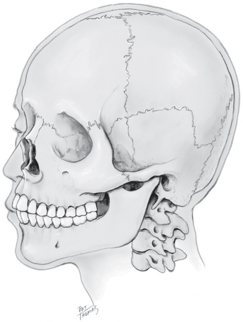
1. State the function of the skull.
_____________________________________________________________
_____________________________________________________________
_____________________________________________________________
_____________________________________________________________
2. Label the figure of the skull with the following:
3. Explain how the cranium is supported and by which structures.
_____________________________________________________________
_____________________________________________________________
_____________________________________________________________
_____________________________________________________________
4. Explain the boundaries of the neck and list the structures contained within the neck.
_____________________________________________________________
_____________________________________________________________
_____________________________________________________________
_____________________________________________________________
5. Describe in detail the two divisions of the nervous system.
_____________________________________________________________
_____________________________________________________________
_____________________________________________________________
_____________________________________________________________
6. List the major function(s) of the following components of the central nervous system:
_____________________________________________________________
_____________________________________________________________
_____________________________________________________________
_____________________________________________________________
_____________________________________________________________
_____________________________________________________________
_____________________________________________________________
_____________________________________________________________
_____________________________________________________________
_____________________________________________________________
_____________________________________________________________
_____________________________________________________________
cerebral cortex—Wernicke’s area
_____________________________________________________________
_____________________________________________________________
_____________________________________________________________
_____________________________________________________________
_____________________________________________________________
_____________________________________________________________
_____________________________________________________________
_____________________________________________________________
_____________________________________________________________
_____________________________________________________________
_____________________________________________________________
_____________________________________________________________
_____________________________________________________________
_____________________________________________________________
_____________________________________________________________
_____________________________________________________________
_____________________________________________________________
_____________________________________________________________
_____________________________________________________________
_____________________________________________________________
_____________________________________________________________
_____________________________________________________________
_____________________________________________________________
_____________________________________________________________
_____________________________________________________________
_____________________________________________________________
_____________________________________________________________
_____________________________________________________________
_____________________________________________________________
_____________________________________________________________
_____________________________________________________________
_____________________________________________________________
_____________________________________________________________
_____________________________________________________________
_____________________________________________________________
_____________________________________________________________
_____________________________________________________________
_____________________________________________________________
_____________________________________________________________
_____________________________________________________________
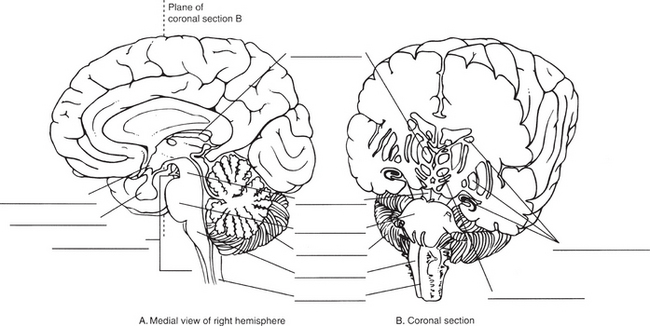
7. Fill in the labels on the following illustration.
8. Sensation travels in the afferent fibres in the peripheral nerve, then through the posterior (dorsal) root, then into the spinal cord. There, the sensation may take one of two routes: 1. The spinothalamic tract or 2. The posterior (dorsal) columns. Identify the sensations each of these pathways transmit and the route they take to mediate a response.
_____________________________________________________________
_____________________________________________________________
_____________________________________________________________
_____________________________________________________________
9. Explain how organ pain is felt from the heart, liver or spleen when there is no representation of these organs on the sensory homunculus.
_____________________________________________________________
_____________________________________________________________
_____________________________________________________________
_____________________________________________________________
10. Identify then describe each of the 3 major motor pathways in the CNS including the type of movements mediated by each.
_____________________________________________________________
_____________________________________________________________
_____________________________________________________________
_____________________________________________________________
11. Differentiate between an upper motor neuron and a lower motor neuron.
_____________________________________________________________
_____________________________________________________________
_____________________________________________________________
_____________________________________________________________
12. List the four types of reflexes and provide an example of each.
_____________________________________________________________
_____________________________________________________________
_____________________________________________________________
_____________________________________________________________
13. Trace the transmission of an impulse from initiation of sensation to response in a deep tendon reflex arc. As you trace the transmission, identify each of the 5 components of a reflex arc.
_____________________________________________________________
_____________________________________________________________
_____________________________________________________________
_____________________________________________________________
14. Fill in the gaps in relation to the spinal nerves:
There are ______________ of spinal nerves that arise from the length of the spinal cord and supply______________.
There are:_______________cervical, _______________thoracic, _______________lumbar, _______________sacral and_______________ coccygeal.
They are_______________ nerves because they contain both_______________ and_______________ fibres.
The nerves enter and exit the cord through roots: sensory afferent fibres through the ______________ or ______________roots; motor efferent fibres through the______________or______________roots. ______________is the cutaneous distribution of the various spinal nerves.
A_______________ is an identified skin area that is supplied mainly from_ through a particular spinal nerve.
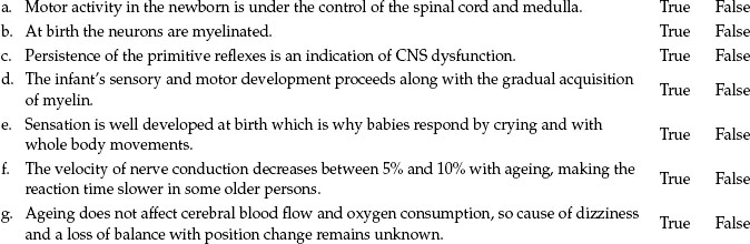
15. Circle True or False to answer the following statements concerning developmental considerations. If the answer is false, state the correct answer
16. Describe the characteristics and duration of each of the following headache types.
_____________________________________________________________
_____________________________________________________________
_____________________________________________________________
_____________________________________________________________
_____________________________________________________________
_____________________________________________________________
_____________________________________________________________
_____________________________________________________________
_____________________________________________________________
_____________________________________________________________
_____________________________________________________________
_____________________________________________________________
_____________________________________________________________
_____________________________________________________________
_____________________________________________________________
17. Identify at least 5 health history questions you would ask a patient concerning headaches they have.
_____________________________________________________________
_____________________________________________________________
_____________________________________________________________
_____________________________________________________________
18. When performing a neurological examination on an infant, why is it important to ask the parent if the infant has had:
_____________________________________________________________
_____________________________________________________________
_____________________________________________________________
_____________________________________________________________
exposure to lead-based paints?
_____________________________________________________________
_____________________________________________________________
_____________________________________________________________
19. Name each of the 3 types of neurological examinations; state when they would be performed, on whom, and what is examined with each.
_____________________________________________________________
_____________________________________________________________
_____________________________________________________________
_____________________________________________________________
20. A previously alert patient’s level of consciousness appears to be deteriorating, as they no longer open their eyes spontaneously. State, in order, how you would increase the stimulus to elicit a response.
_____________________________________________________________
_____________________________________________________________
_____________________________________________________________
_____________________________________________________________
21. Differentiate between localising, decorticate and decerebrate movements.
_____________________________________________________________
_____________________________________________________________
_____________________________________________________________
_____________________________________________________________
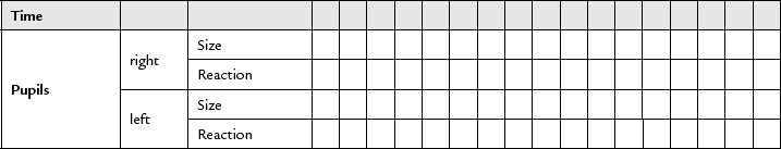
22. When assessing the pupils:
What characteristics should be noted?
_____________________________________________________________
_____________________________________________________________
_____________________________________________________________
_____________________________________________________________
Explain the pupillary light reflex.
_____________________________________________________________
_____________________________________________________________
_____________________________________________________________
_____________________________________________________________
List factors that may affect pupillary size, shape and response.
_____________________________________________________________
_____________________________________________________________
_____________________________________________________________
_____________________________________________________________
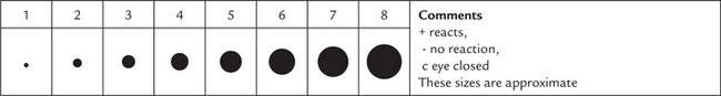
How and why is pupil size measured?
_____________________________________________________________
_____________________________________________________________
_____________________________________________________________
_____________________________________________________________
23. Explain the vital sign changes in the Cushing reflex.
_____________________________________________________________
_____________________________________________________________
_____________________________________________________________
_____________________________________________________________
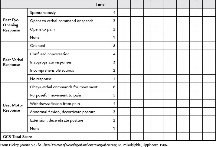
24. Describe the purpose of the Glasgow Coma Scale and each of the three divisions being assessed.
Purpose:_________________________________________
_____________________________________________________________
_____________________________________________________________
_____________________________________________________________
_____________________________________________________________
1. ________________________________________________________
_____________________________________________________________
_____________________________________________________________
_____________________________________________________________
_____________________________________________________________
2. ________________________________________________________
_____________________________________________________________
_____________________________________________________________
_____________________________________________________________
_____________________________________________________________
3. ______________________________________________________
_____________________________________________________________
_____________________________________________________________
_____________________________________________________________
_____________________________________________________________
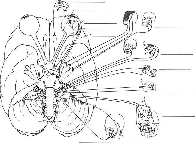
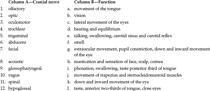
25. List the method of testing for each of the 12 cranial nerves in the adult.
26. Fill in the name of each cranial nerve, and label it as S (sensory), M (Motor), or MX (mixed).
27. Briefly describe each of the following cerebellar tests and state what a positive test may indicate:
_____________________________________________________________
_____________________________________________________________
_____________________________________________________________
_____________________________________________________________
_____________________________________________________________
_____________________________________________________________
_____________________________________________________________
_____________________________________________________________
_____________________________________________________________
_____________________________________________________________
_____________________________________________________________
_____________________________________________________________
28. Briefly describe the method of testing the sensory system for pain, temperature, touch, vibration and position. Hint: pain, temperature, touch test the spinothalamic tract; vibration and position test the posterior column.
_____________________________________________________________
_____________________________________________________________
_____________________________________________________________
_____________________________________________________________
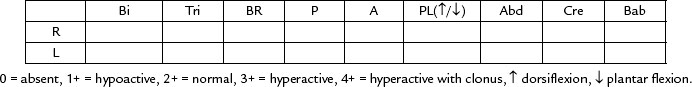
29. Outline the 4-point grading scale for deep tendon reflexes.
_____________________________________________________________
_____________________________________________________________
_____________________________________________________________
_____________________________________________________________
30. State the spinal level that will enable assessment of intactness of the reflex arc associated with the following:
31. Briefly describe testing of each of the following newborn reflexes and state when they disappear.
_____________________________________________________________
_____________________________________________________________
_____________________________________________________________
_____________________________________________________________
_____________________________________________________________
_____________________________________________________________
_____________________________________________________________
_____________________________________________________________
_____________________________________________________________
_____________________________________________________________
_____________________________________________________________
_____________________________________________________________
_____________________________________________________________
_____________________________________________________________
_____________________________________________________________
_____________________________________________________________
_____________________________________________________________
_____________________________________________________________
_____________________________________________________________
_____________________________________________________________
_____________________________________________________________
_____________________________________________________________
_____________________________________________________________
_____________________________________________________________
_____________________________________________________________
_____________________________________________________________
_____________________________________________________________
_____________________________________________________________
_____________________________________________________________
_____________________________________________________________
_____________________________________________________________
_____________________________________________________________
32. Describe patient presentation regarding their level of consciousness that would be graded as:
_____________________________________________________________
_____________________________________________________________
_____________________________________________________________
_____________________________________________________________
_____________________________________________________________
_____________________________________________________________
_____________________________________________________________
_____________________________________________________________
_____________________________________________________________
_____________________________________________________________
_____________________________________________________________
_____________________________________________________________
_____________________________________________________________
_____________________________________________________________
_____________________________________________________________
_____________________________________________________________
33. In question 11 you identified the difference between upper and lower motor neurons. Explain the type of reflex response you would expect to see with an upper motor neuron lesion versus a lower motor neuron lesion.
_____________________________________________________________
_____________________________________________________________
_____________________________________________________________
_____________________________________________________________
REVIEW QUESTIONS
This test is for you to check your own mastery of the content. The answers are provided in Appendix A.
1. The medical record indicates that a person has an injury to Broca’s area. When meeting this person you expect:
2. The control of body temperature is located in:
3. To test for stereognosis, you would:
4. During the examination of an infant, use a cotton-tipped applicator to stimulate the anal sphincter. The absence of a response suggests a lesion of:
5. During a neurological examination, the tendon reflex fails to appear. Before striking the tendon again, the nurse might use the technique of:
6. The National Stroke Foundation Australia (2010) recommends the FAST test as an easy way to recognise and remember the signs of stroke. What does the acronym FAST stand for:
7. Cerebellar function is assessed by which of the following tests?
8. To elicit a Babinski reflex:
9. A positive Babinski sign is:
10. The cremasteric response is:
11. Senile tremors may resemble parkinsonism, except that senile tremors do not include:
12. People who have Parkinson’s disease usually have which of the following characteristic styles of speech?
PRACTICAL SKILLS IN THE LABORATORY/CLINICAL SETTING
Assessment of the neurological system is an important assessment area for nurses. You will perform neurological assessment on a routine basis as an ongoing assessment or use it as a screening tool for your patients. You have reviewed the structure and function of many of the elements involved in the nervous system. Now it is time to practise the knowledge and skills you developed related to performing a comprehensive neurological assessment as you worked through this chapter.
You are now ready for the clinical component of the neurological system.
The purpose of the clinical component is to practise the regional examination on a peer in the skills laboratory or a patient in the clinical setting
CLINICAL OBJECTIVES
At the completion of the clinical laboratory session, with further practice and self-directed learning you should be able to:
1. demonstrate knowledge of the symptoms related to the neurological system by obtaining a neurological health history from a peer or patient
2. demonstrate the techniques used in examination of the neurological system. Beginning practitioners will be able to perform ongoing neurological observations and use of the Glasgow Coma Scale. With more experience and practice you will develop skills to assess the cranial nerves, cerebellar function, sensory system, motor system and deep tendon reflexes
3. record the history and physical examination findings accurately, reach an assessment of the health state and develop a plan of care.
INSTRUCTIONS
2. Prepare the examination setting and gather your equipment for an ongoing neurological examination or as directed by your instructor.
4. Gain consent to perform the examination from either your peer or the patient.
5. Practise the neurological health history interview and the steps of the ongoing neurological examination on a peer in the skills laboratory or a patient in the clinical setting, providing appropriate instructions as you proceed.
6. Record your findings using the regional write-up worksheet.
7. Swap roles and repeat steps 2–6.
8. Discuss your assessment techniques, findings and performance with your peer to develop a complete understanding of the process.
REGIONAL WRITE-UP WORKSHEET-ONGOING NEUROLOGICAL STATUS
Interview conducted by ______________
Patient ____________________________Age_______________ Gender______________
Occupation ________________________________Medical Record Number______________
1. Any unusual frequent or unusually severe headaches?______________
Location_______________ When started? ______________
Pattern_______________ Describe characteristics ______________
Type of pain?_____________How long do they last? ______________
Precipitating factors?_____________Associated factors? ______________
Family history?_____________Coping strategies ______________
2. Do you have neck pain?______________
Onset?_____________Location? ______________
Precipitating factors?_____________Associated factors? ______________
3. Do you have pain anywhere else? ______________
Score?_____________Quality? ______________
4. Ever had any head injury?______________
Show where_______________ Any loss of consciousness? ______________
6. Ever had any seizures?______________
Onset?_____________How often? ______________
Course and duration? ______________
Warning signs?_____________Type? ______________
7. Any tremors in hands or face? ______________
Worse with anxiety?_____________Relieved with rest? ______________
9. Any problem with coordination?______________
Problems with balance when walking?_____________Any falls? ______________
12. Any problem speaking?______________
Problems forming words?_____________Problems getting message across? ______________
13. Significant past history? ______________
Any stroke, spinal cord injury, meningitis, congenital defect, alcoholism? ______________
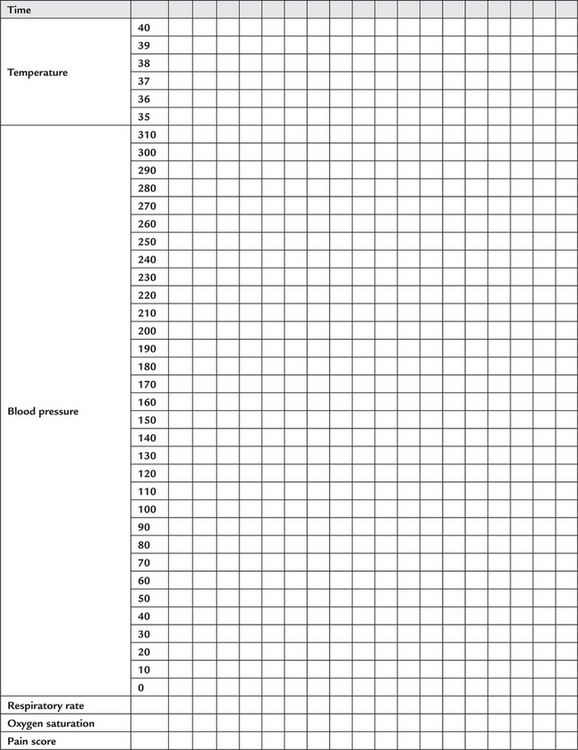
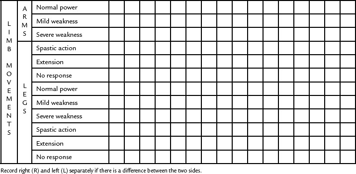
Most hospitals and institutions have specific charts to guide this form of neurological assessment and for ease of recording the data. Generally, the assessment data is presented in graphic form, which usually includes the Glasgow Coma Scale, pupillary response and vital signs. Your instructor may also provide you with an example used in your health service so you can practise recording your findings.
1. Mental status assessment using minimental examination as needed. Refer to MMSE in JF&W, Chapter 21, Table 21.1, pp 562–563.
Ease of arousal/state of awareness
Orientation_______________ Person ______________
Voluntary motor function—obeys commands ______________
Hand grasp—muscle strength ______________
Neuro assessment–motor function
Further assessment for advanced practice
NOTE: Your facilitator may ask you to practise some sections, all sections or none at all. Please check prior to the laboratory session to enable preparation if required.
| Preparation | Equipment needed |
|---|---|
Refer to JF&W, Chapter 22, pp 596–616 for the adult examinations and pages 616–627 for infant and paediatric examinations.
REGIONAL DOCUMENTATION (SOAP)–NEUROLOGICAL SYSTEM
Summarise your findings using the SOAP format.
Subjective (Reason for seeking care, health history)
Objective (Physical exam findings)
Assessment (Assessment of health state or problem, diagnosis)
Plan (Diagnostic evaluation, follow-up care, patient teaching)