Human Biology
At the completion of this chapter, the student should be able to do the following:
1 Discuss the cell theory of human biology.
2 List and describe the molecular composition of the human body.
3 Explain the parts and function of the human cell.
IT IS known beyond the shadow of a doubt that x-rays are harmful. If sufficiently intense, x-rays can cause skin burns, cataracts, cancer, leukemia, and other harmful effects. What is not known for certain is the degree of effect, if any, after exposure to diagnostic levels of x-radiation.
The benefits derived from diagnostic applications of x-rays are enormous. It is the job of radiologic technologists, radiologists, and medical physicists to produce high-quality x-ray images with minimal radiation exposure. This approach results in the greatest benefit with the lowest risk to patients and radiation workers. This is the practice known as ALARA—“as low as reasonably achievable.”
This chapter examines the concepts of human biology and discusses the known radiosensitivity of tissues, organs, and cells.
Human Radiation Response
The effect of x-rays on humans is the result of interactions at the atomic level (see Chapter 9). These atomic interactions take the form of ionization or excitation of orbital electrons and result in the deposition of energy in tissue.
Deposited energy can produce a molecular change, the consequences of which can be measurable if the molecule involved is critical. Figure 29-1 summarizes the sequence of events between radiation exposure and resultant human injury.
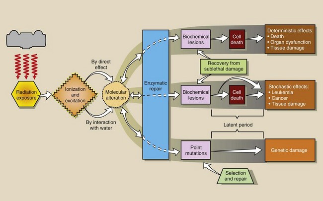
FIGURE 29-1 The sequence of events after radiation exposure of humans can lead to several radiation responses. At nearly every step, mechanisms for recovery and repair are available.
When an atom is ionized, its chemical binding properties change. If the atom is a constituent of a large molecule, ionization may result in breakage of the molecule or relocation of the atom within the molecule. The abnormal molecule may in time function improperly or cease to function, which can result in serious impairment or death of the cell.
This process is reversible. Ionized atoms can become neutral again by attracting a free electron. Molecules can be mended by repair enzymes. Cells and tissues can regenerate and recover from radiation injury.
If the radiation response increases in severity with increasing radiation dose, it is called a deterministic effect and occurs within days after the radiation exposure. On the other hand, if the incidence of the radiation response increases with increasing radiation dose, it is called a stochastic effect and is not observed for months or years.
A general classification scheme of possible deterministic and stochastic human responses to radiation is shown in Box 29-1. In addition, many other radiation responses have been experimentally observed in animals. Most human responses have been observed to occur after exposure to rather large radiation doses. However, we are cautious and assume that even small radiation doses are harmful.
Table 29-1 lists some of the human population groups in which many of these radiation responses have been observed.
TABLE 29-1
Human Populations in Whom Radiation Effects Have Been Observed
| Population | Effect |
| American radiologists | Leukemia, reduced life span |
| Atomic bomb survivors | Malignant disease |
| Radiation accident victims (e.g., Chernobyl) | Acute lethality |
| Marshall Islanders | Thyroid cancer |
| Uranium miners | Lung cancer |
| Radium watch-dial painters | Bone cancer |
| Patients treated with 131I | Thyroid cancer |
| Children treated for enlarged thymus | Thyroid cancer |
| Children of Belarus (downwind from Chernobyl) | Thyroid cancer |
| Patients with ankylosing spondylitis | Leukemia |
| Patients who underwent Thorotrast studies | Liver cancer |
| Irradiation in utero | Childhood malignancy |
| Volunteer convicts | Fertility impairment |
| Cyclotron workers | Cataracts |
The ultimate goal of radiobiologic research is to accurately describe the effects of radiation on humans so that radiation can be used more safely in diagnosis and more effectively in therapy. Most radiobiologic research seeks to develop radiation dose-response relationships so the effects of planned doses can be predicted and the response to accidental exposure managed.
Composition of the Body
At its most basic level, the human body is composed of atoms; radiation interacts at the atomic level. The atomic composition of the body determines the character and degree of the radiation interaction that occurs. The molecular and tissue composition defines the nature of the radiation response. Box 29-2 summarizes the atomic composition of the body and shows that more than 85% of the body consists of hydrogen and oxygen.
Cell Theory
Radiation interaction at the atomic level results in molecular change, which can produce a cell that is deficient in terms of normal growth and metabolism. Robert Hooke, the English schoolmaster, first named the cell as the biologic building block in 1665. Shortly thereafter, in 1673, Anton van Leeuwenhoek accurately described a living cell on the basis of his microscopic observations.
It was more than 100 years later, however, in 1838, that Schneider and Schwann showed conclusively that in all plants and animals, cells are the basic functional units. This is the cell theory.
The 1953 Watson and Crick description of the molecular structure of deoxyribonucleic acid (DNA) as the genetic substance of the cell was a major accomplishment. Precise mapping of the 40,000 human genes, which was the result of the Human Genome Project completed in the year 2000, promises exceptional solutions to the detection and management of human disease.
Molecular imaging is already making significant contributions to human health.
Molecular Composition
Five principal types of molecules are found in the body (Box 29-3). Four of these molecules—proteins, lipids (fats), carbohydrates (sugars and starches), and nucleic acids—are macromolecules.
Proteins, lipids, and carbohydrates are the principal classes of organic molecules. An organic molecule is life supporting and contains carbon. One of the rarest molecules—a nucleic acid concentrated in the nucleus of a cell (DNA)—is considered to be the most critical and radiosensitive target molecule.
Water is the most abundant molecule in the body, and it is the simplest. Water, however, plays a particularly important role in delivering energy to the target molecule, thereby contributing to radiation effects. In addition to water and the macromolecules, some trace elements and inorganic salts are essential for proper metabolism.
Water
The most abundant molecular constituent of the body is water. It consists of two atoms of hydrogen and one atom of oxygen (H2O) and constitutes approximately 80% of human substance. Humans are basically made of structured water.
The water molecules exist both in the free state and in the bound state, that is, bound to other molecules. They provide some form and shape, assist in maintaining body temperature, and enter into some biochemical reactions.
During vigorous exercise, body water is lost through perspiration to stabilize temperature and respiration. Water loss must be replaced to maintain homeostasis, which is the concept of the relative constancy of the internal environment of the human body.
Water and carbon dioxide are end products in the catabolism (breaking down into smaller units) of macromolecules. Anabolism, the production of large molecules from small, and catabolism collectively are referred to as metabolism. Some athletes use anabolic steroids to build muscle mass, but harmful adverse effects may occur.


Proteins
Approximately 15% of the molecular composition of the body is protein. Proteins are long-chain macromolecules that consist of a linear sequence of amino acids connected by peptide bonds. Twenty-two amino acids are used in protein synthesis, the metabolic production of proteins. The linear sequence, or arrangement, of these amino acids determines the precise function of the protein molecule.
Figure 29-2 shows the general chemical form of a protein molecule. The generalized formula for a protein is CnHnOnNnTn, where the subscript “n” refers to the number of atoms of each element in the molecule; T represents trace elements. In general, 50% of the mass of a protein molecule is carbon, 20% oxygen, 17% nitrogen, 7% hydrogen, and 6% other elements.
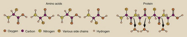
FIGURE 29-2 Proteins consist of amino acids linked by peptide bonds. The creation of the peptide bond requires the removal of a molecule of water.
Proteins have a variety of uses in the body. They provide structure and support. Muscles are very high in protein content. Proteins also function as enzymes, hormones, and antibodies.
Enzymes are molecules that are necessary in small quantities to allow a biochemical reaction to continue even though they do not directly enter into the reaction.
Hormones are molecules that exercise regulatory control over some body functions, such as growth and development. Hormones are produced and secreted by the endocrine glands—the pituitary, adrenal, thyroid, parathyroid, pancreas, and gonads.
Antibodies constitute a primary defense mechanism of the body against infection and disease. The molecular configuration of an antibody may be precise and designed for attacking a particular type of invasive or infectious agent, the antigen.
Lipids
Lipids are organic macromolecules composed solely of carbon, hydrogen, and oxygen. They are represented by the general formula, CnHnOn. Structurally, lipids are seen in the form shown in Figure 29-3, and it is this structure that distinguishes them from carbohydrates. In general, lipids are composed of two types of smaller molecules—glycerol and fatty acid. Each lipid molecule is composed of one molecule of glycerol and three molecules of fatty acid.
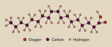
FIGURE 29-3 The structural configuration of a lipid is represented by a molecule of oleic acid: CH2(CH2)7CH = CH(CH2)7COOH.
Lipids are present in all tissues of the body and are the structural components of cell membranes. Lipids often are concentrated just under the skin and serve as a thermal insulator from the environment. Penguins, for instance, have a particularly thick layer of subcutaneous fat (blubber) that protects them from the cold.
Lipids also serve as fuel for the body by providing energy stores. It is more difficult, however, to extract energy from lipids than from the other major fuel source, carbohydrates; this relationship, of course, is associated with one of the major dilemmas in modern nutrition—obesity.
Carbohydrates
Carbohydrates, similar to lipids, are composed solely of carbon, hydrogen, and oxygen, but their structure is different (Figure 29-4). This structural difference determines the contribution of the carbohydrate molecule to body biochemistry. The ratio of the number of hydrogen atoms to oxygen atoms in a carbohydrate molecule is 2 : 1 (as in water), and a large fraction of this molecule consists of these atoms. Consequently, carbohydrates were first considered to be watered, or hydrated, carbons, hence their name.
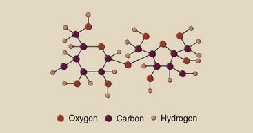
FIGURE 29-4 Carbohydrates are structurally different from lipids, even though their composition is similar. This is a molecule of sucrose, or ordinary table sugar: (C12H22O11).
Carbohydrates also are called saccharides. Monosaccharides and disaccharides are sugars. The chemical formula for glucose, a simple sugar, is C6H12O6. These molecules are relatively small. Polysaccharides are large and include plant starches and animal glycogen. The chemical formula for a polysaccharide is (C6H10O5)n, where n is the number of simple sugar molecules in the macromolecule.
Some carbohydrates are incorporated into the structure of cells and tissues to provide shape and stability. The human polysaccharide, glycogen, is stored in the tissues of the body and is used as fuel only when quantities of the simple sugar, glucose, are inadequate.
Glucose is the ultimate molecule that fuels the body. Lipids can be catabolized into glucose for energy but only with great difficulty. Polysaccharides are much more readily transformed into glucose. This explains why a chocolate bar, which is high in glucose, can provide a quick burst of energy for an athlete.
Nucleic Acids
Two principal nucleic acids are important to human metabolism: deoxyribonucleic acid (DNA) and ribonucleic acid (RNA). Located principally in the nucleus of the cell, DNA serves as the command or control molecule for cell function. DNA contains all the hereditary information that represents a cell and, of course, if the cell is a germ cell, all the hereditary information of the whole individual.
Located principally in the cytoplasm, RNA also is found in the nucleus. Two types of RNA have been identified: messenger RNA (mRNA) and transfer RNA (tRNA). These are distinguished according to their biochemical functions. These molecules are involved in the growth and development of the cell through a number of biochemical pathways, most notably, protein synthesis.
The nucleic acids are very large and extremely complex macromolecules. Figure 29-5 shows the structural composition of DNA and reveals how the component molecules are joined. DNA consists of a backbone composed of alternating segments of deoxyribose (a sugar) and phosphate. For each deoxyribose–phosphate conjugate formed, a molecule of water is removed.
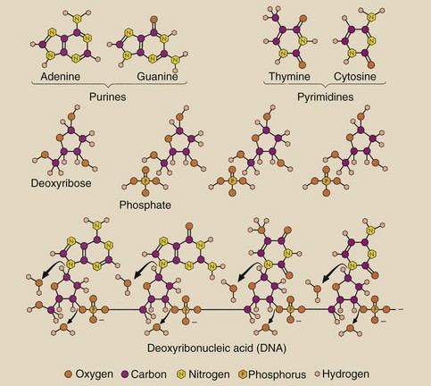
FIGURE 29-5 DNA is the control center for life. A single molecule consists of a backbone of alternating sugar (deoxyribose) and phosphate molecules. One of the four organic bases is attached to each sugar molecule.
Attached to each deoxyribose molecule is one of four different nitrogen-containing or nitrogenous organic bases: adenine, guanine, thymine, or cytosine. Adenine and guanine are purines; thymine and cytosine are pyrimidines.
The base sugar–phosphate combination is called a nucleotide, and the nucleotides are strung together in one long-chain macromolecule. Human DNA exists as two of these long chains attached together in ladder fashion (Figure 29-6). The side rails of the ladder are the alternating sugar–phosphate molecules, and the rungs of the ladder consist of bases joined together by hydrogen bonds.
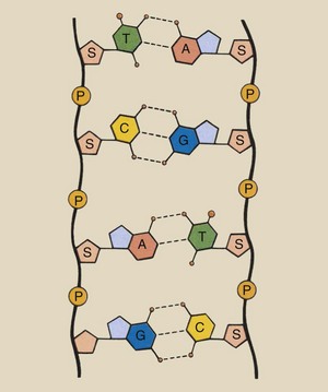
FIGURE 29-6 DNA consists of two long chains of alternating sugar and phosphate molecules fashioned similarly to the side rails of a ladder with pairs of bases as rungs.
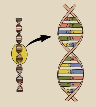
To complete the picture, the ladder is twisted about an imaginary axis such as a spring. This produces a molecule with the double-helix configuration (Figure 29-7). The sequence of base bonding is limited to adenines bonded to thymines and cytosines bonded to guanines.
Structurally, RNA resembles DNA. In RNA, the sugar component is ribose rather than deoxyribose, and uracil replaces thymine as a base component. In contrast, RNA forms a single helix, not a double helix.
The Human Cell
The principal molecular components of the human body are made of intricate cellular structures. The distribution of structures throughout the cell is reminiscent of the way the parts of an automobile are assembled. This assembly ensures proper growth, development, and function of the cell. Figure 29-8 is a cutaway view of a human cell, with its principal structures labeled.
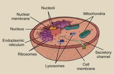
The two major structures of the cell are the nucleus and the cytoplasm. The principal molecular component of the nucleus is DNA, the genetic material of the cell. The nucleus also contains some RNA, protein, and water.
Most of the RNA is contained in a rounded structure, the nucleolus. The nucleolus often is attached to the nuclear membrane, a double-walled structure that at some locations is connected to the endoplasmic reticulum. This connection by its nature controls the passage of molecules, particularly RNA, from nucleus to cytoplasm.
The cytoplasm makes up the bulk of the cell and contains great quantities of all molecular components except DNA. A number of intracellular structures are found in the cytoplasm. The endoplasmic reticulum is a channel or a series of channels that allows the nucleus to communicate with the cytoplasm.
The large bean-shaped structures are mitochondria. Macromolecules are digested in the mitochondria to produce energy for the cell. The mitochondria are therefore called the engine of the cell.
The small, dot-like structures are ribosomes. Ribosomes are the site of protein synthesis and therefore are essential to normal cellular function. Ribosomes are scattered throughout the cytoplasm or the endoplasmic reticulum.
The small pea-like sacs are lysosomes. The lysosomes contain enzymes capable of digesting cellular fragments and sometimes the cell itself. Lysosomes help to control intracellular contaminants.
All of these structures, including the cell itself, are surrounded by membranes. These membranes consist principally of lipid–protein complexes that selectively allow small molecules and water to diffuse from one side to the other. These cellular membranes, of course, also provide structure and form for the cell and its components.
When the critical macromolecular cellular components are irradiated by themselves, a dose of approximately 10 kGyt (1 Mrad) is required to produce a measurable change in any physical characteristic of the molecule.
When a macromolecule is incorporated into the apparatus of a living cell, only a few mGya are necessary to produce a measurable biologic response. The lethal dose in some single-cell organisms, such as bacteria, is measured in Gyt, but human cells can be killed with a dose of less than 1 Gyt.
A number of experiments have shown that the nucleus is much more sensitive than the cytoplasm to the effects of radiation. Such experiments are conducted with the use of precise microbeams of electrons that can be focused and directed to a particular cell part or through incorporation of the radioactive isotopes tritium (3H) and carbon-14 (14C) into cellular molecules that localize exclusively to the cytoplasm or the nucleus.
Cell Function
Every human cell has a specific function in supporting the total body. Some differences are obvious, as in nerve cells, blood cells, and muscle cells. Similarities are also somewhat obvious.
In addition to its specialized function, each cell to some extent absorbs all molecular nutrients through the cell membrane and uses these nutrients in energy production and molecular synthesis. If this molecular synthesis is damaged by radiation exposure, the cell may malfunction and die.
Protein synthesis is a good example of a critical cellular function necessary for survival (Figure 29-9). DNA, located in the nucleus, contains a molecular code that identifies which proteins the cell will make.
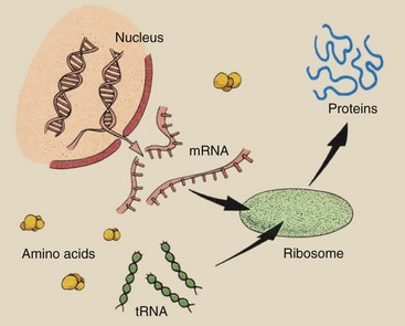
FIGURE 29-9 Protein synthesis is a complex process that involves many different molecules and cellular structures.
This code is determined by the sequence of base pairs (adenine–thymine and cytosine–guanine). A series of three base pairs, called a codon, identifies one of the 22 human amino acids available for protein synthesis.
This genetic message is transferred within the nucleus to a molecule of mRNA. mRNA leaves the nucleus by way of the endoplasmic reticulum and makes its way to a ribosome, where the genetic message is transferred to yet another RNA molecule (tRNA).
tRNA searches the cytoplasm for the amino acids for which it is coded. It attaches to the amino acid and carries it to the ribosome, where it is joined with other amino acids in sequence by peptide bonds to form the required protein molecule.
Interference with any phase of this procedure for protein synthesis could result in damage to the cell. Radiation interaction in which the molecule has primary control over protein synthesis (DNA) is more effective in producing a response than is radiation interaction with other molecules involved in protein synthesis.
Cell Proliferation
Although many Gray (many thousands of rad) are necessary to produce physically measurable disruption of macromolecules in vitro, single ionizing events at a particularly sensitive site of a critical target molecule are thought to be capable of disrupting cell proliferation.
 Cell proliferation is the act of a single cell or group of cells to reproduce and multiply in number.
The human body consists of two general types of cells, somatic cells and genetic cells. The genetic cells include the oogonium of the female and the spermatogonium of the male. All other cells of the body are somatic cells. When somatic cells proliferate or divide, they undergo mitosis. Genetic cells undergo meiosis.
Mitosis
Cell biologists and geneticists view the cell cycle differently (Figure 29-10). Each cycle includes the various states of cell growth, development, and division. Geneticists consider only two phases of the cell cycle, mitosis (M) and interphase.
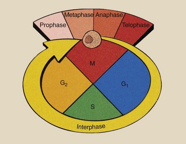
Mitosis, the division phase, is characterized by four subphases: prophase, metaphase, anaphase, and telophase. The portion of the cell cycle between mitotic events is called interphase. Interphase is the period of growth of the cell between divisions.
Cell biologists usually identify four phases of the cell cycle: M, G1, S, and G2. These phases of the cell cycle are characterized by the structure of the chromosomes, which contain the genetic material DNA. The gap in cell growth between M and S is G1. G1 is the pre-DNA synthesis phase.
The DNA synthesis phase is S. During this period, each DNA molecule is replicated into two identical daughter DNA molecules.
During S phase, the chromosome is transformed from a structure with two chromatids attached to a centromere to a structure with four chromatids attached to a centromere (Figure 29-11). The result is two pairs of homologous chromatids, that is, chromatids with precisely the same DNA content and structure.
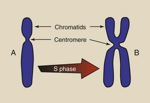
FIGURE 29-11 During the synthesis portion of interphase, the chromosomes replicate from a two-chromatid structure (A) to a four-chromatid structure (B).
The G2 phase is the post-DNA synthesis gap of cell growth.
During interphase, the chromosomes are not visible; however, during mitosis, the DNA slowly takes the form of the chromosomes as seen microscopically. Figure 29-12 schematically depicts the process of mitosis.
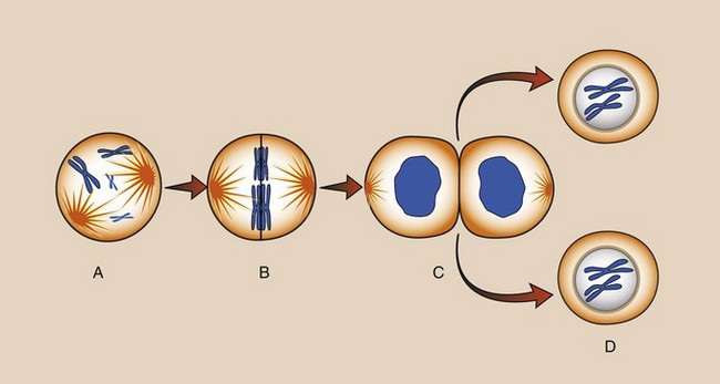
FIGURE 29-12 Mitosis is the phase of the cell cycle during which the chromosomes become visible, divide, and migrate to daughter cells. A, Interphase. B, Prophase. C, Metaphase. D, Anaphase. E, Telophase. F, Interphase.
During prophase, the nucleus swells, and the DNA becomes more prominent and begins to take structural form. At metaphase, the chromosomes appear and are lined up along the equator of the nucleus. It is during metaphase that mitosis can be stopped and chromosomes can be studied carefully under the microscope.
Anaphase is characterized by the splitting of each chromosome at the centromere, so that a centromere and two chromatids are connected by a fiber to the poles of the nucleus. These poles are called spindles, and the fibers are called spindle fibers. The number of chromatids per centromere has been reduced by half, and these newly formed chromosomes migrate slowly toward the spindle.
The final segment of mitosis, telophase, is characterized by the disappearance of structural chromosomes into a mass of DNA and the closing off of the nuclear membrane like a dumbbell into two nuclei. At the same time, the cytoplasm is divided into two equal parts, each of which accompanies one of the new nuclei.
Cell division is now complete. The two daughter cells look precisely the same as the parent cell and contain exactly the same genetic material.
Meiosis
Genetic material can change during the division process of genetic cells, which is called meiosis. Genetic cells begin with the same number of chromosomes as somatic cells—23 pairs (46 chromosomes). However, for a genetic cell to be capable of marriage to another genetic cell, its complement of chromosomes must be reduced by half to 23, so that after conception and the union of two genetic cells, the daughter cells again will contain 46 chromosomes (Figure 29-13).
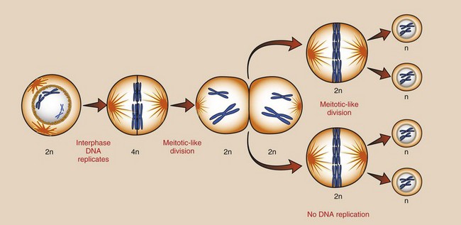
FIGURE 29-13 Meiosis is the process of reduction division, and it occurs only in reproductive cells. n, Number of similar chromosomes.
The genetic cell begins meiosis with 46 chromosomes that appear the same as in a somatic cell that has completed the G2 phase. The cell then progresses through the phases of mitosis into two daughter cells, each containing 46 chromosomes of two chromatids each. The names of the subphases are the same for meiosis and mitosis.
Each of the daughter cells of this first division now progresses through a second division in which all cellular material, including chromosomes, is divided. However, the second division is not accompanied by an S phase. Therefore, no replication of DNA occurs; consequently, no chromosomes are duplicated. Each of the resulting granddaughter cells contains only 23 chromosomes.
Each parent has undergone two division processes, which have resulted in four daughter cells. During the second division, some chromosomal material is exchanged among chromatids through a process called crossing over. Crossing over results in changes in genetic constitution and changes in inheritable traits.
Tissues and Organs
During the development and maturation of a human from two united genetic cells, a number of different types of cells evolve. Collections of cells of similar structure and function form tissues. Box 29-4 is a breakdown of the composition of the body according to its tissue constituents.
These tissues in turn are precisely bound together to form organs. The tissues and the organs of the body serve as discrete units with specific functional responsibilities. Some tissues and organs combine into an overall integrated organization known as an organ system.
The principal organ systems of the body are the nervous system, digestive system, endocrine system, respiratory system, and reproductive system. Effects of radiation that appear at the whole-body level result from damage to these organ systems that occurs as the result of radiation injury to the cells of that system.
The cells of a tissue system are identified by their rate of proliferation and their stage of development. Immature cells are called undifferentiated cells, precursor cells, or stem cells. As a cell matures through growth and proliferation, it can pass through various stages of differentiation into a fully functional and mature cell.
The sensitivity of the cell to radiation is determined somewhat by its state of maturity and its functional role. Table 29-2 lists a number of different types of cells in the body according to their degree of radiosensitivity.
TABLE 29-2
Response to Radiation Is Related to Cell Type
| Radiosensitivity | Cell Type |
| High | Lymphocytes |
| Spermatogonia | |
| Erythroblasts | |
| Intestinal crypt cells | |
| Intermediate | Endothelial cells |
| Osteoblasts | |
| Spermatids | |
| Fibroblasts | |
| Low | Muscle cells |
| Nerve cells |
The tissues and organs of the body include both stem cells and mature cells. Several types of tissue can be classified according to structural or functional features. These features influence the degree of radiosensitivity of the tissue.
Epithelium is the covering tissue, and it lines all exposed surfaces of the body, both exterior and interior. Epithelium covers the skin, blood vessels, abdominal and chest cavities, and gastrointestinal tract.
Connective and supporting tissues are high in protein and are composed principally of fibers that are usually highly elastic. Connective tissue binds tissues and organs together. Bone ligaments and cartilage are examples of connective tissue.
Muscle is a special type of tissue that can contract. It is found throughout the body and is high in protein content.
Nervous tissue consists of specialized cells called neurons that have long, thin extensions from the cell to distant parts of the body. Nervous tissue is the avenue by which electrical impulses are transmitted throughout the body for control and response.
When these various types of tissue are combined to form an organ, they are identified according to two parts of the organ. Whereas the parenchymal part contains tissues that represent that particular organ, the stromal part is composed of connective tissue and vasculature that provide structure to the organ.
The deterministic effects of high-dose radiation may include observable organ damage. The various organs of the body exhibit a wide range of sensitivity to radiation. This radiosensitivity is determined by the function of the organ in the body, the rate at which cells mature within the organ, and the inherent radiosensitivity of the cell type.
Precise knowledge of these various organ radiosensitivities is unnecessary; however, knowledge of general levels of radiosensitivity is helpful toward understanding the effects of whole-body radiation exposure, particularly in the acute radiation syndrome (Table 29-3).
TABLE 29-3
Relative Radiosensitivity of Tissues and Organs Based on Clinical Radiation Oncology
| Level of Radiosensitivity* | Tissue or Organ | Effects |
| High: 2–10 Gyt (200–1000 rad) | Lymphoid tissue | Atrophy |
| Bone marrow | Hypoplasia | |
| Gonads | Atrophy | |
| Intermediate: 10–50 Gyt (1000–5000 rad) | Skin Gastrointestinal tract |
Erythema Ulcer |
| Cornea | Cataract | |
| Growing bone | Growth arrest | |
| Kidney | Nephrosclerosis | |
| Liver | Ascites | |
| Thyroid | Atrophy | |
| Low: >50 Gyt (>5000 rad) | MuscleBrain | FibrosisNecrosis |
| Spinal | Transection |
*The minimum dose delivered at the rate of approximately 2 Gyt/day (200 rad/day), which will produce a response.
Summary
After radiation exposure, the human body responds in predictable ways. Radiobiology is the study of the effects of ionizing radiation on humans conducted to refine knowledge of the expected response to radiation.
If the intensity of the response increases with increasing radiation dose, it is called a deterministic response and occurs within days of exposure. If the frequency of an injury increases with increasing radiation dose, it is called a stochastic effect and is not observable for years.
The cell is the basic functional unit of all plants and animals. At the molecular level, the human body is composed primarily of water, protein, lipid, carbohydrate, and nucleic acid. The two important nucleic acids in human metabolism are DNA and RNA.
DNA contains all the hereditary information in the cell. If the cell is a genetic cell, the DNA contains the hereditary information of the whole individual. DNA is a macromolecule that is made up of two long chains of base sugar–phosphate combinations twisted into a double helix.
Major cellular function consists of protein synthesis and cell division. Mitosis is the growth, development, and division of cells. Meiosis is the term applied to the division of genetic cells.
Cells of similar structure bind together to form tissue. Tissues bind together to form organs. An overall integrated organization of tissue and organs is called an organ system.
The principal organ systems of the body are the nervous, digestive, endocrine, and reproductive systems. The radiosensitivity of various tissue and organ systems varies widely. Reproductive cells are highly radiosensitive; nerve cells are less radiosensitive.
1. Define or otherwise identify the following:
2. At what structural level do x-rays interact with humans to produce a radiation response?
3. How does ionizing radiation affect an atom within a large molecule?
4. List five human groups in which radiation effects have been observed.
5. What are the effects of radiation on the populations mentioned in Question 4?
6. What is the most abundant atom and the most abundant molecule in the body?
8. Why do we say that humans are basically a structured aqueous suspension?
9. What is the meaning of epithelium?
10. How do proteins function in the human body?
11. What do carbohydrates do for us?
12. DNA is the abbreviation for what molecule?
13. Which molecule is considered the genetic material of the cell?
14. What is the function of the endoplasmic reticulum?
15. What is the approximate dose of radiation required to produce a measurable physical change in a macromolecule?
16. List the stages of cell division of a somatic cell.
17. List the stages of cell reduction division of a genetic cell.
18. What cell type is the most radiosensitive?
19. What type of tissue is the least radiosensitive?
20. List three early radiation effects and three late radiation effects in humans.
The answers to the Challenge Questions can be found by logging on to our website at http://evolve.elsevier.com.