Based on traditional considerations, it is not advisable to prescribe Astragalus in acute infections.
In vivo studies suggest that Astragalus may reduce the efficacy of cyclophosphamide (an immunosuppressive agent).231,232 However, the clinical relevance of this is uncertain. In principle, immune-enhancing herbs should not be given long-term to transplant recipients receiving immune-suppressing drugs.
Category B1 – no increase in frequency of malformation or other harmful effects on the fetus from limited use in women. No evidence of increased fetal damage in animal studies for oral doses.
Some species of Astragalus are known to induce locoism (a condition that can cause reproductive alterations), abortion and occasional skeletal deformities in livestock.233,234A. membranaceus has not been identified as one of these species and swainsonine, the indolizidine alkaloid responsible for locoism,235 has not been detected in A. membranaceus. It is unlikely that the Astragalus species used medicinally are teratogenic, since those species that induce teratogenic effects are also known to be toxic. Moreover, a Chinese herbal formula (Man-Shen-Ling) that contains Astragalus did not demonstrate teratogenic activity in animal models.236 Intravenous astragaloside IV was maternally toxic at 1.0 mg/kg in rats and fetotoxic at a dose higher than 0.5 mg/kg, but was devoid of teratogenic effects in rats and rabbits.237
There are no data available concerning the safety of Astragalus in breastfeeding.
Astragalus is official in the Pharmacopoeia of the Republic of China (English edition, 1997) and the Japanese Pharmacopoeia (15th edition, English version 2006).
Astragalus is not covered by a Commission E monograph and is not on the UK General Sale List.
Astragalus does not have GRAS status. However, it is freely available as a ‘dietary supplement’ in the USA under DSHEA legislation (1994 Dietary Supplement Health and Education Act).
Astragalus is not included in Part 4 of Schedule 4 of the Therapeutic Goods Act Regulations of Australia and is freely available for sale.
References
1. Bensky D, Gamble A. Chinese Herbal Medicine Materia Medica. Seattle: Eastland Press, 1986. pp. 457–459
2. Chang H, But P, Pharmacology and Applications of Chinese Materia Medica, Singapore, World Scientific, 1987;vol 2. pp. 1041–1046
3. Leung AY, Foster S. Encyclopedia of Common Natural Ingredients Used in Food, Drugs and Cosmetics, 2nd ed. New York: John Wiley, 1996. p. 52
4. Mabberley DJ, ed. The Plant Book, 2nd ed., Cambridge: Cambridge University Press, 1997. 64, pp. 396–398
5. World Health Organization. Medicinal Plants in China. Manila: World Health Organization, Regional Office for the Western Pacific, 1989. p. 47
6. Ma XQ, Shi Q, Duan JA, et al. J Agric Food Chem. 2002;50(17):4861–4866.
7. Huang DL, Sun SQ, Xu YQ, et al. Guang Pu Xue Yu Guang Pu Fen Xi. 2009;29(9):2396–2400. [Article in Chinese]
8. Tang W, Eisenbrand G. Chinese Drugs of Plant Origin. Berlin: Springer Verlag, 1992. pp. 191–197
9. Katsura E, Katoh Y, Yamagishi T. Hokkaidoritsu Eisei Kenkyushoho. 1983;33:136–137.
10. Mu LX, Zhu L, Zhao AH, et al. Zhong Yao Cai. 2009;32(11):1741–1745. [Article in Chinese]
11. Xu DJ, Xia Q, Wang JJ, et al. Molecules. 2008;13(10):2408–2415.
12. Song JZ, Yiu HH, Qiao CF, et al. J Pharm Biomed Anal. 2008;47(2):399–406.
13. Jing JP, Lin WF. Chin J Microbiol Immunol. 1983;3:293–296.
14. Wang Y, Qian XJ, Hadley HR, et al. Mol Biother. 1992;4(3):143–146.
15. Rittenhouse JR, Lui PD, Lau BHS, et al. J Urol (Paris). 1991;146(2):486–490.
16. Sun Y, Hersh EM, Talpaz M, et al. Cancer. 1983;52(1):70–73.
17. You L, Zhou Y, Zhang Y, et al. Zhongguo Mianyixue Zazhi. 1990;6(1):60–63.
18. Tu LH, Huang DR, Zhang RQ, et al. Chin Med J (Engl). 1994;107(4):300–303.
19. Wang J, Zhang QY, Chen YX. Zhongguo Zhong Xi Yi Jie He Za Zhi. 2009;29(9):794–797. [Article in Chinese]
20. Clement-Kruzel S, Hwang SA, Kruzel MC, et al. J Med Food. 2008;11(3):493–498.
21. Xu HD, You CG, Zhang RL, et al. J Int Med Res. 2007;35(1):84–90.
22. Yang Y, Wang LD, Chen ZB. Zhongguo Dang Dai Er Ke Za Zhi. 2006;8(5):376–378.
23. Wang G, Liu CT, Wang ZL, et al. Chin J Integr Med. 2006;12(4):262–267.
24. Sugiura H, Nishida H, Inaba R, et al. Nippon Eiseigaku Zasshi. 1993;47(6):1021–1031.
25. Jin R, Mitsuishi T, Akuzawa Y, et al. Kitakanto Med J. 1994;44(2):125–133.
26. Jin R, Wan LL, Mitsuishi T. Chung Kuo Chung Hsi I Chieh Ho Tsa Chih. 1995;15(2):101–103.
27. Isotopes laboratory, Beijing institute of tuberculosis. Xinyiyaexue Zazhi 1974;8:12.
28. Wang JY, Zhong JX, Zhou ZH, et al. Chin J Integr Trad West Med. 1987;7(9):543.
29. Chen YC. Chung Kuo Chung Yao Tsa Chih. 1994;19(2):739–741.
30. Kajimura K, Takagi Y, Ueba N, et al. Biol Pharm Bull. 1996;19(9):1166–1169.
31. Du Q, Chen Z, Zhou LF, et al. Can J Physiol Pharmacol. 2008;86(7):449–457.
32. Gao XH, Xu XX, Pan R, et al. J Nat Med. 2009;63(4):421–429.
33. Kang H, Ahn KS, Cho C, et al. Biol Pharm Bull. 2004;27(12):1946–1950.
34. Xu XL, Chen XJ, Ji H, et al. Pharmacology. 2008;81(4):325–332.
35. Ning KJ, Ruan XC, Lu JF, et al. Zhongguo Zhong Yao Za Zhi. 2005;30(21):1670–1672. [Article in Chinese]
36. Yin G, Ardó L, Thompson KD, et al. Fish Shellfish Immunol. 2009;26(1):140–145.
37. Wei X, Zhang J, Li J, et al. Am J Chin Med. 2004;32(5):669–680.
38. Zhang RP, Zhang XP, Ruan YF, et al. World J Gastroenterol. 2009;15(23):2862–2869.
39. Zhu XL, Zhu BD. Phytother Res. 2007;21(7):663–667.
40. Lv Y, Feng X, Zhu B. Zhong Yao Cai. 2005;28(9):791–793. [Article in Chinese]
41. Yang X, Huang S, Chen J, et al. Vaccine. 2010;28(3):737–743.
42. Yang ZG, Sun HX, Fang WH. Vaccine. 2005;23(44):5196–5203.
43. Shen HH, Wang K, Li W, et al. J Ethnopharmacol. 2008;116(2):363–369.
44. Institute of Basic Medical Sciences, Chinese Academy of Medical Sciences. Chung Hua I Hsueh Tsa Chih 1979;59:31–34.
45. Institute of Epidemic Prevention, Chinese Academy of Medical Sciences. Med Commun 1978;4:4.
46. Brush J, Mendenhall E, Guggenheim A, et al. Phytother Res. 2006;20(8):687–695.
47. Qun L, Luo Q, Zhang ZY, et al. Clin Nephrol. 1999;52(5):333–334.
48. Zwickey H, Brush J, Iacullo CM, et al. Phytother Res. 2007;21(11):1109–1112.
49. Chu D, Sun Y, Lin J, et al. Chung Hsi I Chieh Ho Tsa Chih. 1990;10(1):34–36.
50. Chu DT, Sun Y, Lin JR. Chung Hsi I Chieh Ho Tsa Chih. 1989;9(6):351–354.
51. Wang DC. Chung Hua Chung Liu Tsa Chih. 1989;11(3):180–183.
52. Zhao XZ. Chung Kuo Chung Hsi I Chieh Ho Tsa Chih. 1992;12:669–671. 645
53. Chu DT, Lin JR, Wong W. Chung Hua Chung Liu Tsa Chih. 1994;16(3):167–171.
54. Cho WC, Leung KN. J Ethnopharmacol. 2007;113(1):132–141.
55. Shao BM, Xu W, Dai H, et al. Biochem Biophys Res Commun. 2004;320(4):1103–1111.
56. Hao Y, Qiu QY, Wu J. Zhongguo Zhong Xi Yi Jie He Za Zhi. 2004;24(5):427–430.
57. Lee KY, Jeon YJ. Int Immunopharmacol. 2005;5(7–8):1225–1233.
58. Shao P, Zhao LH, Zhi-Chen, et al. Int Immunopharmacol. 2006;6(7):1161–1166.
59. Chen CJ, Li ZL, Fu Q, et al. Nan Fang Yi Ke Da Xue Xue Bao. 2009;29(6):1192–1194. [Article in Chinese]
60. Yin X, Chen L, Liu Y, et al. Biochem Biophys Res Commun. 2010;397(2):232–238.
61. Liu QY, Yao YM, Zhang SW, et al. J Ethnopharmacol, 2010. [Epub ahead of print]
62. Li S, Zhang Y, Zhao J. Int Immunopharmacol. 2007;7(1):23–28.
63. Li RJ, Qiu SD, Chen HX, et al. Biol Pharm Bull. 2007;30(3):470–476.
64. Chen W, Li YM, Yu MH. Exp Clin Endocrinol Diabetes. 2008;116(8):468–474.
65. Chen Y, Wang D, Hu Y, et al. Int J Biol Macromol. 2010;46(4):425–428.
66. Li SP, Zhao XJ, Wang JY. Poult Sci. 2009;88(3):519–525.
67. Wang T, Sun Y, Jin L, et al. Fish Shellfish Immunol. 2009;27(6):757–762.
68. Liu J, Hu X, Yang Q, et al. J Biomed Biotechnol. 2010:12. Article ID 479426
69. Qiu Y, Hu YL, Cui BA, et al. Poult Sci. 2007;86(12):2530–2535.
70. Peng J, Wu SH, Zhang LL, et al. Zhongguo Yixue Kexueyuan Xuebao. 1984;6(2):116–119.
71. Institute of Epidemic Prevention, Chinese Academy of Medical Sciences. Stud on Epidemic Prev 1976;3:204.
72. Zheng MS, et al. Chin Trad Herbal Drugs. 1987;18(10):459–461.
73. Ono K, Nakane H, Meng ZK, et al. Chem Pharm Bull (Tokyo). 1989;37(7):1810–1812.
74. Yuan WL, Chen HZ, Yang YZ, et al. Chin Med J (Engl). 1990;103(3):177–182.
75. Sun Y, Yang J. Di Yi Jun Yi Da Xue Xue Bao. 2004;24(1):57–58. [Article in Chinese]
76. Liu WJ, Liu B, Guo QL, et al. Zhonghua Er Ke Za Zhi. 2004;42(7):490–494.
77. Zhu H, Zhang Y, Ye G, et al. Biol Pharm Bull. 2009;32(1):68–73.
78. Institute of Epidemic Prevention, Chinese Academy of Medical Sciences. Stud Epidemic Prev 1976;2:124.
79. Dang SS, Jia XL, Song P, et al. World J Gastroenterol. 2009;15(45):5669–5673.
80. Wang S, Li J, Huang H, et al. Biol Pharm Bull. 2009;32(1):132–135.
81. Rui T, Yang YZ, Zhou TS. Chung Kuo Chung Hsi I Chieh Ho Tsa Chih. 1994;14(5):292–294. 262
82. Peng TQ, Yang YZ, Kandolf R. Chung Kuo Chung Hsi I Chieh Ho Tsa Chih. 1994;14(11):664–666.
83. Guo Q, Peng TQ, Yang YZ. Chung Kuo Chung Hsi I Chieh Ho Tsa Chih. 1995;15(8):483–485.
84. Chen XJ, Bian ZP, Lu S, et al. Am J Chin Med. 2006;34(3):493–502.
85. Peng T, Yang Y, Riesemann H, et al. Chin Med Sci J. 1995;10(3):146–150.
86. Guo Q, Peng T, Yang Y, et al. Virol Sin. 1996;11(1):40–44.
87. Huang ZQ, Qin NP, Ye W. Chung Kuo Chung Hsi I Chieh Ho Tsa Chih. 1995;15(6):328–330.
88. Xiong ZY. Zhejiang J Trad Chin Med. 1983;18(5):235–238.
89. Wang P, Zhang Z, Sun Y, et al. DNA Cell Biol. 2010;29(1):33–39.
90. Wang P, Zhang Z, Ma X, et al. Mech Ageing Dev. 2003;124(10–12):1025–1034.
91. De Jesus BB, Schneeberger K, Vera E, et al. Aging Cell. 2011;10(4):604–621.
92. Isotopes Section of the Pharmacology Department, Basic Sciences Research Unit, Capital Hospital of the Chinese Academy of Medical Sciences, et al. Med Res Commun. 1977;10:27.
93. Pan SY, Hou JY, Jiang MY, et al. Chung Yao Tung Pao. 1986;11(9):559–561.
94. Hong GX, Qin WC, Huang LS. Chung Kuo Chung Yao Tsa Chih. 1994;19(11):687–688. 704
95. Nagai K, Hanazuka M, Hizume S, et al. Jpn Pharmacol Therapeut. 1992;20(9):47–53.
96. Park HJ, Kim HY, Yoon KH, et al. Korean J Physiol Pharmacol. 2009;13(4):315–319.
97. Liu J, et al. Chin Trad Herbal Drugs. 1981;12(6):264–265.
98. Xu P, Jin G, Shen X. Chung Kuo Chung Yao Tsa Chih. 1991;16(1):49–50.
99. Shi r, He L, Hu Y, et al. J Tradit Chin Med. 2001;21(3):232–235.
100. Institute of Basic Medical Sciences, Chinese Academy of Medical Sciences. Zhonghua Yixue Zazhi 1979;1:23.
101. Wang Q. Chung Kuo Chung Yao Tsa Chih. 1992;17(9):557–559.
102. Meng D, Chen XJ, Bian YY, et al. Am J Chin Med. 2005;33(1):11–20.
103. Zhang WD, Chen H, Zhang C, et al. Planta Med. 2006;72(1):4–8.
104. Luo Z, Zhong L, Han X, et al. Phytother Res. 2009;23(6):761–767.
105. Liu Y, Hua SD, He YG, et al. Zhongguo Zhong Yao Za Zhi. 2008;33(19):2226–2229.
106. Qiu LH, Xie XJ, Zhang BQ. Biol Pharm Bull. 2010;33(4):641–646.
107. Song DJ, Gu DG, Mao SY, et al. Chin Trad Herbal Drugs. 1989;20(8):361–364.
108. Chengdu Institute for Drug Control. Chengdu Medical and Health Information 1971;1:90.
109. Hikino H, Funayama S, Endo K. Planta Med. 1976;30(4):297–302.
110. Li MR, Yu YR, Deng G. Nan Fang Yi Ke Da Xue Xue Bao. 2010;30(1):7–10. [Article in Chinese]
111. Deng G, Yu YR. Sichuan Da Xue Xue Bao Yi Xue Ban. 2009;40(4):608–611. [Article in Chinese]
112. Zhang C, Wang XH, Zhong MF, et al. Clin Exp Pharmacol Physiol. 2007;34(5–6):387–392.
113. Zhang WD, Zhang C, Wang XH, et al. Planta Med. 2006;72(7):621–626.
114. Zhang BQ, Hu SJ, Qiu LH, et al. Biol Pharm Bull. 2005;28(8):1450–1454.
115. Su D, Xu B, Shi HL, et al. Zhongguo Zhong Yao Za Zhi. 2008;33(14):1724–1727. [Article in Chinese]
116. Su D, Li HY, Yan HR, et al. Am J Chin Med. 2009;37(3):519–529.
117. Chen XJ, Meng D, Feng L, et al. Am J Chin Med. 2006;34(6):1015–1025.
118. Ma L, Guan ZZ. Zhongguo Zhong Xi Yi Jie He Za Zhi. 2005;25(7):646–649. [Article in Chinese]
119. Lei ZY, Qin H, Liao JZ. Zhongguo Zhong Xi Yi Jie He Za Zhi. 1994;14(4):199–202. [Article in Chinese]
120. Chen LX, Liao JZ, Guo WQ. Zhongguo Zhong Xi Yi Jie He Za Zhi. 1995;15(3):141–143. [Article in Chinese]
121. Dai JH, Liang ZJ, Qin WZ, et al. Guizhou Med J. 1987;11(1):23–24.
122. Xue JX, Jiang Y, Yan YQ. Chung Kuo Chung Yao Tsa Chih. 1993;18(10):621–630. 640
123. Xue JX, Yan YQ, Jiang Y. Chung Kuo Chung Yao Tsa Chih. 1994;19(2):108–110. 128
124. Zhang YD, Shen JP, Zhu SH, et al. Yao Hsueh Hsueh Pao. 1992;27(6):401–406.
125. Dang SS, Zhang X, Jia XL, et al. Chin Med J (Engl). 2008;121(11):1010–1014.
126. Han DW, Xu RL, Yeung SCS. Abstr Chin Med. 1988;2(1):114–115.
127. Zhang ZL, Wen QZ, Liu CX, et al. J Ethnopharmacol. 1990;30(2):145–150.
128. Yan XW, et al. Symposium of the Chinese Pharmaceutical Association, 1962. 332
129. Sun WY, Wei W, Gui SY, et al. Basic Clin Pharmacol Toxicol. 2008;103(2):143–149.
130. Wang DQ, Critchley JA, Ding BG, et al. Zhongguo Zhong Yao Za Zhi. 2001;26(9):617–620. [Article in Chinese]
131. Wang DQ, Ding BG, Ma YQ, et al. Zhongguo Zhong Yao Za Zhi. 2001;26(7):483–486. [Article in Chinese]
132. Sun WY, Wei W, Wu L, et al. J Ethnopharmacol. 2007;112(3):514–523.
133. Yang Y, Yang S, Chen M, et al. J Ethnopharmacol. 2008;118(2):264–270.
134. Zhang J, Xie X, Li C, et al. J Ethnopharmacol. 2009;126(2):189–196.
135. Zuo C, Xie X, Deng Y, et al. Zhongguo Zhong Yao Za Zhi. 2009;34(2):193–198. [Article in Chinese]
136. Zuo C, Xie X, Deng Y, et al. Wei Sheng Yan Jiu. 2008;37(5):566–570. [Article in Chinese]
137. Zhang GZ, Wu XC, Peng XJ, et al. Zhongguo Dang Dai Er Ke Za Zhi. 2008;10(2):173–178. [Article in Chinese]
138. Huang LM, Shi XQ, Liang H. Zhongguo Zhong Yao Za Zhi. 2007;32(13):1324–1328. [Article in Chinese]
139. Zuo C, Xie XS, Qiu HY, et al. J Zhejiang Univ Sci B. 2009;10(5):389–390.
140. Li X, He DL, Cheng XF, et al. Zhongguo Zhong Yao Za Zhi. 2005;30(20):1606–1609. [Article in Chinese]
141. Zou F, Xq Mao, Wang N, et al. Acta Pharmacol Sin. 2009;30(12):1607–1615.
142. Mao XQ, Yu F, Wang N, et al. Phytomedicine. 2009;16(5):416–425.
143. Liu M, Wu K, Mao X, et al. J Ethnopharmacol. 2010;127(1):32–37.
144. Wu Y, Ou-Yang JP, Wu K, et al. Acta Pharmacol Sin. 2005;26(3):345–352.
145. Chen W, Yu MH, Li YM, et al. Acta Diabetol. 2010;47(suppl 1):35–46.
146. Ma W, Nomura M, Takahashi-Nishioka T, et al. Biol Pharm Bull. 2007;30(11):2079–2083.
147. Yu J, Zhang Y, Sun S, et al. Can J Physiol Pharmacol. 2006;84(6):579–587.
148. Auyeung KK, Mok NL, Wong CM, et al. Int J Mol Med. 2010;26(3):341–349.
149. Auyeung KK, Law PC, Ko JK. Int J Mol Med. 2009;23(2):189–196.
150. Tin MM, Cho CH, Chan K, et al. Carcinogenesis. 2007;28(6):1347–1355.
151. Deng Y, Chen HF. Zhong Xi Yi Jie He Xue Bao. 2009;7(12):1174–1180. [Article in Chinese]
152. Ye MN, Chen HF. Zhong Xi Yi Jie He Xue Bao. 2008;6(4):399–404. [Article in Chinese]
153. Cheng XD, Hou CH, Zhang XJ, et al. Acta Biochim Biophys Sin (Shanghai). 2004;36(3):211–217.
154. Cho WC, Leung KN. Cancer Lett. 2007;252(1):43–54.
155. Kurashige S, Akuzawa Y, Endo F. Cancer Invest. 1999;17(1):30–35.
156. Cui R, He J, Wang B, et al. Cancer Chemother Pharmacol. 2003;51(1):75–80.
157. Wang D, Shen W, Tian Y, et al. Chung Kuo Chung Yao Tsa Chih. 1995;20(4):240–242. 254
158. Liu XJ, Jiang M, Yu Z, et al. Tianran Chanwu Yanjiu Yu Kaifa. 1991;3(4):1–6.
159. Sheng BW, Chen XF, Zhao J, et al. Chin Med J (Engl). 2005;118(1):43–49.
160. Qu XL, Dai Q, Qi YH, et al. Zhong Xi Yi Jie He Xue Bao. 2008;6(5):468–472. [Article in Chinese]
161. Shon YH, Nam KS. Phytother Res. 2003;17(9):1016–1020.
162. Motomura K, Fujiwara Y, Kiyota N, et al. J Agric Food Chem. 2009;57(17):7666–7672.
163. Li HQ, Qiu JD, Yang LH. Yao Xue Xue Bao. 2009;44(7):731–736. [Article in Chinese]
164. Ryu M, Kim EH, Chun M, et al. J Ethnopharmacol. 2008;115(2):184–193.
165. Lee SJ, Oh SG, Seo SW, et al. Biol Pharm Bull. 2007;30(8):1468–1471.
166. Ko JK, Lam FY, Cheung AP. World J Gastroenterol. 2005;11(37):5787–5794.
167. Ko JK, Chik CW. Cytokine. 2009;47(2):85–90.
168. Her X, Li C, Yu S. J Tongji Med Univ. 2000;20(2):126–127.
169. Chan WS, Durairajan SS, Lu JH, et al. Neurochem Int. 2009;55(6):414–422.
170. Yu D, Duan Y, Bao Y, et al. J Ethnopharmacol. 2005;98(1–2):89–94.
171. Lu MC, Yao CH, Wang SH, et al. J Trauma. 2010;68(2):434–440.
172. Aldarmaa J, Liu Z, Long J, et al. Neurochem Res. 2010;35(1):33–41.
173. Zhang JQ, Zhou YP. China J Chin Materia Medica. 1989;14(9):557–559.
174. Mu DW. Yixueyanjiu Tongxun. 1985;10:289–290.
175. Hong CY, Lo YC, Tan FC, et al. Am J Chin Med. 1994;22(1):63–70.
176. Hong CY, Ku J, Wu P. Am J Chin Med. 1992;20(3–4):289–294.
177. Liu J, Liang P, Yin C, et al. Andrologia. 2004;36(2):78–83.
178. Hong CY, Ku J, Wu P. Am J Chin Med. 1992;20(3–4):289–294.
179. Liang P, Li H, Peng X, et al. Zhonghua Nan Ke Xue. 2004;10(1):42–45. 48, [Article in Chinese]
180. Ahn YJ, Kwon JH, Chae SH, et al. Micro Ecol Health Dis. 1994;7(5):257–261.
181. Yan M, Song H, Xie N, et al. Chung Kuo Chung Yao Tsa Chih. 1995;20(10):624–626.
182. Yang DZ. Zhongguo Zhong Xi Yi Jie He Za Zhi. 1993;13(10):616–617. 582, [Article in Chinese]
183. Navarrete A, Arrieta J, Terrones L, et al. J Pharm Pharmacol. 2005;57(8):1059–1064.
184. Zhang WD, Zhang C, Liu RH, et al. Life Sci. 2006;79(8):808–815.
185. Xu F, Zhang Y, Xiao S, et al. Drug Metab Dispos. 2006;34(6):913–924.
186. Weng XS. Chung Kuo Chung Hsi I Chieh Ho Tsa Chih. 1995;15(8):462–464.
187. Matkovic Z, Zivkovic V, Korica M, et al. Phytother Res. 2010;24(2):175–181.
188. Wong EL, Sung RY, Leung TF, et al. J Altern Complement Med. 2009;15(10):1091–1097.
189. Niu GH, sun X, Zhang CM. Zhongguo Zhong Xi Yi Jie He Za Zhi. 2009;29(4):305–308. [Article in Chinese]
190. Wu J, Wang YX, Su WL, et al. Chin J Integr Med. 2006;12(1):29–31.
191. Wang MS, Li J, Di HX, et al. Chin J Integr Med. 2007;13(2):98–102.
192. Liu ZG, Xiong ZM, Yu XY. Zhongguo Zhong Xi Jie He Za Zhi. 2003;23(5):351–353. [Article in Chinese]
193. Cao XY, Xu YL, Lin XJ. Zhongguo Zhong XI Yi Jie He Za Zhi. 2006;26(5):443–445. [Article in Chinese]
194. Zhou QJ. Advances in Chinese Medicinal Materials Research. Singapore: World Scientific, 1985. p. 216
195. Qian ZW, Mao SJ, Cai XC, et al. Chin Med J. 1990;103(8):647–651.
196. Qian ZW, Mao SJ, Cai XC, et al. Chin J Integr Trad West Med. 1987;7(5):268–269. 287
197. Mao SP, Cheng KL, Zhou YF. Zhongguo Zhong Xi Yi Jie He Za Zhi. 2004;24(2):121–123. [Article in Chinese]
198. Tang LL, Sheng JF, Xu CH, et al. J Int Med Res. 2009;37(3):662–667.
199. Kusum M, Klinbuayaem V, Bunjob M, et al. J Med Assoc Thai. 2004;87(9):1065–1070.
200. Sangkitporn S, Shide L, Klinbuayaem V, et al. Southeast Asian J Trop Med Public Health. 2005;36(3):704–708.
201. Liu J, Yang M, Du X. Cochrane Database Syst Rev. (3):2004. :CD003711
202. Li SQ, Yuan RX, Gao H. Chung Kuo Chung Hsi I Chieh Ho Tsa Chih. 1995;15(2):77–80.
203. Lei ZY, Qin H, Liao JZ. Zhongguo Zhong Xi Yi Jie He Za Zhi. 1994;14(4):199–202. 195, [Article in Chinese]
204. Yang QY, Lu S, Sun HR. Zhongguo Zhong Xi Yi Jie He Za Zhi. 2010;30(7):699–701.
205. Yang QY, Lu S, Sun HR. Chin J Integr Med. 2011;17(2):146–149.
206. Liu KZ, Li JB, Lu HL, et al. Zhongguo Zhong Yao Za Zhi. 2004;29(3):264–266. [Article in Chinese]
207. Li ZP, Cao Q, Xing QS. Zhongguo Zhong Xi Yi Jie He Za Zhi. 2003;23(12):891–894. [Article in Chinese]
208. Zhang JG, Yang N, He H. Zhongguo Zhong Xi Yi Jie He Za Zhi. 2005;25(5):400–403. [Article in Chinese]
209. Zhou ZL, Yu P, Lin D. Zhongguo Zhong Xi Yi Jie He Za Zhi. 2001;21(10):747–749. [Article in Chinese]
210. Zhang JG, Gao DS, Wei GH. Zhongguo Zhong Xi Yi Jie He Za Zhi. 2002;22(5):346–348. [Article in Chinese]
211. Wang F, Xiao MD, Liao B, et al. Zhongguo Zhong Xi Yi Jie He Za Zhi. 2008;28(6):495–498. [Article in Chinese]
212. Ai P, Yong G, Dingkun G, et al. J Ethnopharmacol. 2008;116(3):413–421.
213. Ahmed MS, Hou SH, Battaglia MC, et al. Am J Kidney Dis. 2007;50(6):1028–1032.
214. Leehey DJ, Casini T, Massey D. Am J Kidney Dis. 2010;55(4):772.
215. Wang HY, Chen YP. Zhongguo Zhong Xi Yi Jie He Za Zhi. 2004;24(7):589–592. [Article in Chinese]
216. Lu ZM, Yu YR, Tang H, et al. Sichuan Da Xue Xue Bao Yi Xue Ban. 2005;36(4):529–532.
217. Li SM, Yan JX, Yang L. Zhongguo Zhong Xi Yi Jie He Za Zhi. 2006;26(6):504–507.
218. Chen LP, Zhou QL, Yang JH. Zhong Nan Da Xue Xue Bao Yi Xue Ban. 2004;29(2):152–153.
219. McCulloch M, See C, Shu XJ, et al. J Clin Oncol. 2006;24(3):419–430.
220. Taixiang W, Munro AJ, Guanjian L. Cochrane Database Syst Rev. 2005;1:CD004540.
221. Wu P, Dugoua JJ, Eyawo O, et al. J Exp Clin Cancer Res. 2009;28:112.
222. Dong J, Gu HL, Ma CT, et al. Zhongguo Zhong Xi Yi Jie He Za Zhi. 2005;25(10):872–875. [Article in Chinese]
223. Du X, Zhang ZL. Chung Hsi I Chieh Ho Tsa Chih. 1986;5(6): 258–259, 271–274
224. Tan YW, Yin YM, Yu XJ. Zhongguo Zhong Xi Yi Jie He Za Zhi. 2001;21(5):351–353.
225. Cho JH, Cho CK, Shin JW, et al. Complement Ther Med. 2009;17(3):141–146.
226. Yu SY, Ouyang HT, Yang JY, et al. J Ethnopharmacol. 2007;110(2):352–355.
227. Tadaki S, Yamada S, Miyazawa N, et al. Jap J Toxicol Environ Health. 1995;41(6):463–469.
228. Yamamoto H, Mizutani T, Nomura H. Yakugaku Zasshi. 1982;102(6):596–601.
229. Wong BY, Lau BH, Tadi PP, et al. Mutat Res. 1992;279(3):209–215.
230. Su ZZ, He YY, Chen G. Zhongguo Zhong Xi Yi Jie He Za Zhi. 1993;5(13): 259–260, 269–272
231. Chu DT, Wong WL, Mavligit GM. J Clin Lab Immunol. 1988;25(3):125–129.
232. Chu DT, Sun Y, Lin JR. Zhong Xi Yi Jie He Za Zhi. 1989;9(6):351–354. 326
233. James LF, Shupe JL, Binns W, et al. Am J Vet Res. 1967;28(126):1379–1388.
234. James LF, Keeler RF, Binns W. Am J Vet Res. 1969;30(3):377–380.
235. Molyneux RJ, James LF. Science. 1982;216(4542):190–191.
236. Su ZZ, He YY, Chen G. Zhongguo Zhong Xi Yi Jie He Za Zhi. 1993;13(5):259–260. 269–272
237. Jiangbo Z, Xuying W, Yuping Z, et al. J Appl Toxicol. 2009;29(5):381–385.
Bearberry
Arctostaphylos officinalis Wimm., Arbutus uva ursi L. (botanical synonyms), mountain cranberry, green manzanita, uva ursi (Engl), Uvae ursi folium (Lat), Bärentraube (Ger), busserole, raisin d’ours (Fr), uva d’orso, uva ursina (Ital), melbær (Dan).
The Arctostaphylos genus contains 50 species indigenous to western North America; A. uva ursi has circumpolar distribution and is found in central and northern Europe, as well as in North America.1,2 Bearberry leaves have been used as a urinary antiseptic in the UK since the 13th century.1 It is also a traditional herb of the Native Americans, who used the leaves for ceremonial smoking. However, their main use was in the form of a tea to treat venereal disease and inflammation of the genitourinary tract.3 The berries of Arctostaphylos species have provided food, not only for wildlife such as birds and bears but also for humans. Arctostaphylos species suppress the growth of neighbouring plants due to the hydroquinone formed from the arbutin in their leaves, bark and roots.3
Bearberry was traditionally used for its astringent property and was considered of great value in diseases of the bladder and kidneys, strengthening and imparting tone to the urinary passages and alleviating inflammation of the urinary tract.4 Uses by the Eclectic physicians included chronic irritation of the bladder, enuresis, excessive mucus and bloody discharges in the urine, chronic diarrhoea, dysentery, menorrhagia, leucorrhoea, diabetes, chronic gonorrhoea and strangury.5
Cystitis, recurrent cystitis (in conjunction with other herbs).
Urinary infections such as cystitis, urethritis, prostatitis, pyelitis, lithuria, diarrhoea and intestinal irritations, and any condition requiring an astringent action including chronic diarrhoea. The specific indication listed in the British Herbal Pharmacopoeia 1983 is acute catarrhal cystitis with dysuria and highly acid urine.6
Internally and externally as adjuvant treatment of inflammatory conditions such as contact dermatitis, inflammatory oedema and arthritis.
As a whitening agent for the skin and may assist in the control of hyperpigmentary disorders.7
Dried leaves as a cold infusion or liquid extract for internal or external use. Cold water extraction of powdered leaves results in better levels of arbutin and lower levels of tannins compared to hot water extraction.8
• 3 to 12 g dried leaf per day (the latter equivalent to at least 700 mg arbutin) prepared as an infusion or cold macerate
• 4.5 to 8.5 mL of 1:2 liquid extract per day, 11 to 22 mL of 1:5 tincture per day or the equivalent in tablet or capsule form.
Some studies have found that the antimicrobial effect of bearberry is optimal when the urine has an alkaline pH. However, the majority of urinary tract infections produce acid urine. Alkalinisation of the urine may therefore be beneficial in conjunction with herbal therapy using bearberry (although the need for this has been questioned in a recent study). This can be achieved, at least in the short term, by concurrent administration of bicarbonate or a proprietary urinary alkalinising product. An alkaline-forming diet high in fruit and vegetables could also be consumed during treatment. Consumption of plenty of water during treatment is also advised.
Due to its high tannin content, bearberry is not suitable for prolonged internal use at higher doses.
There is a very low risk associated with the short-term administration of bearberry, but its use should be avoided during pregnancy and lactation.
Arctostaphylos uva ursi is a small, evergreen, prostrate, mat-forming shrub belonging to the Ericaceae (heath) family. The leathery leaves are alternate, obovate from a wedge-shaped base, 1 to 2 cm long, dark green on the upper surface and pale green underneath. The small pink flowers with a bell-shaped corolla are arranged in drooping clusters. The fruit is shiny, small, round and scarlet-red.4,9
Substitution with other species of Ericaceae is relatively common in commerce. Vaccinium vitis idaea L., V. uliginosum L., V. myrtillus L. (bilberry), Gaultheria procumbens L. (wintergreen), Arctostaphylos alpinus (L.), Buxus sempervirens L. (box) have all been detected in batches of ‘bearberry leaves’.10 According to the German Pharmacopoeia, samples containing less than 6% arbutin should be considered as adulterated.
Arctostaphylos uva ursi is protected and/or has restrictions for wildcrafting in several areas of Europe.11
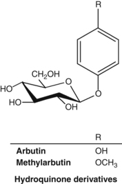
• Hydroquinone glycosides (normally between 6.3% and 9.2% – higher in autumn crops)12 including arbutin and methylarbutin13
• Polyphenols (predominantly gallotannins); phenolic acids, flavonoids, triterpenes.13
Interestingly, arbutin is found at high concentrations in some plants capable of surviving extreme and sustained dehydration.14
There is some debate as to whether the antimicrobial effect of bearberry is due to hydroquinone esters such as arbutin or to free hydroquinone.13 The antimicrobial activities of arbutin and an aqueous extract of bearberry were tested in vitro against bacterial strains implicated in urinary tract infections. The antibacterial activity of arbutin was directly correlated with the beta-glucosidase activity of the bacteria. (This enzyme converts arbutin into free hydroquinone.) The highest enzyme activity was found in Streptococcus, Klebsiella and Enterobacter, the lowest in Escherichia coli.15 Arbutin (128 μg/L) inhibited three of eight clinical isolates of Pseudomonas aeruginosa tested in vitro.16 Arbutin and hydroquinone inhibited the growth of Ureaplasma urealyticum and Mycoplasma hominis in vitro.17 These bacteria are associated with non-gonococcal urethritis.
Piceoside, a glucoside isolated from bearberry, did not demonstrate antibacterial activity in vitro, but its aglycone p-hydroxyacetophenone showed activity against Proteus vulgaris, Enterobacter aerogenes and Bacillus subtilis.18 Another study found antimicrobial activity for bearberry extracts in vitro against E. coli, Proteus vulgaris, Enterobacter aerogenes, Streptococcus faecalis, Staphylococcus aureus, Salmonella typhi and Candida albicans.19 The summer and autumn leaves were more potent than the winter leaves.20
The antibacterial activity of various agents was tested in vitro using 74 different strains of bacteria isolated from the urinary tract including E. coli, Proteus mirabilis, Pseudomonas aeruginosa, Staphylococcus aureus and species of Enterobacter, Citrobacter and Klebsiella. Urine was collected from healthy volunteers 3 h after oral administration of 0.1 g or 1.0 g of arbutin; several synthetic antibiotics were also tested. Of all test substances, only gentamicin, nalidixic acid and urine collected after intake of 1.0 g of arbutin and adjusted to pH 8 were active against every strain used.21
A hydroethanolic extract of bearberry was amongst the most active of 14 traditional Canadian medicinal plants tested against a wide variety of strains of Neisseria gonorrhoeae, including isolates resistant to antibiotics.22 A minimum inhibiting concentration of 32 μg/mL was demonstrated in vitro.
A study of the dry leaf extract of bearberry on the course of acute bacterial pyelonephritis (caused by E. coli) in white rats showed that bearberry extract (25 mg/kg) had a marked antibacterial and nephroprotective effect.23
Samples of both normal urine and urine collected from healthy subjects after consuming bearberry tea were compared for resistance to bacterial contamination. Urine from the bearberry tea drinkers was more bacteriostatic than normal urine samples. However, the addition of arbutin to normal urine did not result in the same bacteriostatic activity. A series of solutions were then tested for their inhibition of the growth of strains of Staph. aureus and E. coli in vitro. The solutions included hydroquinone, methylhydroquinone and arbutin, normal urine and urine from bearberry tea drinkers. They were tested at normal pH and at pH elevated to 8.0 by the addition of potassium hydroxide. Only the hydroquinone and methylhydroquinone solutions inhibited bacterial growth at normal pH. However, incubation of bacteria with bearberry tea drinkers’ urine adjusted to pH 8 also resulted in inhibition of bacterial growth. This inhibitory effect was also seen for urine adjusted to pH 8 from subjects given pure arbutin.24 The authors suggested that antibacterial activity would only occur when the excretion products of arbutin (hydroquinone paired with glucuronate and sulphate) appear in sufficiently high concentrations in an alkaline urine. It was hypothesised that at an alkaline pH these excretion products of arbutin released small amounts of free hydroquinone in the presence of bacteria, thereby conferring antibacterial activity to the urine. The maximum antibacterial effect from the hydroquinone glucuronides and sulphates formed from arbutin was obtained about 3 to 4 h after taking the herb.25 Free hydroquinone is only excreted in trace amounts, which is desirable given the toxic potential of this agent.
A recent paper has expressed a contradictory view. In this study, urine was collected from four healthy individuals after ingestion of 420 mg of arbutin.26 The samples were added to an E. coli suspension and it was noted that the concentration of hydroquinone in bacteria was 20-fold higher than a control. The authors commented that the pH of urine is unlikely to be important, as the intracellular pH of E. coli is not affected by urine alkalinisation. They concluded that deconjugating enzymes such as beta-glucuronidases found in the bacteria enrich and deconjugate hydroquinone glucuronides and/or sulphates regardless of pH. The pH of the test urine was not stated, but in the study design attempts to increase it to more than 6.5 via a vegetarian diet were instituted.
Urine produced by a healthy person consuming a meat and fish diet is typically in the pH range 4.5 to 6.0; a vegetarian diet will make urine more alkaline. A urine of pH >7 during a urinary tract infection indicates infection by a micro-organism capable of splitting urea, with the release of ammonia.27 Urea-splitting organisms include Proteus spp., Klebsiella spp., some Citrobacter spp., some Haemophilus spp., Bilophila wadsworthia, the yeast Cryptococcus neoformans and several other bacteria and fungi.28 Infection with these organisms should be particularly susceptible to treatment with bearberry if alkaline urine does enhance its activity. Alkalinisation of the urine with buffering agents (for example containing sodium bicarbonate, sodium citrate, citric acid and tartaric acid) in conjunction with bearberry intake may prove to be clinically effective for the treatment of cystitis caused by non-urea-splitting bacteria, but given the conflicting findings noted above, this requires further research.
The antibacterial effect of bearberry may also be useful in the gastrointestinal tract. An aqueous extract of bearberry was found to modulate cell surface hydrophobicity and demonstrated antibacterial effects on ten strains of Helicobacter pylori in vitro.29 The research team concluded that the hydrolysable tannins were largely responsible, as pure tannic acid produced comparable results.
Co-administration of arbutin (50 mg/kg, oral) and indomethacin (subcutaneous) showed an inhibitory effect on swelling in a delayed-type hypersensitivity model, which was stronger than that of indomethacin alone.30 In the same model, arbutin (10 and 50 mg/kg, oral) plus prednisolone or dexamethasone showed stronger effects than each of the anti-inflammatory drugs alone.31 Arbutin may therefore have a synergistic anti-inflammatory activity on type IV allergic reaction-induced inflammation. In the same model, oral administration of a bearberry methanolic extract (100 mg/kg) demonstrated an inhibitory effect on swelling. When administered simultaneously with subcutaneous prednisolone, the inhibitory effect was more potent than that of prednisolone alone.32
Although ointments containing 1% and 2% aqueous extract of bearberry did not inhibit the ear swelling caused by experimentally induced contact dermatitis or carrageenan-induced paw oedema in rats and mice, they did increase the anti-inflammatory effect of a steroid ointment (dexamethasone). Co-administration of bearberry did not increase the side effects of dexamethasone.33 Topical doses of bearberry might also increase the anti-inflammatory effects of other steroid-like compounds, such as plant-derived saponins.
Arbutin at a concentration of 5×10⁻⁵ M decreased melanin content to approximately 39% when compared to untreated melanoma cells in vitro, without affecting cell growth. Tyrosinase activity also dropped significantly in the arbutin-treated cells. (This enzyme is involved in melanin synthesis.) Arbutin was not hydrolysed to hydroquinone, suggesting that the observed inhibitory effect was for arbutin itself, not hydroquinone.34 Further studies have revealed that the depigmenting mechanism of arbutin in humans involves inhibition of melanosomal tyrosinase activity, rather than the suppression of expression and synthesis of tyrosinase.35
A 50% methanolic extract of bearberry inhibited melanin synthesis in vitro. Both the bearberry extract and arbutin had an inhibitory effect on tyrosinase activity and inhibited the production of melanin by both tyrosinase and autoxidation.7 Bearberry extract could have a bleaching effect on freckles and may assist in the control of hyperpigmentary disorders.
Oral doses of arbutin (50 mg/kg) suppressed experimentally induced cough reflex. The effect of arbutin was stronger than that of the non-narcotic antitussive dropropizine and comparable to that of codeine.36
Oral administration of a bearberry infusion (3 g/L in drinking water) to healthy rats fed a standard diet containing calcium (8 g/kg) and magnesium (2 g/kg) did not induce significant diuresis, nor affect calcium or citrate concentration levels.37
Aqueous and methanolic extracts of bearberry have demonstrated in vitro molluscicidal activity against the freshwater snail Biomphalaria glabrata (the intermediate host of schistosomiasis). The methanol extract was active at a concentration of 50 ppm.38
A methanol extract of bearberry showed algicidal activity when tested in ponds. It is believed the tannins precipitated the algal proteins.39
Urinary excretion of phenolic metabolites after the oral administration of either bearberry leaf tea or arbutin occurs within 1 to 2 h and reaches a maximum 4 h after administration. In healthy subjects given bearberry tea, 70% to 75% of the administered dose was excreted within 24 h. Arbutin is altered after its passage through the body; it only occurs in trace amounts in urine when high doses are given. Free hydroquinone is only excreted in trace amounts, if at all.
It has been suggested that there are two possible processes for the absorption and metabolism of arbutin.40 The major process involves the absorption of intact arbutin by small intestinal enterocytes via the sodium-glucose pump. On first-pass metabolism in the liver, arbutin is deconjugated to hydroquinone and then reconjugated to sulphate and glucuronide phase II derivatives, which are then excreted via the urine. The minor process involves the conversion to free hydroquinone of arbutin in the colon by the action of bacterial beta-glucosidase. The hydroquinone is then converted by colonic enterocytes into sulphate and glucuronide derivatives which are absorbed into the bloodstream and passed into the urine.
In a crossover study involving six healthy volunteers, enteric-coated bearberry tablets demonstrated the same bioavailability within a 24-h period as an equivalent bearberry extract. The release of arbutin metabolites was retarded by at least 3 h with the tablets. In a pilot study conducted prior to this main study, no free hydroquinone was found in the urine of volunteers, although the above hydroquinone derivatives were found.41 This study was designed to compare the bioavailability of enterically coated tablets containing bearberry extract with uncoated tablets, but it does also add some support to the above metabolic pathways. Additionally, a small, randomised crossover trial in sixteen adults evaluated the bioavailability of an aqueous solution of bearberry as compared to film-coated tablets.42 The maximum mean urinary concentration of hydroquinone equivalents was marginally higher and peaked a little earlier in the tea group, although this was not statistically significant. The authors concluded there were no significant differences between the two groups in terms of metabolites or total amounts of hydroquinone equivalents excreted.
Some other studies investigating the elimination of arbutin in rats have arrived at different conclusions, which cast doubt on their relevance to humans. In these studies, orally administered arbutin was excreted unchanged in urine43 and oral administration of bearberry tea resulted in the excretion of six unidentified phenolic compounds, but no hydroquinone. No degradation products were observed after the perfusion of isolated rat liver with arbutin, thus leading to the conclusion that it was hydrolysed in the kidneys.44 As noted above, the results of these studies are not supported by the human studies. Moreover, in the case of the orally administered arbutin, the authors may have actually measured arbutin metabolites and mistakenly assigned them as arbutin.
In a double blind, placebo-controlled, randomised clinical trial, 57 women who had experienced at least three episodes of cystitis during the preceding year received either herbal medicine or placebo. The herbal medicine consisted of bearberry extract (standardised for arbutin and methylarbutin content) and extract of dandelion root and leaf (dose of individual herbs not specified). Treatment for 1 month significantly reduced the recurrence of cystitis during the 1-year follow-up period, with no incidence of cystitis in the herbal group and a 23% occurrence in the placebo group (p<0.05). No side effects were reported.45
Hydroquinone is a recognised toxic compound. However, arbutin and bearberry extracts are considerably less toxic than hydroquinone, as somewhat evidenced by the studies cited below.
The oral LD50 of hydroquinone as a 2% aqueous solution has been reported as between 320 and 550 mg/kg in various laboratory animals.46 Hydroquinone is non-mutagenic in the Ames test but induces chromosome aberrations and karyotypic effects in eukaryotic cells.47 In contrast, arbutin did not induce mutations in concentrations up to 10−2 M in a gene mutation assay. An increase in mutation frequency was observed with concentrations of 10−3 M and higher when arbutin was preincubated with beta-glucosidase. Hydroquinone, used as a positive control, also exhibited clear effects. In vivo, hydroquinone administered by intraperitoneal injection induced elevated micronucleus incidences. However, there was no induction of micronuclei in bone marrow when arbutin was administered orally (0.5 to 2.0 g/kg). This research suggests that arbutin itself is not mutagenic, but any generated hydroquinone could exert a mutagenic potential.48
According to the British Herbal Compendium, bearberry is contraindicated in kidney disorders13 but there is no evidence to support this, and the contraindication probably arose out of a theoretical caution.
According to the Commission E, bearberry is contraindicated in pregnancy and lactation and for children less than 12 years of age.49
Bearberry is not suitable for prolonged use. Use cautiously in highly inflamed or ulcerated conditions of the gastrointestinal tract.50
In principle, the prolonged use of herbs with high levels of tannins is inappropriate in constipation, iron deficiency anaemia and malnutrition.51
Concomitant acidification of the urine (for instance by medication) may result in a reduction of efficacy,24 although this is hypothetical and of uncertain relevance to the urinary antiseptic mechanism of bearberry.
Oral or topical use of bearberry or arbutin has been observed to augment the anti-inflammatory effects of indomethacin,30 prednisolone,31,32 and dexamethasone31,33 in experimental models. The clinical relevance of these findings is uncertain.
Bearberry extract markedly potentiated the action of beta-lactam antibiotics against methicillin-resistant Staph. aureus in vitro. The constituent corilagin (a polyphenol) was responsible for the activity.52 However, whether this leads to a clinical interaction is uncertain.
The high tannin levels will cause interference with the absorption of various nutrients and drugs, especially metal ions, thiamine and alkaloids. Bearberry should be consumed at least 2 h away from oral thiamine, mineral supplements such as iron and alkaloid-containing drugs.51
Pregnancy category C – has caused or is associated with a substantial risk of causing harmful effects on the foetus or neonate without causing malformations.10
There is a minor theoretical risk to fetal development due to the uterotonic properties of arbutin in vivo.53,54 However, arbutin also occurs in food: wheat products (1 to 10 ppm), pears (4 to 15 ppm), and coffee and tea (0.1 ppm).55
The transfer of arbutin or hydroquinone to breast milk is not advisable, and therefore the herb should be avoided in lactation.
Hydroquinone depigmenting creams may cause exogenous ochronosis (hyperpigmentation)56 and/or allergic contact dermatitis.57 However, these side effects have not been reported for cosmetic creams containing bearberry.56
A case of bilateral bull’s-eye maculopathy has been reported in a 56-year-old woman after ingestion of bearberry tea for 3 years, dose unknown.58 While it is generally acknowledged that bearberry inhibits melanin production, which is present in ocular tissue, therapeutic doses for short periods of time are routinely considered safe.
Due to the high tannin content, internal use of high doses of bearberry may cause cramping, nausea, vomiting and constipation.
Bearberry is official in the European Pharmacopoeia (2006).
Bearberry is covered by a positive Commission E Monograph and has the following application: inflammatory disorders of the lower urinary tract.
Bearberry is on the UK General Sale List and in France the herb is accepted for the internal treatment of benign urinary infections and to promote the renal elimination of water.
Bearberry does not have GRAS status. However, it is freely available as a ‘dietary supplement’ in the USA under DSHEA legislation (1994 Dietary Supplement Health and Education Act). Bearberry has been present in the following over-the-counter (OTC) drug products: weight control drug products and orally administered menstrual drug products. The FDA, however, advises: ‘that based on evidence currently available, there is inadequate data to establish general recognition of the safety and effectiveness of these ingredients for the specified uses’.
Bearberry is not included in Part 4 of Schedule 4 of the Therapeutic Goods Act Regulations of Australia and is freely available for sale.
References
1. Mabberley DJ. The Plant Book, 2nd ed. Cambridge: Cambridge University Press, 1997. p. 53
2. Evans WC. Trease and Evans’ Pharmacognosy, 14th ed. London: WB Saunders, 1996. p. 223
3. Brinker FJ. Eclectic Dispensatory of Botanical Therapeutics, vol 2, Section 1: Native Healing Gifts. Sandy: Eclectic Medical Publications, 1995. pp. 19–23
4. Grieve MI, A Modern Herbal, New York, Dover, 1971;vol 1. pp. 89–90
5. Felter HW, Lloyd JU. King’s American Dispensatory, 18th ed., 3rd rev, vol 2, 1905. Reprinted by Eclectic Medical Publications; 1983. pp. 2038–2040.
6. British Herbal Medicine Association’s Scientific Committee. British Herbal Pharmacopoeia. West York: BHMA, 1983. pp. 29–30
7. Matsuda H, Nakamura S, Shiomoto H, et al. Yakugaku Zasshi. 1992;112(4):276–282.
8. Frohne D. Pharm Ztg. 1980;125:2582–2583.
9. Launert EL. The Hamlyn Guide to Edible and Medicinal Plants of Britain and Northern Europe. London: Hamlyn, 1981. p. 128
10. Mills S, Bone K. The Essential Guide to Herbal Safety. St Louis: Elsevier, 2005. p. 259
11. Lange D. Europe’s Medicinal and Aromatic Plants: Their Use, Trade and Conservation. Traffic International, 1998. p. III
12. Parejo I, Viladomat F, Bastida J, et al. Phytochem Anal. 2001;12(5):336–339.
13. British Herbal Medicine Association, British Herbal Compendium, Bournemouth, BHMA, 1992;vol 1. pp. 211–213
14. Oliver AE, Crowe LM, De Araujo PS, et al. Biochim Biophys Acta. 1996;1302(1):69–78.
15. Jahodar L, Jilek P, Patkova M, et al. Cesk Farm. 1985;34(5):174–178.
16. Ng TB, Ling JM, Wang ZT, et al. Gen Pharmacol. 1996;27(7):1237–1240.
17. Robertson JA, Howard LA. J Clin Microbiol. 1987;25(1):160–161.
18. Jahodar L, Kolb I. Pharmazie. 1990;45(6):446.
19. Holopainen M, Jabodar L, Seppanen-Laakso T, et al. Acta Pharm Fenn. 1988;97(4):197–202.
20. Skvortsov SS, Khan-Fimina VA. Fitontsidy Mater Soveshch. 1969;6:207–209.
21. Kedzia B, Wrocinski T, Mrugasiewicz K, et al. Med Dosw Mikrobiol. 1975;27:305–314.
22. Cybulska P, Thakur SD, Foster BC, et al. Sex Trans Dis. 2011;38(12):1–5.
23. Nikolaev SM, Shantanova LN, Mondodoev AG, et al. Rastitel’Nye Resursy. 1996;32(3):118–123.
24. Frohne D. Planta Med. 1970;18:1–25.
25. German Federal Minister of Justice. German Commission E for Human Medicine Monograph, Bundes-Anzeiger (German Federal Gazette) no. 228, dated 05.12.84 and no. 109, dated 15.06.1994.
26. Siegers C, Bodinet C, Ali SS, et al. Phytomedicine. 2003;10(suppl 4):58–60.
27. Bouchier IAD, Morris JS, eds. Clinical Skills – A System of Clinical Examination, 2nd ed, London: WB Saunders, 1982. p. 243
28. Baron EJ, Peterson LR, Finegold SM. Bailey and Scott’s Diagnostic Microbiology, 9th ed. St Louis: Mosby Year Book, 1994. p. 106
29. Annuk H, Hirmo S, Turi E, et al. FEMS Microbiol Lett. 1999;172(1):41–45.
30. Matsuda H, Tanaka T, Kubo M. Yakugaku Zasshi. 1991;111(4–5):253–258.
31. Matsuda H, Nakata H, Tanaka T, et al. Yakugaku Zasshi. 1990;110(1):68–76.
32. Kubo M, Ito M, Nakata H, et al. Yakugaku Zasshi. 1990;110(1):59–67.
33. Matsuda H, Nakamura S, Tanaka T, et al. Yakugaku Zasshi. 1992;112(9):673–677.
34. Akiu S, Suzuki Y, Asahara T, et al. Nippon Hifuka Gakkai Zasshi. 1991;101(6):609–613.
35. Maeda K, Fukuda M. J Pharmacol Exp Ther. 1996;276(2):765–769.
36. Strapkova A, Jahodar L, Nosalova G. Pharmazie. 1991;46(8):611–612.
37. Grases F, Melero G, Costa-Bauza R, et al. Int Urol Nephrol. 1994;26(5):507–511.
38. Schaufelberger D, Hostettmann K. Planta Med. 1983;48(2):105–107.
39. Ayoub SMH, Yankov LK, Hussein-Ayoub SM. Fitoterapia. 1985;6(4):227–229.
40. Garcia de Arriba S, Stammwitz U, Pickartz S, et al. Z Phytother. 2010;31(2):95–97.
41. Paper DH, Koehler J, Franz G. Pharm Pharmacol Lett. 1993;3:63–66.
42. Schindler G, Patzak U, Brinkhaus B, et al. J Clin Pharmacol. 2002;42(8):920–927.
43. Jahodar L, Leifertova I, Lisa M. Pharmazie. 1983;38(11):780–781.
44. Leifertova I, Lisa M, Jahodar L, et al. Rozvoj Farm Ramci Ved-Tech Revoluce, Sb Prednasck Sjezdu Cesk Farm Spol, 7th ed. Prague: University of Karlova, 1979. Meeting date 1977; pp. 41–43
45. Larsson B, Jonasson A, Fianu S. Curr Ther Res Clin Exp. 1993;53(4):441–443.
46. Woodard G, Hagan CE, Radomski JL. Fed Proc. 1949;8:348.
47. Devillers J, Boule P, Vasseur P, et al. Ecotoxicol Environ Saf. 1990;19(3):327–354.
48. Mueller L, Kasper P. Mutat Res. 1996;360(3):291–292.
49. German Federal Minister of Justice. German Commission E for Human Medicine Monograph, Bundes-Anzeiger (German Federal Gazette) no. 109, dated 15.06.94; no. 19, dated 28.01.1994.
50. Blumenthal M, Busse WR, Goldberg A, et al. The Complete German Commission E Monographs. Texas: American Botanical Council, 2000. pp. 224–225
51. Mills S, Bone K. The Essential Guide to Herbal Safety. St Louis: Elsevier, 2005. p. 260
52. Shimizu M, Shiota S, Mizushima T, et al. Antimicrob Agents Chemother. 2001;45(11):3198–3201.
53. Shipochliev T. Vet Med Nauki. 1981;18(4):94–98.
54. Itabashi M, Aihara H, Inoue T, et al. Iyakuhin Kenkyu. 1988;19:282–297.
55. Deisinger PJ, Hill TS, English JC. J Toxicol Environ Health. 1996;47(1):31–46.
56. Howard KL, Ferner BB. Cutis. 1990;45(3):180–182.
57. Engasser PG, Maibach HI. J Am Acad Dermatol. 1981;5(2):143–147.
58. Wang L, Del Priore LV. Am J Ophthalmol. 2004;137(6):1135–1137.
59. Stübler M, Krug E. Leesers Lehrbuch der Homöopathie, Pflanzliche Arzneistoffe II. Heidelberg: Haug-Verlag, 1988. pp. 403–406
60. Standardzulassung für Fertigarzneimittel. Pharmazeutischer Verlag, Deutscher Apotheker, Frankfurt/Main, 1987/89, Verlag.
Berberis bark and Hydrastis root
Berberis vulgaris: barberry (Engl), Berberidis cortex (Lat), Berberitze, Sauerdorn (Ger), epinevinette, vinettier (Fr), berberi (Ital), almindelig Berberis (Dan). Hydrastis canadensis: golden seal (Engl), Hydrastidis rhizoma (Lat), Goldsiegel, Kanadische Gelbwurzel (Ger), guldsegl (Dan).
There are more than 500 species of the Berberis genus. Berberis vulgaris, the common or European barberry, is indigenous to Europe and naturalised in Britain. Many parts of the plant have been utilised: the fine wood for turning, the root and stems providing dyestuff for fabrics, leather and wood (also formerly a hair dye) and the fruit for jams.1 The root and stem bark are used medicinally.
Hydrastis canadensis was known to the Cherokee nation long before the settlement of America by Europeans. They employed its underground portion for dyeing and as an internal remedy, and acquainted the early settlers with most of its properties.2 Hydrastis became a very prominent herb in the Eclectic tradition. The plant is indigenous to central and eastern North America, but its population is now much reduced through overexploitation.3,4 Hence, it is preferable to use cultivated sources of Hydrastis because of its endangered status. The high price commanded by the root and rhizome means that Hydrastis is susceptible to adulteration.
The activities of Berberis and Hydrastis are thought to be mainly due to their isoquinoline alkaloids, in particular berberine and hydrastine (the latter occurs only in Hydrastis). Other plants also contain berberine, but will generally not be examined in this monograph unless they provide some relevant insights regarding this phytochemical. However, most of the research cited here for berberine will be relevant to its other herbal sources such as Coptis chinensis and Phellodendron amurense.
Berberis vulgaris: controls gastrointestinal infections; improves the flow of bile.
Hydrastis canadensis: in addition to the above, restores the integrity of mucous membranes of the respiratory and digestive tract, and promotes gastric digestive processes.
Berberis vulgaris has had a long history of use in Western herbalism. A decoction was taken in the spring months as a blood purifier and used externally as a mouth and eyewash. The Eclectics regarded Berberis primarily as a tonic, but it was also used for conditions affecting the liver and gallbladder, and for diarrhoea, dysentery and parasitic infestations including malaria.5,6
Hydrastis canadensis was specifically indicated for catarrhal states of the mucous membranes when unaccompanied by acute inflammation (except in the case of acute purulent otitis media, where it was considered to work better than in the chronic condition). Muscular debility was another key traditional indication. As a bitter stomachic, it was used to sharpen appetite and aid digestion, and was considered valuable for disordered states of the digestive apparatus, especially when functional in character. Hydrastis was considered a valuable local agent in affections of the nose and throat. It was also used in cutaneous diseases, especially when dependent upon gastric difficulties; concurrent internal and external use was said to hasten the cure.7 In addition, Hydrastis was recommended for submucosal myoma, haemorrhagic endometriosis and heavy menstrual bleeding.8
Berberis vulgaris: antimicrobial, cholagogue, choleretic, antiemetic, mild laxative, bitter.
Hydrastis canadensis: stomachic, reputed oxytocic, antihaemorrhagic, anticatarrhal, trophorestorative for mucous membranes, antimicrobial, bitter, anti-inflammatory, depurative, vulnerary, choleretic.
From clinical trials on berberine: acute infectious diarrhoea; trachoma (as eyedrops); giardiasis; hypertyraminaemia; type 2 diabetes mellitus; elevated blood lipids; protection from radiation injury; cutaneous leishmaniasis (topically).
Berberis vulgaris: jaundice (when there is no obstruction of the bile ducts); biliousness, cholecystitis, gallstones; functional derangement of the liver; digestive stimulant, diarrhoea; in larger doses for constipation.5,6,9
Hydrastis canadensis: catarrhal states of the mucous membranes when unaccompanied by acute inflammation (except in acute purulent otitis media); disordered states of the gastrointestinal tract (particularly gastritis, gastric ulcer, diarrhoea) including conditions with hepatic symptoms; as a tonic during convalescence; haemorrhagic conditions of the uterus and pelvis (but it was considered too slow for active postpartum haemorrhage); internally and externally for skin disorders including eczema and acne, especially with gastrointestinal involvement; discharges from the genitourinary tract (e.g. leucorrhoea, gonorrhoea); disorders of the ear, nose, mouth, throat; externally for superficial disorders of the eye (but has no suggested value in intraocular infection).7
Berberine-containing herbs may also be used for bacterial and fungal infections, protozoal infections (cutaneous and visceral leishmaniasis, amoebic dysentery, malaria, giardiasis, trichomoniasis), tapeworm infestation; possibly as an adjunct in treatment of congestive heart failure, arrhythmia; possibly in prevention of cancer; thrombocytopenia.
Hydrastis: the above indications plus anorexia and conditions requiring increased flow of gastric juices; conditions of visceral and/or smooth muscle spasm.
Hydrastis was used as a component of eyewashes, and both Hydrastis and Berberis are used in bitter tonic preparations. Berberine salts are used in ophthalmic products, usually in eyedrops and eyewashes.10 Despite the traditional contraindication for Hydrastis in acute respiratory infections such as the common cold, it is often used in this way, particularly in modern practice in the USA. Notwithstanding some popular use, Hydrastis has no value in masking drug-screening tests.
Dried or fresh stem bark or root bark (Berberis) or rhizome and rootlets (Hydrastis) for decoction, liquid extract, tincture, tablets and capsules for internal or external use.
• Berberis vulgaris: 1.5 to 3 g/day of the dried root or stem bark or 3 to 6 mL/day of the 1:2 liquid extract; 7 to 14 mL/day of the 1:5 tincture
• Hydrastis canadensis: 0.7 to 2 g/day of the dried rhizome/root or 2 to 5 mL/day of the 1:3 tincture; 3.5 to 8.0 mL/day of the 1:5 tincture.
Equivalent herb doses can also be taken in tablet or capsule form.
Higher doses of both herbs are necessary in acute conditions and to achieve the clinical effects for berberine noted in most of the clinical trials.
No adverse effects from ingestion of either Berberis or Hydrastis are expected when used within the recommended dosage. Berberine-containing plants are not recommended for use during pregnancy, although there is a view that this concern is overstated. High doses of berberine increase the bioavailability of cyclosporin.
Berberis vulgaris, a member of the Berberidaceae family, is a deciduous shrub 0.75 to 1.75 m tall with thick, creeping roots and a much-branched, greyish stem. The leaves are arranged in clusters on short axillary shoots, obovate to oblong-obovate, up to 4 cm long with spiny-toothed margins and short petioles. Its yellow flowers are six-sepalled and six-petalled, falling in loose clusters. The edible berries are red, oblong and about 1 cm in size.11,12
Hydrastis canadensis, a member of the Ranunculaceae (buttercup) family, is a small perennial. The stems are purplish and hairy above ground, but below the soil the root hairs and rhizome are yellow. The yellow rhizome is characteristically marked with depressions caused by the falling away of the annual stems (hence golden seal, as in the impression in wax once used to seal letters). The rhizome is about 5 cm in length, producing a profusion of yellow roots at its sides, 30 cm or more in length. The stems bear two or three large, slightly hairy five part leaves. The small solitary, greenish white or rose-coloured flower develops into a berry-like fruiting head, bright red in colour when fully ripe, resembling a raspberry and containing 10 to 30 black seeds.13
The Ranunculaceae and Berberidaceae are part of the same order (Ranunculales).
Due to the price of genuine Hydrastis, commercial products have been found not to contain the authentic plant material.14,15 Hydrastis was listed on Appendix II of the Convention on International Trade in Endangered Species (CITES) as of 18 September 199716 and is currently listed. It is preferable to use cultivated sources (rather than wildcrafted sources) of Hydrastis because of its endangered status.
A 1933 source indicated that Berberis aristata was often confused with other Berberis spp. (such as B. lycium, and B. vulgaris) in India.17 Commercially available barberry root bark may contain branch and stem bark. Berberis vulgaris is a protected species in one or more regions of France.18
Berberis vulgaris (root bark):
• Alkaloids (up to 13%), including those of the isoquinoline group: protoberberines (berberine (up to 6%), jatrorrhizine, palmatine) and bisbenzylisoquinolines (total <5%, including oxyacanthine).19 Levels are much lower in the stem bark.
The activities of Berberis and Hydrastis are thought to be largely due to the presence of their isoquinoline alkaloids. An earlier pharmacological review indicated that berberine has the following activities:21
• Antimicrobial, antifungal, antiparasitic
• Antidiarrhoeal, intestinal antisecretory, inhibits enterotoxins, cholera toxin antagonist
• Antiarrhythmic, positive inotropic (cardiotonic)
• Cytotoxic, antimitotic, antitumoral, increases the action of antitumoral agents, inhibits the action of carcinogens
• Cholagogue, choleretic, increases bilirubin excretion
• Mydriatic (dilates the pupil), increases lacrimal secretion
More recently discovered properties (see below) include hypolipidaemic, antidepressant and anticonvulsant activities.
Hydrastine has the following activities:22
Berberine possesses extensive antimicrobial activity and does appear, in general, to be more active against Gram-positive bacteria (see Table 1). However, most of the research has been published before 2000, with relatively few studies conducted since.
Berberine sulphate blocked the adhesion of a uropathogenic strain of E. scherichia coli, in vitro. The reduction in adherence is related to the loss of the synthesis and expression of fimbriae (hairlike appendages) on the surface of the berberine-treated bacteria. Inhibition of microbial adherence results in termination of infection and may explain the anti-infectious activity of berberine in E. coli urinary tract infections, since the direct antimicrobial activity of berberine against E. coli is relatively low (see Table 1).23 Berberine reduces FtsZ (a protein involved in bacterial cell division) in E. coli, thereby inhibiting the replication of this organism.24
Berberine has also demonstrated antibacterial activity against methicillin-resistant Staphylococcus aureus (MRSA) in vitro.25 It was effective against all strains of MRSA, with 90% growth inhibition obtained at concentrations of 64 μg/mL or less. Berberine also restored the efficacy of antibiotics commonly used for MRSA. It was found to have an additive effect when combined with ampicillin, and a synergistic activity with oxacillin. Another study found that berberine chloride exhibited antimicrobial activity against all 43 tested strains of Staph. aureus.26 Biofilm formed by Staphylococcus epidermidis is a common cause of infection in orthopaedic joint prostheses. Berberine was found to inhibit Staph. epidermidis adhesion to titanium alloy at a concentration of 45 μg/mL and prevented biofilm formation.27,28
Berberine sulphate has demonstrated antimycotic activity against several fungal species (see Table 1). Relatively high concentrations of 10 to 25 mg/mL inhibited the growth of Alternaria, Aspergillus flavus, Asp. fumigatus, Candida albicans, Curvularia, Drechslera, Fusarium, Mucor, Penicillium, Rhizopus oryzae and Scopulariopsis. The growth of Syncephalastrum was inhibited by a concentration of 50 mg/mL.29 The minimum inhibitory concentration (MIC) of the antifungal drug fluconazole against Candida albicans was 1.9 μg/mL. However, this decreased to 0.48 μg/mL in the presence of just 1.9 μg/mL berberine.30
Berberine and protoberberine derivatives exhibited a potency comparable to that of quinine in vitro against two clones of human malaria: Plasmodium berghei and P. falciparum. None of the compounds, however, were active in P. berghei-parasitised mice.31 Berberine sulphate inhibited the growth of Entamoeba histolytica, Giardia lamblia and Trichomonas vaginalis in vitro and induced morphological changes in the parasites.32 Berberine chloride (1 μg/mL) significantly inhibited the growth of Leishmania donovani promastigotes by approximately 50%, in vitro. A concentration of 5 μg/mL resulted in complete inhibition of growth. L. donovani causes visceral leishmaniasis.33 A recent study confirmed a half maximal inhibitory concentration (IC50) for berberine chloride of 2.6 μg/mL against L. donovani promastigotes.34 The mechanism involved was apoptosis following enhanced oxidative damage.
In vivo berberine demonstrated significant activity (greater than 50% suppression of lesion size) against Leishmania braziliensis panamensis in golden hamsters.35 In both the 8-day and long-term models of L. donovani infection in hamsters, berberine markedly diminished the parasitic load, rapidly improved the haematological picture and was less toxic than pentamidine. Berberine sulphate administered into the lesion (1% four times per week) was found to be highly effective against cutaneous leishmaniasis in dogs. Cutaneous leishmaniasis (oriental sore) is caused by the protozoan Leishmania tropica.36
Berberine hydrochloride demonstrated a high degree of activity against E. histolytica in vitro with a minimum amoebicidal concentration of 10 μg/mL. Oral administration of berberine (100 mg/kg) to rats with experimental amoebiasis (protozoal infection) reduced the infection by 83%. Berberine also reduced the level of infection to 20% in infected hamsters.37
Studies have shown that certain antimicrobial agents can block the adherence of micro-organisms to host cells at doses much lower than those needed to kill cells or inhibit cell growth. At concentrations below the MIC, berberine caused an increase in release of lipoteichoic acid (LTA) from streptococci. LTA is the major ligand responsible for the adherence of the bacteria to host cells, including host cell receptors (fibronectin). Release of LTA from the streptococcal cells means a reduction in the capacity of the bacteria to adhere to the host. Berberine also interfered with bacterial adherence by directly preventing the complexing of LTA with fibronectin or by dissolving the complexes once they were formed.38 (See also the berberine research above regarding biofilms.)
Antimicrobial research on hydrastine is more limited. It exhibited relatively weaker killing activity than berberine against Staph. aureus and Streptococcus sanguis.39 An MIC50 of 100 μg/mL was demonstrated against Helicobacter pylori (see also later under Pharmacodynamics of the herbs).40 The effect of hydrastine on the protoscolices (larvae) of the tapeworm Echinococcus granulosus was measured in vitro and in vivo. Hydrastine at 0.3% concentration produced 70% mortality of the larvae in both experiments.41
Berberine demonstrated in vitro antiviral activity against herpes simplex virus types 1 and 2 with an IC50 of 82 and 90 μg/mL, respectively.42 The in vitro antiviral activity (IC50) of berberine chloride against human cytomegalovirus using the plaque assay was 0.68 μM (0.25 μg/mL), which was similar to the drug ganciclovir.43
Oral doses of berberine (>25 mg/kg) and Geranium extract (a source of tannins) showed significant inhibition of diarrhoea in mice, and both substances inhibited spontaneous peristalsis in rat intestine. Comparison with atropine and papaverine indicated that the antidiarrhoeal activity of berberine differs from that of Geranium extract.47 Intragastric administration of berberine sulphate reduced the purging effect of castor oil or Cassia angustifolia (senna) leaf in mice. It did not affect the gastrointestinal transport of Chinese ink in normal mice.48
A goal of therapy for diarrhoeal diseases is to decrease stool water. This can be done in two ways: by increasing the absorption of water and electrolytes or decreasing the stimulated secretion of fluid. Berberine probably does the latter.49 It did not significantly alter normal ileal water and electrolyte transport as measured in vivo50 and in vitro.51 However, it inhibited secretion caused by Vibrio. cholerae and E. coli heat-labile enterotoxins, even when administered after the enterotoxin had bound to intestinal mucosa. The antisecretory effect was therefore not dependent on the type of enterotoxin.52 In contrast, while simultaneous oral administration of berberine (0.1 mg/rat) and E. coli enterotoxin resulted in a significant reduction in fluid accumulation (p<0.01), treatment with berberine prior to or after the enterotoxin was ineffective.53 Two studies are contradictory as to whether berberine alters cholera toxin-induced stimulation of the adenylate cyclase–cAMP system.44,54 Hence, the exact mechanism of action of berberine is not certain, but its site of action appears to be distal to second messenger production and may be at a level common to all stimuli of colonic chloride secretion.55
A more recent investigation of the antidiarrhoeal activity of berberine used a model of thyroid hormone-induced diarrhoea in rats and measured gastrointestinal peptides.56 While both plasma motilin and gastrin were elevated by thyroxine, they were normalised by the berberine (60 mg/kg/day for 7 days, oral). As a result, berberine also normalised increases in the number and volume of intestinal goblet cells.
A review concluded that berberine possesses a range of cardiovascular properties, including positive inotropic, negative chronotropic, antiarrhythmic and vasodilatory activities.57 An in vitro study indicated that berberine inhibits voltage-dependent and ATP-sensitive potassium channels, hence the hypoglycaemic and antiarrhythmic activity of berberine might be due to its potassium channel-blocking effect.58 Intravenous administration of berberine (1 mg/kg) decreased the amplitude of delayed after-depolarisations and blocked arrhythmias in rabbit ventricular muscles. The mechanism of antiarrhythmic activity of berberine might therefore instead be due to suppression of delayed after-depolarisations caused by a decrease in sodium influx.59
Berberine prevented the development of pressure-overload-induced left ventricular hypertrophy in vivo after aortic banding.60 Oral administration of 10 mg/kg decreased left ventricular and diastolic pressures, and whole heart and left ventricular weights were lower after 8 weeks of treatment. A follow-up study was conducted to investigate the effects of berberine on catecholamine levels in a similar experimental model.61 Plasma and left ventricular levels of adrenaline and noradrenaline were decreased after treatment with berberine. Berberine (5 to 10 mg/kg) improved cardiac contractility and inhibited left ventricular remodelling (especially myocardial fibrosis) in a rat model of hypertension.62 Such effects might be partially associated with increased nitric oxide (NO) and cAMP in left ventricular tissue.
Administration of berberine sulphate increased the number of thrombocytes, decreased the activity of factor XIII and promoted blood coagulation in intact and gamma-irradiated rats and mice.63 Berberine inhibited platelet aggregation and platelet adhesiveness in rats with reversible middle cerebral artery occlusion. Thromboxane B2 levels after treatment with berberine were lower than levels in untreated ischaemic controls. The decline of platelet aggregation and decrease of thromboxane B2 may be one of the important factors behind the anti-ischaemic activity of berberine.64 Berberine also markedly inhibited clot retraction in vitro, which may be due to direct inhibition of calcium ion influx.65
Recent research has also focused on the cholesterol-lowering activity of berberine and its mechanism of action. The compound upregulates hepatic LDL (low density lipoprotein) receptor expression in vitro via post-transcriptional mRNA stabilisation.66 This appears to be mediated by the extracellular signal-regulated kinase signalling pathway. Canadine, along with two other constituents of Hydrastis, have also demonstrated the same ability.67 While berberine and the whole root preparation of Hydrastis both upregulated the expression of LDL receptors in a human liver cell line (HepG2 cells), the latter preparation was more effective (see also later in this monograph). Berberine was also found to inhibit cholesterol and triglyceride synthesis in HepG2 cells by increasing AMP-activated protein kinase (AMPK).68
Oral administration of berberine (50 and 100 mg/kg/day) to hyperlipidaemic hamsters for 10 days resulted in 26% and 42% decreases in total cholesterol and LDL-cholesterol, respectively. A 3.5-fold increase in hepatic LDL receptor mRNA and a 2.6-fold increase in LDL protein was seen in hamsters taking the higher dose.69 LDL receptors in the liver increase the uptake and clearance of cholesterol from the bloodstream. The same researchers found that a combination of berberine (90 mg/kg/day, oral) and simvastatin (6 mg/kg/day, oral) in food-induced hyperlipidaemic rats was more effective than either agent alone at reducing serum LDL-cholesterol (p<0.01). A more effective reduction of serum triglyceride levels was also demonstrated.70 The combination also upregulated LDL receptor mRNA in the rat livers to a level 1.6 times higher than either monotherapy, with a corresponding reduction of liver fat storage and improvement in hepatic histology.
A different research group investigated the effect of berberine and plant stanols (inhibitors of cholesterol absorption from the diet) on plasma cholesterol levels in rats with diet-induced hyperlipidaemia. Berberine (100 mg/kg/day, oral) slightly, but not significantly, lowered plasma total cholesterol, while the plant stanols (1% in diet) showed a borderline significant reduction (18%, p=0.067).71 However, the combination markedly and significantly lowered plasma total cholesterol (41%, p=0.0002). Follow-up research by the same investigators sought to understand the mechanisms involved. Diet-induced hyperlipidaemic hamsters were chosen as the experimental model, since these animals are regarded as a good correlate for human cholesterol metabolism.72 Using the same doses as above, it was again demonstrated that total plasma cholesterol was most reduced by the combination (43%, p=0.0001).73 Neither berberine nor the plant stanols affected plasma triglyceride levels, whereas their combination resulted in a significant 37% reduction (p<0.001). As to the possible sites of action of berberine, a series of tests suggested that it interfered with intestinal cholesterol absorption in a different manner to the stanols, possibly by interfering with micelle formation. In addition bile acid synthesis was increased, which can increase cholesterol clearance via the digestive tract. In contrast, cholesterol synthesis by the liver was also increased (this is a known property of other cholesterol absorption inhibitors), but not by enough to counter the cholesterol-lowering mechanisms. Contrary to research cited above, the authors found that berberine did not affect hepatic LDL receptor mRNA expression.
Tissue factor (TF) plays an essential role in coagulation by binding factor VII, which activates factor X. TF is expressed in cells within the atherosclerotic vessel wall, is induced by inflammatory mediators and enhances plaque thrombogenicity. Human aortic endothelial cells treated with berberine (1 to 30 μM) exhibited enhanced TF expression following inflammatory stimulants.74 This was via stabilisation of TF mRNA. Also berberine countered the inhibitory effects of statins on endothelial TF in vitro. Berberine also enhanced TF and inhibited TFPI (tissue factor pathway inhibitor), a TF antagonist, in ApoE−/− mice after oral doses of 100 mg/kg/day. The authors suggested that this prothrombotic effect of berberine indicates it should be considered with caution as a cholesterol-lowering agent and that large-scale clinical trials are needed to prove its safety in this regard. In addition, berberine might promote atherosclerosis and foam cell formation by inducing scavenger receptor-A expression in macrophages (which mediates LDL uptake), as determined by in vitro and in vivo (mice, 5 mg/kg/day, ip) experiments.75
On the other hand, berberine might reduce the incidence or pathogenesis of arterial disease by other mechanisms: in vitro berberine inhibited lysophosphatidylcholine-induced vascular smooth muscle cell (VSMC) proliferation and migration via inhibition of intracellular reactive oxygen species, suggesting potential in the prevention of atherosclerosis.76 Berberine also inhibited the in vitro growth and migration of VSMC induced by platelet-derived growth factor.77 LDL oxidation and LDL-induced cytotoxic effects on human endothelial cells were also reduced by berberine in vitro.78 Atherosclerotic plaque rupture is usually the initiating event in a heart attack. Overproduction of MMPs (matrix metalloproteinases) by macrophages can lead to plaque rupture by degrading the extracellular matrix. Berberine reduced MMP-9 expression in activated macrophages in vitro.79
Berberine might benefit vascular endothelial cell integrity and function by improving protective mechanisms, such as increasing the production of NO and enhancing resistance to hyperglycaemic-induced injury. This was observed in blood vessels and vascular endothelial cells taken from mice and rats.80 In human studies, 1.2 g/day (oral) of berberine for 30 days mobilised circulating endothelial progenitor cells (improving small artery elasticity),81 augmented their function (a result of enhanced plasma NO levels)82 and reduced circulating endothelial microparticles in parallel with improved flow-mediated vasodilation.83 This suggests berberine has beneficial effects on human vascular endothelial health.
There have been numerous investigations into the anticancer properties of berberine, mainly via in vitro studies using human and animal cancer cell lines. A 2009 systematic review cited more than 100 studies, with most published since the year 2000.84 Antiproliferative activity at concentrations typically up to 100 μM (about 34 μg/mL) has been demonstrated in vitro for a range of tumour cell lines including liver, lung, breast, uterus, melanoma, prostate and leukaemia. According to the review, the antiproliferative effect of berberine is relatively slow and gentle, requiring exposure times of at least 24 h for significant effects. Various mechanisms underlying this antiproliferative/cytotoxic activity of berberine have been proposed, but the most widely investigated mechanism is its role in cell cycle arrest (with sometimes conflicting results). Induction of apoptosis is another possible key mechanism, via regulation of reactive oxygen species production, mitochondrial transmembrane potential and NF-kappaB activation. Other possible anticancer mechanisms according to the review include transcriptional regulation of some oncogene and carcinogenesis-related gene expression, interaction with DNA and RNA, and inhibition of key enzymes such as DNA topoisomerase.
The review also notes studies, relatively few in number, where berberine has inhibited metastasis, invasion and angiogenesis in vitro, especially via MMP inhibition. Despite earlier studies suggesting that berberine might be relatively inactive against cancer in vivo, later studies have demonstrated significant effects. The review cites studies for Dalton’s lymphoma ascites tumour cells in mice (ip treatment was more effective than oral) and human tongue squamous carcinoma, also in mice (berberine 10 mg/kg, ip). Berberine has also prevented cancer development in models of chemical carcinogenesis.
Another 2009 review of the antineoplastic activity of berberine noted many of the same studies and came to similar conclusions regarding mechanisms of action.85 The review also included additional positive in vivo studies for berberine including inhibition of leukaemia cells and lung tumours inoculated into mice (berberine dose 100 mg/kg, oral). Berberine also strongly suppressed the growth of human prostate cancer xenografts in mice. This review did caution that antagonistic activity of berberine on chemotherapeutic drugs has been observed in vitro, due to the upregulation of multidrug resistance pumps in cancer cells. Hence there is a risk that co-administered berberine might compromise cancer chemotherapy. However, in vivo research is needed to clarify this issue.
Berberine was shown to inhibit COX-2 transcription in human colon cancer cells (at >0.3 μM)86 and reduce COX-2 protein (but not enzyme activity) in an oral cancer cell line in vitro (1, 10 and 100 μM).87 Activator protein 1 (AP-1) activity was also decreased in human hepatoma cells88 and oral cancer cells treated with berberine.87 AP-1 is a pro-inflammatory transcription factor also involved in carcinogenesis.
Berberine inhibited cellular proliferation of human peripheral lymphocytes in vitro. Some effects of berberine, especially its anti-inflammatory activity, may arise in part from the inhibition of DNA synthesis in activated lymphocytes.89 The phytochemical also suppressed pro-inflammatory responses via AMPK activation in macrophages in vitro.90 Berberine suppressed the induction of the interleukin IL-1beta and tumour necrosis factor (TNF)-alpha by inflammatory agents in human lung cells in vitro.91
The alkaloid berbamine demonstrated suppressive effects via ip administration on delayed hypersensitivity and mixed lymphocyte reactions and significantly prolonged allograft survival, compared with untreated transplanted mice.92 In an early study berberine significantly inhibited the proliferative response of spleen cells to mitogens in vitro and reduced the amount of haemolytic plaque-forming cells. The ratio of CD4+ to CD8+ cells was decreased, which may be one of the mechanisms of action.93
An ethanol extract of Berberis, three alkaloidal fractions and isolated alkaloids (berberine and oxyacanthine) were applied by the ip route in acute inflammation models. The ethanol extract demonstrated the highest anti-inflammatory activity. It also demonstrated activity in chronic inflammation (adjuvant arthritis). The two fractions containing only protoberberines and berberine suppressed the delayed-type hypersensitivity reaction. The other fraction (containing protoberberines and bisbenzylisoquinoline alkaloids) and berberine reduced the antibody response against sheep red blood cells in vivo.94
Researchers assessed the effect of berberine in a colitis model in rats.95 Oral berberine (15 mg/kg/day for 7 days) reduced histological lesions, tissue damage and myeloperoxidase activity. IL-8 was also reduced in vitro when berberine was added to inflamed rectal mucosa tissue.95
The isoquinoline alkaloids berberine, berbamine, palmatine, oxyacanthine, magnoflorine and columbamine were isolated from the root bark of a Turkish Berberis plant (B. crataegina) and investigated for their anti-inflammatory and antinociceptive effects in various in vivo models.96 Berberine, berbamine and palmatine were found to be effective in a dose-dependent manner in mice at oral doses of 100 and/or 200 mg/kg. The antipyretic activity of the three alkaloids in mice was particularly marked, with doses of 25 to 50 mg/kg proving active.
Berberine pretreatment (5 and 10 mg/kg, ip) inhibited the production of exudate and the prostaglandin PGE2 in carrageenan-induced inflammation in rats.90 The acute inflammatory response induced by lipopolysaccharide (LPS) in broiler chickens was largely mitigated by berberine (15 mg/kg, oral).97 Berberine also attenuated LPS-induced acute lung injury by inhibiting TNF-alpha production and expression and activation of phospholipase A2 in mice (50 mg/kg, oral).98
A recent review examined the neurological potential of berberine.99 The authors noted that, like many alkaloids, it readily crosses the blood-brain barrier and has exhibited neuroprotective, antidepressant and anxiolytic activities in various test models. The alkaloid is reported to modulate neurotransmitters and their receptor systems in the brain in vitro and in vivo, exhibiting D2 dopamine antagonist activity and noradrenaline, serotonin and dopamine transporter inhibition. In one study berberine (50 mg/kg, oral) significantly reversed amyloid-beta memory damage in rats100 and in another it exhibited antidepressant activity in mice (5 to 20 mg/kg, ip).101 Oral doses of berberine (mice, 20 mg/kg) potentiated the effect of antidepressant drugs in a depression model and increased noradrenaline and serotonin levels in the hippocampus and frontal cortex.102 Berberine (100 and 500 mg/kg, oral) displayed anxiolytic activity in mice, with effects comparable to diazepam (1 mg/kg) and buspirone (2 mg/kg).103
Anticonvulsant activity has also been demonstrated in various models in mice (10 and 20 mg/kg, ip).104 This was attributed to a modulation of neurotransmitter systems. Berberine also significantly improved scopolamine-induced amnesia in rats. This anti-amnesic effect of berberine may be related to an increase in activity in the peripheral and central cholinergic neuronal systems.105
Much recent research has focused on the antidiabetic properties of berberine. One in vitro mechanistic study compared the glucose-lowering effects of berberine with metformin and troglitazone.106 Similar to metformin, berberine reduced the uptake of glucose by hepatocytes in an insulin-independent manner. Another in vitro study found that it increased glucose uptake by adipocytes via an insulin-independent mechanism that may involve AMPK.107 Further to this berberine and dihydroberberine were found to inhibit mitochondrial respiratory complex I in vitro, thereby activating AMPK and improving insulin action.108 Results from another in vitro study suggested that inhibition of DPP IV (human dipeptidyl peptidase IV) is another possible hypoglycaemic mechanism of berberine.109
In vitro models of insulin resistance have provided further information. Berberine reversed free-fatty-acid (FFA) induced insulin resistance in adipocytes.110 It also reduced insulin resistance through protein kinase C-dependent upregulation of the insulin receptor in cultured human liver cells111 and improved FFA-induced insulin resistance in muscle cells by inhibiting fatty acid uptake.112 Berberine also modulated insulin signalling transduction in insulin-resistant muscle cells.113
In cultured rat glomerular mesangial cells exposed to high glucose, incubation with berberine significantly decreased cell proliferation and aldose reductase activity.114 Aldose reductase facilitates the conversion of excess cellular glucose into toxic metabolites, which probably facilitates the development of diabetic nephropathy. In the same model, berberine also inhibited fibronectin and collagen accumulation, suggesting a renoprotective effect.115
The hypoglycaemic activity of berberine has been demonstrated in a number of in vivo studies. One in vivo study investigated the activity of berberine in streptozotocin-induced diabetic rats and found that oral doses (187.5 and 562.5 mg/kg) significantly reduced fasting blood glucose, triglycerides, total cholesterol, FFAs and apolipoprotein B, and increased HDL-cholesterol and apolipoprotein AI in a dose-dependent manner.116 A follow-up in vivo experiment in normal mice found that berberine increased serum insulin and decreased blood glucose after oral doses of 93.75, 187.5 and 562.5 mg/kg.116
One study examined the metabolic effects of berberine in models of diabetes and insulin resistance in vivo, and in insulin-responsive cell lines in vitro.117 In a diabetic mouse model, berberine (5 mg/kg/day, ip) reduced body weight and significantly improved glucose tolerance. Similarly, berberine (380 mg/kg/day, oral) reduced body weight and plasma triglycerides and improved the activity of insulin in Wistar rats fed a high fat diet. Berberine also reduced lipogenesis by downregulating the genes involved, and was found to upregulate those involved in energy expenditure in both muscle and adipose tissue. The in vitro arm of the study showed once again that berberine increased AMPK activity. It also increased GLUT4 (glucose transporter type 4) translocation and reduced lipid accumulation in adipocytes. Another study explored the impact of berberine on diabetes induced by alloxan and a high fat and cholesterol diet in rats.118 Oral administration of berberine (100 and 200 mg/kg) significantly decreased fasting blood glucose levels, total cholesterol, LDL-cholesterol and triglycerides, whilst increasing HDL-cholesterol, NO, superoxide dismutase and glutathione peroxidase. Histopathological results demonstrated that berberine was able to restore damaged pancreatic tissue,118 which was also found in another study.119 Similar favourable effects on lowering insulin and plasma glucose and on metabolism (including lipid levels) have been demonstrated in other in vivo studies.111,119,120–123
As touched on above, berberine has also demonstrated favourable effects in animal models of insulin resistance, suggesting a possible role in the management of metabolic syndrome. In dietary obese rats, berberine (250 mg/kg/day, oral for 5 weeks) markedly increased insulin sensitivity.124 Associated in vitro experiments suggested that it stimulates glycolysis, and the observed increase in AMPK activation is a consequence of the associated mitochondrial inhibition.124 In high-fat-diet rats, berberine (150 mg/kg/day, oral for 6 weeks) significantly decreased plasma glucose and insulin levels, along with a reduction in body weight and improvement of blood lipid profiles.125 In rats with fructose-induced insulin resistance, berberine (187.5 mg/kg/day, oral for 4 weeks) reduced plasma insulin, insulin resistance and triglyceride levels, but did not change plasma glucose.126
One possible antidiabetic mechanism of berberine could be via the inhibition of intestinal disaccharidases, thereby retarding postprandial sugar absorption. Berberine (100 mg/kg/day, oral) attenuated intestinal disaccharidase activities in streptozotocin-induced diabetic rats,127 a finding that was also observed in vitro.128 These results were supported in another investigation that found berberine significantly lowered postprandial blood glucose (PBG) following sucrose or maltose loading in normal rats.129
Berberine also inhibits aldose reductase activity in vivo. It ameliorated renal injury in streptozotocin-induced diabetic rats at 200 mg/kg/day (oral) by suppressing both oxidative stress and aldose reductase activity, as well as lowering blood glucose.114,130 The alkaloid also demonstrated some protective activity against retinal pathology in a diabetic rat model.131
Berberine reduced the tonic contraction induced by carbachol in isolated longitudinal muscle of gastric fundus. It mainly acted by inhibiting extracellular calcium entry induced by both carbachol and potassium chloride.132 Comparative examinations were made of the relative activity of various isoquinoline alkaloids on isolated mouse intestine and uterus. Berberine, palmatine, jatrorrhizine, dihydroberberine and dihydropalmatine caused marked contraction of uterus. Only tetrahydroberberine, tetrahydropalmatine and tetrahydrojatrorrhizine showed strong papaverine-like spasmolytic activity on intestine.133 Berberine was also found to inhibit the influx of extracellular calcium and calcium-release from intracellular stores in smooth muscle cells of guinea pig colon in a dose-dependent manner.134
Acute doses of berberine (2.5 mg/rat, oral) significantly increased the secretion of bilirubin in experimental hyperbilirubinaemia (p<0.05), without affecting the functional capacity of the liver. Chronic administration of 5 mg/day for 8 days abolished the effect, resulting in normal biliary bilirubin excretion.135
An in vitro study conducted on the sebaceous glands of the hamster ear found that lipogenesis was suppressed 63% by 100 μM berberine (p<0.01). Lipogenesis was also suppressed by wogonin, a flavonoid in Scutellaria baicalensis. Herbal medicines containing berberine and/or wogonin may therefore be useful in the treatment of acne vulgaris, especially topically. It is likely that berberine inhibits lipogenesis at the level of synthesis of triglyceride from FFA.136
Intragastric administration of berberine sulphate significantly inhibited the increased vascular permeability induced by intraperitoneal acetic acid in mice. Subcutaneous administration markedly inhibited the increased vascular permeability in rats and inhibited mouse ear swelling.37
Berberine inhibited osteoclast formation and survival in vitro137 and promoted osteoblast differentiation.138 In another in vitro study it inhibited osteoclast formation, differentiation and bone resorption.139
Berberine (3 mg/kg/day, ip for 36 days) reduced weight gain and food intake, inhibited adipogenesis and lowered serum glucose, triglycerides and total cholesterol in high-fat-diet-induced obese mice.140 It improved lipid dysregulation in obese mice at 5 mg/kg/day ip by controlling central and peripheral AMPK activity.141
Berberine has an antiurolithic effect that is mediated through multiple pathways. In Wistar rats, berberine (5 to 20 mg/kg, ip) increased urine output accompanied by increased pH and sodium and potassium excretion and decreased calcium excretion, similar to the diuretic drug hydrochlorothiazide.142 At 10 mg/kg (ip) it also prevented and eliminated chemically induced calcium oxalate crystal deposition in renal tubules and protected against the deleterious effects of lithogenic treatment, including weight loss and oxidative stress.
A more efficient inhibition of growth was observed for a 1:4 tincture of Berberis compared with 0.2% berberine chloride solution against a variety of microorganisms in vitro. This effect of the Berberis tincture was the result of a higher concentration of berberine (0.31%) and the presence of other active components, including alkaloids.143
A 70% alcohol–water extract of Hydrastis and the major isolated alkaloids berberine, hydrastine, canadine and canadaline all demonstrated antibacterial activity against Staph. aureus, E. coli, Streptococcus sanguis and Pseudomonas aeruginosa in vitro.39 The whole extract was as active or more than pure berberine in terms of MIC values, despite the lower content of alkaloids. However, its killing activity was weaker than berberine. The crude methanolic extract of Hydrastis was found to inhibit Helicobacter pylori in vitro, with an MIC50 of 12.5 μg/mL.40 Berberine and beta-hydrastine were considered to be the most active constituents with MIC50 values of 12.5 and 100 μg/mL, respectively.
Hydrastis was among the most potent of 21 herbal extracts screened for in vitro activity against Helicobacter pylori, but sage (Salvia officinalis) was more active.144 The Hydrastis extract exhibited only relatively modest activity against Campylobacter jejuni.
Berberine caused a slow diminution of tone, amplitude, rate and response to acetylcholine in rat uterus. Hydrastine increased the rate of uterine contraction, with slowly decreasing tone and amplitude. Berberine and hydrastine together produced a rapid decrease in tone and amplitude, similar to that produced by Hydrastis extract.145 The author commented that, although commonly regarded as a uterine stimulant, Hydrastis was in fact a uterine sedative in this model. Hydrastis extract and the total crude alkaloids of Hydrastis demonstrated spasmolytic activity on isolated mouse intestine and uterus.146
In contrast, an alcohol extract of Hydrastis exerted a vasoconstrictive activity in rabbit aorta. It also inhibited the contraction of rabbit aorta induced by adrenaline, serotonin and histamine in vitro. However, berberine and hydrastine did not show this vasoconstrictive effect. Berberine demonstrated some inhibitory activity on aortic contraction induced by adrenaline, but hydrastine was inactive.147 The observed vasoconstrictive activity of Hydrastis extract may be due to the presence of hydrastinine, a decomposition product of hydrastine.
The four major alkaloids of Hydrastis (berberine, hydrastine, canadine and canadaline) were tested on rabbit aorta strips for adrenolytic activity (inhibition of adrenaline-induced contraction). The total extract had a lower adrenolytic potency than the alkaloid mixture. The authors suggested that berberine, canadine and canadaline acted synergistically and the presence of other compounds (in particular hydrastine) probably counteracted their activity.148
Another study by the same research group found that the major alkaloids of Hydrastis evoked contractile activity in isolated guinea pig ileum through an indirect cholinergic mechanism, acting on acetylcholine release from nerve endings. They demonstrated differing contractile potencies for the alkaloids, depending on chemical structure.149
In confirmation of earlier research, an ethanolic extract of Hydrastis exhibited reversible relaxant activity on spontaneous contractions in non-pregnant rat uterus and also on contractions induced by serotonin, oxytocin and acetylcholine.150 The extract also relaxed carbachol precontracted guinea pig trachea. An ethanolic extract of Hydrastis induced strong relaxation in rabbit bladder detrusor muscle, but the four major individual alkaloids were inactive.151
Berberis tincture increased contractions in isolated rabbit intestine and demonstrated cholagogue activity in guinea pigs and cholekinetic activity in rats.152
An extract of Berberis aristata root inhibited the PAF-induced aggregation of rabbit platelets in a dose-dependent manner in vitro. It also inhibited the binding of radiolabelled PAF to rabbit platelets in a competitive manner.153
Conflicting results have been recorded for Berberis in pyresis. An early study demonstrated an antipyretic effect for Berberis decoction in rabbits with fever.154 However, in a later study water, chloroform and hexane extracts showed no activity.155
A study of the action of bitters was conducted in 1956 on the stomach of a man named Tom, who had an occluded oesophagus and a gastric fistula.2 Bitters were administered by mouth and swallowed into the blind oesophagus; the resulting salivary volume and gastric secretion were compared with direct administration into the stomach. In the 96 experiments conducted it was found that there was considerable variation in effects of the bitters. Hydrastis was the most active herb and gentian was virtually inactive at the levels tested (see also Chapter 2).156
Hydrastis tincture (0.06 mL/mouse) showed some anti-carcinogenic and hepatoprotective activity in p-dimethylaminoazobenzene-induced hepatocarcinogenesis in mice.157
Hypercholesterolaemic hamsters received either berberine (15 mg/kg/day, ip) or Hydrastis extract (containing berberine 7.5 mg/kg/day, ip).67 The Hydrastis significantly lowered plasma total cholesterol, LDL-cholesterol, triglycerides and FFAs, and to the same extent as the berberine. It also significantly reduced liver fat storage, and hepatic LDL receptor mRNA was increased. The observation that berberine from Hydrastis has a longer intracellular retention time in hepatocytes than berberine alone suggests the existence of an unknown multidrug resistance pump inhibitor in the herb.67
The innate effects of Hydrastis and Astragalus on pro-inflammatory cytokines produced by cultured macrophages were examined using two different commercial preparations.158 Both Hydrastis and Astragalus were found to exhibit little to no direct effect on stimulation of mouse macrophages, with only Astragalus able to affect production of TNF-alpha when used in high concentrations. However, both herbs were able to modify responses from LPS-stimulated macrophages, reducing production of TNF-alpha, IL-6, IL-10 and IL-12 in a dose-dependent manner.
The antigen-specific immunomodulatory potential of Hydrastis (6.6 g/L of drinking water) was assessed in rats over 6 weeks.159 The rats were periodically injected with an antigen (keyhole limpet haemocyanin) over this time period. Hydrastis caused an increase in the primary IgM response in the first 2 weeks only, but had no major impact on IgG production.
Berberis vulgaris is widely used in Pakistan for the treatment of kidney stones. To evaluate its antiurolithic potential, the aqueous-methanol extract of Berberis root bark was tested in an animal model of urolithiasis in male Wistar rats.160 The extract (50 mg/kg/day, ip) inhibited calcium crystal deposition in renal tubules and protected against associated changes including polyuria, weight loss, impaired renal function and the development of oxidative stress in the kidneys (see also a study by the same group in the berberine section of this monograph).
An early study found that oral administration of 500 mg/kg of berberine to rabbits resulted in a maximum level in the blood after 8 h. Berberine was still found in the blood after 72 h. Levels were highest in the heart, pancreas and liver and it was excreted through the stools and urine.161 Another early study investigated the concentration of berberine in rat plasma after oral administration of aqueous extracts of Coptis spp. Co-administration with aqueous extract of Glycyrrhiza did not influence the bioavailability of berberine from the Coptis extract.162
Human studies have established that the oral bioavailability of berberine is relatively low, but the phytochemical has a long residence time in the body.163,164 One study found that the average half-life of a single oral 400 mg dose of berberine in 20 healthy volunteers was 28.6±9.5 h.163 The same study recorded a Cmax of 0.44±0.42 ng/mL with a corresponding Tmax of 9.8±6.6 h.
The metabolism of berberine has also been studied. An investigation in rats found that intravenously administered berberine mainly underwent hepatobiliary excretion after metabolism in the liver with phase I demethylation and phase II glucuronidation.165 A study in conventional and germ-free rats (treated with antibiotics) found that orally administered berberine was converted into four main metabolites that were present in the free state or as glucuronide conjugates.166 These were the products of phase I demethylation (berberrubine and thalifendine) or ring cleavage (demethyleneberberine and jatrorrhizine). These metabolites achieved significantly higher concentrations than berberine and were more persistent in plasma (as a result of enterohepatic circulation), suggesting that they might be more important than berberine itself for any systemic pharmacological activity. Interestingly, the four metabolites and their conjugates were much lower in the germ-free rats, which the authors suggested indicated a significant role of intestinal flora in their enterohepatic circulation.
Despite the fact that berberine and its metabolites are largely excreted via the bile, some studies have determined the main urinary metabolites of the alkaloid. An earlier study found sulphates of jatrorrhizine, demethyleneberberine and thalifendine in the urine of healthy volunteers following the administration of 900 mg/day berberine chloride for 3 days.167 This was confirmed in humans and rats in a later study, which additionally found free thalifendine and other metabolites (including glucuronides).168
One reason for the low bioavailability of berberine could be due to P-glycoprotein (P-gp) in the intestinal wall, which might pump absorbed berberine back into the lumen. This was supported by an in vitro study which found that P-gp inhibitors improved the intestinal uptake of berberine by a factor of 6.169
As noted previously, berberine possesses broad antimicrobial activity in vitro. Of interest to intestinal health, Chinese researchers found that berberine may have a selective antimicrobial effect. Using the more comprehensive microcalorimetric method (which measures the energy changes of bacterial growth), they measured IC50 values of berberine towards Bacillus shigae (a harmful bacterium), Bifidobacterium adolescentis (a commensal or probiotic) and E. coli (a bacterium of the intermediate flora). IC50 values were 75 μg/mL for Bacillus shigae, 101 μg/mL for E. coli and 806 μg/mL for Bifidobacterium adolescentis. This suggests that, at an intestinal concentration sufficient to inhibit the growth of harmful and intermediate flora, berberine would have little effect on probiotic flora.170 Due to the low oral bioavailability of berberine, the intestine is exposed to most of the dose and hence relevant enteric antimicrobial concentrations could well be achieved.
Results from an early uncontrolled clinical study suggested that berberine hydrochloride therapy (50 mg orally every 8 h for the first 2 days, and then tapering off) might be useful for cholera and severe diarrhoea.171 Data collected for 620 patients suggested berberine was superior to chloramphenicol in some respects. A parallel, open clinical trial comparing berberine and a variety of antidiarrhoeal drug combinations was conducted in 100 children suffering from gastroenteritis of less than 5 days’ duration. Fifty children up to 6 months old received 25 mg of berberine four times a day; older children received 50 mg initially and 25 mg every 6 h; another 50 children received one of several antidiarrhoeal drugs. The state of hydration and number of stools passed were used to assess the treatment. Berberine demonstrated effective antidiarrhoeal action and compared well with the standard antidiarrhoeal drugs. Patients on berberine improved faster, although the lack of a placebo group weakens the value of this finding.172
In a randomised, placebo-controlled, double blind clinical trial, the effects of berberine, tetracycline and tetracycline plus berberine were studied in 400 patients presenting with acute watery diarrhoea. Of this number, 185 patients had cholera and 215 non-cholera diarrhoea. At the dosage used (100 mg four times daily) berberine did not show significant anti-secretory activity in either group. A reduction in stool volume and cAMP concentrations in stools was observed, although not significant.173 In a later trial, a larger dose of berberine of 200 mg four times daily plus tetracycline (2 g/day) was compared with tetracycline alone using a randomised, double blind clinical design involving 74 patients infected with V. cholerae. No statistically significant differences were observed between the two groups.174
One hundred and sixty-five adult men with diarrhoea caused by enterotoxigenic E. coli or Vibrio cholerae were treated in a randomised, controlled clinical trial. In patients with E. coli diarrhoea, mean stool volume decreased significantly (p<0.05) in the first 8 h after treatment with 400 mg berberine sulphate, compared with controls. Over the first 24 h period significantly more patients taking berberine stopped having diarrhoea compared with controls (42% versus 20%, p<0.05). Only limited effects against diarrhoea caused by V. cholerae were observed, with no significant difference found between patients treated with 1200 mg berberine sulphate plus tetracycline and those treated with tetracycline alone.175
The conclusion to draw from these clinical studies is that berberine is probably valuable for some forms of acute infectious diarrhoea, particularly E. coli infection, but has no value in the treatment of V. cholerae infection (cholera).
Small intestinal transit time in 20 healthy human volunteers was significantly delayed after oral administration of 1.2 g of berberine (p<0.01). Hence the antidiarrhoeal property of berberine might be further mediated by its ability to delay small intestinal transit.176
A clinical trial compared the effects of the antiulcer drug ranitidine and four antibacterial drugs, one of which was berberine (300 mg twice daily), in patients with H. pylori-associated duodenal ulcer disease. Although the antibacterial drugs were more effective at H. pylori clearance and improving gastritis, ranitidine was more effective for ulcer healing.177
Fifty-one patients with clinically active trachoma lesions (stages I and II) were treated for 8 weeks with eyedrops containing either 0.2% berberine chloride or 20% of the antitrachoma drug sulfacetamide in an open label trial. Sulfacetamide eyedrops gave the better clinical results, but the infective agent (Chlamydia trachomatis) remained present in the conjunctiva and relapses of symptoms occurred. Berberine-treated patients showed only very mild ocular symptoms after treatment and were negative for the infective agent. No relapses occurred among these patients.178
A single blind, placebo-controlled clinical trial was conducted in 96 children with trachoma stage IIa or IIb over a period of 3 months. Berberine eyedrops (0.2%) were compared with berberine plus neomycin ointment, sulfacetamide or a placebo. In patients treated with berberine alone, 83% were clinically cured (p<0.001), but only 50% were microbiologically cured. The response rate was higher in those treated with berberine plus neomycin (88%) and lower in the sulfacetamide (73%) and placebo (0%) groups (no p values provided). The berberine treatment was better tolerated than sulfacetamide.179
After berberine was administered at a dose of 5 mg/kg/day for 6 days to 25 giardiasis patients between the ages of 1 and 10 years, 68% became negative for the presence of Giardia cysts. In a similar group receiving placebo, only 25% experienced a parasitological cure. Metronidazole at a dose of 10 mg/kg/day for 6 days was 100% effective in another nine patients.180
A clinical trial involving children (ages from 5 months to 14 years) with giardiasis compared the effect of berberine with established antigiardial drugs. Of the group of 42 patients who received 10 mg/kg/day of berberine orally for 10 days, 90% had negative stool specimens upon completion of treatment, although a small number of cases relapsed 1 month later. This result compared favourably with the three other antigiardial drugs investigated, including metronidazole.181
Patients with cirrhosis of the liver have high plasma concentrations of tyramine, resulting in cardiovascular and neurologic complications. An uncontrolled clinical trial investigated the effect of oral berberine on hypertyraminaemia in cirrhotic patients over several months. Oral administration of berberine (600 to 800 mg/day) corrected hypertyraminaemia and prevented the elevation of plasma tyramine levels following chemical tyramine stimulation. This effect was probably due to inhibition by berberine of bacterial tyrosine decarboxylase in the intestine.182
In an early uncontrolled clinical trial, the effect of berberine was investigated in 60 patients with varying levels of type 2 diabetes mellitus. Oral doses (300 to 500 mg three times a day) were prescribed for 1 to 3 months, together with a therapeutic diet for 1 month. Major symptoms of diabetes disappeared, strength improved, blood pressure normalised and blood lipids decreased. Fasting glycaemic levels in 60% of patients were better controlled.183
More recent trials have delivered impressive results, although they did involve the use of relatively high doses of berberine. In a randomised, double blind, placebo-controlled trial in 116 type 2 diabetes patients with dyslipidaemia, berberine (1000 mg/day) for 3 months significantly improved a range of metabolic parameters.184 Relative to placebo, berberine significantly lowered body weight, systolic blood pressure, fasting blood glucose (FBG), postload blood glucose, HbA1c, triglycerides, total and LDL-cholesterol, liver transaminases and IL-6 (p<0.0001 to p=0.038). Compared with baseline, berberine lowered fasting plasma glucose by 20%, HbA1c by 12%, triglycerides by 36% and LDL-cholesterol by 21%. No serious adverse events occurred, but five patients in the berberine group complained of constipation, compared to just one in the placebo group (p=0.207 for frequency of constipation between the two groups).
Two different trials were described in the one publication.185 In the first controlled (and presumably open label) trial, 36 adults with newly diagnosed type 2 diabetes were randomly assigned to berberine or metformin (1500 mg/day of each) over 3 months, with 31 patients completing the study. Berberine demonstrated similar activity to metformin in terms of reductions in HbA1c, FBG and PBG and had reduced these significantly by the end of the trial, compared with baseline (p<0.01). In the second open label trial, 48 adults with poorly controlled type 2 diabetes received berberine (1500 mg/day) in addition to their current treatments for 3 months. If gastrointestinal side effects occurred, the dose of berberine was reduced to 900 mg/day. Compared with baseline, by the end of the trial there were significant reductions (p<0.01 to p<0.001) in HbA1c (10%), FBG (21%), PBG (34%), fasting insulin (28%), HOMA-IR (a measure of insulin resistance, 45%) and LDL-cholesterol (14%). The incidence of functional gastrointestinal adverse events in both trials for berberine was 34.5% (mainly in the first 4 weeks), with flatulence the most common complaint (19%). Berberine was decreased in 14 (24%) patients in the second trial as a consequence of gastrointestinal side effects, with a successful resolution.
In a randomised, double blind, three-arm controlled trial involving 97 patients with type 2 diabetes, berberine treatment (1000 mg/day) was compared with metformin (1500 mg/day) and rosiglitazone (4 mg/day) over a 2-month period.186 All three treatments significantly lowered FBG and HbA1c by a similar amount, but only berberine significantly lowered plasma triglycerides compared with baseline. Following berberine therapy, liver transaminases were significantly lowered (p<0.01), serum insulin had declined by 28.2% (p<0.01) and insulin receptor expression on peripheral blood lymphocytes was significantly elevated by 3.6-fold (p<0.01). A second trial in patients with liver disease was also included in this publication, motivated by the observation that current drugs for type 2 diabetes can cause adverse hepatic effects. This open label trial included 35 patients with either type 2 diabetes or insulin resistance (impaired fasting glucose) in conjunction with chronic hepatitis B or C. They were administered berberine (1000 mg/day) for 2 months. Berberine significantly lowered FBG, triglycerides and transaminases in all patient subgroups (p<0.01 or p 0.001).
A comprehensive metabonomic analysis was performed to understand the potential mechanisms of action of berberine in patients with both type 2 diabetes and dyslipidaemia.187 Sixty patients were randomly selected for analysis from the trial described above.184 Compared with placebo, patients before and after berberine treatment could be separated into distinct clusters in terms of changes in global serum metabolites. There was a highly significant decrease in the serum concentration of 13 FFAs following berberine, 10 of these also differed statistically from placebo. These results suggest that berberine downregulates the high level of FFAs seen in type 2 diabetes.
Twelve patients with refractory congestive heart failure (CHF) were studied before and during berberine intravenous infusion (at rates of 0.02 and 0.2 mg/kg/min for 30 min) in an acute single-dose study. The lower infusion dose produced no significant circulatory changes, apart from a reduction in heart rate. The higher dose produced marked and significant changes indicative of cardiotonic activity.188 Some time later, a randomised, placebo-controlled clinical trial was instigated to evaluate the safety and efficacy of oral berberine in congestive heart failure.189 One hundred and 56 patients were randomised into two groups and all received appropriate conventional medications such as ACE (angiotensin- converting enzyme) inhibitors, diuretics, digoxin and nitrates. The active group was also given 1.2 to 2.0 g/day of berberine. After 8 weeks, the berberine group displayed a significant improvement in left ventricular ejection fraction and exercise capacity, less dyspnoea and a decrease in the frequency and complexity of ventricular premature complexes (a marker of CHF). Additionally, the mortality rate of patients in the berberine group was almost half that of the control group during the 24-month follow-up period. A prior 2-week study in 56 patients with CHF found more significant decreases in the frequency and complexity of ventricular premature complexes and increases in left ventricular ejection fraction when plasma concentrations of berberine reached levels higher than 0.11 μg/mL (after oral doses of berberine of 1200 mg/day).190
Berberine also lowers elevated blood lipids, including those in patients with type 2 diabetes (see above). A single blind clinical trial compared the effects of daily oral administration of berberine (500 mg/day) with a combination including berberine, policosanol, red yeast extract, folic acid and astaxanthin for 4 weeks in 40 patients with moderate hypercholesterolaemia. Compared with baseline readings, the berberine and the berberine combination significantly reduced total cholesterol (16% and 20%), LDL-cholesterol (20% and 25%), apolipoprotein B (15% and 29%) and triglycerides (22% and 26%). Both treatments also increased HDL-cholesterol (by 6.6% and 5.1%, respectively, p<0.05 for both).191
Even more striking results were obtained in an earlier randomised, placebo-controlled trial that investigated the effects of oral berberine (1000 mg/day) in 43 hypercholesterolaemic patients. After 3 months of treatment, berberine reduced total cholesterol by 29%, LDL-cholesterol by 25% and triglycerides by 35% (p<0.0001 compared with baseline).69
One clinical trial assessed the value of combining berberine with simvastatin. In this randomised, controlled (presumably open) three-arm trial, 63 patients with hyperlipidaemia received either berberine (1000 mg/day), simvastatin (20 mg/day) or their combination for 2 months.70 All patient groups exhibited significant reductions in total cholesterol, LDL-cholesterol and triglycerides compared with baseline readings (p<0.05 to p<0.001). The effect of the combination on these parameters was significantly better than either berberine (p<0.05) or simvastatin (p<0.01) alone. In terms of LDL-cholesterol, the reductions after berberine, simvastatin and their combination were 23.8%, 14.3% and 31.8%, respectively.
In an open label comparative trial, 86 hyperlipidaemic patients also suffering from chronic hepatitis B (n=51), chronic hepatitis C (n=18) or alcoholic liver cirrhosis (n=17) were given either berberine (1000 mg/day, n=70) or silymarin (210 mg/day, n=16 and only in hepatitis B patients) for 3 months.192 Compared with baseline, berberine significantly reduced total cholesterol, LDL-cholesterol and triglycerides in all patient groups (p<0.001 or p<0.0001), whereas silymarin in the hepatitis B patients only lowered triglycerides (p<0.05). Both silymarin and berberine significantly lowered hepatic transaminases (p<0.001 or p<0.0001). The authors concluded that, unlike some current conventional treatments, berberine appeared well-suited as a treatment for lipid dysfunction in liver patients. It should be noted that the dose of silymarin used in the trial was relatively low.
The lipid-lowering effects of berberine (500 mg/day) and ezetimibe (10 mg/day) were compared in 195 patients with hypercholesterolaemia in a 3-month (presumably open label) clinical trial.193 Berberine was more effective than ezetimibe in terms of lowering total cholesterol (24.1% versus 21.7%, p=0.002) and LDL-cholesterol (31.7% versus 29.3%, p=0.03) from baseline. Both treatments lowered triglycerides by about 17%. Only two patients reported gastrointestinal intolerance to berberine. No significant adverse effects, including AST, ALT and CPK levels, were observed during the two treatments.
Given the traditional role of berberine-containing herbs in China for the treatment of diarrhoea, the impact of berberine on radiation-induced acute intestinal syndrome (RIAIS) was explored.194 RIAIS includes side effects such as nausea, vomiting, abdominal pain, loss of appetite, diarrhoea, colitis and proctitis. Thirty-six patients with seminoma (a type of testicular cancer) or lymphoma, and another 42 with cervical cancer, were randomly administered either berberine (900 mg/day) or a placebo for 4 and 5 weeks, respectively. The berberine was given just prior to and during abdominal or pelvic radiotherapy. Berberine had significantly improved the incidence and severity of RIAIS in both patient groups by the end of the trial (p<0.05). Berberine (900 mg/day) also reduced existing RIAIS in a separate group of eight patients. The treatment was well tolerated.
In a prior randomised, double blind, placebo-controlled study, the effect of 6 weeks of berberine (20 mg/kg/day) on radiation-induced lung injury (RILI) was assessed in 90 patients undergoing treatment for non-small cell lung cancer.195 The incidence of RILI was significantly lower in the berberine group compared with the placebo group at 6 weeks and at a 6-month follow-up (45.2% versus 72.1% and 35.7% versus 65.1%, respectively, both p<0.05). Two measures of radiation-induced lung tissue damage (soluble intercellular adhesion molecule-1 and transforming growth factor beta 1) were also significantly reduced at 6 weeks compared with the placebo (p<0.01 for both), and two measures of lung function were significantly improved at 6 months.
Ten patients with cutaneous leishmaniasis received a 1% berberine salt solution intralesionally at weekly intervals for a period of 2 months. A blood sample was taken from the patients before and after treatment. The lesions showed evidence of healing after the second injection. Two patients dropped out of this uncontrolled trial, and of the remaining eight, healing was complete by 4 to 8 weeks.196
Berberine sulphate, given on its own and in a combined treatment at a dose of 15 mg/day for 15 days, increased platelet count in patients with primary and secondary thrombocytopenia.197
Two hundred and fifteen patients with chloroquine-resistant malaria were randomised into three groups: 82 patients received pyrimethamine and berberine chloride (1500 mg/day), 64 patients received pyrimethamine and tetracycline, and 69 patients received pyrimethamine and co-trimoxazole, all for 3 days. The clearance rate of asexual parasitaemia after treatment was 74% in the berberine group, 67% in the tetracycline group and 48% in the co-trimoxazole group. Berberine was more effective in clearing the parasite than the other antimicrobial agents when used in conjunction with pyrimethamine.198
Early acute toxicological studies have established the following LD50 values: berberine 329 mg/kg (oral, mice) and 18 mg/kg (sc, mice),146 berberine 23 mg/kg (ip, mice),199 hydrastine 104 mg/kg (ip, rats)200 and Hydrastis extract 1.62 g/kg (oral, mice).146
A recent study in mice suggests that berberine has extremely low oral toxicity as a result of its relatively low gastrointestinal absorption. LD50 values for berberine chloride after iv and ip injection were 9.0 and 57.6 mg/kg, respectively, but no LD50 could be determined for intragastric (oral) administration.201 A 30% mortality rate was found among mice in the two highest oral dose groups (41.6 and 83.2 g/kg), which was related to the level achieved in their blood (with around 0.43 μg/mL being a potentially toxic concentration). An oral dose of 20.8 g/kg of berberine exhibited no acute toxic effects in mice, which corresponds to about 3 g/kg for a human. This is more than 100 times above the typical doses used in published clinical trials (up to around 30 mg/kg). A much earlier study found an oral LD50 for berberine sulphate in rats of >1 g/kg, confirming its low toxicity by this route.202 A single oral dose of berberine given to mice (4 mg/kg) 1 h prior to the administration of a sub-lethal dose of strychnine enhanced the effect of strychnine, causing 100% mortality. The authors suggest that the berberine had an inhibitory effect upon cytochrome P450 enzymes.203
Berberine has been found to partially insert into DNA in vitro.204 Berberine hydrochloride was weakly mutagenic in Salmonella typhimurium strain TA98 in the absence of S9 mix, but was inactive towards strain TA100 under these conditions. In the presence of S9 mix (metabolic activation) it was non-mutagenic towards both strains.205 Berberine did not show genotoxic activity with or without metabolic activation in the SOS chromotest. It was inactive in treatments performed under non-growth conditions, but showed mild mutagenic activity in dividing cells.206
An in vitro study demonstrated that berberine exhibits phosphorescence in ethanol. UVA irradiation of keratinocytes in the presence of berberine resulted in a decrease in cell viability and an increase in DNA damage.207 The effect of the topical application of berberine or berberine-containing preparations and subsequent skin exposure to UVA irradiation or to natural light has not been studied.
In vitro berberine has caused mitochondrial depolarisation and fragmentation, with a simultaneous increase on oxidative stress.208 This has relevance to its antitumour activity as well as any potential toxicity.
Human lens epithelial cells were damaged when incubated with berberine and exposed to UVA. Under the same conditions palmatine was less phototoxic and hydrastine, canadine and hydrastinine were inactive.209 At much higher concentrations berberine also damaged human retinal pigment epithelial cells following irradiation with visible light. The authors suggested that eyewashes and lotions derived from Hydrastis or containing berberine should be used with caution when the eyes are exposed to bright sunlight, but oral preparations are unlikely to cause ocular phototoxicity.
The US National Institute of Environmental Health Services (NIEHS) has published the results of its short- and long-term genetic toxicology and carcinogenesis studies of Hydrastis.210 A significant incidence of liver tumours was observed in rats and male mice at doses of 50 000 and 25 000 ppm of diet, respectively, corresponding to doses at least 10 times higher than normal human doses over the lifespan of the animals. Moreover, the vast majority of the tumours were benign, and it is debated if such tumours do actually demonstrate carcinogenic potential. In support of this, the same study also found that Hydrastis lacked mutagenic and genotoxic activities using standard in vitro and in vivo tests. Nonetheless, the National Toxicology Program concluded that there was ‘clear evidence’ of liver carcinogenicity in the rats and ‘some evidence’ for the male mice. The short-term toxicity studies found evidence of Hydrastis inducing hepatocellular hypertrophy in rats and mice, but without apparent adverse health effects.
Use of berberine-containing plants is best avoided during pregnancy211,212 or for jaundiced neonates.211 The British Herbal Pharmacopoeia 1983 advises that Hydrastis is contraindicated in pregnancy and hypertensive conditions.6 The contraindication for hypertension may be based on the cardiovascular activity of berberine. The British Herbal Compendium 1992 does not list hypertension as a contraindication.22 An Eclectic text notes that ‘the whole drug … arterial tension is augmented, and blood pressure in the capillaries increased, rendering it valuable, like belladonna and ergot, in overcoming blood stasis’.7 This contraindication of Hydrastis in hypertension is unjustified on current information.
Although berberine-containing plants have been used in traditional Chinese medicine for the treatment of jaundiced neonates, berberine is thought to cause severe acute haemolysis and neonatal jaundice in babies with glucose-6-phosphate dehydrogenase (G6PD) deficiency.213 However, a review published in 2001 questioned the causal relationship between the berberine-containing herb Coptis chinensis and haemolysis in G6PD-deficient infants (see also the Pregnancy and lactation section below).214
The British Herbal Pharmacopoeia 1983 also advises that Berberis is contraindicated in diarrhoea and early pregnancy.6 The contraindication for diarrhoea is probably not valid. Berberis bark was used by Eclectic practitioners for the treatment of chronic diarrhoea and dysentery,5 and berberine has been administered in clinical trials for the treatment of diarrhoea (see previously).
A Chinese study investigating the effect of berberine on the protein binding of bilirubin in vitro found that berberine exerted a 10-fold effect compared with phenylbutazone, a potent displacer of bilirubin. Chronic ip administration of berberine (10 to 20 mg/kg/day for 7 days) to rats resulted in a significant decrease in mean bilirubin serum protein binding.211 Hence, berberine may reinforce the effects of other drugs that displace the protein binding of bilirubin. This might also explain the contraindication for pregnancy, rather than any reputed uterine-contracting effects (see also the discussion under Pregnancy and lactation).
Berberine appears to interact with cyclosporin. A randomised, placebo-controlled clinical trial investigated the effect of berberine (600 mg/day) in 52 renal transplant patients and found that levels of cyclosporin A were increased by 29.3% in the berberine group.215 In a separate pharmacokinetic study, co-administration of berberine with cyclosporin (6 mg/kg/day) in six patients for 12 days resulted in a 34.5% increase in plasma cyclosporin.215 Berberine (600 mg/day for 10 days) did not increase blood levels of cyclosporin A (6 mg/kg/day) in six healthy male volunteers, but in another six volunteers 300 mg/day berberine increased the bioavailability of 3 mg/kg/day of cyclosporin A.216 Combined use of cyclosporin A and berberine increased the blood concentration of cyclosporin A in heart transplant recipients and reduced the dosage required of the drug.217
The notion that ingestion of Hydrastis could mask illicit drugs in urinalysis has appeared in the popular literature since the late 1970s. A number of scientific studies have verified that this is a fallacy.218,219 For example, the performance of a particular assay (CEDIA) for screening amphetamines, barbiturates, benzodiazepines, cocaine, opiates, phencyclidine and tetrahydrocannabinol in urine was evaluated. Only minimal or selective interferences were observed with the presence of adulterants, which were added to urine to potentially invalidate the screening results. These included Hydrastis tea, lemon juice, Visine and low concentrations of bleach and Drano.220 This notion is likely to have come from a novel written in 1900 by the well-known American herbalist John Uri Lloyd.219 It has been suggested that the copious water consumed with Hydrastis tea might in fact lead to false negative drug tests.221
Chronic use of Hydrastis is said to decrease absorption of vitamin B.222 There is no further information available to support this claim.
In vitro, Hydrastis decreased the activity of several cytochrome P450 enzymes, including CYP2C8, CYP2C9, CYP2D6, CYP2E1 and CYP3A4.223–227 When individual alkaloids were tested, hydrastine was substantially more potent than berberine, but both were active.
These findings have led to in vivo and human studies. Berberine (30 and 100 mg/kg/day, oral for 14 days) did not influence the pharmacokinetics of oral carbamazepine or iv-administered digoxin in rats.228 However, berberine did substantially increase the bioavailabilities of oral digoxin and cyclosporin A, probably by inhibition of intestinal P-gp. No significant changes in CYP3A activity were observed.
In 12 healthy volunteers given Hydrastis (2700 mg/day of dried extract) for 28 days, probe drug cocktails determined that CYP2D6 and CYP3A4/5 were moderately but significantly inhibited.229 Similarly, Hydrastis extract (3969 mg/day containing 132 mg hydrastine and 77 mg berberine) for 14 days moderately inhibited CYP3A4 in 16 healthy volunteers.230 The extract (3210 mg/day, similar alkaloid doses) for 14 days also significantly inhibited CYP2D6 in 18 healthy volunteers.231 However, Hydrastis root (2280 mg/day) for 14 days had no influence on the pharmacokinetics of indinavir.232 Based on this finding the authors suggested that the interaction of Hydrastis with other drugs metabolised by CYP3A4 is unlikely. The apparent discrepancy between this and the above three studies can be explained by dose (use of extract, which is more concentrated than the root by at least a factor of 3, versus use of the root). Furthermore, the above positive studies (in terms of CYP inhibition) employed doses of Hydrastis much higher than those recommenced in this monograph, notwithstanding the authors’ adherence to label recommendations for dosage.
In 20 healthy volunteers given Hydrastis extract (3210 mg/day) for 14 days, no impact on the pharmacokinetics of 0.5 mg digoxin was observed, other than a 14% increase in Cmax.233 Based on these findings the authors suggested that Hydrastis is not a potent modulator of P-gp in humans at the dosage tested.
Category C – Berberine-containing plants have caused or are associated with a substantial risk of causing harmful effects on the fetus or neonate without causing malformations (on the basis of the presence of berberine and related alkaloids).
On this basis, Hydrastis is best avoided during pregnancy except for short-term use to assist labour (see above and below). Hydrastine (500 mg) induced labour when taken orally by pregnant women.234
At the high oral dosage of 1.86 g/kg (65 times normal human dose) administered from days 1 to 15 of gestation, Hydrastis did not have any adverse effect on reproductive outcome in rats. Fetal weights were slightly increased when the herb was administered from days 1 to 8 and days 8 to 15 of gestation.235,236 There was no difference in placental weight, the number of resorptions or litter size and there were no externally visible malformations. The herb was administered as an ethanol extract and the dose administered was the highest possible at which the ethanol remained below the teratogenic threshold.237 Hydrastis did induce toxicity when embryos were cultured for 26 h in rat serum to which the extract was added.236 Poor oral absorption of the alkaloids probably explains this discrepancy.
A reduction in average fetal body weight per litter was observed in the offspring of mated mice fed Hydrastis root powder (7.7 g/kg/day) from days 6 to 17. No significant developmental toxicity was observed below this dosage. Maternal liver weights were increased at greater than 2 g/kg/day, but histopathological lesions were absent.238
Berberine has caused uterine contraction in both non-pregnant and pregnant experimental models.212 In another study that investigated 10 berberine-containing plant extracts, stimulation or relaxation of isolated uterus occurred depending upon the extract tested. Results did not correlate with berberine content. This suggests that a berberine-containing herb will not necessarily produce uterine contractions merely because of the presence of berberine.146 An alcohol extract of the bark of branches and stem of Berberis enhanced the contractility of isolated uterus.239 Also see p. 414.
The maternal LOAEL (lowest observed adverse effect level) for mated rats fed berberine chloride dihydrate from gestational day 6 to day 20 was measured at 531 mg/kg/day. Maternal NOAEL (no observed adverse effect level) was 282 mg/kg/day. The developmental LOAEL was 1313 mg/kg/day and the NOAEL was 531 mg/kg/day.240 A follow-up study using the same protocol (except for administration by gavage) found similar results, but there was an absence of a significant effect for berberine on developmental toxicity. It was suggested the developmental toxicity NOAEL could be raised in rats to approximately 1100 mg/kg/day berberine.241,242 This indicates that doses less than 1 g/kg of berberine chloride dihydrate had no observable effect on offspring in the model used.
In mice fed berberine chloride dihydrate (BCD) by gavage on gestation days 6 to 17, a maternal toxicity LOAEL of 841 mg/kg/day was determined, with a developmental toxicity LOAEL of 1000 mg/kg/day. Corresponding NOAEL values were 569 and 841 mg/kg/day, respectively.242 Levels for berberine chloride would be about 20% lower (accounting for the waters of hydration in BCD).
The incidence of kernicterus in premature Chinese infants with neonatal jaundice has been reported to be associated in some cases with exposure to Coptis chinensis, either by direct administration, transplacental absorption or via breast milk.211 (Coptis contains 7% to 9% berberine.) This suggests that berberine-containing plants are best avoided during pregnancy and lactation. However, this association with neonatal jaundice has been challenged by two Chinese authors, who provided data from their experiments in newly born rats and mice indicating that Coptis did not increase serum bilirubin.243 More research is needed on this topic.
No adverse reactions have been documented for Berberis. In popular literature Hydrastis is often described as toxic in large doses and/or that it should be restricted to short-term use. It is possible that this information comes from a misinterpretation of the writings of the homeopath Dr Edwin Hale, who noted side effects (such as exhaustion of the mucous membranes) after many homeopathic provings in the mid-nineteenth century.218 The results of homeopathic provings do not necessarily translate to herbal practice.
Native Americans and Eclectic physicians have used golden seal topically, particularly for ophthalmias (eye inflammation). One objection to this use was its ability to stain the conjunctiva.7
A case has been reported of the hospitalisation of an 11-year-old girl with diabetic ketoacidosis and severe hypernatraemia in conjunction with newly diagnosed type 1 diabetes.244 Despite the fact that diabetic ketoacidosis is commonly associated with hypernatraemia, its severe nature led the authors to link it to the concurrent use of Hydrastis (1000 to 1500 mg/day for at least 2 weeks prior to admission). The Hydrastis was used to treat her polyuria, which the family attributed to a bladder infection. The authors incorrectly attributed aquaretic properties to Hydrastis and also failed to confirm the identity of the herb being taken.
The use of berberine for diarrhoea in children has resulted in cases of poor tolerance due to emesis. However, the berberine was often given in combination with other compounds that might have contributed to this adverse reaction.212
At doses higher than 500 mg, berberine may cause dizziness, nose bleeds, dyspnoea, skin and eye irritation, gastrointestinal irritation, nausea, diarrhoea, nephritis and urinary tract disorders.245 However, apart from moderate gastrointestinal side effects, it was generally well tolerated in clinical trials.
Hydrastis has been said to cause irritation of the mouth, throat and stomach as well as convulsions when taken in toxic (undefined) doses.222
The Commission E notes that no reports of poisoning with Berberis are known, but that death from berberine poisoning has (possibly) occurred.245
Berberine has been used to treat diarrhoea and giardiasis in children, which suggests that berberine-containing plants, such as Hydrastis and Berberis, may also be used in this way. Treatment of neonates and very young children should be avoided until further information becomes available.
Berberis bark is covered by a negative Commission E monograph. In the opinion of the Commission E there is no evidence for the efficacy of the herb and there are risks associated with plant parts containing berberine. Side effects are possible if more than 500 mg of berberine is ingested.
Berberis is on the UK General Sale List.
Berberis does not have GRAS status. However, it is freely available as a ‘dietary supplement’ in the USA under DSHEA legislation (1994 Dietary Supplement Health and Education Act).
Berberis is not included in Part 4 of Schedule 4 of the Therapeutic Goods Act Regulations of Australia and is freely available for sale.
Hydrastis canadensis is official in the United States Pharmacopea – National Formulary USP31 NF26 2008.
Hydrastis is not covered by a Commission E monograph but it is on the UK General Sale List.
Hydrastis does not have GRAS status. However, it is freely available as a ‘dietary supplement’ in the USA under DSHEA legislation (1994 Dietary Supplement Health and Education Act). Hydrastis has been present in the following over-the-counter (OTC) drug products: digestive aid products, weight control products, orally administered menstrual products, antiseptic products and counterirritant products. Hydrastis has also been present as an ingredient in products offered OTC for use as an aphrodisiac. However, the FDA advises: ‘that based on evidence currently available, there is inadequate data to establish general recognition of the safety and effectiveness of these ingredients for the specified uses’.
Hydrastis is not included in Part 4 of Schedule 4 of the Therapeutic Goods Act Regulations of Australia and is freely available for sale.
References
1. Mabberley DJ. The Plant Book, 2nd ed. Cambridge: Cambridge University Press; 1997. p. 84
2. Osol A, Farrar GE, et al. The Dispensatory of the United States of America, 24th ed. Philadelphia: JB Lippincott, 1947. pp. 543–545
3. Mabberley DJ. The Plant Book, 2nd ed. Cambridge: Cambridge University Press; 1997. p. 352
4. Concannon JA, DeMeo TE. Endangered Species Bull. 1997;22(6):10–12.
5. Felter HW, Lloyd JU. King’s American Dispensatory. 18th ed., rev 3, vol. 1, 1905. Portland, reprinted by Eclectic Medical Publications; 1983. pp. 345–346.
6. British Herbal Medicine Association’s Scientific Committee. British Herbal Pharmacopoeia. West York: BHMA, 1983. pp. 39–40
7. Felter HW, Lloyd JU. King’s American Dispensatory, 18th ed., vol. 2, Portland; Eclectic Medical Publications; 1983. pp. 1020–1030.
8. Spaich W. Moderne Phytotherapie. Heidelberg: Karl F. Haug Verlag, 1977. pp. 232–234
9. Grieve M, A Modern Herbal, New York, Dover Publications, 1971;vol. 1. pp. 82–84
10. Leung AY, Forster S. Encyclopedia of Common Natural Ingredients Used in Food, Drugs and Cosmetics, 2nd ed. New York: John Wiley, 1996. pp. 67, 282
11. Chiej R. The Macdonald Encyclopaedia of Medicinal Plants. London: Macdonald, 1984. Entry no. 54
12. Launert EL. The Hamlyn Guide to Edible and Medicinal Plants of Britain and Northern Europe. London: Hamlyn, 1981. p. 24
13. Veringa L, Zaricor BR. Goldenseal/etc. A Pharmacognosy of Wild Herbs. Santa Cruz: Ruka Publications, 1978. pp. 20–22
14. Govindan M, Govindan G. Fitoterapia. 2000;71(3):232–235.
15. Weber HA, Zart MK, Hodges AE, et al. J Agric Food Chem. 2003;51(25):7352–7358.
16. Bannerman J. HerbalGram. 1997;41:51–52.
17. Chopra RN, Chopra IC, Handa KL, et al. Chopra’s Indigenous Drugs of India, 2nd ed. Calcutta; 1958. Reprinted Academic Publishers; 1982. p. 289.
18. Lange D. Europe’s Medicinal and Aromatic Plants: Their Use, Trade and Conservation. TRAFFIC International, 1998. p. IV
19. Wagner H, Bladt S. Plant Drug Analysis: A Thin Layer Chromatography Atlas, 2nd ed. Berlin: Springer-Verlag, 1996. p. 10
20. Brown PN, Roman MC. J AOAC Int. 2008;91(4):694–701.
21. Simeon S, Rios JL, Villar A. Plantes Méd Phytothér. 1989;23(3):202–250.
22. British Herbal Medicine Association, British Herbal Compendium, Bournemouth, BHMA, 1992;vol. 1. pp. 119–120
23. Sun D, Abraham SN, Beachey EH. Antimicrob Agents Chemother. 1988;32(8):1274–1277.
24. Domadia PN, Bhunia A, Sivaraman J, et al. Biochemistry. 47(10):3225–3234.
25. Yu HH, Kim KJ, Cha JD, et al. J Med Food. 2005;8(4):454–461.
26. Wang D, Yu L, Xiang H, et al. FEMS Microbiol Lett. 2008;279(2):217–225.
27. Wang X, Qiu S, Yao X, et al. J Orthop Res. 2009;27(11):1487–1492.
28. Wang X, Yao X, Zhu Z, et al. Int J Antimicrob Agents. 2009;34(1):60–66.
29. Mahajan VM, Sharma A, Rattan A. Sabouraudia. 1982;20(1):79–81.
30. Iwazaki RS, Endo EH, Ueda-Nakamura T, et al. Antonie Van Leeuwenhoek. 2010;97(2):201–205.
31. Vennerstrom JL, Klayman DL. J Med Chem. 1988;31(6):1084–1087.
32. Kaneda Y, Torii M, Tanaka T, et al. Ann Trop Med Parasitol. 1991;85(4):417–425.
33. Ghosh AK, Rakshit MM, Ghosh DK. Indian J Med Res. 1983;78:407–416.
34. Saha P, Sen R, Hariharan C, et al. Free Radic Res. 2010;43(11):1101–1110.
35. Vennerstrom JL, Lovelace JK, Waits VB, et al. Antimicrob Agents Chemother. 1990;34(5):918–921.
36. Ahuja A, Purohit SK, Yadav JS, et al. Indian J Public Health. 1993;37(1):29–31.
37. Dutta NK, Iyer SN. J Indian Med Assoc. 1968;50(8):349–352.
38. Sun D, Courtney HS, Beachey EH. Antimicrob Agents Chemother. 1988;32(9):1370–1374.
39. Scazzocchio F, Cometa MF, Tomassini L, et al. Planta Med. 2001;67(6):561–564.
40. Mahady GB, Pendland SL, Stoia A, et al. Phytother Res. 2003;17(3):217–221.
41. Chen QM, Ye YC, Chai FL, et al. Chung Kuo Chi Sheng Chung Hsueh Yu Chi Sheng Chung Ping Tsa Chih. 1991;9(2):137–139.
42. Chin LW, Cheng YW, Lin SS, et al. Arch Virol, 2010. [Epub ahead of print]
43. Hayashi K, Minoda K, Nagaoka Y, et al. Bioorg Med Chem Lett. 2007;17(6):1562–1564.
44. Amin AH, Subbaiah TV, Abbasi KM. Can J Microbiol. 1969;15:1067–1076.
45. Palasuntheram C, Sangara Iyer K, De Silva LB, et al. Indian J Med Res. 1982;76(suppl):71–76.
46. Pizzorno JE, Murray MT, A Textbook of Natural Medicine, Seattle, John Bastyr College Publications, 1987;vol. 1. p. V. Hydras-2
47. Takase H, Yamamoto K, Ito K, et al. Nippon Yakurigaku Zasshi. 1993;102(2):101–112.
48. Zhang MF, Shen YQ. Chung Kuo Yao Li Hsueh Pao. 1989;10(2):174–176.
49. Donowitz M, Wicks J, Sharp GW. Rev Infect Dis. 1986;8(suppl 2):S188–S201.
50. Swabb EA, Tai YH, Jordan L. Am J Physiol. 1981;241(3):G248–G252.
51. Tai YH, Feser JF, Marnane WG, et al. Am J Physiol. 1981;241(3):G253–G258.
52. Sack RB, Froehlich JL. Infect Immun. 1982;35(2):471–475.
53. Khin-Maung U, Nwe-Nwe-Wai J. Diarrhoeal Dis Res. 1992;10(4):201–204.
54. Palasuntheram C, Sangara IK, De Silva LB, et al. Indian J Med Res. 1982;76(suppl):71–76.
55. Taylor CT, Baird AW. Br J Pharmacol. 1995;116(6):2667–2672.
56. Cheng ZF, Zhang YQ, Liu FC. Regul Pept. 2009;155(1–3):145–149.
57. Lau CW, Yao XQ, Chen ZY, et al. Cardiovasc Drug Rev. 2001;19(3):234–244.
58. Hua Z, Wang XL. Yao Hsueh Hsueh Pao. 1994;29(8):576–580.
59. Wang YX, Yao XJ, Tan YH. J Cardiovasc Pharmacol. 1994;23(5):716–722.
60. Hong Y, Hui SC, Chan TY, et al. Am J Chin Med. 2002;30(4):589–599.
61. Hong Y, Hui SC, Chan TY, et al. Life Sci. 2003;72(22):2499–2507.
62. Zhao HP, Hong Y, Xie JD, et al. Yao Xue Xue Bao. 2007;42(3):336–341.
63. Ziablitskii VM, Romanovskaia VN, Umurzakova RZ, et al. Eksp Klin Farmakol. 1996;59(1):37–39.
64. Wu JF, Liu TP. Yao Hsueh Hsueh Pao. 1995;30(2):98–102.
65. Chu ZL, Huang CG, Lai FS. Chin Pharmacol Bull. 1994;10(2):114–116.
66. Abidi P, Zhou Y, Jiang JD, et al. Arterioscler Thromb Vasc Biol. 2005;25(10):2170–2176.
67. Abidi P, Chen W, Kraemer FB, et al. J Lipid Res. 2006;47(10):2134–2147.
68. Brusq JM, Ancellin N, Grondin P, et al. J Lipid Res. 2006;47(6):1281–2188.
69. Kong W, Wei J, Abidi P, et al. Nat Med. 2004;10(12):1344–1351.
70. Kong WJ, Wei J, Zuo ZY, et al. Metabolism. 2008;57(8):1029–1037.
71. Jia X, Chen Y, Zidichouski J, et al. Atherosclerosis. 2008;201(1):101–107.
72. Suckling KE, Jackson B. Prog Lipid Res. 1993;32(1):1–24.
73. Wang Y, Jia X, Ghanam K, et al. Atherosclerosis. 2010;209(1):111–117.
74. Holy EW, Akhemdov A, Luscher TF, et al. J Mol Cell Cardiol. 2009;46(2):234–240.
75. Li K, Yao W, Zheng X, et al. Cell Res. 2009;19(8):1006–1017.
76. Cho BJ, Im EK, Kwon JH, et al. Mol Cells. 2005;20(3):429–434.
77. Liang KW, Yin SC, Ting CT, et al. Eur J Pharmacol. 2008;590(1–3):343–354.
78. Hsieh YS, Kuo WH, Lin TW, et al. J Agric Food Chem. 2007;55(25):10437–10445.
79. Huang Z, Wang L, Meng S, et al. Int J Cardiol, 2009. [Epub ahead of print]
80. Wang Y, Huang Y, Lam KS, et al. Cardiovasc Res. 2009;82(3):484–492.
81. Xu MG, Wang JM, Chen L, et al. J Hum Hypertens. 2008;22(6):389–393.
82. Xu MG, Wang JM, Chen L, et al. Cardiology. 2009;112(4):279–286.
83. Wang JM, Yang Z, Xu MG, et al. Eur J Pharmacol. 2009;614(1–3):77–83.
84. Sun Y, Xun K, Wang Y, et al. Anticancer Drugs. 2009;20(9):757–769.
85. Tang J, Feng Y, Tsao S, et al. J Ethnopharmacol. 2009;126(1):5–17.
86. Fukuda K, Hibiya Y, Mutoh M, et al. J Ethnopharmacol. 1999;66(2):227–233.
87. Kuo CL, Chi CW, Liu TY. Cancer Lett. 2004;203(2):127–137.
88. Fukuda K, Hibiya Y, Mutoh M, et al. Planta Med. 1999;65(4):381–383.
89. Ckless K, Schlottfeldt JL, Pasqual M, et al. J Pharm Pharmacol. 1995;47(12A):1029–1031.
90. Jeong HW, Hsu KC, Lee JW, et al. Am J Physiol Endocrinol Metab. 2009;296(4):E955–E964.
91. Lee CH, Chen JC, Hsiang CY, et al. Pharmacol Res. 2007;56(3):193–201.
92. Luo CN, Lin X, Li WK, et al. J Ethnopharmacol. 1998;59(3):211–215.
93. Luo CN, Lin X, Li WK, et al. Phytother Res. 1997;11(8):585–587.
94. Ivanovska N, Philipov S. Int J Immunopharmacol. 1996;18(10):553–561.
95. Zhou H, Mineshita S. J Pharmacol Exp Ther. 2000;294(3):822–829.
96. Kupeli E, Kosar M, Yesilada E, et al. Life Sci. 2002;72(6):645–657.
97. Shen YB, Piao XS, Kim SW, et al. Poult Sci. 2010;89(1):13–19.
98. Zhang HQ, Wang HD, Lu DX, et al. Shock. 29(5):617–622.
99. Kulkarni SK, Dhir A. Phytother Res. 2010;24(3):317–324.
100. Zhu F, Qian C. BMC Neurosci. 2006;7:78.
101. Kulkarni SK, Dhir A. Eur J Pharmacol. 2008;589(1–3):163–172.
102. Peng WH, Lo KL, Lee YH, et al. Life Sci. 2007;81(11):933–938.
103. Peng WH, Wu CR, Chen CS, et al. Life Sci. 2004;75(20):2451–2462.
104. Bhutada P, Mundhada Y, Bansod K, et al. Epilepsy Behav. 2010;18(3):207–210.
105. Peng WH, Hsieh MT, Wu CR. Jpn J Pharmacol. 1997;74(3):261–266.
106. Yin J, Hu R, Chen M, et al. Metabolism. 2002;51(11):1439–1443.
107. Zhou L, Yang Y, Wang X, et al. Metabolism. 2007;56(3):405–412.
108. Turner N, Li JY, Gosby A, et al. Diabetes. 2008;57(5):1414–1418.
109. Al-Masri IM, Mohammad MK, Tahaa MO. J Enzyme Inhib Med Chem. 2009;24(5):1061–1066.
110. Yi P, Lu FE, Xu LJ, et al. World J Gastroenterol. 2008;14(6):876–883.
111. Kong WJ, Zhang H, Song DQ, et al. Metabolism. 2009;58(1):109–119.
112. Chen Y, Li Y, Wang Y, et al. Metabolism. 2009;58(12):1694–1702.
113. Liu LZ, Cheung SC, Lan LL, et al. Mol Cell Endocrinol. 2010;317(1–2):148–153.
114. Liu W, Liu P, Tao S, et al. Arch Biochem Biophys. 2008;475(2):128–134.
115. Liu W, Tang F, Deng Y, et al. Mol Cell Biochem. 2009;325(1–2):99–105.
116. Leng SH, Lu FE, Xu LJ. Acta Pharmacol Sin. 2004;25(4):496–502.
117. Lee YS, Kim WS, Kim KH, et al. Diabetes. 2006;55(8):2256–2264.
118. Tang LQ, Wei W, Chen LM, et al. J Ethnopharmacol. 2006;108(1):109–115.
119. Zhou J, Zhou S, Tang J, et al. Eur J Pharmacol. 2009;606(1–3):262–268.
120. Zhou JY, Zhou SW, Zhang KB, et al. Biol Pharm Bull. 2008;31(6):1169–1176.
121. Zhang W, Xu YC, Guo FJ, et al. Chin Med J (Engl). 2008;121(21):2124–2128.
122. Zhang M, Lv XY, Li J, et al. Exp Diabetes Res, 2008; 2008:. 704045
123. Wang Y, Campbell T, Perry B, et al. Metabolism, 2010. [Epub ahead of print]
124. Yin J, Gao Z, Liu D, et al. Am J Physiol Endocrinol Metab. 2008;294(1):E148–E156.
125. Zhou L, Wang X, Shao L, et al. Endocrinology. 2008;149(9):4510–4518.
126. Gao Z, Leng S, Lu F, et al. J Huazhong Univ Sci Technolog Med Sci. 2008;28(3):261–265.
127. Liu L, Deng Y, Yu S, et al. Pharmazie. 2008;63(5):384–388.
128. Pan GY, Wang GJ, Sun JG, et al. Yao Xue Xue Bao. 2003;38(12):911–914.
129. Liu L, Yu YL, Yang JS, et al. Naunyn Schmiedebergs Arch Pharmacol. 2010;381(4):371–381.
130. Liu WH, Hei ZQ, Nie H, et al. Chin Med J (Engl). 2008;121(8):706–712.
131. Zhou JY, Zhou SW. Yao Xue Xue Bao. 2007;42(12):1243–1249.
132. Lin WC, Change HL. Res Commun Mol Pathol Pharmacol. 1995;90(3):333–346.
133. Imaseki I, Kitabatake Y, Taguchi H. Yakugaku Zasshi. 1961;81:1281–1284.
134. Cao JW, Luo HS, Yu BP, et al. Digestion. 2001;64(3):179–183.
135. Chan MY. Comp Med East West. 1977;5(2):161–168.
136. Seki T, Morohashi M. Skin Pharmacol. 1993;6(1):56–60.
137. Hu JP, Nishishita K, Sakai E, et al. Eur J Pharmacol. 2008;580(1–2):70–79.
138. Lee HW, Suh JH, Kim HN, et al. J Bone Miner Res. 2008;23(8):1227–1237.
139. Wei P, Jiao L, Qin LP, et al. Zhong Xi Yi Jie He Xue Bao. 2009;7(4):342–348.
140. Hu Y, Davies GE. Fitoterapia. 2010;81(5):358–366.
141. Kim WS, Lee YS, Cha SH, et al. Am J Physiol Endocrinol Metab. 2009;296(4):E812–E819.
142. Bashir S, Gilani AH. Eur J Pharmacol. 2011;651(1–3):168–175.
143. Pepeljnjak S, Petricic J. Pharmazie. 1992;47(4):307–308.
144. Cwikla C, Schmidt K, Matthias A, et al. Phytother Res. 2010;24(5):649–656.
145. Gibbs OS. Fed Proc. 1947;6:322.
146. Haginiwa J, Harada M. Yakugaku Zasshi. 1962;82:726–731.
147. Palmery M, Leone MG, Pimpinella G, et al. Pharmacol Res. 1993;27(suppl 1):73–74.
148. Palmery M, Cometa MF, Leone MG. Phytother Res. 1996;10(suppl 1):S47–S49.
149. Cometa MF, Galeffi C, Palmery M. Phytother Res. 1996;10(suppl 1):S56–S58.
150. Cometa MF, Abdel-Haq H, Palmery M. Phytother Res. 1998;12(suppl 1):S83–S85.
151. Bolle P, Cometa MF, Palmery M, et al. Phytother Res. 1998;12(suppl 1):S86–S88.
152. Rentz E. Arch Exp Pathol Pharmakol. 1948;205:332–338.
153. Tripathi YB, Shukla SD. Phytother Res. 1996;10(7):628–630.
154. Nikoronow M. Acta Polon Pharm. 1939;3:23–56.
155. Khattak SG, Gilani SN, Ikram M. J Ethnopharmacol. 1985;14(1):45–51.
156. Wolf S, Mack M. Drug Stand. 1956;24(3):98–101.
157. Karmakar SR, Biswas SJ, Khuda-Bukhsh AR. Asian Pac J Cancer Prev. 2010;11(2):545–551.
158. Clement-Kruzel S, Hwang SA, Kruzel MC, et al. J Med Food. 2008;11(3):493–498.
159. Rehman J, Dillow JM, Carter SM. Immunol Lett. 1999;68(2–3):391–395.
160. Bashir S, Gilani AH, Siddiqui AA, et al. Phytother Res. 2010;24(8):1250–1255.
161. Bhide MB, Chavan SR, Dutta NK. Indian J Med Res. 1969;57(11):2128–2131.
162. Ozaki Y, Suzuki H, Satake M. Yakugaku Zasshi. 1993;113(1):63–69.
163. Hua W, Ding L, Chen Y, et al. J Pharm Biomed Anal. 2007;44(4):931–937.
164. Yu C, Zhang H, Ren JY, et al. Chinese J Clin Pharm. <http://en.cnki.com.cn/Article_en/CJFDTotal–GLYZ200001010.htm>. Accessed 5.11.10.
165. Tsai PL, Tsai TH. Drug Metab Dispos. 2004;32(4):405–412.
166. Zuo F, Nakamura N, Akao T, et al. Drug Metab Dispos. 2006;34(12):2064–2072.
167. Pan JF, Yu C, Zhu DY, et al. Acta Pharmacol Sin. 2002;23(1):77–82.
168. Qiu F, Zhu Z, Kang N, et al. Drug Metab Dispos. 2008;36(11):2159–2165.
169. Pan GY, Wang GJ, Liu XD, et al. Pharmacol Toxicol. 2002;91(4):193–197.
170. Yan D, Wei L, Xiao XH, et al. Chin Sci Bull. 2009;54(3):369–373.
171. Lahiri SC, Dutta NK. J Indian Med Assoc. 1967;48(1):1–11.
172. Sharda DC. J Indian Med Assoc. 1970;54:22–24.
173. Khin-Maung U, Myo-Khin, Nyunt-Nyunt-Wai, et al., et al. Br Med J (Clin Res Ed). 1985;291(6509):1601–1605.
174. Khin-Maung-U, Myo-Khin, Nyunt-Nyunt-Wai, et al. J Diarrhoeal Dis Res. 1987;5(3):184–187.
175. Rabbani GH, Butler T, Knight J, et al. J Infect Dis. 1987;155(5):979–984.
176. Yuan J, Shen XZ, Zhu XS. Chung Kuo Chung Hsi I Chieh Ho Tsa Chih. 1994;14(12):718–720.
177. Hu FL. Chung Hua I Hsueh Tsa Chih. 1993;73(4):217–219. 253
178. Babbar OP, Chhatwal VK, Ray IB, et al. Indian J Med Res. 1982;76(suppl):83–88.
179. Mohan M, Pant CR, Angra SK, et al. Indian J Ophthalmol. 1982;30(2):69–75.
180. Choudhry VP, Sabir M, Bhide VN. Indian Pediatr. 1972;9(3):143–146.
181. Gupte S. Am J Dis Child. 1975;129(7):866.
182. Watanabe A, Obata T, Nagashima H. Acta Med Okayama. 1982;36(4):277–281.
183. Ni YX, Liu AQ, Gao YF, et al. Chin J Integr Med. 1988;8(12):71–713.
184. Zhang Y, Li X, Zou D, et al. J Clin Endocrinol Metab. 2008;93(7):2559–2565.
185. Yin J, Xing H, Ye J. Metabolism. 2008;57(5):712–717.
186. Zhang H, Wei J, Xue R, et al. Metabolism. 2010;59(2):285–292.
187. Gu Y, Zhang Y, Shi X, et al. Talanta. 2010;81(3):766–772.
188. Marin-Neto JA, Maciel BC, Secches AL, et al. Clin Cardiol. 1988;11(4):253–260.
189. Zeng XH, Zeng XJ, Li YY. Am J Cardiol. 2003;92(2):173–176.
190. Zeng X, Zeng X. Biomed Chromatogr. 1999;13(7):442–444.
191. Cicero AF, Rovati LC, Setnikar I. Arzneimittelforschung. 2007;57(1):26–30.
192. Zhao W, Xue R, Zhou ZX, et al. Biomed Pharmacother. 2008;62(10):730–731.
193. Pisciotta L, Ivaldi C, Borrini C, et al. Nutr Metab Cardiovasc Dis. 2009;19(suppl 1):S22.
194. Li GH, Wang DL, Hu YD, et al. Med Oncol. 2010;27(3):919–925.
195. Liu Y, Yu H, Zhang C, et al. Eur J Cancer. 2008;44(16):2425–2432.
196. Purohit SK, Kochar DK, Lal BB, et al. Indian J Public Health. 1982;26(1):34–37.
197. Chekalina SI, Umurzakova RZ, Saliev KK, et al. Gematol Transfuziol. 1994;39(5):33–35.
198. Sheng WD, Jiddawi MS, Hong XQ, et al. East Afr Med J. 1997;74(5):283–284.
199. Schmeller T, Latz-Bruning B, Wink M. Phytochemistry. 1997;44(2):257–266.
200. Poe CF, Johnson CC. Acta Pharmacol Toxicol. 1954;10:338–346.
201. Kheir MM, Wang Y, Hua L, et al. Food Chem Toxicol. 2010;48(4):1105–1110.
202. Kowalewski Z, Mrozikiewicz A, Bobkiewicz T, et al. Acta Pol Pharm. 1975;32(1):113–120.
203. Janbaz KH, Gilani AH. Fitoterapia. 2000;71(1):25–33.
204. Saran A, Srivastava S, Coutinho E, et al. Indian J Biochem Biophys. 1995;32(2):74–77.
205. Nozaka T, Watanabe F, Tadaki S, et al. Mutat Res. 1990;240(4):267–279.
206. Pasqual MS, Lauer CP, Moyna P, et al. Mutat Res. 1993;286(2):243–252.
207. Inbaraj JJ, Kukielczak BM, Bilski P, et al. Chem Res Toxicol. 2001;14(11):1529–1534.
208. Pereira CV, Machado NG, Oliveira PJ. Toxicol Sci. 2008;105(2):408–417.
209. Chignell CF, Sik RH, Watson MA, et al. Photochem Photobiol. 2007;83(4):938–943.
210. National Toxicology Program. Natl. Toxicol Program Tech Rep Ser. 2010;562:1–188.
211. Chan E. Biol Neonate. 1993;63(4):201–208.
212. De Smet P.A.G.M., Keller K, Hansel R, et al, eds., Adverse Effects of Herbal Drugs, Berlin, Springer-Verlag, 1992;vol. 1:97–104.
213. Ho NK. Singapore Med J. 1996;37(6):645–651.
214. Fok TF. J Perinatol. 2001;21(suppl 1):S98–S100.
215. Wu X, Li Q, Xin H, et al. Eur J Clin Pharmacol. 2005;61(8):567–572.
216. Xin HW, Wu XC, Li Q, et al. Methods Find Exp Clin Pharmacol. 2006;28(1):25–29.
217. Huang XS, Yang GF, Pan YC. Zhongguo Zhong Xi Yi Jie He Za Zhi. 2008;28(8):702–704.
218. Bergner P. Med Herbalism. 1996–1997;8(4):1, 4–6. Available via <www.mediherb.com.au>.
219. Foster S. HerbalGram. 1989;21:735.
220. Wu AHB, Forte E, Casella G, et al. J Forensic Sci. 1995;40(4):614–618.
221. Cone EJ, Lange R, Darwin WD. J Anal Toxicol. 1998;22(6):460–473.
222. Hamon NW. Can Pharm J. 1990;123(11):508–510.
223. Budzinski JW, Foster BC, Vandenhoek S, et al. Phytomedicine. 2000;7(4):273–282.
224. Budzinski JW, Trudeau VL, Drouin CE, et al. Can J Physiol Pharmacol. 2007;85(9):966–978.
225. Raner GM, Cornelious S, Moulick K, et al. Food Chem Toxicol. 2007;45(12):2359–2365.
226. Etheridge AS, Black SR, Patel PR, et al. Planta Med. 2007;73(8):731–741.
227. Chatterjee P, Franklin MR. Drug Metab Dispos. 2003;31(11):1391–1397.
228. Qiu W, Jiang XH, Liu CX, et al. Phytother Res. 2009;23(11):1553–1558.
229. Gurley BJ, Gardner SF, Hubbard MA, et al. Clin Pharmacol Ther. 2005;77(5):415–426.
230. Gurley BJ, Swain A, Hubbard MA, et al. Clin Pharmacol Ther. 2008;83(1):61–69.
231. Gurley BJ, Swain A, Hubbard MA, et al. Mol Nutr Food Res. 2008;52(7):755–763.
232. Sandhu RS, Prescilla RP, Simonelli TM, et al. J Clin Pharmacol. 2003;43(11):1283–1288.
233. Gurley BJ, Swain A, Barone GW, et al. Drug Metab Dispos. 2007;35(2):240–245.
234. Grismondi GL, Scivoli L, Cetera C. Min Ginecol. 1979;31(1/2):19–32.
235. Ernst E, Schmidt K. BMC Cancer. 2005;5:69.
236. Yao M, Ritchie HE, Brown-Woodman PD. Birth Defects Res B Dev Reprod Toxicol. 2005;74(5):399–404.
237. Yao M, Brown-Woodman PDC, Ritchie H. Teratology. 2001;64(6):323–324.
238. NTP Study TER99004. Final Study Report: Developmental Toxicity Evaluation for Goldenseal (Hydrastis canadensis) Root Powder Administered in the Feed to Swiss (CD1®) Mice on Gestational Days 6–17. Available from the National Toxicology Program website: <http://ntp-server.niehs.nih.gov>. Accessed November, 2010.
239. Aliev RK, Yuzbashinskaya NA. Doklady Akad Nauk Azerbaidzhan SSR. 1953;9(4):231–237.
240. Price CJ, George JD, Marr MC, et al. Teratology. 2001;63(6):279.
241. NTP Study TER20102. Final Study Report: Developmental Toxicity Evaluation for Berberine Chloride Dihydrate (Cas No. 5956–60–5) Administered by Gavage to Sprague-Dawley (CD®) Rats on Gestational Days 6 through 19. Available from the National Toxicology Program website: <http://ntp-server.niehs.nih.gov>. Accessed November, 2010.
242. Jahnke GD, Price CJ, Marr MC, et al. Birth Defects Res B Dev Reprod Toxicol. 2006;77(3):195–206.
243. Yang S, Wang X. J Tradit Chin Med. 2008;28(3):235–240.
244. Bhowmick SK, Hundley OT, Rettig KR. Clin Pediatr (Phila). 2007;46(9):831–834.
245. Blumenthal M, ed. The Complete German Commission E Monographs: Therapeutic Guide to Herbal Medicines. Austin: American Botanical Council, 1998. pp. 309–310
Bilberry fruit
Whortleberry (Engl), Myrtilli fructus (Lat), Heidelbeeren, Blaubeeren (Ger), Petit myrte, Baies de myrtille (Fr), Baceri mirtillo (Ital), Blåbær (Dan).
The Vaccinium genus contains hundreds of species, many with edible berry-like fruits (including the American cranberry, V. macrocarpon). Bilberry fruit is well known as a food, and in particular as a jam. The name is derived from a Danish word meaning dark berry, possibly because, unlike the blueberry, its pulp as well as skin is dark blue. Bilberry has been used in Europe to colour wine and to dye wool. During World War II, bilberry jam or wine was consumed by RAF pilots to improve their night vision. Both the leaves and the ripe blue-black fruits are used medicinally, but this monograph only covers the use of the fruit. It is available as fresh or dried fruit, fluid extract or in solid dose form as a tablet or capsule.
Assists vision, decreases vascular permeability, supports the microcirculation, protects against oxidative stress; astringent and anti-inflammatory to mucosa of the gastrointestinal tract.
Bilberry fruit was used to treat diarrhoea, dysentery, gastrointestinal inflammation, haemorrhoids and vaginal discharges and to ‘dry up’ breast milk. It has also been used to treat scurvy and for urinary complaints.1
Peripheral vascular disorders of various origin, including Raynaud’s syndrome; venous insufficiency, especially of the lower limbs; venous disorders during pregnancy, including haemorrhoids; symptoms caused by decreased capillary resistance; conditions involving increased capillary fragility, such as nosebleed; diabetic and hypertensive retinopathies; post-operative complications of minor surgery (such as ear, nose, throat); vision disorders due to impaired photosensitivity or altered microcirculation of the retina, including myopia, retinitis, hemeralopia and simple glaucoma. Many of the studies supporting these uses are open label trials.
For capillary repair and to protect damaged capillaries; to treat and protect against ischaemic injury; antioxidant including inhibition of lipid peroxidation; wound healing (internal and topical); stabilisation of connective tissue; gastric disorders requiring repair of gastric mucosal barrier (ulceration, gastritis, oesophagitis).
Dried or fresh fruit as a decoction, fluid extract and tablets or capsules for internal use. Decoction or extract for topical use. Available as a concentrated extract standardised for anthocyanin content and prepared from the fresh fruit. Due to the instability of the anthocyanins on drying, preparations made from the fresh fruit are preferable.
Testing of solid and liquid forms of purified bilberry extract under drastic conditions (such as mild and strong acidic solutions at elevated temperatures) did not produce important changes in the relative anthocyanin composition. Mild conditions did not result in significant degradation of the anthocyanins.4 However, a more recent study found heating at 100°C or more rapidly degraded bilberry anthocyanins.5 One in vitro study found that exposure of bilberry anthocyanins to physiological conditions that mimic those in the body may stimulate the conversion of monomeric anthocyanins to their polymeric forms.6
Tablets or capsules containing dried extracts of the fresh berries delivering 60 to 160 mg or more of anthocyanins per day (equivalent to about 20 to 50 g of fresh fruit).
No significant adverse effects from ingestion of bilberry are expected, although ingestion of the whole fresh fruit (as opposed to extracts) may irritate the intestinal lining in sensitive individuals. High doses may have clinical antiplatelet activity.
Vaccinium myrtillus, a member of the Ericaceae (heath) family, is a small shrub approximately 30 to 40 cm high with erect, branched flowering stems. The alternate, light green leaves are flat, oval and pointed with a toothed margin; flowers contain four to five white or pink petals. The fruit is a deep violet, fleshy berry enclosing crescent-shaped seeds.7
High-performance liquid chromatography (HPLC) mass spectroscopy and nuclear magnetic resonance have been used to detect adulteration of bilberry extracts with a synthetic dye amaranth. This adulteration is not detected by simple analytical techniques.8
Vaccinium uliginosum and V. vitis-idaea have been noted as adulterant species.
• Anthocyanins (0.5%), also known as anthocyanosides, including C-3 glucosides of the anthocyanidins delphinidin, malvidin, pelargonidin, cyanidin and petunidin;9 some of these are blue pigments responsible for the colour of the ripe fruits.
• Catechin, epicatechin, condensed tannins,10 oligomeric procyanidins (procyanidin B1–B4).11
Analysis of bilberry fruits collected in different locations from cultivated and wild plants in Sweden revealed a significant variation in anthocyanin content.15 Higher values were observed for fruit from northern latitudes or from a more northerly origin of parent plants.
Anthocyanins improved functional disturbances of the fine blood vessels, especially capillaries,16 were more effective in protecting damaged capillaries than flavonoids17 and stimulated capillary repair.18 Anthocyanin-rich bilberry extracts demonstrated endothelium-dependent relaxation in coronary arteries in vitro. Extract concentrations too low to directly alter coronary vascular tone still protected the arteries from reactive oxygen species.19
Oral administration of bilberry extract in vivo reduced microvascular impairments due to ischaemia-reperfusion injury, resulting in preservation of endothelium, attenuation of leucocyte adhesion and improvement of capillary perfusion.20 A separate study indicated that increased arteriolar vasomotion might also contribute to these effects.21
Oral administration to rats of bilberry extract (equivalent to 180 mg/kg anthocyanins per day) for 12 days maintained normal permeability of the blood-brain barrier and limited the increase in vascular permeability in the skin and aorta wall after induced hypertension. Interaction with collagen within blood vessel walls is believed to be partly responsible for this vasoprotective activity.22
Oral administration of bilberry extract (equivalent to 72 to 144 mg/kg anthocyanins) demonstrated significant vasoprotective and anti-oedema effects in vivo. Activity was stronger and longer lasting than that of rutin, and was not due to a specific antagonism of inflammatory mediators such as histamine or bradykinin. The anti-oedema activity was also observed after topical application (but resulted in persistent colouration due to the anthocyanins).23
Anthocyanins have demonstrated an affinity for the pigmented epithelium of the retina in vitro.24 Bilberry hastened the regeneration of rhodopsin (visual purple) in vitro and in vivo after injection.25
Senescence-accelerated OXYS rats were given either control diets or those supplemented with 25% bilberry extract (equivalent to 20 mg/kg body weight). After 3 months, more than 70% of control rats had cataract and macular degeneration, while the bilberry completely prevented such damage in the lens and retina.26
Bilberry extract improved the viability of cultured human corneal epithelial cells.27 Neuroprotective activity against retinal neuronal damage was demonstrated in vitro and in mice for bilberry and its main anthocyanin constituents. The bilberry extract was administered to the mice by intravitreous injection (10 or 100 μg per eye).
Anthocyanins have demonstrated in vitro antioxidant activity in a number of models, including scavenging of superoxide anions, inhibition of lipid peroxidation,28 and upregulation of haeme-oxygenase and glutathione-S-transferase-pi.29 Fifteen bilberry anthocyanins, particularly as aglycones,30 and bilberry catechins and procyanidins31 have all shown appreciable activity.
Bilberry extract also demonstrated antioxidant activity in vitro by inhibiting the potassium ion loss induced by free radicals in human erythrocytes, and by inhibiting cellular reactions induced by oxidant compounds.32,33 A concentration-dependent inhibition of oxysterol formation (cholesterol oxides) was observed in vitro for bilberry extract after photo-induced oxidation of human low-density lipoproteins (LDL).34 Aqueous extract of bilberry also demonstrated potent protective activity on human LDL particles during in vitro copper-mediated oxidation.35 Dietary bilberry powder supplementation (1.6 g/mouse) protected against intestinal oxidative damage following ischaemia-reperfusion injury.36
A US team investigating the effects of antioxidant foods on mental ageing found that, while some fruits such as strawberries gave promising results, blueberry supplementation was particularly effective at reversing the negative effects of ageing on balance and coordination.37 The team found that anthocyanins in blueberries showed the most activity in penetrating cells and providing antioxidant protection,38 and were even present in the brains of elderly rats after they were fed blueberries for 8 weeks. The more anthocyanins found in the brains of the rats, the better they were at negotiating a complex maze.39 In fact, blueberries in the diet appeared to exert a rejuvenating effect on the brain cells of elderly rats, making them behave more like young rats.40 Such effects are also likely for the anthocyanins in bilberry.
Anthocyanins have demonstrated collagen-stabilising activity in vitro.41 Two anthocyanins from bilberry protected collagen against non-enzymatic proteolytic activity in vitro and therefore may protect collagen from degradation during inflammatory processes.42 Anthocyanins stimulated mucopolysaccharide biosynthesis in experimentally induced granuloma in vivo.43 Bilberry extract induced active phagocytosis and intense cell regeneration from human umbilical cord and demonstrated growth-promoting activity for fibroblasts and smooth muscle cells in vitro.44,45
Bilberry extract was reported to accelerate the process of spontaneous healing of experimental wounds after topical application.46 Topical bilberry extract significantly improved the healing of experimental skin wounds after it had been delayed by a steroidal anti-inflammatory agent. The wound healing activity was more potent than that of the selected triterpenoids of Centella asiatica (gotu kola).47
Bilberry extract exhibited a significant, dose-dependent inhibition of angiotensin-converting enzyme in cultured endothelial cells from human umbilical veins.48 The anthocyanin myrtillin chloride was also active, but not anthocyanin aglycones (anthocyanidins).
Addition of either bilberries or yeast-fermented bilberries at 0.02% to the diet of apoE-deficient mice led to a significant inhibition of atherosclerotic plaque development, whereas no effect on oxidative stress or lipid profiles was observed.49 Later work by the same group using just a fermented bilberry extract at 0.02% of diet demonstrated a reduction in serum cholesterol together with altered expression of aortic genes related to atherosclerosis development.50
Bilberry anthocyanins (0.01 to 1.0 mg/L) significantly attenuated ischaemia-reperfusion injury in isolated rat hearts.51
Bilberry extract demonstrated strong antiplatelet activity in vitro and, at dosages equivalent to up to 144 mg/kg anthocyanins, prolonged bleeding time without affecting blood coagulation in vivo.52 Inhibition of platelet aggregation was demonstrated from the blood of healthy volunteers after oral administration of an extract equivalent to 173 mg anthocyanins/day for 30 to 60 days. The mechanism of action may depend on an increase in the concentration of cAMP and/or a decrease in the concentration of platelet thromboxane.53
Several biomarkers of colon cancer including colonic cell proliferation were reduced in Fischer 344 male rats by a diet supplemented with a bilberry extract.54 These findings were supported by another study in mice that supplemented bilberry at the high level of 10% of diet. Similarly, dietary bilberry extract reduced colon tumour number in a mouse model, an effect that was not related to ellagic acid.55
Cyanidin chloride (an anthocyanin from bilberry) demonstrated promising antiulcer activity in vivo. Gastric mucus production was increased without affecting gastric secretion.56,57 Bilberry extract (equivalent to 9 to 72 mg/kg anthocyanins) demonstrated significant dose-dependent antiulcer activity, in some cases exhibiting stronger activity than carbenoxolone or cimetidine.47 More recently, oral administration of bilberry anthocyanins (30 and 100 mg/kg) significantly protected against ethanol-induced gastric ulceration in mice.
Bilberry extract (1, 5 and 10 mg/kg, ip) helped to induce the resolution of liver fibrosis in mice with chemical liver injury, partially by downregulating fibrogenic cytokines.58 The extract (50 to 200 mg/kg, oral) also reduced restraint-induced liver injury in mice.59 Attenuation of mitochondrial dysfunction was identified as a key mechanism. In addition, the bilberry extract increased glutathione and ascorbate levels and decreased oxidative parameters in the same model at the same doses.60
Phenolic compounds present in bilberries and other berries (cranberry, cloudberry, raspberry and strawberry) selectively inhibit the growth of human gastrointestinal pathogens such as salmonella and staphylococcus.61 An effect in reducing bacterial adherence to gut epithelium may be a factor.
Oral administration of 500 mg/kg bilberry extract for 10 days to mice treated with doxorubicin (10 mg/kg) partially restored the toxicity-induced changes by increasing red blood cell and bone marrow counts. Dietary supplementation with the extract at 1% also significantly suppressed doxorubicin-induced myocardial damage.62
Five-day oral administration of bilberry extract (50 to 200 mg/kg) attenuated potassium bromate-induced kidney damage in mice.63 Oxidative parameters were also improved, indicating reduced oxidative stress. Bilberry extract (2.7% of diet) reduced blood glucose and enhanced insulin sensitivity in type 2 diabetic mice via activation of AMP-activated protein kinase.64
Animal pharmacokinetic studies following oral administration of bilberry extract suggest that, although the anthocyanins have low bioavailability, plasma peak levels are within the therapeutic range.65,66 Absorption from the gastrointestinal tract was about 5% and there was no hepatic first-pass effect. Plasma concentrations of anthocyanins reached peak levels after 15 minutes and then rapidly declined within 2 h. Elimination occurred mainly through the bile.67
A more recent study in mice found that, after oral administration of bilberry extract (100 mg/kg) both unmodified and methylated anthocyanins appeared in plasma.68 The concentration of total anthocyanins reached a maximum of 1.18 μM after 15 minutes and then sharply decreased. About 1.9% of the total dose of bilberry anthocyanins was excreted in the urine, all within 24 h. Malvidin-3-glucoside and malvidin-3-galactoside were the main anthocyanins observed. With prolonged dosing, anthocyanins were detected in body tissues, but only in the liver, kidney, testes and lung.
General human studies on anthocyanin bioavailability (not specifically related to bilberry) have shown that only a small percentage (<1%) is found in plasma or in urine, typically as phase II conjugates via glucuronidation or methylation pathways.69,70 More recently, a bilberry-lingonberry puree was given to six healthy volunteers in order to investigate the absorption of phenolic acids resulting from the bowel flora fragmentation of anthocyanins.71 Methylated phenolic acids such as vanillic and homovanillic acids were the most abundant metabolites in urine, although other expected fragments were not found, suggesting a significant part of anthocyanin metabolism remains unknown.
In a later pilot study, anthocyanins and their metabolites were analysed in the urine of two patients with colorectal liver metastases, each given a single dose of 1.88 g of bilberry extract.72 The dose was administered via either nasogastric or nasojejeunal tube intraoperatively during liver resection. More anthocyanins and metabolites were observed in the urine of the patient who received the extract via the stomach (greater than 20 times), suggesting that the stomach is a primary site of absorption (consistent with rodent studies). Intact glycosides were identified in urine, suggesting that a percentage of each anthocyanin is absorbed and excreted unchanged. Absorption efficiency was influenced by the nature of the aglycone and the sugar moiety. In addition, the bilberry anthocyanins underwent metabolic conjugation via glucuronidation or methylation pathways (consistent with the above).
In open trials, bilberry extract (equivalent to 86 to 173 mg anthocyanins per day) improved oedema and subjective symptoms of lower limb varicose syndrome,73 reduced protein exudate of varicose ulcers43 and decreased the total drainage time after reactive hyperaemia in chronic venous insufficiency.74 Bilberry extract (57 to 115 mg anthocyanins per day for 2 to 3 months) provided relief for venous disorders including haemorrhoids during pregnancy.75,76 A review of open trials from 1979 to 1985 involving a total of 568 patients with venous insufficiency of the lower limbs concluded that bilberry extract caused rapid disappearance of symptoms and improvements in venous microcirculation and lymph drainage.77 Mobilisation of finger joints was improved in patients with Raynaud’s syndrome.78
Bilberry extract (equivalent to 173 mg anthocyanins per day) or placebo was administered for 30 days in a single blind, placebo-controlled clinical trial in 60 patients with venous insufficiency. Significant reduction in the severity of symptoms (oedema, sensation of pain, paraesthesia, cramping pain) was observed for the treated group after 4 weeks’ treatment (p<0.01).79
In a double blind, placebo-controlled trial, 47 patients with peripheral vascular disorders of various origins were treated with bilberry extract (equivalent to 173 mg anthocyanins per day) or placebo for 30 days. The treated group experienced a reduction in subjective symptoms including paraesthesia, pain, heaviness and oedema.78
In open trials, bilberry extract (equivalent to 57 to 288 mg anthocyanins per day) improved symptoms caused by decreased capillary resistance (petechiae, bruising and faecal occult blood),44 reduced the microcirculatory changes induced by cortisone therapy in patients with asthma and chronic bronchitis,80 and improved diabetic retinopathy with a marked reduction or even disappearance of retinic haemorrhages.81 Post-operative complications from surgery of the nose were reduced in patients who received bilberry extract (equivalent to 115 mg anthocyanins per day) administered for 7 days before and 10 days after surgery.82
In a placebo-controlled trial, bilberry extract (equivalent to 115 mg anthocyanins per day for 12 months) improved early phase diabetic retinopathy, as indicated by a reduction of hard exudate at the posterior pole.83
In a double blind, placebo-controlled clinical trial, 14 patients with diabetic and/or hypertensive retinopathy received bilberry extract (equivalent to 115 mg anthocyanins per day) or placebo for 1 month. Significant improvements in the ophthalmoscopic and angiographic patterns were observed in 77% to 90% of treated patients.84
In open trials conducted as early as 1964, bilberry extract (including isolated anthocyanins), alone or in combination with beta-carotene and retinol, improved vision in healthy subjects and in patients with visual disorders such as myopia.85,86 Enlargement of visual range was observed for patients with pigmentary retinitis87 and retinal sensitivity was improved in patients with hemeralopia (defective vision in bright light).88
Visual perception improved in 76% of myopic patients receiving 150 mg/day bilberry extract and retinol for 15 days.89 Similar results were obtained for patients with simple glaucoma.90
Three placebo-controlled trials91–93 and two open studies85,94 have demonstrated improvement in night vision in a number of settings. However more recent research95 and a systematic review96 have cast doubt on the validity of such findings.
In open trials, bilberry extract was administered post-operatively in conjunction with anti-inflammatory and analgesic drugs to patients who had undergone haemorrhoidectomy. Bilberry reduced post-operative symptoms (itching and oedema).97,98
In a placebo-controlled trial, bilberry extract (equivalent to 115 mg anthocyanins per day for 180 days) was effective in reducing nosebleed caused by abnormal capillary fragility of the mucous membranes.99
In a double blind, placebo-controlled trial, 30 women with chronic primary dysmenorrhoea were treated with bilberry extract (equivalent to 115 mg anthocyanins per day) for 3 days before and during menstruation. Bilberry significantly reduced symptoms of dysmenorrhoea such as pelvic and lumbosacral pain, mammary tension, headache, nausea and heaviness of lower limbs (p<0.01).100
In a controlled trial, 62 volunteers at increased risk of cardiovascular disease were randomised to receive either 330 mL/day of bilberry juice or water for 4 weeks.101 Supplementation with bilberry juice resulted in significant decreases in plasma levels of several inflammatory factors or measures regulated by NF-kappaB. Unexpectedly, an increase of TNF-alpha was observed. Plasma quercetin and p-coumaric acid increased in the bilberry group.
Naturally occurring anthocyanins possess colorectal cancer chemopreventive properties in rodent models. Hence, a human investigation was undertaken to determine if an anthocyanin-rich standardised bilberry extract caused pharmacodynamic changes consistent with chemopreventive efficacy, and generated measurable levels of anthocyanins in blood, urine and target tissue.102 Twenty-five colorectal cancer patients scheduled to undergo resection of the primary tumour or liver metastases received extract containing 0.5 to 2.0 g anthocyanins daily for 7 days before surgery. Anthocyanins and their methyl and glucuronide metabolites were identified in plasma, colorectal tissue and urine, but not in liver. Anthocyanin concentrations in plasma and urine were roughly dose-dependent, reaching approximately 179 ng/g in tumour tissue at the highest dose. Proliferation in tumour tissue was decreased by an average of 7% for all patients on bilberry compared with pre-intervention values. The authors concluded that repeated administration of bilberry anthocyanins exerts pharmacodynamics effects and generates concentrations of anthocyanins in humans resembling those active in a murine adenoma model sensitive to the chemopreventive properties of anthocyanins. Studies of doses containing less than 0.5 g bilberry anthocyanins are necessary to determine if they may be appropriate as colorectal cancer chemopreventive agents.
LD50 data recorded in rats and mice for bilberry extract and its anthocyanins indicate very low oral acute toxicity.103 Oral administration of a single dose of bilberry extract (equivalent to 1.08 g/kg anthocyanins) to dogs did not result in adverse effects, apart from darkening of the urine and faeces which was attributed to absorption of the extract.104 No toxic effects were observed in chronic oral toxicity studies where anthocyanins (600 mg/day) were administered to rats for 90 days and to guinea pigs for 15 days,105 or where bilberry extract was administered to rats and dogs for 6 months at doses equivalent to 45 to 180 mg/kg/day and 29 to 115 mg/kg/day anthocyanins, respectively.104
Weak activity was observed for bilberry extract in the Ames mutagenicity test, probably due to the presence of quercetin.106 However, bilberry extract (standardised to 36% anthocyanins) failed to demonstrate mutagenic activity in other in vitro studies with or without metabolic action, or in vivo after oral administration of up to 5 g/kg to rats.104,107
High doses (>100 mg/day anthocyanins) should be used cautiously in patients with haemorrhagic disorders and in those taking warfarin or antiplatelet drugs, because of the observed human antiplatelet activity.
Possible interaction with warfarin and antiplatelet drugs, but only for high doses. (See Special warnings and precautions.)
Category A – no proven increase in the frequency of malformation or other harmful effects on the fetus despite consumption by a large number of women.
A number of uncontrolled studies involving over 200 pregnant women have reported that bilberry extract is a safe and effective treatment for venous disorders, including haemorrhoids, with no adverse effects observed in mothers or infants. Doses of extract equivalent to 57 to 173 mg/day of anthocyanins were administered for 60 to 102 days.75,76,108,109
Bilberry extract (standardised to 36% anthocyanins) did not demonstrate teratogenic activity or adversely influence fertility in rats.104 Oral administration of anthocyanins (360 mg/kg) failed to demonstrate teratogenic activity in three successive generations of rats and rabbits.105
Bilberry is listed in one traditional Western herbal as an anti-galactogogue.1 This is not otherwise supported and it is considered compatible with breastfeeding.110
A post-marketing surveillance study was conducted on 2295 patients with venous disorders and retinal microcirculation disorders who consumed bilberry extract (standardised to 36% anthocyanins). Dosages ranged from the equivalent of 29 to 288 mg/day of anthocyanins for 14 to 60 days, with most patients (69.5%) consuming 115 mg/day of anthocyanins for 30 to 60 days. Ninety-four patients (4.1%) reported mild side effects affecting the gastrointestinal system (gastric pain, pyrosis and nausea in 3.3% of cases), skin (0.2%) or nervous system (0.2%).111
Ingestion of the whole fruit (as opposed to extracts) may irritate the intestinal lining of sensitive individuals due to the presence of fruit fibre and acids.