Anti-inflammatory activity

The anti-inflammatory activity of the saikosaponins appears to be related, at least in part, to their ability to both induce secretion of endogenous corticosterone and potentiate its anti-inflammatory activity. Saikosaponins and some of their gastric and intestinal metabolites have demonstrated this activity following oral doses in mice.10 Saikosaponin d caused an increase in serum corticosterone in rats when administered orally.9 Combined oral administration of Bupleurum extract and corticosterone acetate increased anti-inflammatory activity compared with corticosterone alone, as indicated by the increase in activity of liver tyrosine aminotransferase (LTA).4 This effect was also demonstrated clinically4 and confirmed in a later study on rats that found individual injection of saikosaponins a, d or f (ssa, ssd, ssf) or cortisone acetate did not induce LTA activity in adrenalectomised rats.12 However, co-administration of the same dose of cortisone acetate with either ssa, ssd or ssf significantly induced LTA activity, suggesting that these saikosaponins potentiate the action of cortisone.11

Saikosaponins a, b1, b2, b3, b4, d and saikogenins F and G were all found to increase plasma corticosterone levels in rats 30 minutes after ip injection.9 Ssa and ssd had the strongest effect and the activity of the saikosaponins was considerably greater than the saikogenins. Saikosaponin c (ssc) was inactive.9

Injection of ssa and ssd, but not ssc, raised plasma corticosterone and ACTH levels in rats pretreated with dexamethasone.13 Dexamethasone is a potent and long-lasting inhibitor of ACTH secretion via negative feedback to the hypothalamus and pituitary. This suggests that the site of action of saikosaponins is the hypothalamic-pituitary system. The use of an antihistamine showed that the saponin-induced corticosterone secretion was not due to histamine release.12 Another study found that injection of saikogenin A (sgA) increased plasma ACTH levels, possibly through its effect of increasing cyclic AMP levels in the pituitary, but not the hypothalamus.14 Injections of ssd significantly increased adrenal weight and decreased thymus weight in both dexamethasone-treated and normal rats.15 However, this effect was abolished in rats with no pituitary, suggesting a mechanism of action involving the pituitary or hypothalamus.14 The authors suggested that saikosaponins may be used to reduce the dose of glucocorticoid drugs and to prevent glucocorticoid-induced adrenal suppression.

The anti-inflammatory action of oral saikosaponins has been demonstrated in several experimental models. Oral doses of a mixture of saikosaponins were shown to have significant anti-inflammatory activity using the granuloma pouch,4,16 dextran-induced oedema15 and cotton pellet methods.8 (In one study plasma corticosteroid levels decreased and adrenal weight remained unchanged, contrary to previous findings.)8 However, some negative in vivo results have also been recorded for oral doses of saikosaponins in tests for anti-inflammatory activity.4

In contrast, saikosaponins administered by injection demonstrated potent anti-inflammatory activities in several models.6,8 Saikosaponins a and d antagonised the inflammatory effects of implanted cotton pellets and were as active as prednisolone.6 Combined injection of doses of ssd and dexamethasone produced an anti-inflammatory effect on cotton-induced granuloma in rats.17 However, individual administration of the same doses of ssd and dexamethasone were inactive.16 An in vitro study found that some saikosaponins inhibit the production of prostaglandins18,19 while another found that they increased prostaglandin E2 (PGE2).20 Saikogenin D inhibited PGE2 production in vitro.21

The ability of saikosaponins to raise blood glucose levels was demonstrated in several pharmacological studies following both oral and injected doses.6,9,12 This is probably a direct consequence of their ability to increase levels of endogenous glucocorticoids. Since saikosaponins also increase liver glycogen stores,6 Bupleurum might prove to be useful in the treatment of reactive hypoglycaemia (dysglycaemia).

Immune-modulating activity

Saikosaponins and saikogenins given by injection in vivo stimulate immune function. However, some in vitro studies on saikosaponins suggest immune suppression.

Injections of ssa, ssd and saikogenin D caused a marked increase in the number and activation of macrophages in the peritoneum of mice.22 Since saikogenin D is a metabolite of ssd, it is probable that ssd also possesses oral activity in this regard. The same compounds were found to enhance the non-specific resistance of mice infected with Pseudomonas aeruginosa, but not Listeria monocytogenes.23 Peritoneal macrophages from mice treated with ssd showed intense spreading and significantly increased phagocytic activity.24 In contrast ssd suppressed murine T cell activation in vitro via multiple mechanisms.25,26 Also ssa inhibited the proliferation and activation of stimulated T cells in vitro and induced apoptosis of activated T cells, suggesting immunosuppressive activity.27

Pretreatment of mice with ssd injections increased the antibody response after immunisation with sheep red blood cells (SRBC).28 Ssd also enhanced spleen cell proliferative responses to Tcell mitogens both before and after immunisation with SRBC, but decreased the response to B-cell mitogens.21 Macrophages from ssd-treated mice showed increased spreading activity and lysosomal enzyme activity. Interleukin-1 production was also increased in a dose-dependent manner.21 Similar results were also found in another study by the same research group, who also observed increased intracellular killing of yeasts by macrophages obtained from ssd-treated mice.29

Hot water extracts of Bupleurum demonstrated mitogenic activity on lymphocytes in vitro. The mitogenic substances were likely to be large molecular weight polyphenolic compounds and polysaccharides.30 Further testing determined that the mitogenic substance was indeed a polyphenolic compound and the structure had been modified by the extraction to increase the stable free radical components.31 Bupleurum polysaccharides (bupleurans) have also demonstrated T cell-independent B cell mitogenic activity and antibody stimulation in vitro.32,33 Bupleuran 2IIc appears to be most active.34,35 In contrast crude Bupleurum polysaccharides (15 or 30 mg/kg/day for 35 days, oral) prevented glomerular injury and lowered serum autoantibodies in a murine model of autoimmune disease.36

Hepatoprotective activity

Although Bupleurum is traditionally used as a liver treatment, the hepatoprotective activity of saikosaponins after oral doses has only been recently demonstrated.4 Oral administration of ssa (at 0.004% of diet) improved hepatic antioxidant capacity and protected against carbon tetrachloride-induced liver injury in rats.37 A similar model and dose found that ssa significantly reduced hepatic collagen and inhibited NF-kappaB expression. Hepatic pro-inflammatory cytokines were also inhibited.38 Injected doses of ssa and ssd have demonstrated marked hepatoprotective activity in several animal models.3942 Decoctions of Bupleurum administered subcutaneously were also hepatoprotective.43 Saponins isolated from B. scorzonerifolium demonstrated hepatoprotective activity on D-galactosamine-induced cytotoxicity in cultured rat hepatocytes.44 Saikosaponin d attenuated liver fibrosis in vitro45 and in vivo following subcutaneous injection at 1 to 2 mg/kg.46

The hepatoprotective mechanism of the saikosaponins is not known. However, they have been shown to increase hepatic protein synthesis in vivo and in vitro.47,48 Increased protein synthesis may enhance the ability of the hepatocyte to withstand a toxic insult. The anti-inflammatory and antioxidant effects noted above may also play a role.

Nephroprotective activity

Injection of ssd significantly decreased urinary protein excretion in rats with chemically induced proteinuria (and symptoms similar to nephrotic syndrome).49 Urinary protein was reduced by up to 48% after ssd treatment and the degree of abnormality in glomerular epithelial cells was lowered. It was suggested that ssd protects the basement membrane of the glomerulus.32

Injection of various saikosaponins demonstrated antinephritic activity following administration of an antiglomerular basement membrane serum to rats.50 In the same model (but using oral doses) ssd at 10 mg/kg/day suppressed the increased urinary protein excretion and histopathological changes in adrenalectomised rats, possibly by an agonistic action on the glucocorticoid receptor.51 Other experimentation suggested that the antinephritic mechanisms of saikosaponins were partly due to antiplatelet and corticosterone-releasing activities and an inhibition of the decrease in free radical scavengers such as glutathione peroxidase.33 Ssd (1.8 mg/kg/day, ip) inhibited the progression of experimental mesangioproliferative glomerulonephritis in rats via a reduction in both transforming growth factor beta-1 and the infiltration of macrophages and CD8+ T lymphocytes.52

Ssd has also demonstrated antiproliferative activity on rat glomerular mesangial cells and inhibited synthesis of extracellular matrix proteins in vitro.53,54 Bupleurum inhibited cytokine production in human mesangial cells in vitro, but there was no additional effect from concurrent angiotensin II receptor blockade.55

Antineoplastic activity

Research into the antineoplastic activity of Bupleurum components is at an early stage and largely based on in vitro models. Ssa and ssd induced differentiation of rat C6 glioma cells in vitro.56 In vitro ssd upregulated the expression of the glucocorticoid receptor, inhibited cell growth and induced apoptosis in HL-60 cells.57 The potent cytotoxicity of ssd against HepG2 human hepatocellular carcinoma cells was due to induction of apoptosis.58,59 Similar activity was observed for ssa on human breast cancer cell lines.60 Ssd inhibited the proliferation of human non-small cell lung cancer A549 cells in vitro by inducing apoptosis and blocking cell cycle progression in the G1 phase.61 Bupleurum extract increased the cytotoxicity of 5-fluorouracil in HepG2 cells in vitro, but protected normal blood lymphocytes against this agent.62 Ssb2 induced differentiation without growth inhibition in cultured B16 melanoma cells.63 However, in an earlier study ssb2 inhibited the proliferation of B16 melanoma cells in vitro. Apoptosis induced by this compound may be due to down-regulation of protein kinase C activity.64,65

Ssc lacks growth-inhibiting activity in vitro and promoted angiogenesis in human umbilical vein endothelial cells.66 In contrast, ssd inhibited angiogenesis in chicken embryos in vitro.67 Bupleurum was among the six most active of 232 Chinese herbs tested for inhibition of adhesion of solid tumour cell lines in vitro.68 Ssa, ssd and sse also exhibited potent anti-cell adhesive activity and a strong haemolytic action in this study.

The formulation Sho-saiko-to contains Bupleurum and has shown cancer preventative properties in vivo, but did not reduce the risk of spontaneous hepatocarcinogenesis in rats after oral dosing via the diet.69

Other activity

Both bupleurans and saikosaponins exhibit decreased gastric ulcer development in a number of models.5,6,70 The saikosaponins may act by inhibiting gastric secretion.71 Saikosaponins have also been found to decrease the corrosive and protein-denaturing effects of tannic acid and to improve the integrity of the gastric mucosa of rats.72 However, as might be expected of saponins, high doses of saikosaponins cause gastric irritation.6 The antiulcer polysaccharide BR-2 from Bupleurum healed chronic ulcers in rats (50 to 200 mg/kg, oral).73

Many saponins are known to lower cholesterol and this activity has also been demonstrated for the saikosaponins.6,30 It has been suggested that saikosaponins and saikogenins lower cholesterol by increasing cholesterol excretion in bile.6

Oral administration of Bupleurum decoction and saikosaponins demonstrated antipyretic activity.6 Injection of saikosaponins demonstrated a potent antitussive effect on guinea pigs.74 Intranasal administration of a Bupleurum essential oil gel decreased body temperature in rabbits, suggesting possible antipyretic activity.75

Ssd inactivated enveloped viruses such as measles and herpes viruses but had no effect on a naked virus (polio).76 Despite this, a toxic effect on host cells was noted, leading to the conclusion that ssd is not a useful antiviral agent.38 In contrast ssc was effective against hepatitis B virus (HBV) in vitro58 and ssb2 exhibited potent anticoronaviral activity by possibly interfering with the early stage of viral replication.77 The crude saponin fraction of Bupleurum also inhibited HBV replication in vitro.78

Oral doses of saikosaponins from Bupleurum inhibited slow kindling and seizures (an epilepsy model) induced in rats by pentetrazole.79 Proposed mechanisms included correction of neurotransmitter anomalies80 and inhibition of glial fibrillary acidic protein overexpression.81 Bupleurum (150 to 600 mg/kg, oral) countered the stress-induced impairment of spatial working memory in rats and demonstrated anxiolytic activity.82 After oral administration a Bupleurum extract (150 and 300 mg/kg) exhibited antidepressant activity in a rat model via a mechanism that possibly involves the serotonergic and noradrenergic systems.83

Ssa at doses more than 1 mg/kg (iv) significantly inhibited passive cutaneous anaphylaxis in rats in a dose-dependent manner.84 At 3 and 10 mg/kg it also suppressed asthmatic bronchoconstriction in sensitised guinea pigs. Ssa administered during the induction phase inhibited contact sensitivity in a murine model.85

Ssd has a similar structure to oestradiol and stimulated the proliferation of MCF-7 cells in vitro by acting as a weak phyto-oestrogen, possibly mainly via oestrogen receptor alpha.86 Ssd also demonstrated weak oestrogenic activity in ovariectomised mice (5 and 50 mg/kg, ip).87

Pharmacokinetics

The pharmacokinetics of oral doses of saponins are complex and not completely understood. While low levels of triterpenoid saponins may be absorbed into the bloodstream unchanged, it is also likely on current evidence that significant quantities of triterpenoid saponin metabolites are absorbed in most cases. These metabolites are usually formed by the action of gastric and intestinal secretions and/or bowel flora. The pharmacological implications of these observations are not fully understood.

Oral administration of saikosaponins at a dosage which was 10 times that of intramuscular injection demonstrated a similar anti-inflammatory activity in the granuloma pouch method.8 This indicates that the saponins and/or their metabolites probably exhibit poor absorption profiles (about 10% absorption). Ssa, ssc and ssd are transformed into 27 metabolites in the gastrointestinal tract of mice.10,88 Therefore 30 compounds are potentially available for absorption into the bloodstream. However, only ssa and its monoglycoside (prosaikogenin F) and aglycone (saikogenin F) were detected in the plasma of rats when ssa was administered orally.31 The plasma concentration of ssa peaked after 30 minutes at 74 μg/L and ssa was undetectable after 1.5 h.31 In contrast, the ssa metabolites were both found to peak after 8 h at about 2 μg/L and were undetectable after 12 h.31 This implies that after oral doses of ssa there is a short but intense plasma concentration (and therefore activity) of ssa itself, followed by more prolonged and less intense activities of its metabolites. These findings need to be confirmed in human studies. Total faecal excretion of injected doses of saikosaponins after 2 and 7 days accounted for about 50% and 85% respectively of the administered dose.6 This suggests that saikosaponin metabolites undergo enterohepatic recycling.

An in vitro study found that ssc is transformed into four saikogenins by human intestinal bacteria.89 Bacterial enzymes responsible for saikosaponin metabolism could include a beta-D-glucosidase and a beta-D-fucosidase isolated from Eubacterium spp.90 Supporting this concept, the oral administration of ssb1 to germ-free and Eubacterium spp. A-44-infected rats was investigated.91 After a dose of 50 mg/kg, no metabolite was detected in the plasma, caecal contents or faeces of the germ-free rats. In contrast, considerable quantities of the metabolites prosaikogenin A and saikogenin A were found in the plasma of the rats containing the Eucobacterium spp.

Clinical trials

Despite the considerable number of pharmacological studies on the saikosaponins, controlled clinical studies on both Bupleurum and saikosaponins are lacking. However, formulations containing Bupleurum have been tested in controlled clinical trials such as the prevention of hepatocellular carcinoma in hepatitis C patients.92,93

In an uncontrolled clinical study of 143 patients treated with B. chinensis, fever subsided within 24 h in 98.1% of influenza cases and 87.9% of common cold cases. In another study of 40 patients with pathological fever, Bupleurum produced an antipyretic effect in 97.5% and achieved a reduction of 1 to 2°C in body temperature in 77.5%.6 Intravenous injection of B. chinensis (10 to 20 mL, one to two times a day for adults, 5 to 10 mL per day for children) was found to give satisfactory therapeutic effects in 100 cases of infectious hepatitis in an uncontrolled study.6 In chronic hepatitis with hepatomegaly, an injection of Bupleurum and Salvia miltiorrhiza was used, with vitamins B and C taken orally. Each course lasted 10 days, with a 4- to 5-day interval between courses. After treatment, patients usually showed marked improvement in mental state, appetite and subjective symptoms. Amelioration or disappearance of pain over the liver area was achieved in 4 to 5 days in most patients.6

Toxicology and other safety data

Toxicology

Like most saponins, crude saikosaponins show moderate toxicity after ip administration (LD50 112 mg/kg in mice, 58.3 mg/kg in guinea pigs) but low toxicity by the oral route (LD50 4.7 g/kg in mice).16,72 Aqueous extract of Bupleurum root did not show any toxic effect in rats and mice at oral doses of 6 g/kg.65 Repeated oral administration at 1.5 and 3.0 g/kg/day over 21 days caused slight decreases in red blood cell count and liver weight.94

Bupleurum methanolic extract did not demonstrate mutagenic activity in vitro in the Ames test.95 Bupleurum extract decreased the mutagenicity of the mutagens benzo(a)-pyrene and aflatoxin B in the Ames test in one study,65 but hot water extracts enhanced the mutagenic activity of benzo(a)pyrene in other studies.96,97 Hot water extracts of Bupleurum also demonstrated antimutagenic activity in vivo.98

Chronic oral administration of Sho-saiko-to (2 g/kg/day) containing Bupleurum to male and female rats did not affect their general condition, body weight, food consumption, reproductive ability or gross anatomy. Neither did it cause any abnormalities in the F1 generation or F2 fetuses.99

Contraindications

Bupleurum is contraindicated in Deficient Yin cough (i.e. cough in debility) or Liver Fire ascending to the head (i.e. some cases of headache and hypertension). Bupleurum can occasionally cause nausea or vomiting; if this happens, use the smallest dose possible.1

Special warnings and precautions

Saponin-containing herbs are best kept to a minimum in patients with pre-existing cholestasis. Bupleurum has a sedative effect in some patients and is used in a popular insomnia formulation in Taiwan.100

Interactions

None known.

Use in pregnancy and lactation

Category B1 – no increase in frequency of malformation or other harmful effects on the fetus from limited use in women. No evidence of increased fetal damage in animal studies.

Subcutaneous administration of Bupleurum aqueous extract (0.1 to 0.4 mL/day) for 5 days did not affect fertility or exhibit teratogenic effects in mice.101 Oral administration to female rats, from 2 weeks before mating until day 21 after delivery, of a TCM herbal formula (2 g/kg/day) containing Bupleurum did not exert teratogenic effects in the F1 generation or F2 fetuses.99 This formula, also known as Sho-saiko-to, contains Bupleurum as the main ingredient (26 to 29% by weight) as well as six other herbs. Another Japanese herbal formula containing Bupleurum abolished the teratogenicity of sodium valproate in rats. Oral administration of this formula (up to 3 g/kg/day) to rat fetuses during organogenesis also resulted in no abnormalities.102

Bupleurum is compatible with breastfeeding. Oral administration of Sho-saiko-to to female rats from 2 weeks prior to mating until day 21 after delivery (which includes the lactation period) did not result in any abnormalities in growth, development, behaviour or reproductive ability in the F1 generation or F2 fetuses.99

Effects on ability to drive and use machines

Bupleurum has a sedative effect in some patients, especially in larger doses,6 therefore sensitive patients should exhibit caution when driving or operating machinery.

Side effects

Large doses of Bupleurum may have a sedative effect, increase bowel movements and flatulence or decrease appetite in some patients.6 As with all herbs rich in saponins, oral use may cause irritation of the gastric mucous membranes and reflux.

Three cases of allergic reaction have been reported in patients given intramuscular injections of Bupleurum.6

The formula Sho-saiko-to has been associated with eosinophilic pneumonia,103 pulmonary oedema,104 liver damage105 and multiple cases of pneumonitis.106108 In Japan, Sho-saiko-to is mainly used to treat liver disease and is the most frequently used herbal remedy for this condition, which may explain the high number of reported reactions. Pneumonitis induced by herbs is suspected to be caused by an allergic mechanism rather than a toxic mechanism.106 A review of the literature found that, in addition to reports of hepatotoxicity, Sho-saiko-to is hepatoprotective in humans and animals and has beneficial effects on the liver.109 Cases of liver toxicity110 and adult respiratory distress syndrome111 have also been reported after the use of other TCM herbal preparations containing Bupleurum. However, Sho-saiko-to and these preparations contain seven or more different herbs, and the particular herb or herbs responsible for these adverse reactions are currently unknown. A provocation test with Baical skullcap (Scutellaria baicalensis) suggested that this herb was responsible for a case of drug-induced pneumonitis caused by a TCM formula that also contained Bupleurum.112 Baical skullcap is also present in Sho-saiko-to. A case was reported of a man who developed acute hepatitis with jaundice after he was given a Japanese herbal medicine, Sairei-to (Bupleurum and Hoelen combination).113 Unusually, the component thought to be responsible for the observed drug-induced liver injury was able to be identified. Lymphocyte migration inhibition testing indicated that Pinellia ternata was the causative agent.

A study was undertaken to determine the association between the use of TCM formulas containing Bupleurum and the risk of hospitalisation related to liver injury among HBV-infected patients in Taiwan.114 From data for a total of 639 779 patients between 1997 and 2004, case-control and case-crossover designs were used to assess the risk of hospitalisation. Cumulative doses of TCM formulas and Bupleurum were assessed for any dose-response relationship. An odds ratio of 1.9 was found, indicating an increased risk of liver damage from TCM Bupleurum formulas. The risk from prescribing Sho-saiko-to was significantly high and a dose-response relationship was observed.

Overdosage

No incidents found in the published literature.

Safety in children

No adverse effects anticipated, although specific information is lacking.

Regulatory status in selected countries

Bupleurum is official in the Pharmacopoeia of the People’s Republic of China (English edition, 2000) and the Japanese Pharmacopoeia (English 14th edition, 2001).

Bupleurum is not covered by a Commission E Monograph and is not on the UK General Sale List.

Bupleurum does not have GRAS status. However, it is freely available as a ‘dietary supplement’ in the USA under DSHEA legislation (1994 Dietary Supplement Health and Education Act).

Bupleurum is not included in Part 4 of Schedule 4 of the Therapeutic Goods Act Regulations of Australia and is freely available for sale.

References

1. Bensky D, Gamble A. Chinese Herbal Medicine Materia Medica. Seattle: Eastland Press, 1986. pp. 68–69

2. Mabberley DJ. The Plant Book, 2nd ed. Cambridge: Cambridge University Press, 1997. 107

3. World Health Organization. Medicinal Plants in China. Manilla: Regional Office for the Western Pacific, World Health Organization, 1989. 61

4. Tang W, Eisenbrand G. Chinese Drugs of Plant Origin. Berlin: Springer Verlag, 1992. 223–232

5. Yamada H, Sun XB, Matsumoto T, et al. Planta Med. 1991;57(6):555–559.

6. Chang H, But P, Pharmacology and Applications of Chinese Materia Medica, Singapore, World Scientific, 1987;Vol 2. pp. 967–974

7. Dong YY, Luo SQ. Chung Kuo Chung Yao Tsa Chih. 1989;14(11):678–681. 703–704

8. Pharmacopoeia Commission of the People’s Republic of China, English edn, Vol 1. Beijing: Chemical Industry Press. 1997. pp. 143–144.

9. Yamamoto M, Kumagai A, Yamamura Y. Arzneimittelforschung. 1975;25(7):1021–1023.

10. Yokoyama H, Hiai S, Oura H. Chem Pharm Bull (Tokyo). 1981;29(2):500–504.

11. Nose M, Amagaya S, Ogihara Y. Chem Pharm Bull (Tokyo). 1989;37(10):2736–2740.

12. Hashimoto M, Inada K, Ohminami H, et al. Planta Med. 1985;51(5):401–403.

13. Hiai S, Yokoyama H, Nagasawa T, et al. Chem Pharm Bull (Tokyo). 1981;29(2):495–499.

14. Cheng JT, Tsai CL. Biochem Pharmacol. 1986;35(15):2483–2487.

15. Hiai S, Yokoyama H. Chem Pharm Bull (Tokyo). 1986;34(3):1195–1202.

16. Shibata M. Yakugaku Zasshi. 1970;90(3):398–404.

17. Abe H, Sakaguchi M, Arichi S. Nippon Yakurigaku Zasshi. 1982;80(2):155–161.

18. Ohuchi K, Watanabe M, Ozeki T, et al. Planta Med. 1985;51(3):208–212.

19. Kumazawa Y, Takimoto H, Nishimura C, et al. Int J Immunopharmacol. 1989;11(1):21–28.

20. Kyo R, Nakahata N, Kodama Y, et al. Biol Pharm Bull. 1999;22(12):1385–1387.

21. Kodama Y, Xiaochuan L, Tsuchiya C, et al. Planta Med. 2003;69(8):765–767.

22. Kumazawa Y, Kawakita T, Takimoto H, et al. Int J Immunopharmacol. 1990;12(5):531–537.

23. Ushio Y, Abe H. Int J Immunopharmacol. 1991;13(5):493–499.

24. Ushio Y, Oda Y, Abe H. Int J Immunopharmacol. 1991;13(5):501–508.

25. Wong VK, Zhou H, Cheung SS, et al. J Cell Biochem. 2009;107(2):303–315.

26. Leung CY, Liu L, Wong RN, et al. Biochem Biophys Res Commun. 2005;338(4):1920–1927.

27. Sun Y, Cai TT, Zhou XB, et al. Int Immunopharmacol. 2009;9(7–8):978–983.

28. Ushio Y, Abe H. Jpn J Pharmacol. 1991;56(2):167–175.

29. Oka H, Ohno N, Iwanaga S, et al. Biol Pharm Bull. 1995;18(5):757–765.

30. Ohtsu S, Izumi S, Iwanaga S, et al. Biol Pharm Bull. 1997;20(1):97–100.

31. Abe H, Sakaguchi M, Yamada M, et al. Planta Med. 1980;40(4):366–372.

32. Sakurai MH, Matsumoto T, Kiyohara H, et al. Immunology. 1999;97(3):540–547.

33. Guo Y, Matsumoto T, Kiluchi Y, et al. Immunopharmacology. 2000;49(3):307–316.

34. Matsumoto T, Hosono-Nishiyama K, Yamada H. Biol Pharm Bull. 2008;31(5):931–934.

35. Matsumoto T, Moriya M, Sakurai MH, et al. Int Immunopharmacol. 2008;8(4):581–588.

36. Wang Z, Li H, Xu H, et al. J Ethnopharmacol. 2009;124(3):481–487.

37. Wu SJ, Lin YH, Chu CC, et al. J Med Food. 2008;11(2):224–229.

38. Wu SJ, Tam KW, Tsai YH, et al. Am J Chin Med. 2010;28(1):99–111.

39. Abe H, Sagaguchi M, Odashima S, et al. Naunyn Schmiedebergs Arch Pharmacol. 1982;320(3):266–271.

40. Abe H, Orita M, Konishi H, et al. J Pharm Pharmacol. 1985;37(8):555–559.

41. Lin CC, Chiu HF, Yen MH, et al. Am J Chin Med. 1990;18(3–4):105–112.

42. He Y, Hu ZF, Li P, et al. Zhongguo Zhong Yao Za Zhi. 2008;33(8):915–919.

43. Matsuda H, Murakami T, Ninomiya K, et al. Bioorg Med Chem Lett. 1997;7(17):2193–2198.

44. Yamamoto M, Kumagai A, Yamamura Y. Arzneimittelforschung. 1975;25(8):1240–1243.

45. Fan J, Li X, Li P, et al. Biochem Cell Biol. 2007;85(2):189–195.

46. Dang SS, Wang BF, Cheng YA, et al. World J Gastroenterol. 2007;13(4):557–563.

47. Fujiwara K, Ogihara Y. Life Sci. 1986;39(4):297–301.

48. Abe H, Orita M, Konishi H, et al. Eur J Pharmacol. 1986;120(2):171–178.

49. Hattori T, Ito M, Suzuki Y. Nippon Yakurigaku Zasshi. 1991;97(1):13–21.

50. Sun XB, Matsumoto T, Yamada H. J Pharm Pharmacol. 1991;43(10):699–704.

51. Hattori T, Hishimura H, Kase Y, et al. Nephron Physiol. 2008;109(2):19–27.

52. Li P, Gong Y, Zu N, et al. Nephron Exp Nephrol. 2005;101(4):3111–3118.

53. Zu N, Li P, Li N, et al. Biochem Cell Biol. 2007;85(2):169–174.

54. Zu N, Dong X, Fu GX. Zhongguo Zhong Xi Yi Jie He Za Zhi. 2007;27(4):321–325.

55. Cho BS, Kim SD, Park JK, et al. Phytother Res. 2010;24(3):339–343.

56. Tsai YJ, Chen IL, Horng LY, et al. Phytother Res. 2002;16(2):117–121.

57. Bu S, Xu J, Sun J. Zhonghua Xue Ye SXue Za Zhi. 1999;20(7):354–356.

58. Chiang LC, Ng LT, Liu LT, et al. Planta Med. 2003;69(8):705–709.

59. Hsu YL, Kuo PL, Chiang LC, et al. Cancer Lett. 2004;213(2):213–221.

60. Chen JC, Chang NW, Chung JG, et al. Am J Chin Med. 2003;31(3):363–377.

61. Hsu YL, Kuo PL, Lin CC. Life Sci. 2004;75(10):1231–1242.

62. Kang SJ, Lee YJ, Kim BM, et al. Basic Clin Pharmacol Toxicol. 2008;103(4):305–313.

63. Zong Z, Fujikawa-Yamamoto K, Ota T, et al. Cell Struct Funct. 1998;23(5):265–272.

64. Shimizu K, Amagaya S, Ogihara Y. J Pharmacobiodyn. 1985;8(9):718–725.

65. Tanaka S, Takahashi A, Onoda K, et al. Yakugaku Zasshi. 1986;106(8):671–686.

66. Shyu KG, Tsai SC, Wang BW, et al. Life Sci. 2004;76(7):813–826.

67. Wang BF, Cheng YA, Dang SS. Zhongguo Zhong Xi Yi Jie He Za Zhi. 2009;29(5):425–429.

68. Ahn BZ, Yoon YD, Lee YH, et al. Planta Med. 1998;64(3):220–224.

69. Watanabe S, Kitade Y, Masaki T, et al. Nutr Cancer. 2001;39(1):96–101.

70. Shibata M, Yoshida R, Motoashi S, et al. Yakugaku Zasshi. 1973;93(12):1660–1667.

71. Hung CR, Wu TS, Chang TY. Chin J Physiol. 1993;36(4):211–217.

72. Takagi K, Shibata M. Yakugaku Zasshi. 1969;89(5):712–720.

73. Matsumoto T, Sun XB, Hanawa T, et al. Phytother Res. 2002;16(1):91–93.

74. Ushio Y, Abe H. Planta Med. 1992;58(2):171–173.

75. Cao SL, Chen E, Zhang QZ, et al. Arch Pharm Res. 2007;30(8):1014–1019.

76. Motoo Y, Sawabu N. Cancer Lett. 1994;86(1):91–95.

77. Cheng PW, Ng LT, Chiang LC, et al. Clin Exp Pharmacol Physiol. 2006;33(7):612–616.

78. Chang JS, Wang KC, Liu HW, et al. Am J Chin Med. 2007;35(2):341–351.

79. Xie W, Bao Y, Yu LJ, et al. Nan Fang Yi Ke Da Xue Xue Bao. 2006;26(2):177–180.

80. Xie W, Li CZ, Bao Y, et al. Nan Fang Yi Ke Da Xue Xue Bao. 2006;26(8):1132–1135.

81. Xie W, Bao Y, Yu LJ, et al. Nan Fang Yi Ke Da Xue Xue Bao. 2006;26(4):452–455.

82. Lee B, Shim I, Lee H, et al. Biol Pharm Bull. 2009;32(8):1392–1398.

83. Kwon S, Lee B, Kim M, et al. Prog Neuropsychopharmacol Biol Psychiatry. 2010;34(2):265–270.

84. Park KH, Park J, Koh D, et al. Phytother Res. 2002;16(4):359–363.

85. Zhang L, Dong Y, Sun Y, et al. Pharm Pharmacol. 2006;58(9):1257–1264.

86. Wang P, Ren J, Tang J, et al. Eur J Pharmacol. 2010;626(2–3):159–165.

87. Li Y, Wang P, Ren JL, et al. Zhong Xi Yi Jie He Xue Bao. 2009;7(7):657–660.

88. Kim JM, Ji HW, Jung YM. Korean J Vet Public Health. 1994;18(3):261–268.

89. Yu KU, Jang IS, Kang KH, et al. Arch Pharm Res. 1997;20(5):420–424.

90. Kida H, Akao T, Meselhy MR, et al. Biol Pharm Bull. 1997;20(12):1274–1278.

91. Kida H, Akao T, Meselhy MR, et al. Biol Pharm Bull. 1998;21(6):588–593.

92. Shimizu I. J Gastroenterol Hepatol. 2000;15(suppl):D84–D90.

93. Ikegami F, Sumino M, Fujii Y, et al. Hum Exp Toxicol. 2006;25(8):481–494.

94. Tsukiyama K, Tasaka Y, Nakajima M, et al. Nippon Kyobu Shikkan Gakkai Zasshi. 1989;27(12):1556–1561.

95. Yamamoto H, Mizutani T, Nomura H. J Pharm Soc Jap. 1982;102(6):596–601.

96. Sakai Y, Nagase H, Ose Y, et al. Mutat Res. 1988;206(3):327–334.

97. Niikawa M, Sakai Y, Ose Y, et al. Chem Pharm Bull (Tokyo). 1990;38(7):2035–2039.

98. Liu DX. Chung Kuo Tung Yao Tsa Chih. 1990;15:640–642.

99. Shimazu H, Katsumata Y, Takamatsu T, et al. Yakuri To Chiryo. 1997;25(6):29–42.

100. Chen LC, Chen IC, Wang BR, et al. J Clin Pharm Ther. 2009;34(5):555–560.

101. Matsui AS, Rogers J, Woo YK, et al. Med Pharmacol Exp. 1967;16:414–424.

102. Minematsu S, Taki M, Watanabe M, et al. Nippon Yakurigaku Zasshi. 1990;96(5):265–273.

103. Kobashi Y, Nakajima M, Niki Y, et al. Nihon Kyobu Shikkan Gakkai Zasshi. 1997;35(12):1372–1377.

104. Miyazaki E, Ando M, Ih K, et al. Nihon Kokyuki Gakkai Zasshi. 1998;36(9):776–780.

105. Itoh S, Marutani K, Nishimina T, et al. Dig Dis Sci. 1995;40(8):1845–1848.

106. Mizushima Y, Oosake R, Kobayashi M. Phytother Res. 1997;11(4):295–298.

107. Sato A, Toyoshima M, Kondo A, et al. Nihon Kyobu Shikkan Gakkai Zasshi. 1997;35(4):391–395.

108. Hatakeyama S, Tachibana A, Morita M, et al. Nihon Kyobu Shikkan Gakkai Zasshi. 1997;35(5):505–510.

109. Stickel F, Egerer G, Seitz HK. Public Health Nutr. 2000;3(2):113–124.

110. Kane JA, Kane SP, Jain S. Gut. 1995;36(1):146–147.

111. Shiota T, Wilson JG, Matsumoto H, et al. Intern Med. 1996;35(6):494–496.

112. Takeshita K, Saisho Y, Kitamura K, et al. Intern Med. 2001;40(8):764–768.

113. Aiba T, Takahashi T, Suzuki K, et al. J Gastroenterol Hepatol. 2007;22(5):762–763.

114. Lee CH, Wang JD, Chen PC. PLoS One. 2011;6(1):e16064.

Butcher’s broom

(Ruscus aculeatus L.)

Synonyms

Knee holly, box holly, pettigree, R. ponticus Woron., R. hyrcanus Stankov & Taliev, Mäusedorn (Ger), petit houx (Fr).

What is it?

The butcher’s broom (Ruscus aculeatus) is a small evergreen shrub native to western Europe.1,2 The tough leaf-like branches (known as phylloclades) have been used to assemble makeshift brooms, hence the common name. However, the part used therapeutically is the rootstock and rhizome.

In recent times, butcher’s broom has been the subject of several scientific investigations, including clinical trials. The research has focused on its anti-oedema and venotonic properties, which make it an effective therapy for symptoms associated with varicose veins and haemorrhoids (similar to the horsechestnut with which it combines well). In Europe, proprietary preparations of butcher’s broom are commonly used for varicose veins and haemorrhoids. The extract is often combined with other ingredients, especially hesperidin methylchalcone and ascorbic acid. (Hesperidin is a flavonoid that has beneficial activity on capillaries.)

Effects

Improves venous tone, decreases oedema associated with venous stagnation and inflammation.

Traditional view

Butcher’s broom root was traditionally regarded in Western herbal medicine as a diuretic, diaphoretic, laxative and expectorant, and was used to treat dropsy, urinary obstruction or gravel, dysuria, oedema, ascites, jaundice, difficult breathing and for the removal of phlegm. For the treatment of haemorrhoids it was used both orally and locally.3,4 The oedema-reducing effect of this plant is clearly highlighted in the traditional literature, with reference to butcher’s broom as ‘much recommended by Dioscorides and other ancient physicians as an aperient and diuretic’.3

Summary actions

Venotonic, antio-edematous, anti-inflammatory.

Can be used for

Indications supported by clinical trials

Chronic venous insufficiency, varicose veins, varicose ulcers, lymphoedema, haemorrhoids, congestive symptoms of premenstrual syndrome (most of these trials in combination with other agents). Possibly useful in cases of retinopathy.

Traditional therapeutic uses

Oedema and haemorrhoids.

May also be used for

Extrapolation from pharmacological studies

May be useful in orthostatic hypotension.

Preparations

Decoction of the dried root, liquid extract and tablets or capsules for internal use. Extracts of the herb can also be applied topically as a cream, ointment or gel.

Dosage

• 1.5 to 3 g/day of dried root

• Butcher’s broom tablets or capsules (200 mg of a 4:1 concentrate standardised to contain 20 mg of saponins expressed as ruscogenin), two to three per day

• 3 to 6 mL/day of 1:2 liquid extract; 7.5 to 15 mL/day of 1:5 tincture

In most of the clinical trials, butcher’s broom extract was used in combination. The typical daily doses of the components used in many trials were 300 mg of butcher’s broom extract, 300 mg of hesperidin methylchalcone and 200 mg of ascorbic acid (corresponding usually to two capsules per day).

Duration of use

There is no suggestion that the long-term use of butcher’s broom should be limited.

Summary assessment of safety

In rare cases the herb has been implicated in the development of lymphocytic colitis after long-term use. However, these cases were all recorded for the combination product and the identification of butcher’s broom root and rhizome as the causative agent has not been confirmed.

Technical data

Botany

A sub-shrub which, in some instances, may grow to 1 m. It has a fibrous, oblique, whitish rhizome from which emerge green, striate stems. The branches take on a leaf-like form and are known as cladodes or phylloclades; they are green, leathery and ovate-lanceolate with a spine at the top. The true leaves are represented by scales. The greenish-white, six-petalled flowers are borne on a lanceolate bract on the first half of the cladode. The fruit is an almost spherical, red berry with one to two seeds and a viscous pulp. This unusual type of shrub can often be seen wild throughout Britain and Europe on the outskirts of woods and in moist, uncultivated ground. It is collected in September, but as it is a protected plant special permission is needed.2

Adulteration

No adulterants known.

Key constituents

The rhizome contains steroidal compounds (0.5% to 1.5%) comprising the aglycones ruscogenin and neoruscogenin and their glycosides (which are of the spirostanol and furostanol saponin types).5 One study has also identified the presence of triterpene and sterol compounds.6

Pharmacodynamics

A preparation containing butcher’s broom extract, hesperidin methylchalcone and ascorbic acid has been extensively studied in recent decades. Throughout this monograph this formulation will be referred to as ‘butcher’s broom extract combination’.

A 1994 review of published pharmacological studies indicated that butcher’s broom extract exerted activity on the three circulatory compartments involved in chronic venous insufficiency. The venoconstrictor effect is explained by its peripheral post-synaptic alpha-noradrenergic action. This action also possibly explains the lymphatic activity observed in experimental models assessing lymphatic flow. A microcirculatory activity is also involved: this combines an inhibitory effect on capillary permeability, a protective action on the endothelium against hypoxia via stimulation of mitochondrial enzymes, and an inhibition of the endothelial leukocyte adhesion observed in models of ischaemia/reperfusion. The other components in the proprietary butcher’s broom extract combination, namely hesperidin methylchalcone and ascorbic acid, exert their effects mainly by reducing capillary permeability and increasing capillary resistance.7

Anti-inflammatory activity

In research conducted in the 1950s and 1960s, butcher’s broom extract demonstrated activity against experimental paw inflammation and peritonitis in vivo.8

Saponin constituents isolated from butcher’s broom displayed anti-inflammatory activity on rat paw oedema, but did not influence capillary fragility. A strong vasoconstrictor activity was observed on isolated blood vessels and the ruscogenins also decreased capillary permeability in a rabbit model.9 However, the ruscogenins failed to inhibit hyaluronidase activity in vitro, but did exhibit a remarkable anti-elastase activity.10 An in vivo study in mice found that ruscogenin significantly suppressed zymosin A-evoked leukocyte migration, possibly through an anti-adhesive activity via inhibition of the NF-kappaB pathway.11

Venotonic activity

In an experimental microcirculation model, butcher’s broom extract caused constriction in venules, but not arterioles, after intravenous administration.12 The inhibitory effect of butcher’s broom extract on histamine-induced microvascular permeability was attributed to the blocking of calcium and a selective activation of alpha1-adrenoceptors (and to a lesser extent alpha2-receptors).1214 Butcher’s broom extract potentiated the activity of norepinephrine (noradrenaline) released at nerve endings.15 The vasoconstriction is independent of the endothelium and, as indicated above, is mediated by the activation of smooth muscle adrenergic receptors, but not by endothelin receptors.16 This adrenergic action of butcher’s broom extract enhances venous return.17

The venoconstriction induced by butcher’s broom in isolated vessels was increased by heat.12,18 This effect of temperature on the response to butcher’s broom resembled that noted during partial alpha1-adrenergic activation.19 A venotonic effect was also observed in diabetic animals, and treatment with butcher’s broom extract combination reversed an experimentally impaired venoarteriolar reflex (which is a hallmark of diabetic microangiopathy).20

Studies utilising isolated human veins indicated that butcher’s broom extract caused dose-dependent contractions in leg veins from patients with primary varicosity. Varicose tributaries were more sensitive to the contractile effects of butcher’s broom extract than segments of the greater saphenous vein removed from the same patient. Contractions following butcher’s broom exposure were independent of the endothelium and mediated by the activation of adrenergic receptors on the smooth muscle, but not endothelin-A.21

Butcher’s broom extract increased the cyclic AMP concentration in isolated human varicose veins, but did not affect cyclic GMP levels. In the presence of the extract, the ratios of 6-ketoprostaglandin-F1a to thromboxane B2 in varicose veins were rendered identical to normal vein tissue. (Segments of vein were obtained from patients who had undergone surgery.)22 Contractions initiated by butcher’s broom extract were enhanced following chronic exposure to progesterone, an effect that could be reversed by oestrogen. These contractions were mediated by an adrenergic and a non-adrenergic component. The adrenergic component was enhanced by progesterone and decreased by oestrogen.23

A venoconstrictive activity of a butcher’s broom cream under orthostatic conditions was demonstrated by ultrasonography in a randomised, double blind study involving 18 healthy volunteers. Within 2.5 h of applying a quantity of butcher’s broom cream containing 64 to 96 mg of extract, the diameter of the femoral vein decreased by an average of 1.25 mm, compared with a diameter increase of 0.5 mm for the placebo cream (p<0.05).24

As noted earlier, the mechanism of action of the combination of butcher’s broom extract and hesperidin methylchalcone in the treatment of venous diseases is thought to involve an increase in venous tone (by butcher’s broom extract) and protection of capillary integrity (by hesperidin methylchalcone). In a double blind study involving 20 healthy volunteers, the efficacy of the individual agents, their combination and a placebo were assessed. Butcher’s broom decreased venous capacity (p<0.01), reduced the blood pool in the lower leg under orthostatic conditions and decreased tissue volume in the foot and ankle (p<0.01). Hesperidin methylchalcone lowered the capillary filtration rate (p<0.01) and increased the blood pool (due to dehydration of the lower leg tissue). Blood volume after use of the combination was between the values for butcher’s broom and hesperidin methylchalcone alone, and effects on other parameters corresponded to those obtained for the individual substances.25 Some of the 20 volunteers administered the butcher’s broom extract combination showed a significantly reduced venous capacity, whereas others developed an increase in venous storage ability.

Haemorheological and haemostasis parameters

The influence of combined treatment with butcher’s broom extract (450 mg/day containing 11.16 mg ruscogenin) and hesperidin methylchalcone (450 mg/day) on fibrinolytic activity of the vein wall was investigated ex vivo in patients with venous insufficiency.26 Patients received treatment for 14 days in a double blind, placebo-controlled design: 10 were given active treatment and 10 were given placebo. The fibrinolytic activity of the intima of the greater saphenous vein was significantly increased in the active group (p<0.01). Activity in the adventitia layer also showed a significant increase after an incubation time of 60 minutes (p<0.05). These results suggest that the combination might reduce the risk of venous thrombosis in patients with varicose veins.

The effect of an extract of butcher’s broom on haemorheological parameters was also investigated under double blind, placebo-controlled conditions.27 Forty-five participants took part in the clinical trial. There were 20 volunteers who acted as a normal, untreated control group, 13 patients with venous insufficiency who received oral doses of butcher’s broom extract (amount not specified) and another 12 patients who were given a placebo. Venous blood samples were collected before and after 30 days of treatment, each time before and after 10 minutes of venous stasis induced by a hyperpressure of 10 mmHg. Results demonstrated that butcher’s broom treatment was accompanied by a significant improvement in several rheological parameters under these conditions of venous stasis, including a decrease in plasma viscosity (p<0.01), a reduction in red cell deformability (p<0.001), a reduction in red cell aggregation (p<0.01), an increase in red cell aggregation time (p<0.01) and a reduction in the red cell disaggregation shear rate (p<0.05).

Anti-oedema activity

Oral administration of the butcher’s broom extract combination inhibited histamine-induced plasma exudation in hamsters with mild diabetes, without affecting blood glucose.28 In addition to reducing experimentally induced capillary permeability, a combination of butcher’s broom extract, hesperidin methylchalcone, methylesculetin and ascorbic acid decreased experimentally induced hyperaemia, UV erythema and dextran or carrageenin local oedemas after either oral dosing or intraperitoneal injection.29

The anti-oedematous effect of a combination containing butcher’s broom extract and hesperidin methylchalcone was tested in a randomised, double blind study using water plethysmography.30 Forty patients with permanent oedema caused by chronic venous insufficiency were treated for 15 days orally with 450 mg butcher’s broom extract and 450 mg hesperidin methylchalcone (n=20) or placebo (n=20). After placebo, the oedema volume caused by orthostatic stress increased slightly (16.3 mL). However, in patients receiving active treatment the extent of the provoked venous oedema was significantly reduced (53.4 mL, p<0.01).

Other activity

Butcher’s broom extract inhibited the activation of endothelial cells induced by hypoxia (which mimics venous blood stasis), as evidenced by its effect on several parameters. Hesperidin methylchalcone was also able to inhibit the hypoxia-induced decrease in ATP content.31 Butcher’s broom extract combination and each of its components demonstrated a dose-dependent protection against hypoxia in endothelial cells in vitro. A synergistic effect between the components was observed.32

Pharmacokinetics

The oral bioavailability of butcher’s broom extract was found to be ‘good’ after administration of a radiolabelled extract in an experimental model. The sapogenins were excreted mainly in the urine.33 The relative bioavailability of butcher’s broom extract following oral, intravenous or topical administration was assessed in rats. Absorption was estimated at 65% by the oral route and 25% after topical application. Biliary excretion was also observed, with enterohepatic cycling.34,35

The major spirostanol glycosides of butcher’s broom (degluconeoruscin and deglucoruscin) were detected in human plasma after oral administration of 1 g of butcher’s broom extract. Three healthy volunteers took part in the study.36

Clinical trials

Chronic venous insufficiency/varicose veins

More than 30 clinical trials over the past 30 years have demonstrated the benefit of the butcher’s broom extract combination in the management of chronic venous insufficiency. A recent meta-analysis included 20 placebo-controlled, randomised, double blind studies, five studies randomised against a comparison treatment and six single-arm surveillance studies.37 (Several of the individual studies included in this meta-analysis are also reviewed below.) In total there was information from 10 246 patients. On a 4-point symptom severity scale, the butcher’s broom extract combination significantly reduced pain severity (0.44 ± 0.12), cramps (0.26 ± 0.08), heaviness (0.53 ± 0.11) and paraesthesia (0.29 ± 0.10) compared with placebo. There was also a significant reduction in venous capacity of 0.7 ± 0.19 mL/100 mL. Reductions in the severity of oedema and decreases in calf and ankle circumference did not achieve statistical significance. The authors concluded that their analysis was a strong and objective demonstration of the clinical efficacy of the butcher’s broom extract combination in chronic venous insufficiency.

A study published after this meta-analysis examined the activity of butcher’s broom extract alone. In a well-designed multicentre, double blind, randomised, placebo-controlled trial, 166 women suffering from chronic venous insufficiency received either butcher’s broom extract (72 to 75 mg/day of a 15 to 20:1 extract) or a placebo for 12 weeks.38 Analysis of the 148 women who completed the trial revealed the herbal treatment significantly reduced leg volume, ankle and leg circumferences (all p<0.001) and subjective symptoms such as heavy tired legs (p=0.0022). Tolerability of both treatments was assessed as good to very good. The authors concluded that butcher’s broom was a safe and effective treatment for patients suffering chronic venous insufficiency.

Significant decreases in the venous diameter of deep veins (for example p=0.02 for the common femoral vein), a significant increase in flow parameters in these veins (p=0.05) and a non-significant decrease in the flow parameters in superficial veins was observed when 12 patients with primary varicose vein disease were treated with butcher’s broom extract combination and assessed in the standing position.39 Measurements were taken using ultrasonography at baseline, 2 h after the intake of the combination on the first day of the study and at the end of the 7-day study period. (Most of the above venotonic outcomes occurred at the 2-h measurement.) In terms of the primary endpoint (venous diameter of the greater saphenous vein in the supine position after 7 treatment days), the study failed to demonstrate a significant effect. (Two hours was assumed to be the time of the peak concentration of the herbal treatment.)

During a 2-month treatment with butcher’s broom extract combination, a significant decrease or regression of clinical symptoms and a reduction in ankle circumference was observed.40 This randomised, double blind, placebo-controlled trial involved 60 patients with uncomplicated chronic venous insufficiency. Clinical symptoms that were significantly reduced included heavy legs (p<0.01 at 30 and 60 days), tired legs (p<0.01 at 30 and 60 days), sensation of evening oedema (p<0.01 at 30 and 60 days, p<0.05 at 15 days), pain (p<0.01 at 30 and 60 days), pruritus (p<0.01 at 60 days) and cramp (p<0.05 at 30 and 60 days). The reduction in average ankle circumference for the treatment group compared with placebo was significant at the end of the trial (p<0.05). Global assessment of efficacy in the 30 patients in the butcher’s broom group showed an excellent result in 15 patients, good in 13 and satisfactory in two. Of the 30 patients receiving placebo, results were rated as excellent in four, good in 17, satisfactory in eight and insufficient in one. The tolerability of butcher’s broom extract combination was found to be similar to the placebo.

In a double blind, placebo-controlled, crossover trial, 40 patients with chronic venous disorders of the lower limbs evaluated butcher’s broom extract combination.41 The trial involved two treatment periods of 2 months, with an interim washout phase of 15 days. During active treatment, patients received capsules each day containing butcher’s broom extract (99 mg), hesperidin (450 mg) and ascorbic acid (300 mg). An overall tendency for improvement occurred that was more distinct during active treatment than during placebo treatment. Pruritus and plethysmographic parameters changed significantly with active treatment compared to placebo (p<0.01 or p<0.05, depending on the parameter). Tolerability was deemed to be excellent.

A significant improvement in venous tone (p<0.05) was measured after 2 weeks in a randomised, double blind, placebo-controlled trial involving 50 patients with varicose veins.42 Active treatment consisted of the butcher’s broom extract combination. To assess venous function, venous capacity, venous distensibility and expelled blood volume were measured by plethysmography. The authors suggested that, despite the improvements observed, the treatment period was probably too short to obtain a full therapeutic effect since not all parameters achieved statistical significance.

In patients with post-thrombotic syndrome, the median blood velocity in the femoral vein increased by 24% on the diseased side 2 h after the oral administration of butcher’s broom extract combination.43 The ratio of the middle arterial inflow to venous outflow velocities showed a favourable decrease of 40% after medication (p<0.05). Measurements were conducted using ultrasound and the study was an open design, with results compared against baseline.

In a double blind, placebo-controlled trial involving 100 patients, butcher’s broom extract combination for a period of 1 to 3 months improved the following symptoms: heavy and painful legs, nocturnal cramps, oedema of the lower limbs.44 The overall improvement was significant, as measured by global assessment (p<0.05).

A clinical trial comparing the therapeutic efficacy of butcher’s broom extract combination with micronised diosmin found the two treatments produced similar effects by the conclusion of the trial (2 months).45 The onset of action for the butcher’s broom extract combination appeared to be earlier, since benefits were observed at 15 days.

In a randomised, multicentre study, butcher’s broom extract combination was more effective than hydroxyethyl rutoside after 90 days of treatment for chronic venous insufficiency.46,47 More rapid and complete regression of symptoms occurred for the butcher’s broom group (p<0.01). A significant reduction in the affected limb size was observed in both groups, but only persisted for 90 days with the butcher’s broom extract combination (p<0.01).

Several other double blind, placebo-controlled or comparative trials published in the 1980s or early 1990s demonstrated significant clinical benefit for the butcher’s broom extract combination in the management of chronic venous insufficiency.4851 Similarly, a group of more recent open label trials from Mexico and South America have shown positive clinical outcomes in patients with chronic venous disorders.5255

The safety and efficacy of the butcher’s broom extract combination was evaluated in a multicentre surveillance study involving 886 cases.56 After 30 and 60 days of treatment, the number of patients reporting mild and absent symptoms (pain, cramping, paraesthesia, heaviness) was substantially increased. Tolerance was rated as excellent for 85.4% of patients and side effects were reported in 5.6%. These were essentially minor digestive troubles or transient problems that did not necessitate the cessation of treatment except in three cases.

Topical and oral use during pregnancy

A significant reduction in the dilatation of the femoral vein (p<0.05) in pregnancy was observed in an uncontrolled study in 18 women that assessed the use of a butcher’s broom cream.57 Similar beneficial results were also observed in an earlier study.58

A therapeutic trial involving 20 pregnant women (21 to 24 weeks) found that the oral use of butcher’s broom extract combination improved symptoms of venous insufficiency and was innocuous to the fetus, as assessed by the usual clinical and ultrasonographic criteria of pregnancy surveillance, including umbilical artery flow and the state of the infant and placenta following birth.59 In a much larger earlier study, the efficacy and tolerability of the butcher’s broom extract combination were assessed in an uncontrolled multicentre clinical trial.60 The product was administered for 2 months to 214 women who presented with chronic venous insufficiency as a result of their pregnancy. Symptoms (including pain, cramps and paraesthesia) and signs (such as varices and oedema) were significantly improved compared to baseline at 30 and 60 days of treatment (no p values were provided). Tolerability was rated as excellent, with only nine patients reporting mild gastrointestinal or skin reactions. No information regarding pregnancy outcomes was provided.

Varicose ulcers

In a randomised, double blind trial, 23 patients with venous leg ulcers were treated over a period of 6 weeks with six capsules daily of the butcher’s broom extract combination (n=12) or a placebo (n=11).61 The treatment was in addition to basic ulcer therapy. In the placebo group five patients did not show any improvement or healing of the ulcer during the observation period, whereas in 11 of 12 patients treated with the active combination the ulcer area was markedly reduced (p<0.001). Parallel to the change in the ulcer size, venous haemodynamics were improved and venous drainage increased significantly (p<0.05). The results showed that this additional therapy favourably affects the healing rate of venous ulcers.

Lymphoedema

Fifty-seven patients with secondary lymphoedema of the upper limb after previous treatment for breast cancer participated in a double blind, placebo-controlled trial of butcher’s broom extract combination for a period of 3 months. All patients also underwent manual lymphatic drainage twice a week for at least 1 month. A significant reduction in the volume of arm oedema (12.9%, p<0.01) was observed in the treatment group compared with placebo. Decreased oedema was more marked in the forearm compared to the upper arm, where there was increased fat deposition.62

Haemorrhoids

The efficacy of butcher’s broom extract combination for the treatment of acute attacks of haemorrhoids was investigated in an open label multicentre study.63 One hundred and twenty-four patients were treated for 1 week. The treatment protocol was six capsules of the combination daily for the first 3 days, then four capsules daily. Each capsule contained 150 mg of butcher’s broom extract, 150 mg of hesperidin methylchalcone and 100 mg of ascorbic acid. Parameters studied were painful symptoms (discomfort, sensation of heaviness, burning, pruritus, tenesmus), accompanying symptoms (rectal bleeding, altered intestinal motility, abdominal pains), local signs (prolapse, congestive state, inflammation), overall severity score of symptoms and clinical efficacy and safety. Analysis of results demonstrated a statistically significant decrease in all scores between day 0 and day 7. The main results were: an average decrease of 4.9 points in painful symptoms (p=0.0001 compared against baseline), a decrease of 1.3 points in the assessment of accompanying symptoms (p=0.0001), a decrease of 3.6 points in local signs (p=0.0001) and a decrease of 9.8 points in the overall severity score of the symptoms (p=0.0001). Sixty-nine per cent of the patients rated the efficacy of the treatment as good or excellent (>75% for the physicians’ rating).

Premenstrual syndrome

In a randomised, double blind trial, 40 women suffering premenstrual syndrome received either butcher’s broom extract combination (n=20) or an identical placebo (n=20) for 90 days.64 Mastalgia (p<0.02), menstrual pain (p<0.05) and mood (p<0.05) were significantly improved in the treatment group compared with placebo.

Orthostatic hypotension

It has been postulated that butcher’s broom may counter the blood pooling in the lower limbs associated with orthostatic hypotension by virtue of its vasoconstrictive and venotonic properties.65

Diabetic retinopathy

The effects of a combination of buckwheat leaf (1.5 g/day) and troxerutin (90 mg/day) or butcher’s broom extract (75 mg/day) on ophthalmological and biochemical parameters in patients with non-proliferative diabetic retinopathy were compared with that of the synthetic rutin derivative troxerutin (1 g/day).66 During the study period of 3 months, 60 diabetic patients were divided into three equal groups: group I received troxerutin, group II received butcher’s broom and group III received buckwheat plus troxerutin. The amplitude of oscillating potentials decreased in patients receiving only the troxerutin, and increased in group II and III patients. Regression of changes in the fundus of the eye was demonstrated in 23.1% to 27.8% of all treated patients; however, deterioration in 5.6% of patients in group I, 3.3% of those in group III and none in group II was also observed. Troxerutin seemed to be less effective than the herbs, especially when oscillating potentials were considered.

Toxicology and other safety data

Toxicology

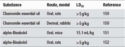
The following LD50 data have been recorded for butcher’s broom extract and its constituents:

image

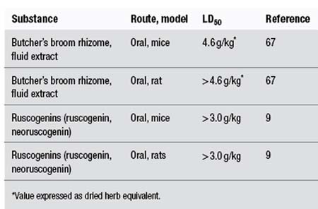
Intravenous administration of dried butcher’s broom extract to dogs resulted in a slowing of the heart rate, a marked drop in arterial pressure, and at high doses increased respiration and blood glucose levels. Intraperitoneal doses of 1.5 to 2.0 g/kg of dried ethanolic extract of butcher’s broom were lethal in guinea pigs. The authors estimated that the upper level of a safe dose for a human would be about 10 g of extract by injection.68

Prolonged oral administration of high doses (300 mg/kg) of saponosides, prosapogenins and ruscogenins isolated from butcher’s broom were well tolerated by rats.9

Contraindications

Because of the irritant effect of the saponins, butcher’s broom should not be applied to broken or ulcerated skin.

Special warnings and precautions

The use of herbs rich in saponins is possibly inappropriate in coeliac disease, fat malabsorption and vitamin A, D, E and K deficiency, and some upper digestive irritations. Saponin-containing herbs are best kept to a minimum in patients with pre-existing cholestasis.

Interactions

None identified in the literature.

Use in pregnancy and lactation

Category B1 – no increase in frequency of malformation or other harmful effects on the fetus from limited use in women. No evidence of increased fetal damage in animal studies.

Animal studies have not produced any evidence of embryotoxic activity for butcher’s broom extract. Butcher’s broom combined with sweet clover (Melilotus officinalis) or hesperidin methylchalcone has been used topically to treat varicose veins in pregnant women.57,67 An uncontrolled trial (noted earlier) observed that oral administration of butcher’s broom combined with hesperidin methylchalcone and ascorbic acid to pregnant women (21 to 24 weeks’ gestation) had no adverse effect on their infants. The 20 women involved in the study received the herbal treatment for at least 6 weeks and not more than 13 weeks. Postprandial digestive heaviness was noted by eight patients and was ascribed to the herbal treatment.59

The herb is rated as compatible with breastfeeding.

Effects on ability to drive and use machines

No adverse effects expected.

Side effects

As with all herbs rich in saponins, oral use may cause irritation of the gastric mucous membranes and reflux. The use of an enteric-coated preparation is preferred in sensitive patients.

Contact allergy to ruscogenins occurs rarely, but may be underestimated. In the 10 years to 1998 eight cases were reported to the French medical authorities.69 Contact allergy to butcher’s broom extract has also been reported.70,71 In one of these cases the cream contained butcher’s broom extract and the excipient thimerosal, and positive patch test results were obtained for both ingredients.70

Adverse reactions associated with the oral intake of butcher’s broom extract combined with hesperidin methylchalcone and ascorbic acid have been reported. In particular, lymphocytic colitis (one case associated with ileal villous atrophy and most with chronic diarrhoea) were observed, many after long-term use of the product.7275 Faecal fluid measurements were not consistent with an osmotic mechanism.75 Other cases of chronic diarrhoea7679 including diarrhoea without mucus or blood,80 and watery diarrhoea mimicking coeliac disease, have been reported.81 These earlier reports are probably also cases of lymphocytic colitis that were recorded before this association was more widely recognised. The lymphocytic colitis may be secondary to a chronic activation of the mucosal immune system by one or several components of the combination.82 It has not been determined if the butcher’s broom extract is implicated in this adverse reaction.

A case of cytolytic hepatitis associated with the ingestion of a preparation containing ruscogenins, hesperidin, ascorbic acid and aesculetol has been reported.83

All the clinical trials noted previously indicate that the risk of side effects for butcher’s broom is relatively low and that such side effects are likely to be minor.

Overdosage

No incidents found in the literature.

Safety in children

No information available.

Regulatory status in selected countries

In the UK butcher’s broom is not included on the General Sale List. In Germany it is covered by a positive Commission E Monograph. Butcher’s broom is official in the European Pharmacopoeia 2006.

In the USA butcher’s broom does not have GRAS status. However, it is freely available as a ‘dietary supplement’ in the USA under DSHEA legislation (Dietary Supplement Health and Education Act of 1994).

In Australia butcher’s broom is not included in Part 4 of Schedule 4 of the Therapeutic Goods Regulations and is freely available for sale.

References

1. Mabberley DJ. The Plant Book, 2nd ed. Cambridge: Cambridge University Press, 1997. p. 628

2. Chiej R. The Macdonald Encyclopedia of Medicinal Plants. London: Macdonald; 1984. Entry No. 269

3. Grieve M, A Modern Herbal, New York, Dover Publications, 1971;Vol 1. pp. 128–129

4. Leclerc H. Précis de Phytothérapie, 5th ed. Paris: Masson, 1983. pp. 46–47

5. Wagner H, Bladt S. Plant Drug Analysis: A Thin Layer Chromatography Atlas, 2nd ed. Berlin: Springer-Verlag, 1996. p. 309

6. Dunouau C, Bellé R, Oulad-Ali A, et al. Planta Med. 1996;62:189–190.

7. Domange JR, Bougaret S, Yubero L. Clin Hemorheol. 1994;14(suppl 1):S7–S13.

8. Chevillard L, Ranson M, Senault B. Med Pharmacol Exp. 1965;12(2):109–114.

9. Capra C. Fitoterapia. 1972;43(4):99–113.

10. Facino RM, Carini M, Stefani R, et al. Arch Pharm (Weinheim). 1995;328(10):720–724.

11. Huang YL, Kou JP, Ma L, et al. J Pharmacol Sci. 2008;108(2):198–205.

12. Bouskela E, Cyrino FZ, Marcolon G. J Cardiovasc Pharmacol. 1993;22(2):221–224.

13. Bouskela E, Cyrino FZ, Marcolon G. J Cardiovasc Pharmacol. 1994;24(2):281–285.

14. Bouskela E, Cyrino FZ, Marcolon G. J Cardiovasc Pharmacol. 1994;24(1):165–170.

15. Marcelon G, Vanhoutte PM. Phlebology. 1988;3(suppl 1):51–54.

16. Miller VM, Rud K, Gloviezki P. Clin Hemorheol. 1994;14(suppl 1):S34–S37.

17. Anon. Dtsch Apoth Ztg. 1985;125(6):295.

18. Rubanyi G, Marcelon G, Vanhoutte PM. Gen Pharmacol. 1984;15(5):431–434.

19. Barton D, Ollis WD, eds. Advances in Medicinal Phytochemistry. London: John Libbey Eurotext, 1986. pp. 187–194

20. Bouskela E, Cyrino FZ, Bougaret S. Clin Hemorheol Microcirc. 1997;17(5):351–356.

21. Miller VM, Rud K, Gloviczki P. Clin Hemorheol. 1994;14(suppl 1):S37–S45.

22. Nemcova S, Gloviczki P, Rud KS, et al. J Vasc Surg. 1999;30(5):876–883.

23. Miller VM, Marcelon G, Vanhoutte PM. Phlebology. 1991;6(4):261–268.

24. Berg D. Fortschr Med. 1990;108(24):473–476.

25. Rudofsky C. Fortschr Med. 1989;107(19):52. 55–58

26. Haas S, Lill G, Stiller A, et al, Vanhoutte PM, ed. Return Circulation and Norepinephrine: An Update, Paris-London, John Libbey Eurotext, 1991:157–162.

27. Le Devehat C, Khodabandehlou T, Vimeux M, Bondoux G, Vanhoutte PM, ed. Return Circulation and Norepinephrine: An Update, Paris-London, John Libbey Eurotext, 1991:225–236.

28. Svensjo E, Bouskela E, Cyrino FZ, et al. Clin Hemorheol Microcirc. 1997;17(5):385–388.

29. Tarayre JP, Lauressergues H. Ann Pharm Franc. 1976;34:375–382.

30. Strauss AL, Rieger H. Phlebologie. 1992;21:247–251.

31. Bouaziz N, Michiels C, Janssens D, et al. Int Angiol. 1999;18(4):306–312.

32. Baurain R, Dom G, Trouet A. Clin Hemorheol. 1994;14(suppl 1):S15–S21.

33. Benard P, Cousse H, Rico AG, et al. Ann Pharm Fr. 1986;43(6):573–584.

34. Chanal JL, Mbatchi B, Sicart MT, et al. Trav Soc Pharm Montpellier. 1981;41(4):263–272.

35. Chanal JL, Cousse H, Sicart MT, et al. Trav Soc Pharm Montpellier. 1978;38(1):43–48.

36. Rauwald HW, Grunwidl J. Planta Med. 1991;57(suppl 2):A75–A76.

37. Boyle P, Diehm C, Robertson C. Int Angiol. 2003;22(3):250–262.

38. Vanscheidt W, Jost V, Wolna P, et al. Arzneimittelforschung. 2002;52(4):243–250.

39. Jager K, Eichlisberger R, Jeanneret Ch, et al. Clin Drug Invest. 1999;17(4):265–273.

40. Parrado F, Buzzi A. Clin Drug Invest. 1999;18(4):255–261.

41. Cappelli R, Nicora M, Di Perri T. Drugs Exp Clin Res. 1988;14(4):277–283.

42. Weindorf N, Schultz-Ehrenburg U. Z Hautkr. 1987;62(1):28–38.

43. Marshall M. Fortschr Med. 1984;102(29–30):772–774.

44. Elbaz C, Nebot F, Reinharez D. Phlebology. 1976;29(1):77–84.

45. Monteil-Seurin J. C R Ther Pharmacol Clin. 1993;109:3–7.

46. Beltramino R, Penenory A, Buceta AM. Int Angiol. 1999;18(4):337–342.

47. Beltramino R, Penenory A, Buceta AM. Angiology. 2000;51(7):535–544.

48. Rudofsky G, Diehm C, Gruss JD, et al. MMW. 1990;132:205–210.

49. Le Devehat C, Lemoine A, Roux E, et al. Angiologie. 1984;34(3):119–122.

50. Lozes A, Boccalon H. Int Angiol. 1984;3:99–102.

51. Braun R, Hirche H, Van Laak H-H. Z Allgemeinm. 1985;61:309–313.

52. Guex JJ, Avril L, Enrici E, et al. Int Angiol. 2010;29(6):525–531.

53. Guex JJ, Enriquez Vega DM, Avril L, et al. Phlebology. 2009;24(4):157–165.

54. Porto CL, Milhomens AL, Pires CE, et al. Int Angiol. 2009;28(3):222–231.

55. Guex JJ, Enrici E, Boussetta S, et al. Dermatol Surg. 2008;34(12):1666–1675.

56. Leutenegger E, Martinaggi P. Gazette Med. 1988;95(13):66–69.

57. Berg D. Fortschr Med. 1992;110(3):67–68. 71–72

58. Berg D, Vanhoutte PM, ed. Return Circulation and Norepinephrine: An Update, Paris, John Libbey Eurotext, 1991:55–61.

59. Baudet JH, Collet D, Aubard Y, et al, Vanhoutte PM, ed. Return Circulation and Norepinephrine: An Update, Paris, John Libbey Eurotext, 1991:63–71.

60. Leutenegger E, Martinaggi P. Gazette Med. 1988;95(33):83–85.

61. Leyh F. Therapiewoche. 1988;36:2325–2331.

62. Cluzan RV, Alliot F, Ghabboun S, et al. Lymphology. 1996;29(1):29–35.

63. Bennani A, Biadillah MC, Cherkaoui A, et al. Phlebologie. 1999;52(1):89–95.

64. Monteil-Seurin J, Ladure Ph. Efficacy of Ruscus extract in the treatment of the premenstrual syndrome. In: Vanhoutte PM, ed. Return Circulation and Norepinephrine: An Update. Paris: John Libbey Eurotext; 1991:43–53.

65. Redman DA. J Altern Complement Med. 2000;6(6):539–549.

66. Archimowicz-Cyrylowska B, Adamek B, Drozdzik M, et al. Phytother Res. 1996;10(8):659–662.

67. Seidenberger AV, Muller I, Heindl HJH. Therapiewoche. 1974;24(8):866–881.

68. Caujolle F, Meriel P, Stanislas E. Ann Pharm Fr. 1953;11:109–120.

69. Elbadir S, El-Sayed R, Renaud F, et al. Rev Fr Allergol. 1998;38(1):37–40.

70. Breuil K, Patte F, Meurice JC, et al. Rev Fr Allergol Immunol Clin. 1989;29(4):215.

71. Landa N, Aguirre A, Goday J, et al. Contact Dermatitis. 1990;22(5):290–291.

72. Dharancy S, Dapvril V, Dupont-Evrard F, et al. Gastroenterol Clin Biol. 2000;24(1):134–135.

73. Pierrugues R, Saingra B. Gastroenterol Clin Biol. 1996;20(10):916–917.

74. Bouaniche M, Chassagne P, Landrin I, et al. Rev Med Interne. 1996;17(9):776–778.

75. Ouyahya F, Codjovi P, Machet MC, et al. Gastroenterol Clin Biol. 1993;17(1):65–66.

76. Mornet M, Boiserie P, Jonville AP, et al. Therapie. 1991;46(3):254.

77. Anon. Presc Int. 1993;2(7):123.

78. Gendreau-Tranquart C, Barbieux JP, Furet Y, et al. Presse Med. 1989;18(29):1439.

79. Oliver JM, Bacq Y, Dorval ED, et al. Gastroenterol Clin Biol. 1991;15(2):160–162.

80. Thomas-Anterion C, Guy C, Vial F, et al. Rev Med Interne. 1993;14(4):215–217.

81. Widgren S, De Peyer R, Geissbuhler P, et al. Schweiz Med Wochenschr. 1994;124(8):313–318.

82. Beaugerie L, Luboinski J, Brousse N, et al. Gut. 1994;35(3):426–428.

83. Sgro C, Taque R, Zoll A, et al. J Hepatol. 1995;22(2):251.

Chamomile, German

(Matricaria recutita (L.) Rauschert)

Synonyms

Matricaria chamomilla L., Chamomilla recutita (L.) Rauschert (botanical synonyms), German chamomile, wild chamomile, matricaria (Engl), Matricariae flos (Lat), Kamillenblüten, Feldkamille (Ger), fleur de camomile, matricaire (Fr), camomilla (Ital), kamille (Dan).

What is it?

A number of plants have ‘chamomile’ as part of their common name, such as the corn chamomile (Anthemis arvensis). Although sweet or Roman chamomile (Chamaemelum nobile = Anthemis nobilis) is also used medicinally, the German chamomile (Matricaria recutita) is the medicinal chamomile covered by this monograph. German chamomile has been known and utilised since ancient times: the flower heads were widely included in pharmaceutical and medicinal preparations, beverages and cosmetics, and the essential oil in perfumery. In the Mediterranean it is common to order chamomile tea in restaurants or bars, even in a concentrated ‘espresso’ form. The Eclectic physicians preferred the use of German over Roman chamomile; the latter is also known to cause allergic reactions such as contact dermatitis and anaphylaxis. The use of German chamomile chemotypes rich in bisabolol (levomenol) is preferred, as this will confer maximum anti-inflammatory and spasmolytic activities.

Effects

Anti-inflammatory; inhibits spasm in the digestive tract; inhibits the occurrence of ulceration; promotes wound healing; stimulates skin metabolism.

Traditional view

Chamomile was considered to have two specific fields of action: the nervous system (as a calming treatment) and the gastrointestinal tract (decreasing irritation and as a carminative and spasmolytic). It is believed to affect both sensory and motor nerves and was used to treat nervous manifestations of dentition, and conditions with a morbid susceptibility to pain. Other important applications were amenorrhoea and nervous conditions involving the gastrointestinal tract. Large doses of chamomile infusion produce free diaphoresis, which was even said to relieve dysmenorrhoea and prevent clotting.1 Chamomile was also considered to be anticatarrhal and used to treat catarrhal conditions of the ears, nose and eyes.1,2 It was commonly employed in external applications for haemorrhoids, mastitis, mammary abscess, leucorrhoea and leg ulcers. The traditional use in children indicates chamomile was considered a very gentle and safe herb.2,3 Eclectic physicians also prescribed chamomile for therapeutic use during pregnancy.1

Summary of actions

Anti-inflammatory, spasmolytic, mild sedative, antiulcer, carminative, vulnerary, diaphoretic.

Can be used for

Indications supported by clinical trials

• Topical application: eczema, including neurodermatitis; wound healing.

• Internal use: anxiety, non-specific gastrointestinal complaints; in combination with pectin for the treatment of acute, non-complicated diarrhoea; in combination with dong quai for treatment of menopause; in combination with other herbs for the treatment of infantile colic.

Traditional therapeutic uses

Flatulent and nervous dyspepsia, travel sickness, nervous diarrhoea; nasal catarrh; dysmenorrhoea, amenorrhoea; restlessness and anxiety; during dentition.

May also be used for

Extrapolations from pharmacological studies

Spasm or ulceration of the gastrointestinal tract; restlessness, anxiety, mild sleep disorders.

Other applications

Cosmetics (for sensitive skin, anti-inflammatory and anti-acne products), bath preparations and hair care products.4

Preparations

Infusion of dried herb or liquid extract for internal use; infusion of dried herb, liquid extract or essential oil for external use or as an ingredient in ointments, creams, bath additives, mouthwashes and sprays. As with all essential oil-containing herbs, use of the fresh plant or carefully dried herb is advised. Keep covered when infusing the herb to retain the essential oil.

Topical use studies have generally used chamomile extracts from high-bisabolol chemotypes. Given the pharmacological properties of (–)-alpha-bisabolol and matricine (chamazulene), it is important to prefer these varieties.

Dosage

• 2 to 4 g three times per day of dried flower heads or in an infusion

• 3 to 6 mL/day of 1:2 liquid extract, 7 to 14 mL/day of 1:5 tincture; 50% ethanol is the preferred extraction solvent

• Infusions or semisolid preparations containing 3% to 10% (w/w) of the flowers or equivalent for external use as a compress, wash or gargle.

Duration of use

No restriction on long-term use.

Summary assessment of safety

Except in extremely rare cases of allergy and contact dermatitis in susceptible patients, chamomile is a safe herb.

Technical data

Botany

Matricaria recutita, a member of the Compositae (Asteraceae, daisy) family, is an annual that can grow as tall as 1 m in the right soil (usually up to 60 cm). The alternate leaves are bipinnatisect, delicately lobed and thread-like, ending in a point. The flower consists of 12 to 16 white ligules (ray florets, 10 to 15 mm long) surrounding the central mound of tiny yellow flowers (disc florets, five-lobed), which are embedded in a hollow, conical receptacle. The flower head is 1.2 to 2.4 cm in diameter. The light-coloured fruits are very small achenes, without outside oil glands.5,6

Adulteration

There has been much contention over the appropriate botanical classification of chamomile. There are various genotypes and it is also easily confused with related species of similar appearance or odour, especially from the genera Anthemis, Matricaria, Chamomilla, Chrysanthemum and Tanacetum. One authoritative review of the literature suggested that most cases of allergic contact dermatitis involving chamomile may actually be from related species, particularly Anthemis cotula (stinking dog fennel), which contains much higher levels of a particularly potent potential allergen, anthecotulide.7,8

Key constituents

• Essential oil (0.5% to 1.5%), containing (–)-alpha-bisabolol (also known as levomenol), chamazulene, bisabolol oxides A, B, C and cis- and trans-en-yn-dicycloethers.9 The intensely blue phytochemical chamazulene is an artefact formed from matricine during steam distillation10

• Flavonoids (0.5% to 3%), particularly apigenin-7-glucoside, flavonoid aglycones,9 coumarins (herniarin and umbelliferone), phenolic acids, mucilage,11 GABA (gamma-aminobutyric acid).12

The content of these constituents varies considerably between different chemical races or varieties of chamomile.13 Weather and where the plant is grown also affect the content and composition of the essential oil.14,15 The British Pharmacopoeia recommends that chamomile contain not less than 4 mL/kg of blue essential oil.16

image

Pharmacodynamics

Anti-inflammatory activity

Chamazulene inhibited the formation of leukotriene B4 in intact neutrophils in a dose-dependent manner and blocked the chemical peroxidation of arachidonic acid. Matricine did not show these effects, even at higher concentrations. Matricine had no effect on cyclo-oxygenase (COX) and 12-lipoxygenase activities in human platelets.17 However, other in vitro studies have determined that at least part of the anti-inflammatory activity of chamomile extracts is due to constituents that inhibit the formation of 5-lipoxygenase and COX, and have antioxidant activity. Bisabolol and apigenin appear to be responsible for this.18

An aqueous chamomile extract inhibited the release of prostaglandin E2 from lipopolysaccharide (LPS)-activated macrophages in vitro. The activity was due to a dose-dependent inhibition of COX-2 activity. Chamomile reduced COX-2 mRNA and protein expression, without affecting COX-1 expression or activity. The species was not defined, but is likely to be Matricaria recutita.19 Apigenin also inhibited COX-2 and nitric oxide synthase (NOS) activity in LPS-activated macrophages.20

In terms of a possible influence on cytokine activity, apigenin inhibited LPS-induced interleukin-6 production in macrophages. Oral pretreatment (50 mg/kg) inhibited LPS-induced interleukin-6 and tumour necrosis factor-alpha production in mice.21

Oral intake of matricine demonstrated anti-inflammatory activity in the carrageenan test on rat paw.22 Matricine was nearly as effective as (–)-alpha-bisabolol up to 3 h after oral administration. Chamazulene was significantly less active than both these compounds.23 Matricine, together with most of the components in chamomile oil but especially bisabolol, demonstrated anti-inflammatory activity when tested topically on skin.24

(–)-alpha-Bisabolol demonstrated anti-inflammatory activity in a number of experimental inflammatory models: rat paw oedema, adjuvant arthritis of the rat, ultraviolet erythema of the guinea pig and yeast fever in rats.25cis-En-yn-dicycloether inhibited the development of dextran-induced oedema in rats.26 Several polysaccharides in chamomile have demonstrated anti-inflammatory activity when applied topically.27

The anti-inflammatory activity of an aqueous-alcoholic extract of fresh chamomile, an aqueous-alcoholic extract of dried chamomile, the essential oil and isolated components of the essential oil were investigated by topical administration using croton oil-induced dermatitis in mouse ear. The extract prepared from dried flowers showed a mild but significant anti-inflammatory activity (24%), but the extract based on fresh chamomile was more active (32%) and equalled the activity of the reference drug, benzydamine. The essential oil solution did not show any significant inhibition of oedema. The anti-inflammatory activity of apigenin was 10 times higher than matricine, which was 10 times higher than chamazulene. The differences in anti-inflammatory activity of the various preparations might be attributable to the varying concentrations of matricine.28

An aqueous-alcoholic extract of dried chamomile demonstrated a significant reduction of oedema (23.4%) compared with controls after topical administration to mice in the croton oil ear test. The non-steroidal anti-inflammatory agent benzydamine produced a similar reduction at a dose corresponding to twice its usual clinical dose. Hydrocortisone was the most active agent (56.4%).29 Anti-inflammatory effects were demonstrated for topical administration of standardised chamomile extract, the flavone fraction and isolated flavones (apigenin, luteolin and quercetin) in the same model. The lipophilic flavone fraction demonstrated stronger activity than the total chamomile extract, and the action of apigenin was superior to the reference drugs indomethacin and phenylbutazone.30

The anti-inflammatory efficacies of topical compounds were measured directly and objectively on the skin of healthy volunteers using the Tesafilm stripping technique. Three pharmaceutical formulations were investigated: a chamomile cream, its cream base and a hydrocortisone ointment. Chamomile cream exhibited 70% of the activity of the hydrocortisone ointment.31 Experimentally induced toxic contact dermatitis was topically treated with a chamomile ointment and compared with ointment base and 1% hydrocortisone acetate. The chamomile ointment demonstrated a superior soothing effect in comparison to the cortisone cream on human skin.32

Spasmolytic activity

Chamomile extract and some of its constituents demonstrated a dose-dependent spasmolytic effect on isolated guinea pig ileum. (–)-alpha-Bisabolol, bisabolol oxides A and B and chamomile oil showed a papaverine-like antispasmodic activity, with the essential oil showing the lowest effect. The cis-en-yn-dicycloether component also showed activity, although it was not dose dependent. The flavones apigenin, luteolin, patuletin and quercetin demonstrated marked antispasmodic effects, with apigenin significantly more potent than the other flavones, but less active than papaverine. The coumarins only demonstrated a weak effect.33 Freeze-dried, ethanol-free extract of chamomile completely blocked slow wave activity in isolated mouse small intestine.34 Inhibition of cAMP phosphodiesterase was a likely mechanism for the spasmolytic activity.35

Spasmolytic activity has also been observed following oral administration of apigenin.36cis-En-yn-dicycloether demonstrated a more pronounced antispasmodic effect than papaverine on isolated guinea pig and rabbit intestine.26 Chamomile extract (25 mL/L)37 and chamomile oil (0.2 mL diluted in 400 mL methanol)38 inhibited acetylcholine- and histamine-induced contractions in guinea pig ileum in vitro.

Chamomile extract showed moderate inhibitory activity in experimentally induced hyperperistalsis. The inhibition (56%) was greater than that of the control drug loperamide (34% at doses of 10 mg/kg). Rats were treated orally with the methanol extract of the aerial parts (5:1, 300 mg/kg).39

Sedative and CNS activity

Several fractions from the aqueous extract of chamomile, including apigenin, have demonstrated significant affinity for the central benzodiazepine receptor in vitro. Apigenin also demonstrated clear antianxiety activity (10 mg/kg) and slight sedative activity without muscle relaxant effects after intraperitoneal administration in mice.40 However, a later study in rats found that apigenin (doses from 0.5 to 10 mg/kg, ip) did not demonstrate antianxiety activity. In vitro investigations suggest that any sedative effect demonstrated in vivo for apigenin (by injection), is not mediated by benzodiazepine receptors.41 Fractions isolated from the methanol extract of chamomile were able to selectively bind to both central and peripheral benzodiazepine receptors in vitro. The displacing activity on radiolabelled muscimol binding observed for several of the fractions was found to be due to the presence of GABA in micromolar concentrations. The identity of other active compounds was not established. However, some of the fractions not containing GABA produced a statistically significant reduction of locomotor activity in rats after intracerebroventricular injection.12 Chamomile extract significantly inhibited GAD (glutamic acid decarboxylase) activity in vitro (brain homogenate). GAD is a rate-limiting enzyme that determines GABA levels in normal nervous tissues, such as the brain.42

Oral administration of chamomile extract (300 mg/kg) caused a significant shortening in sleep latency in rats. Flumazenil (a benzodiazepine receptor antagonist) at a dose that caused no obvious effect when used alone showed a significant antagonistic effect on the shortening of sleep latency induced by the chamomile extract.43

A dried preparation of a chamomile infusion demonstrated a depressive effect on the CNS after intraperitoneal administration in mice. A dose-dependent decrease of basal locomotor activity was observed (90 to 360 mg/kg) without involving motor coordination and muscle relaxation. A significant sleeping-time potentiating effect was only observed at the two highest doses (160 and 320 mg/kg).44 Inhalation of chamomile oil vapour decreased stress-induced increases in plasma ACTH (adrenocorticotropic hormone) in ovariectomised rats, compared with placebo. The plasma ACTH level decreased further when diazepam was administered with chamomile oil vapour, and the decrease was blocked by flumazenil. This suggests that chamomile oil may have an effect on GABA ergic systems in rat brain and perhaps an activity similar to benzodiazepine agonists.45

A series of clinical tests in Japan found that an increase in mental activity was observed just after eating warmed chamomile jelly. There was also a decrease in heart rate and an increase in ‘pleasantness’ and good mood. Compared with control nights, eating the warmed jelly improved aspects of sleep in men and women. Skin temperature increased more when drinking chamomile tea than when drinking hot water.4648 No exact details of dosage were provided in these studies.

The effect of olfactory stimulation on fluency, vividness of imagery and associated mood was studied with 22 volunteers after exposure to either chamomile oil or placebo. Participants were asked to visualise positive and negative images after exposure to one oil then the other. Chamomile oil did not affect the vividness of imagery ratings, but significantly prolonged the time taken for participants to visualise both positive and negative phrases, suggesting a sedative effect. Negative mood ratings following negative phrase presentation were less extreme after chamomile (p=0.042). Overall mood rating was significantly higher (mean=+1) after chamomile compared with placebo (mean=0) (p=0.001).49

Co-administration of extract of Matricaria chamomilla (25 mg/kg) with morphine (both by ip injection) greatly reduced the development of morphine dependence in rats. Administration of chamomile extract before induction of withdrawal syndrome by naloxone injection inhibited the expression of abstinence syndrome in morphine-dependent animals.50 Pretreatment with an aqueous-methanolic extract of chamomile (200 mg/kg, ip) increased the latency of seizure onset in experimentally induced seizure in mice.51

Antiulcer activity

Chamomile extract demonstrated antipeptic activity in vitro.52 (–)-alpha-Bisabolol inhibited the occurrence of ulceration induced by indomethacin, stress or ethanol in rats. A decrease in healing time of ulcers was also observed. A standardised chamomile extract also demonstrated ulcer-protective activity.53 Oral administration of (–)-alpha-bisabolol demonstrated a significant protective effect (p<0.05) against gastric toxicity produced by acetylsalicylic acid in rats.54 Several more recent studies have confirmed that an aqueous-ethanolic extract of chamomile and alpha-bisabolol can reduce experimentally induced gastric lesions.5557 The mechanism of gastroprotection may involve a reduction in oxidation (chamomile extract, alpha-bisabolol),55,57 and activation of ATP-sensitive potassium channels (alpha-bisabolol).56

In an interesting series of experiments, the antiulcerogenic activity of chamomile in rats was compared with a number of other herbs.58 A liquid aqueous-ethanolic extract of chamomile demonstrated a dose-dependent protection (2.5, 5 and 10 mL/kg, oral) against indomethacin-induced ulcers, but was not as active as many of the other herbs, including licorice and caraway. However, chamomile extract (10 mL/kg, oral) was the most potent herb at decreasing gastric acidity and acid output. It also significantly increased gastric mucin content, activity shared with licorice, St Mary’s thistle and caraway.

Antimicrobial activity

Antimicrobial activity has been demonstrated for (–)-alpha-bisabolol in vitro59 and chamomile oil (<0.05%, v/v) against Staphylococcus aureus, Bacillus subtilis and Candida albicans60 and for herniarin in the presence of near UV light. Chamomile extracts demonstrated similar activity against Escherichia.61 The growth of Staphylococcus aureus, Streptococcus mutans, group B Streptococcus and Streptococcus salivarius was completely inhibited by chamomile extract at 10 mg/mL. The extract showed strong antibacterial activity against Bacillus megatherium and Leptospira icterohaemorrhagiae. The effective concentration of each component (alpha-bisabolol, bisabolol oxides, dicycloethers and chamazulene) was much lower when the component was in combination with the others, as in the natural extract, indicating a possible synergistic effect.62

The antibacterial activity of chamomile against Helicobacter pylori was tested in vitro. Activity ranged from no activity (infusion,63 aqueous-ethanolic extract standardised for alpha-bisabolol,64 methanol extract65) to some activity (olive oil extract,66 essential oil,67 concentrated aqueous-methanolic extract68). Test methods varied, making comparison difficult. The methanol extract was described as having no activity (MIC greater than 100 μg/mL). In comparison, the MIC of amoxicillin was 0.002 to 0.06 μg/mL.65 The MIC90 of chamomile olive oil extract (1:10) was 125 mg/mL (no standard comparison),66 while the MIC for the essential oil was 36 to 70 μg/mL (in comparison, the MIC of amoxicillin was 0.02 μg/mL).67 (The minimum inhibitory concentration (MIC) is the lowest concentration at which there is no visible bacterial growth. MIC90 is the minimum concentration at which 90% of the bacterial growth is inhibited.)

An aqueous-ethanolic extract of chamomile, standardised for alpha-bisabolol, inhibited the growth of Campylobacter jejuni by approximately 100% at both 1:25 and 1:100 dilutions. The IC50 (for dry weight of herb) against the bacteria was 2 mg/mL. In comparison, the IC50 for gentamicin was 0.08 μg/mL. (IC50 is the concentration of a compound needed to reduce the growth of an organism by 50%.64)

Quorum sensing is a process of intercellular communication among bacterial cells, which occurs with the use of signalling molecules. Inhibition of quorum sensing may reduce infections. Chamomile essential oil was ineffective in inhibiting quorum sensing of two sensor strains in vitro.69

Chamomile essential oil demonstrated significant virucidal activity towards herpes simplex virus type 2 (HSV-2) in vitro with an IC50 of 1.5 μg/mL.70 The oil was also active against an acyclovir-sensitive HSV-1 strain and against acyclovir-resistant clinical HSV-1 isolates.71 Chamomile essential oil affected the viruses by interrupting the adsorption of virus onto the host cell.70,71

Bisabolol (500 and 1000 μg/mL) achieved 100% inhibition of Leishmania infantum promastigote in vitro.72

Wound healing, itching

Aqueous extract of chamomile (120 mg/kg/day) in the drinking water of rats resulted in an increased rate of wound contraction, increased wound-breaking strength and hydroxyproline content, and improved histology of experimentally induced wounds.73 Topical application of chamomile extract accelerated the healing of burns and incisions in rats.74,75 A chamomile ointment was effective in the treatment of oral mucositis in a hamster model. The ointment contained a fluid extract of chamomile (10%) and tincture of myrrh.76 The same preparation promoted faster wound healing of tongue lesions compared with topical corticosteroids in rats.77

The wound-healing activity of chamomile is closely linked to its anti-inflammatory activity. (–)-alpha-Bisabolol promotes granulation and tissue regeneration.59 Application of chamomile extract altered the metabolism of guinea pig skin cells, indicating possible stimulation of cellular regeneration and inhibition of inflammation.78 Extracts of chamomile showed moderate activity in stimulating proliferation and migration of fibroblasts in vitro.79

The impact of oral administration of chamomile in reducing experimentally induced itch was investigated in mice. A diet containing 30% (w/w) of dried, powdered chamomile for 11 days reduced itch by about 30%, which was not significant. Of the other extracts tested, significant inhibition of itch occurred for an ethyl acetate extract (dosage adjusted to 30% (w/w) of dried chamomile). Removal of water-soluble substances from the ethanol extract appeared to significantly enhance its antipruritic activity.80 In the same model, oral administration of ethyl acetate extract of chamomile extract (25:1, 300 mg/kg) combined with antihistamines was more effective for the suppression of scratching behaviour in mice than administration of antihistamines alone. At least part of the antipruritic activity was due to the essential oil, present at approximately 30% in the extract.81 Topical application of chamomile essential oil (3% oil, 70 μL, daily for six times per week) alleviated some altered immune parameters in mice with experimentally induced atopic dermatitis. The elevation in serum immunoglobulin E (IgE), IgG1 and histamine were significantly reduced compared with control groups. Scratching frequency was significantly lower than in either control group (saline, jojoba oil).82

Other activity

Chamazulene and (–)-alpha-bisabolol, within the concentration range 10−9 to 10−5 M, demonstrated very little influence on histamine release in isolated rat mast cells. At concentrations higher than 10−5 M they stimulated histamine release. En-yn-dicycloether exhibited a moderate stimulating effect at concentrations lower than 10−4 M and a strong inhibiting effect on histamine release at higher concentrations.83

Extracts and infusion (species undefined) of chamomile have shown antioxidant activity in vitro. The activity was moderate compared with other herbs.84,85 Chamazulene inhibited membrane lipid peroxidation in vitro in a concentration- and time-dependent manner. It also demonstrated hydroxyl radical scavenging activity.86 Chamomile may protect against global cerebral ischaemia/reperfusion-induced brain injury in rats. Oral administration of methanol extract decreased lipid peroxidation and increased superoxide dismutase, catalase, glutathione and total thiol levels compared with the untreated group. Cerebral infarction area was also significantly reduced in chamomile-treated groups. The activity was dose-dependent (100 to 300 mg/kg body weight, 9.3:1 extract).87

Chamomile extract strongly stimulated the proliferation of lymphocytes, as determined by the mixed lymphocyte reaction, an in vitro stimulatory system involving T cell activation.88

Diets containing chamomile flower (1.5% and 7%), several chamomile oils (0.2% to 0.35%) or guaiazulene (0.2%) stimulated liver regeneration in rats.89 Intragastric administration of a mixture of flavonoids of chamomile normalised sphingolipid metabolism in the liver of old rats. (An increase in sphingolipid turnover in the liver is associated with elevation of free radical production and state of chronic inflammation in old age.) Animals were fed 160 mg/kg of a mixture consisting of apigenin, luteolin, apigenin-7-glucoside, luteolin-7-glucoside, isorhamnetin and quercetin.90 The same mixture and dosage normalised the elevated ceramide content in carbon tetrachloride-damaged livers of rats via neutral sphingomyelinase inhibition and ceramidase activation. Alterations in sphingolipid turnover induced by administration of the flavonoids coincided with the stabilisation of the damaged hepatocyte membranes.91 This mixture (40 to 80 mg/kg) induced a dose-dependent increase in bile flow and bile acid secretion in adult and old rats. Cholesterol bile secretion was increased and hepatic lipid content reduced in old rats, but not in adult rats.92

In a long-term feeding test, the hot water extract of chamomile (500 mg/kg/day) significantly suppressed blood glucose levels in streptozotocin-induced diabetic rats. The hypoglycaemic effect of chamomile was independent of the inhibition of intestinal alpha-glycosidases, but depended on the inhibition of hepatic glycogen phosphorylase. Chamomile extract also showed strong inhibition against aldose reductase in vitro.93 In the same model, oral administration of an aqueous-ethanolic extract of the aerial parts of chamomile reduced hyperglycaemia and oxidative stress in a dose-dependent manner. Significant results were obtained for daily dosages of 280 and 560 mg/kg (dried herb equivalent). Histological investigation indicated that treatment with chamomile protected the majority of the pancreatic islet cells, in comparison with the control group.94

An ethanol extract of chamomile showed weak activity against a cancer cell line in vitro.95 In another test, a significant decrease in cell viability was observed in several cancer cell lines for chamomile extracts. Chamomile exposure resulted in differential apoptosis in cancer cells.96 Bisabolol oxide A at a concentration of 10 μM inhibited the growth of leukaemia cells in vitro. Combination with 5-fluorouracil further inhibited the growth of leukaemia.97 Bisabolol oxide A at 5 to 10 μM did not exert a cytotoxic action on normal non-proliferative cells (rat thymocytes) in a previous study.98 Luteolin (5 mg/kg, ip) suppressed prostate tumour growth in mice by inhibiting insulin-like growth factor 1 receptor signalling.99 Luteolin inhibited platelet-derived growth factor (PDGF)-induced proliferation by inhibiting PDGF receptor phosphorylation in vascular smooth muscle cells.100 Bisabolol efficiently and selectively induced apoptosis in malignant tumour cells in vitro by targeting lipid rafts on cell membranes.101

Chamomile extract stimulated osteoblastic cell differentiation and exhibited an anti-oestrogenic effect on breast cancer cells in vitro, without proliferative effects on cervical adenocarcinoma cells.102 Using an in vitro tissue culture indicator system, chamomile (species undefined) was found to have weak oestrogen agonist activity and weak progestogenic activity.103

An aqueous-ethanolic extract of chamomile was superior to 2.5% sodium hypochlorite solution as a cleaning agent to remove the smear layer of teeth. A smear layer is produced by cutting the enamel or dentin in cavity preparation. Extracted, single-rooted teeth were used for this in vitro investigation. Chamomile extract was not better than sodium hypochlorite solution combined with EDTA (ethylenediaminetetraacetic acid), but was more effective than tea tree oil.104

Pharmacokinetics

Chamazulene is formed from matricine in the gut of rats by the action of gastric acid following oral administration.23 When volunteers were given matricine orally (500 mg, in suspension), peak plasma levels of chamazulene carboxylic acid were determined at 0.9 to 2.2 μg/mL.105

Topical application of radioisotope-labelled (–)-alpha-bisabolol to the skin of nude mice resulted in half the radioactivity being found on the skin, with the remainder present in tissue and organs. Of the level measured in the tissues and organs, 90% was intact (–)-alpha-bisabolol. Further measurements indicated that (–)-alpha-bisabolol penetrated quickly into the skin. Five hours after the topical application, it was displaced from outermost to innermost areas. Hence a fast cutaneous absorption and a long therapeutic effect might be expected from (–)-alpha-bisabolol.106 In contrast, a similar earlier study found that most of the bisabolol had been metabolised to a polar metabolite. Bisabolol was mainly excreted in the urine in the form of polar metabolites and to a slight extent as carbon dioxide in exhaled air.107

Skin penetration studies of the chamomile flavones apigenin, luteolin and apigenin 7-O-beta-glucoside were carried out with nine healthy female volunteers. Apigenin showed the greatest flux (the greatest amount of flavonoid per time and area), followed by luteolin. Penetration of apigenin 7-O-beta-glucoside was negligible. A steady state was attained after 3 h, indicating that the flavonoids penetrated through further skin layers. It was concluded that these flavonoids are not only adsorbed at the skin surface, but penetrate into deeper skin layers.108,109

Clinical trials

Eczema, dermatitis and ulceration

Topical application of chamomile preparations helped relieve eczema110 and varicose eczema.111 In a survey conducted in the early 1980s, 95% of 2477 general practitioners described good tolerance and therapeutic efficacy for a chamomile cream (containing 2% standardised chamomile extract) in the treatment of eczema.110 The herbal treatment decreased inflammation111 and facilitated a reduction in the level of topical corticosteroids used.110

Chamomile cream was compared to steroidal and non-steroidal dermal preparations in the maintenance therapy of hand, forearm and lower leg eczema in an open bilateral comparative trial involving 161 patients. During maintenance therapy over 3 to 4 weeks, the chamomile cream showed similar efficacy to 0.25% hydrocortisone and superior activity to the non-steroidal anti-inflammatory agent (5% bufexamac) and a glucocorticoid preparation (0.75% fluocortin butyl ester).112 After 2 weeks of treatment, a standardised chamomile cream showed mild superiority over 0.5% hydrocortisone cream and marginal improvement compared with placebo in medium-degree atopic eczema. This was a partially blinded, randomised trial carried out as a half-side comparison (one side of the body compared with the other).113 Eight patients with neurodermatitis in the stage of subacute eczema were treated with ointments containing 1% of a carbon dioxide extract of chamomile or 1% hydrocortisone, in a randomised, double blind trial. Global clinical impression was used to assess the results. Hydrocortisone was significantly more effective than chamomile (p<0.001). Nearly all the patients had been pretreated topically with steroids. In cases of pretreatment with weak or moderately potent steroids, hydrocortisone was much more effective than chamomile, but not in patients pretreated with potent or very potent steroids. The carbon dioxide extract contained constituents similar to the essential oil, with the exception of chamazulene, which was absent.114

In an open, uncontrolled clinical trial, patients with varicose ulcers were treated with a chamomile ointment alone or the ointment and a chamomile wash. The therapeutic response was good at 83% and 78%, respectively.115

Wound healing

Standardised chamomile ointment had similar efficacy to 5% dexpanthenol cream in healing episiotomy wounds in an open, controlled, randomised trial.116 An open, randomised trial compared several procedures for second degree haemorrhoid treatment and found best results in the group receiving application of a standardised chamomile ointment in conjunction with surgical procedures (ligature and anal dilation).117

Chamomile extract demonstrated a statistically significant improvement in wound healing in a double blind, placebo-controlled clinical trial involving 14 patients. The significant decrease in weeping of the wound area and the drying tendency after dermabrasion of tattoos (p<0.05) indicated the therapeutic efficacy of chamomile.118 In a randomised, single blind, controlled trial involving 48 women, chamomile cream was not statistically more effective than almond cream at reducing erythema and moist desquamation acquired after receiving radiotherapy for breast cancer. However, there was a trend to fewer and later appearances of grade 2 reactions for the chamomile treatment.119

A physiotherapist and a medical doctor carried out a small study involving five patients with chronic wounds. The patients were treated with a mixture of chamomile and Lavandula angustifolia essential oils in a grape seed oil carrier base and compared with three controls. One of the controls, who had wounds on his left and right legs, started the study as both a control and a member of the experimental group. Fifty-six days into treatment he discontinued conventional treatment and used essential oils only. The recipients of the essential oils did better than those who only received conventional treatments.120

Colostomy patients seeking treatment for a peristomal skin lesions were assigned to one of two treatments: hydrocortisone (1%) ointment applied once a day or chamomile compress in an open label trial involving 72 patients.121 A chamomile solution was prepared by infusing dried and powdered flowers (6 g) in 150 mL of boiling water for 10 minutes in a closed glass container. The filtered solution was applied to gauze and the compress placed on the wound for 1 hour twice a day. The lesions were evaluated every 3 days for a maximum of 28 days. Lesions healed significantly faster in the chamomile than in the hydrocortisone group (mean time to healing: 8.9 and 14.5 days respectively; p=0.001). Complete healing was achieved in 100% of the chamomile group by day 15, while 76% of the hydrocortisone group achieved complete healing by day 21. Pain intensity and itching were lower in the chamomile group.

Oral inflammation

In an uncontrolled trial involving 36 patients, a chamomile mouthwash provided a cooling and astringent effect in the treatment of chronic oral inflammation, except in the case of glossodynia.122 A chamomile preparation was used therapeutically and prophylactically as an oral rinse in the treatment of oral mucositis caused by head and neck irradiation and systemic chemotherapy in an uncontrolled trial. Resolution of mucositis was accelerated by the chamomile rinse and prophylactic use was also favourable.123

In a double blind, placebo-controlled clinical trial involving 164 patients, a chamomile mouthwash did not decrease the incidence of stomatitis resulting from 5-fluoruracil-based chemotherapy. The mouthwash was administered three times per day for 2 weeks.124 This may have been too short a period to detect improvement.125 A later, randomised trial conducted in Iran found that allopurinol and chamomile mouthwashes were superior to placebo (saline) for the treatment of chemotherapy-induced stomatitis. Both active mouthwashes were equally effective. The chamomile mouthwash was prepared from an infusion (8 g powdered flower per 50 mL water) and filtered.126 A case of methotrexate-induced oral mucositis in a patient with rheumatoid arthritis was successfully treated with a chamomile mouthwash. The patient refused standard mouthwash treatment and was instead administered an infusion of Matricaria recutita (8 g dried flower in 1 litre of boiling water). The suggested dosage was 20 mL kept in the mouth or gargled for 1 to 2 minutes and repeated four times daily. By the 13th day the oral mucositis had reduced from grade 3 (oral ulcers, liquid diet only) to grade 2 (oral erythema, ulcers, able to eat solids) and was completely healed within 4 weeks.127

Fluid extract of chamomile provided an analgesic effect in patients with aphthous stomatitis and other painful ulcers of the mouth in an open label trial. The analgesic effect was considered excellent by 82% and good by 18% of patients. The efficacy of chamomile was considered excellent in 67% of patients and good in 33%. Pain was assessed using a visual analogue scale and 34 patients were evaluated.128

Anxiety

Fifty-seven outpatients with mild to moderate generalised anxiety disorder were randomised via a double blind design to receive chamomile (Matricaria recutita) extract or placebo for 8 weeks. Treatment was initiated at one capsule per day for the first week and increased to two per day during the second week. Patients with a 50% reduction or less in total Hamilton Anxiety Rating (HAM-A) score (compared with baseline) were increased to three capsules per day during week 3, and then to four per day during week 4. Patients who continued to only have a 50% reduction or less in baseline HAM-A score were increased to five capsules/day during weeks 5 to 8. The chamomile extract (220 mg/capsule, undefined strength) was standardised to contain 1.2% apigenin. There was a significantly greater reduction over time in the mean total HAM-A score for chamomile versus placebo (p=0.047). Secondary outcomes included changes in the Beck Anxiety Inventory, Psychological Well Being and Clinical Global Impression Severity scores and the proportion of patients with 50% reduction or more in baseline HAM-A score. There were no statistically significant differences observed between the groups for any secondary outcome measure. One patient in each treatment group discontinued therapy because of adverse events. The proportion of patients experiencing adverse events was not significantly different between groups (p=0.417).129

Gastrointestinal conditions

In an uncontrolled study, 104 patients with non-specific gastrointestinal complaints (probably functional dyspepsia) were treated with an aqueous-ethanolic preparation of chamomile (standardised for alpha-bisabolol and apigenin-7-glucoside) at 25 drops four times a day (in a cup of hot water) for a period of 6 weeks. The following symptoms were assessed: stomach and abdominal pain, burping, fullness, heartburn, loss of appetite, nausea and vomiting. Symptoms completely disappeared in 44.2% of patients. The success rate for single symptoms ranged from 61% for loss of appetite to 84.5% for stomach and abdominal pain and 88.7% for nausea.130

In a prospective, double blind, randomised, multicentre, parallel group study, 79 children with acute, non-complicated diarrhoea received either a preparation containing apple pectin and chamomile extract or placebo for 3 days in addition to the usual rehydration and realimentation diet. At the end of treatment, the diarrhoea had stopped significantly more frequently (p<0.05) in the pectin/chamomile group (85%) compared with the placebo group (58%). The pectin/chamomile combination also significantly reduced the duration of diarrhoea by at least 5.2 h (p<0.05). In contrast to placebo, a trend of continuous improvement was observed by parents for the pectin/chamomile group. The parents expressed their satisfaction more frequently (82%) with pectin/chamomile than with placebo (60%).131 A follow-up trial (also of double blind design) with a larger number of patients found that the combination significantly reduced stool frequency compared with placebo. This multicentre, randomised trial enrolled 255 children aged between 6 months and 6 years with acute diarrhoea. Two hundred and forty-one children were available for final analysis: 121 received the active medication and 120 received placebo. Stool frequency on the third day was significantly lower in the treatment group compared with placebo (decreases of 49% and 36%, respectively; p=0.0012). Each child also received a glucose-electrolyte solution on the first day of treatment.132

The effect of a herbal instant tea preparation containing chamomile, vervain, licorice, fennel and lemon balm on infantile colic was assessed in a prospective, double blind study on babies about 3 weeks old. Tea or placebo up to 150 mL per dose was given to each infant with every episode of colic, but not more than three times a day. After the 7 days of the trial, the colic improvement scores were significantly better in the herbal tea group: 1.7 versus 0.7 for the placebo group (p<0.05). In addition, more babies in the treatment group had their colic eliminated: 57% compared with 26% for placebo (p<0.01).133 A randomised, double blind, placebo-controlled trial showed that colic in infants improved within 1 week of treatment with a combination of chamomile, fennel (Foeniculum vulgare) and lemon balm (Melissa officinalis). The preparation also contained vitamin B1, calcium pantothenate and vitamin B6.134 An experimental study found that the activity of the combination in reducing upper gastrointestinal motility was due to the chamomile and lemon balm.135

Other activity

Oral administration of chamomile tea during cardiac catheterisation induced a deep sleep in 10 of 12 patients tested, despite the pain and anxiety experienced from the procedure. The chamomile tea had essentially no cardiac effects.136

Fifty-five postmenopausal women were randomly assigned to receive tablets containing Matricaria recutita (150 mg/day of an undefined extract) and dong quai (Angelica sinensis, 375 mg/day of an undefined extract) or placebo for a period of 12 weeks. There were significant and dramatic differences observed between the study and control groups in terms of the number and intensity of hot flushes (both p<0.001). In the herbal treatment group, a response was already in evidence by the first month (68% reduction of hot flushes during the day and 74% during the night). There was also a marked alleviation of sleep disturbance and fatigue.137

Mean scores improved for Conners’ hyperactivity, inattention and immaturity factors in three young men (14 to 16 years old) diagnosed with attention-deficit hyperactivity disorder who received treatment with chamomile. Two participants received chamomile for 4 weeks and then placebo for 4 weeks; the others received placebo first followed by chamomile. The observed improvement was lower than reported in stimulant trials, but was similar to that found in non-stimulant trials, such as for desipramine.138 Treatment with chamomile also led to a slight improvement in irritability and stereotypic behaviour in three male patients with autism. The standardised extract used provided 200 mg/day of alpha-bisabolol.139

A randomised, double blind trial found no significant impact on post-operative sore throat and hoarseness for a herbal spray containing standardised chamomile extract administered before intubation, compared with a normal saline spray (placebo). One hundred and sixty-one elective surgery patients were recruited for this study.140

Sixty outpatients with uncomplicated common cold were randomly assigned to test whether a chamomile inhalation would provide symptomatic relief in an open label, controlled trial. The control group used a solution containing 35% alcohol, while three different treatment groups used increasing doses of an alcoholic extract of chamomile. The steam inhalation was prepared with boiled water cooled to 50°C, with the chamomile extract then added. Patients inhaled slowly and deeply, breathing through the nose and mouth for 10 minutes, with their heads above the container and below a towel. Chamomile reduced symptoms of the common cold in a dose-dependent manner, especially those of the upper and middle respiratory tract. Onset of action was within 15 minutes, the maximum effect was reached between 20 to 30 minutes. The benefit then declined over 2 to 3 h.141

A preliminary study found that use of chamomile baths and a chamomile bladder wash as an adjunct to antibiotic treatment provided a faster alleviation of the symptoms of haemorrhagic cystitis than use of antibiotics alone.142

A study in the UK investigated the use of aromatherapy within a healthcare setting for 8058 women in childbirth. A total of 10 essential oils were used, plus carrier oil, and were administered via skin absorption and inhalation. Two essential oils, namely chamomile and clary sage, were effective in alleviating pain.143 It is not clear whether the oils were administered individually or in combination. They were described only by common name.

A metabonomic strategy was applied to the study of human biological responses to chamomile tea ingestion. Daily urine samples were collected from 14 volunteers during a 6-week period incorporating a 2-week baseline period, 2 weeks of daily chamomile tea ingestion, and a 2-week post-treatment phase. Although strong intersubject variation in metabolite profiles was observed, a clear differentiation between the samples obtained before and after chamomile ingestion was achieved.144 It was found that depletion of creatinine and elevation of hippurate, glycine and other molecules in urine were strongly associated with chamomile intake. In addition, the metabolic consequences of chamomile ingestion were prolonged in the 2-week post-dosing period, implying a persistent change of resident gut microflora activities. The clinical significance of these findings is not known.

Twenty-five cancer patients with phlebitis from intravenous infusion of antineoplastic chemotherapy were divided into five treatment groups using an open label design.145 The patients received compresses of chamomile infusion (1.25%, 2.5%, 5.0% or 10.0%) or water (control group) three times a day for 20 minutes each time. Phlebitis (erythema) regression times were significantly shorter for the 1.25%, 2.5% and 5% concentration groups compared with the control group (p<0.001). The average regression time for the 2.5% concentration group was 29.2 h versus 110.4 h for the control group.

Toxicology and other safety data

Toxicology

No toxic effects were observed in mice administered up to 1440 mg/kg of a dried preparation of chamomile infusion by intraperitoneal injection. (Reversible depressive effects on the CNS occurred after long-term use at doses beyond 90 mg/kg.) There were no gastrointestinal effects observed in rats for doses up to 5 g/kg.44 See Table 1 below for LD50 values recorded for chamomile essential oil and alpha-bisabolol.

Table 1 LD50 data recorded for chamomile essential oil and alpha-bisabolol

image

Long-term oral administration of chamomile extract produced no observable toxicity and no teratogenicity in rats. Daily cutaneous application of chamomile to rabbits, or inhalation of chamomile extract by guinea pigs, produced no observable toxicity over 3 weeks.111

Chamomile infusion did not show significant genotoxicity in the Somatic Mutation and Recombination Test in Drosophila melanogaster, and may have exerted an antimutagenic action against hydrogen peroxide (used as the oxidative genotoxicant in the test).146 Daunorubicin and methyl methane sulphonate may potentially cause genotoxic damage. Oral administration of chamomile essential oil (5, 50 and 500 mg/kg) to mice demonstrated a dose-dependent inhibitory effect on sister chromatid exchanges produced by daunorubicin and methyl methane sulphonate in bone marrow cells.147 The methodology of this experiment has been questioned.148

The mutagenicity and antimutagenicity of alpha-bisabolol were evaluated in the Salmonella/microsome assay. The phytochemical was not found to be mutagenic and inhibited the effects of aflatoxin B1 and some indirect-acting mutagens. The antimutagenic effect demonstrated by alpha-bisabolol might be explained, at least in part, by inhibition of the activity of some cytochrome P450 enzymes (CYP1A and CYP2B subfamilies) involved in the metabolic activation of promutagens.149

Chamomile essential oil did not cause irritation when applied undiluted to the highly sensitive hen’s egg chorioallantoic membrane.71

Contraindications

Despite reports of skin reactions and dermatitis from topical use of chamomile, the likelihood of chamomile preparations causing contact allergy is low. However, persons with known sensitivity to other members of the Compositae family (such as ragweed, daisies and chrysanthemums) should avoid topical application of chamomile or chamomile products.

Since ingestion of chamomile has been linked to anaphylaxis, its intake should clearly be avoided in cases of known allergy.

Special warnings and precautions

Best avoided in patients with known hypersensitivity to plants in the Compositae family.

Interactions

The activity of CYP1A2 in the liver microsomes of rats receiving chamomile tea (2%, w/v) was significantly decreased by 39%. No alterations were observed in the activities of CYP2D and CYP3A.153 The effect of chamomile essential oil and its major constituents on cytochrome P450 enzymes was investigated using microsomes (in vitro). The essential oil inhibited each of the enzymes, with CYP1A2 being more sensitive than the other isoforms. Three constituents of the oil, namely chamazulene, cis-spiroether and trans-spiroether, strongly inhibited CYP1A2, and were active towards CYP3A4. CYP2C9 and CYP2D6 were less affected and only chamazulene and alpha-bisabolol significantly inhibited the latter.154 The clinical relevance of such in vitro findings is uncertain.

Three cases of an interaction between herbal teas and cyclosporin in renal transplant patients have been reported. The dosages were 2 L/day of herbal tea (containing peppermint (Mentha piperita), rose hip (Rubus fruticosus), chamomile (Matricaria recutita), lemon balm (Melissa officinalis), coriander (Coriandrum sativum), sandalwood (Santalum album), orange peel (Citrus aurantium), ratanhia root (Krameria triandra) and anise (Pimpinella anisum)); 1 to 1.5 L/day of chamomile tea (species undefined), and ‘large quantities’ of fruit tea containing hibiscus (2%) and rose hip extract (0.1%), and another drink containing black tea extract (1.3%)). The interaction was confirmed by rechallenge in one case, but there were no signs of rejection.155

Speculative interactions with NSAIDs and analgesics,156 antiepileptics157 and warfarin158 are based on a misunderstanding of the implications of the coumarin content of the herb. Hence, the following case report needs to be viewed with caution. A patient on warfarin was admitted to hospital with internal haemorrhage. She had been using chamomile tea (five cups/day; one teaspoon dried chamomile leaves) and a chamomile-based body lotion. The patient was taking several drug medications, which were ruled out as the cause of the adverse event by the authors.159

A clinical study indicated a potential interaction between Matricaria recutita ingestion and reduced iron absorption. An infusion of chamomile reduced the absorption of iron by 47% from a bread meal (compared to a water control) in adult volunteers. The inhibition was dose dependent and related to polyphenol content (phenolic acids, monomeric flavonoids, polymerised polyphenols). Inhibition by black tea was 79% to 94%.160 A randomised clinical trial conducted in Latin America measured the iron bioavailability of meals based on wheat flour and fortified with ferrous sulphate in 13 women of child-bearing age. In contrast to the above trial, iron absorption from bread given alone or with chamomile infusion was not significantly different. The infusion was sweetened with panela, a traditional unrefined whole sugar cane preparation, and the authors noted that it cannot be discounted that panela may have a promoting effect on iron absorption that could have counteracted any inhibitory effect from the chamomile.161

Until further information becomes available, chamomile should not be taken simultaneously with meals or iron supplements in anaemia and cases where iron supplementation is required.

Use in pregnancy and lactation

Category A – no credible proven increase in the frequency of malformation or other harmful effects on the fetus despite consumption by a large number of women.

Chamomile is widely consumed as a tea in many countries and adverse effects on pregnancy have not been credibly documented. Three hundred and 92 Italian women from the maternity wards of two hospitals were interviewed during January to October 2009. One hundred and nine women (27.8%) reported taking one or more herbal products during pregnancy. The most frequently used herbs were chamomile (oral and topical), licorice, fennel, aloe (oral and topical), valerian, Echinacea, almond oil (topical), propolis (oral and inhalatory) and cranberry. Forty women were defined as ‘regular users’, as they consumed herbs (chamomile, licorice, fennel) every day throughout their pregnancies. A statistically significant difference in morbidities was found between users of herbs and nonusers (52.3% versus 37.8%, p=0.013). Neonates of users were more frequently small for their gestational age (11.9% versus 5.3%, p=0.039). By examining separate herb usage, a higher frequency of threatening miscarriages (21.6%) and preterm labours (21.6%) was observed among the 37 regular users of chamomile.162 The statistical significance of this finding in such a small group was not provided, and the species of the herbs involved were not verified.

In contrast, after adjusting for potential confounders no association was found in another study between chamomile use in the last two trimesters of pregnancy and prematurity. This was a more rigorous case-control study conducted in Canada involving 3191 women.163 The species of the herbs were also not verified.

Two cases of premature constriction of the fetal ductus arteriosus have been incorrectly reported as associated with maternal consumption of ‘camomile tea’. However, the tea was apparently made from the leaves of Camellia sinensis, which is green tea.164 The authors further confused the issue by discussing the properties of green tea root extract.

Long-term oral administration of chamomile extracts to rats produced no teratogenicity or signs of changes in prenatal development.165 A herbal formula containing chamomile, sage and yeast plasmolysate did not produce teratogenic effects in rats. The administered dose of chamomile extract was 0.4 mL/day.166,167

Prenatal development was not affected in rats and rabbits orally administered up to 1 mL/kg of alpha-bisabolol. Teratogenic effects were also not observed at this dosage. A dose of 3 mL/kg did increase the number of resorptions.151 alpha-Bisabolol administered orally (250 to 500 mg/kg) to pregnant rats had no effect on the fetus.

There is a reference to chamomile infusion having a stimulating effect on the uterus,168 but the relevance of this to normal human use is uncertain.

Chamomile use is compatible with breastfeeding.

Effects on ability to drive and use machines

No negative influence is expected.

Side effects

An observational study recruited German physicians to evaluate prescribing patterns and adverse reactions (ARs) for remedies containing plants from the Asteraceae (Compositae, daisy) family.169 From September 2004 to September 2006 all prescriptions and suspected ARs for both conventional and complementary therapies were documented. Thirty-eight physicians participated by reporting any suspected serious ARs, and a subgroup of seven reported minor and serious ARs. Overall assessment of the results concluded that treatment with Compositae-containing remedies is not associated with a high risk of ARs, including allergic reactions. A total of 50 115 patients were evaluated and 344 ARs for conventional and complementary remedies were reported. Altogether, 18 830 patients (58.0% female, 60.3% children) received 42 378 Compositae-containing remedies. The most frequently prescribed were German chamomile (Matricaria recutita, 23%), followed by Calendula officinalis (20%) and Arnica montana (20%, homeopathic). No serious ARs for Compositae-containing remedies were reported and the few minor ARs were not indicative of any increased risk.

Contact allergy and hypersensitivity reactions

Hypersensitivity reactions have been confirmed as the basis for contact dermatitis to chamomile,170,171 but are relatively rare. Chamomile tea allergy,172 and contact dermatitis,173176 as well as urticaria,177 including from cosmetics, have been reported.178 Occasional hypersensitivity reactions, including asthma, to chamomile dust have been reported among tea packers.179,180 A 20-year-old woman with a proven allergy to chamomile suffered from short-lasting rhinitis when using a chamomile-scented toilet paper. The plant species was not disclosed.181 Contact dermatitis with asthma and rhinitis was reported in a beautician using chamomile in face masks.182 Recurrent facial dermatitis from drinking German chamomile tea has been reported.183

Chamomile was described as a trigger for eczema as early as 1921. However, varying reports of chamomile causing iatrogenic contact dermatitis have been published since. In a 1936 study on 539 patients, 3.1% displayed sensitivity to chamomile on epidermal testing. No occurrences were reported in 260 patients over a period from 1957 to 1963. From 1964 to 1967, 1.3% of 237 patients demonstrated an adverse reaction to external applications containing chamomile. Other authors concluded that chamomile was not a causative factor in their studies of 265 and 240 cases of iatrogenic contact dermatitis. However, a 1967 report indicated that 7.1% of 338 patients demonstrated contact dermatitis from chamomile preparations. It is unknown whether the chamomile preparation was the single identifiable harmful substance.184

Later studies suggest that genuine contact dermatitis from chamomile has a lower incidence. One clinical investigation was undertaken to clarify possible adverse reactions to chamomile preparations. Among 200 patients, 21 (10.5%) reacted positively on epicutaneous tests to chamomile preparations, though only three patients reacted to chamomile extract. Only one patient had exhibited a clinical contact reaction to a chamomile ointment used previously. Of the other 18 patients, 13 had a provable hypersensitivity to the preservatives and five patients probably to ointment bases and/or other constituents of the preparations. The authors felt that genuine chamomile allergy was not as prevalent as first thought.185

This conclusion was also supported in a study of 830 patients with contact dermatitis. Only one patient with very high exposure to chamomile tested positive when patch tested with chamomile extract. One hundred and fifty-two patients were tested with chamomile cream and chamomile ointment. Results were negative, even in one patient with oral allergy syndrome and immediate hypersensitivity to many plants, including chamomile.186

A high (–)-alpha-bisabolol-containing chamomile extract demonstrated negative results in a contact allergy study in 540 patients with eczema. These results suggest that the extract does not contain allergenic components, such as might be found in other chamomile products.185 However, allergic contact dermatitis to bisabolol has been reported twice in European adults (1995 and 2008). Bisabolol was also regarded as the likely allergen in three children who presented with recalcitrant atopic dermatitis.187 The coumarin herniarin, which is present in German chamomile, may also be a sensitiser. Among 36 patients with known or suspected Compositae contact allergy who were patch tested, there was one positive and three doubtful positive reactions to herniarin.188

To investigate Compositae dermatitis, sesquiterpene lactones and ether extracts of five European Compositae plants (Arnica, feverfew, German chamomile, yarrow and tansy) were added to routine patch testing of patients over 1 year; 4.5% of the 686 patients demonstrated Compositae sensitivity. Testing with the individual ingredients of the extract in these 31 Compositae-allergic patients resulted in 75% positive reaction to chamomile.189 In a later study, patch testing in 190 Danish Compositae-allergic patients found that feverfew caused positive reactions most frequently (81%), followed by tansy (77%), German chamomile (64%), yarrow (41%) and Arnica (23%). A large proportion of the positive reactions to feverfew, tansy and chamomile are likely to be due to chrysanthemum (Dendranthema cultivar) sensitivity, that is they are cross-reactions.190

Chamomile allergies are also likely to follow previous sensitisation to mugwort (Artemisia vulgaris). Cross-reactivity with mugwort has been confirmed by skin patch tests using chamomile in 21 out of 24 affected individuals. Positive inhalation reactions to chamomile pollen occurred in 16 of these individuals, and positive reactions to oral consumption of chamomile in 13 out of the 24.191

A study of 30 patients with atopic dermatitis found that nine were positive in patch tests to a mix of Compositae or Compositae mix and sesquiterpene lactone mix. Five were positive to chamomile. Among Compositae-sensitive patients, 78.8% had other contact allergies, most often to nickel.192 When chamomile-sensitive patients were patch tested with a range of preparations, although positive reactions occurred most frequently to the plant, fragrances, emulsifiers and preservatives also tested positive. Avocado oil and witch hazel tincture were unexpectedly detected as sensitisers as well.193

Allergic conjunctivitis was observed in seven hay fever sufferers who had used chamomile tea as an eyewash; all cases had positive skin tests to chamomile, but there were no reactions after oral consumption. The pollens were judged to be the sensitising components.194

The above data suggest that the likelihood of chamomile preparations causing a contact allergy is low, but persons with known sensitivity to other members of the Compositae family should generally avoid contact with chamomile or chamomile products. However, it may be that chamomile is in part a victim of misidentification, as suggested in the Adulteration section above.

Anaphylactic reaction

Severe anaphylaxis occurred in a man 1 h after consuming chamomile tea. Laboratory examination demonstrated a total serum IgE of 123 kU/L, with specific IgE against chamomile (4.94 kU/L, class 3). Skin prick test and labial provocation test with chamomile both showed a strong positive reaction.195

Two cases of anaphylactic shock associated with the use of chamomile as an enema have been reported (1998 and 2001). The enemas were prepared using an oily extract of chamomile (Matricaria recutita) and tea (species unknown).196,197 One case of severe anaphylactic reaction was observed in an atopic child after taking chamomile tea for the first time. Mugwort was identified as one of the predisposing allergens.198

Over a 2-year period, 14 patients with sensitivity to chamomile (as proven by RAST (radioallergoabsorbent test) and/or the skin prick test) were identified in an allergy unit serving approximately 4000 patients/year. Ten of these patients had a positive history of anaphylaxis to chamomile. The most severe symptoms were observed in two women who had received chamomile-containing enemas, in one case an oily extract of chamomile (case mentioned above). Eleven of the 14 patients exhibited positive RAST and skin prick test reactions to mugwort; six patients had a history of a mugwort-celery-spice syndrome and eight were sensitised to birch pollen. Bet v 1 (homologue of birch pollen allergen) and non-carbohydrate high molecular weight proteins were found to be allergens and responsible for cross-reactivity with other foods and pollen.199

Given the widespread consumption of chamomile tea and the few reported cases of anaphylaxis, this type of reaction is quite rare. Moreover, the use of ethanolic extracts denatures the proteins in chamomile and renders this type of reaction unlikely.

Other reactions

One thousand, four hundred and twenty people completed questionnaires regarding their use of herbal medicines from pharmacies in a north-eastern town in Italy. Two consumers noted an adverse reaction (hypotension, tachycardia) to a product containing chamomile, valerian and Melissa.200 The species of the herbs were not verified.

Overdosage

No incidents found in published literature.

Safety in children

Chamomile is judged as generally benign when taken by children.201 As discussed above, a chamomile and pectin combination was beneficial for the treatment of diarrhoea in children aged between 6 months and 5.5 years in a placebo-controlled clinical trial.131

Regulatory status in selected countries

Chamomile is official in the European Pharmacopoeia 7.0 (2011). Chamomile is also listed in the United States Pharmacopeia-National Formulary (USP31–NF26, 2008).

Chamomile is covered by a positive Commission E Monograph and has the following applications:

• Externally: inflammation of the skin and mucous membranes and bacterial skin conditions, including those of the mouth and gums

• Inhalations: inflammation and irritation of the respiratory tract

• Baths and washes: complaints in the anal and genital regions

• Internally: gastrointestinal spasms and inflammatory conditions of the gastrointestinal tract.

Chamomile is on the UK General Sale List.

Chamomile and chamomile oil have GRAS status. Chamomile is also freely available as a ‘dietary supplement’ in the USA under DSHEA legislation (1994 Dietary Supplement Health and Education Act). Chamomile has been present in over-the-counter (OTC) digestive aid drug products. The FDA, however, advises: ‘based on evidence currently available, there is inadequate data to establish general recognition of the safety and effectiveness of these ingredients for the specified uses’.

Chamomile is not included in Part 4 of Schedule 4 of the Therapeutic Goods Act Regulations of Australia and is freely available for sale.

References

1. Felter HW, Lloyd JU. King’s American Dispensatory, ed 18, rev 3, vol 2, 1905. Portland, Reprinted by Eclectic Medical Publications; 1983: pp. 1246–1247.

2. British Herbal Medicine Association’s Scientific Committee. British Herbal Pharmacopoeia. West York: BHMA, 1983. pp. 139–140

3. Grieve M, A Modern Herbal, New York, Dover Publications, 1971;Vol 1. pp. 187–188

4. Smeh NJ. Creating Your Own Cosmetics – Naturally. Garrisonville: Alliance Publishing, 1995. pp. 81, 82, 137

5. Chiej R. The Macdonald Encyclopedia of Medicinal Plants. London: Macdonald, 1984. Entry no. 191

6. Launert EL. The Hamlyn Guide to Edible and Medicinal Plants of Britain and Northern Europe. London: Hamlyn, 1981. pp. 192–194

7. De Smet PAGM, Keller K, Hansel R, et al, eds., Adverse Effects of Herbal Drugs, Berlin, Springer-Verlag, 1993;Vol 1. pp. 243–248

8. Hausen BM, Busker E, Carle R. Planta Med. 1984;50:229–234.

9. Wagner H, Bladt S. Plant Drug Analysis: A Thin Layer Chromatography Atlas, 2nd ed. Berlin: Springer-Verlag, 1996. pp. 159, 199

10. Schmidt PC, Weibler K, Soyke B. Dtsch Apoth Ztg. 1991;131:175–181.

11. British Herbal Medicine Association. British Herbal Compendium, Vol 1. BHMA, Bournemouth; 1992. pp. 154–157.

12. Avallone R, Zanoli P, Corsi L, et al. Phytother Res. 1996;10(suppl 1):S177–S179.

13. Carle R, Isaac O. Dtsch Apoth Ztg. 1985;125(43 suppl 1):2–8.

14. Gosztola B, Sárosi S, Németh E. Nat Prod Commun. 2010;5(3):465–470.

15. Orav A, Raal A, Arak E. Nat Prod Res. 2010;24(1):48–55.

16. British Pharmacopoeia vol IV Herbal Drugs, Herbal Drug Preparations and Herbal Medicinal Products – Matricaria Flowers, 2011 [online].

17. Safayhi H, Sabieraj J, Sailer E–R, et al. Planta Med. 1994;60(5):410–413.

18. Ammon HPT, Sabieraj J, Kaul R. Dtsch Apoth Ztg. 1996;136(22):17–30.

19. Srivastava JK, Pandey M, Gupta S. Life Sci. 2009;85(19–20):663–669.

20. Liang YC, Huang YT, Tsai SH, et al. Carcinogenesis. 1999;20(10):1945–1952.

21. Smolinski AT, Pestka JJ. Food Chem Toxicol. 2003;41(10):1381–1390.

22. Shipochliev T, Dimitrov A, Aleksandrova E. Vet Med Nauki. 1981;18:87–94.

23. Jakovlev V, Isaac O, Flaskamp E. Planta Med. 1983;49(10):67–73.

24. Della Loggia R. 24th International Symposium on Essential Oils. Berlin. July 1993.

25. Jakovlev V, Isaac K, Thiemer K, et al. Planta Med. 1979;35(2):125–140.

26. Breinlich J, Scharnagel K. Arzneimittelforschung. 1968;18:429–431.

27. Fuller E, Sosa S, Tubaro A, et al. Planta Med. 1993;59(suppl 1):A666–A667.

28. Della Loggia R, Carle R, Sosa S, et al. Planta Med. 1990;56(6):657–658.

29. Tubaro A, Zilli C, Redaelli C, et al. Planta Med. 1984;50(4):359.

30. Della Loggia R. Dtsch Apoth Ztg. 1985;125(suppl 1):9–11.

31. Albring M, Albrecht H, Alcorn G, et al. Meth Find Exp Clin Pharmacol. 1983;5(8):575–577.

32. Nissen HP, Biltz H, Kreysel HW. Z Hautkr. 1988;63(3):184–190.

33. Achterrath-Tuckermann U, Kunde R, Flaskamp E, et al. Planta Med. 1980;39(1):38–50.

34. Sibaev A, Yuece B, Kelber O, et al. Phytomedicine. 2006;13(suppl 5):80–89.

35. Maschi O, Cero ED, Galli GV, et al. J Agric Food Chem. 2008;56(13):5015–5020.

36. Redaelli C. Formentini L., Santaniello E. Cited in Carle R, Isaac O. Z Phytother 1987;8:67–77.

37. Forster HB, Niklas H, Lutz S. Planta Med. 1980;40(4):309–319.

38. Lis-Balchin M, Hart S, Deans SG, et al. J Herbs Spices Med Plants. 1996;4(2):69–86.

39. Calzada F, Arista R, Pérez H. J Ethnopharmacol. 2010;128(1):49–51.

40. Viola H, Wasowski C, Levi de Stein M, et al. Planta Med. 1995;61:213–215.

41. Avallone R, Zanoli P, Puia G, et al. Biochem Pharmacol. 2000;59(11):1387–1394.

42. Awad R, Levac D, Cybulska P, et al. Can J Physiol Pharmacol. 2007;85(9):933–942.

43. Shinomiya K, Inoue T, Utsu Y, et al. Biol Pharm Bull. 2005;28(5):808–810.

44. Della Loggia R, Traversa U, Scarcia V, et al. Pharmacol Res Commun. 1982;14(2):153–162.

45. Yamada K, Miura T, Mimaki Y, et al. Biol Pharm Bull. 1996;19(9):1244–1246.

46. Kakuta H, Yano E, Maeda T, et al. Aroma Res. 2002;3(2):131–136.

47. Yano E, Hashimoto K, Haeno H, et al. J Physiol Anthropol Appl Human Sci. 2004;23(5):183–184.

48. Nakamura H, Moriya K, Oda S, et al. Aroma Res. 2002;3(3):251–255.

49. Roberts A, Williams JM. Br J Med Psychol. 1992;65(2):197–199.

50. Gomaa A, Hashem T, Mohamed M, et al. J Pharmacol Sci. 2003;92(1):50–55.

51. Heidari MR, Dadollahi Z, Mehrabani M, et al. Ann N Y Acad Sci. 2009;1171:300–304.

52. Thiemer K, Stadler R, Isaac O. Arzneimittelforschung. 1972;22(6):1086–1087.

53. Szelenyi I, Isaac O, Thiemer K. Planta Med. 1979;35(3):218–227.

54. Torrado S, Torrado S, Agis A, et al. Pharmazie. 1995;50(2):141–143.

55. Cemek M, Yilmaz E, Büyükokurolu ME. Pharm Biol. 2010;48(7):757–763.

56. Bezerra SB, Leal LK, Nogueira NA, et al. J Med Food. 2009;12(6):1403–1406.

57. Moura Rocha NF, Venâncio ET, Moura BA, et al. Fundam Clin Pharmacol. 2010;24(1):63–71.

58. Khayyal MT, el-Ghazaly MA, Kenawy SA. Arzneimittelforschung. 2001;51(7):545–553.

59. Carle R, Isaac O. Z Phytother. 1987;8:67–77.

60. Aggag ME, Yousef RT. Planta Med. 1972;22:140–144.

61. Ceska O, Chaudhary SK, Warrington PJ, et al. Fitoterapia. 1992;58(5):387–394.

62. Cinco M, Banfi E, Tubaro A, et al. Int J Crude Drug Res. 1983;21(4):145–151.

63. Annuk H, Hirmo S, Türi E, et al. FEMS Microbiol Lett. 1999;172(1):41–45.

64. Cwikla C, Schmidt K, Matthias A, et al. Phytother Res. 2010;24(5):649–656.

65. Mahady GB, Pendland SL, Stoia A, et al. Phytother Res. 2005;19(11):988–991.

66. Shikov AN, Pozharitskaya ON, Makarov VG, et al. Phytother Res. 2008;22(2):252–253.

67. Weseler A, Geiss HK, Saller R, et al. Pharmazie. 2005;60(7):498–502.

68. Stamatis G, Kyriazopoulos P, Golegou S, et al. J Ethnopharmacol. 2003;88(2–3):175–179.

69. Szabó MA, Varga GZ, Hohmann J, et al. Phytother Res. 2010;24(5):782–786.

70. Koch C, Reichling J, Schneele J, et al. Phytomedicine. 2008;15(1–2):71–78.

71. Koch C, Reichling J, Kehm R, et al. J Pharm Pharmacol. 2008;60(11):1545–1550.

72. Morales-Yuste M, Morillas-Márquez F, Martín-Sánchez J, et al. Phytomedicine. 2010;17(3–4):279–281.

73. Nayak BS, Raju SS, Rao AV. J Wound Care. 2007;16(7):298–302.

74. Jarrahi M. Nat Prod Res. 2008;22(5):422–427.

75. Jarrahi M. Nat Prod Res. 2010;24(8):697–702.

76. Pavesi VC, Lopez TC, Martins MA, et al. Support Care Cancer. 2011;19(5):639–646.

77. Martins MD, Marques MM, Bussadori SK, et al. Phytother Res. 2009;23(2):274–278.

78. Thiemer K, Stadler R, Isaac O. Arzneimittelforschung. 1973;23(6):756–759.

79. Fronza M, Heinzmann B, Hamburger M, et al. J Ethnopharmacol. 2009;126(3):463–467.

80. Kobayashi Y, Nakano Y, Inayama K, et al. Phytomedicine. 2003;10(8):657–664.

81. Kobayashi Y, Takahashi R, Ogino F. J Ethnopharmacol. 2005;101(1–3):308–312.

82. Lee SH, Heo Y, Kim YC. J Vet Sci. 2010;11(1):35–41.

83. Miller T, Wittstock U, Lindequist U, et al. Planta Med. 1996;62(1):60–61.

84. Pereira RP, Fachinetto R, de Souza Prestes A, et al. Neurochem Res. 2009;34(5):973–983.

85. Campanella L, Bonanni A, Tomassetti M. J Pharm Biomed Anal. 2003;32(4–5):725–736.

86. Rekka EA, Kouroundakis AP, Kourounakis PN. Res Commun Mol Pathol Pharmacol. 1996;92(3):361–364.

87. Chandrashekhar VM, Ranpariya VL, Ganapaty S, et al. J Ethnopharmacol. 2010;127(3):645–651.

88. Amirghofran Z, Azadbakht M, Karimi MH. J Ethnopharmacol. 2000;72(1–2):167–172.

89. Gershbein LL. Food Cosmet Toxicol. 1977;15:173–181.

90. Babenko NA, Shakhova EG. Exp Gerontol. 2006;41(1):32–39.

91. Babenko NA, Shakhova EG. Lipids Health Dis. 2008;28(7):1.

92. Babenko NO, Shakhova OH. Fiziol Zh. 2005;51(4):65–69.

93. Kato A, Minoshima Y, Yamamoto J, et al. J Agric Food Chem. 2008;56(17):8206–8211.

94. Cemek M, Kağa S, Simşek N, et al. J Nat Med. 2008;62(3):284–293.

95. Mazzio EA, Soliman KF. Phytother Res. 2009;23(3):385–398.

96. Srivastava JK, Gupta S. J Agric Food Chem. 2007;55(23):9470–9478.

97. Ogata–Ikeda I, Seo H, Kawanai T, et al. Phytomedicine, 2010. [Epub ahead of print]

98. Ogata I, Kawanai T, Hashimoto E, et al. Arch Toxicol. 2010;84(1):45–52.

99. Fang J, Zhou Q, Shi XL, et al. Carcinogenesis. 2007;28(3):713–723.

100. Kim JH, Jin YR, Park BS, et al. Biochem Pharmacol. 2005;69(12):1715–1721.

101. Darra E, Abdel-Azeim S, Manara A, et al. Arch Biochem Biophys. 2008;476(2):113–123.

102. Kassi E, Papoutsi Z, Fokialakis N, et al. J Agric Food Chem. 2004;52(23):6956–6961.

103. Rosenberg Zand RS, Jenkins DJ, Diamandis EP. Clin Chim Acta. 2001;312(1–2):213–219.

104. Sadr Lahijani MS, Raoof Kateb HR, Heady R, et al. Int Endod J. 2006;39(3):190–195.

105. Ramadan M, Goeters S, Watzer B, et al. J Nat Prod. 2006;69(7):1041–1045.

106. Hahn B, Hölzl J. Arzneimittelforschung. 1987;37(6):716–720.

107. Hölzl J, Hahn B. Dtsch Apoth Ztg. 1985;125(suppl 1):32–38.

108. Merfort I, Heilmann J, Hagedorn-Leweke U, et al. Pharmazie. 1994;49(7):509–511.

109. Heilmann J, Merfort I, Hagedorn U, et al. Planta Med. 1993;59(suppl 1):A638.

110. Patzelt-Wenczler R. Dtsch Apoth Ztg. 1985;125(suppl 1):12–13.

111. Homberg Pharma Germany, Division of Degussa: Kamillosan Scientific Information, Frankfurt, Main.

112. Aertgeerts P, Albring M, Klaschka F, et al. Z Hautkr. 1985;60(3):270–277.

113. Patzelt-Wenczler R, Ponce-Poschl E. Eur J Med Res. 2000;5(4):171–175.

114. Hempel B. Acta Hort. 1999;503:15–17.

115. Aertgeerts J. Arztl Kosmetol. 1984;14(6):502–504.

116. Kaltenbach FJ, Nasemann Th, Patzelt-Wenczler R, eds. Kamillosan im Spiegel der Literatur. Frankfurt: Pmi Verlag, 1991.

117. Forster CF, Sussmann HE, Patzelt-Wenczler R. Schweiz Rundsch Med Prax. 1996;85(46):1476–1481.

118. Glowania HJ, Raulin Chr, Swoboda M. Z Hautkr. 1987;62(17):1262–1271.

119. Maiche AG, Grohn P, Maki-Hokkonen H. Acta Oncol. 1991;30:395–396.

120. Hartman D, Coetzee JC. J Wound Care. 2002;11(8):317–320.

121. Charousaei F, Dabirian A, Mojab F. Ostomy Wound Manage. 2011;57(5):28–36.

122. Nasemann T. Z Allgemeinmed. 1975;51(25):1105–1106.

123. Carl W, Emrich LS. J Prosthet Dent. 1991;66:361–369.

124. Fidler P, Loprinzi CL, O’Fallon JR, et al. Cancer. 1996;77(3):522–525.

125. O’Hara M, Kiefer D, Farrell K, et al. Arch Fam Med. 1998;7(6):523–536.

126. Shabanloei R, Ahmadi F, Vaez J, et al. J Clin Diagn Res. 2009;3(3):1537–1542.

127. Mazokopakis EE, Vrentzos GE, Papadakis JA, et al. Phytomedicine. 2005;12(1–2):25–27.

128. Ramos-e-Silva M, Ferreira AF, Bibas R, et al. J Drugs Dermatol. 2006;5(7):612–617.

129. Amsterdam JD, Li Y, Soeller I, et al. J Clin Psychopharmacol. 2009;29(4):378–382.

130. Stiegelmeier H. Kassenarzt. 1978;18:3605–3606.

131. De La Motte S, Bose-O’Reilly S, Heinisch M, et al. Arzneimittelforschung. 1997;47(11):1247–1249.

132. Becker B, Kuhn U, Hardewig–Budny B. Arzneimittelforschung. 2006;56(6):387–393.

133. Weizman Z, Alkrinawi S, Goldfarb D, et al. J Pediatr. 1993;122(4):650–652.

134. Savino F, Cresi F, Castagno E, et al. Phytother Res. 2005;19(4):335–340.

135. Capasso R, Savino F, Capasso F. Phytother Res. 2007;21(10):999–1101.

136. Gould L, Reddy RCV, Gomprecht RF. J Clin Pharmacol. 1973;13:475–479.

137. Kupfersztain C, Rotem C, Fagot R, et al. Clin Exp Obstet Gynecol. 2003;30(4):203–206.

138. Niederhofer H. Phytomedicine. 2009;16(4):284–286.

139. Niederhofer H. Z Phytother. 2008;29(6):275–276.

140. Kyokong O, Charuluxananan S, Muangmingsuk V, et al. J Med Assoc Thai. 2002;85(suppl 1):S180–S185.

141. Saller R, Beschorner M, Hellenbrecht D, et al. Eur J Pharmacol. 1990;183(3):728–729.

142. Barsom VS, Moosmayr A, Sakka M. Erfahungsheilkunde. 1993;3:159–164.

143. Burns E, Blamey C, Ersser SJ, et al. Complement Ther Nurs Midwifery. 2000;6(1):33–34.

144. Wang Y, Tang H, Nicholson JK, et al. J Agric Food Chem. 2005;53(2):191–196.

145. Reis PE, Carvalho EC, Bueno PC, et al. Rev Lat Am Enfermagem. 2011;19(1):3–10.

146. Romero-Jiménez M, Campos-Sánchez J, Analla M, et al. Mutat Res. 2005;585(1–2):147–155.

147. Hernandez-Ceruelos A, Madrigal-Bujaidar E, de la Cruz C. Toxicol Lett. 2002;135(1–2):103–110.

148. Nersesyan AK. Toxicol Lett. 2004;150(2):229–230.

149. Gomes-Carneiro MR, Dias DM, De-Oliveira AC, et al. Mutat Res. 2005;585(1–2):105–112.

150. Opdyke DLJ. Food Cosmet Toxicol. 1974;12:851–852.

151. Habersang S, Leuschner F, Isaac C, et al. Planta Med. 1979;37(10):115–123.

152. Bhatia SP, McGinty D, Letizia CS, et al. Food Chem Toxicol. 2008;46(suppl 11):S72–S76.

153. Maliakal PP, Wanwimolruk S. J Pharm Pharmacol. 2001;53(10):1323–1329.

154. Ganzera M, Schneider P, Stuppner H. Life Sci. 2006;78(8):856–861.

155. Nowack R, Nowak B. Nephrol Dial Transplant. 2005;20(11):2554–2556.

156. Abebe W. J Clin Pharm Ther. 2002;27:391–401.

157. Spinella M. Epilepsy Behav. 2001;2:524–532.

158. Heck AM, DeWitt BA, Lukes AL. Am J Health Syst Pharm. 2000;57:1221–1227.

159. Segal R, Pilote L. CMAJ. 2006;174(9):1281–1282.

160. Hurrell RF, Reddy M, Cook JD. Br J Nutr. 1999;81:289–295.

161. Olivares M, Pizarro F, Hertrampf E, et al. Nutrition. 2007;23(4):296–300.

162. Cuzzolin L, Francini-Pesenti F, Verlato G, et al. Pharmacoepidemiol Drug Saf, 2010. [Epub ahead of print]

163. Moussally K, Bérard A. Eur J Obstet Gynecol Reprod Biol. 2010;150(1):107–108.

164. Sridharan S, Archer N, Manning N. Ultrasound Obstet Gynecol. 2009;34(3):358–359.

165. Mann C, Staba EJ. The Chemistry, Pharmacology, and Commercial Formulations of Chamomile. Herbs, Spices, and Medicinal Plants, Phoenix, Oryx Press, 1986;Vol 1. p. 265

166. Leslie GB, Salmon G. Swiss Med. 1979;1:43–45.

167. Bio-Strath AG. Personal communication; September 2003.

168. Shipochliev T. Vet Med Nauki. 1981;18:94–98.

169. Jeschke E, Ostermann T, Luke C, et al. Drug Saf. 2009;32(8):691–706.

170. Anliker MD, Borelli S, Wuthrich B. Contact Dermatitis. 2002;46(2):72–74.

171. Bossuyt L, Dooms-Goossens A. Contact Dermatitis. 1994;31(2):131–132.

172. Casterline CL. JAMA. 1980;244:330–331.

173. Giordano-Labadie F, Schwarze HP, Bazex J. Contact Dermatitis. 2000;42(4):247.

174. Pereira F, Santos R, Pereira A. Contact Dermatitis. 1997;36(6):307.

175. Rodriguez-Serna M, Sanchez-Motilla JM, Ramon R, et al. Contact Dermatitis. 1998;39(4):192–193.

176. Rudzki E, Rebandel P. Contact Dermatitis. 1998;38(3):164–184.

177. Foti C, Nettis E, Cassano N, et al. Contact Dermatitis. 2000;42(6):360–361.

178. Schempp CM, Schopf E, Simon JC. Hautarzt. 2002;53:93–97.

179. Abramson MJ, Sim MR, Fritschi L, et al. Occup Med (Lond). 2001;51:259–265.

180. Vandenplas O, Pirson F, D’Alpaos V, et al. Allergy. 2008;63(8):1090–1092.

181. Scala G. Int Arch Allergy Immunol. 2006;139(4):330–331.

182. Rudzki E, Rapiejko P, Rebandel P. Contact Dermatitis. 2003;49(3):162.

183. Rycroft RJ. Contact Dermatitis. 2003;48(4):229.

184. Von Beetz D, Cramer H, Melhorn HC. Derm Mschr. 1971;157:505–510.

185. Jablonska S, Rudzki E. Z Hautkr. 1996;71(7):542–546.

186. Rudzki E, Jablonska S. J Dermatol Treat. 2000;11(3):161–163.

187. Jacob SE, Hsu JW. Pediatr Dermatol. 2010;27(1):103–104.

188. Paulsen E, Otkjaer A, Andersen KE. Contact Dermatitis. 2010;62(6):338–342.

189. Paulsen E, Andersen KE, Hausen BM. Contact Dermatitis. 1993;29(1):6–10.

190. Paulsen E, Andersen KE, Hausen BM. Contact Dermatitis. 2001;45(4):197–204.

191. de la Torre Morin F, Sanchez Machin I, Garcia Robaina JC, et al. J Investig Allergol Clin Immunol. 2001;11:118–122.

192. Jovanović M, Poljacki M, Duran V, et al. Med Pregl. 2004;57(5–6):209–218.

193. Paulsen E, Chistensen LP, Andersen KE. Contact Dermatitis. 2008;58(1):15–23.

194. Subiza J, Subiza JL, Alonso M, et al. Ann Allergy. 1990;65:127–132.

195. Andres C, Chen WC, Ollert M, et al. Allergol Int. 2009;58(1):135–136.

196. Jensen-Jarolim E, Reider N, Fritsch R, et al. J Allergy Clin Immunol. 1998;102:1041–1042.

197. Thien FC. Med J Aust. 2001;175:54.

198. Subiza J, Subiza JL, Hinojosa M, et al. J Allergy Clin Immunol. 1989;84:353–358.

199. Reider N, Sepp N, Fritsch P, et al. Clin Exp Allergy. 2000;30(10):1436–1443.

200. Cuzzolin L, Benoni G. Phytother Res. 2009;23(7):1018–1023.

201. Fugh-Berman A. Nutr Today. 2002;37:122–124.

Chaste tree

(Vitex agnus-castus L.)

Synonyms

Monk’s pepper (Engl), Agni casti fructus (Lat), Keuschlammfrüchte (Ger), agneau chaste, gatillier (Fr), kyskhedstræ (Dan).

What is it?

The ripe berries of chaste tree have long been regarded as a symbol of chastity, and were used in the Middle Ages and earlier to suppress sexual excitability. The name ‘agnus’ derives from the Greek ‘agnes’ meaning pure or chaste.1 The dried fruits have a peppery taste and were apparently used in monasteries instead of pepper, supposedly to suppress libido. Hence the common name ‘monk’s pepper’. Another account suggests that monks carried the seeds with them.1 Hippocrates, Theophrastus and Dioscorides all mentioned the plant.1 As a remedy it was considered primarily a herb for women’s complaints, especially from the mid-1900s throughout Europe. Modern scientific interest developed in Germany in the 1930s.

Effects

Enhances corpus luteal development (thereby correcting a relative progesterone deficiency) via a dopaminergic activity on the anterior pituitary (which inhibits prolactin); normalises the menstrual cycle, encourages ovulation. Indicated for any kind of premenstrual aggravation. The inhibition of prolactin contrasts with the traditional use to promote breast milk.

Traditional view

A tincture of the fresh berries was traditionally used by the Eclectics as a galactagogue and emmenagogue and was also said to ‘repress the sexual passions’. Other suggested uses from the Eclectics included impotence (paradoxical, but there are indirect references to such properties in ancient Spartan culture1 and chaste tree could indeed be relevant here if hyperprolactinaemia is the cause), sexual melancholia, sexual irritability with nervousness, melancholia or mild dementia.2 The main traditional use of chaste tree occurred in Europe, where it was widely used by women for a variety of gynaecological problems. Details of such use by women are scant (as is often the case with such remedies). However, the herb appears to have enjoyed a wide variety of applications and was an archetypal ‘women’s herb’ in some Mediterranean traditions. Ironically, the most persistent traditional indication is insufficient lactation.3,4

In addition to dysmenorrhoea, chaste tree is used traditionally in France to relieve minor sleep disorders in adults and children.5 In Jordan and Israel, it is used for headaches, stomach-ache and joint pains.6,7

A survey of current practice among 155 herbalists conducted in the UK and Ireland and published in 1997 found the following therapeutic uses (in order of decreasing prevalence): premenstrual syndrome (PMS), perimenopausal complaints, female acne, uterine fibroids, breast cysts, fibrocystic breast disease, female infertility, menstrual irregularities, endometriosis, male acne and PCOS (polycystic ovary syndrome).8 Interestingly, only around 3% of those who responded to the survey noted milk flow disturbance as a potential use. The average dose used was 2.2 mL/day of a 1:5 tincture, which corresponds to 440 mg of the dried berry.

Summary actions

Prolactin inhibitor, dopamine agonist, indirectly progesterogenic, possible galactagogue.

Can be used for

Indications supported by clinical trials

Menstruation disorders including secondary amenorrhoea, metrorrhagia (from functional causes), oligomenorrhoea (lengthened cycle), polymenorrhoea (shortened cycle), especially when marked by progesterone deficiency (cystic hyperplasia of the endometrium) and latent hyperprolactinaemia; PMS (except perhaps type C, which is characterised by symptoms such as headache, craving for sweets, palpitations and dizziness), especially premenstrual mastalgia and fluid retention; other premenstrual aggravations (such as mouth ulcers, orofacial herpes, epilepsy); insufficient lactation (very low doses only, certainly less than 150 mg/day); infertility due to decreased progesterone levels or hyperprolactinaemia; acne (in both sexes); perimenopausal PMS-like syndromes; poor sleep maintenance, insomnia (indirect evidence via melatonin stimulation); restless legs syndrome.

Most of these indications come from low-level, uncontrolled trials, with the most robust data being for PMS.

Traditional therapeutic uses

Promotion of lactation; menstrual irregularities; to decrease male libido; perimenopausal symptoms; to enhance female fertility.

May also be used for

Extrapolations from pharmacological studies

Possible roles in relative progesterone deficiency or conditions where unopposed oestrogen plays a role: uterine fibroids, endometriosis, follicular ovarian cysts; additional conditions caused by hyperprolactinaemia (such as some cases of erectile dysfunction or infertility in men, hypogonadism, galactorrhoea or irregular menstruation) and conditions in which raised prolactin secretion is implicated: breast cysts, fibrocystic breast disease, benign prostatic hyperplasia. Any activity for chaste tree in Parkinson’s disease could depend on whether its dopaminergic compounds cross the blood-brain barrier in sufficient quantity.

Several autoimmune diseases have been linked to higher levels of prolactin in the blood, including systemic lupus erythematosus (SLE),9 rheumatoid arthritis (RA),10 Sjögren’s syndrome11 and juvenile arthritis.12 For SLE, serum prolactin concentrations have been correlated with both clinical activity and remission.9,13 Studies on dopaminergic drugs such as bromocriptine suggest that lowering elevated prolactin levels of pituitary origin can lead to clinical improvement in SLE,14 RA,15,16 psoriatic arthritis and Reiter’s syndrome.16 (Prolactin is also produced peripherally in inflammatory diseases.) One study involving 79 patients with SLE, 23 with RA and 28 healthy controls, found significant elevations in around 40% of patients in a pattern resembling latent hyperprolactinaemia.17 In other words, the stress of taking the first blood sample significantly raised prolactin, which was lower in subsequent samples. This pattern was not observed in healthy controls. Hence chaste tree at lower doses (see below) may be of value for the above autoimmune diseases.

Preparations

Dried or fresh ripe berries for decoction, or tinctures, tablets or capsules for internal use.

Dosage

Low doses are typically used in Germany, equivalent to 30 to 40 mg/day of herb as an aqueous-ethanolic extract. However, clinicians in English-speaking countries tend to use the following doses: daily dose of 1 to 5 mL of a 1:5 tincture or 1 to 4 mL of a 1:2 extract, or equivalent doses in tablet or capsule form. In severe cases or certain disorders, higher doses may be used in the short term. Prolonged use of high doses can also be appropriate for some health problems (see below). Table 1 lists suggested dosages for a number of different conditions.

Table 1 Recommended doses of chaste tree for various health conditions

Condition Dosage (dry herb equivalent) Rationale
Mastalgia, premenstrual symptoms, menstrual cycle irregularities and perimenopausal complaints 200 to 500 mg/day Based on clinical trials and clinical observation
Corpus luteal insufficiency 200 to 500 mg/day Based on clinical trials and clinical observation
Latent hyperprolactinaemia (LHP) and infertility associated with LHP or other issues 200 to 500 mg/day Based on clinical trials and clinical observation
Uterine fibroids, endometriosis, acne, excessive male sex drive Up to 2500 mg/day in divided doses given two to three times per day This higher dosage may be required to exert a significant oestrogen/testosterone antagonist effect. Based on clinical observation
Hyperprolactinaemia, breast cysts Up to 2500 mg/day in divided doses given two to three times per day (the dosage will be influenced by the prolactin level) To exert a significant dopaminergic effect, thereby reducing prolactin secretion. Based on clinical observation
Polycystic ovary syndrome 200 to 1000 mg/day In cases of moderate prolactin/androgen excess, from clinical observation
1000 to 2500 mg/day in divided doses given two to three times per day In cases of higher prolactin or androgen levels. Based on clinical observation
Sleep disorders including poor sleep maintenance and problems associated with shift work and jet lag 1000 to 2400 mg/day in divided doses given two to three times per day Based on a clinical trial assessing melatonin secretion

In Table 1 the term ‘clinical observation’ refers to traditional use, the experience of the authors and their colleagues and surveys of usage in the UK8 and Australia.18 The authors do not recommend and have no clinical experience of the low doses used in some clinical trials and referred to above and below.

It is common practice, and also recommended by some German manufacturers, that chaste tree is taken as a single dose each morning before breakfast throughout the cycle. It is considered that hormonal regulation via the pituitary is more receptive to this regime, although there is no pharmacological or clinical evidence to support this proposition.

Duration of use

May be used long term if prescribed within the recommended therapeutic range. Chaste tree should be discontinued or the dose reduced if the length of the menstrual cycle is excessively changed.

Summary assessment of safety

Only mild adverse effects from ingestion of chaste tree are expected when taken within the recommended dosage range.

Technical data

Botany

Vitex agnus-castus, a member of the Verbenaceae (verbena) family, was indigenous to southern Europe but is now widely cultivated.19 The shrub grows to 3 to 5 m and produces large dark green leaves radiating from a long, hairy stalk. The shoots terminate in a slender spike and are composed of whorls of violet flowers. The black spherical berries are about 5 mm in size and contain four seeds.20

Adulteration

Common adulterants of V. agnus-castus are V. negundo, V. rotundifolia and V. trifolia, which are Asiatic species used in Chinese or Ayurvedic medicine.21

Key constituents

• Essential oil, about 0.7%, containing monoterpenes and sesquiterpenes such as sabinene, cineole, beta-caryophyllene and trans-beta-farnesene22,23

• Flavonoids including methoxylated flavones such as casticin, eupatorin and penduletin; together with other flavonoids including vitexin and orientin24

• Iridoid glycosides, including aucubin (0.3%) and agnuside (0.6%).25

• Diterpenes of the labdane and halimane types including rotundifuran (0.04% to 0.3%), vitexilactone (0.016% to 0.167%),26 vitetrifolin B and C24 and viteagnusins A to I27-29

• Other constituents include vitexlactam A, triterpenic acids, phenolic acids29 and cleroda-7,14-dien-13-ol.30

image