References

1. Rogers, KS. Transmissible venereal tumor. Compend Contin Educ Pract Vet. 1997;19:1036–1045.

2. Das, U, Das, AK. Review of canine transmissible venereal sarcoma. Vet Res Comm. 2000;24:545–556.

3. Gurel, A, Kuscu, B, Gulanber, EG, et al. Transmissible venereal tumors detected in the extragenital organs of dogs. Israel J Vet Med. 2002;57:23–26.

4. Mukaratirwa, S, Gruys, E. Canine transmissible venereal tumour: cytogenetic origin, immunophenotype, and immunobiology: a review. Vet Q. 2003;25:101–111.

5. Nielsen, SW, Kennedy, PC. Tumors of the genital systems. In: Moulton JE, ed. Tumors in domestic animals. Berkeley: University of California Press, 1990.

6. Cohen, D. The canine transmissible venereal tumor: a unique result of tumor progression. Adv Cancer Res. 1985;43:75–112.

7. de Lorimier, LP, Fan, TM. Canine transmissible venereal tumor. In Withrow SJ, Vail DM, eds.: Small animal clinical oncology, ed 4, Philadelphia: WB Saunders Elsevier, 2007.

8. Higgins, DA. Observations on the canine transmissible venereal tumour as seen in the Bahamas. Vet Rec. 1966;79:67–71.

9. Hayes, HM, Biggar, RJ, Pickle, LW, et al. Canine transmissible venereal tumor: a model for Kaposi’s sarcoma? Am J Epidemiol. 1983;117:108–109.

10. Mikaelian, I, Girard, C, Ivascu, I. Transmissible venereal tumor: a consequence of sex tourism in a dog. Can Vet J. 1998;39:591.

11. Rogers, KS, Walker, MA, Dillon, HB. Transmissible venereal tumor: a retrospective study of 29 cases. J Am Anim Hosp Assoc. 1998;34:463–470.

12. Papazoglou, LG, Koutinas, AF, Plevraki, AG, et al. Primary intranasal transmissible venereal tumour in the dog: a retrospective study of six spontaneous cases. J Vet Med A Physiol Pathol Clin Med. 2001;48:391–400.

13. Brandao, CV, Borges, AG, Ranzani, JJ, et al. Transmissible venereal tumour in dogs: a retrospective study of 127 cases (1998-2000). Rev Educ Contin. 2002;5:25–31.

14. Albanese, E, Poli, A, Millanta, F, et al. Primary cutaneous extragenital canine transmissible venereal tumor with Leishmania-laden neoplastic cells: a further suggestion of histiocytic origin? Vet Dermatol. 2002;13:243–246.

15. Murgia, C, Pritchard, JK, Kim, SY, et al. Clonal origin and evolution of a transmissible cancer. Cell. 2006;126:477–487.

16. Murchison, EP. Clonally transmissible cancers in dogs and Tasmanian devils. Oncogene. 2009;27:S19–S30.

17. Rebbeck, CA, Thomas, R, Breen, M, et al. Origins and evolution of a transmissible cancer. Evolution. 2009;63(9):2340–2349.

18. Pereira, JS, Silva, AB, Martins, AL, et al. Immunohistochemical characterization of intraocular metastasis of a canine transmissible venereal tumor. Vet Ophthalmol. 2000;3:43–47.

19. Ferreira, AT, Jaggy, A, Varejao, AP, et al. Brain and ocular metastases from a transmissible venereal tumour in a dog. J Small Anim Pract. 2000;41:165–168.

20. Kang, MS, Park, MS, Kim, DY. Malignant transmissible venereal tumor with multiorgan metastases in a mastiff. Vet Pathol. 2004;41:560.

21. Mozos, E, Méndez, A, Gómez-Villamandos, JC, et al. Immunohistochemical characterization of canine transmissible venereal tumor. Vet Path. 1996;33:257–263.

22. Marchal, T, Chabanne, L, Kaplanski, C, et al. Immunophenotype of the canine transmissible venereal tumour. Vet Immunol Immunopathol. 1997;57:1–11.

23. Catone, G, Marino, G, Poglayen, G, et al. Canine transmissible venereal tumor parasitized by Leishmania infantum. Vet Res Comm. 2003;27:549–553.

24. Rebbeck, CA, Leroi, AM, Burt, A. Mitochondrial capture by a transmissible cancer. Science. 2011;331(21):303.

25. Katzir, N, Rechavi, G, Cohen, JB, et al. “Retroposon” insertion into the cellular oncogene c-myc in canine transmissible venereal tumor. Proc Natl Acad Sci. 1985;82:1054–1058.

26. Katzir, N, Arman, E, Cohen, D, et al. Common origin of transmissible venereal tumors (TVT) in dogs. Oncogene. 1987;1:445–448.

27. Amariglio, EN, Hakim, I, Brok-Simoni, F, et al. Identity of rearranged LINE/c-MYC junction sequences specific for the canine transmissible venereal tumor. Proc Natl Acad Sci. 1991;88:8136–8139.

28. Choi, Y, Ishiguro, N, Shinagawa, M, et al. Molecular structure of canine LINE-l elements in canine transmissible venereal tumor. Anim Genet. 1999;30:51–53.

29. Choi, YK, Kim, CJ. Sequence analysis of canine LINE-l elements and p53 gene in canine transmissible venereal tumor. J Vet Sci. 2002;3:285–292.

30. vonHoldt, BM, Ostrander, EA. The singular history of a canine transmissible tumor. Cell. 2006;126:445–447.

31. Liao, KW, Lin, ZY, Pao, HN, et al. Identification of canine transmissible venereal tumor cells using in situ polymerase chain reaction and the stable sequence of the long interspersed nuclear element. J Vet Diagn Invest. 2003;15:399–406.

32. Portela, RF, Spim, JS, Castelli, EC, et al. The use of molecular approaches in the diagnosis of canine transmissible venereal tumor in Brazil. Vet Pathol. 2004;41:560.

33. Sánchez-Servín, A, Martínez, S, Cárdova-Alarcon, E, et al. TP53 polymorphisms allow for genetic sub-grouping of the canine transmissible venereal tumor. J Vet Sci. 2009;10(4):353–355.

34. Stockman, D, Ferrari, HF, Andrade, AL, et al. Detection of the tumour suppressor gene TP53 and expression of p53, Bcl-2 and p63 proteins in canine transmissible venereal tumour. Vet Comp Oncol. 2011;9(4):251–259.

35. Fenton, MA, Yang, TJ. Role of humoral immunity in progressive and regressive and metastatic growth of the canine transmissible venereal sarcoma. Oncology. 1988;45:210–213.

36. Perez, J, Day, MJ, Mozos, E. Immunohistochemical study of the local inflammatory infiltrate in spontaneous canine transmissible venereal tumour at different stages of growth. Vet Immunol Immunopathol. 1998;64:133–147.

37. Hsiao, YW, Liao, KW, Hung, SW, et al. Effect of tumor infiltrating lymphocytes on the expression of MHC molecules in canine transmissible venereal tumor cells. Vet Immunol Immunopathol. 2002;87:19–27.

38. Chu, RM, Sun, TJ, Yang, HY, et al. Heat shock proteins in canine transmissible venereal tumor. Vet Immunol Immunopathol. 2001;82:9–21.

39. Liao, KW, Hung, SW, Hsiao, YW, et al. Canine transmissible venereal tumor cell depletion of B lymphocytes: molecule(s) specifically toxic for B cells. Vet Immunol Immunopathol. 2003;92:149–162.

40. Hsiao, YW, Liao, KW, Hung, SW, et al. Tumor-infiltrating lymphocyte secretion of IL-6 antagonizes tumor-derived TGF-βl and restores the lymphokine-activated killing activity. J ImmunoI. 2004;172:1508–1514.

41. Yang, TJ. Immunobiology of a spontaneously regressive tumor, the canine transmissible venereal sarcoma (review). Anticancer Res. 1988;8:93–95.

42. Gonzalez, CM, Griffey, SM, Naydan, DK, et al. Canine transmissible venereal tumour: a morphological and immunohistochemical study of 11 tumours in growth phase and during regression after chemotherapy. J Comp Path. 2000;122:241–248.

43. Mukaratirwa, S, Chimonyo, M, Obwolo, M, et al. Stromal cells and extracellular matrix components in spontaneous canine transmissible venereal tumour at different stages of growth. Histol Histopathol. 2004;9:1117–1123.

44. Chu, RM, Lin, CY, Liu, CC, et al. Proliferation characteristics of canine transmissible venereal tumor. Anticancer Res. 2001;21:4017–4024.

45. Liu, CC, Wang, YS, Lin, CY, et al. Transient downregulation of monocyte-derived dendritic-cell differentiation, function, and survival during tumoral progression and regression in an in vivo canine model of transmissible venereal tumor. Cancer Immunol Immunother. 2008;57:479–491.

46. Fassati, A, Mitchison, NA. Testing the theory of immune selection in cancers that break the rules of transplantation. Cancer Immunol Immunother. 2010;59:643–651.

47. Hsiao, YW, Liao, KW, Chung, TF, et al. Interactions of host IL-6 and IFN-γ and cancer-derived TGF-ß1 on MHC molecule expression during tumor spontaneous regression. Cancer Immunol Immunother. 2008;57:1091–1104.

48. Cohen, D. Detection of humoral antibody to the transmissible venereal tumour of the dog. Int J Cancer. 1972;10:207–212.

49. Yang, TJ, Palker, TJ, Harding, MW. Tumor size, leukocyte adherence inhibition and serum level of tumor antigen in dogs with the canine transmissible venereal sarcoma. Cancer Immunol Immunother. 1991;33:255–262.

50. Batamuzi, EK, Kristensen, F. Urinary tract infection: the role of canine transmissible venereal tumour. J Small Anim Pract. 1996;37:276–279.

51. Calvert, CA, Leifer, CE, MacEwen, EG. Vincristine for treatment of transmissible venereal tumor in the dog. J Am Vet Med Assoc. 1982;181:163–164.

52. Amber, EI, Henderson, RA, Adeyanju, JB, et al. Single-drug chemotherapy of canine transmissible venereal tumor with cyclophosphamide, methotrexate, or vincristine. J Vet Intern Med. 1990;4:144–147.

53. Singh, J, Rana, JS, Sood, N, et al. Clinico-pathological studies on the effect of different anti-neoplastic chemotherapy regimens on transmissible venereal tumours in dogs. Vet Res Comm. 1996;20:71–81.

54. Nak, D, Nak, Y, Cangul, IT, et al. A clinico-pathological study on the effect of vincristine on transmissible venereal tumour in dogs. J Vet Med A. 2005;52:366–370.

55. Scarpelli, KC, Valladão, ML, Metze, K. Predictive factors for the regression of canine transmissible venereal tumor during vincristine therapy. Vet J. 2010;183:362–363.

56. Brown, NO, Calvert, C, MacEwen, EG. Chemotherapeutic management of transmissible venereal tumors in 30 dogs. J Am Vet Med Assoc. 1980;176:983–986.

57. Thrall, DE. Orthovoltage radiotherapy of canine transmissible venereal tumors. Vet Radiol. 1982;23:217–219.

58. Idowu, AL. A retrospective evaluation of four surgical methods of treating canine transmissible venereal tumour. J Small Anim Pract. 1984;25:193–198.

59. Amber, EI, Henderson, RA. Canine transmissible venereal tumor evaluation of surgical excision of primary and metastatic lesions in Zaria-Nigeria. J Am Anim Hosp Assoc. 1982;18:350–352.

60. Knapp, DW, Richardson, RC, Bottoms, GD, et al. Phase I trial of piroxicam in 62 dogs bearing naturally occurring tumors. Chemother Pharmacol. 1992;29:214–218.

61. Ahmed, M, Liu, Z, Afzal, KS, et al. Radiofrequency ablation: effect of surrounding tissue composition on coagulation necrosis in a canine tumor model. Radiology. 2004;230:761–767.

62. Spugnini, EP, Dotsinsky, I, Mudrov, N, et al. Biphasic pulses enhance bleomycin efficacy in a spontaneous canine genital tumor model of chemoresistance: sticker sarcoma. J Exp Clin Cancer Res. 2008;27:58.

63. Chou, PC, Chuang, TF, Jan, TR, et al. Effects of immunotherapy of IL-6 and IL-15 plasmids on transmissible venereal tumor in beagles. Vet Immunol Immunopathol. 2009;130:25–34.

64. Pai, CC, Kuo, TF, Mao, SJT, et al. Immunopathogenic behaviors of canine transmissible venereal tumor in dogs following an immunotherapy using dendritic/tumor cell hybrid. Vet Immunol Immunopathol. 2011;139:187–199.

65. Belov, K. The role of the major histocompatibility complex in the spread of contagious cancers. Mamm Genome. 2011;22:83–90.

image Section D

Mesothelioma

Laura D. Garrett

Incidence and Risk Factors

Mesothelioma is a rare neoplasm of dogs and cats affecting the cells lining the coelomic cavities of the body. In 1962, Gerb et al cited reports of one case of mesothelioma in 1000 dogs and three cases in 5315 dogs.1 In dogs, primary mesothelial tumors affecting the thoracic cavity, abdominal cavity, pericardial sac, and vaginal tunics of the scrotum have been reported.2-6 In the cat, primary mesotheliomas have been reported in the pericardium, pleura, and peritoneum, as well as throughout the abdomen with lung and mediastinal lymph node metastases.7-12 Exposure to asbestos may be an important contributory factor to mesothelioma development in pet dog populations. Affected dogs often live with owners who have occupations or hobbies for which exposure to asbestos is a known risk.13 The level of asbestos fibers in lung tissues of affected dogs has been documented to be greater than controls.13,14 Asbestos refers to a family of silicate minerals that crystallize into long, flexible fibers. The fibers are categorized into two groups: thin rodlike amphibole and long curly serpentine, the main type being chrysotile. In humans, much greater risk has been related to amphibole asbestos compared to chrysotile exposure.15 Chrysotile now accounts for 90% of asbestos used worldwide.15

The underlying mechanisms of the neoplastic transformation of mesothelial cells, despite its association with asbestos, are not completely understood. Asbestos interacts with mesothelial cells via direct and indirect mechanisms and is associated with both phenotypic and genotypic changes in the affected cells. Chromosomal missegregation, aneuploidy, and deletions are reported.15,16 Loss of tumor suppressor gene products is thought to contribute to the transformation of mesothelial cells.16 Also, reactive oxygen and nitrogen species generated by macrophages as the cellular response to asbestos fiber phagocytosis and by the fibers themselves add to the genetic damage in the tumor precursor cells.15 Numerous growth factors (e.g., insulin-like growth factor-1 [IGF-1], platelet-derived growth factor [PDGF], and VEGF) produced by stimulated macrophages or mesothelial cells, as well as tumor suppressor genes, are likely important in the pathogenesis of mesothelioma.16-18 A recent report of five golden retrievers that developed pericardial mesothelioma after a long-term (30 to 54 months) history of idiopathic hemorrhagic pericardial effusion (IHPE) supports the concept that chronic inflammation may lead to neoplastic transformation in canine mesothelial cells.19

Mesothelial tumors occur most often in older animals; however, in cattle and sheep, newborn or young animals may be affected.20 Juvenile mesothelioma has been reported in two mixed-breed dogs under 1 year of age; no underlying etiology was identified.21,22 A report of a 7-week-old puppy with mesothelioma suggests a congenital form may exist.23

Pathology and Natural Behavior

The normal mesothelium is a monolayer of flattened mesothelial cells. These cells are distinguished by the presence of microvilli, desmosomes, and evidence of phagocytic potential. Disease conditions associated with inflammation or irritation of the lining of body cavities commonly result in a marked physiologic proliferation of mesothelial cells. Fluid accumulation in a body cavity promotes exfoliation and implantation of mesothelial cells. Mesotheliomas are considered malignant due to their ability to seed the body cavity, resulting in multiple tumor growths. Distant metastasis is rare.

Mesothelial cells appear morphologically as epithelial cells; however, their derivation is from mesoderm. Mesothelioma can appear histologically as epithelial, mesenchymal, or biphasic, which is a combination of the two.24 The epithelial form, which resembles carcinoma or adenocarcinoma, is by far the most common form in small animals. There are also several reports of a variation of the mesenchymal form, which resembles sarcoma and is referred to as sclerosing mesothelioma.4,25,26 The biphasic form of mesothelioma has been reported in two dogs.27,28 A cystic peritoneal mesothelioma has also been reported in the dog. This is a rare, benign, slowly progressive form of mesothelioma in humans, which is treated with surgical excision when the disease is localized.29

History and Clinical Signs

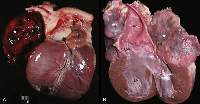
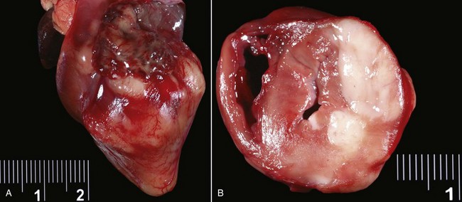
Classic mesotheliomas occur as a diffuse nodular mass or multifocal masses covering the surfaces of the body cavity (Figure 33-8). Extensive effusions occur due to exudation from the tumor surface or from tumor-obstructed lymphatics; therefore the most common presenting sign is dyspnea from pleural effusion or a distended abdomen from peritoneal effusion. Dogs with pericardial or heart-base mesotheliomas can present with acute tamponade and right-sided heart failure.30-32

image

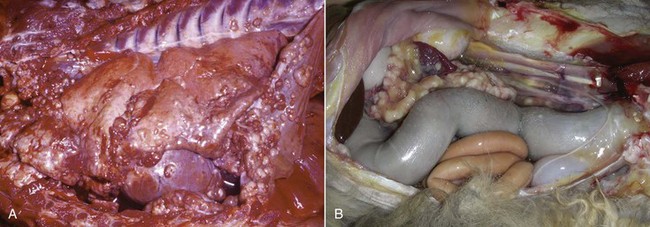
Figure 33-8 A, Pleural, parietal, and pericardial surfaces of a dog at necropsy illustrating nodular lesions histologically confirmed as mesothelioma. B, Mesothelioma involving the peritoneal surfaces of a cat at necropsy.

Sclerosing mesothelioma is a variation of mesothelial tumor seen primarily in male dogs, with German shepherd dogs being overrepresented.4,25,26,33 These tumors present as thick fibrous linings in the abdominal and/or pleural cavities. Restriction occurs around organs in the affected area, and in the abdomen such changes can impinge on organs and lead to vomiting and urinary tract signs.

Diagnostic Techniques and Work-Up

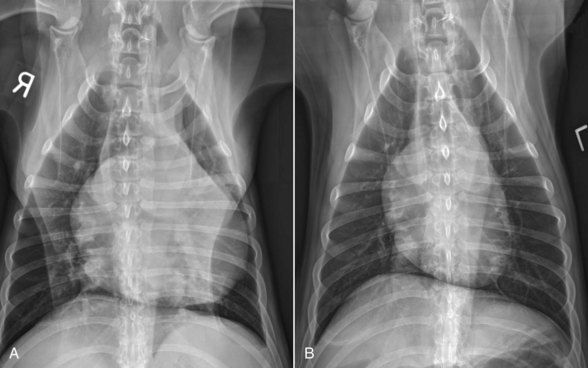
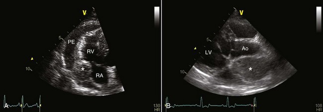
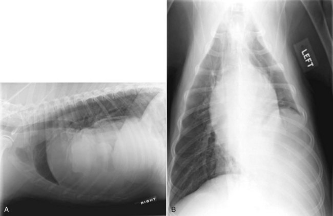
Mesothelioma should be suspected in adult dogs presenting with a history of chronic, nonspecific disease and fluid accumulation in any of the body cavities. Routine echocardiography and abdominal ultrasound are not typically helpful because the tumor cells cling to epithelial surfaces and a mass lesion is rarely noted.34 In a recent study of echocardiography of dogs with pericardial effusion, only 5 of 15 dogs with pericardial effusion due to mesothelioma had a discrete cardiac mass identified.32 Thoracic CT may be of benefit in identification of nodular lesions and in assessment of lung parenchyma in the face of pleural effusion35,36 (Figure 33-9).

image

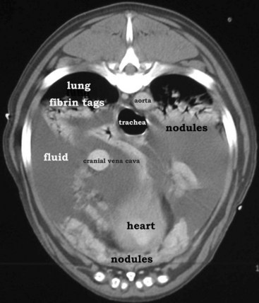
Figure 33-9 Thoracic CT (with contrast) from a dog with histologically confirmed mesothelioma. The effusion resolved following the first of five doxorubicin chemotherapy (30 mg/m2 every 3 weeks, IV) treatments.

Cytologic evaluation of fluid can be diagnostic for other disease processes such as infection or lymphoma but will not conclusively diagnose mesothelioma. Mesothelial cells proliferate under any circumstance associated with fluid accumulation in the body cavity, making the distinction between physiologic mesothelial proliferation and neoplasia difficult. Although malignant mesothelial cells easily exfoliate into effusion fluid, they are hard to distinguish from reactive hypertrophic mesothelial cells cytologically. Reactive mesothelial cells display many cytologic features of malignancy, making a definitive diagnosis of neoplasia via cytology impossible in most cases. Although one study found pericardial fluid pH analysis to be a discriminatory test to differentiate benign from malignant effusions, subsequent studies found too much overlap in the pH values for the test to be of benefit.37-39 Fibronectin concentrations have also been evaluated in pleural effusions in dogs and cats and were found to differentiate malignant or inflammatory causes from cardiogenic effusions. Elevation in fibronectin levels is a sensitive but nonspecific test for malignant effusions, and mesothelioma can be ruled out if fibronectin levels are not increased.40

Establishing a definitive diagnosis of malignant mesothelioma may be difficult, particularly early in the disease. The diagnosis of mesothelioma requires adequate tissue sampling, preferably from an open, visually directed biopsy. Increasing availability of thoracoscopy and laparoscopy for small animals provides a less invasive means to evaluate these cases.41 In either procedure, the clinician is encouraged to biopsy any body cavity lining when an obvious cause for fluid accumulation is not found. Sclerosing mesothelioma must be distinguished from chronic inflammatory diseases of the body cavity, such as chronic peritonitis, and histologic examination of biopsy material is essential to establish the diagnosis. Additionally, embolized, nonneoplastic mesothelial cells within lymph nodes is a rare finding in humans with cavity effusions and has been reported in dogs affected with idiopathic hemorrhagic pericardial effusion; therefore care must be taken so as not to overinterpret these cells as indicative of a metastatic process.42

The most useful criteria in establishing a diagnosis of mesothelioma is to demonstrate that the tumor is primarily a neoplasm of the coelomic cavity lining and that the method of tumor spread is by transcoelomic implantation. Therefore mesothelioma should be considered when the bulk of the neoplastic tissue exists on the coelomic surface. Histologically, mesotheliomas need to be differentiated from carcinomas, adenocarcinomas, or sarcomas, depending on the morphologic type of the mesothelioma. Unfortunately, there are no cellular markers that conclusively define the mesothelial cell. Recent advances in IHC staining have provided additional ways to examine neoplastic cells to help differentiate mesothelioma from other epithelial or mesenchymal tumors in humans. Podoplanin and D2-40 were found to be highly sensitive IHC markers for sarcomatoid mesotheliomas.43 Differentiation of malignant epithelial mesotheliomas from adenocarcinomas can be aided by application of a panel of different immunohistochemical stains, including calretinin, Wilms’ tumor-1, and cytokeratin 5/6 for which mesotheliomas, but not adenocarcinomas, are strongly positive.44

Treatment and Prognosis

No satisfactory treatment exists for mesothelioma. Radical excision may benefit some animals, but usually the tumors are too advanced locally and have spread by implantation early in the course of disease. In one case report, a 2-year-old Siberian Husky with a solitary sclerosing mesothelioma affecting the left thoracic diaphragmatic surface with pericardial and mediastinal adhesions was treated with aggressive surgical resection and diaphragmatic reconstruction using the transversus abdominis muscle.45 The dog recovered well, but subcutaneous masses at the surgery site, as well as hepatic and renal masses, were noted 54 days postoperatively, leading to euthanasia. Pericardiectomy may palliate mesothelioma patients that present with cardiac tamponade; two dogs treated with surgery alone survived 4 and 9 months in one study.46 In another report, the median survival in five dogs treated with pericardiectomy was 13.6 months; three of these dogs received adjuvant IV chemotherapy (two DOX, one mitoxantrone).47 A dog treated with pericardiectomy, intrathoracic and IV cisplatin, and IV DOX remained free of disease at 27 months.48 In a report of eight dogs with pericardial mesothelioma, the MST was 60 days(range 15 to 300 days) following partial pericardiectomy. The one dog that survived 300 days was treated with DOX and intracavitary cisplatin for the 4 months preceding death.34 Thoracoscopic partial pericardiectomy is a less invasive procedure than open thoracic surgical pericardiectomy and has been successfully performed in dogs with malignant pericardial effusions, including four dogs with mesotheliomas.49 Portal site seeding with the mesothelioma is a potential complication of this procedure.50 Median survival for animals with untreated mesotheliomas in any location is difficult to assess from reports because the tumors are rare and animals frequently are euthanized at the time of diagnosis.

Intracavitary cisplatin has shown palliative potential in the dog; it was well tolerated and greatly decreased mesothelioma-associated thoracic fluid accumulation in three dogs in one study.51 The treatments also appeared to arrest tumor growth for a limited time. Two doses of intracavitary carboplatin were safely administered to a cat with suspected pleural mesothelioma and resulted in transient resolution of clinical signs for a total of 54 days, at which point the owners discontinued therapy.52 Unfortunately, local penetration of intracavitary chemotherapy only occurs to a limited depth (2 to 3 mm); thus large masses will not be affected significantly, other than from ultimate exposure to the systemically absorbed intracavitary drug. In such cases, combining debulking surgery or systemic chemotherapy such as DOX or mitoxantrone with intracavitary cisplatin may be beneficial. For peritoneal mesothelioma, four doses of intracavitary cisplatin in two dogs and carboplatin in one cat, combined with piroxicam administration, resolved the effusion in all cases.53 One of the dogs had debulking surgery first; this dog was still in remission at 2 years, whereas the other dog and the cat lived 8 and 6 months, respectively. IV chemotherapy may provide benefit in some patients; single-agent IV cisplatin administered every 3 weeks was reported to improve clinical signs in a dog with bicavitary epithelial mesothelioma, until sudden death occurred 5 months after treatment initiation.54

Comparative Aspects

In humans, mesothelioma is closely linked to exposure to aerosolized asbestos fibers. Approximately 70% to 80% of cases have a history of occupational exposure, with the type of employment significantly affecting relative risk.17,55,56 Occupations such as construction work, ship building, heating trades, asbestos mining, and insulation work are strong risk factors in the development of mesothelioma in humans.17 Family members of exposed industrial workers are at risk due to asbestos fiber exposure from the workers’ clothing. Affected individuals routinely have greatly increased counts of asbestos fibers in parenchymal lung tissue.55 The latency period from time of exposure to tumor development is long, with reports ranging from 12 to 50 years.17 Other risk factors discussed in the development of mesotheliomas include past radiation, exposure to certain chemicals or nonasbestos fibers, and genetic tendency.15,17 In addition, the DNA tumor virus Simian 40 (SV40) is suspected to act as a cocarcinogen with asbestos in causing mesotheliomas.16,57,58 SV40 was introduced into a large percentage of humans through contaminated polio vaccines between 1955 and 1963. SV40 can transform cells and experimentally leads to mesothelioma development in laboratory animals. However, there is strong evidence against the proposed connection between SV40 and mesothelioma and data support that the high prevalence of SV40 found in human mesotheliomas in previous studies was likely due to false-positive molecular assays.59,60

In humans, the median survival is approximately 1 year from symptom onset. A prognostic index, based on a combination of prognostic factors and gene expression testing, allows for the determination of high- and low-risk patients with respective median survivals of 6.9 versus 31.9 months.56 There is no consensus in the literature regarding surgery and RT for treatment, although recently several chemotherapy options have been shown to improve survival time and quality of life.56 The current gold standard drugs are antifolates, specifically pemetrexed and raltitrexed, often combined with cisplatin or carboplatin.56 Gemcitabine and vinorelbine have also shown some efficacy in humans with mesothelioma. Several novel therapies are also under investigation. Unfortunately, some therapies (e.g., epidermal growth factor receptor [EGFR] inhibitors) performing well in preclinical models did not show activity in clinical mesothelioma patients.61 Trials of other novel therapies, including anti-VEGF antibody, cyclin-dependent kinase (CDK) inhibitor, antimesothelin antibody, and proteasome inhibitors, are ongoing. Multimodality therapy, including novel agents, will hopefully improve survival time in the future.57,61

References

1. Geib, LW, DeNarvaez, F, Eby, CH. Pleural mesothelioma in a dog. J Am Vet Med Assoc. 1962;140:1317–1319.

2. Cihak, RW, Roen, DR, Klaassen, J. Malignant mesothelioma of the tunica vaginalis in a dog. J Comp Pathol. 1986;96:459–462.

3. Vascellari, M, Carminato, A, Camali, G, et al. Malignant mesothelioma of the tunica vaginalis testis in a dog: histological and immunohistochemical characterization. J Vet Diagn Invest. 2011;23:135–139.

4. Dubielzig, RR. Sclerosing mesothelioma in five dogs. J Am Anim Hosp Assoc. 1979;15:745–748.

5. Thrall, DE, Goldschmidt, MH. Mesothelioma in the dog: six case reports. J Am Vet Radial Soc. 1978;19:107–115.

6. Morini, M, Bettini, G, Morandi, F, et al. Deciduoid peritoneal mesothelioma in a dog. Vet Pathol. 2006;43:198–201.

7. Schaer, M, Meyer, D. Benign peritoneal mesothelioma, hyperthyroidism, nonsuppurative hepatitis, and chronic disseminated intravascular coagulation in a cat: a case report. J Am Anim Hosp Assoc. 1988;24:195–202.

8. Kobayashi, Y, Usuda, H, Ochiai, K, et al. Malignant mesothelioma with metastases and mast cell leukaemia in a cat. J Comp Path. 1994;111:453–458.

9. Carpenter, JL, Andrews, LK, Holzworth, J. Tumors and tumor-like lesions. Philadelphia: WB Saunders; 1987.

10. Tilley, LP, Owens, JM, Wilkins, RJ, et al. Pericardial mesothelioma with effusion in a cat. J Am Anim Hosp Assoc. 1975:60–65.

11. Umphlet, RC, Bertoy, RW. Abdominal mesothelioma in a cat. Mod Vet Pract. 1988;69:71–73.

12. Bacci, B, Morandi, F, De Meo, M, et al. Ten cases of feline mesothelioma: an immunohistochemical and ultrastructural study. J Comp Pathol. 2006;134:347–354.

13. Glickman, LT, Domanski, LM, Maguire, TG, et al. Mesothelioma in pet dogs associated with exposure of their owners to asbestos. Environ Res. 1983;32:305–313.

14. Harbison, ML, Godleski, JJ. Malignant mesothelioma in urban dogs. Vet Pathol. 1983;20:531–540.

15. Hughes, RS. Malignant pleural mesothelioma. Am J Med Sci. 2005;329:29–44.

16. Jaurand, MC, Fleury-Feith, J. Pathogenesis of malignant pleural mesothelioma. Respirology. 2005;10:2–8.

17. Pistolesi, M, Rusthoven, J. Malignant pleural mesothelioma: update, current management, and newer therapeutic strategies. Chest. 2004;126:1318–1329.

18. Belli, C, Anand, S, Tassi, G, et al. Translational therapies for malignant pleural mesothelioma. Expert Rev Respir Med. 2010;4:249–260.

19. Machida, N, Tanaka, R, Takemura, N, et al. Development of pericardial mesothelioma in golden retrievers with a long-term history of idiopathic haemorrhagic pericardial effusion. J Comp Pathol. 2004;131:166–175.

20. Head, KW. Tumors of the alimentary tract, ed 3. Berkeley: University of Calif Press; 1990.

21. Kim, JH, Choi, YK, Yoon, HY, et al. Juvenile malignant mesothelioma in a dog. J Vet Med Sci. 2002;64:269–271.

22. Vural, SA, Ozyildiz, Z, Ozsoy, SY. Pleural mesothelioma in a nine-month-old dog. Ir Vet J. 2007;60:30–33.

23. Leisewitz, AL, Nesbit, JW. Malignant mesothelioma in a seven-week-old puppy. J S Afr Vet Assoc. 1992;63:70–73.

24. Corson, JM. Pathology of mesothelioma. Thorac Surg Clin. 2004;14:447–460.

25. Geninet, C, Bernex, F, Rakotovao, F, et al. Sclerosing peritoneal mesothelioma in a dog - a case report. J Vet Med A Physiol Pathol Clin Med. 2003;50:402–405.

26. Schoning, P, Layton, CE, Fortney, WD, et al. Sclerosing peritoneal mesothelioma in a dog evaluated by electron microscopy and immunoperoxidase techniques. J Vet Diagn Invest. 1992;4:217–220.

27. Dias Pereira, P, Azevedo, M, Gartner, F. Case of malignant biphasic mesothelioma in a dog. Vet Rec. 2001;149:680–681.

28. Sato, T, Miyoshi, T, Shibuya, H, et al. Peritoneal biphasic mesothelioma in a dog. J Vet Med A Physiol Pathol Clin Med. 2005;52:22–25.

29. DiPinto, MN, Dunstan, RW, Lee, C. Cystic, peritoneal mesothelioma in a dog. J Am Anim Hosp Assoc. 1995;31:385–389.

30. Cobb, MA, Brownlie, SE. Intrapericardial neoplasia in 14 dogs. J Small Anim Pract. 1992;33:309–316.

31. McDonough, SP, MacLachlan, NJ, Tobias, AH. Canine pericardial mesothelioma. Vet Pathol. 1992;29:256–260.

32. MacDonald, KA, Cagney, O, Magne, ML. Echocardiographic and clinicopathologic characterization of pericardial effusion in dogs: 107 cases (1985-2006). J Am Vet Med Assoc. 2009;235:1456–1461.

33. Gumber, S, Fowlkes, N, Cho, DY. Disseminated sclerosing peritoneal mesothelioma in a dog. J Vet Diagn Invest. 2011;23:1046–1050.

34. Stepien, RL, Whitley, NT, Dubielzig, RR. Idiopathic or mesothelioma-related pericardial effusion: clinical findings and survival in 17 dogs studied retrospectively. J Small Anim Pract. 2000;41:342–347.

35. Echandi, RL, Morandi, F, Newman, SJ, et al. Imaging diagnosis—canine thoracic mesothelioma. Vet Radiol Ultrasound. 2007;48:243–245.

36. Reetz, JA, Buza, EL, Krick, EL. CT features of pleural masses and nodules. Vet Radiol Ultrasound. 2011. [November 18. Epub ahead of print].

37. Edwards, NJ. The diagnostic value of pericardial fluid pH determination. J Am Anim Hosp Assoc. 1996;32:63–67.

38. Fine, DM, Tobias, AH, Jacob, KA. Use of pericardial fluid pH to distinguish between idiopathic and neoplastic effusions. J Vet Intern Med. 2003;17:525–529.

39. de Laforcade, AM, Freeman, LM, Rozanski, EA, et al. Biochemical analysis of pericardial fluid and whole blood in dogs with pericardial effusion. J Vet Intern Med. 2005;19:833–836.

40. Hirschberger, J, Pusch, S. Fibronectin concentrations in pleural and abdominal effusions in dogs and cats. J Vet Intern Med. 1996;10:321–325.

41. Reggeti, F, Brisson, B, Ruotsalo, K, et al. Invasive epithelial mesothelioma in a dog. Vet Pathol. 2005;42:77–81.

42. Peters, M, Tenhundfeld, J, Stephan, I, et al. Embolized mesothelial cells within mediastinal lymph nodes of three dogs with idiopathic haemorrhagic pericardial effusion. J Comp Pathol. 2003;128:107–112.

43. Chirieac, LR, Pinkus, GS, Pinkus, JL, et al. The immunohistochemical characterization of sarcomatoid malignant mesothelioma of the pleura. Am J Cancer Res. 2011;1:14–24.

44. Chirieac, LR, Corson, JM. Pathologic evaluation of malignant pleural mesothelioma. Semin Thorac Cardiovasc Surg. 2009;21:121–124.

45. Liptak, JM, Brebner, NS. Hemidiaphragmatic reconstruction with a transversus abdominis muscle flap after resection of a solitary diaphragmatic mesothelioma in a dog. J Am Vet Med Assoc. 2006;228:1204–1208.

46. Kerstetter, KK, Krahwinkel, DJ, Millis, DL, et al. Pericardiectomy in dogs: 22 cases (1978-1994). J Am Vet Med Assoc. 1997;211:736–740.

47. Dunning, D, Monnet, E, Orton, EC, et al. Analysis of prognostic indicators for dogs with pericardial effusion: 46 cases (1985-1996). J Am Vet Med Assoc. 1998;212:1276–1280.

48. Closa, JM, Font, A, Mascort, J. Pericardial mesothelioma in a dog: long-term survival after pericardiectomy in combination with chemotherapy. J Small Anim Pract. 1999;40:383–386.

49. Jackson, J, Richter, K, Launer, D. Thoracoscopic partial pericardiectomy in 13 dogs. J Vet Intern Med. 1999;13:529–533.

50. Brisson, BA, Reggeti, F, Bienzle, D. Portal site metastasis of invasive mesothelioma after diagnostic thoracoscopy in a dog. J Am Vet Med Assoc. 2006;229:980–983.

51. Moore, AS, Kirk, C, Cardona, A. Intracavitary cisplatin chemotherapy experience with six dogs. J Vet Intern Med. 1991;5:227–231.

52. Sparkes, A, Murphy, S, McConnell, F, et al. Palliative intracavitary carboplatin therapy in a cat with suspected pleural mesothelioma. J Feline Med Surg. 2005;7:313–316.

53. Spugnini, EP, Crispi, S, Scarabello, A, et al. Piroxicam and intracavitary platinum-based chemotherapy for the treatment of advanced mesothelioma in pets: preliminary observations. J Exp Clin Cancer Res. 2008;27:6.

54. Seo, KW, Choi, US, Jung, YC, et al. Palliative intravenous cisplatin treatment for concurrent peritoneal and pleural mesothelioma in a dog. J Vet Med Sci. 2007;69:201–204.

55. Zellos, L, Christiani, DC. Epidemiology, biologic behavior, and natural history of mesothelioma. Thorac Surg Clin. 2004;14:469–477. [viii].

56. Campbell, NP, Kindler, HL. Update on malignant pleural mesothelioma. Semin Respir Crit Care Med. 2011;32:102–110.

57. Carbone, M, Fisher, S, Powers, A, et al. New molecular and epidemiological issues in mesothelioma: role of SV40. J Cell Physiol. 1999;180:167–172.

58. Fisher, SG, Weber, L, Carbone, M. Cancer risk associated with simian virus 40 contaminated polio vaccine. Anticancer Res. 1999;19:2173–2180.

59. Lopez-Rios, F, Illei, PB, Rusch, V, et al. Evidence against a role for SV40 infection in human mesotheliomas and high risk of false-positive PCR results owing to presence of SV40 sequences in common laboratory plasmids. Lancet. 2004;364:1157–1166.

60. Gee, GV, Stanifer, ML, Christensen, BC, et al. SV40 associated miRNAs are not detectable in mesotheliomas. Br J Cancer. 2010;103:885–888.

61. Raja, S, Murthy, SC, Mason, DP. Malignant pleural mesothelioma. Curr Oncol Rep. 2011;13:259–264.

image Section E

Neoplasia of the Heart

William C. Kisseberth

Incidence and Risk Factors

Neoplasia of the heart and pericardium is rare in the dog and even less common in the cat. The proportional morbidity of canine heart tumors was 0.19% in a Veterinary Medical Data Base (VMDB) search.1 In two necropsy series, the overall frequency of primary or metastatic cardiac tumors was 0.12% to 5.73%2-9 and 11.74% to 28.3% of neoplasms involving intrapericardial tissues, respectively.2,3 Cardiac tumors (excluding lymphoma) occur most frequently in middle-aged to older (7 to 15 years) dogs.1 Overwhelmingly, HSA is the most common primary cardiac tumor in the dog, followed by aortic body tumors (chemodectoma, paraganglioma) and then miscellaneous other tumors.1 HSA represented 69% and aortic body tumor 8% of the histologically diagnosed cardiac tumors in the VMDB.1 Breeds reported to be at increased risk or predisposed for cardiac HSA include the German shepherd dog and golden retriever.3,10,11 Aortic body tumors occur most commonly in older brachycephalic dogs,3,12-15 including Boxers, Boston terriers, English bulldogs, and also German shepherd dogs.1 It has been suggested that chronic hypoxia may stimulate development of chemoreceptor tumors in both dogs and humans12,13 and that this factor could explain the increased occurrence of aortic body chemodectomas in brachycephalic breeds; however, not all dogs diagnosed with aortic body tumors are brachycephalic nor do all brachycephalic breeds appear to be at increased risk, and thus other factors (e.g., genetic) presumably contribute to pathogenesis. Based on the VMDB study, 12 breeds were identified as having a significantly higher cardiac tumor incidence compared to all other breeds. Many of the breeds previously reported to be at increased risk for cardiac tumors were identified; however, other breeds were also identified with a relatively high incidence. Most notably, the golden retriever had a high incidence of cardiac tumors (primarily HSA) and total number of recorded tumors.1

A similar VMDB search determined an overall incidence of 0.0275% for feline cardiac tumors.16 The only primary tumors of the cardiovascular system found in a series of 4933 feline necropsies were one case of mesothelioma of the pericardium and two cases of chemodectoma.17 Overall, the most common primary or metastatic cardiac tumor in cats is lymphoma.16

Pathology and Natural Behavior

Neoplasms affecting the heart may occur in intracavitary, intramural, or pericardial locations or at the heart base. Primary tumors may be benign or malignant, with most in the dog occurring in the right atrium and auricle. The most common primary cardiac tumor in the dog is HSA (Figure 33-10, A), followed by aortic body tumor (Figure 33-10, B).1-15 Cardiac HSAs often are associated with hemorrhagic pericardial effusion, cardiac tamponade, and metastatic disease.10,11,18,19 Aortic body tumors arise from chemoreceptor cells at the heart base. Most frequently, they present as a single mass at the base of the heart; however, occasionally they present as a localized collection of focal masses or are infiltrative into the myocardium. Aortic body tumors are primarily locally invasive but occasionally metastasize. Functional tumors have not been found in domestic animals but have been reported in humans.20 Other primary cardiac tumors reported in the dog include lymphoma, undifferentiated sarcoma, myxoma, ectopic thyroid carcinoma, fibroma, fibrosarcoma, rhabdomyosarcoma, chondrosarcoma, mesothelioma, granular cell tumor, osteosarcoma, myxosarcoma, leiomyoma, thyroid adenoma, thyroid carcinosarcoma, leiomyosarcoma, lipoma, peripheral nerve sheath tumor, and mixed mesenchymal tumor.21-48 In contrast to humans, primary cardiac tumors are more common than metastatic tumors in the dog.1 This is due largely to the high incidence of HSA in dogs. HSA accounts for 40% to 69% of cardiac neoplasms in the dog.1-3 In most studies, cardiac HSA is considered a primary site, although dogs often have evidence of disease at other locations at the time of diagnosis, and it usually is impossible to determine which is the primary site and which are the metastatic sites. In a recent necropsy case series, 24 of 66 dogs (36%) with extracardiac malignant tumors had metastases found in the heart (15 carcinomas, 6 lymphomas, 3 HSAs), suggesting that cardiac metastases may be underdiagnosed in dogs with malignant, especially advanced, cancers.49 In individual reports, metastatic mammary carcinoma, melanoma, malignant histiocytosis, pheochromocytoma, granulosa cell tumor, gastric adenocarcinoma, lymphoma, and liposarcoma have been reported to occur in the heart of the dog.50-58 Primary cardiac HSA, aortic body tumors, myxoma, and rhabdomyosarcoma have been reported in the cat16,59-64; however, metastatic tumors are more common—lymphoma (Figure 33-11), mammary gland carcinoma, pulmonary carcinoma, salivary gland adenocarcinoma, oral melanoma, rhabdomyosarcoma, sweat gland adenocarcinoma, squamous cell carcinoma, and mast cell tumor have been reported.16,17,49,65-70

image

Figure 33-10 A, Gross postmortem specimen from a dog with right auricular HSA. Note the large, hemorrhagic mass effacing the right auricle (*). B, Gross postmortem specimen from a dog with an aortic body tumor (chemodectoma). Note the large, multilobulated mass (*) surrounding the aorta and compressing the atria. LV, Left ventricle; AoV, aortic valve; Ao, ascending aorta. (Courtesy Brian A. Scansen, DVM, MS, DACVIM [Cardiology], the Ohio State University, College of Veterinary Medicine.)

image

Figure 33-11 Gross postmortem specimen from a cat with cardiac lymphoma. A, The external surface of the heart shows several raised, white masses. B, A cross-section of the ventricles shows a large expansile infiltrate determined to be lymphoma by histopathology. (Courtesy Paul C. Stromberg DVM, PhD, DACVP, the Ohio State University, College of Veterinary Medicine.)

History and Clinical Signs

Tumors involving the heart cause varied clinical signs. Cardiac tumors disrupt the normal function of the tissues from which they arise, leading to altered cardiovascular function. In general, signs result from (1) the physical presence of the mass causing obstruction of blood flow into or out of the heart, (2) external compression of the heart that impedes filling (e.g., pericardial effusion) and resulting cardiac tamponade, and (3) disruption of normal heart rhythm or contractility if myocardial infiltration occurs or ischemia develops. Clinical signs produced by cardiac tumors are more closely related to their precise anatomic location than their histologic type. Specific clinical signs observed in an individual patient are influenced by the tumor’s size and location and the presence of pericardial effusion. Acute death from rupture of a tumor with subsequent blood loss, with or without cardiac tamponade, is a common sequela of cardiac HSA. Sudden death due to cardiac arrhythmias may also occur. Cardiac HSAs, as well as the majority of reported primary sarcomas of the right heart in dogs, often produce signs of right heart failure due to the presence of cardiac tamponade and inflow obstruction. These signs include ascites, pleural effusion, jugular venous distension, abnormal jugular pulsations, exercise intolerance, dyspnea, pulse deficits, muffled heart sounds, and syncope. Tumors that cause pericardial effusion and cardiac tamponade have been reported the most often.1 Cardiac or pericardial tumors are responsible for approximately 60% of cases of pericardial effusion in dogs.71 The most common tumors to cause pericardial effusion are right atrial HSA, aortic body tumors, and mesothelioma.71 Associated clinical signs resulting from cardiac tamponade include restricted ventricular filling secondary to external cardiac compression, venous congestion, and poor cardiac output. Heart base tumors are most often associated with pericardial involvement of the tumor and accompanying pericardial effusion.16 Edema, ascites, cough, dyspnea, weight loss, and vomiting are the signs most commonly reported with aortic body tumors in dogs.14,16,72 Heart base masses are a common cause for cranial vena cava syndrome (edema of the head, neck, and forelimbs) due to tumor pressure on the cranial vena cava. Sometimes, these tumors may be present without causing clinical signs or are incidental findings at necropsy. Cardiac tumors that do not cause pericardial effusion can cause signs of congestive heart failure or low cardiac output by obstructing blood flow within the heart or great vessels and by inducing arrhythmias. Syncope and weakness with exertion or excitement are common signs in animals with cardiac tumors. These signs of low cardiac output can result from cardiac tamponade, blood flow obstruction, arrhythmias, impaired myocardial function, and hemorrhage.1 Clinical signs may be absent if the tumor is small or in a location that does not affect cardiac function.

Diagnostic Techniques and Work-Up

The diagnosis of cardiac neoplasia in the dog and cat is usually based on clinical history, physical examination, and radiographic and echocardiographic findings. Occasionally, tumors, especially aortic body tumors, are an incidental finding at necropsy. In many instances, cytologic or histologic confirmation of neoplasia is not obtained antemortem; however, FNA or tissue biopsy should be obtained when indicated and technically feasible. An electrocardiogram may be normal in patients with cardiac tumors or may show any of a variety of arrhythmias, which may correlate with the underlying site of the primary or metastatic tumor or may be secondary to myocardial ischemia or hypoxia. Low-amplitude QRS complexes and electrical alternans may be seen in animals with pericardial effusion.73 Sinus tachycardia is common with cardiac tamponade. Conduction and rhythm abnormalities are especially common with infiltrative tumors of the myocardium. Animals with a large volume of pericardial effusion may have a rounded (“globoid”) cardiac silhouette (Figure 33-12). Smaller fluid accumulations may allow visualization of atrial and tumor shadows.16 Thoracic radiographs may reveal cardiomegaly or effusions associated with cardiac tamponade. Mass lesions, if seen, are most common in the areas of the right atrium and heart base. Lung metastases may also be seen.

image

Figure 33-12 Thoracic radiographs from an 8-year-old golden retriever with moderate pericardial effusion before (A) and 5 days after pericardiocentesis (B). Note the globoid cardiac silhouette prior to pericardiocentesis, compared to the normal cardiac silhouette seen after the pericardial fluid is removed. (Courtesy Brian A Scansen, DVM, MS, DACVIM [Cardiology], the Ohio State University, College of Veterinary Medicine.)

Echocardiography is the most valuable diagnostic procedure for identifying tumors of the heart in cats and dogs (Figure 33-13).74-76 In a recent study of 107 dogs with pericardial effusion, the sensitivity and specificity of echocardiography were 82% and 100%, respectively, for detection of a cardiac mass; 82% and 99%, respectively, for detection of a right atrial mass; and 74% and 98%, respectively, for detection of a heart base mass.77 The positive and negative predictive values of echocardiography were 100% and 75%, respectively, for detection of a cardiac mass; 100% and 87%, respectively, for the detection of a right atrial mass; and 89% and 93%, respectively, for detection of a heart base mass.77 In another study of histologically confirmed HSA of the right atrium/auricle, echocardiography had a positive predictive value of 92% (11/12) and a negative predictive value of 64% (9/14) in dogs.76 Tumor location (extrapericardial, noncavitary pericardial, and small auricular masses) and size appear to be the most important factors for false-negative results with echocardiography.76 Pericardial effusions are a common finding associated with cardiac tumors in both cats and dogs.36,76-78 Echocardiographic diagnosis of mesothelioma is difficult. Results of echocardiographic examination may be negative unless there are discrete mass lesions associated with the pericardium. Advanced imaging modalities, including CT, MRI, positron emission tomography (PET), and PET/CT, may be useful for selected cases.79-82

image

Figure 33-13 A, Echocardiographic image from a 12-year-old golden retriever presented for acute collapse. There is mild pericardial effusion (PE) and a circular mass of mixed echogenicity at the right atrioventricular groove (*) consistent with HSA. RA, Right atrium, RV, right ventricle. B, Echocardiographic image from an 8-year-old Boxer. A large mass (*) of mixed echogenicity is seen adjacent to the ascending aorta (Ao) consistent with an aortic body tumor. LV, Left ventricle. (Courtesy Brian A Scansen, DVM, MS, DACVIM [Cardiology], the Ohio State University, College of Veterinary Medicine.)

Other clinical diagnostic methods for the evaluation of cardiac or pericardial masses include pneumopericardiography, selective and nonselective angiography, gated radionuclide imaging, and endomyocardial biopsy in selected patients.71,83,84 Cytologic evaluation of pericardial fluid and pericardial fluid pH has proved to be of limited usefulness in diagnosing or discriminating between neoplastic and nonneoplastic causes of pericardial effusion.85-87

Cardiac troponin I (cTnI) appears to be useful for diagnosing cardiac HSA in dogs.88,89 cTnI and cardiac troponin T (cTnT) are sensitive and specific markers for myocardial ischemia and necrosis. In one study, dogs with pericardial effusion had significantly higher serum concentrations of cTnI but not cTnT than normal dogs.88 Furthermore, dogs with cardiac HSA had significantly higher concentrations of cTnI than did dogs with idiopathic pericardial effusion.88 In another study, the median plasma cTnI concentration was higher in dogs with cardiac HSA compared with the median concentration in dogs with HSA at other sites, dogs with other neoplasms, and dogs with pericardial effusion not caused by HSA. Furthermore, dogs with cTnI concentrations higher than 0.25 ng/mL were found likely to have cardiac HSA, and a plasma cTnI higher than 2.45 ng/mL indicated that cardiac involvement is likely in dogs with HSA.89

For dogs and cats with a cardiac mass and suspected neoplasia, every effort should be made to determine the extent of disease and the existence of primary or metastatic sites elsewhere in the patient. In addition to echocardiography and possibly other advanced imaging, a minimum database, including a CBC, serum biochemical profile, coagulation profile, thoracic radiographs, and abdominal ultrasound or radiographs, should be obtained. A pathologic diagnosis may be obtained by FNA cytology, endomyocardial biopsy, or open surgical or thoracoscopic biopsy, although many animals are treated based on anatomic location of the mass (e.g., right auricle HSA).

Therapy

Initially, treatment of patients with cardiac tumors consists of treating existing arrhythmias and clinical signs of heart failure, if present. Unfortunately, without effective antitumor treatment, the hemodynamic consequences of the mass often are refractory to medical management. Surgical resection may be indicated for some primary cardiac tumors, especially those of the right auricular appendage.10,19,26,90-94 However, surgical resection of right auricular masses in dogs with cardiac HSA must be considered a palliative procedure due to the high probability of metastatic disease, although a pericardiectomy may improve signs. Dogs with aortic body tumors generally benefit from pericardiectomy, independent of the presence or absence of pericardial effusion at the time of surgery.14,72 Thoracoscopy has been recommended as an alternative to open thoracotomy for biopsy and pericardiectomy because almost all aortic body tumors and many (≈50%) right auricular masses are unresectable. Thoracoscopy requires advanced training and special instrumentation if it is to attain the goals of tissue diagnosis, decreased operative time, and decreased morbidity.93,95-97 DOX-based chemotherapy protocols, including DOX alone, DOX and cyclophosphamide, and DOX-cyclophosphamide-vincristine have been used alone98,99 and in combination with surgery19 for the treatment of cardiac HSA.

Prognosis

The prognosis for primary cardiac tumors generally is poor. Most reported cases responded poorly to medical management. Mean survival of dogs with cardiac HSA treated with surgical resection alone is reported to be 46 days to 5 months.10,19,90 Survival time was significantly longer in dogs treated with tumor resection and adjuvant chemotherapy, with a reported mean of 164 days,19 comparable to dogs with splenic HSA treated with splenectomy and adjuvant chemotherapy.99 Complete surgical resection of aortic body tumors usually is not possible; however, dogs that receive a pericardiectomy survive longer (median survival 730 days) than those that do not have a pericardiectomy (median survival 42 days).72 Other cardiac tumors found during exploratory thoracotomy commonly are judged to be unresectable; however, a few cases with longer disease-free intervals following resection of tumors other than HSA have been reported.26,28 In one study, dogs with HSA involving the pericardium survived a median of 16 days, whereas those with mesothelioma survived a median of 15.3 months following surgery.100

Comparative Aspects

In humans, primary tumors of the heart and pericardium are rare. The vast majority of such tumors are metastatic.101 Primary cardiac tumors occur with a frequency of approximately 0.02% in pooled autopsy series.102 Primary tumors are usually cavitary, and 75% are benign.103 Familial cardiac myxomas occur and appear to have an autosomal dominant transmission and are caused by mutations in the PRKAR1alpha gene that encodes a regulatory subunit of protein kinase A.104 Myxomas constitute nearly 50% of all histologically benign tumors of the heart in humans. Seventy-five percent of myxomas occur in the left atrium. Systemic tumor embolization occurs with high frequency. Surgical resection of myxoma is the treatment of choice, with recurrence of sporadic atrial myxomas being rare following excision. Rhabdomyoma is the most common cardiac tumor of infants and children.103 Other benign primary cardiac tumors that have been reported in humans include fibroma, papillary fibroelastoma, lipoma, cystic tumors of the atrioventricular node, hemangioma, lymphangioma, and intrapericardial paraganglioma.102,103

Almost all primary malignant cardiac tumors are sarcomas, most frequently angiosarcomas.103,105,106 As is the case with HSA in the dog, angiosarcoma of the heart in humans most commonly originates in the right atrium or pericardium.103 Rhabdomyosarcoma and mesothelioma rank second and third, respectively, in frequency among primary malignant tumors of the heart and pericardium in humans. Palliative and local control of malignant primary tumors can be achieved with extensive resection. Adjuvant chemotherapy and RT have been used; however, their routine use for primary cardiac sarcomas has been questioned.107 Cardiac transplantation has been utilized on occasion.

Metastatic tumors involving the heart and pericardium occur up to 100 times more frequently than primary tumors.108 Incidence rates ranging from 2.3% and 18.3% are reported in autopsy studies of patients who died of cancer.109 Malignant melanoma metastasizes most frequently to the myocardium, occurring in 46% of autopsies of cancer patients.108 Cardiac metastasis also frequently occurs with bronchogenic carcinoma and carcinoma of the breast.109

References

1. Ware, WA, Hopper, DL. Cardiac tumors in dogs: 1982-1995. J Vet Intern Med. 1999;13:95–103.

2. Girard, C, Helie, P, Odin, M. Intrapericardial neoplasia in dogs. J Vet Diagn Invest. 1999;11:73–78.

3. Walter, JH, Rudolph, R. Systemic, metastatic, eu- and heterotope tumours of the heart in necropsied dogs. Zentralbl Veterinarmed An. 1996;43:31–45.

4. Prange, H, Falk-Junge, G, Katenkamp, D, et al. Zur Verbreitung, Epizootiologie und Röntgendiagnostik intrathorakaler Geschwülste beim Hund. Arch Exper Vet Med. 1988;42:637–649.

5. Cammarata, G, Caramelli, M, Cavazzini, E, et al. Neoplasie cardiache e della base del cuore nel cane: Contributo casistico e studio istologico. Clinica Veterinaria. 1987;110:97–110.

6. Priester, WA, McKay, FW. The occurrence of tumors in domestic animals, National Cancer Institute Monograph 54. Bethesda: National Institutes of Health; 1980.

7. Detweiler, DK, Patterson, DF. The prevalence and types of cardiovascular disease in dogs. Ann N Y Acad Sci. 1965;127:481–516.

8. Luginbuhl, H, Detweiler, K. Cardiovascular lesions in dogs. Ann N Y Acad Sci. 1965;127:517–540.

9. Detweiler, DK. Wesen und Häufigkeit von Herzkrankheiten bei Hunden. Zentralbl Veterinärmed [A]. 1962;9:317–357.

10. Arosohn, M. Cardiac hemangiosarcoma in the dog: A review of 38 cases. J Am Vet Med Assoc. 1985;187:922–926.

11. Kleine, W, Zook, BC, Munson, TO. Primary cardiac hemangiosarcoma in dogs. J Am Vet Med Assoc. 1970;157:326–337.

12. Hayes, HM, Jr. A hypothesis for the aetiology of canine chemoreceptor neoplasms, based upon an epidemiological study of 73 cases among hospital patients. J Small Anim Pract. 1975;16:337–343.

13. Patnaik, AK, Liu, SK, Hurvitz, AI, et al. Canine chemodectoma (extra-adrenal paragangliomas)-a comparative study. J Small Anim Pract. 1975;16:785–801.

14. Vicari, ED, Brown, DC, Holt, DE, et al. Survival times of and prognostic indicators for dogs with heart base masses: 25 cases (1986-1999). J Am Vet Med Assoc. 2001;219:485–487.

15. Noszczyk-Nowak, A, Nowak, M, Paslawska, U, et al. Cases with manifestation of chemodectoma diagnosed in dogs in Department of Internal Diseases with Horses, Dogs and Cats Clinic, Veterinary Medicine Faculty, University of Environmental and Life Sciences, Wroclaw, Poland. Acta Vet Scand. 2010;52:35.

16. Ware, WA. Cardiac neoplasia. In: Bonagura JD, ed. Kirk’s current veterinary therapy XII. Philadelphia: WB Saunders, 1995.

17. Tilley, LP, Bond, B, Patnaik, AK, et al. Cardiovascular tumors in the cat. J Am Anim Hosp Assoc. 1981;17:1009–1021.

18. Berg, J. Pericardial disease and cardiac neoplasia. Semin Vet Med Surg (Small Anim). 1994;9:185–191.

19. Weisse, C, Soares, N, Beal, MW, et al. Survival times in dogs with right atrial hemangiosarcoma treated by means of surgical resection with or without adjuvant chemotherapy: 23 cases (1986-2000). J Am Vet Med Assoc. 2005;226:575–579.

20. Yates, WD, Lester, SJ, Mills, JH. Chemoreceptor tumors diagnosed at the Western College of Veterinary Medicine 1967-1979. Can Vet J. 1980;21:124–129.

21. Swartout, MS, Ware, WA, Bonagura, JD. Intracardiac tumors in two dogs. J Am Anim Hosp Assoc. 1987;23:533–538.

22. Machida, N, Hoshi, K, Kobayashi, M, et al. Cardiac myxoma of the tricuspid valve in a dog. J Comp Pathol. 2003;129:320–324.

23. Roberts, SR. Myxoma of the heart in a dog. J Am Vet Med Assoc. 1959;134:185–188.

24. Bright, JM, Toal, RL, Blackford, LM. Right ventricular outflow obstruction caused by primary cardiac neoplasia. J Vet Intern Med. 1990;4:12–16.

25. Sims, CS, Tobias, AH, Hayden, DW, et al. Pericardial effusion due to primary cardiac lymphosarcoma in a dog. J Vet Intern Med. 2003;17:923–927.

26. Ware, WA, Merkley, DF, Riedesel, DH. Intracardiac thyroid tumor in a dog: diagnosis and surgical removal. J Am Anim Hosp Assoc. 1994;30:20–23.

27. Madarame, H, Sato, K, Ogihara, K, et al. Primary cardiac fibrosarcoma in a dog. J Vet Med Sci. 2004;66:979–982.

28. Lombard, CW, Goldschmidt, MH. Primary fibroma in the right atrium of a dog. J Small Anim Pract. 1980;21:439–448.

29. Atkins, CE, Badertscher, RR, II., Greenlee, P, et al. Diagnosis of an intracardiac fibrosarcoma using two-dimensional echocardiography. J Am Anim Hosp Assoc. 1984;20:131–137.

30. Vicini, DS, Didier, PJ, Ogilvie, GK. Cardiac fibrosarcoma in a dog. J Am Vet Med Assoc. 1986;189:1486–1488.

31. Perez, J, Perez-Rivero, A, Montoya, A, et al. Right-sided heart failure in a dog with primary cardiac rhabdomyosarcoma. J Am Anim Hosp Assoc. 1998;34:208–211.

32. Krotje, LJ, Ware, WA, Niyo, Y. Intracardiac rhabdomyosarcoma in a dog. J Am Vet Med Assoc. 1990;197:368–371.

33. Caro-Vadillo, A, Pizarro-Diaz, M, Martinez-Merlo, E, et al. Clinical and pathological features of a cardiac chondrosarcoma in a dog. Vet Rec. 2004;155:678–680.

34. Southerland, EM, Miller, RT, Jones, CL. Primary right atrial chondrosarcoma in a dog. J Am Vet Med Assoc. 1993;203:1697–1698.

35. Greenlee, PG, Liu, SK. Chondrosarcoma of the mitral leaflet in a dog. Vet Pathol. 1984;21:540–542.

36. Cobb, MA, Brownlie, SE. Intrapericardial neoplasia in 14 dogs. J Small Anim Pract. 1992;33:309–316.

37. Sanford, SE, Hoover, DM, Miller, RB. Primary cardiac granular cell tumor in a dog. Vet Pathol. 1984;21:489–494.

38. Schelling, SH, Moses, BL. Primary intracardiac osteosarcoma in a dog. J Vet Diagn Invest. 1994;6:396–398.

39. Sato, T, Koie, H, Shibuya, H, et al. Extraskeletal osteosarcoma in the pericardium of a dog. Vet Rec. 2004;155:780–781.

40. Foale, RD, White, RA, Harley, R, et al. Left ventricular myxosarcoma in a dog. J Small Anim Pract. 2003;44:503–507.

41. Gallay, J, Bélanger, MC, Hélie, P, et al. Cardiac leiomyoma associated with advanced atrioventricular block in a young dog. J Vet Cardiol. 2011;13:71–77.

42. Di Palma, S, Lombard, C, Kappeler, A, et al. Intracardiac ectopic thyroid adenoma in a dog. Vet Rec. 2010;167:709–710.

43. Almes, KM, Heaney, AM, Andrews, GA. Intracardiac ectopic thyroid carcinosarcoma in a dog. Vet Pathol. 2008;45:500–504.

44. Fews, D, Scase, TJ, Battersby, IA. Leiomyosarcoma of the pericardium, with epicardial metastases and peripheral eosinophilia in a dog. J Comp Pathol. 2008;138:224–228.

45. Ben-Amotz, R, Ellison, GW, Thompson, MS, et al. Pericardial lipoma in a geriatric dog with an incidentally discovered thoracic mass. J Small Anim Pract. 2007;48:596–599.

46. Brambilla, PG, Roccabianca, P, Locatelli, C, et al. Primary cardiac lipoma in a dog. J Vet Intern Med. 2006;20:691–693.

47. Wohlsein, P, Cichowski, S, Baumgartner, W. Primary endocardial malignant spindle-cell sarcoma in the right atrium of a dog resembling a malignant peripheral nerve sheath tumour. J Comp Pathol. 2005;132:340–345.

48. Machida, N, Kobayashi, M, Tanaka, R, et al. Primary malignant mixed mesenchymal tumour of the heart in a dog. J Comp Pathol. 2003;128:71–74.

49. Aupperle, H, März, I, Ellenberger, C, et al. Primary and secondary heart tumours in dogs and cats. J Comp Pathol. 2007;136:18–26.

50. Wilkerson, MJ, Dolce, K, DeBey, BM, et al. Metastatic balloon cell melanoma in a dog. Vet Clin Pathol. 2003;32:31–36.

51. Hilbe, M, Hauser, B, Zlinszky, K, et al. Haemangiosarcoma with a metastasis of a malignant mixed mammary gland tumour in a dog. J Vet Med A Physiol Pathol Clin Med. 2002;49:443–444.

52. Sako, T, Kitamura, N, Kagawa, Y, et al. Immunohistochemical evaluation of a malignant pheochromocytoma in a wolf dog. Vet Pathol. 2001;38:447–450.

53. Sabocanec, R, Culjak, K, Vrbanac, L, et al. A case of metastasizing ovarian granulosa cell tumour in the myocardium of a bitch. Acta Vet Hung. 1996;44:189–194.

54. Uno, Y, Momoi, Y, Watari, T, et al. Malignant histiocytosis with multiple skin lesions in a dog. J Vet Med Sci. 1993;55:1059–1061.

55. Guglielmini, C, Civitella, C, Malatesta, D, et al. Metastatic pericardial tumors in a dog with equivocal pericardial cytological findings. J Am Anim Hosp Assoc. 2007;43:284–287.

56. Lowe, AD. Alimentary lymphosarcoma in a 4-year-old Labrador retriever. Can Vet J. 2004;45:610–612.

57. Ogilvie, GK, Brunkow, CS, Daniel, GB, et al. Malignant lymphoma with cardiac and bone involvement in a dog. J Am Vet Med Assoc. 1989;194:793–796.

58. Wang, FI, Liang, SL, Eng, HL, et al. Disseminated liposarcoma in a dog. J Vet Diagn Invest. 2005;17:291–294.

59. Merlo, M, Bo, S, Ratto, A. Primary right atrium haemangiosarcoma in a cat. J Feline Med Surg. 2002;4:61–64.

60. Campbell, MD, Gelberg, HB. Endocardial ossifying myxoma of the right atrium in a cat. Vet Pathol. 2000;37:460–462.

61. Paltrinieri, S, Riccaboni, P, Rondena, M, et al. Pathologic and immunohistochemical findings in a feline aortic body tumor. Vet Pathol. 2004;41:195–198.

62. Venco, L, Kramer, L, Sola, LB, et al. Primary cardiac rhabdomyosarcoma in a cat. J Am Anim Hosp Assoc. 2001;37:159–163.

63. Willis, R, Williams, AE, Schwarz, T, et al. Aortic body chemodectoma causing pulmonary oedema in a cat. J Small Anim Pract. 2001;42:20–23.

64. George, C, Steinberg, H. An aortic body carcinoma with multifocal thoracic metastases in a cat. J Comp Pathol. 1989;101:467–469.

65. Klausner, JS, Bell, FW, Hayden, DW, et al. Hypercalcemia in two cats with squamous cell carcinomas. J Am Vet Med Assoc. 1990;196:103–105.

66. Bortnowski, HB, Rosenthal, RC. Gastrointestinal mast cell tumors in two cats. J Am Anim Hosp Assoc. 1992;28:271–275.

67. Venco, L, Kramer, L, Sola, LB, et al. Primary cardiac rhabdomyosarcoma in a cat. J Am Anim Hosp Assoc. 2001;37:159–163.

68. Meschter, CL. Disseminated sweat gland adenocarcinoma with acronecrosis in a cat. Cornell Vet. 1991;81:195–203.

69. Wilkinson, GT. Lymphosarcoma of the heart of a cat. Vet Rec. 1967;80:381–382.

70. Carter, TD, Pariaut, R, Snook, E, et al. Multicentric lymphoma mimicking decompensated hypertrophic cardiomyopathy in a cat. J Vet Intern Med. 2008;22:1345–1347.

71. Berg, RJ, Wingfield, W. Pericardial effusion in the dog: a review of 42 cases. J Am Anim Hosp Assoc. 1984;20:131–137.

72. Ehrhart, N, Ehrhart, EJ, Willis, J, et al. Analysis of factors affecting survival in dogs with aortic body tumors. Vet Surg. 2002;31:44–48.

73. Bonagura, JD. Electrical alternans associated with pericardial effusion in the dog. J Am Vet Med Assoc. 1981;178:574–579.

74. Gidlewski, J, Petrie, JP. Pericardiocentesis and principles of echocardiographic imaging in the patient with cardiac neoplasia. Clin Tech Small Anim Pract. 2003;18:131–134.

75. Thomas, WP, Sisson, D, Bauer, TG, et al. Detection of cardiac masses in dogs by two-dimensional echocardiography. Vet Radiol. 1984;25:65–71.

76. Fruchter, AM, Miller, CW, O’Grady, MR. Echocardiographic results and clinical considerations in dogs with right atrial/auricular masses. Can Vet J. 1992;33:171–174.

77. MacDonald, KA, Cagney, O, Magne, ML. Echocardiographic and clinicopathologic characterization of pericardial effusion in dogs: 107 cases (1985-2006). J Am Vet Med Assoc. 2009;235:1456–1461.

78. Rush, JE, Keene, BW, Fox, PR. Pericardial disease in the cat: a retrospective evaluation of 66 cases. J Am Anim Hosp Assoc. 1990;26:39–46.

79. De Rycke, LM, Gielen, IM, Simoens, PJ, et al. Computed tomography and cross-sectional anatomy of the thorax in clinically normal dogs. Am J Vet Res. 2005;66:512–524.

80. Mai, W, Weisse, C, Sleeper, MM. Cardiac magnetic resonance imaging in normal dogs and two dogs with heart base tumor. Vet Radiol Ultrasound. 2010;51:428–435.

81. Hansen, AE, McEvoy, F, Engelholm, SA, et al. FDG PET/CT imaging in canine cancer patients. Vet Radiol Ultrasound. 2011;52:201–206.

82. Naudé, SH, Miller, DB. Magnetic resonance imaging findings of a metastatic chemodectoma in a dog. J S Afr Vet Assoc. 2006;77:155–159.

83. Ogilvie, GK, Brunkow, CS, Daniel, GB, et al. Malignant lymphoma with cardiac and bone involvement in a dog. J Am Vet Med Assoc. 1989;194:793–796.

84. Keene, BW, Rush, JE, Cooley, AJ, et al. Primary left ventricular hemangiosarcoma diagnosed by endomyocardial biopsy in a dog. J Am Vet Med Assoc. 1990;197:1501–1503.

85. Sisson, D, Thomas, WP, Ruehl, WW, et al. Diagnostic value of pericardial fluid analysis in the dog. J Am Vet Med Assoc. 1984;184:51–55.

86. Fine, DM, Tobias, AH, Jacob, KA. Use of pericardial fluid pH to distinguish between idiopathic and neoplastic effusions. J Vet Intern Med. 2003;17:525–529.

87. de Laforcade, AM, Freeman, LM, Rozanski, EA, et al. Biochemical analysis of pericardial fluid and whole blood in dogs with pericardial effusion. J Vet Intern Med. 2005;19:833–836.

88. Shaw, SP, Rozanski, EA, Rush, JE. Cardiac troponin I and T in dogs with pericardial effusion. J Vet Intern Med. 2004;18:322–324.

89. Chun, R, Kellihan, HB, Henik, RA, et al. Comparison of plasma cardiac troponin I concentrations among dogs with cardiac hemangiosarcoma, noncardiac hemangiosarcoma, other neoplasms, and pericardial effusion of nonhemangiosarcoma origin. J Am Vet Med Assoc. 2010;237:806–811.

90. Wykes, PM, Rouse, GP, Orton, C. Removal of five canine cardiac tumors using a stapling instrument. Vet Surg. 1986;15:103–106.

91. Brisson, BA, Holmberg, DL. Use of pericardial patch graft reconstruction of the right atrium for treatment of hemangiosarcoma in a dog. J Am Vet Med Assoc. 2001;218:723–725.

92. Morges, M, Worley, DR, Withrow, SJ, et al. Pericardial free patch grafting as a rescue technique in surgical management of right atrial HSA. J Am Anim Hosp Assoc. 2011;47:224–228.

93. Crumbaker, DM, Rooney, MB, Case, JB. Thoracoscopic subtotal pericardiectomy and right atrial mass resection in a dog. J Am Vet Med Assoc. 2010;237:551–554.

94. Rioja, E, Beaulieu, K, Holmberg, DL. Anesthetic management of an off-pump open-heart surgery in a dog. Vet Anaesth Analg. 2009;36:361–368.

95. Walsh, PJ, Remedios, AM, Ferguson, JF, et al. Thoracoscopic versus open partial pericardectomy in dogs: comparison of postoperative pain and morbidity. Vet Surg. 1999;28:472–479.

96. Jackson, J, Richter, KP, Launer, DP. Thoracoscopic partial pericardiectomy in 13 dogs. J Vet Intern Med. 1999;13:529–533.

97. Kovak, JR, Ludwig, LL, Bergman, PJ, et al. Use of thoracoscopy to determine the etiology of pleural effusion in dogs and cats: 18 cases (1998-2001). J Am Vet Med Assoc. 2002;221:990–994.

98. de Madron, E, Helfand, SC, Stebbins, KE. Use of chemotherapy for treatment of cardiac hemangiosarcoma in a dog. J Am Vet Med Assoc. 1987;190:887–891.

99. Clifford, CA, Mackin, AJ, Henry, CJ. Treatment of canine hemangiosarcoma: 2000 and beyond. J Vet Intern Med. 2000;14:479–485.

100. Dunning, D, Monnet, E, Orton, EC, et al. Analysis of prognostic indicators for dogs with pericardial effusion: 46 cases (1985-1996). J Am Vet Med Assoc. 1998;212:1276–1280.

101. Reynen, K, Köckeritz, U, Strasser, RH. Metastases to the heart. Ann Oncol. 2004;15:375–381.

102. Reynen, K. Frequency of primary tumors of the heart. Am J Cardiol. 1996;77:107.

103. McAllister, HA, Jr., Hall, RJ, Cooley, DA. Tumors of the heart and pericardium. Curr Probl Cardiol. 1999;24:57–116.

104. Casey, M, Vaughan, CJ, He, J, et al. Mutations in the protein kinase A R1alpha regulatory subunit cause familial cardiac myxomas and Carney complex. J Clin Invest. 2000;106:R31–R38.

105. Raaf, HN, Raaf, JH. Sarcomas related to the heart and vasculature. Semin Surg Oncol. 1994;10:374–382.

106. Donsbeck, AV, Ranchere, D, Coindre, JM, et al. Primary cardiac sarcomas: an immunohistochemical and grading study with long-term follow-up of 24 cases. Histopathology. 1999;34:295–304.

107. Llombart-Cussac, A, Pivot, X, Contesso, G, et al. Adjuvant chemotherapy for primary cardiac sarcomas: the IGR experience. Br J Cancer. 1998;78:1624–1628.

108. Burke, A, Virmani, R. Tumors of the cardiovascular system. In: Atlas of tumor pathology, 3rd series, fascicle 16. Washington DC: Armed Forces Institute of Pathology; 1996.

109. Bussani, R, De-Giorgio, F, Abbate, A, et al. Cardiac metastases. J Clin Pathol. 2007;60:27–34.

image Section F

Histiocytic Diseases

Craig A. Clifford, Katherine A. Skorupski, and Peter F. Moore

Background

There are at least four well-defined histiocytic proliferative diseases that have been recognized in dogs. The challenge in some instances is to differentiate them from granulomatous, reactive inflammatory diseases or from lymphoma by examination of routine stains on paraffin sections alone. The clinical presentation and behavior and responsiveness to therapy vary tremendously between the syndromes observed.

Histiocytic Differentiation and Canine Histiocytosis

The development of canine-specific markers for differentiation molecules of macrophages and dendritic cells (DCs) has enabled the identification of the cell lineages involved in canine histiocytic diseases.1-11 The majority of canine histiocytic diseases involve proliferations of cells of various DC lineages1,2 (Figure 33-14).

image

Figure 33-14 Lineage of histiocytic cells and the corresponding histiocytic disease syndromes.

The term histiocyte has been used to generically describe cells of DC or macrophage lineage. Histiocytes differentiate from CD34+ stem cell precursors into macrophages and several DC lineages. Intraepithelial DCs are also known as Langerhans cells (LC). Interstitial DCs occur in perivascular locations in many organs except the brain, although they do occur in the meninges. Perhaps the most studied interstitial DCs are the dermal DCs. DCs that occur in T-cell domains in peripheral lymphoid organs (lymph node and spleen) are known as interdigitating DCs. Interdigitating DCs in lymph nodes are comprised of resident DCs and migratory DCs. The migratory DCs arrive in lymphatics from tissues and consist of LCs and interstitial DCs.6 Cytokines and growth factors that influence DC development include FLT3 ligand, granulocyte-macrophage colony-stimulating factor (GM-CSF), tumor necrosis factor-α (TNF-α), IL-4, and TGF-β.6,7 Macrophage development from CD34+ precursors is influenced by GM-CSF and M-CSF. Blood monocytes can differentiate either into macrophages under the influence of M-CSF or into DCs under influence of GM-CSF and IL-4.6-9

DCs are the most potent antigen-presenting cells (APCs) for induction of immune responses in naïve T cells. The development of canine-specific monoclonal antibodies (MAbs) for functionally important molecules of DCs and macrophages has enabled their identification in canine tissues, especially skin.1,10 DCs occur in two major locations: within the epidermis (LC) and within the dermis, especially adjacent to postcapillary venules (dermal interstitial DCs).11 Canine DCs abundantly express CD1a molecules, which together with MHC class I and MHC class II molecules, are responsible for presentation of peptides, lipids, and glycolipids to T-cells.1,3,12 Hence DCs are best defined by their abundant expression of molecules essential to their function as APCs. Of these, the family of CD1 proteins is largely restricted in expression to DCs in skin, whereas MHC class I and II are more broadly expressed.

The beta-2 integrins (CD11/CD18) are critically important adhesion molecules, which are differentially expressed by all leukocytes. CD11/CD18 expression is highly regulated in normal canine macrophages and DCs. CD11c is expressed by LCs and interstitial DCs, whereas macrophages predominately express CD11b (or CD11d in the splenic red pulp and bone marrow). A subset of dermal interstitial DCs also express CD11b.13-15 In diseased tissues, these beta-2 integrin expression patterns may be broadened. LCs and dermal interstitial DCs are also distinguishable by their differential expression of E-cadherin (LC+) and Thy-1 (CD90) (dermal interstitial DC+). LCs localize within epithelia via E-cadherin homotypic adhesion with E-cadherin expressed by epithelial cells.1

Migration of DCs (as veiled cells) beyond the skin to the paracortex of lymph nodes, where they join forces with interdigitating DCs, occurs following contact with antigen. Successful interaction of DCs and T-cells in response to the antigenic challenge also involves the orderly appearance of co-stimulatory molecules (B7 family—CD80 and CD86) on DCs and their ligands (CD28 and CTLA-4) on T-cells.16-18 In situ DCs have low expression of MHC II and co-stimulatory molecules and are more receptive to antigen uptake. Migratory DCs upregulate MHC class II and B7 family members and become more adept at antigen presentation to T-cells.17,18

Aspects of the developmental and migratory program of DCs are recapitulated in canine histiocytic diseases. Defective interaction of DCs and T-cells appears to contribute to the development of reactive histiocytoses (cutaneous and systemic histiocytosis), which are related interstitial DC disorders arising out of disordered immune regulation. The distant migratory potential of DCs is of immense clinical significance in the adverse prognosis of histiocytic sarcomas, which largely originate in interstitial DCs and rapidly disseminate via metastasis.

Immunophenotyping

To classify hematopoietic neoplasia according to the WHO system as applied to the canine, it is important to have access to markers for IHC analysis.19,20 Table 33-4 lists the markers of value for determining cell lineages in leukocytic proliferations in dogs. These markers are suitable for use in formalin-fixed paraffin-embedded tissues with appropriate antigen retrieval protocols. Markers are available for the detection of B- and T-cells. However, markers for the unequivocal detection of NK cells are not available, so the existence of NK cell lymphomas in dogs is not easily assessable. Determination of T-cell receptor usage and major subsets of T-cells (CD4 or CD8), as well as dissecting the lineages of histiocytes (macrophages, interstitial type DCs, and LCs), is best done in unfixed cell smears or snap-frozen tissues or by flow cytometry. Important markers for the dissection of the histiocytic lineage that are only detectable in fresh smears or snap-frozen tissues include CD1a, CD11b, CD11c, MHC class II, CD80, and CD86. However, it is still possible to presumptively identify histiocytes in formalin-fixed tissues by using combinations of lymphoid markers coupled with CD18 staining (as indicated in Table 33-4) in an appropriate morphologic context.

Table 33-4

Cell Markers of Importance in the Diagnosis of Leukocytic Proliferative Diseases in Dogs

CD3ε Signaling component of the T-cell antigen receptor. Expressed by αβ T-cells and γδ T-cells. Cytoplasmic expression by natural killer (NK) cells is possible, especially if activated.
CD79a Signaling component of the B-cell antigen receptor. Expressed by all stages of B-cell differentiation. Expression is less in plasma cells.
CD20 Surface molecule expressed at all stages of B-cell differentiation except for plasma cells. CD20 plays a role in regulation of B-cell activation and proliferation. CD20 is not lineage specific and has been observed uncommonly in T-cell lymphomas. Caution is advised in interpretation of diffuse cytoplasmic expression, which can occur in several cell types.
Pax5 Transcription factor essential for maintenance of B-cell differentiation. Useful B-cell marker.
MUM1/IRF4 Transcription factor essential for plasma cell differentiation. Useful plasma cell marker.
CD11d αD subunit of beta-2 integrin (CD18) family. Expressed by macrophages and T-cells in hemopoietic environments, especially splenic red pulp. Bone marrow and lymph node medullary sinus macrophages express CD11d. CD11d is consistently expressed in diseases emanating from splenic red pulp (large granulated lymphoma [LGL] form of chronic lymphocytic leukemia [CLL], hepatosplenic lymphoma, and hemophagocytic histiocytic sarcoma).
CD18 β-subunit of the beta-2 integrin family of leukocyte adhesion molecules. Expressed as a heterodimer of CD11a, CD11b, CD11c, or CD11d with CD18. Leukocytes express at least one form of the heterodimer. Hence CD18 is expressed on all leukocytes—the expression level on myeloid cells is especially high compared to normal lymphocytes. CD18 has been used as a marker of histiocytes, but this depends on exclusion of lymphocyte differentiation by the use of other markers (CD3 and CD79a).
CD30 CD30 is an integral membrane glycoprotein and a member of the tumor necrosis factor receptor (TNFR) superfamily. It is not expressed by resting lymphocytes but is expressed by mitogen-activated T- and B-cells.
CD45 Surface molecule expressed by all leukocytes, formerly known as leukocyte common antigen. Antibodies to CD45 bind to the extracellular domain outside of the 3 variably spliced exons (A, B, and C).
CD45RA Splice variant of CD45 in which the A exon is present. Expressed by B-cells and naïve T-cells. Not typically expressed by histiocytes.
CD90 (Thy-1) Cell surface molecule with broad cell and tissue distribution. CD90 is expressed by interstitial-type dendritic cells (DC) but not by Langerhans cells (LC).
c-Kit Surface molecule and member of the receptor tyrosine kinase family (type III). Expressed by most hemopoietic progenitor cells and by mast cells. Expression level is high in high-grade mast cell tumors.
E-cadherin Adhesion molecule expressed by epithelia and by some leukocytes, especially useful in cutaneous round cell tumors to identify LC indicative of cutaneous histiocytoma.
Granzyme B (GrB) Serine protease located in the granules of cytotoxic T-cells (CD8+) and NK cells. GrB is expressed at high levels in activated cells and leads to rapid target cell death by apoptosis.
Myeloperoxidase (MPO) MPO is a lysosomal protein stored in the azurophilic granules of neutrophils (and monocytes). MPO is an important marker of myeloid differentiation.
Ki67 Cell proliferation marker (nuclear) expressed in all phases of the cell cycle except G0 and early G1. Excellent marker for determining the growth fraction of a cell population.

Once IHC stains are performed, it may also be necessary to run molecular clonality analyses to rule out lymphoma. This is particularly so with inflamed T-cell lymphomas, which are readily mistaken for reactive histiocytoses when they arise in skin.

Cutaneous Histiocytoma

Cutaneous histiocytoma (CH) is a benign tumor that often occurs as a single lesion in young dogs (<3 years of age), although histiocytomas do occur in dogs of all ages. In a retrospective review from the United Kingdom of histopathologic diagnoses of neoplasia in dogs less than 1 year, of 20,280 submissions, CH was the most common diagnosis, representing 89%.21 These tumors typically present as a solitary lesion often in the cranial portion of the body. The growth of the lesion can be quite rapid (1 to 4 weeks) and oftentimes will spontaneously regress within 1 to 2 months of presentation.22-31 The regression is lymphocyte mediated.

Multiple tumors and metastatic histiocytomas have been reported, and the presence of multiple histiocytomas, especially in aged dogs, can present a diagnostic dilemma. Distinction from epitheliotropic and nonepitheliotropic cutaneous T-cell lymphoma may be challenging unless IHC stains for lymphoid and histiocytic markers are conducted. Definitive diagnosis of histiocytoma depends on IHC, which is best performed on frozen sections of tumor or cytologic preparations. Studies have demonstrated histiocytomas are of epidermal LC origin via expression of CD1a, CD11c, E-cadherin, and MHC class II molecules; inconsistent lysozyme immunoreactivity; and lack of expression of Thy-1 and CD4 (marker of activated DCs).1,2,4,23,24 Among skin leukocytes, E-cadherin expression is largely restricted to LCs. LCs utilize E-cadherin to localize in the epidermis via homotypic interaction, with E-cadherin expressed by keratinocytes. E-cadherin expression has only rarely been observed in histocytic sarcoma (HS) in canine skin and subcutis, although a recent report challenges the specificity of E-cadherin for diagnosis of cutaneous round cell tumors.32 The expression of E-cadherin is unique to histiocytomas, and negative expression of Thy-1 and CD4 helps to differentiate histiocytomas (CH) from reactive histiocytoses—systemic histiocytosis (SH) and cutaneous histiocytosis (CHS).2,4 This differentiation can be made based on IHC or flow cytometry.33 It is interesting to note that CH cells not only have the immunophenotypic characteristics of LCs but also the functional capability of being potent stimulators of the mixed leukocyte reaction.

The factors that determine the onset of regression in canine histiocytomas are unknown. Evidence of regression may be rapid (weeks), although regression can be delayed for many months. Regression is mediated by CD8+ αβ T-cells; only scant numbers of CD4+T-cells are observed in histiocytoma lesions. Migration of tumor histiocytes and/or tumor-infiltrating reactive interstitial DCs to draining lymph nodes likely activate CD4+ T-cells, which would assist in CD8+ cytotoxic T-cell recruitment. Because massive CD8+ T-cell infiltration is observed in all instances of histiocytoma regression, therapeutic intervention with the aim of immunosuppression should be avoided once a definitive diagnosis of histiocytoma has been reached to avoid interference with cytotoxic T-cell function. Recent evidence suggests E-cadherin staining correlates with the stage of regression. In more superficial regions of the tumor, membranous staining was noted versus deeper regions in which staining was decreased to nonexistent. The decrease in E-cadherin expression is associated with lymphoid infiltration and is linked to its spontaneous regression.

Cutaneous Langerhans Cell Histiocytosis

The presence of multiple histiocytomas is uncommon. Lesions may be limited to skin or involve skin and draining lymph nodes. Rarely, internal organ involvement also occurs. This spectrum of disease best fits under the umbrella of cutaneous Langerhans cell histiocytosis (LCH) because skin is invariably involved.

Cutaneous LCH limited to skin appears to be more common in Shar-Pei dogs (35% of cases) but can occur in any breed. Delayed regression of cutaneous LCH can occur, and lesions can persist for up to 10 months before onset of regression. In about 50% of instances, dogs with cutaneous LCH are euthanized due to lack of regression of lesions and complications in management of the extensive ulcerated lesions that are often present.

Cutaneous LCH with lymph node metastasis has an even poorer prognosis because spontaneous regression has not been encountered, and all of the dogs with this lesion distribution and stage have been euthanized. Cutaneous LCH may spread beyond skin and draining lymph nodes to involve internal organs. This is a more serious and rapidly progressive disease. This spectrum of disease manifestation and diverse clinical behavior is most like LCH of humans.34,35 In veterinary medicine, anecdotal responses to this form have been noted with lomustine (CCNU) and the tyrosine kinase inhibitor masitinib (AB Science, Paris).

Reactive Histiocytosis

Reactive histiocytosis can be separated into CHS and SH. CHS represents a benign, diffuse aggregation of histiocytes that grows rapidly into infiltrating nodules, plaques, and crusts within the skin and subcutaneous tissue. 4,31,36 This disease tends to occur in younger dogs; however, one study noted a range of 2 to 11 years. A breed predisposition has yet to be identified, although, in one study of 32 dogs, golden retrievers, Great Danes, and Bouvier des Flandres were more common.37 A study of 18 dogs with CHS noted a male predilection; however, in a second larger study, no sex predilection was identified.4,37 Interestingly, a study of 32 dogs noted a previous history of dermatologic disease, with allergic dermatitis (n = 11) being most common. The duration from appearance of lesions to diagnosis via biopsy in one study was 1.75 months (range 0 to 30 months).37

This disease is limited to the skin and subcutis but can be multifocal. The head, pinna, limb, and scrotum are commonly reported sites.4,31,36 Lesions may also be found on the nasal planum and within nasal mucosa, the gross appearance of which has been described as a “clown nose.”38 In a recent study, 10 of 32 dogs had nasal planum/nares involvement, which presented as swelling, erythema, depigmentation, and stertorous respiration. However, extension into the nasal mucosa would classify this disease as SH. Histologically, lesions contain a pleocellular histiocytic infiltrate, often perivascular within the dermis and subcutaneous tissue. Lymphoid infiltrates (T-cell predominance) and some neutrophils are common. Vascular invasion may be present. These histiocytes express CD1a, CD11c, MHC class II molecules, Thy-1, and CD4 but are negative for E-cadherin. The expression of Thy-1 and CD4 aids in differentiation of this disease, which appears to be of interstitial DC origin from CH, which is of epidermal LC origin.4,31,36 Definitive diagnosis of CHS is typically based on history, clinical signs, histopathologic features, and ruling out infectious causes. Immunohistochemistry on fresh, snap-frozen tissue is required to differentiate between macrophages and DCs and to identify dermal DC origin.

CHS follows a benign course and is often responsive to immunosuppressive therapy, although spontaneous regressions have been reported. Surgical excision may be successful in a minority of cases as lesions typically recur in other locations. Antibiotics are generally ineffective in treating CHS. Systemic steroids are a standard of care, and PRs are seen in the majority of dogs.4,36,38 However, recent findings and the author’s impression are a 50% response rate with steroids. Many of these dogs require continuous therapy to prevent recurrence. A remission was reported in one dog receiving intralesional corticosteroids. Spontaneous regression occurred in 2 of 13 dogs and surgery was curative in another. In another study of 32 dogs, all had complete resolution of lesions within a median number of 45 days (range 10 to 162 days) from initial therapy.37 Initial therapy included prednisone alone or in combination with antibiotics (n = 12), prednisone with tetracycline/doxycycline and niacinamide (n = 4), prednisone and azathioprine (n = 3), and tetracycline/niacinamide either alone or in combination with vitamin E and essential fatty acids (n = 6). Of the 19 dogs receiving prednisone, dosages ranged from 0.5 mg/kg to 2 mg/kg.37

Long-term maintenance therapies may be warranted to prevent recurrence; however, affected dogs may have a prolonged survival. In 32 dogs obtaining complete resolution of lesions, 17 were maintained on a variety of medications, including 12 with tetracycline/niacinamide, either alone (n = 7) or in various combinations with safflower oil, essential fatty acids, or vitamin E.37 Other maintenance therapies included cyclosporine/ketoconazole, azathioprine alone, prednisone and azathioprine, or prednisone alone. Immunosuppressive agents such as leflunomide and cyclosporine A/ketoconazole and azathioprine have demonstrated efficacy in steroid refractory cases.

In a recent study with a median follow-up time of 25 months (mean 32 months, range 6 to 108 months), 26 of 32 dogs were alive with no lesions. Interestingly, 10 dogs were on a maintenance protocol, the majority of which entailed tetracycline/niacinamide alone or in combination with essential fatty acids, azathioprine, or vitamin E.

Systemic Histiocytosis

SH is a nonneoplastic disease of proliferative lymphocytes occurring in Bernese mountain dogs, Rottweilers, golden retrievers, and Irish wolfhounds.4,31,38-42 In the Bernese mountain dog, this appears to be a familial disease, suggesting a genetic predisposition.4,31,38,40 Earlier work also noted a male predilection in the Bernese breed.3,38,40 The age of onset for SH is 3 to 9 years. Dermal lesions manifest in the skin with similar site predilection as CHS; however, other sites, including subcutaneous tissue, lymph node, bone marrow, spleen, liver, lung, and mucous membranes (nasal and ocular tissue), can be involved. Ocular involvement includes the conjunctiva, sclera, ciliary body, extraocular muscles, and retrobulbar tissue.42 One distinguishing feature from CH is the presence of palpably enlarged peripheral lymph nodes and the presence of organ involvement.4,31 Clinical signs vary, depending on the affected tissue and severity of disease; however, depression, anorexia, weight loss, conjunctivitis, and harsh respiration are common. In one study, 2 of 26 dogs were hypercalcemic at presentation.4 This disease appears similar to some forms of the LC histiocytosis in humans, which are reactive disorders likely secondary to immune system dysregulation. Clinicopathologic features of SH are varied; however, anemia, monocytosis, and lymphopenia are consistently reported.31,43

Cytologically, SH lesions are similar to granulomatous inflammation or CHS, characterized by a predominance of benign histiocytes with occasional multinucleated giant cells.43 Other inflammatory cells including lymphocytes, eosinophils, and neutrophils can be interspersed. Erythrophagia is reported but rare. Histiocytic cells are large, contain voluminous cytoplasm, and have indented nuclei with variable nucleoli.43 Histologically, these lesions are characterized by multicentric, nodular, angiocentric histiocytic infiltrates within the deep dermis and panniculus. To a lesser degree, infiltration of lymphocytes, plasma cells, eosinophils, and neutrophils is present, with small lymphocytes making up the greatest proportion of nonhistiocytic cells.1,4,31,38 Blood and lymphatic vessel invasion may also be noted. In some cases, vascular wall degeneration, thrombosis, and ischemic necrosis may be present.4,31,38,40 The histologic appearance of lesions within other organs consists of nodular, perivascular accumulation of histiocytes, lymphocytes, and neutrophils.4,31,38 With IHC, SH lesions express CD1a, CD11c, MHC class II, Thy-1, and CD4, similar to that of CHS.3,4 This expression pattern suggests these cells are of activated interstitial DCs and not epidermal LCs (histiocytoma).1,44 The majority of the small lymphocytes present within the lesions have been demonstrated to be of T-cell origin (CD3 and TCRαβ positive) and 50% were CD8 positive.3,4 Unlike cutaneous histiocytomas, the presence of T-cells is not associated with regression but likely secondary to cytokine-induced migration. Interestingly, ocular involvement appears similar to lesions described with fibrous histiocytoma.42,43

Lesions may have a waxing and waning presentation but generally do not spontaneously resolve and thus require long-term therapy. Corticosteroids alone appear ineffective in controlling this disease long term.4,31,38,42 Experimentally, bovine thymosin fraction-5 demonstrated some efficacy in two dogs.4,40 The use of azathioprine, cyclosporine A, or leflunomide (Hoechst Marion Roussel, Wiesbaden, Germany) has yielded long-term control in some cases.4 Cyclosporine and leflunomide both have the ability to inhibit T-cells. The successful treatment with either agent suggests a significant role of T-cell lymphocytes in this disease. Two proposed mechanisms for immune dysregulation include an increase in proinflammatory cytokine (TNF-α, IL-6, IL-12, IFN-γ) production by T-cells due to persistent DC accumulation and inappropriate dendritic and T-cell interaction due to abnormal regulation of accessory ligands on both cell types.4,11 These molecules are needed for induction of the immune response and the subsequent downregulation of the response. Without a proper interaction, the cells may remain within the area.11 Although no infectious cause has been identified, it is important to rule out such etiologies with either a culture or immunohistochemistry of histopathology samples.4,11 The clinical course of this disease is often prolonged but rarely results in death. Generally, there are episodic periods of response followed by recrudescence, with most dogs euthanized due to repeated relapses of the clinical condition or failure to respond to therapeutics.4,31,40

Histiocytic Sarcoma

Pathology and Natural Behavior

Malignant proliferations of histiocytic cells were first reported in the dog in the late 1970s. A predisposition in Bernese mountain dogs was reported in 1986 in a group of 11 dogs affected with the disease, 9 of which were related.45 In that study group, a male predisposition was present (10 of 11 dogs) and the majority had pulmonary involvement. Histiocytic sarcoma (HS) has since been identified in a variety of breeds; however, flat-coated retrievers and Rottweilers also appear to be overrepresented.46,47 Dogs are commonly middle aged or older, but HS has been reported in dogs as young as 3 years of age. HS may present with either localized organ involvement or disseminated, multiorgan involvement.

HS is the preferred term identifying malignant tumors of histiocytic origin and the older term malignant histiocytosis refers to the disseminated form of the disease. Reported anatomic sites include the lungs, lymph nodes, liver, spleen, stomach, pancreas, mediastinum, skin, skeletal muscle, central nervous system (CNS), bone, bone marrow, nasal cavity, and eyes.46-53 In a clinical population, 5% of primary brain tumors were HS, as were 4.5% of secondary brain tumors in a necropsy population.53,54 In a series of 26 dogs with ocular HS, ocular involvement was usually found in association with multisystem disease.8 HS also appears to be the most common synovial tumor in dogs, with 18 of 35 tumors previously diagnosed as synovial cell sarcomas reclassified as HS based on IHC staining patterns.47 Eleven of these 18 dogs with synovial HS were Rottweilers.

Hemophagocytic HS is a subtype of HS that arises from macrophages rather than DCs. The hemophagocytic HS variant can be definitively differentiated through confirmation of an IHC staining pattern consistent with macrophages.5 Clinically, these tumors appear to behave more aggressively due to their cellular ability to phagocytose material, including host red blood cells.

History and Clinical Signs

Presenting complaints and clinical signs vary, depending on site(s) of tumor involvement, but nonspecific symptoms such as lethargy, inappetence, and weight loss are common. Other common signs include a visible mass, lameness, cough, vomiting, and lymphadenopathy.51 Lymphadenopathy is sometimes the only clinical sign and can appear at a site distant to other tumor lesions. Patients may also present with clinical signs related to severe anemia or thrombocytopenia, especially in dogs with the hemophagocytic variant.5,51

Diagnosis and Staging

A diagnosis of HS can be obtained via cytologic or histologic examination of tumor tissue; however, definitive diagnosis can be challenging in pleomorphic tumors that have morphologic characteristics similar to carcinomas or round cell tumors. HS cells are large, discrete, mononuclear cells that often display marked anisocytosis and anisokaryosis. Nuclei are round, oval, or reniform with prominent nucleoli, and cytoplasm is moderate to abundant, lightly basophilic, and vacuolated. Mitotic figures are common, and some tumor cells may display erythrophagocytosis and/or multinucleated giant cells (Figure 33-15).55 Evidence to support a diagnosis of HS may be acquired through immunocytochemistry or immunohistochemistry on formalin-fixed tissues using antibodies to CD18, CD3, and CD79a.2 Macrophages and granulocytes express tenfold more CD18 than lymphocytes; thus for differentiation purposes, lymphomas express low or undetectable levels and usually express either CD3 or CD79a.1 If fresh or frozen tissue is available, further confirmation and subclassification of the cell of origin can be performed using antibody staining for CD1 or the CD11 α subunits.2

image

Figure 33-15 Cytology of histiocytic sarcoma in the liver of a dog. Note individualized and loosely cohesive moderately pleomorphic spindle cells with prominent nucleoli (arrowheads). Some demonstrate phagocytic activity (arrow). (Wright-Giemsa, 100× objective.) (Courtesy Elizabeth Little, VMD, DACVP, IDEXX Laboratories, Langhorne, PA.)

Because the disease is multifocal or disseminated in most dogs, complete staging is recommended. CBC and biochemical screens are often abnormal. Anemia is common and usually regenerative when caused by erythrophagocytosis by neoplastic cells. Leukocytosis, thrombocytopenia, increased liver enzymes, hypoalbuminemia, and hypocholesterolemia are frequent findings, and hypercalcemia occurs occasionally.5,51 HS was the second most common cause of pancytopenia in dogs in a retrospective study of 51 dogs at a veterinary teaching hospital.56 Hyperferritinemia has also been documented in dogs with HS and is theorized to be the result of ferritin production by tumor cells.57,58 Thoracic radiography and abdominal ultrasonography commonly reveal abnormalities. Pulmonary involvement may appear as a diffuse interstitial infiltrate, patchy consolidated areas, or focal or multifocal mass lesions (Figure 33-16). Radiographic evidence of sternal, cranial mediastinal, or tracheobronchial lymphadenopathy may also be noted. Hepatosplenomegaly, splenic or hepatic mottling, or discrete nodules or masses in these organs are the most common abdominal ultrasonographic abnormalities.59

image

Figure 33-16 A, Right lateral radiograph of a 6-year-old female spayed German shepherd dog demonstrating a large, multilobular soft tissue mass and an adjacent yet separate smaller mass in the left caudal thorax. The mass displaces the caudal portion of the left lung bronchus and completely obscures the caudal bronchus. B, Ventrodorsal radiograph of the same patient demonstrating a soft tissue density in the left caudal thorax. Histopathology confirmed histiocytic sarcoma. (Picture courtesy Dr. LP de Lorimier, Hôpital Vétérinaire Rive-Sud, Québec Canada; radiographic description courtesy Dr. Anthony Fischetti, the Animal Medical Center, New York City.)

Bone marrow aspiration cytology may reveal tumor infiltrate, especially in patients with cytopenias. In addition, flow cytometry has been used to differentiate etiology of hemophagocytosis in bone marrow samples containing over 5% macrophages and cytologic evidence of hemophagocytosis (Figure 33-17).60 Results suggested that cellular distribution in scatter plots and the number of histiocytes may help differentiate neoplastic from nonneoplastic causes of hemophagocytosis.

image

Figure 33-17 Cytology of histiocytic sarcoma in the bone marrow of a dog. Neoplastic cells (arrows) and erythroid progenitors (arrowheads) are visible. Note phagocytic activity of neoplastic cells. (Wright-Giemsa, 100× objective.) (Courtesy Elizabeth Little, VMD, DACVP, IDEXX Laboratories, Langhorne, PA.)

Treatment and Prognosis

The clinical course of disseminated HS, left untreated, is rapid and fatal, whereas the localized form may be more slowly progressive. Few reports documenting survival duration after surgical excision of localized HS exist. However, in a series of 18 synovial or periarticular HSs confirmed with CD18 staining, the MST for dogs undergoing amputation was 6 months and the metastatic rate was 91%.47 The periarticular form of HS may be associated with a better prognosis than other locations. In one study, dogs with periarticular HS treated in a variety of ways had a MST of 391 days compared to 128 days in dogs with the nonperiarticular form.61 Fewer dogs with periarticular HS had distant metastasis; however, it is not clear whether the improved survival may be due to primary tumor location or due to earlier stage of disease at diagnosis.61

CCNU appears to be the most effective chemotherapy agent against HS in dogs. One study reported a 46% response rate to CCNU in 56 dogs with gross measurable disease.51 Median remission duration in dogs achieving CR or PR was 85 days, and the MST of responders was 172 days. In this study, anemia, thrombocytopenia, hypoalbuminemia, and splenic involvement, all factors associated with the hemophagocytic subtype of HS, were associated with a grave prognosis. Corticosteroids did not improve response to therapy in this study. In a prospective study of 21 dogs with HS treated with 90 mg/m2 of CCNU, the response rate was lower at 29%, although 67% of dogs received only one dose of CCNU.62 The median response duration was 96 days. CCNU therapy used as an adjuvant to surgery and/or RT may result in lengthy survival times in dogs with localized HS. One study documented a MST of 19 months in 16 dogs with localized HS treated with aggressive combination therapy.63

There is limited information in the literature regarding alternative chemotherapeutic options to treat HS, but reports of responses to liposomal DOX and paclitaxel chemotherapy exist.64,65 In addition, a case report of a dog with cutaneous disseminated HS documented temporary remissions resulting from multiple protocols, including cyclophosphamide, vincristine, prednisone, mitoxantrone, dacarbazine, and etoposide.66 The efficacy of the bisphosphonate, clodronate, has been recently studied in histiocytic cell lines and in five dogs with HS.67 Two of the five dogs experienced tumor regression with this therapy, and further study is warranted. The efficacy of DOX against HS has not been published, but preliminary reports suggest that tumor responses can occur.68 Additionally, the authors have observed responses to a nanoparticle formulation of paclitaxel in dogs with HS.

The efficacy of RT against HS has not been fully studied; however, preliminary evidence suggests that HS is radiosensitive. In a report of 37 flat-coated retrievers with mostly joint origin HS, dogs undergoing RT lived longer than those not having RT, with a MST of 182 days.69 Dogs treated with a set combined protocol of a palliative radiation and CCNU had a MST of 208 days. Further study into the optimal radiation protocol for HS is necessary.

Hemophagocytic Histiocytic Sarcoma

Hemophagocytic HS is a variant of HS that originates from the tissue macrophage, not the DC.5 This more aggressive form of HS invariably involves the spleen, but dogs may also have liver, bone marrow, lymph node, and/or lung involvement. Splenic involvement is usually diffuse, resulting in gross enlargement with diffuse infiltrates. In one study, common hematologic findings in dogs with the hemophagocytic variant of HS included a regenerative anemia (94%), thrombocytopenia (88%), hypoalbuminemia (94%), and hypocholesterolemia (69%).5 A presumptive diagnosis of hemophagocytic HS may be obtained through splenic cytology, which shows infiltration with atypical to highly pleomorphic macrophages displaying phagocytosis of red blood cells, splenic origin red cell precursors, and white blood cells. However, definitive diagnosis and differentiation from nonhemophagocytic HS requires immunophenotyping. To date, effective treatment of hemophagocytic HS has not been described. Reported survival times are extremely short, ranging from days to 1 to 2 months.5,51

Feline Histiocytic Diseases

Histiocytic neoplasms are much rarer in cats than dogs, but three distinct forms have been documented in the species to date. These include HS, with features similar to the canine disease; feline progressive histiocytosis, a cutaneous form of histiocytic neoplasia with indolent but progressive behavior; and LC histiocytosis, with disease localized primarily to the lungs.

Feline Histiocytic Sarcoma

HS of DC origin and hemophagocytic HS of macrophage origin have both been documented in cats.70-72 With both variants, cats usually present with multifocal or disseminated disease. Spleen, liver, and bone marrow involvement are most common, but lymph node, lung, trachea, mediastinum, kidney, bladder, and CNS involvement are also reported.70-77 Bone marrow involvement appears commonly in cats, and all three cats in a case series were found to have positive bone marrow on postmortem evaluation.71 Severe anemia and thrombocytopenia are also common findings and may indicate bone marrow involvement and/or hemophagocytic HS, which can be confirmed through immunophenotyping of tumor tissue samples.70-72 The localized form of HS is extremely rare in cats but has been reported in the tarsus of a cat with local lymph node metastasis present at diagnosis.78 An aggressive clinical course is typical of HS in cats, particularly in those with anemia and suspected hemophagocytic HS. Effective treatment options for feline HS have not been studied, although treatment with CCNU chemotherapy has been reported anecdotally.72

Feline Progressive Histiocytosis

Feline progressive histiocytosis is a recently described neoplasm of DCs that occurs initially on the skin and progresses over time to involve multiple organs. Lesions appear on the skin as multiple firm, haired or hairless, dermal papules or nodules with a predilection for the head, feet, and legs. The lesions may enlarge gradually and coalesce into plaques and can become ulcerated and painful over time. An example of a cat with advanced lesions is shown in Figure 33-18. The disease is usually progressive over months or years (median 13.4 months) with involvement of lymph nodes, lungs, and abdominal visceral organs. Females appear to be overrepresented.79

image

Figure 33-18 A cat with advanced feline progressive histiocytosis. The lesions consist of multiple coalescing hairless dermal nodules on the head, some of which have become ulcerated. (Courtesy Emily Rothstein, DVM, DACVD, Animal Allergy and Dermatology Service of Connecticut, Plantsville, CT.)

A diagnosis of feline progressive histiocytosis is made through biopsy and histopathologic evaluation of skin lesions. Lesions appear histologically as poorly circumscribed multinodular aggregates or masses of round cells with or without epitheliotropism in the dermis and, occasionally, invading subcutis. Cells have mild-to-moderate anisocytosis and anisokaryosis, and mitotic figures are rare. Immunohistochemistry may be necessary to confirm the diagnosis and rule out other round cell tumors.79 Staging test results are usually negative for internal organ involvement early in the course of disease, but tumors may be found in lymph nodes, lung, and/or abdominal viscera as the disease progresses. Surgical excision may control solitary, superficial skin lesions early in the course of disease, but development of additional skin lesions is expected. Skin lesions do not appear to respond to corticosteroid therapy, and effective medical treatment of diffuse skin or visceral lesions has not yet been described.79

Langerhans Cell Histiocytosis

A single case series exists describing an aggressive neoplasm arising from LCs in three cats.80 All three cats presented for respiratory compromise or distress, with symptom duration ranging from 5 days to 7 months. Thoracic radiographs showed a diffuse, severe bronchointerstitial lung pattern with diffuse miliary to nodular opacities in all lung fields. Symptomatic therapy was unsuccessful in all three cases and the diagnosis of LCH was made on necropsy through the use of extensive immunostaining and electron microscopy confirming the presence of Birbeck’s granules. At necropsy, metastasis to pancreas, kidneys, liver, and/or visceral lymph nodes was noted in all three cats.

Malignant Fibrous Histiocytoma

The term malignant fibrous histiocytoma (MFH) refers to a group of tumors with histologic characteristics resembling both histiocytes and fibroblasts often displaying a storiform pattern with foam cells and multinucleated tumor giant cells.81 In some cases, the term may be designated to poorly differentiated or pleomorphic forms of other soft tissue sarcomas.82 With the widespread use of IHC techniques to delineate tumor cell lineage, the malignant fibrous histiocytomas have become separated from tumors of true histiocytic origin based on positive vimentin, desmin, and S100 staining but lack of CD18 and CD11 subunit staining.82,83 Further discussion on the clinical behavior of soft tissue sarcomas such as MFH are located elsewhere in this text (see Chapter 21).

References

1. Moore, PF, Affolter, V, Olivry, T, et al. The use of immunological reagents in defining the pathogenesis of canine skin disease involving proliferation of leukocytes. In: Kwochka KW, Wilemse T, von Tscharner C, eds. Advances in veterinary dermatology. Oxford: Butterworth-Heinemann, 1998.

2. Affolter, VK, Moore, PF. Localized and disseminated histiocytic sarcoma of dendritic cell origin in dogs. Vet Pathol. 2002;39:74–83.

3. Moore, PF, Schrenzel, MD, Affolter, VK, et al. Canine cutaneous histiocytoma is an epidermotropic Langerhans cell histiocytosis that expresses CD1 and specific beta 2-integrin molecules. Am J Pathol. 1996;148:1699–1708.

4. Affolter, VK, Moore, PF. Canine cutaneous and systemic histiocytosis of dermal and dendritic origin. Am J Dermatopathol. 2000;22:40–48.

5. Moore, PF, Affolter, VK. Canine hemophagocytic histiocytic sarcoma: a proliferative disorder of CD11d+ macrophages. Vet Pathol. 2006;43:632–645.

6. Shortman, K, Naik, SH. Steady-state and inflammatory dendritic-cell development. Nat Rev Immunol. 2007;7:19–30.

7. Larregina, AT, Morelli, AE. Dermal-resident CD14+ cells differentiate into Langerhans cells. Nat Immunol. 2001;2:1151–1158.

8. Shortman, K, Caux, C. Dendritic cell development: multiple pathways to nature’s adjuvants. Stem Cells. 1997;15:409–419.

9. Shortman, K, Liu, YJ. Mouse and human dendritic cell subtypes. Nat Rev Immunol. 2002;2(3):151–161.

10. Ricklin, ME, Roosje, P. Characterization of canine dendritic cells in healthy, atopic, and non-allergic inflamed skin. J Clin Immunol. 2010;30:845–854.

11. Zaba, LC, Krueger, JG. Resident and inflammatory dendritic cells in human skin. J Invest Dermatol. 2009;129:302–308.

12. Looringh van Beeck, FA, Zajonc, DM. Two canine CD1a proteins are differentially expressed in skin. Immunogenetics. 2008;60:315–324.

13. Danilenko, DM, Moore, PF, Rossitto, PV. Canine leukocyte cell adhesion molecules (LeuCAMS): characterization of the CD11/CD18 family. Tissue Antigens. 1992;40:13–21.

14. Danilenko, DM, Rossitti, PV, Van der Vieren, M, et al. A novel canine leukointegrin, alpha d beta 2, is expressed by specific macrophage subpopulations in tissue and a minor CD8+ lymphocyte subpopulation in peripheral blood. J Immunol. 1995;155:35–44.

15. Ricklin, ME, Roosje, P. Characterization of canine dendritic cells in healthy, atopic, and non-allergic inflamed skin. J Clin Immunol. 2010;30:845–854.

16. Scalapino, KJ, Daikh, DI. CTLA-4: a key regulatory point in the control of autoimmune disease. Immunol Rev. 2008;223:143–155.

17. Banchereau, J, Steinman, RM. Dendritic cells and the control of immunity. Nature. 1998;392:245–252.

18. Banchereau, J, Briere, F, Caux, C, et al. Immunology of dendritic cells. Ann Rev Immunol. 2000;18:767–811.

19. Swerdlow, SH, Campo, E. WHO classification of tumors of the haematopoietic and lymphoid tissues. Lyon: International Agency for Research on Cancer (IARC); 2008.

20. Valli, VE, Jacobs, RM. Histological classification of hematopoietic tumors of domestic animals: World Health Organization international histological classification of tumors of domestic animals. Washington, DC: Armed Forces Institute of Pathology, American Registry of Pathology; 2002.

21. Schmidt, JM, North, SM, Freeman, KP, et al. Canine paediatric oncology: retrospective assessment of 9522 tumours in dogs up to 12 months (1993-2008). Vet Comp Oncol. 2010;8:283–292.

22. Glick, AD, Holscher, M, Campbell, GR. Canine cutaneous histiocytoma: ultrastructural and cytochemical observations. Vet Pathol. 1976;13:374–380.

23. Kelly, DF. Canine cutaneous histiocytoma: a light and microscopic study. Vet Pathol. 1970;7:12–27.

24. Moore, PF, Schrenzel, MD, Affolter, VK, et al. Canine cutaneous histiocytoma is an epidermotropic Langerhans cell histiocytosis that expresses CD1 and specific beta 2-integrin molecules. Am J Pathol. 1996;148:1699–1708.

25. Bostock, DE. Neoplasms of the skin and subcutaneous tissues in dogs and cats. Br Vet J. 1986;142:1–19.

26. Rothwell, TLW, Howlett, CR, Middleton, DJ, et al. Skin neoplasms of dogs in Sydney. Aust Vet J. 1987;64:161–164.

27. Brodey, RS. Canine and feline neoplasias. Adv Vet Sci Comp Med. 1970;14:309–354.

28. Gross, TL, Affolter, VK. Advances in skin oncology. In: Kwochka KW, Willemse T, von Tscharner C, eds. Advances in veterinary dermatology. Oxford, UK: Butterworth-Heinemann, 1998.

29. Gross, TL, Ihrke, PJ, Walder, EJ. Veterinary dermatopathology; a macroscopic evaluation of canine and feline skin diseases. St Louis: Mosby Year Book; 1992.

30. Yager, JA, Wilcock, BP. Color atlas and text of surgical pathology of the dog and cat: dermatopathology and skin tumors. London: M. Wolfe; 1994. [p 320].

31. Angus, JC, de Lorimier, LP. Lymphohistiocytic neoplasms. In: Campbell KL, ed. Small animal dermatology secrets. Philadelphia: Hanley and Belfus; 2004:425–442.

32. Ramos-Vara, JA, Miller, MA. Immunohistochemical expression of E-cadherin does not distinguish canine cutaneous histiocytoma from other canine round cell tumors. Vet Pathol. 2011;48:758–763.

33. Baines, SJ, McInnes, EF, McConnell, I. E-cadherin expression in canine cutaneous histiocytomas. Vet Rec. 2008;162:509–513.

34. Schmitz, L, Favara, BE. Nosology and pathology of Langerhans cell histiocytosis. Hematol Oncol Clin North Am. 1998;12:221–246.

35. Munn, S, Chu, AC. Langerhans cell histiocytosis of the skin. Hematol Oncol Clin North Am. 1998;12:269–286.

36. Mays, MB, Bergeron, JA. Cutaneous histiocytosis in dogs. J Am Vet Med Assoc. 1986;188:377–381.

37. Palmeiro, BS, Morris, DO, Goldschmidt, MH, et al. Cutaneous reactive histiocytosis in dogs: a retrospective evaluation of 32 cases. Vet Dermatol. 2007;18:332–340.

38. Scott, DW, Angurano, DK, Suter, MM. Systemic histiocytosis in 2 dogs. Canine Pract. 1987;14:7–12.

39. Moore, PF. Malignant histiocytosis of Bernese mountain dogs. Vet Pathol. 1986;23:1–10.

40. Moore, PF. Systemic histiocytosis of Bernese mountain dogs. Vet Pathol. 1984;21:554–563.

41. Scott, DW, Miller, WH, Griffin, CE. Lymphohistiocytic neoplasms. In: Muller and Kirk’s small animal dermatology. Philadelphia: WB Saunders; 2000:1130–1357.

42. Scherlie, PH, Smedes, SL, Feltz, T, et al. Ocular manifestations of systemic histiocytosis in a dog. J Am Vet Med Assoc. 1992;201:1229–1232.

43. DeHeer, HL, Grindem, CB. Histiocytic disorders. In Jain NC, ed.: Schalms veterinary hematology, ed 4, Philadelphia: Lea & Febiger, 1986.

44. Pires, I, Queiroga, FL, Alves, A, et al. Decrease of E-cadherin expression in canine cutaneous histiocytoma appears to be related to its spontaneous regression. Anticancer Res. 2009;29:2713–2717.

45. Rosin, A, Moore, P, Dubielzig, R. Malignant histiocytosis in Bernese Mountain dogs. J Am Vet Med Assoc. 1986;188:1041–1045.

46. Dobson, J, Hoather, T, McKinley, TJ, et al. Mortality in a cohort of flat-coated retrievers in the UK. Vet Comp Oncol. 2009;7:115–121.

47. Craig, LE, Julian, ME, Ferracone, JD. The diagnosis and prognosis of synovial tumors in dogs: 35 cases. Vet Pathol. 2002;39:66–73.

48. Hayden, DW, Waters, DJ, Burke, BA, et al. Disseminated malignant histiocytosis in a golden retriever: clinicopathologic, ultrastructural, and immunohistochemical findings. Vet Pathol. 1993;30:256–264.

49. Kohn, B, Arnold, P, Kaser-Hotz, B, et al. Malignant histiocytosis of the dog: 26 cases (1989-1992). Kleintierpraxis. 1993;38:409–424.

50. Fant, P, Caldin, M, Furlanello, T, et al. Primary gastric histiocytic sarcoma in a dog—a case report. J Vet Med. 2004;51:358–362.

51. Skorupski, K, Clifford, C, Paoloni, M, et al. CCNU for the treatment of dogs with histiocytic sarcoma. J Vet Intern Med. 2007;21:121–126.

52. Naranjo, C, Dubielzig, R, Friedrichs, K. Canine ocular histiocytic sarcoma. Vet Ophthalmol. 2007;10:179–185.

53. Vernau, KM, Higgins, RJ, Bollen, AW, et al. Primary canine and feline nervous system tumors: intraoperative diagnosis using the smear technique. Vet Pathol. 2001;38:47–57.

54. Snyder, J, Lipitz, L, Skorupski, K, et al. Secondary intracranial neoplasia in the dog: 177 cases (1986-2003). J Vet Intern Med. 2008;22:172–177.

55. Brown, DE, Thrall, MA, Getzy, DM, et al. Cytology of canine malignant histiocytosis. Vet Clin Pathol. 1994;23:118–123.

56. Weiss, DJ, Evanson, OA, Sykes, J. A retrospective study of canine pancytopenia. Vet Clin Pathol. 1999;28:83–88.

57. Newlands, CE, Houston, DM, Vasconcelos, DY. Hyperferritinemia associated with malignant histiocytosis in a dog. J Am Vet Med Assoc. 1994;205:849–851.

58. Friedrichs, K, Thomas, C, Plier, M, et al. Evaluation of serum ferritin as a tumor marker for canine histiocytic sarcoma. J Vet Intern Med. 2010;24:904–911.

59. Cruz-Arambulo, R, Wrigley, R, Powers, B. Sonographic features of histiocytic neoplasms in the canine abdomen. Vet Radiol Ultrasound. 2004;45:554–558.

60. Weiss, DJ. Flow cytometric evaluation of hemophagocytic disorders in canine. Vet Clin Pathol. 2002;31:36–41.

61. Klahn, SL, Kitchell, B, Dervisis, N. Evaluation and comparison of outcomes in dogs with periarticular and nonperiarticular histiocytic sarcoma. J Am Vet Med Assoc. 2001;239:90–96.

62. Rassnick, K, Moore, A, Russell, D, et al. Phase II, open-label trial of single-agent CCNU in dogs with previously untreated histiocytic sarcoma. J Vet Intern Med. 2010;24:1528–1531.

63. Skorupski, K, Rodriguez, C, Krick, E, et al. Long-term survival in dogs with localized histiocytic sarcoma treated with CCNU as an adjuvant to local therapy. Vet Comp Oncol. 2009;7:139–144.

64. Vail, DM, Kravis, LD, Cooley, AJ, et al. Preclinical trial of doxorubicin entrapped in sterically stabilized liposomes in dogs with spontaneously arising malignant tumors. Cancer Chemother Pharmacol. 1997;39:410–416.

65. Poirier, VJ, Hershey, AE, Burgess, KE, et al. Efficacy and toxicity of paclitaxel (Taxol) for the treatment of canine malignant tumors. J Vet Intern Med. 2004;18:219–222.

66. Uno, Y, Momio, Y, Watari, T, et al. Malignant histiocytosis with multiple skin lesions in a dog. J Vet Med Sci. 1993;55:1059–1061.

67. Hafeman, S, London, C, Elmslie, R, et al. Evaluation of liposomal clodronate for treatment of malignant histiocytosis in dogs. Cancer Immunol Immunother. 2010;59:441–452.

68. Higuchi, T, Dervisis, N, Kitchell, B, Efficacy of doxorubicin for histiocytic sarcoma in dogs. 30th Annual Veterinary Cancer Society Meeting Conference. San Diego 2010.

69. Fidel, J, Schiller, I, Hauser, Y, et al. Histiocytic sarcomas in flat-coated retrievers: a summary of 37 cases (November 1998-March 2005). Vet Comp Oncol. 2006;4:63–74.

70. Friedrichs, K, Young, K. Histiocytic sarcoma of macrophage origin in a cat: case report with a literature review of feline histiocytic malignancies and comparison with canine hemophagocytic histiocytic sarcoma. Vet Clin Pathol. 2008;37:121–128.

71. Walton, RM, Brown, DE, Burkhard, MJ, et al. Malignant histiocytosis in a domestic cat: cytomorphologic and immunohistochemical features. Vet Clin Pathol. 1997;26:56–60.

72. Kraje, AC, Patton, CS, Edwards, DF. Malignant histiocytosis in 3 cats. J Vet Intern Med. 2001;15:252–256.

73. Smoliga, J, Schatzberg, S, Peters, J, et al. Myelopathy caused by a histiocytic sarcoma in a cat. J Small Anim Pract. 2005;46:34–38.

74. Bell, R, Philbey, A, Martineau, H, et al. Dynamic tracheal collapse associated with disseminated histiocytic sarcoma in a cat. J Small Anim Pract. 2006;47:461–464.

75. Court, E, Earnest-Koons, K, Barr, S, et al. Malignant histiocytosis in a cat. J Am Vet Med Assoc. 1993;203:1300–1302.

76. Ide, T, Uchida, K, Tamura, S, et al. Histiocytic sarcoma in the brain of a cat. J Vet Med Sci. 2010;72:99–102.

77. Ide, K, Setoguchi-Mukai, A, Nakagawa, T, et al. Disseminated histiocytic sarcoma with excessive hemophagocytosis in a cat. J Vet Med Sci. 2009;71:817–820.

78. Pinard, J, Wagg, C, Girard, C, et al. Histiocytic sarcoma in the tarsus of a cat. Vet Pathol. 2006;43:1014–1017.

79. Affolter, V, Moore, P. Feline progressive histiocytosis. Vet Pathol. 2006;43:646–655.

80. Busch, M, Reilly, C, Luff, J, et al. Feline pulmonary Langerhans cell histiocytosis with multiorgan involvement. Vet Pathol. 2008;45:816–824.

81. Pulley, LT, Stannard, AA. Tumors of the skin and soft tissues. In Moulton JE, ed.: Tumors in domestic animals, ed 3, Los Angeles: University of California Press, 1990.

82. Morris, JS, McInnes, EF, Bostock, DE, et al. Immunohistochemical and histopathologic features of 14 malignant fibrous histiocytomas from Flat-Coated Retrievers. Vet Pathol. 2002;39:473–479.

83. Thoolen, RJ, Vos, JH, van der Linde-Sipman, JS, et al. Malignant fibrous histiocytomas in dogs and cats: an immunohistochemical study. Res Vet Sci. 1992;53:198–204.


*References 12, 14, 61, 66, 83, 85.

*References 128, 129, 131, 132, 134, 136, 137.

*References 3, 5, 10, 11, 13-21.

*References 3-6, 10, 14, 21, 37.

*References 1, 2, 11-13, 42, 51-55.