Chapter 99 The Umbilicus
Umbilical Cord
The umbilical cord contains the two umbilical arteries, the umbilical vein, the rudimentary allantois, the remnant of the omphalomesenteric duct, and a gelatinous substance called Wharton jelly. The sheath of the umbilical cord is derived from the amnion. The muscular umbilical arteries contract readily, but the vein does not. The vein retains a fairly large lumen after birth. The normal cord at term is 55 cm long. Abnormally short cords are associated with antepartum abnormalities, including fetal hypotonia, oligohydramnios, and uterine constraint, and with increased risk for complications of labor and delivery for both mother and infant. Long cords (>70 cm) increase risk for true knots, wrapping around fetal parts (neck, arm), and/or prolapse. Straight untwisted cords are associated with fetal distress, anomalies, and intrauterine fetal demise.
When the cord sloughs after birth, portions of these structures remain in the base. The blood vessels are functionally closed but anatomically patent for 10-20 days. The arteries become the lateral umbilical ligaments; the vein, the ligamentum teres; and the ductus venosus, the ligamentum venosum. During this interval, the umbilical vessels are potential portals of entry for infection. The umbilical cord usually sloughs within 2 wk. Delayed separation of the cord, after more than 1 mo, has been associated with neutrophil chemotactic defects and overwhelming bacterial infection (Chapter 124).
A single umbilical artery is present in about 5-10/1,000 births; the frequency is about 35-70/1,000 in twin births. Approximately 30% of infants with a single umbilical artery have congenital abnormalities, usually more than one; many such infants are stillborn or die shortly after birth. Trisomy 18 is one of the more frequent abnormalities. Because abnormalities may not be apparent on physical examination, it is important that at every delivery, the cut cord and the maternal and fetal surfaces of the placenta be inspected. The number of arteries present should be recorded as an aid to the early suspicion and identification of abnormalities in the infants. For infants with a single umbilical artery but no other anomalies, the need for renal ultrasonography is controversial.
Patency of the omphalomesenteric (vitelline) duct may be responsible for intestinal obstruction, intestinal fistula with fecal or bilious draining, prolapse of the bowel, a polyp (cyst), or a Meckel diverticulum (Chapter 323.2). Therapy is surgical excision of the anomaly.
A persistent urachus (urachal cyst, sinus, patent urachus, or diverticulum) is due to failure of closure of the allantoic duct and is associated with bladder outlet obstruction. Patency should be suspected if a clear, light yellow, urine-like fluid is being discharged from the umbilicus. Symptoms include drainage, a mass or cyst, abdominal pain, local erythema, and infection. Urachal anomalies should be investigated by ultrasonography and a cystogram. Therapy is surgical excision of the anomaly and correction of any bladder outlet obstruction if present.
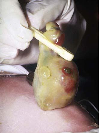
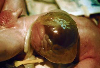
Congenital Omphalocele
An omphalocele is a herniation or protrusion of the abdominal contents into the base of the umbilical cord (Figs. 99-1 and 99-2). In contrast to the more common umbilical hernia, the sac is covered with peritoneum without overlying skin. The size of the sac that lies outside the abdominal cavity depends on its contents. Herniation of intestines into the cord occurs in about 1/5,000 births, and herniation of liver and intestines in 1/10,000 births. The abdominal cavity is proportionately small because the impetus to grow and develop is deficient. Immediate surgical repair, before infection has taken place and before the tissues have been damaged by drying (saline-soaked sterile dressings should be applied immediately) or by rupture of the sac, is essential for survival. Mersilene mesh or similar synthetic material may be used to cover the viscera if the sac has ruptured or if excessive mobilization of the skin would be necessary to cover the mass and its intact sac. The majority (≈75%) of infants with omphalocele have associated congenital anomalies/syndromes, including Beckwith-Wiedemann syndrome (omphalocele, macrosomia, hypoglycemia), and other chromosomal (29%, including trisomies 13 and 18) and nonchromosomal (45%) multiple and isolated congenital anomalies (musculoskeletal, 24%; urogenital, 20%; cardiovascular, 15%; and central nervous system, 9%). The survival rate is about 80% overall, but in infants with isolated omphalocele, the survival rate is > 90%.
Tumors
Tumors of the umbilicus are rare and include angioma, enteroteratoma, dermoid cyst, myxosarcoma, and cysts of urachal or omphalomesenteric duct remnants.
Hemorrhage
Hemorrhage from the umbilical cord may be due to trauma, inadequate ligation of the cord, or failure of normal thrombus formation. It may also indicate hemorrhagic disease of the newborn or other coagulopathies (especially factor XIII deficiency), septicemia, or local infection. The infant should be observed frequently during the first few days of life so that if hemorrhage does occur, it will be detected promptly.
Granuloma
The umbilical cord usually dries and separates within 6-8 days after birth. The raw surface becomes covered by a thin layer of skin; scar tissue forms, and the wound is usually healed within 12-15 days. The presence of saprophytic organisms delays separation of the cord and increases the possibility of invasion by pathogenic organisms. Mild infection or incomplete epithelialization may result in a moist granulating area at the base of the cord with a slight mucoid or mucopurulent discharge. Good results are usually obtained by cleansing with alcohol several times daily.
Persistence of granulation tissue at the base of the umbilicus is common. The tissue is soft, 3-10 mm in size, vascular and granular, and dull red or pink, and it may have a seropurulent secretion. Treatment is cauterization with silver nitrate, repeated at intervals of several days until the base is dry.
Umbilical granuloma must be differentiated from umbilical polyp, a rare anomaly resulting from persistence of all or part of the omphalomesenteric duct or the urachus. The tissue of the polyp is firm and resistant, is bright red, and has a mucoid secretion. If the polyp is communicating with the ileum or bladder, small amounts of fecal material or urine may be discharged intermittently. Histologically, the polyp consists of intestinal or urinary tract mucosa. Treatment is surgical excision of the entire omphalomesenteric or urachal remnant.
Infections
Although aseptic delivery and routine cord care (application of triple dye and other antiseptics to the umbilical stump and surrounding skin) decrease bacterial colonization and umbilical infection, the necrotic tissue of the umbilical cord is an excellent medium for bacterial growth. In a meta-analysis of 4 randomized controlled trials, triple dye was found to be more effective than alcohol in reducing omphalitis. Soap and water or dry care is not as effective in the prevention of omphalitis. Omphalitis may remain localized or may spread to the abdominal wall, the peritoneum, the umbilical or portal vessels, or the liver. Infants with abdominal wall cellulitis or those with necrotizing fasciitis have a high incidence of associated bacteremia. Portal vein phlebitis may develop and result in the later onset of extrahepatic portal hypertension. The general manifestations may be minimal (periumbilical erythema), even when septicemia or hepatitis has resulted. Treatment includes prompt antibiotic therapy (with agents effective against Staphylococcus aureus and Escherichia coli) and, if abscess formation has occurred, surgical incision and drainage. Necrotizing fasciitis is often polymicrobial and has a high mortality. Changes in newborn bathing practices that replace antiseptics with nonantiseptic pH-balanced soap may be associated with increased risk of omphalitis.
Umbilical Hernia
Often associated with diastasis recti, an umbilical hernia is due to imperfect closure or weakness of the umbilical ring. Predisposing factors include black race and low birthweight. The hernia appears as a soft swelling covered by skin that protrudes during crying, coughing, or straining and can be reduced easily through the fibrous ring at the umbilicus. The hernia consists of omentum or portions of the small intestine. The size of the defect varies from < 1 cm in diameter to as much as 5 cm, but large defects are rare. Most umbilical hernias that appear before the age of 6 mo disappear spontaneously by 1 yr of age. Even large hernias (5-6 cm in all dimensions) have been known to disappear spontaneously by 5-6 yr of age. Strangulation is extremely rare. It is generally agreed that “strapping” is ineffective. Surgery is not advised unless the hernia persists to the age of 4-5 yr, causes symptoms, becomes strangulated, or becomes progressively larger after the age of 1-2 yr. Defects exceeding 2 cm are less likely to close spontaneously.
Ameh EA, Nmadu PT. Major complications of omphalitis in neonates and infants. Pediatr Surg Int. 2002;18:413-416.
Deshpande SA, Jog S, Watson H, et al. Do babies with isolated single umbilical artery need routine postnatal renal ultrasonography? Arch Dis Child Fetal Neonatal Ed. 2009;94:F265-F267.
Hwang P, Kousseff B. Omphalocele and gastroschisis: an 18-year review study. Genetics Med. 2004;6:232-236.
Krakowiak P, Smith EN, de Bruyn G, et al. Risk factors and outcomes associated with a short umbilical cord. Obstet Gynecol. 2004;103:119-127.
Mullany LC, Darmstadt GL, Khatry SK. Topical applications of chlorhexidine to the umbilical cord for prevention of omphalitis and neonatal mortality in southern Nepal: a community-based, cluster-randomized trial. Lancet. 2006;367:910-918.
Pomeranz A. Anomalies, abnormalities, and care of the umbilicus. Pediatr Clin North Am. 2004;51:819-827.
Salihu HM, Emusu D, Aliyu ZY, et al. Omphalocele and gastroschisis: black-white disparity in infant survival. Am J Med Genet A. 2005;1(135):161-165.
Stoll C, Alembik Y, Dott B, et al. Omphalocele and gastroschisis and associated malformations. Am J Med Genet A. 2008;146A:1280-1285.
Weber DM, Freeman NV, Elhag KM. Periumbilical necrotizing fasciitis in the newborn. Eur J Pediatr Surg. 2001;11:86-91.
Zupan J, Garner P, Omari AA: Topical umbilical cord care at birth, Cochrane Database Syst Rev (3):CD001057, 2004.