Chapter 644 Hyperpigmented Lesions
Disorders of Pigment
Normal pigmentation requires migration of melanoblasts from the neural crest to the dermal-epidermal junction, enzymatic processes to form pigment, structural components to contain the pigment (melanosomes), and transfer of pigment to the surrounding keratinocytes. Increased skin color may be generalized or localized and may result from various defects in any of these requirements. Some of these aberrations are a manifestation of systemic disease, others represent generalized or focal developmental or genetic defects, and still others may be nonspecific and the result of cutaneous inflammation.
Ephelides (Freckles)
Ephelides are light or dark brown macules usually <3 mm in diameter, with a poorly defined margin, that occur in sun-exposed areas such as the face, upper back, arms, and hands. They are induced by exposure to sun, particularly during the summer, and may fade or disappear during the winter. They are more common in redheads and fair-haired individuals and first appear in the preschool years. Histologically, they are marked by increased melanin pigment in epidermal basal cells, which have more numerous and larger dendritic processes than the melanocytes of the surrounding paler skin. The lack of melanocytic proliferation or elongation of epidermal rete ridges distinguishes them from lentigines. Freckles have been identified as a risk factor for melanoma independent of melanocytic nevi.
Lentigines
Lentigines, often mistaken for freckles or junctional nevi, are small (<3 cm), round, dark brown macules that can appear anywhere on the body. They are unrelated to sun exposure and remain permanently. Histologically, they have elongated, club-shaped, epidermal rete ridges with increased numbers of melanocytes and dense epidermal deposits of melanin. No nests of melanocytes are found. The lesions are benign and, when few, may be viewed as a normal occurrence and are seen most commonly on the lower lip.
Lentiginosis profusa involves innumerable small, pigmented macules that are present at birth or appear during childhood. There are no associated abnormalities, and mucous membranes are spared. LAMB syndrome (Carney complex), a multiple endocrine neoplasia syndrome, consists of lentigines of the face and vulva, atrial myxoma, mucocutaneous myxomas, and blue nevi (type 1, PRKAR1 gene; type 2, gene map locus 2p16-gene as yet to be identified). The Carney complex variant is associated with distal arthrogryposis (MYH8 gene). The multiple lentigines (LEOPARD) syndrome is an autosomal dominant entity consisting of a generalized, symmetric distribution of lentigines (Fig. 644-1) in association with electrocardiogram abnormalities, ocular hypertelorism, pulmonary stenosis, abnormal genitals (cryptorchidism, hypogonadism, hypospadias), growth retardation, and sensorineural deafness (type 1, PTPN11 gene; type 2, RAF1 gene). Other features include hypertrophic obstructive cardiomyopathy and pectus excavatum or carinatum.
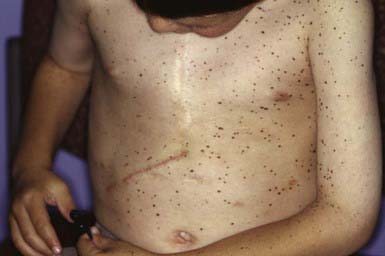
Figure 644-1 Multiple lentigines in LEOPARD (lentigines in association with electrocardiogram abnormalities, ocular hypertelorism, pulmonary stenosis, abnormal genitals [cryptorchidism, hypogonadism, hypospadias], growth retardation, and sensorineural deafness) syndrome.
The Peutz-Jeghers syndrome is characterized by melanotic macules on the lips and mucous membranes and by gastrointestinal (GI) polyposis. It is inherited as an autosomal dominant trait (STK11 gene). Onset is noted in infancy and early childhood when pigmented macules appear on the lips and buccal mucosa. The macules are usually a few millimeters in size but may be as large as 1-2 cm. Macules also appear occasionally on the palate, gums, tongue, and vaginal mucosa. Cutaneous lesions may develop on the nose, hands, and feet; around the mouth, eyes, and umbilicus; and as longitudinal bands or diffuse hyperpigmentation of the nails. Pigmented macules often fade from the lips and skin during puberty and adulthood but generally do not disappear from mucosal surfaces. Buccal mucosal macules are the most constant feature of the disorder; in some families, occasional members may be affected only with the pigmentary changes. Indistinguishable pigmentary changes beginning in adult life, without intestinal involvement, also occur sporadically in individuals.
Polyposis usually involves the jejunum and ileum but may also occur in the stomach, duodenum, colon, and rectum (Chapter 337). Episodic abdominal pain, diarrhea, melena, and intussusception are frequent complications. Patients have a significantly increased risk of GI tract and non–GI tract tumors at a young age. GI cancer has been reported in ≈ 2-3% of patients; the lifetime relative risk for GI malignancy is 13. The relative risk of non–GI tract malignancies, including ovarian, cervical, and testicular tumors, is 9. Peutz-Jeghers syndrome must be differentiated from other syndromes associated with multiple lentigines (Laugier-Hunziker syndrome), from ordinary freckling, from Gardner syndrome, and from Cronkhite-Canada syndrome, a disorder characterized by GI polyposis, alopecia, onychodystrophy, and diffuse pigmentation of the palms, volar aspects of the fingers, and dorsal hands. Treatment of Peutz-Jeghers melanotic macules with multiple different lasers has been successful, in some cases.
Café-Au-Lait Spots
Café-au-lait spots are uniformly hyperpigmented, sharply demarcated macular lesions, the hues of which vary with the normal degree of pigmentation of the individual: They are tan or light brown in white individuals and may be dark brown in black children (Figs. 644-2 and 644-3). Café-au-lait spots vary tremendously in size and may be large, covering a significant portion of the trunk or limb. Generally the borders are smooth, but some have exceedingly irregular borders. The lesions are characterized by increased numbers of melanocytes and melanin in the epidermis but lack the clubbed rete ridges that typify lentigines. One to 3 café-au-lait spots are common in normal children; ≈ 10% of normal children have café-au-lait macules. The spots may be present at birth or may develop during childhood.
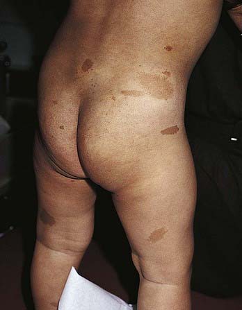
Figure 644-2 Multiple café au lait macules on a child with neurofibromatosis type 1.
(From Eichenfield LF, Frieden IJ, Esterly NB: Textbook of neonatal dermatology, Philadelphia, 2001, WB Saunders, p 372.)
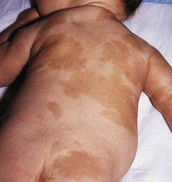
Figure 644-3 Multiple patterned café-au-lait spots in a child with McCune-Albright syndrome.
(From Eichenfield LF, Frieden IJ, Esterly NB: Textbook of neonatal dermatology, Philadelphia, 2001, WB Saunders, p 373.)
Large, often asymmetric café-au-lait spots with irregular borders are characteristic of patients with McCune-Albright syndrome (GNAS1 gene) (Chapter 556.6). This disorder includes polyostotic fibrous dysplasia of bone, leading to pathologic fractures; precocious puberty; and numerous hyperfunctional endocrinopathies. The macular hyperpigmentation may be present at birth or may develop late in childhood (see Fig. 644-3). Cutaneous pigmentation is typically most extensive on the side showing the most severe bone involvement.
Neurofibromatosis Type 1 (Von Recklinghausen Disease)
The café-au-lait spot is the most familiar cutaneous hallmark of the autosomal dominant neurocutaneous syndrome known as neurofibromatosis type 1 (neurofibromin gene) (see Fig. 644-2 and Chapter 589.1). The lesions also occur with certain other disorders, including other types of neurofibromatosis, but in these disorders the café-au-lait spots are not a major feature of the disorder and do not aid in diagnosis (Table 644-1). Included in the criteria for this diagnosis is the presence of 5 or more café-au-lait spots >5 mm in diameter in prepubertal patients or 6 or more café-au-lait spots >15 mm in diameter in postpubertal patients. Multiple café-au-lait macules commonly produce a freckled appearance of non–sun-exposed areas such as the axillae (Crowe sign), the inguinal and inframammary regions, and under the chin.
Incontinentia Pigmenti (Bloch-Sulzberger Disease)
See Chapter 589.7 for a full discussion of this condition.
Postinflammatory Pigmentary Changes
Either hyperpigmentation or hypopigmentation can occur as a result of cutaneous inflammation. Alteration in pigmentation usually follows a severe inflammatory reaction but may result from mild dermatitis. Dark-skinned children are more likely to show these changes than fair-skinned ones. Although altered pigmentation may persist for weeks to months, patients can be reassured that these lesions are usually temporary.
Boikos SA, Stratakis CA. Carney complex: the first 20 years. Curr Opin Oncol. 2007;19:24-29.
McGarrity TJ, Amos C. Peutz-Jeghers syndrome: clinicopathology and molecular alterations. Cell Mol Life Sci. 2006;63:2135-2144.
Sarkozy A, Digilio MC, Dallapiccola B. Leopard syndrome. Orphanet J Rare Dis. 2008;3:13.
Williams VC, Lucas J, Babcock, et al. Neurofibromatosis type 1 revisited. Pediatrics. 2009;123:124-133.
Zacharin M. The spectrum of McCune Albright syndrome. Pediatr Endocrinol Rev. 2007;Suppl 4:412-418..