Radiation Characteristics
After completion of this chapter, the student will be able to do the following:
• Define the key words associated with radiation characteristics
• Describe the effect that the kilovoltage peak has on the quality of the x-ray beam
• Describe how milliamperage influences the quantity of the x-ray beam
• Identify the range of kilovoltage and milliamperage required for dental radiography
• Describe how increasing and decreasing exposure factors affect the density and contrast of the image
• State the rules governing kilovoltage, milliamperage, distance, and exposure time that are used when changing exposure variables
• Describe how kilovoltage, milliamperage, exposure time, and source-to-receptor distance influence the intensity of the x-ray beam
• Calculate an example of radiation intensity using the inverse square law
• Explain how the half-value layer determines the penetrating quality of the x-ray beam
Radiation characteristics include x-ray beam quality, quantity, and intensity. Variations in the character of the x-ray beam influence the quality of the resulting radiographs.
The dental radiographer must have a working knowledge of radiation characteristics. The purpose of this chapter is to (1) detail the concepts of x-ray beam quality and quantity, (2) define the concept of beam intensity, and (3) discuss how exposure factors influence these radiation characteristics.
Advances in dental radiographic equipment have produced control panels with predetermined settings for the various anatomic areas of the maxilla and mandible (Figure 3-1). On older radiographic units, the individual radiation characteristics of kilovoltage peak, milliamperage, and time could all be manually changed. On today’s units, adjustments are not possible for milliamperage and kilovoltage peak. Although the modern equipment is easily understood and convenient, the concepts of these three radiation characteristics must still be reviewed and understood.
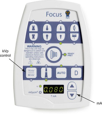
FIGURE 3-1 Kilovoltage peak (kVp) and milliamperage (mA) controls are located on the dental x-ray machine. (Courtesy Instrumentarium Dental, Inc. Milwaukee, WI.)
X-Ray Beam Quality
Wavelength determines the energy and penetrating power of radiation. X-rays with shorter wavelengths have more penetrating power, whereas those with longer wavelengths are less penetrating and more likely to be absorbed by matter. In dental radiography, the term quality is used to describe the mean energy or penetrating ability of the x-ray beam. The quality, or wavelength and energy of the x-ray beam, is controlled by kilovoltage.
Voltage and Kilovoltage
Voltage is a measurement of force that refers to the potential difference between two electrical charges. Inside the dental x-ray tubehead, voltage is the measurement of electrical force that causes electrons to move from the negative cathode to the positive anode. Voltage determines the speed of electrons that travel from cathode to anode. When voltage is increased, the speed of the electrons is increased. When the speed of the electrons is increased, the electrons strike the target with greater force and energy, resulting in a penetrating x-ray beam with a short wavelength.
Voltage is measured in volts or kilovolts. The volt (V) is the unit of measurement used to describe the potential that drives an electrical current through a circuit. Dental x-ray equipment requires the use of high voltages. Most radiographic units operate using kilovolts; 1 kilovolt (kV) is equal to 1000 volts.
Dental radiography requires the use of 65 to 100 kV. The use of less than 65 kV does not allow adequate penetration, whereas the use of more than 100 kV results in overpenetration.
Kilovoltage can be adjusted according to the individual diagnostic needs of patients. The use of 85 to 100 kV produces more penetrating dental x-rays with greater energy and shorter wavelengths, whereas the use of 65 to 75 kV produces less penetrating dental x-rays with less energy and longer wavelengths. A higher kilovoltage should be used when the area to be examined is dense or thick.
Kilovoltage Peak
On dental radiographic equipment that allows for the adjustment of individual radiation characteristics, kilovoltage is controlled by the kilovoltage peak adjustment dial on the x-ray control panel (Figure 3-2). Kilovoltage peak (kVp) can be defined as the maximum or peak voltage. The voltage meter on the control panel measures the x-ray tube voltage, which is actually the peak voltage of an alternating current (AC) (see Figure 3-3). This peak voltage is measured in kilovolts, and thus the term “kilovoltage peak” is used. For example, when 90 kVp is used to expose a receptor, the peak voltage of the tube current is 90,000 volts. As a result of varying kilovoltages occurring in the tube current, a polychromatic x-ray beam, or a beam that contains many different wavelengths of varying intensities, is produced.
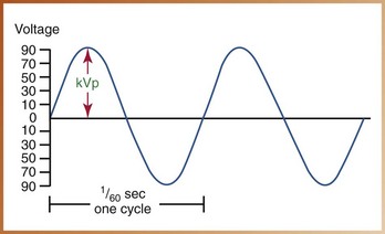
FIGURE 3-2 Kilovoltage peak (kVp) controls the quality of the x-ray beam and measures the peak voltage of the current.
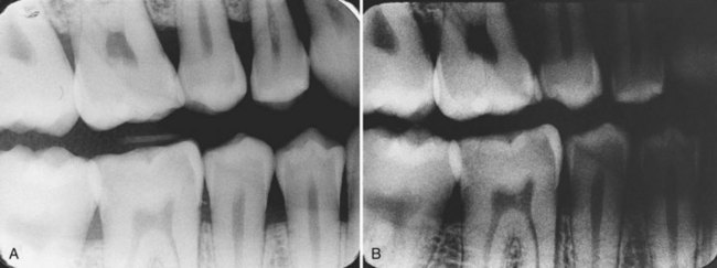
FIGURE 3-3 A, Diagnostic radiograph. B, Increase in kilovoltage results in an image that exhibits increased density; the image appears darker.
The quality, or wavelength and energy of the x-ray beam, is controlled by the kilovoltage peak. The kilovoltage peak regulates the speed and energy of the electrons and determines the penetrating ability of the x-ray beam. Increasing the kilovoltage peak results in a higher energy x-ray beam with increased penetrating ability.
Density and Kilovoltage Peak
Density is the overall darkness or blackness of an image. An adjustment in kilovoltage peak results in a change in the density of a dental radiograph. When the kilovoltage peak is increased while other exposure factors (milliamperage, exposure time) remain constant, the resultant image exhibits an increased density and appears darker (Figure 3-4A). If kilovoltage peak is decreased, the resultant image exhibits a decreased density and appears lighter (Figure 3-4B). Table 3-1 summarizes the effect of kilovoltage peak on density (also see Chapter 8).
Contrast and Kilovoltage Peak
Contrast refers to how sharply dark and light areas are differentiated or separated on an image. An adjustment in kilovoltage peak results in a change in the contrast of a dental radiograph. When low kilovoltage peak settings are used (65–70 kVp), a high-contrast image will result. An image with “high” contrast has many black areas and many white areas and few shades of gray (Figure 3-5). An image with high contrast is useful for the detection and progression of dental caries.

FIGURE 3-5 Image produced with lower kilovoltage exhibits high contrast; many light and dark areas are seen, as demonstrated by the use of the stepwedge.
With high kilovoltage peak settings (>90 kVp), low contrast results. An image with “low” contrast has many shades of gray instead of black and white. An image with low contrast is useful for the detection of periodontal or periapical disease (Figure 3-6). Mounted radiographs that demonstrate low contrast and that are viewed properly on an illuminated surface with masked extraneous light are preferred in dental radiography.

FIGURE 3-6 Image produced with higher kilovoltage exhibits low contrast; many shades of gray are seen instead of black and white.
A compromise between high contrast and low contrast is desirable. See Table 3-1 for a summary of the effect of kilovoltage peak on contrast (also see Chapter 8).
Exposure Time and Kilovoltage Peak
Exposure time refers to the interval of time during which x-rays are produced. Exposure time is measured in impulses because x-rays are created in a series of bursts or pulses rather than in a continuous stream. One impulse occurs every 1/60 of a second; therefore, 60 impulses occur in 1 second.
To compensate for the penetrating power of the x-ray beam, an adjustment in exposure time is necessary when kilovoltage peak is increased (Box 3-1).
For example, a receptor is exposed using 90 kVp and 0.5 second. If the kilovoltage peak setting is decreased from 90 to 75, the exposure time must be increased from 0.5 to 1.0 second to maintain proper density and contrast.
X-Ray Beam Quantity
Quantity of the x-ray beam refers to the number of x-rays produced in the dental x-ray unit.
Amperage and Milliamperage
Amperage determines the amount of electrons passing through the cathode filament. An increase in the number of electrons available to travel from the cathode to the anode results in production of an increased number of x-rays. The quantity of the x-rays produced is controlled by milliamperage.
The ampere (A) is the unit of measure used to describe the number of electrons, or current flowing through the cathode filament. The number of amperes needed to operate a dental x-ray unit is small; therefore, amperage is measured in milliamperes. One milliampere (mA) is equal to 1/1000 of an ampere. Some dental x-ray units have a fixed milliampere setting, whereas others have a milliampere adjustment on the control panel (see Figure 3-2). In dental radiography, the use of 7 to 15 mA is required; a setting above 15 mA is not recommended because of the resultant excessive heat production in the x-ray tube.
Milliamperage regulates the temperature of the cathode filament. A higher milliampere setting increases the temperature of the cathode filament and consequently increases the number of electrons produced. An increase in the number of electrons that strike the anode increases the number of x-rays emitted from the tube.
The quantity, or number of x-rays emitted from the tubehead, is controlled by milliamperage. Milliamperage controls the amperage of the filament current and the amount of electrons that pass through the filament. As the milliamperage is increased, more electrons pass through the filament, and more x-rays are produced. For example, if the milliamperage is increased from 5 to 10 mA, twice as many electrons travel from the cathode to the anode, and twice as many x-rays are produced.
Milliampere-Seconds
Both milliamperes and exposure time have a direct influence on the number of electrons produced by the cathode filament. The product of milliamperes and exposure time is termed milliampere-seconds (mAs), as follows:
When milliamperage is increased, the exposure time must be decreased, and vice versa, if the density of the exposed radiograph is to remain the same.
If a patient has difficulty holding still during the exposure, for example, the dental radiographer can increase the milliamperage and decrease the exposure time to compensate for the patient’s movement.
Density and Milliamperage
Milliamperage, as with kilovoltage peak, has an effect on the density of a dental radiograph. An increase in milliamperage increases the overall density of the radiograph and results in a darker image. Conversely, a decrease in milliamperage decreases the overall density and results in a lighter image. Table 3-2 summarizes the effect of milliamperage on density.
Exposure Time and Milliamperage
Milliamperage and exposure time are inversely related. When altering milliamperage, the exposure time must be adjusted to maintain the diagnostic density of an image film. When milliamperage is increased, the exposure time must be decreased. When milliamperage is decreased, the exposure time must be increased.
Table 3-3 lists guidelines for adjusting kilovoltage peak, milliamperage, and exposure time.
TABLE 3-3
Guidelines for Adjusting Kilovoltage Peak (kVp), Milliamperage (mA), and Exposure Time
| Adjustment | Exposure* |
| ↑ kVp by 15 | ↓ Exposure time by 1/2 |
| ↓ kVp by 15 | ↑ Exposure time by 2 |
| ↑ mA | ↓ Exposure time |
| ↓ mA | ↑ Exposure time |
*Adjustment in exposure time needed to maintain diagnostic density of image.
X-Ray Beam Intensity
Quality refers to the energy or penetrating ability of the x-ray beam; quantity refers to the number of x-ray photons in the beam. Quality and quantity are described together in a concept known as intensity. Intensity is defined as the product of the quantity (number of x-ray photons) and quality (energy of each photon) per unit of area per unit of time of exposure, as follows:
Intensity of the x-ray beam is affected by a number of factors, including kilovoltage peak, milliamperage, exposure time, and distance.
Kilovoltage Peak
Kilovoltage peak regulates the penetrating power of the x-ray beam by controlling the speed of the electrons traveling between the cathode and the anode. Higher kilovoltage peak settings produce an x-ray beam with more energy and shorter wavelengths; higher kilovoltage levels increase the intensity of the x-ray beam.
Milliamperage
Milliamperage controls the penetrating power of the x-ray beam by controlling the number of electrons produced in the x-ray tube and the number of x-rays produced. Higher milliampere settings produce a beam with more energy, increasing the intensity of the x-ray beam.
Exposure Time
Exposure time, as with milliamperage, affects the number of x-rays produced. A longer exposure time produces more x-rays. An increase in exposure time produces a more intense x-ray beam.
Distance
The distance traveled by the x-ray beam affects the intensity of the beam. Distances that must be considered when exposing a dental radiograph include the following (Figure 3-7):
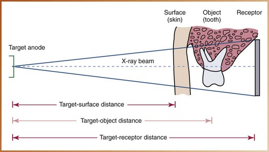
FIGURE 3-7 Distances to consider when exposing dental radiographs: target-surface, target-object, and target-receptor distance.
• Target-surface distance: The distance from the source of radiation to the patient’s skin
• Target-object distance: The distance from the source of radiation to the tooth
• Target-receptor distance: The distance from the source of radiation to the receptor
The distance between the source of radiation and the receptor has a marked effect on the intensity of the x-ray beam. As x-rays travel from their point of origin or away from the target anode, they diverge like waves of light and spread out to cover a larger surface area. As x-rays travel away from their source of origin, the intensity of the beam lessens. Unless a corresponding change is made in one of the other exposure factors (kilovoltage peak, milliamperage), the intensity of the x-ray beam is reduced as the distance increases.
The x-ray beam that exits from an 8-inch position-indicating device (PID) is more intense than one that exits from a 16-inch PID. The inverse square law is used to explain how distance affects the intensity of the x-ray beam.
Inverse Square Law
The inverse square law is stated as follows:
“Inversely proportional” means that as one variable increases, the other decreases. When the source-to-receptor distance is increased, the intensity of the beam is decreased.
For example, when the PID length is changed from 8 to 16 inches, the source-to-receptor distance is doubled. According to the inverse square law, the resultant beam is one-fourth as intense (Figure 3-8). When the PID length is changed from 16 to 8 inches, the source-to-receptor distance is reduced by half. According to the inverse square law, the resultant beam is four times as intense.
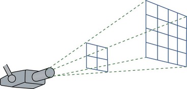
FIGURE 3-8 The inverse square law states that the intensity of radiation is inversely proportional to the square of the distance from the source. Note that as the source-to-receptor distance is doubled, the intensity of radiation is one fourth as intense. (Modified from White SC, Pharoah MJ: Oral radiology principles and interpretation, ed 5, St Louis, 2004, Mosby.)
The following mathematical formula is used to calculate the inverse square law.
Example
If the PID length is changed from 8 inches to 16 inches, how does this increase in source-to-receptor distance affect the intensity of the beam?


This mathematical formula reveals that the intensity of the beam will be one fourth as intense if the source-to-receptor distance is changed from 8 to 16 inches (assuming that kilovoltage peak and milliamperage remain constant). In this example, the inverse square law reveals that doubling the distance from the source of radiation to the receptor (from an 8-inch to a 16-inch PID) results in a beam that is one fourth as intense. Remember: The intensity of the radiation is inversely proportional to the square of the distance.
Half-Value Layer
To reduce the intensity of the x-ray beam, aluminum filters are placed in the path of the beam inside the dental x-ray tubehead. Aluminum filters are used to remove the low-energy, less penetrating, longer-wavelength x-rays. Aluminum filters increase the mean penetrating capability of the x-ray beam while reducing the intensity. When placed in the path of the x-ray beam, the thickness of a specified material (e.g., aluminum) that reduces the intensity by half is termed the half-value layer (HVL).
For example, if an x-ray beam has an HVL of 4 mm, a thickness of 4 mm of aluminum would be necessary to decrease its intensity by half. Measuring the HVL determines the penetrating quality of the beam. The higher the half-value layer, the more penetrating is the beam. (Filtration of the x-ray beam is discussed further in Chapter 5.)
Summary
• Radiation characteristics include x-ray beam quality, quantity, and intensity.
• X-ray units may or may not have adjustable dials or buttons for kilovoltage peak, milliamperage, and time.
• Quality refers to the mean (average) energy or penetrating ability of the x-ray beam and is controlled by the kilovoltage peak.
• Increased kilovoltage peak produces x-rays with increased energy, shorter wavelength, and increased penetrating power; kilovoltage peak affects density and contrast.
• Quantity refers to the number of x-rays produced and is controlled by the milliamperage.
• Increased milliamperage produces an increased number of x-rays; milliamperage affects density.
• Exposure time also influences the number of x-rays produced.
• Intensity is the total energy contained in the x-ray beam in a specific area at a given time; intensity is affected by kilovoltage peak, milliamperage, exposure time, and distance.
• Increased kilovoltage peak, milliamperage, or exposure time results in increased intensity of the x-ray beam.
• Intensity of the x-ray beam is reduced with increased distance. The inverse square law is used to explain how distance affects the intensity of the x-ray beam.
• An aluminum filter is placed in the path of the x-ray beam to reduce the intensity and remove the low-energy x-rays from the beam.
• The thickness of aluminum placed in the path of the x-ray beam that reduces the intensity by half is termed the half-value layer (HVL).
Frommer, HH, Savage-Stabulas, JJ, Image formation. Radiology for the dental professional, ed 9, St Louis, Mosby, 2011.
Frommer, HH, Savage-Stabulas, JJ, Image receptors. Radiology for the dental professional, ed 9, St Louis, Mosby, 2011.
Frommer, HH, Savage-Stabulas, JJ, Ionizing radiation and basic principles of x-ray generation. Radiology for the dental professional, ed 9, St Louis, Mosby, 2011.
Johnson, ON, Thomson, EM, The dental x-ray machine: components and functions. Essentials of dental radiography for dental assistants and hygienists, ed 9, Pearson Prentice Hall, Upper Saddle River, NJ, 2007.
Johnson, ON, Thomson, EM, Producing quality radiographs. Essentials of dental radiography for dental assistants and hygienists, ed 8, Upper Saddle River, NJ, Pearson Prentice Hall, 2007.
Miles, DA, Van Dis, ML, Williamson, GF, Jensen, CW, Image characteristics. Radiographic imaging for the dental team, ed 4, St Louis, Saunders, 2009.
Miles, DA, Van Dis, ML, Williamson, GF, Jensen, CW, X-ray properties and the generation of x-rays. Radiographic imaging for the dental team, ed 4, St Louis, Saunders, 2009.
White, SC, Pharoah, MJ, Radiation physics. Oral radiology: principles and interpretation, ed 6, St Louis, Mosby, 2009.
Multiple Choice
____1. In dental radiography, the quality of the x-ray beam is controlled by:
____2. Identify the kilovoltage range for most dental x-ray machines:
____3. A higher kilovoltage produces x-rays with:
____4. Identify the unit of measurement used to describe the amount of electric current flowing through the x-ray tube:
____5. Radiation produced with high kilovoltage results in:
____6. In dental radiography, the quantity of radiation produced is controlled by:
____7. Increasing milliamperage results in an increase in:
____8. Identify the milliamperage range for dental radiography:
____9. The overall blackness or darkness of an image is termed:
____10. If kilovoltage is decreased with no other variations in exposure factors, the resultant image will:
____11. Identify the term that describes how dark and light areas are differentiated on an image:
____12. A radiograph that has many light and dark areas with few shades of gray is said to have:
____13. The radiograph described in question 12 was produced with:
____14. Increasing milliamperage alone results in an image with:
____15. A diagnostic image is produced using 90 kVp and 0.25 second. What exposure time is needed to produce the same image at 75 kVp?
____16. A diagnostic image is produced using 10 mA and 0.45 second. What exposure time is needed to produce the same image at 15 mA?
____17. The total energy contained in the x-ray beam in a specific area at a given time is termed:
____18. Increasing which of these four exposure controls will increase the intensity of the x-ray beam: (1) kilovoltage, (2) milliamperage, (3) exposure time, (4) source-to-receptor distance?
____19. The length of the position-indicating device is changed from 16 inches to 8 inches. The resultant intensity of the beam will be: