Chapter 123Lameness in the Arabian and Half-Arabian Show Horse
History of the Arabian
The Arabian is one of the oldest breeds in the world. The horse originated in the deserts of the Middle East and was used by the Bedouins for transportation and in battle. The Arabian breed was noted for its speed and endurance. Three Arabian stallions (the Godolphin Barb, Byerley Turk, and Darley Arabian) imported to Europe during the late 1600s and early 1700s became the foundation of a new breed of horse, the Thoroughbred. Today, 93% of all modern Thoroughbreds can be traced to these three sires. In the 1800s many royal families of Europe established Arabian stud farms. Two of the most notable were the Polish National Arabian Stud in Poland and the Crabbet Arabian Stud in England. The Arabian is thought to have an influence on many of the light horse breeds that have developed throughout history. A typical Arabian ranges from 14.1 to 15.1 hands in height. The American Horse Show Association breed standards describe the Arabian as having a small, slightly dished face with large eyes set well apart, small ears, deep and wide jowls, a small muzzle, and large nostrils. The horse should have a long, arched neck; a long, sloping shoulder; well-sprung ribs; a short back with a relatively horizontal croup; and natural, high tail carriage. The limbs should have large, well-defined joints, short cannon bones, sloping pasterns of good length, and round feet of proportionate size.1
The Half-Arabian studbook originated with the United States Army Remount Service after World War II and was acquired by the International Arabian Horse Association in 1951. Half-Arabians must have a registered purebred Arabian sire or dam. The Anglo-Arabian is a cross between an Arabian and a Thoroughbred, whereas the more recently developed National Show Horse is a cross between an Arabian and a Saddlebred. Many Half-Arabians are double registered.
History of the Sporting Event
The International Arabian Horse Association was created in 1950 to join the local and regional clubs across America into one united association. The International Arabian Horse Association promotes and coordinates all Arabian and Half-Arabian horse show activities and develops horse show rules. The International Arabian Horse Association also maintains the Half-Arabian and Anglo-Arabian registries, whereas the Arabian Horse Registry of America maintains the registry and pedigree records for purebred Arabian horses in the United States and Mexico.
The first United States National Arabian and Half-Arabian Show was held in 1966. The United States National Show is held in October in Tulsa, Oklahoma. A separate Youth National show for riders under 18 years old is held in July of each year in Albuquerque. The Canadian National Horse Show is held in August in Regina, Saskatchewan. The Sport Horse Nations, an event for Arabians and Half-Arabians competing in dressage, working hunter-jumper, and carriage-driving categories, is held at a different time and location each year. The United States and Canada are separated into 18 regions, each of which holds an annual show. Horses can qualify for the national show by placing in the top five at a regional show or by accumulating points at Class A shows. Some of the major shows in the United States other than the regionals and nationals are the Scottsdale Show (Arizona), the Buckeye Show (Ohio), the Pacific Slopes Show (California), the East Coast Championship (Pennsylvania), and the Pro-Am Challenge (Texas). Major international shows are held in England, France, South America, and Australia.
Performance classes for Arabian and Half-Arabian horses cover a broad spectrum and are listed in Box 123-1. Each of these classes is held separately for Arabians and Half-Arabians. They may be divided further into sections for junior owner, adult amateur owner, amateur owner, junior exhibitor, and amateur. The Park horse has a strong animated trot, with the forearm horizontal and the limb extending fully forward. The hock has a well-raised driving action. The walk and canter are animated and collected. The English Pleasure horse is shown at a walk, trot, strong trot (faster and more animated than the normal trot), canter, and hand gallop. Its gaits are less animated than those of the Park horse, although the forearm, at the trot, is horizontal. The same gaits are used in the Country English Pleasure class, but horses have lower limb action, and high action is penalized. The Country English Pleasure horse must also halt, stand quietly, back, and walk off on a loose rein. With all pleasure classes the horse must give the appearance of being a pleasure to ride. Park horses and English Pleasure horses are shown with the head carried high and considerable flexion at the poll. Saddle seat attire is required.
In the English Show Hack class a horse must perform each gait (walk, trot, and canter) in a normal, collected, and extended manner. A transition between gaits should be noticeable, and high knee action is not expected. Horses in the Hunter Pleasure division are shown under saddle at the walk, trot, canter, and hand gallop. The neck should be carried lower, the head should be carried with less bend at the poll, and the horse should be in a generally longer frame than the English Pleasure or Show Hack horse. Working Hunters are shown over a course of fences set at levels of 0.6 to 1 m and are judged on performance, manners, and soundness. Jumpers are shown over courses of jumps that vary in height from 0.9 to 1.05 m. The maximum width (spread) is 1.5 m.
Horses in driving classes are shown pulling a four-wheeled (Formal and Pleasure) or two-wheeled (Pleasure and Country Pleasure) vehicle. The gaits judged in the Formal, Pleasure, and Country Pleasure driving classes correspond to the Park, English Pleasure, and Country English Pleasure classes under saddle. The Roadster is a driving class that focuses on the trot at three different speeds.
The Western Pleasure horse is shown at the walk, jog (trot), lope (slow canter), and hand gallop. Ideally, contact with the reins is light, the head is carried low (approximately at the level of the withers), and the jog and lope are slow, easy gaits. The Working Western horse classes include reining, working cow horse, trail, cutting, and Western riding.
Training: Impact of Industry
The Arabian and Half-Arabian are versatile breeds, as shown by the many sports in which they compete. These include halter, endurance (see Chapter 118), pleasure, jumping, dressage, reining, cutting, and racing (see Chapter 111). Young performance horses are not shown under saddle until they are 3 years old. They then compete in futurity classes for horses 3 years of age or junior horse classes for horses 5 years of age or younger. Because these horses do not compete in performance classes until 3 years of age, this allows more time for adequate skeletal development compared with racehorses and Quarter Horses that start training before 2 years of age. The reason that training of Arabian show horses is started later than some other breeds may be partly smaller size and late maturation, but it is also related to the fact that no performance classes are available for 2-year-olds, and therefore no economic incentives exist to start intensive training early.
Early training and conditioning typically involve a substantial amount of work in a round pen or by lunging. Excessive training in small circles causes increased torque on the joints and support structures of the distal aspect of the limbs. Young horses trained in this manner commonly develop bilateral distal forelimb lameness involving numerous structures. These problems tend to be exacerbated by uneven and excessively hard or deep footing.
Rules govern the shoeing of Arabian and Half-Arabian show horses. The rules vary with age, breed, discipline, and the country of the competition. Foot length, shoe weight and shape, and pad usage are individualized for each horse to optimize the height and arc of flight of the forelimbs and hindlimbs. In English Pleasure horses, a common shoe is the toe-weighted shoe, constructed by forging more steel in the toe of the shoe. The long foot and weighted shoes are used to enhance forelimb motion. Unfortunately this can contribute to strain on the suspensory ligament (SL) and joints of the distal aspect of the limb.
In all performance divisions horses are shown in a collected frame. In each division the type of work performed, the body position required, and the conformational defects of an individual horse contribute to the common lameness conditions. Differences are apparent in gaits, degrees of collection, and head and neck position in the various divisions. In the English Pleasure division, for example, the degree of collection, neck elevation, and poll flexion required shift weight to the hindlimbs and increase the work of the back and abdominal muscles. These positional factors can cause hindlimb lameness (especially involving the stifle and SL) and back pain. In the Western Pleasure and Reining divisions, similar problems are seen because of the amount of collection required. These horses also incur a variety of lameness conditions because they commonly are worked for longer periods than English Pleasure horses. In the Western Pleasure and Reining classes, because any departure from a quiet, steady position is penalized, fatigue can be part of the class preparation.
Conformation and Lameness
Mild-to-moderate carpus valgus and toed-out conformation commonly are seen and do not appear to have a major impact on soundness (Figure 123-1). One reason is that the carpus is not a common location for lameness. If these conformational faults are severe, horses are at risk of suspensory desmitis. Horses with long sloping pasterns, back-at-the-knee conformation, or offset knees are also predisposed to suspensory desmitis. These conformational faults are more common in the Half-Arabian and National Show Horse than in purebred Arabians and are more common in certain pedigrees.

Fig. 123-1 A 4-year-old Half-Arabian with toed-out, carpus valgus, back-at-the-knee conformation. These are common conformation faults in the Arabian and Half-Arabian breeds and may predispose the horses to suspensory desmitis.
Horses with low, underrun heels certainly are prone to lameness from palmar foot pain. This fault can be difficult to correct, even with careful attention to shoeing and trimming. A small, upright, contracted foot (club foot) can be a source of lameness and appears to be increasing in incidence. Inflammation of the soft tissues such as the SL, accessory ligament of the deep digital flexor tendon (ALDDFT), palmar foot structures, and distal sesamoidean ligaments tend to be more common in horses with club foot conformation.
A long, weak (sway) back and a short croup may predispose horses to soreness in the thoracolumbar, sacroiliac, and gluteal areas. Because problems in these areas are a common cause of poor performance, this type of conformation is a serious fault. Horses with cow-hocked conformation are the rule rather than the exception, but this conformation seems to have little effect on soundness.
Lameness Examination
History
When describing lameness, trainers often comment that problems occur or are more noticeable during the first direction or the second direction of the show ring. This refers to the directional order in which rail classes are run. In the first direction horses enter the ring and travel counterclockwise (on the left rein), and in the second direction horses travel clockwise (on the right rein). Important questions regarding history include the division in which the horse competes; onset, degree, and progression of the lameness; previous or current treatment; and response to therapy. Additional information that can be helpful includes knowing which direction is harder for the horse at the trot (jog) and canter (lope), whether the horse pulls unevenly on the reins, whether the horse tracks straight in each direction, whether the horse falls out of leads behind (in the hindlimbs) in corners (i.e., becomes disunited in canter or breaks from canter to trot), and whether the rider rides the correct or incorrect diagonal in each direction (the horse may throw the rider up on one diagonal preferentially).
The age of the horse is important, because osteochondrosis is more likely to affect young horses recently started into training than older horses, but in older horses osteoarthritis (OA) is common. It is important to find out when the horse was last shod, and if any recent shoeing changes have been made. Altering medial-to-lateral hoof balance or hoof angle may increase pressure in certain areas and lead to bruising of the heel or sole. Increasing the hoof angle by raising the heel may increase load on the SL, which may lead to suspensory desmitis. The type and condition of the footing the horse has been working on is important to consider. Often footing at shows is less than ideal and in many cases is too hard, leading to the development of bruised feet. Conversely, if footing is too deep, it may lead to tendon and ligament injuries.
Static Examination
Visual Examination
Stepping back to visually examine the horse for overall symmetry of the limbs and upper body is helpful. Asymmetry of upper limb muscle groups may be a sign of atrophy from denervation, chronic lameness, or neurological disease. Asymmetry in the height and position of the points of the shoulders, the tubera sacrale, the tubera coxae, or the tubera ischii can indicate subluxations or fractures. The size, shape, and symmetry of the feet are important to note. Abnormalities such as club feet, an underrun or sheared heel, growth rings, dished dorsal hoof walls, and a contracted heel are some of the more common problems. Joint swelling, soft tissue swellings, poor conformation, scars, and abnormal structures such as splints are recognized quickly.
Palpation
Thorough palpation of the distal aspect of the limbs is performed with the horse in weight-bearing and non–weight-bearing positions. Degree of joint and tendon sheath filling; abnormal contours of bones, tendons, and ligaments; and intensity of the digital pulse amplitudes are best evaluated while the horse is bearing weight. With the limb elevated, painful response to palpation of tendons, ligaments (origins and insertions), and splints; pain on joint flexion; and range of motion of joints are assessed. Effusion of the distal interphalangeal (DIP) and stifle joints (especially the medial femorotibial joint) is common. To evaluate filling in the medial femorotibial joint, it is helpful to have the horse bearing weight with the limb being palpated slightly cranial to the opposite limb and perpendicular to the ground.
Response of an Arabian or Half-Arabian to palpation of the SL varies greatly, depending on the horse. Differences between limbs should be considered important, and change in the response over time is noteworthy. Many horses have a pain response to palpation of the SL but have primary lameness localized to the foot. Possibly the SL is painful because of a compensatory gait caused by a primary foot problem. In contrast, absence of inducible pain, especially in the proximal suspensory region, does not rule out this area as a source of pain causing lameness.
Careful palpation of the cervical, thoracic, and lumbosacral regions is useful in diagnosing the reason for poor performance and lameness. Asymmetry, abnormal contours, and painful response to palpation are important to assess. Particular attention should be paid to the thoracolumbar musculature, the spinous processes, and the sacral tuberosities, because these areas commonly are involved in horses that perform poorly.
Hoof Tester Examination
Many Arabian and Half-Arabian show horses wear full pads in front during training and showing. Although sometimes inconvenient, especially at a competition, removing the shoe and pad for a complete hoof tester examination is helpful if the veterinarian suspects a foot problem. Some indication of painful areas may be obtained with the shoe and pad on, but many areas can be missed. Bruised heels and soles are common, especially at shows where footing may be too hard and horses are being worked longer than normal. Bruised heels and soles are two of the most common sources of lameness. Improvement in lameness can be dramatic if areas of bruising can be trimmed to reduce pressure or if the shoe is modified to eliminate weight bearing on a bruised area. Many horses show pain when hoof testers are applied across the heel but no pain with an individual heel bulb, the sole, or the bars of the hoof. These horses are in as much pain with the shoe and pad on, and therefore this situation may relate to structures deeper in the heel of the foot, rather than simply bruising of the sole.
Medial-to-lateral hoof imbalances may contribute to lameness and should be addressed whenever lameness exists and sore feet have been identified with hoof testers. In general, pain is associated with the high side of the foot or that area making ground contact first.
Pain in the toe region occurs less commonly than in the heel, especially for horses in the English and Park divisions, and likely is related to the way the foot is trimmed. These horses usually have a long toe and thick sole that may protect sensitive structures from bruising and exaggerate heel-first landing in the forelimbs. When pain over the middle of the frog is detected, bruising, palmar foot pain, or navicular-related pain should be considered. Diagnostic analgesia should be performed to confirm the relevance of hoof tester examination, because false-positive reactions occur.
Dynamic Examination
Flexion Tests
Flexion tests are important, but responses should be interpreted carefully. A painful response to flexion of joints and the degree of lameness should be assessed. Many false-positive distal limb flexion tests occur in Arabian and Half-Arabian show horses. Many horses without lameness show pain to static flexion and a positive response when trotted. In a lame horse a distal limb flexion test is not specific. For example, in a lame horse with a positive response to a distal limb flexion test, lameness may be localized anywhere from the foot to the distal metacarpal region. Carpal flexion usually is performed only in a static situation because we do not feel that substantial additional information is gained by trotting the horse. Horses that resent carpal flexion may have proximal suspensory desmitis (PSD), superficial digital flexor tendonitis, or carpal tenosynovitis, because bony injury of the carpus itself is unusual in Arabian and Half-Arabian show horses.
Initially the entire hindlimb is held in flexion and then individual distal limb flexion and proximal limb flexion tests are performed. We attempt to stress the stifle independently of the tarsus by holding the distal tibia upward and behind the horse for 60 seconds. This test can also elicit pain from the lumbosacral and sacroiliac joint regions. These flexion tests are not specific, but they may increase the index of suspicion in a certain area. For example, horses with hindlimb PSD or distal hock joint pain respond positively to proximal limb flexion and must be differentiated based on the results of other tests. Mildly positive hindlimb flexion test results can be seen in sound horses that are actively training and showing. These mildly positive flexion test results may be related to subtle lameness or subclinical pain, or could be a normal response. For horses that have been competing successfully to have a moderate-to-severe positive response to a distal limb flexion test after a lengthy show is not unusual. This fact needs to be taken into account when prepurchase examinations are performed directly after horses have competed.
Examination in Hand and under Saddle
Horses should be examined at the walk and at the trot on the straight in hand. It is also helpful to examine horses at the walk in a figure-eight pattern. Examination on both hard and soft surfaces can be helpful. If possible, examining the horse at all appropriate gaits on the lunge line (or long line) and under saddle or in a cart is helpful. Multiple-limb lameness, especially contralateral forelimb and hindlimb lameness and bilateral front foot lameness, is common, which is important to keep in mind. The possibility of subtle neurological deficits should be considered during all phases of the lameness examination. In some horses lameness may not be apparent unless the horse is under tack and working near the higher end of its performance capabilities. This type of lameness is more difficult to diagnose, and diagnostic analgesia is usually required. Subtle lameness can be masked by a rider restricting free head movement or by controlling body position. The diagonal on which the rider sits can alter the appearance of lameness. When riding the left diagonal, the rider sits when the left forelimb and right hindlimb bear weight, and on the right diagonal the rider sits when the right forelimb and left hindlimb bear weight. The correct diagonal is the left when trotting clockwise and the right when trotting counterclockwise. In general a horse appears more lame behind when the rider sits when the lame limb is bearing weight, and the horse may try to throw the rider to the opposite diagonal. A horse with right hindlimb lameness appears more lame when the rider sits on the left diagonal. In Park, English, or Country English Pleasure classes, where riders are not penalized for riding the incorrect diagonal, horses often are ridden on the diagonal that minimizes lameness. Often a lameness is involved when a horse is seen being ridden on the incorrect diagonal when traveling in one direction and on the correct diagonal going the other way. These horses are referred to as right or left diagonal horses. An inconsistent relationship exists between the degree of forelimb lameness and the diagonal on which the rider sits, but lameness may be modified by switching diagonals.
It is important to note whether the observed lameness is constant regardless of direction or pattern. It is important to note if lameness varies, whether the involved limb is on the inside or outside of a circle, whether the horse is on the straight or in a turn, and whether lameness is worse when the horse is on hard or soft ground. The only gait deficit visible in horses with subtle forelimb lameness may be a slight difference in the height or arc of flight of the involved limb. Often a head nod is absent, but the horse appears unsteady in the face (movement of the nose from a fixed position) because of the discomfort of the limb from landing or from pushing off the ground.
Diagnostic Analgesia
If possible, it is important to perform diagnostic analgesia to confirm the source of pain. For example, many horses that appear to have foot pain because of sensitivity to hoof testers or pain on palpation of the SL actually have pain elsewhere. Under United States Equestrian Federation (USEF) rules, with a properly filed medication report, local anesthetic solution such as mepivacaine can be administered up to 24 hours before a class.
If lameness is to be reassessed with the horse under saddle or in a cart, intraarticular rather than perineural nerve blocks are preferred, because of possible loss of proprioception, which may lead to tripping or stumbling. After perineural blocks, horses may pull shoes or show an altered or awkward gait regardless of the lameness. If perineural blocks are performed in horses under saddle or in a cart, the rider or driver should be cautioned. Hindlimb nerve blocks can be difficult in uncooperative horses, and a tranquilizer (10 to 15 mg acepromazine intravenously) may be administered to fractious horses. Lameness may be more obvious after a tranquilizer is administered, but the risk of permanent penile prolapse with the use of acepromazine in stallions or geldings should be considered. In some horses, diagnostic analgesia of the hindlimbs may be impossible. On occasion the response to intraarticular medication, administered under heavy sedation, may be assessed as a diagnostic aid. Other diagnostic procedures such as nuclear scintigraphy, radiographic and ultrasonographic examinations, and magnetic resonance imaging (MRI) may be employed.
Neurological Examination
An Arabian or Half-Arabian show horse with neurological disease may show subtle to overt clinical signs of lameness, weakness, or ataxia, which may be mistaken easily for signs of musculoskeletal pain. Watching the horse turn in tight circles, back up, and walk a figure eight can be performed quickly and may pinpoint subtle deficits that were not noted or were less noticeable earlier in the examination.
Traumatic injuries caused by fractious horses falling during handling and training and equine protozoal myelitis are common causes of neurological disease and dysfunction in Arabian and Half-Arabian show horses in North America. OA of the caudal cervical facets is common, but is not necessarily of clinical significance. Clinical signs can include focal muscle atrophy, reduced lateral flexion of the neck, and subtle or overt forelimb lameness and ataxia. Radiographic examination and nuclear scintigraphy may be helpful in diagnosis. Ultrasound-guided injections of the cervical facet joints are commonly performed. Two rare neurological conditions that may be confused with lameness and are much more common in Arabians than in any other breed are cerebellar abiotrophy and occipito-atlantoaxial malformation. Cerebellar abiotrophy is a congenital neurological abnormality that may have an inherited susceptibility and has been reported only in the Arabian horse and Gotland pony. Typical signs occur around 2 to 4 months of age and include head tremor, incoordination, hypermetria, and proprioceptive deficits. Horses may live into adulthood, and anecdotal evidence suggests that the condition may stabilize or improve, although for these horses to improve sufficiently to be useful performance horses is unlikely. Occipito-atlantoaxial malformation is an inherited congenital malformation that has been reported most frequently in Arabians. The severity of clinical signs varies greatly, from splinting of the neck to progressive ataxia and weakness to congenital tetraparesis. Reduced atlantooccipital movement and abnormal head carriage with the neck extended are common findings.2 Diagnosis is confirmed radiologically (see Figure 53-4, B). Developmental cervical vertebral anomalies also may cause ataxia.
Undiagnosed Lameness
When a diagnosis of the cause of lameness cannot be made after all available diagnostic procedures have been performed, a veterinarian should consider the following:
If these problems are ruled out and a diagnosis still cannot be made, the veterinarian could try empirical treatment including a period of rest, a course of nonsteroidal antiinflammatory drugs (NSAIDs), systemic corticosteroids, intravenously administered hyaluronan, oral or systemic polysulfated glycosaminoglycans (PSGAGs), acupuncture, or chiropractic manipulation.
Diagnosis and Management of Lameness
Bruised and Inflamed Feet
Differential diagnosis for lameness in the foot of an Arabian or Half-Arabian show horse includes sole bruising, osteitis of the distal phalanx, foot abscess, penetrating wounds, fractures, laminitis, navicular syndrome, OA of the DIP joint, collateral desmitis of the DIP joint, and palmar foot pain. The most common foot problems are bruising, OA of the DIP joint, and palmar foot pain.
Bruising is diagnosed by finding localized or generalized pain in the sole and by ruling out other lameness conditions. Digital pulse amplitudes are usually normal at rest unless bruising is severe and commonly increase abnormally with exercise. Discoloration of the sole by hemorrhage usually occurs later and may not be seen with acute sole bruising. The most common causes of bruising are hard footing or poor medial-to-lateral hoof balance. The high side (longer when viewed from the palmar aspect) is usually the bruised side, and treatment may be as simple as balancing the hoof. If hoof balance is judged to be adequate, then a pad can be cut out or the shoe can be beveled to reduce pressure on the bruised area. A common problem is a low, underslung heel. One foot may be affected, whereas the other tends to be upright or club footed. Horses with low, underslung hoof conformation commonly develop a bruised heel. Although raising the heel angle with a degree pad may appear desirable, this correction actually may cause further heel bruising by concentrating the force on the heel. We recommend the use of both a bar shoe and frog pads or a pad cut out over a heel bulb. The shoe may also be gently rockered into the pad (by grinding the heel portion of the pad) to reduce impact at the heel.
Horses with club foot conformation also may develop a contracted heel. Treatment involves identification and medical treatment of any existing palmar foot pain and shoeing fully in the quarters. Some horses with a low, underslung heel or with club foot conformation may not be lame, and therefore attempts at correction should be tempered.
In a flat-footed horse, sole bruising may occur in areas underlying the shoe, where the sole is not concave enough to prevent contact with the shoe or pad. In these horses a concave inner surface shoe should be used. Pads and packing material are important in treating and preventing bruised feet, but bruising can still be a problem. Silicone, tar, and oakum and newer products such as advanced cushion support are commonly used packing materials. However, if the packing is too firm, it may create rather than prevent sole bruising. Radiographs should be obtained to measure sole depth. A minimum of 15 to 20 mm of sole at the toe region of the distal phalanx is required. Venograms are useful for identifying areas and severity of vascular compression to aid in shoeing and in deciding aftercare. Osteitis of the distal phalanx refers to a noninfectious inflammation of the distal phalanx, which in many horses appears to occur secondary to chronic sole bruising. The diagnosis is made radiologically by observing radiolucent changes and modeling along the solar margins and proliferative new bone along the dorsal aspect of the distal phalanx. Horses that have these radiological changes in the toe region also may have chronic bruising, laminitis, solar margin fractures, or club-footed conformation. In the heel the margins of the distal phalanx are normally irregular and less well defined radiologically, and a distinction between bruising and osteitis is harder, if not impossible, to make. In most horses, however, the treatment is similar.
Treatment of horses with acutely bruised feet also includes the administration of NSAIDs and physical therapy. If treatment is necessary during or close to a show in North America, NSAIDs must be given according to the USEF rules for therapeutic medication under which the Arabian and Half-Arabian divisions operate. The 2008 guidelines state that horses can receive phenylbutazone at a dose of 4.4 mg/kg once daily for 5 days in a row no closer than 12 hours before a class. If 2.2 mg of phenylbutazone per kilogram is given by mouth every 12 hours, then the drug can be administered at any time before a class. Under USEF rules, two NSAIDs from a list of seven (flunixin meglumine, ketoprofen, naproxen, meclofenamic acid, firocoxib, diclofenac, and phenylbutazone) are allowed to be given at the same time, except that phenylbutazone and flunixin meglumine may not be given to the same horse within 7 days of a class. If bruising is severe and a second NSAID is required, intravenously administered ketoprofen (2.2 mg/kg sid) is helpful. This should be given no closer than 6 hours before a class. Specifics of NSAID use at USEF competitions are listed in the USEF handbook and can be viewed online. Isoxsuprine (400 mg orally [PO] bid) can be used to increase blood flow to the foot and, although controversial, appears to be helpful in many horses.
Physical therapy includes standing the horse in ice (15 minutes in and 15 minutes out for three repetitions, repeated several times a day) for the first 24 hours, followed by soaks in hot water and Epsom salts (15 to 20 minutes, three times daily). After soaking, the bottom of the foot can be packed with a poultice and wrapped. An effective poultice is a slurry of dimethyl sulfoxide (DMSO) and Epsom salts. If areas of the sole are particularly soft, painting on a mixture of formaldehyde hardens the sole. Feet with weak or damaged walls should not be soaked.
Osteoarthritis of the Distal Interphalangeal Joint and Palmar Foot Pain
Synovitis or acute inflammation of the DIP joint is common and likely represents an early form of OA. Lameness is usually mild to moderate and frequently is bilateral. Lameness tends to be more obvious in circles or tight turns, frequently with the affected limb on the outside of a circle. When horses are affected bilaterally, the only signs may be a shortened cranial phase of the stride and reluctance to go forward. Effusion of the DIP joint can be palpated just proximal to the coronary band on the dorsal aspect of the limb. Horses may show pain when hoof testers are applied across the heel, but examination of the sole of the foot is unremarkable, unless other problems such as bruising or navicular syndrome are present. Many young horses develop DIP synovitis early in training and lameness improves with a short period (14 to 28 days) of rest and the administration of NSAIDs. DIP synovitis is also common in older show horses, in which it may be associated with poor foot conformation or a heavy show schedule.
No radiological abnormalities are detected unless the condition is chronic and advanced OA develops. Lameness is abolished by analgesia of the DIP joint or palmar nerve blocks at the base of the proximal sesamoid bones (PSBs). Mepivacaine (3 mL) deposited over the medial and lateral palmar digital nerves just above the bulbs of the heel also substantially alleviates pain originating from the DIP joint.3 Analgesia of the DIP joint is not specific, but use of a small volume (6 mL) of local anesthetic solution and evaluation of the response within 6 to 8 minutes may minimize the effects of diffusion into the digital nerves and inadvertent misdiagnosis.
It is important to remember that several problems may coexist in the foot, and sorting out a single specific diagnosis can be difficult. Horses with a low, underrun heel often have bruising, osteitis of the distal phalanx, and OA of the DIP joint. Horses with navicular syndrome may have bruising in the toe area related to decreased weight bearing on the heel. Scintigraphic examination may be useful in differentiating these potential causes of lameness.
Horses with early, acute OA of the DIP joint should be evaluated clinically for abnormalities of hoof balance and hoof angle. Shortening and rolling the toe of the shoe or rockering the shoe into the pad to ease breakover can be helpful. Shortening the toe in English show horses may not be well accepted by trainers because they think it decreases the desired forelimb action. If the lameness is moderate or severe, the horse should be allowed to rest for 30 days or the workload should be reduced drastically. Intraarticular medication with hyaluronan (20 mg) and a corticosteroid (40 mg methylprednisolone acetate or 6 to 10 mg triamcinolone acetonide) and systemic phenylbutazone (2.2 mg/kg PO bid for 5 days) are recommended. Intraarticular medication with interleukin-1 receptor antagonist protein (IRAP), usually as a three-dose series, is a useful alternative treatment. If lameness is mild, training can resume 2 to 3 days after the injections. Treatment with hyaluronan (40 mg intravenously once a week for 3 weeks) or PSGAGs (500 mg intramuscularly once every 5 days for four to seven treatments) sometimes is used also, but it should not be a substitute for intraarticular therapy. These products frequently are used as “maintenance” medications (one dose every 2 to 4 weeks) and for preshow medication (one dose 24 to 72 hours before a class) for joint-related lameness.
Palmar foot pain is a frequent cause of lameness. Horses may or may not show pain on hoof tester examination, but lameness is eliminated by palmar digital analgesia. Causes include navicular syndrome, deep digital flexor tendonitis, distal sesamoidean impar desmitis, collateral sesamoidean ligament injury, inflammation of the navicular bone, fragmentation of the distal border of the navicular bone, and congenital bipartite or tripartite navicular bones. Reaching a specific diagnosis in some horses may be difficult. Radiology, ultrasonography, and MRI may be used to determine the specific cause(s) of lameness.
Although it is important to identify the source of lameness as specifically as possible, the treatment options in horses with palmar foot pain are somewhat limited and similar regardless of the cause. Rest, systemic corticosteroid and NSAID administration, therapeutic shoeing, extracorporeal shock wave therapy (ESWT), medication of the DIP joint or navicular bursa and the administration of tiludronate in horses with navicular involvement, and palmar digital neurectomy are the most common treatments. Mesenchymal stem cell or platelet-rich plasma (PRP) injections may be used to treat horses with distal deep digital flexor tendon (DDFT) lesions. Natural balance shoes are not used routinely for English and Park horses because they decrease forelimb motion.
Suspensory Desmitis
PSD is one of the most common lameness conditions of the metacarpal and metatarsal regions. Although more prevalent in the forelimb, PSD is a common hindlimb problem and typically is an insidious lameness but may have an acute onset. Lameness is more obvious when the affected limb is on the outside of a circle and is worse on turns than in straight lines. Usually no swelling is detectable, and the response to palpation is unreliable. Definitive diagnosis is based on the response to diagnostic analgesia, combined with radiological and ultrasonographic examinations. Many treatments are available, including the use of systemic antiinflammatory agents; local injections of antiinflammatory agents or counterirritants; intralesional injections of mesenchymal stem cells or PRP; topical agents with support wraps; ESWT, magnetic, laser, and ultrasound therapies; and various surgical procedures.
A decision must be made whether the horse is sound enough to remain in work, as in horses with a chronic low-grade PSD, or whether the horse should be allowed to rest. If the horse is to remain in work and local injections are to be part of the treatment regimen, a veterinarian has two options. An antiinflammatory agent such as a short-acting corticosteroid, possibly mixed with hyaluronan or a PSGAG, may be used, followed by a few days of handwalking, before the horse resumes work. Alternatively, a counterirritant such as 1 to 2 mL of 2% iodine in almond oil is injected, followed by continued work. In both instances the work is limited to graduated periods of walking and low-speed trotting for several weeks. The walking and trotting generally are performed in escalating timed intervals. The canter or lope is not recommended initially, because the affected limb is the only limb on the ground for a portion of the gait sequence. The weight of the shoes and pads may be reduced during the initial portion of the recovery period. If the injury is too severe for the horse to remain in work, the horse is confined and handwalked until recovery is sufficient to begin a regimen of low-impact, controlled exercise. These horses are not turned out, because they tend to reinjure themselves with free exercise.
PSD tends to recur with prolonged intense exercise. Horses with bilateral club feet, low underrun heels, one club foot and one foot with a low heel, or substantial rotational and angular deformities are predisposed to PSD. Horses with a chronic lameness in the contralateral limb or the diagonal limb also are predisposed to lameness because of compensatory loading. It is important to identify other problems, because resolution of lameness in another limb may be crucial to the long-term resolution of PSD.
Suspensory branch desmitis also occurs frequently. An acute injury causes pain, heat, and swelling over the affected branch and a positive response to distal limb flexion. Usually some distention of the fetlock joint capsule occurs. Diagnosis is confirmed by ultrasonography, and the PSBs should be evaluated radiologically. Horses with acute injuries are treated symptomatically with ice, poulticing, NSAIDs, and systemic corticosteroids. If sesamoiditis is present, isoxsuprine or pentoxifylline treatment may be helpful. The shoeing should be evaluated, and imbalance (frequently a low heel bulb on the affected side) corrected. If possible, the toe should be shortened, or a rolled toe shoe should be used to ease breakover. Horses with chronic active suspensory branch desmitis may benefit from injection of corticosteroids (methylprednisolone acetate and isoflupredone acetate) subcutaneously in the affected area. Most horses are managed similarly to horses with PSD. Suspensory branch desmitis tends to recur, can be difficult to manage successfully, and may be a career-ending injury.
Suspensory desmitis in the hindlimbs also occurs frequently. Horses with camped-out conformation may be predisposed. Diagnosis and treatment are similar to those described for forelimb desmitis. In hindlimb PSD compartment syndrome may be identified. Transection of the proximal metatarsal retinaculum (fasciotomy) can be helpful. Hindlimb PSD is often chronic, and horses require periodic therapy to remain sound.
Osteoarthritis of the Stifle Joints
The stifle is a common source of lameness, and apart from occasional acute injuries most often horses have chronic, low-grade lameness that may not be evident when examined in hand. Riders may complain that horses are hard on one side of the mouth, tracking with a shoulder or hip in or out, falling out of a hind lead in the corners (becoming disunited in canter or breaking to trot), and bending poorly. Horses with stifle pain also have difficulties in downward transitions, because they cannot maintain a collected frame and tend to fall out behind. If a horse is forced to maintain collection, the head may be raised. Horses exhibit increased discomfort going downhill. If the work area has even a slight grade, an affected horse appears more comfortable going up the grade than down. Horses also fatigue easily in deep footing, with loss of collection, diving forward in the bridle (English divisions), raising out of the bridle (Western and Hunter divisions), and loss of hindlimb cadence at the trot. Lameness is worse in turns and improves in the straight portions of the arena.
The medial femorotibial and femoropatellar joint capsules may be distended, and fibrous thickening from a previous medial patellar desmotomy or desmoplasty (splitting) and other abnormalities from previous injuries may be apparent. Various flexion tests, such as pulling the limb caudally, may exacerbate lameness, but these tests are not reliable and may be dangerous to perform. Intraarticular analgesia usually is required. The medial femorotibial joint, the femoropatellar joint, and the lateral femorotibial joint are affected with decreasing frequency and generally are blocked individually in that order. Radiographic examination should include caudocranial, lateromedial, and oblique images. Ultrasonographic examination, nuclear scintigraphy, and diagnostic arthroscopy are sometimes required.
Most commonly lameness is improved by intraarticular analgesia of the medial femorotibial joint, but radiological findings are negative or equivocal. Affected horses are thought to have chronic inflammation of the soft tissues of the joint and synovitis. Some of these horses eventually develop an enthesophyte on the proximal medial aspect of the tibia. Lameness often recurs, because lameness reflects the type of work the horse performs, the collected frame required, and conformational weaknesses, such as lack of angulation in the hip, stifle, and hock, camped-out conformation, and weak hindquarters in general. The condition is managed by adapting the work level between shows and the show schedule of the horse to accommodate for lameness, by corrective shoeing, and by using systemic antiinflammatory agents and intraarticular medication. Corrective shoeing is individualized but involves trimming the foot short and using a light shoe that assists breakover. Increasing the hoof angle in a long-toed low-heeled foot can be helpful. Many horses break over the dorsomedial aspect of the toe, and that portion of the shoe can be rolled or shaped to ease breakover. A worn shoe can be examined for breakover location (Figure 123-2). Some horses are more sound barefooted behind and compete that way. To maintain soundness at shows, horses are maintained at a work level between shows that does not require intraarticular treatment. Systemic administration of hyaluronan and a PSGAG may help. Systemic and intraarticular antiinflammatory therapy is begun before competition to minimize lameness during the competition. If a horse requires frequent therapy between shows, it has a poor chance of enduring a competition in good form.
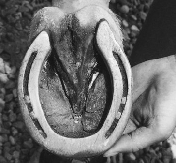
Fig. 123-2 A left hind shoe indicating natural breakover medial to the toe area (medial is right). Future shoes will be formed to allow breakover in this area rather than directly at the center of the toe.
Counterirritant therapy in the form of 2% iodine in oil commonly is injected in various patterns along the patellar and collateral ligaments of young horses, in horses with loose stifles, and in horses that have been rested as the intensity of training increases and that exhibit nonspecific pain or weakness in the stifle area. Counterirritants are not used as a replacement for intraarticular therapy but may be used concurrently. Injections usually are limited to 1 to 5 mL to reduce postinjection inflammation and to decrease scarring from numerous injections. Work is continued, but the intensity is reduced for a short time as the horse recovers.
Osteochondrosis is the second most common problem seen in the stifle. Osteochondral fragments from the femoral trochleas can be removed arthroscopically; usually horses have a favorable prognosis. Horses with lameness caused by subchondral bone cysts in the medial femoral condyle have a poorer prognosis. Some horses respond to intraarticular or intralesional corticosteroids. Surgical treatment may fail to resolve the lameness.
Thoracolumbar (Back), Sacroiliac, and Gluteal (Croup) Pain
Work-related back and croup pain is common in horses because of working in a collected frame frequently or for extended periods, compensatory altered carriage caused by injuries to the distal aspect of the limb, poor saddle fit, poor balance and timing of a rider, injuries from falling and weakness from immaturity, lack of conditioning, or poor conformation (long, weak back).
Back pain should be suspected when behavioral changes such as kicking out during lead changes in canter and uncharacteristic bucking occur. The horse is observed for muscle asymmetry while it stands with the body straight and the limbs squared on a level surface. The back and croup are palpated for areas of pain, using gradual pressure with the flat surface of the digits. Flexion, extension, and lateral flexion of each area of the back should be induced, and ease of mobility evaluated. Unilateral abnormalities may indicate concurrent injuries to the distal aspect of the limb. Unilateral lumbar pain often results from compensatory carriage of the hindlimbs to that side. Unilateral sacroiliac inflammation or dorsal ligamentous thickening often results from overbearing on that side from a chronic contralateral or diagonal limb injury. For example, horses with a chronic left front or left hind lameness may have swelling and pain in the area of the right tuber sacrale. Rectal examination is used to evaluate pelvic symmetry and ventral lumbar muscular pain. Other diagnostic modalities include radiography, ultrasonography, nuclear scintigraphy, and thermography, but physical examination remains the most important and reliable method of diagnosis. Myositis also occurs commonly and is diagnosed by history, observation of altered gait, palpation, and serum enzyme assay.
A variety of treatments is used, and these are discussed thoroughly elsewhere. Usually a period of rest is indicated, along with resolution of any associated lameness, followed by a period of reduced exercise. Systemic and local antiinflammatory agents are administered. Other therapies include cold and heat therapy, acupuncture, chiropractic therapy, massage therapy, and magnetic, electromagnetic, laser, and ultrasound therapy.
Successful treatment of horses with back and croup injuries depends on identifying initiating causes and making appropriate changes to alleviate these causes if possible. Educating the rider is also helpful, because changes in training routines or tack may be helpful in preventing recurrence.
Osteoarthritis of the Metacarpophalangeal and Metatarsophalangeal Joints
Synovitis of the metacarpophalangeal and metatarsophalangeal joints, an early form of OA, causes effusion, pain on static fetlock flexion, and a positive response to a distal limb flexion test. The condition is frequently bilateral. Because mild effusion in sound Arabian and Half-Arabian show horses is seen commonly, it is important to perform intraarticular or perineural analgesia to confirm the source of pain when OA of the metacarpophalangeal or metatarsophalangeal joint is suspected. Radiological assessment is performed to determine the extent of osteoarthritic abnormalities. Many Arabian and Half-Arabian show horses perform well in spite of mild OA of the fetlock (Figure 123-3). Treatment is similar to that recommended for OA of the DIP joint, except that a shorter-acting corticosteroid such as triamcinolone acetonide or betamethasone is used for intraarticular medication.
Distal Hock Joint Pain
OA of the centrodistal and tarsometatarsal joints, called distal tarsitis, is common. Tarsal lameness is often bilateral, although one limb usually is affected more severely. Affected horses have reduced hock flexion at the trot and tend to travel with a hip off to one side, especially in the corners (drift away from the lame limb). Rider complaints are similar to those regarding horses with stifle lameness. Western horses may tend to rate poorly and lope (canter) with a four-beat rather than a three-beat action. Reining horses tend to raise up in spins to the affected direction, bend poorly, stop unevenly or on the forehand, and change leads late behind in flying changes. Diagnosis is based on palpation, including the Churchill test, flexion tests, diagnostic analgesia, radiography, and occasionally ultrasonography and nuclear scintigraphy. Treatment consists of systemic administration of antiinflammatory agents and intraarticular medication with corticosteroids, possibly with hyaluronan (60 to 80 mg of methylprednisolone acetate and 10 mg of hyaluronan per joint), repeated as necessary, often two or three times during a show season. If medical therapy fails, chemical, laser, and surgical fusion techniques are available. ESWT and intravenous tiludronate administration have both been used successfully. Cunean tenectomy usually is not performed because the procedure commonly does not resolve lameness. Corrective shoeing involves inspecting the old shoe and squaring the new shoe to accommodate the breakover, setting the shoe back, shortening the toe, reducing the weight of the hind shoe, and occasionally using asymmetrical trailers.
Splint Bone Injuries
Proliferative periostitis, referred to as splints, and fractures of the second and fourth metacarpal bones and to a lesser extent fractures of the second and fourth metatarsal bones are common, especially in young horses. Diagnosis of splint-induced lameness is made using palpation, diagnostic analgesia, and radiography. Associated suspensory desmitis should be considered and assessed by ultrasonographic examination. Predisposing causes include conformational defects such as offset knees or base narrow and toed out. Heavy shoes and pads can exacerbate a tendency to wing in and cause interference. Poor footing that is excessively deep or hard can contribute to the development of splints. Immature horses in excessive work, or wearing shoes and pads that are too heavy for their level of fitness, are predisposed to splints. Shoeing with improper medial-to-lateral balance, obesity, and dietary imbalances can be contributory causes.
Proliferative periostitis originates from injury to the interosseous ligament, resulting in the development of proliferative fibrous connective tissue that subsequently mineralizes. Radiology is used to assess the injury and to determine the presence of fractures. Osteolysis of the third metacarpal or metatarsal bone and of the splint bone at the site of the injury is a sign of substantial inflammation and can indicate a prolonged recovery. Treatment includes rest (2 to 4 weeks), cold therapy, systemic and local administration of antiinflammatory agents, topical agents with support wraps, ESWT, and magnetic therapy. Large exostoses that interfere with surrounding soft tissue structures can be excised surgically.
Work-related fractures are predominantly distal, closed fractures and are thought to result from overexcursion of the SL during fetlock hyperextension. These fractures frequently are displaced, heal by nonunion, and tend to irritate surrounding soft tissues. For these reasons they commonly are removed surgically. Lameness in horses with mid–splint bone nondisplaced fractures often resolves with conservative therapy. Initial therapy is similar to the treatment of a true splint, except that local administration of corticosteroids is not performed. If conservative therapy fails, surgical excision of the exostosis and distal portion of the splint bone may be performed. Horses with fractures of the proximal aspect of a splint bone are evaluated on an individual basis, because some respond to conservative therapy, whereas others require surgical intervention.
Osteoarthritis of the Proximal Interphalangeal Joint
Incidental radiological changes (lipping and spurring) indicating mild OA of the proximal interphalangeal (PIP) joint in Arabian and Half-Arabian show horses are not uncommon. Moderate-to- severe OA, however, would be expected to cause chronic lameness. In most horses OA of the PIP joint is caused by chronic repetitive trauma, although OA secondary to a single severe injury is possible. In young horses osteochondrosis in the form of osseous cystlike lesions and fragmentation may be involved. The importance of any radiological abnormalities should be validated using diagnostic analgesia. Distal limb flexion is usually positive, and lameness should improve with palmar nerve blocks at the base of or at the level of the PSBs or intraarticular analgesia of the PIP joint. Treatment is similar to that recommended for horses with OA of the DIP joint.
Desmitis of the Accessory Ligament of the Deep Digital Flexor Tendon
Desmitis of the ALDDFT occurs less frequently than does PSD and is usually diagnosed in horses with club feet or low, underrun heels. Footing that accumulates under the shoe, creating a rocking motion of the foot during the weight-bearing phase of the stride, may predispose horses to desmitis. Lameness is usually acute in onset, and palpation often reveals a painful thickening along the length of the ligament that can be confused with the DDFT. If the condition is chronic and low grade, diagnostic analgesia may be required. Diagnosis is confirmed with ultrasonography. Treatment involves prolonged rest and systemic and local administration of antiinflammatory agents, PRP, or mesenchymal stem cells. Adjuncts include cold therapy, topical agents with support wraps, and magnetic, laser, ESWT, and ultrasound therapy. Because the predisposing conformational defect remains, the condition tends to recur.