KEY POINTS
•
The muscular system comprises striated or skeletal muscle, i.e. muscle which is attached to the skeleton and brings about movement of a region.
•
Each striated muscle fibre is filled with myofibrils made of two contractile proteins, actin and myosin. At the cellular level, muscle contraction results from the formation of cross-bridges between the actin and myosin molecules.
•
Muscle fibres are stimulated to contract by nerve impulses carried by nerve fibres. The number of muscle fibres supplied by a single nerve fibre is called a motor unit. In muscles which perform accurate and delicate movements, a nerve fibre will supply a few muscle fibres, but in muscles which perform less accurate movements a nerve fibre will supply many muscle fibres.
•
Muscle tissue is always under a degree of tension, known as muscle tone. The tone increases when an animal is alert or frightened and decreases when it is relaxed or asleep.
•
All muscles consist of a central belly and, at the point of attachment to a bone, an origin (often called the head), and an insertion.
•
Skeletal muscles may be either:
•
Extrinsic, i.e. attached from one major structure, such as the trunk, to another structure, such as a limb. These muscles bring about movement of the
whole limb in relation to other body parts
•
Intrinsic, i.e. attached at both ends within the one structure, such as a limb. These muscles bring about movement
within the individual limb, e.g. bending an elbow.
•
Each area has a range of specialised muscles designed to bring about the specific types of movement necessary for the animal’s normal function.
Muscle structure and function
Contraction
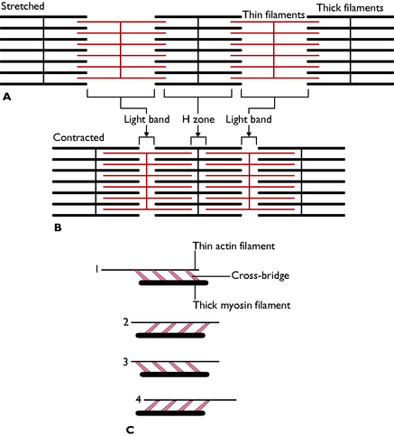
Muscle is stimulated to contract when it receives a nerve impulse from the central nervous system. Each striated muscle fibre is composed of myofibrils made of thin actin filaments and thick myosin filaments. These fibres overlap in such a way that under the microscope muscle has the appearance of alternating light and dark bands or striations. These bands are separated into units called sarcomeres, which are the units of contraction.
During contraction, the actin and myosin filaments slide over one another and cross-bridges form between the heads of the myosin filaments and the heads of the actin filaments. The cross-bridges swing through an arc, pulling the thin filaments past the thick ones, and the sarcomere shortens. Once this movement is completed the cross-bridge detaches itself from the thin filament and reattaches itself further away – in other words, the cross-bridges between the myosin and actin filaments act as a ratchet mechanism, thus shortening the muscle (Fig. 4.1). This process requires energy input, which is provided by adenosine triphosphate (ATP) molecules; calcium ions are also essential to the process of muscle contraction.
The nerve that stimulates the muscle to contract enters the muscle and then splits up into many fibres to innervate the bundles of muscle fibres. The number of muscle fibres supplied by one nerve fibre will vary depending on the type of movement for which the muscle is responsible. If it is a delicate movement then a nerve fibre may only innervate a small number of muscle fibres. However, in larger movements, such as those made by the muscles of the limbs, one nerve fibre may supply 200 or more muscle fibres. A single nerve together with the muscle fibres that it supplies is called a motor unit.
Muscle tone
Many of the skeletal muscles in the body are always in a slight state of tension, known as muscle tone. Even when an animal is at rest, muscles, such as those responsible for maintaining posture, will not be truly relaxed. Muscle tone is achieved by a proportion of the motor units within the muscle being activated so that some of the muscle fibres are contracting while others are relaxed. The nervous system can adjust this, and the number of motor units stimulated will increase when the animal is in an anxious state, i.e. the muscles become ‘twitchy’ or ‘jumpy’. Thus, muscles undergo two types of contraction:
•
Isometric contraction – when tension is generated in the muscle, i.e. muscle tone is increased, but the muscle does not shorten
•
Isotonic contraction – the muscle actually moves or shortens.
The more a muscle is used or exercised, the larger it will become – it is said to be hypertrophied. However, if a muscle is not used for some reason, e.g. due to injury or illness when the animal may be recumbent, or if a limb is in a cast, it will wither or shrink in size – it is said to be atrophied.
Muscle atrophy or wasting may be the result of many factors, e.g. lack of use as a result of lameness, fracture, denervation (injury to the nerve that supplies it) or a generalised disease condition, such as neoplasia. Muscle enlargement or hypertrophy may result from overexercise or overuse. This may be seen where a patient has fractured a limb and the muscles in the other limb must work harder to support it.
Anatomy of a muscle
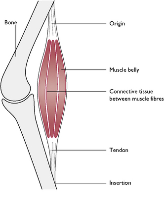
A ‘classically’ shaped muscle (Fig. 4.2) has a thick, fleshy central part called the belly and tapers at each end – the head. Here, the connective tissue muscle sheath is continuous with the dense fibrous connective tissue of the tendon that attaches the muscle to a bone. A muscle is attached to a bone at two points: its starting point is called its origin; this moves least during contraction. The opposite end, where the muscle inserts on the bone, is called the insertion. However, a muscle can have more than one belly, all inserting at one point, in which case the muscle is said to have a number of heads, e.g. the biceps muscle has two heads. The length of the tendon attaching the muscle to a bone will vary, and in some cases tendons are far longer than the muscle itself, e.g. flexor and extensor tendons running over the digits.
Not all muscles take the ‘classical’ shape described above. Sometimes they are present in flat sheets, in which case the tendon is also drawn out into a flat sheet of connective tissue – this type of arrangement is called an aponeurosis, e.g. the muscles of the abdominal wall. Some muscles form a circular ring and serve to control the entrance or exit to a structure, e.g. the stomach and the bladder. These are called sphincter muscles.
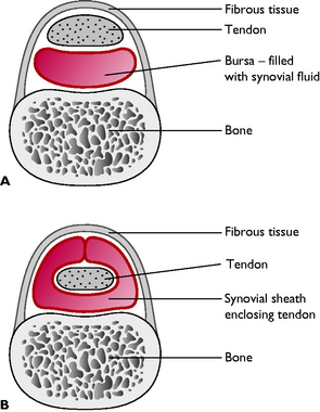
A bursa is a connective tissue sac lined with synovial membrane and filled with synovial fluid. These typically develop between a bony prominence and a tendon, ligament or muscle and their function is to reduce friction between the associated structure and the bone. Sometimes a bursa completely wraps around a tendon forming a synovial or tendon sheath (Fig. 4.3).
The skeletal muscles of the body can be classed as either intrinsic or extrinsic:
•
Intrinsic muscles lie completely within one region of the body where they have their origin and insertion. They act on the joints in that part only; for example, when a dog bends its elbow it is using the intrinsic muscles of the forelimb.
•
Extrinsic muscles run from one region of the body to another and alter the position of the whole part, e.g. a limb, in relation to the other. The muscles that attach the foreleg of the dog to the trunk are extrinsic muscles; they move the whole foreleg in relation to the trunk.
The skeletal muscles
To simplify the study of these muscles we can look at one area of the body at a time.
Muscles of the head
Muscles of facial expression
The muscles of facial expression are intrinsic muscles that move the lips, cheeks, nostrils, eyelids and external ears. A number of muscles are responsible for these movements, all of which are innervated by the facial nerve (cranial nerve VII).
Muscles of mastication
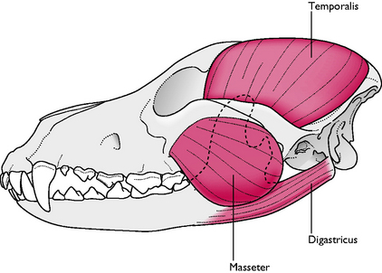
The main muscles responsible for the masticatory or ‘chewing’ action of the jaw (Fig. 4.4) are:
•
Digastricus – this muscle opens the jaw, aided by gravity, and is located on the caudoventral surface of the mandible. Its origin is the jugular process of the occipital bone and it inserts on the angle and ventral surface of the mandible
•
Masseter – this muscle closes the jaw and lies lateral to the mandible. It originates from the zygomatic arch and inserts on the masseteric fossa on the lateral surface of the mandible
•
Temporalis – this muscle also closes the jaw and is the largest and strongest muscle of the head. It covers much of the dorsal and lateral surfaces of the skull. It fills the temporal fossa of the skull and inserts on the coronoid process of the mandible
•
Medial and lateral pterygoids – these are deep muscles that lie medial to the mandible. They aid the temporalis and masseter muscles in closing the jaw, but they are also responsible for the side to side movements of the mouth.
The tone in the muscles of the jaw responsible for closure, i.e. the temporalis and the masseter, are what keeps the mouth closed when it is not in use.
Muscles of the eye
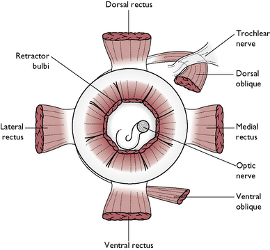
The extrinsic muscles of the eye (Fig. 4.5), also called the extraocular muscles, are responsible for moving the eye within its bony socket. These muscles are:
•
Four
RECTUS muscles:
dorsal rectus, ventral rectus, medial rectus, lateral rectus – these four muscles all insert on the
sclera of the eyeball near the ‘equator’, at the surface that corresponds to their name, i.e. the dorsal rectus muscle inserts on the upper part of the sclera. Their name also reflects their action – the dorsal rectus muscle turns the eye upwards; the ventral rectus turns the eye downwards; the medial rectus turns the eye inwards; the lateral muscle turns the eye outwards
•
Two
OBLIQUE muscles: –
dorsal oblique and
ventral oblique – these two muscles are positioned as their name describes and they act to rotate the eye about its visual axis
•
Retractor bulbi – this muscle forms a muscular cone around the optic nerve at the back of the eye and its action is to pull the eye deeper into the socket.
Other muscles of the head
These include:
•
Intrinsic and extrinsic muscles in the tongue, which enable the tongue to carry out a wide range of delicate movements
•
Many muscles are found in the pharynx, larynx and soft palate, which enable these structures to carry out their specific functions, e.g. swallowing and sound production
•
Extrinsic muscles attach the head to the neck and move it in relation to the neck.
Muscles of the trunk
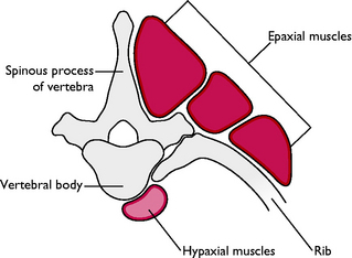
Muscles of the vertebral column
Groups of muscles lie above and below the vertebral column (Fig. 4.6). These are:
•
Epaxial muscles – this group lie dorsal to the transverse processes of the vertebrae, i.e. they are above the vertebral column. The epaxial muscles are numerous and are arranged in three longitudinal groups, which together span the length of the vertebral column. Their functions are to support the spine, extend the vertebral column and allow lateral flexion
•
Hypaxial muscles – this group lies ventral to the transverse processes of the vertebrae, i.e. they are below the vertebral column. One region is associated with the neck, another with the back. The hypaxial muscles flex the neck and tail, and contribute to flexion of the vertebral column.
Muscles of the thorax
The muscles of the thoracic wall are mostly involved in respiration. The main muscles are:
•
External intercostals – the most superficial, which originate from the caudal border of one rib and insert on the cranial border of the rib behind it. Thus, each muscle is confined to one intercostal space. These muscles assist in inspiration
•
Internal intercostals – lie below the external intercostals within the intercostal spaces and originate from the cranial surface of one rib and insert on the caudal border of the rib in front of it. These muscles assist in expiration, which is a largely passive movement.
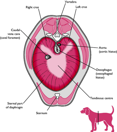
The diaphragm
The diaphragm is the sheet of muscle that separates the thoracic and abdominal cavities (Fig. 4.7). It consists of a central tendon and a muscular periphery that arises from the lumbar vertebrae, caudal ribs and sternum. The lumbar portion of the muscle consists of thickened muscular bundles called the right and left crura (sing. crus).
The diaphragm is the main muscle of inspiration. When it contracts, the lungs expand and draw in air (see Ch. 8). In the centre of the diaphragm there are three openings that allow through structures that pass from the thoracic region into the abdomen. These openings are:
•
Aortic hiatus – this transmits the aorta, azygous vein and thoracic duct
•
Oesophageal hiatus – transmits the oesophagus and vagal nerve trunks
•
Caval foramen – lies within the central tendon and transmits the caudal vena cava.
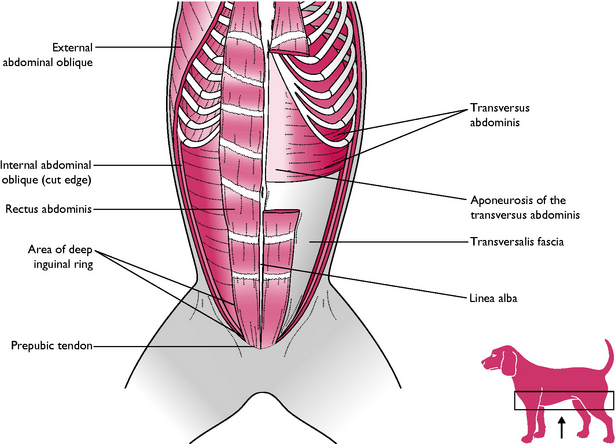
Abdominal muscles
The abdominal wall on each side of the body is composed of four muscles, which form broad, flat sheets (Fig. 4.8). Their fibres run in all directions, which gives the abdominal wall great strength. Their function is to protect the organs and structures of the abdomen. The muscles of the abdominal wall are:
•
External abdominal oblique – the most superficial of the lateral abdominal muscles. It originates from the lateral surfaces of the ribs and lumbar fascia. It terminates in a wide aponeurosis in the midline known as the
linea alba.
•
Internal abdominal oblique – the intermediate muscle of the lateral abdominal wall, which also terminates in an aponeurosis on the linea alba.
•
Transversus abdominis – the deepest of the lateral abdominal muscles which also terminates in an aponeurosis on the linea alba.
•
Rectus abdominus – this is a broad band of muscle on each side of the linea alba that forms the floor of the abdomen. The rectus abdominus originates on the first rib and sternum and inserts on the pubis by means of the
prepubic tendon.
The linea alba, or white line, is the combined aponeuroses of the three lateral abdominal muscles and it extends along the ventral midline from the xiphoid process of the sternum to the pubic symphysis.
The linea alba can easily be seen when a vet makes a midline incision in the abdomen, e.g. during a bitch spay or any type of laparotomy. Because it is tough, it provides a good tissue in which to place sutures without the risk of them pulling out.
The inguinal ring is a slit-like opening in the aponeuroses of the abdominal muscles, in the region of the groin. This allows the passage of vessels from the abdomen to the external genitalia and mammary glands, and transmits the structures of the spermatic cord to the scrotum. Occasionally the inguinal ring is damaged causing it to enlarge and allow structures such as a piece of intestine or a pad of fat to pass through, forming an inguinal hernia.
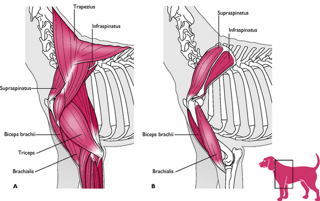
Muscles of the forelimb
The muscles of the forelimb can be divided into extrinsic and intrinsic muscles.
Extrinsic group
The extrinsic muscles of the forelimb (Fig. 4.9) attach the forelimb to the trunk forming a synsarcosis (the attachment of a structure to the skeleton by muscles instead of the more conventional joint).
•
Trapezius – this is a triangular sheet of muscle that originates from the dorsal midline from C2 to C7 and inserts on the spine of the scapula
ACTION – draws the leg forward and protracts the limb
•
Pectorals – these muscles run from the ribs and sternum and insert on the humerus
ACTION – adduct the limb and hold the forelimb against the body wall
•
Latissimus dorsi – this is a large, fan-shaped muscle, which has a very broad origin on the thoracic spine and inserts on the humerus
ACTION – retracts the forelimb
•
Brachiocephalicus – this muscle runs from the base of the skull to an insertion on the cranial aspect of the humerus
ACTION – when the limb is on the ground, it flexes the neck and bends the neck laterally; when the limb is not taking weight, it draws the foreleg forwards or protracts the limb.
Intrinsic group
The intrinsic muscles of the forelimb all originate and insert on the forelimb. They include:
•
Supraspinatus – arises from and fills the supraspinous fossa of the scapula and inserts on the greater tubercle of the humerus
ACTION – extends the shoulder and stabilises the shoulder joint
•
Infraspinatus – arises from and fills the infraspinous fossa of the scapula and inserts on the greater tubercle of the humerus
ACTION – helps to stabilise the shoulder joint and flexes the shoulder joint.
•
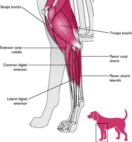
Triceps brachii – this muscle has four heads with separate origins. Three of the heads originate from the proximal humerus and the fourth head originates from the scapula. However, they all share a common insertion on the olecranon of the ulna. A bursa lies between the bone and the tendon to prevent friction
ACTION – extends the elbow joint
•
Biceps brachii – originates from the supraglenoid tubercle of the scapula and inserts on the radius and ulna
ACTION – flexes the elbow joint
•
Brachialis – this muscle originates from the humerus and inserts on the radius and ulna
ACTION – flexes the elbow joint
Carpus and digits
There are many muscles responsible for flexing and extending the carpus and the digits. The muscle bellies are grouped around the radius and ulna and communicate with the digits by means of long tendons.
The main flexors and extensors are:
•
Two carpal extensors – originating from the humerus and inserting on the carpals; they run in front of the lower limb and foot
•
Two digital extensors – originating from the humerus and inserting on the third phalanx; they run in front of the lower limb and foot
•
Two carpal flexors – run behind the carpus and foot
•
Two digital flexors – the
superficial digital flexor inserts on the second phalanx and the
deep digital flexor inserts on the third phalanx.
Muscles of the hindlimb
The hindlimb is attached to the pelvic girdle by a mobile ball and socket joint, the hip joint. The sublumbar hypaxial muscles of the vertebral column are extrinsic muscles attached to the vertebral column and to the pelvic girdle.
The intrinsic muscles of the hindlimb all insert on the hindlimb itself or on the pelvic girdle and are described below.
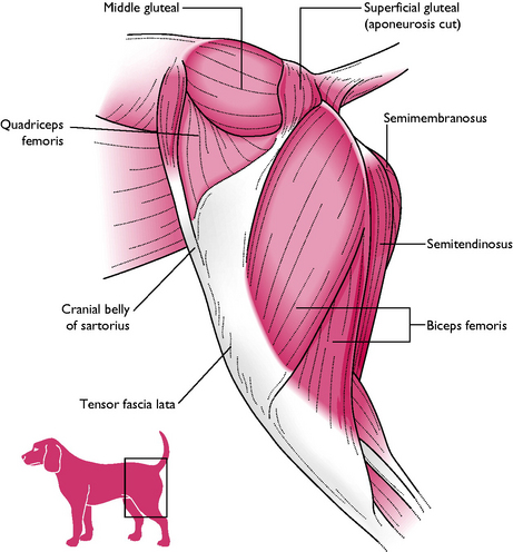
Muscles of the thigh
These originate on the pelvic girdle and insert on the femur.
•
Gluteals – three muscles, the
superficial, middle and
deep gluteals, and the
tensor fascia latae form the curve of the rump and are powerful extensors of the hip joint
ACTION – extend the hip joint and abduct the thigh
 Hamstring group
 – form the caudal aspect of the thigh and act together to propel and extend the whole limb backwards (
Fig. 4.11). They provide the main propulsive force of the animal and consists of three muscles:
Biceps femoris – is the most lateral muscle in this group (
Fig. 4.11). It originates from the pelvis and runs over the femur to the tibia and inserts on the calcaneus of the hock
ACTION – extends the hip, flexes the stifle and extends the hock
Semitendinosus – runs from the pelvis and inserts on the tibia and calcaneus
ACTION – extends the hip, flexes the stifle and extends the hock
•
Semimembranosus – is the most medial muscle of the hamstring group. It runs from the pelvis to the femur and tibia
ACTION – extends the hip and flexes the stifle
•
Quadriceps femoris – this large muscle runs down the cranial aspect of the thigh. It consists of four parts, all of which insert at the same point on the tibial tuberosity or crest. The tendon of this muscle contains the
patella, which articulates with the femur at the stifle joint
ACTION – extends the stifle joint
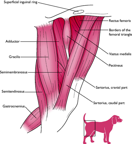
•
Adductor muscles – lie on the medial aspect of the thigh and hold the limb close into the body (
Fig. 4.12):
The tendon of the pectineus muscle is sometimes transected to relieve pressure on the hip joint of dogs with hip dysplasia. Pectineotomy does not cure the problem but may provide a degree of pain relief.
Pectineus – this muscle runs from the pubis to the distal femur
ACTION – adducts the limb
Sartorius – inserts on the cranial border of the tibia with the gracilis muscle
ACTION – adducts the limb
Gracilis – forms the caudal half of the medial surface of the thigh
ACTION – adducts the limb
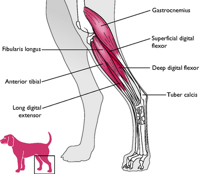
Muscles of the lower hindlimb
These muscles act mainly on the hock joint (Fig. 4.13). They include:
•
Gastrocnemius – this muscle originates from the caudal aspect of the femur and inserts on the calcaneus of the hock. The
tendons of this muscle contain two small sesamoid bones or fabellae that articulate with the caudal aspect of the stifle joint
ACTION – extends the hock and flexes stifle
•
Achilles tendon – this is the large, strong tendon that runs down the back of the leg to the point of the hock. It includes the tendons of insertion of the gastrocnemius, biceps femoris and semitendinosus, all of which insert on the calcaneus, and also of the superficial digital flexor muscle, which continues over the point of the hock and down to its insertion on the digits. There is a bursa at the point of insertion on the calcaneus.
The popliteal lymph node lies within the fibres of the gastrocnemius muscle and can be palpated just caudal to the stifle joint. Normally it is about 2 cm long but if it is enlarged it may indicate some form of infection (or tumour) in the distal extremity.
Muscles of the hock and digits
As is seen in the forelimb, there are a number of muscles responsible for flexing and extending the hock and the digits:
•
Anterior tibialis – runs from the proximal end of the tibia to the tarsus
ACTION – flexes the hock and rotates the paw medially
•
Three digital extensors – run in front of the hock and foot
•
Two digital flexors – run behind the foot. The
superficial digital flexor runs from the femur to the phalanges and is one of the components of the Achilles tendon.