Chapter 2 Tissues and body cavities
Within the body individual cells are grouped together to form tissues and organs. Thus:
Body tissues
Each tissue type consists of three main components:
There are four main types of tissue:
Epithelial tissue
Epithelial tissue or epithelium covers the surface of the body and the organs, cavities and tubes within it – it covers the internal and external surfaces of the body. Its main function is to protect delicate structures lying beneath it but in some areas the epithelium may be secretory, e.g. glands, or absorbent, e.g. in the small intestine. The epithelium lining structures such as the inside of the heart, blood vessels and lymph vessels is referred to as endothelium.
Epithelium may be described according to the number of layers of cells, i.e. its thickness:
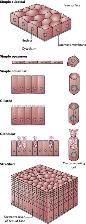
The thickness of the epithelium reflects its ability to protect: the more layers of cells, the more protection is provided. The epithelium on the footpads consists of many layers of cells providing protection when walking on rough surfaces, while the epithelium over the abdominal wall is only a few cells thick and additional protection is provided by fur. Further protection may be provided by the presence of the protein keratin. The epithelium is described as being a keratinised stratified epithelium and this type can be seen in claws and nails.
Epithelium may also be described according to the shape of the cells within it (Fig. 2.1). There are three basic shapes of epithelial cell:
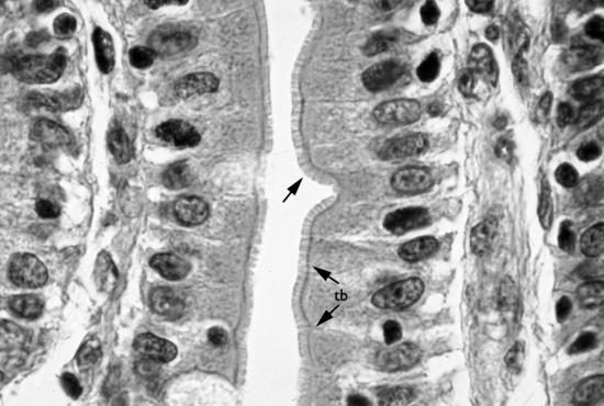
Fig. 2.2 Light micrograph of the mucosa of the small intestine showing a simple columnar epithelium with a border of microvilli that increase the surface area for absorption.
(Taken from D. Samuelson. Textbook of Veterinary Histology. Saunders. 2007, p 42.)
The full classification of the type of epithelium is based upon the shape of the cell and the number of layers present. There are a number of different types of epithelial tissue in the body, these include:
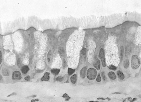
Fig. 2.3 Photomicrograph of the lining of the trachea showing a pseudostratified ciliated columnar epithelium
(Taken from D. Samuelson. Textbook of Veterinary Histology. Saunders. 2007, p 43.)
Glands
Glandular tissue is a modification of epithelial tissue. The epithelium, in addition to its protective function, may also be a secretory membrane. Glands are either:
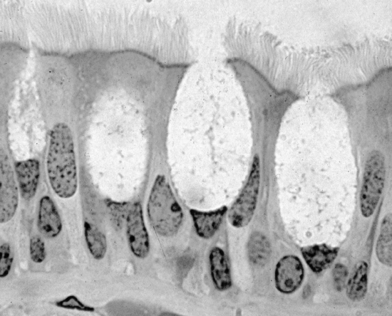
Fig. 2.4 Photomicrograph of goblet cells within a pseudostratified ciliated columnar epithelium
(Taken from D. Samuelson. Textbook of Veterinary Histology. Saunders, p 64.)
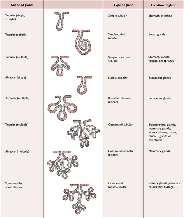
Glands may be categorised as either:
Connective tissue
Connective tissue is responsible for supporting and holding all the organs and tissues of the body in place. It also provides the transport system within the body, carrying nutrients to the tissues and waste products away. Connective tissue consists of cells embedded in an extracellular matrix or ground substance. The properties of this ground substance depend on the type of connective tissue. There are many types of connective tissue which, in order of increasing density are:
Blood
Blood is a specialised connective tissue that circulates through the blood vessels to carry nutrients and oxygen to the cells, and waste products to the organs of excretion. It consists of a number of different types of blood cell within a fluid ground substance – the plasma. (This is covered in more detail in Ch. 7).
Haemopoietic tissue
This jelly-like connective tissue forms the bone marrow within the long bones and is responsible for the formation of the blood cells (see Ch. 7).
Areolar tissue
Areolar (meaning spaces) or loose connective tissue (Figs 2.6, 2.7) is the most widely distributed type of connective tissue and is found all over the body, e.g. beneath the skin, around blood vessels and nerves, between and connecting organs and between muscle bundles. The ground substance contains a loose weave work of two types of protein fibre: collagen fibres, with a high tensile strength secreted by the main cell type (the fibroblast) and elastic fibres, which enable the tissue to stretch and return to its former shape. Fat cells may be present in varying quantities depending on location and the degree of obesity of the animal. Macrophages, cells which are capable of phagocytosis, are also present.
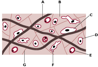
Fig. 2.6 Composition of areolar tissue. A Collagen fibres: flexible but very strong and resistant to stretching. B Ground substance: this contains the different fibres and cells of the tissue. C Fibroblast: long, flat cell that produces collagen and elastic fibres. D Mast cell: secretes an anticoagulant. E Fat cell: stores fat. F Macrophage: large cell capable of phagocytosis of foreign particles. G Elastic fibres: form a loose and stretchable network.
Adipose tissue
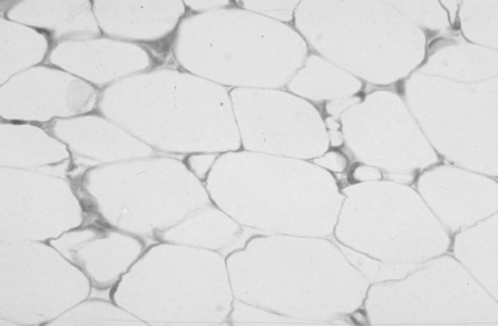
Adipose tissue (Fig. 2.8) is similar to areolar tissue but its matrix contains mainly fat-filled cells, closely packed together, giving it the name fatty tissue. These fat cells act as an energy reserve and, in the dermis of the skin, the tissue insulates the body to reduce heat loss. In some areas, such as around the kidney, adipose tissue provides a protective layer.
Dense connective tissue
Dense or fibrous connective tissue consists of densely packed collagen fibre bundles with relatively few fibroblasts and other cells in between them (Fig. 2.9). The fibres may be arranged in two ways:
Cartilage
Cartilage is a specialised connective tissue which is rigid but flexible and resilient and is able to bear weight (Fig. 2.10). It is composed of cells (known as chondrocytes) and fibres within a gel-like ground substance. Cartilage has no blood supply and its nutrition is supplied by the fibrous sheath or perichondrium that surrounds it.
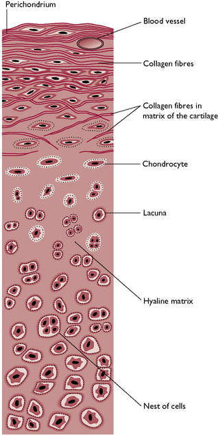
There are three types of cartilage:
Bone
Bone is a living tissue that is capable of remodelling and repairing itself when damaged. It is a specialised type of connective tissue, which provides the rigid supportive framework of the body and forms a system of levers for locomotion.
Bone consists of an extracellular matrix or ground substance that contains the protein osteonectin and collagen fibres. Together, these form osteoid, within which crystals of insoluble calcium phosphate are deposited as the bone tissue becomes calcified. Calcification gives bone its characteristic rigidity and hardness. As the ground substance becomes calcified the bone cells or osteocytes are trapped in spaces called lacunae. Running through the bone matrix are fine channels, called Haversian canals, which carry the blood vessels and nerves of the bone(Figs 2.11, 2.12). Each Haversian canal is surrounded by a series of concentric cylinders of matrix material called lamellae and the osteocytes within their lacunae. Each series of these cylinders, together with the canal, is called a Haversian system (Figs 2.11, 2.12). A fibrous membrane, the periosteum, covers the outer surface of all types of bone.
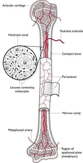
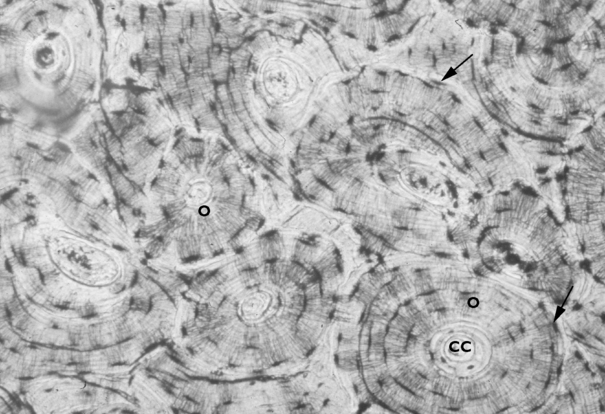
Fig. 2.12 Light micrograph (×100) of compact bone showing Haversian canals (CC) surrounded by lamellae of osteocytes (O) within their lacunae.
(Taken from D. Samuelson. Textbook of Veterinary Histology. Saunders, p 122.)
Muscle tissue
Muscle tissue is responsible for organised movement in the body.
Skeletal or striated muscle
This type of muscle is found attached to the skeleton and brings about movement (Fig. 2.13). It is under voluntary or conscious control, i.e. an animal uses its brain to move its limbs.

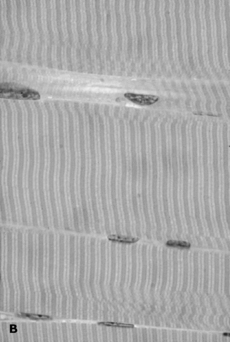
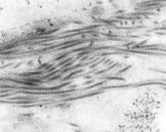
Fig. 2.13 A Skeletal muscle, as found attached to the bones. It is voluntary and consists of striped (striated) cells, which are long and cylindrical in shape. Each cell has several nuclei. B. Light micrograph (×1000) of skeletal muscle showing characteristic bands and lines and nuclei located at the edges of the fibres.
(Taken from D. Samuelson. Textbook of Veterinary Histology. Saunders, p 165.)
The muscle cells or fibres are long and cylindrical and lie parallel to each other. Each individual muscle fibre is composed of bundles of microfilaments known as myofibrils that are made of two contractile proteins called actin (thin filaments) and myosin (thick filaments). It is their highly regular arrangement that gives the muscle its striated or striped appearance when viewed under a microscope. Each fibre has several nuclei, which lie on the outer surface of the cell as the presence of the myofibrils pushes all the cell structures to the outer margins.
The muscle fibres are grouped together in bundles or fascicles by connective tissue. Groups of fascicles are then held together by connective tissue and form a large muscle. The whole muscle is surrounded by the muscle sheath, which is continuous with the tendons that connect the muscle to a bone.
Smooth muscle
Smooth muscle is also called unstriated, involuntary and visceral muscle. It is found in regions of the body that are under involuntary control, e.g. in the walls of the blood vessels, digestive tract, respiratory tract, bladder and uterus. It is therefore responsible for moving food through the digestive system, controlling the flow of blood through blood vessels, and other unconscious processes. Smooth muscle is controlled by the autonomic nervous system.
The cells of smooth muscle are long and spindle-shaped (Fig. 2.14) and are surrounded by small amounts of connective tissue that bind the cells into sheets, or layers. The nucleus in each cell lies in its centre. Smooth muscle does not appear ‘striped’ when viewed under the microscope – hence its name.
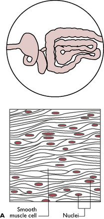
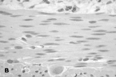
Fig. 2.14 A Smooth muscle, as found in the gastrointestinal tract and other viscera. It is involuntary and consists of small spindle shaped cells without striations (hence ‘smooth’). Each cell has one nucleus in the centre. B. Light micrograph (×1000) of smooth muscle cells showing the long spindle shaped cells with no striations.
(Taken from D. Samuelson. Textbook of Veterinary Histology. Saunders, p 161.)
Cardiac muscle
This type of muscle is found only in the heart and forms the myocardium. It is responsible for the rhythmic and automatic contraction of the heart that continues throughout an animal’s life. This inherent contractibility is increased or slowed down by nerves supplying the heart according to the requirements of the body. Control of cardiac muscle is therefore involuntary or unconscious.
Cardiac muscle cells are striated and cylindrical in shape (Fig. 2.15). Unlike the cells of striated muscle, they branch to create a network of fibres, which are linked by intercalated discs. These enable nerve impulses to be conveyed across the myocardium extremely quickly, producing a rapid response to the changing needs of the body.
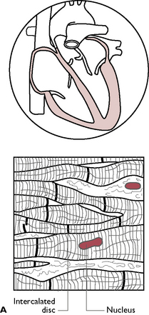
Fig. 2.15 A Cardiac muscle, found in the heart. It is involuntary and consists of striated, cylindrical cells. Cells are connected by intercalated discs. B. Light micrograph (×1000) of cardiac muscle cells. Arrow indicates intercalated discs.
(Taken from D. Samuelson. Textbook of Veterinary Histology. Saunders, p 173.)
Nervous tissue
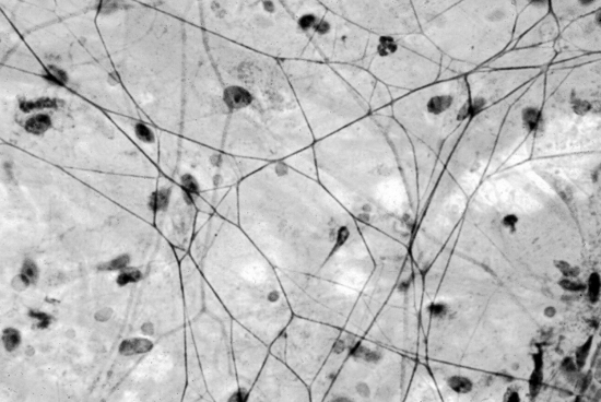
The main cell of nervous tissue is the neuron (Fig. 2.16), whose function is to transmit nerve impulses from one area to another. Each neuron consists of a cell body, containing the nucleus, several short processes known as dendrons and one long process known as an axon. Dendrons carry information towards the cell body, while the axon carries information away from it and towards its destination. Many axons within the body are covered in fatty material known as myelin. This is secreted by specialised cells wrapped around each axon – the Schwann cells – and it increases the speed of transmission of nerve impulses from one place to another.
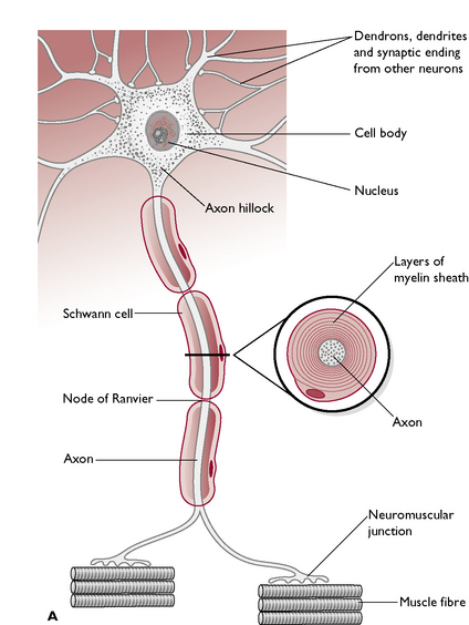
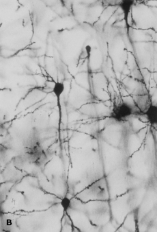
Fig. 2.16 A. The structure of a neuron (nerve cell). B Light micrograph of nervous tissue.
(Taken from D. Samuelson. Textbook of Veterinary Histology. Saunders, p 182.)
Dendrons and axons are referred to as nerve fibres and the whitish structures identified by the naked eye as ‘nerves’ within the body are collections of large numbers of nerve fibres.
Nerve impulses are transferred from one neuron to another by means of button-like structures known as synapses. All nerve pathways consist of neurons and synapses. (Nervous tissue is covered in more detail in Ch. 5.)
The body cavities
The body is divided into separate areas referred to as the body cavities (Fig. 2.17). They are described as ‘potential’ spaces because, although they are completely filled with the visceral organs and fluid, there is only a very small amount of free space. All the body cavities are lined with a serous membrane or endothelium, which is a single continuous layer of epithelium that produces a watery or serous lubricating fluid. This is different from the thicker, more proteinaceous secretion mucus, which has a protective function. Serous fluid acts as a lubricant between the surfaces of the cavity and the organs and structures within it.

Each part of the serous membrane is a continuous layer, named according to its position within the cavity:
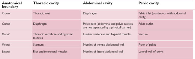
There are three body cavities:
The thoracic cavity
The thoracic cavity (Table 2.1) contains the heart, lungs and other associated structures. Its skeletal walls are formed by the bony thoracic cage consisting of the ribs, thoracic vertebrae and sternum. The ‘entrance’ into the cavity is known as the cranial thoracic inlet and is formed by the first thoracic vertebra, the first pair of ribs and the manubrium. The exit or caudal border is filled by the diaphragm.
The serous membrane lining the thoracic cavity and covering the organs within it is called the pleura (Fig. 2.18). The parietal pleura lines the inside of the thoracic cavity but is named according to which part of the walls it covers, i.e. the diaphragmatic pleura covers the diaphragm and the costal pleura covers the ribs. The thoracic cavity is divided into two pleural cavities by a continuation of the parietal pleura. Each cavity contains one of the lungs and serous pleural fluid. The lungs themselves are coveredin visceral pleura, called the pulmonary pleura. Between the two pleural cavities, the thorax is divided into right and left sides by a vertical connective tissue septum called the mediastinum, which is covered in the mediastinal pleura. The mediastinum is the potential space formed by the double layer of parietal pleura that separates the two pleural cavities. It contains the pericardial cavity, containing the heart, aorta, trachea, oesophagus and the thymus gland in young animals.
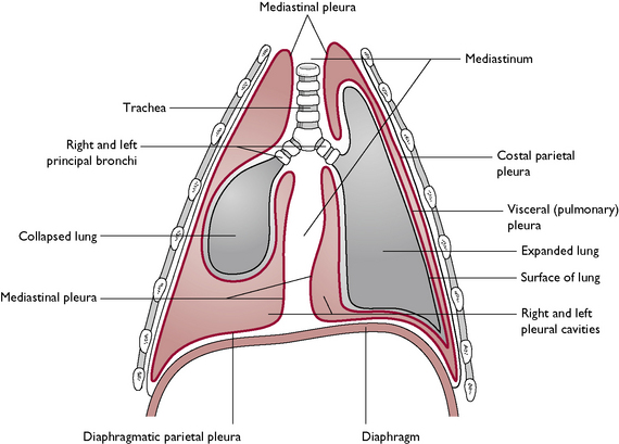
The pericardial cavity lies within the mediastinum in the thoracic cavity and is the space in which the heart sits. The heart is contained within the pericardium, which is a double-layered membranous sheath that completely surrounds it. Between the two layers of membrane is serous fluid that acts as a lubricant, enabling the heart to beat freely.
The abdominal cavity
The abdominal cavity (Table 2.1) lies caudal to the thoracic cavity and contains the abdominal viscera (Fig. 2.17). These include the organs of the digestive system and related glands, the urogenital system and all the associated vessels and nerves that supply these systems. The abdominal cavity is bounded cranially by the diaphragm and caudally by the pelvic inlet (there is no actual physical division between the abdominal and pelvic cavities). Dorsally, the boundary is the lumbar vertebrae and the hypaxial muscles. The muscles of the abdominal walls form the dorsolateral, ventral and lateral limits.
The internal surface of the abdominal cavity is lined with a serous membrane called the peritoneum (Fig. 2.19). This is a continuous sheet that forms a closed cavity, the peritoneal cavity. The peritoneal cavity is the potential space between the parietal peritoneum that lines the abdominal walls and the visceral peritoneum that covers the organs. The peritoneal cavity contains only a small volume of lubricating serous fluid known as peritoneal fluid, which allows friction-free movement of the organs and prevents adhesions forming between the organs and the peritoneum.
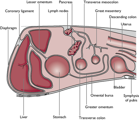
Fig. 2.19 Sagittal section through the abdominal cavity to show the reflections (folds) of the peritoneum.
The visceral peritoneum is folded on itself in a way that keeps the organs separate, suspending the organs within the abdominal cavity and carrying the various vessels and nerves that serve the viscera. This area of peritoneum is collectively known as the mesentery. The folds of mesentery have different names depending on their position, e.g. the mesentery suspending the duodenum is called the mesoduodenum, while the one suspending the ovary is the mesovarium. The omentum is a mobile fold of peritoneum that contains a ‘lacy’ network of fine vessels and fat and is divided into the greater omentum, arising from the greater curvature of the stomach, and the lesser omentum, arising from the lesser curvature of the stomach.
The pelvic cavity
The pelvic cavity (Table 2.1) lies caudal to the abdominal cavity (Fig. 2.17) and contains the urinary bladder, the rectum and the reproductive organs. There is no physical separation between the two as there is between the thoracic and abdominal cavities. The cavity is bounded cranially by the pelvic inlet and caudally by the caudal pelvic aperture. The sacrum and the first few coccygeal vertebrae form the dorsal boundary and the pubis and ischium form the floor of the cavity. The walls are formed by muscles and ligaments. A continuation of the peritoneum extends into the cranial parts of the cavity and covers the organs which lie in both cavities, e.g. bladder and reproductive tract.