1 McIlwraith CW. What is developmental orthopaedic disease, osteochondrosis, osteochondritis, metabolic bone disease?. Am Assoc Equine Pract. 1993;39:35.
2 Ineson AC, Rogers CW, Firth EC, et al. A comparison of subjective and quantitative methods for detection of physitis in Thoroughbred foals. N Z Vet J. 2004;52:65.
3 Jeffcott LB. Problems and pointers in equine osteochondrosis. Equine Vet J Suppl. 1993;16:1.
4 Smith BP. Large animal internal medicine, ed 3. St Louis: Mosby, 2003.
5 Orsini JA, Kreuder C. Musculoskeletal disorders of the neonate. Vet Clin North Am. 1994;10:137.
6 Ross MW, Dyson SJ. Diagnosis and management of lameness in the horse. Saunders: St Louis, 2003.
7 Pearce SG. Copper nutrition in pasture-fed New Zealand Thoroughbreds, and its role in developmental orthopedic disease. Massey University: PhD Thesis, 1997.
8 Pearce SG, Firth EC, Grace ND, et al. Effect of copper supplementation on the evidence of developmental orthopedic disease in pasture-fed New Zealand Thoroughbreds. Equine Vet J. 1998;30:211.
9 McIlwraith CW. What is developmental orthopaedic disease, osteochondrosis, osteochondritis, metabolic bone disease?. Am Assoc Equine Pract. 1993;39:35.
10 Poulos P. Radiologic manifestations of developmental problems. In: McIlwraith CW, editor. Proceedings: Panel on Developmental Orthopedic Disease. Amarillo, Texas: American Quarter Horse Association, 1986.
11 Kadarmideen HN, Schworer D, Ilahi H, et al. Genetics of osteochondral disease and its relationship with meat quality and quantity, growth, and feed conversion traits in pigs. J Anim Sci. 2004;82:3118.
12 Olsson SE, Reiland S. The nature of osteochondrosis in animals: summary and conclusions with comparative aspects on osteochondritis dissecans in man. Acta Radiol Suppl. 1978;358:299.
13 Reiland S. Pathology of so-called leg weakness in the pig. Acta Radiol Suppl. 1978;358:23.
14 Hill MA. Causes of degenerative joint disease (osteoarthrosis) and dyschondroplasia (osteochondrosis) in pigs. J Am Vet Med Assoc. 1990;197:107.
15 Watkins JP. Osteochondrosis. In Auer JA, Stick JA, editors: Equine surgery, ed 2, Philadelphia: Saunders, 1999.
16 Tryon KA, Farrow CS. Osteochondrosis in cattle. Vet Clin North Am Food Anim Pract. 1999;15:265.
17 McIlwraith CW. Inferences from referred clinical cases of osteochondritis dissecans. Equine Vet J Suppl. 1993;16:27.
18 Dabareiner RM, Sullins KE, White NA. Progression of femoropatellar osteochondrosis in nine young horses. Vet Surg. 1993;22:515.
19 Dik KJ, Enzerink E, van Weeren PR. Radiographic development of osteochondral abnormalities in the hock and stifle of Dutch Warmblood foals, from age 1 to 11 months. Equine Vet J Suppl. 1999;31:9.
20 Bertone AL, Bramlage LR, McIlwraith CW, et al. Comparison of proteoglycan and collagen in articular cartilage of horses with naturally developing osteochondrosis and healing osteochondral fragments of experimentally induced fractures. Am J Vet Res. 2005;66:1881.
21 Barneveld A, van Weeren PR. Conclusions regarding the influence of exercise on the development of the equine musculoskeletal system with special reference to osteochondrosis. Equine Vet J Suppl. 1999;31:112.
22 Ytrehus B, Ekman S, Carlson CS, et al. Focal changes in blood supply during normal epiphyseal growth are central in the pathogenesis of osteochondrosis in pigs. Bone. 2004;35:1294.
23 Trostle SS, Nicoll RG, Forrest LJ, et al. Clinical and radiographic findings, treatment, and outcome in cattle with osteochondrosis: 29 cases (1986-1996). J Am Vet Med Assoc. 1997;211:1566.
24 Savage CJ, McCarthy RN, Jeffcott LB. Effects of dietary energy and protein on induction of dyschondroplasia in foals. Equine Vet J Suppl. 1993;16:74.
25 Van Weeren PR, Knaap J, Firth EC. Influence of liver copper status of mare and newborn foal on the development of osteochondrotic lesions. Equine Vet J. 2003;35:67.
26 Philipsson J, Andreasson E, Sandgren B, et al. Osteochondrosis in the tarsocrural joint and osteochondral fragments in the fetlock joints in Standardbred trotters. II. Heritability. Equine Vet J Suppl. 1993;16:38.
27 Grondahl AM, Dolvik NI. Heritability estimations of osteochondrosis in the tibiotarsal joint and bony fragments in the palmar/plantar portion of the metacarpo- and metatarsophalangeal joints of horses. J Am Vet Med Assoc. 1993;203:101.
28 Fortier LA, Nixon AJ. New surgical treatments for osteochondritis dissecans and subchondral bone cysts. Vet Clin North Am Equine Pract. 2005;21:673.
29 Auer JA, Martens RJ, Morris EL. Angular limb deformities in foals. Part I. Congenital factors. Compend Cont Educ (Pract Vet). 1982;4:S330.
30 Auer JA, Martens RJ, Morris EL. Angular limb deformities in foals. Part II. Developmental factors. Compend Cont Educ (Pract Vet). 1983;5:S27.
31 Leitch M. Angular limb deformities arising at the carpal region in foals. Compend Cont Educ (Pract Vet). 1979;1:S39.
32 Turner AS. Diseases of bones and related structures. In: Stashaked TS, editor. Adams’ lameness in horses. ed 4. Philadelphia: Lea & Febiger; 1987:293.
33 Hidiroglou M, Dukes TW, Ho SK, et al. Bent-limb syndrome in lambs raised in total confinement. J Am Vet Med Assoc. 1978;173:1572.
34 Anderson KL, Adams WM. Epiphysitis and recumbency in a yearling prepartum goat. J Am Vet Med Assoc. 1983;183:226.
35 Bertone AL, Turner AS, Park RD. Periosteal transection and stripping for treatment of angular limb deformities in foals: clinical observations. J Am Vet Med Assoc. 1985;187:145.
36 Bertone AL, Park RD, Turner AS. Periosteal transection and stripping for treatment of angular limb deformities in foals: radiographic observations. J Am Vet Med Assoc. 1985;187:153.
37 Fretz PB, Cymbaluk NK, Pharr JW. Quantitative analysis of long-bone growth in the horse. Am J Vet Res. 1984;45:1602.
38 Auer JA, Martens RJ, Williams EH. Periosteal transection for correction of angular limb deformities in foals. J Am Vet Med Assoc. 1982;181:459.
39 Culham A. The spider lamb syndrome: where are we today, where shall we go tomorrow?. Suffolk Banner. 1988;11:54.
40 Rook JS, Trapp AL, Krehbiel J, et al. Diagnosis of hereditary chondrodysplasia (spider lamb syndrome) in sheep. J Am Vet Med Assoc. 1988;193:713.
41 Berg PT, Alstad AD, Moore BL, et al. The mode of inheritance of the “spider” lamb syndrome in Suffolk sheep. Sheep Industry Res Dig, Fall. 1987.
42 Oberbauer AM, East NE, Pool R, et al. Developmental progression of the spider lamb syndrome. Small Rumin Res. 1995;18:179.
43 Troyer DL, Thomas DL, Stein LE. A morphologic and biochemical evaluation of the spider syndrome in Suffolk sheep. Anat Histol Embryol. 1988;17:289.
44 Vanek JA, Blvseier WJ, Whited DA, et al. Comparison of G-banded chromosomes from clinically normal lambs and lambs affected with ovine hereditary chondrodysplasia (spider syndrome). Am J Vet Res. 1988;49:1164.
45 Osborne JM, Thomas DL, White ME. Insulin-like growth factor-I levels and gene expression in ovine hereditary chondrodysplasia (spider lamb syndrome). Dom Anim Endocrinol. 1992;9:25.
46 Cohen ND. Causes of and farm management factors associated with disease and death in foals. J Am Vet Med Assoc. 1994;204:1644.
47 Meijer MC, van Weeren PR, Rijkenhuizen AB. Clinical experiences of treating septic arthritis in the equine by repeated joint lavage: a series of 39 cases. J Vet Med A Physiol Pathol Clin Med. 2000;47(6):351.
48 Schneider R, Bramlage L, Moore R, et al. A retrospective study of 192 horses affected with septic arthritis/tenosynovitis. Equine Vet J. 1992;24:436.
49 Robinson JA, Allen GK, Green EM, et al. A prospective study of septicaemia in colostrum-deprived foals. Equine Vet J. 1993;25(3):214.
50 Gustafson S, McIlwraith C. Intra-articular infection following intra-articular injection of medication: diagnosis, possible etiologic factors and prevention. Proc Am Assoc Equine Pract. 1988.
51 Gustafson SB, McIlwraith CW, Jones RL. Comparison of the effect of polysulfated glycosaminoglycan, corticosteroids, and sodium hyaluronate in the potentiation of a subinfective dose of Staphylococcus aureus in the midcarpal joint of horses. Am J Vet Res. 1989;50(12):2014.
52 Firth E. Current concepts of infectious polyarthritis in foals. Equine Vet J. 1983;15:5.
53 Hunt R. Lameness in foals. In: Ross MW, Dyson S, editors. Diagnosis and management of lameness in the horse. St Louis: Saunders; 2003:1084.
54 Gibson KT, McIlwraith CW, Turner AS, et al. Open joint injuries in horses: 58 cases (1980-1986). J Am Vet Med Assoc. 1989;194(3):398.
55 Janicek JC, Dabareiner RM, Honnas CM, et al. Heel bulb lacerations in horses: 101 cases (1988-1994). J Am Vet Med Assoc. 2005;226(3):418.
56 Madison JB, Reid BV, Raskin RE. Amphotericin B treatment of Candida arthritis in two horses. J Am Vet Med Assoc. 1995;206(3):338.
57 Cuny C, Kuemmerle J, Stanek C, et al. Emergence of MRSA infections in horses in a veterinary hospital: strain characterisation and comparison with MRSA from humans. Eur Surveill. 2006;11(1):44.
58 Weese JS, Caldwell F, Willey BM, et al. An outbreak of methicillin-resistant Staphylococcus aureus skin infections resulting from horse to human transmission in a veterinary hospital. Vet Microbiol. 2006;114(1–2):160.
59 Olds AM, Stewart AA, Freeman DE, et al. Evaluation of the rate of development of septic arthritis after elective arthroscopy in horses: 7 cases (1994-2003). J Am Vet Med Assoc. 2006;229(12):1949.
60 Van Amstel S, Shearer J. Manual for the treatment and control of lameness in cattle. Ames, Iowa: Blackwell, 2006.
61 Bulgin M. Diagnosis of lameness in sheep. Compend Cont Educ (Pract Vet). 1986;8:F122.
62 Perez-Martinez J, Storz J. Chlamydial infection in cattle. Mod Vet Pract. 1985;66:517.
63 Tulamo RM, Bramlage LR, Gabel AA. Sequential clinical and synovial fluid changes associated with acute infectious arthritis in the horse. Equine Vet J. 1989;21(5):325.
64 Madison JB, Sommer M, Spencer PA. Relations among synovial membrane histopathologic findings, synovial fluid cytologic findings, and bacterial culture results in horses with suspected infectious arthritis: 64 cases (1979-1987). J Am Vet Med Assoc. 1991;198:1655.
65 Firth EC. Diagnosis of metaphysitis in foals. N Z Vet J. 1990;38:66.
66 Lugo J, Gaughan EM. Septic arthritis, tenosynovitis, and infections of hoof structures. Vet Clin North Am Equine Pract. 2006;22(2):363.
67 Yapar Z, Kibar M, Yapar AF, et al. The efficacy of technetium-99m ciprofloxacin (Infecton) imaging in suspected orthopaedic infection: a comparison with sequential bone/gallium imaging. Eur J Nucl Med. 2001;28(7):822.
68 Crabill MR, Cohen ND, Martin LJ, et al. Detection of bacteria in equine synovial fluid by use of the polymerase chain reaction. Vet Surg. 1996;25(3):195.
69 Stewart AJ, Hinchcliff KW, Saville WJ, et al. Actinobacillus sp. bacteremia in foals: clinical signs and prognosis. J Vet Intern Med. 2002;16(4):464.
70 Cohen ND. Treating foals with Rhodococcus equi infection: what do you recommend?. Compend Cont Educ (Pract Vet). 2006;28:14.
71 Egerbacher M, Edinger J, Tschulenk W. Effects of enrofloxacin and ciprofloxacin hydrochloride on canine and equine chondrocytes in culture. Am J Vet Res. 2001;62(5):704.
72 Egerbacher M, Wolfesberger B, Gabler C. In vitro evidence for effects of magnesium supplementation on quinolone-treated horse and dog chondrocytes. Vet Pathol. 2001;38(2):143.
73 Wright IM, Smith MR, Humphrey DJ, et al. Endoscopic surgery in the treatment of contaminated and infected synovial cavities. Equine Vet J. 2003;35(6):613.
74 Lloyd K, Stover S, Pascoe J, et al. Plasma and synovial fluid concentrations of gentamicin in horses after intra-articular administration of buffered and unbuffered gentamicin. Am J Vet Res. 1988;49:644.
75 Mills ML, Rush BR, St Jean G, et al. Determination of synovial fluid and serum concentrations, and morphologic effects of intraarticular ceftiofur sodium in horses. Vet Surg. 2000;29(5):398.
76 Booth TM, Butson RJ, Clegg PD, et al. Treatment of sepsis in the small tarsal joints of 11 horses with gentamicin-impregnated polymethylmethacrylate beads. Vet Rec. 2001;148(12):376.
77 Farnsworth KD, White NA2nd, Robertson J. The effect of implanting gentamicin-impregnated polymethylmethacrylate beads in the tarsocrural joint of the horse. Vet Surg. 2001;30(2):126.
78 Holcombe S, Schneider R, Bramlage L, et al. Use of antibiotic-impregnated polymethylmethacrylate in horses with open or infected fractures or joints: 19 cases (1987-1995). J Am Vet Med Assoc. 1997;211:889.
79 Ethell MT, Bennett RA, Brown MP, et al. In vitro elution of gentamicin, amikacin, and ceftiofur from polymethylmethacrylate and hydroxyapatite cement. Vet Surg. 2000;29(5):375.
80 Gallo J, Kolar M, Florschutz AV, et al. In vitro testing of gentamicin-vancomycin loaded bone cement to prevent prosthetic joint infection. Biomed Pap Med Fac Univ Palacky Olomouc Czech Repub. 2005;149(1):153.
81 Lee CG, Fu YC, Wang CH. Simulation of gentamicin delivery for the local treatment of osteomyelitis. Biotechnol Bioeng. 2005;91(5):622.
82 Lewis G, Janna S. The in vitro elution of gentamicin sulfate from a commercially available gentamicin-loaded acrylic bone cement, VersaBond AB. J Biomed Mater Res B Appl Biomater. 2004;71(1):77.
83 Merkhan IK, Hasenwinkel JM, Gilbert JL. Gentamicin release from two-solution and powder-liquid poly(methyl methacrylate)-based bone cements by using novel pH method. J Biomed Mater Res A. 2004;69(3):577.
84 Neut D, de Groot EP, Kowalski RS, et al. Gentamicin-loaded bone cement with clindamycin or fusidic acid added: biofilm formation and antibiotic release. J Biomed Mater Res A. 2005;73(2):165.
85 Phillips H, Boothe DM, Shofer F, et al. In vitro elution studies of amikacin and cefazolin from polymethylmethacrylate. Vet Surg. 2007;36(3):272.
86 Ramos JR, Howard RD, Pleasant RS, et al. Elution of metronidazole and gentamicin from polymethylmethacrylate beads. Vet Surg. 2003;32(3):251.
87 Seldes RM, Winiarsky R, Jordan LC, et al. Liquid gentamicin in bone cement: a laboratory study of a potentially more cost-effective cement spacer. J Bone Joint Surg Am. 2005;87(2):268.
88 Cook V, Bertone A, Kowalski J, et al. Biodegradable drug delivery systems for gentamicin release and treatment of synovial membrane infection. Vet Surg. 1999;28:233.
89 Cook V, Bertone A, Kowalski J, et al. Gentamicin-impregnated biodegradable polymer for the treatment of equine joint infection in vivo: preliminary study. Proceedings of the 7th Annual American College of Veterinary Surgeons Symposium. 1997:4. Lake Buena Vista, Fla
90 Hirsbrunner G, Steiner A. Treatment of infectious arthritis of the radiocarpal joint of cattle with gentamicin-impregnated collagen sponges. Vet Rec. 1998;142(15):399.
91 Werner LA, Hardy J, Bertone AL. Bone gentamicin concentration after intra-articular injection or regional intravenous perfusion in the horse. Vet Surg. 2003;32(6):559.
92 Whitehair K, Blevins W, Fessler J, et al. Regional perfusion of the equine carpus for antibiotic delivery. Vet Surg. 1992;21:279.
93 Lescun TB, Adams SB, Wu CC, et al. Continuous infusion of gentamicin into the tarsocrural joint of horses. Am J Vet Res. 2000;61(4):407.
94 Desjardins MR, Vachon AM. Surgical management of Rhodococcus equi metaphysitis in a foal. J Am Vet Med Assoc. 1990;197(5):608.
95 Markel MD, Meagher DM, Ford TS. Use of cancellous bone graft in treatment of navicular bone osteomyelitis in a foal. J Am Vet Med Assoc. 1985;187(3):278.
96 McLaren AC. Alternative materials to acrylic bone cement for delivery of depot antibiotics in orthopaedic infections. Clin Orthop. 2004;427:101.
97 Honnas C, Crabill M, Mackie J, et al. Use of autogenous cancellous bone grafting in the treatment of septic navicular bursitis and distal sesamoid osteomyelitis in horses. J Am Vet Med Assoc. 1995;206:1191.
98 Brusie RW, Sullins KE, White NA2nd, et al. Evaluation of sodium hyaluronate therapy in induced septic arthritis in the horse. Equine Vet J Suppl. 11(18), 1992.
99 Smith CL, MacDonald MH, Tesch AM, et al. In vitro evaluation of the effect of dimethyl sulfoxide on equine articular cartilage matrix metabolism. Vet Surg. 2000;29(4):347.
100 Peloso JG, Cohen ND, Walker MA, et al. Case-control study of risk factors for the development of laminitis in the contralateral limb in Equidae with unilateral lameness. J Am Vet Med Assoc. 1996;209(10):1746.
101 Desrochers A. Septic arthritis. In: Fubini S, Ducharme NG, editors. Farm animal surgery. St Louis: Saunders; 2005:330.
102 St Jean G, Desrochers A. Treatment of pathological diseases; Foot and digits. In: Fubini S, Ducharme NG, editors. Farm animal surgery. St Louis: Saunders; 2004:323.
103 Steel CM, Hunt AR, Adams PL, et al. Factors associated with prognosis for survival and athletic use in foals with septic arthritis: 93 cases (1987-1994). J Am Vet Med Assoc. 1999;215(7):973.
104 Smith LJ, Marr CM, Payne RJ, et al. What is the likelihood that Thoroughbred foals treated for septic arthritis will race?. Equine Vet J. 2004;36(5):452.
105 Wereszka M, White NI, Furr M. Factors associated with outcome following treatment of horses with septic tenosynovitis: 51 cases (1986-2003). J Am Vet Med Assoc. 2007;230:1195.
105a Verschooten F, DeMoor A, Steenhaut M. Surgical and conservative treatment of infectious arthritis in cattle. J Am Vet Med Assoc. 1974;165:271.
105b Verschooten F, Vermeiren D, Devriese L. Bone infection in the bovine appendicular skeleton: a clinical, radiographic, and experimental study. Vet Radiol Ultrasound. 2000;41(3):250.
106 Steiner A. Arthroscopic lavage and implantation of gentamicin-impregnated collagen sponges for treatment of chronic septic arthritis in cattle. Vet Comp Orthop Traumatol. 1999;12:64.
107 Rosendal S, Erno H, Wyand DS. Mycoplasma mycoides subspecies mycoides as a cause of polyarthritis in goats. J Am Vet Med Assoc. 1979;175:378.
108 East NE, DaMassa AJ, Logan LL, et al. Milkbone outbreak of Mycoplasma mycoides subspecies mycoides infection in a commercial goat dairy. J Am Vet Med Assoc. 1983;182:1338.
109 DaMassa AJ, Brooks DL, Holmberg CA. Introduction of mycoplasmosis in goat kids by oral inoculation with Mycoplasma mycoides subspecies mycoides. Am J Vet Res. 1986;47:2084.
110 DaMassa AJ. Preliminary report: prevalence of mycoplasma and mites in the external auditory meatus of goats. Calif Vet. 1983;37(12):10.
111 Crawford TB, Adams DS. Caprine arthritis-encephalitis: clinical features and presence of antibody in selected goat populations. J Am Vet Med Assoc. 1981;178:713.
112 Rimstad E, East N, DeRock E, et al. Detection of antibodies to caprine arthritis-encephalitis virus using recombinant gag proteins. Arch Virol. 1994;134:345.
113 Rimstad E, East NE, Torten M, et al. Delayed seroconversion following naturally acquired caprine arthritis-encephalitis virus infection in goats. Am J Vet Res. 1993;54:1858.
114 Adams DS, Kevljer-Anderson P, Carlson JL, et al. Transmission and control of caprine arthritis-encephalitis virus. Am J Vet Res. 1983;44:1670.
115 East NE, Rowe JD, Dahlberg JE, et al. Modes of transmission of caprine arthritis-encephalitis virus infection. Small Ruminant Res. 1993;10:251.
116 East NE, Rowe JD, Madewell BR, et al. Serologic prevalence of caprine arthritis-encephalitis virus in California goat dairies. J Am Vet Med Assoc. 1987;190:182.
117 Rowe JD, East NE, Frant CE, et al. Risk factors associated with the incidence of seroconversion to caprine arthritis-encephalitis virus in goats on California dairies. Am J Vet Res. 1992;53:2396.
118 Fieni F, Rowe J, Van Hoosear K, et al. Presence of caprine arthritis-encephalitis virus (CAEV) proviral DNA in genital tract tissues of superovulated dairy goat does. Theriogenology. 2003;59:1515.
119 Ali Al Ahmad MZ, Fieni F, Guiguen F, et al. Cultured early goat embryos and cells are susceptible to infection with caprine encephalitis virus. Virology. 2006;353:307.
120 Le Jan C, Bellaton C, Greenland T, et al. Mammary transmission of caprine arthritis-encephalitis virus: a 3D model for in vitro study. Reprod Nutr Dev. 2005;45:513.
121 Shah C, Huder JB, Boni J, et al. Direct evidence for natural transmission of small-ruminant lentiviruses of subtype A4 from goats to sheep and vice versa. J Virol. 2004;78:7518.
122 Cutlip RC, Lehmkuhl HD, Sacks JM, et al. Prevalence of antibody to caprine arthritis-encephalitis virus in goats in the United States. J Am Vet Med Assoc. 1992;200:802.
123 Cork LC, Hadlow WJ, Gorham RC, et al. Pathology of viral leukoencephalomyelitis goats. Acta Neuropathol. 1974;29:281.
124 Dolf G, Ruff G. A DNA fingerprinting band associated with the susceptibility to CAE virus-induced arthritis in goats. Br Vet J. 1994;150:349-353.
125 Rowe JD, East NE. Risk factors for transmission and methods for control of caprine arthritis-encephalitis virus infection. Vet Clin North Am. 1997;13:35.
126 McIlwraith CW, Vachon A. Review of pathogenesis and treatment of degenerative joint disease. Equine Vet J Suppl. 1988;6:3.
127 McIlwraith CW. General pathobiology of the joint and response to injury. In: McIlwraith CW, Trottereds GW, editors. Joint disease in the horse. Philadelphia: Saunders; 1996:40.
128 Pool RR. Pathologic manifestations of joint disease in the athletic horse. In: McIlwraith CW, Trotter GW, editors. Joint disease in the horse. Philadelphia: Saunders; 1996:87.
129 Bulgin MS. Ovine progressive pneumonia, caprine arthritis-encephalitis, and related lentiviral diseases of sheep and goats. Vet Clin North Am Food Anim Pract. 1990;6:691.
130 Phelps SL, Smith MC. Caprine arthritis-encephalitis virus infection. J Am Vet Med Assoc. 1993;203:1663.
131 Hill MA. Causes of degenerative joint disease (osteoarthrosis) and dyschondroplasia (osteochondrosis) in pigs. J Am Vet Med Assoc. 1990;197:107.
132 Vailey JV. Bovine arthritides: classification, diagnosis, prognosis and treatment. Vet Clin North Am Food Anim Pract. 1985;1:39.
133 Gaughan E. Arthroscopy in food animal practice. Vet Clin North Am Food Anim Pract. 1996;1:233.
134 McIlwraith CW. Intra-articular and systemic medications for the treatment of equine joint disease. Proceedings of the Forty-second Annual Convention of the American Association of Equine Practitioners. 1996:101.
135 Trotter GW. Intra-articular corticosteroids. In: McIlwraith CW, Trotter GW, editors. Joint disease in the horse. Philadelphia: Saunders; 1996:237.
136 Howard RD, McIlwraith CW. Hyaluronan and its use in the treatment of equine joint disease. In: McIlwraith CW, Trotter GW, editors. Joint disease in the horse. Philadelphia: Saunders; 1996:257.
137 Trotter GW. Polysulfated glycosaminoglycan (Adequan). In: McIlwraith CW, Trotter GW, editors. Joint disease in the horse. Philadelphia: Saunders; 1996:270.
138 Jones WE. A nutritional approach to osteoarthritis. J Equine Vet Sci. 2000;20:160.
139 Nixon AJ. Advances in promoting cartilage healing. Proc Am Coll Vet Surg. 1999;9:221.
140 McIlwraith WC. Diseases of joints, tendons, ligaments, and related structures. In Stashak TS, editor: Textbook of Adams’ lameness in horses, ed 4, Philadelphia: Lea & Febiger, 1987.
141 Goodship AE, Birch HL, Wilson AM. The pathobiology and repair of tendon and ligament injury. Vet Clin North Am Equine Pract. 1994;10:322.
142 Starke A, Herzog K, Sohrt J, et al. Diagnostic procedures and surgical treatment of craniodorsal coxofemoral luxation in calves. Vet Surg. 2007;36:99.
143 Tulleners EP, Nunamaker DM, Richardson DW. Coxofemoral luxations in cattle: 22 cases. J Am Vet Med Assoc. 1987;191:569.
144 Semevolos SA, Nixon AJ, Goodrich LR, Ducharme NG. Shoulder joint luxation in large animals: 14 cases. J Am Vet Med Assoc. 1998;213:1608.
145 Dyson SJ, Murray R, Schramme M, Branch M. Collateral desmitis of the distal interphalangeal joint in 18 horses. Equine Vet J. 2004;36:160.
146 Dyson SJ, Murray R, Shramme MC. Lameness associated with foot pain: results of magnetic resonance imaging in 199 horses (January 2001-December 2003) and response to treatment. Equine Vet J. 2005;37:113.
147 Zubrod CJ, Farnsworth KD, Tucker RL, Ragle CA. Injury of the collateral ligaments of the distal interphalangeal joint diagnosed by magnetic resonance. Vet Radiol Ultrasound. 2005;46:11.
148 Russell RG, Doige CE, Oteruleo FT, et al. Variability in limb malformations and possible significance in the pathogenesis of Charolais cattle (syndrome of arthrogryposis and palatoschisis). Vet Pathol. 1985;22:2.
149 Nes N, Lomo OM, Bjerkas I. Hereditary lethal arthrogryposis (“muscle contracture”) in horses. Nord Vet Med. 1982;34:425.
150 Morley FHW. A new lethal factor in Australian merino sheep. Aust Vet J. 1954;30:237.
151 Roberts JAF. The inheritance of a lethal muscle contracture in the sheep. J Genet. 1929;21:57.
152 Pritchard JT, Voss JL. Fetal ankylosis in horses associated with hybrid Sudan pasture. J Am Vet Med Assoc. 1967;150:871.
153 Livingston CW, Crandell RA, Collisson E, et al. Sheep arthrogryposis and hydranencephaly. Foreign Anim Dis Rep. 1987;15(2):2.
154 Van Huffel X, De Moor A. Congenital multiple arthrogryposis of the forelimbs in calves. Compend Cont Educ (Pract Vet). 1987;9:F333.
155 Mayhew IG. Neuromuscular arthrogryposis multiplex congenita in a thoroughbred foal. Vet Pathol. 1987;21:187.
156 Zubrod CJ, Schneider RK. Arthrodesis techniques in horses. Vet Clin North Am Equine Pract. 2005;21:691.
157 Honnas CM, Welch RD, Ford TS, et al. Septic arthritis of the distal interphalangeal joint in 12 horses. Vet Surg. 1992;21:261.
158 Honnas CM, Schumacher J, Kuesis BS. Ankylosis of the distal interphalangeal joint in a horse after septic arthritis and septic navicular bursitis. J Am Vet Med Assoc. 1992;200:964.
159 Laughton KW, Fisher KR, Halina WG, et al. Schistosomus reflexus syndrome: a heritable defect in ruminants. Anat Histol Embryol. 2005;34:312.
160 Haussler K. Ankylosis. In Smith BP, editor: Large animal internal medicine, ed 3, St Louis: Mosby, 2002.
161 Gorse MJ, Early TD, Aron DN. Tarsocrural arthrodesis: long-term functional results. J Am Anim Hosp Assoc. 1991;27:231.
162 Shoemaker RW, Allen AL, Richardson CE, et al. Use of intra-articular administration of ethyl alcohol for arthrodesis of the tarsometatarsal joint in healthy horses. Am J Vet Res. 2006;67:850.
163 Dechant JE, Baxter GM, Southwood LL, et al. Use of a three-drill-tract technique for arthrodesis of the distal tarsal joints in horses with distal tarsal osteoarthritis: 54 cases (1990-1999). J Am Vet Med Assoc. 2003;223:1800.
164 Zubrod CJ, Schneider RK, Hague BA, et al. Comparison of three methods for arthrodesis of the distal intertarsal and tarsometatarsal joints in horses. Vet Surg. 2005;34:372.
165 Dowling BA, Dart AJ, Matthews SM. Chemical arthrodesis of the distal tarsal joints using sodium monoiodoacetate in 104 horses. Aust Vet J. 2004;82:38.
166 Johnson KA. Osteomyelitis in dogs and cats. J Am Vet Med Assoc. 1994;205:1882.
167 Rudd RG. A rational approach to the diagnosis and treatment of osteomyelitis. Compend Cont Educ (Pract Vet). 1986;8:225.
168 Wagner PC, Watrous BJ, Darien BJ. Septic arthritis and osteomyelitis. In: Robinsoned NE, editor. Current therapy in equine medicine. ed 3. Philadelphia: Saunders; 1992:455.
169 Clem MF, Yovich JV, Douglass JP. Osseous sequestration in horses. Compend Cont Educ (Pract Vet). 1987;9:1219.
170 Mader JT, Landon GC, Calhoun J. Antimicrobial treatment of osteomyelitis. Clin Orthop. 1993;295:87.
171 Moore RN, Schneider RK, Kowalski J, et al. Antimicrobial susceptibility of bacterial isolates from 233 horses with musculoskeletal infection during 1979-1989. Equine Vet J. 1992;24:450.
172 Whitehair KJ, Adams SB, Parker JE, et al. Regional limb perfusion with antibiotics in three horses. Vet Surg. 1992;21:286.
173 Murphey ED, Santschi EM, Papich G. Local antibiotic perfusion of the distal limb of horses. Proceedings of the Fortieth Annual Convention of the American Association of Equine Practitioners. 1994:141.
174 Calhoun JH, Henry SL, Anger DM, et al. The treatment of infected nonunions with gentamicin-polymethylmethacrylate antibiotic beads. Clin Orthop. 1993;295:23.
175 Honnas CM, Crabill MR, Mackie JT, et al. Use of autogenous cancellous bone grafting in the treatment of septic navicular bursitis and distal sesamoid osteomyelitis in horses. J Am Vet Med Assoc. 1995;206:1191.
176 Rijkenhuizen ABM. Navicular disease: a review of what’s new. Equine Vet J. 2006;38:82.
177 Turner TA. Navicular disease management: shoeing principles. Am Assoc Equine Pract. 1986;24:625.
178 Dabareiner RM, Carter GK, Honnas CM. Injection of corticosteroids, hyaluronate, and amikacin into the navicular bursa in horses with signs of navicular area pain unresponsive to other treatments: 25 cases (1999-2002). J Am Vet Med Assoc. 2003;223:1469.
179 Schumacher J, Steiger R, Schumacher J, et al. Effects of analgesia of the distal interphalangeal joint or palmar digital nerves on lameness caused by solar pain in horses. Vet Surg. 2000;29:54.
179 Turner TA. Use of navicular bursography in 97 horses. Proc Am Assoc Eq Pract. 1995;41:234.
180 Dyson SJ, Murray R, Schramme MC. Lameness associated with foot pain: results of magnetic resonance imaging in 199 horses (January 2001-December 2003) and response to treatment. Equine Vet J. 2005;37:113.
181 Crisman MV, Furr MO, Ley WB, et al. Evaluation of polysulfated glycosaminoglycan for the treatment of navicular disease: a double blind study. Proc Am Assoc Pract. 1993;39:219.
181 Eddington N, Du J, White N. Evidence of the oral absorption of chondroitin sulfate as determined by total disaccharide content after oral and intravenous administration to horses. Proc Am Assoc Eq Pract. 2001;47:326.
182 Denoix JM, Thibaud D, Riccio B. Tiludronate as a new therapeutic agent in the treatment of navicular disease: a double-blind placebo-controlled clinical trial. Equine Vet J. 2003;35:407.
183 Brown KE, Nickels FA, Caron JP, et al. Investigation of the immediate analgesic effects of extracorporeal shock wave therapy for treatment of navicular disease in horses. Vet Surg. 2005;34:554.
184 Markel MD, Madigan JE, Lichtensteiger CA, et al. Vertebral body osteomyelitis in the horse. J Am Vet Med Assoc. 1986;188:632.
185 Braun U, Fluckiger M, Gerspach C. Clinical and radiographic findings in six cattle with cervical diskospondylitis. Vet Rec. 2003;152:630.
186 Levine GJ, Bissett WT, Cole RC, et al. Imaging diagnosis: bacterial diskospondylitis in a goat. Vet Radiol Ultrasound. 2006;47:585.
187 Adams SB, Steckel R, Blevins W. Diskospondylitis in five horses. J Am Vet Med Assoc. 1985;186:270.
188 Furr MO, Anver M, Wise M. Intervertebral disk prolapse and diskospondylitis in a horse. J Am Vet Med Assoc. 1991;198:2095.
189 Colbourne CM, Raidal SL, Yovich JV, et al. Cervical diskospondylitis in two horses. Aust Vet J. 1997;75:477.
190 Hillyer MH, Innes JF, Patteson MW, et al. Discospondylitis in an adult horse. Vet Rec. 1996;139:519.
191 Alward AL, Pease AP, Jones SL. Thoracic discospondylitis with associated epaxial muscle atrophy in a Quarter Horse gelding. Equine Vet Educ. 2007;19:67.
192 Sweers L, Carstens A. Imaging features of discospondylitis in two horses. Vet Radiol Ultrasound. 2006;47:159.
193 Chaffin MK, Honnas CM, Crabill MR, et al. Cauda equine syndrome, diskospondylitis, and a paravertebral abscess caused by Rhodococcus equi in a foal. J Am Vet Med Assoc. 1995;206:215.
194 Giguere S, Lavoie JP. Rhodococcus equi vertebral osteomyelitis in 3 Quarter Horse colts. Equine Vet J. 1994;26:74.
195 Hammond G, VanWinden W, Philbey A. Diskospondylitis and umbilical abscessation in a calf. Vet Rec. 2006;158:600.
196 Haussler K. Spondylitis. In Smith BP, editor: Large animal internal medicine, ed 3, St Louis: Mosby, 2002.
197 Figueiredo MD, Perkins GA, Opsina PA, et al. Case report: diskospondylitis in two first-calf heifers. Bovine Pract. 2003;38:31.
198 Collins JD, Kelly WR, Tomey T, et al. Brucella-associated vertebral osteomyelitis in a Thoroughbred mare. Vet Rec. 1971;88:321.
199 Kelly WR, Collins JD, Farrelly BT, et al. Vertebral osteomyelitis in a horse associated with Mycobacterium tuberculosis var bovis (M bovis) infection. Am Vet Radiol Soc. 1972;13:59.
200 Olchowy TWJ. Vertebral body osteomyelitis due to Rhodococcus equi in two Arabian foals. Equine Vet J. 1994;26:79.
201 Richardson DW. Eikenella corrodens osteomyelitis of the axis in a foal. J Am Vet Med Assoc. 1986;188:298.
202 Finley GG. A survey of vertebral abscesses in domestic animals in Ontario. Can Vet J. 1975;16:114.
203 Haussler K. Spondylosis. In Smith BP, editor: Large animal internal medicine, ed 3, St Louis: Mosby, 2002.
204 Jeffcott LB. Disorders of the thoracolumbar spine of the horse: a survey of 443 cases. Equine Vet J. 1980;12:197.
205 Denoix JM. Discovertebral pathology in horses. Equine Vet Educ. 2007;19:72.
206 Thomson RG. Vertebral body osteophytes in culls. Pathol Vet Suppl. 1969;6:1.
207 Weisbrode SE, Monke DR, Dodaro ST, et al. Osteochondrosis, degenerative joint disease, and vertebral osteophytes in middle-aged bulls. J Am Vet Med Assoc. 1982;181:700.
208 Rooney JR. The horse’s back: biomechanics of lameness. Equine Pract. 1982;4:17.
209 McEntee K, Hall CE, Dunn HO. The relationship of calcium intake to the development of vertebral osteophytosis and ultimobranchial tumors in bulls. In Proceedings of the Eighth Technical Conference on AI and Reproduction. NAAB: Columbia, Mo; 1980.
210 Almquist JO, Thomson RG. Relation of sexual behavior and ejaculation frequency to severity of vertebral body osteophytes in dairy and beef bulls. J Am Vet Med Assoc. 1973;163:163.
211 Adams OR. Lameness in horses, ed 3, Philadelphia: Lea & Febiger; 1974:247.
212 Garner HE. Update on equine laminitis. Vet Clin North Am Large Anim Pract. 1980;2:25.
213 Hood DM. Current concepts of the physiopathology of laminitis. Proc Am Assoc Equine Pract. 1979;25:13.
214 Colles CM, Jeffcott LB. Laminitis in the horse. Vet Rec. 1977;100:262.
215 Farrow CS, Payne RM. Equine laminitis: clinico-radiologic considerations. Calif Vet. 1981;35:25.
216 Muylle E, Oyaert W. Lung function tests in obstructive pulmonary disease in horses. Equine Vet J. 1973;5:37.
217 Eyre P, Elmes PJ, Strickland S. Corticosteroid-potentiated vascular responses of the equine digit: a possible pharmacologic basis for laminitis. Am J Vet Res. 1979;40:135.
218 Minnick PD, Brown CM, Braselton WE, et al. The induction of equine laminitis with an aqueous extract of the heartwood of black walnut (Juglans nigra). Vet Hum Toxicol. 1987;29:230.
219 Greenough PR, MacCallum FJ, Weaver AD. Lameness in cattle, ed 2, Bristol, England: Wright Scientechnica; 1981:219.
220 Garner HE, Sprouse RF, Green EM. Active and passive immunization for blockade of endotoxemia. Proc Am Assoc Equine Pract. 1985;31:525.
221 Linford RL. A radiographic, morphometric, histological, and ultrastructural investigation of lamellar function, abnormality, and the associated radiographic changes in sound and footsore Thoroughbreds, and horses with experimentally induced traumatic and alimentary laminitis. University of California Davis: PhD Thesis, 1987.
222 Belknap JK, Giguere S, Pettigrew A, et al. Lamellar pro-inflammatory cytokine expression patterns in laminitis at the developmental stage and at the onset of lameness: innate vs. adaptive immune response. Equine Vet J. 2007;39:42.
223 Hurley DJ, Parks RJ, Reber AJ, et al. Dynamic changes in circulating leukocytes during the induction of equine laminitis with black walnut extract. Vet Immunol Immunopathol. 2006;110:195.
224 Black SJ, Lunn DP, Yin C, et al. Leukocyte emigration in the early stages of laminitis. Vet Immunol Immunopathol. 2006;109:161.
225 Riggs LM, Franck T, Moore JN, et al. Neutrophil myeloperoxidase measurements in plasma, laminar tissue, and skin of horses given black walnut extract. Am J Vet Res. 2007;68:81.
226 Loftus JP, Belknap JK, Black SJ. Matrix metalloproteinase-9 in laminae of black walnut extract treated horses correlates with neutrophil abundance. Vet Immunol Immunopathol. 2006;113:267.
227 Mungall BA, Pollitt CC. Zymographic analysis of equine laminitis. Histochem Cell Biol. 1999;112:467.
228 Mungall BA, Pollitt CC, Collins R. Localisation of gelatinase activity in epidermal hoof lamellae by in situ zymography. Histochem Cell Biol. 1998;110:535.
229 Pollitt CC, van Eps AW. Prolonged, continuous distal limb cryotherapy in the horse. Equine Vet J. 2004;36:216.
230 Obel N. Studies on the histopathology of acute laminitis. Uppsala, Sweden Almqvist, & Wiksells Boktryckeri: PhD Thesis, 1948.
231 Nilsson SA. Laminitis in cattle in comparison with equine laminitis. In: Espinasse J, editor. Fourth International Symposium on Disorders of the Ruminant Digit. Maisons-Alfort: Societe Francaise De Buiatrie, 1982.
232 Coffman JR, Hammond LS, Garner HE, et al. Haematology as an aid to prognosis of chronic laminitis. Equine Vet J. 1980;12:30.
233 Linford RL, O’Brien TR, Trout DR. Qualitative and morphometric radiographic findings in the distal phalanx and digital soft tissues of sound thoroughbred racehorses. Am J Vet Res. 1993;54:38.
234 Peloso JG, Cohen ND, Walker MA, et al. Case-control study of risk factors for the development of laminitis in the contralateral limb in Equidae with unilateral lameness. J Am Vet Med Assoc. 1996;209:1746.
235 Dorn CR, Garner HE, Coffman JR, et al. Castration and other factors affecting the risk of equine laminitis. Cornell Vet. 1975;65:57.
236 Redden RF. Eighteen-degree elevation of the heel as an aid to treating acute and chronic laminitis in the equine. Proc Am Assoc Equine Pract. 1992;38:375.
237 Hansen N, Buchner HH, Haller J, et al. Evaluation using hoof wall strain gauges of a therapeutic shoe and a hoof cast with a heel wedge as potential supportive therapy for horses with laminitis. Vet Surg. 2005;34:630.
238 Cohen ND, Parson EM, Seahorn TL, et al. Prevalence and factors associated with development of laminitis in horses with duodenitis/proximal jejunitis: 33 cases (1985-1991). J Am Vet Med Assoc. 1994;204:250.
239 Redden RF. Laminitis. Calif: Second Annual Charles Heumphreus Memorial Lecture Davis, 1988.
240 Shupe JL, Miner ML, Greenwood DA, et al. The effect of fluorine on dairy cattle. II. Clinical and pathologic effects. Am J Vet Res. 1963;24:964.
241 Shupe JL, Harris LE, Greenwood DA, et al. The effect of fluorine on dairy cattle. V. Fluorine in the urine as an estimator of fluorine intake. Am J Vet Res. 1963;24:300.
242 Suttie JW, Kolstad DL. Sampling of bones for fluoride analysis. Am J Vet Res. 1974;35:1375.
243 Suttie JW. Vertebral biopsies in the diagnosis of bovine fluoride toxicosis. Am J Vet Res. 1967;28:709.
244 National Research Council. Nutrients and toxic substances in water for livestock and poultry. Washington, DC: National Academy of Science, p 63, 1974.
245 National Research Council. Mineral tolerance of domestic animals. Washington, DC: National Academy of Science, p 184, 1980.
246 Shupe JL, Olson AE, Sharma RP. Fluoride toxicity in domestic and wild animals. Clin Toxicol. 1972;5:195.
247 Chaffin MK, Ruoff WW, Schmitz DG, et al. Regression of hypertrophic osteopathy in a filly following successful management of an intrathoracic abscess. Equine Vet J. 1990;22:62.
248 Mair TS, Dyson SJ, Fraser JA, et al. Hypertrophic osteopathy (Marie’s disease) in Equidae: a review of twenty-four cases. Equine Vet J. 1996;28:256.
249 Mair TS, Tucker RL. Hypertrophic osteopathy (Marie’s disease) in horses. Equine Vet J. 2004;1:308.
250 Schleining JA, Voss ED. Hypertrophic osteopathy secondary to gastric squamous cell carcinoma in a horse. Equine Vet J. 2004;16:304.
251 Sweeney CR, Stebbins KE, Schelling CG, et al. Hypertrophic osteopathy in a pony with a pituitary adenoma. J Am Vet Med Assoc. 1989;195:103.
252 Cotchin E. Marie’s disease associated with tuberculosis in a horse. Br Vet J. 1944;100:261.
253 Lavoie JP, Carlson GP, George L. Hypertrophic osteopathy in three horses and a pony. J Am Vet Med Assoc. 1992;201:1900.
254 Wright JJ, Reinertson E, Tennant B. Hypertrophic osteodystrophy in the horse: a report of two cases. J Equine Med Surg. 1979;3:230.
255 Godber LM, Brown CM, Mullaney TP. Polycystic hepatic disease, thoracic granular cell tumor and secondary hypertrophic osteopathy in a horse. Cornell Vet. 1993;83:227.
256 Leach MW, Pool RR. Hypertrophic osteopathy in a Shetland pony attributable to pulmonary squamous cell carcinoma metastases. Equine Vet J. 1992;24:247.
257 Heinola T, Heikkila M, Ruohoniemi M, et al. Hypertrophic pulmonary osteopathy associated with granular cell tumor in a mare. Vet Rec. 2001;149:307.
258 Alexander JE, Keown GH, Palotay JL. Granular cell myoblastoma with hypertrophic pulmonary osteoarthropathy in a mare. J Am Vet Med Assoc. 1965;146:703.
259 Messer NT, Powers BE, Snyder SP. Hypertrophic osteopathy associated with pulmonary infarction in a horse. Compend Cont Educ (Pract Vet). 1983;5:S636.
260 Holmes JR. A case of hypertrophic osteoarthropathy in a mare. Vet Rec. 1961;73:333.
261 Long MT, Foreman JH, Wallig MA. Hypertrophic osteopathy characterized by nuclear scintigraphy in a horse. Vet Radiol Ultrasound. 1993;34:289.
262 Van der Kolk JH, Geelen NJ, Jonker FH, et al. Hypertrophic osteopathy associated with ovarian carcinoma in a mare. Vet Rec. 1998;143:172.
263 Meuten DJ, Rendano V. Hypertrophic osteopathy in a mare with dysgerminoma. J Equine Med Surg. 1978;2:445.
264 McLennan M, Leslie A. Hypertrophic osteopathy and dysgerminoma in a mare. Aust Vet J. 1977;53:144.
265 Bacon CW, Lyons PC, Porter JK, et al. Ergot toxicity from endophyte-infected grasses: a review. Agronomy. 1986;78:106.
266 Putnam MR, Bransby DI, Schumacher J, et al. Effects of fungal endophyte Acremonium coenophialum in fescue pasture on pregnant mares and foal viability. Am J Vet Res. 1992;52:2071.
267 Lyons PC, Plattner RD, Bacon CW. Occurrence of peptide and clavine ergot alkaloids in tall fescue grass. Science. 1986;232:487.
268 Garner GB, Cornell CN, Yates SG, et al. Fescue foot: assay of extracts and compounds identified in extracts of toxic tall fescue herbage. J Anim Sci. 1982;55:185.
269 Yates SG. Tall fescue toxins. In: Rechcigl MJr, editor. Handbook of naturally occurring food toxicants. Chicago: CRC Press; 1983:249.
270 Davis CB, Camp BJ. The vasoactive potential of halostacine on alkaloid of tall fescue in cattle. Vet Hum Toxicol. 1983;25:408.
271 Strickland JR, Oliver JW, Cross DL. Fescue toxicosis and its impact on animal agriculture. Vet Hum Toxicol. 1993;35:454.
272 Cornell CN, Garner GB, Yates SG, et al. Comparative fescue foot potential of fescue varieties. J Anim Sci. 1982;55:180.
273 Shelby RA, Dalrymple LW. Incidence and distribution of the tall fescue endophyte in the United States. Plant Dis. 1987;71:783.
274 Groodman AA. Fescue foot in cattle in Colorado. J Am Vet Med Assoc. 1952;122:289.
275 Walter MR, Morck DW. Chemotaxis, phagocytosis, and oxidative metabolism in bovine macrophages exposed to a novel interdigital phlegmon (footrot) lesion isolate. Porphyromonas levii, Am J Vet Res. 2002;63:757.
276 Clark BL, Stewart DJ, Emery DL. The role of Fusobacterium necrophorum and Bacteroides melaninogenicus in the aetiology of interdigital necrobacillosis in cattle. Aust Vet J. 1985;62:47.
277 Rebhun WC, Pearson EG. Clinical management of bovine foot problems. J Am Vet Med Assoc. 1982;181:574.
278 Step DL, Smith RA. Nonrespiratory diseases of stocker cattle. Vet Clin North Am Food Anim Pract. 2006;22:43.
279 Bailey J, Bargai U, Bergsten C, et al. Lameness in cattle. Philadelphia: Saunders, 1997.
280 Bovine interdigital necrobacillosis (foul in the foot, foot rot, interdigital phlegmon). Radostits OM, Gay CC, Hinchcliff KW, et al, editors. Veterinary medicine: a textbook of the diseases of cattle, horses, sheep, pigs, and goats, ed 10, St Louis: Saunders, 2007.
281 Cook NB, Cutler KL. Treatment and outcome of a severe form of foul-in-the-foot. Vet Rec. 1995;136:19.
282 Van Amstel S, Shearer J. Manual for treatment and control of lameness in cattle. Ames, Iowa: Blackwell, 2006.
283 Daniel R, Davies H, Davies A, et al. Severe foul-in-the-foot and BDV [bovine diarrhoea virus] infection. Vet Rec. 1995;137:647.
284 Baird AN. Interdigital phlegmon (interdigital necrobacillosis). In Howard JL, Smith RA, editors: Current veterinary therapy: food animal practice, ed 4, Philadelphia: Saunders, 1999.
285 Nagaraja TG, Narayanan SK, Stewart GC, et al. Fusobacterium necrophorum infections in animals: pathogenesis and pathogenic mechanisms. Anaerobe. 2005;11:239.
286 Zhang F, Nagaraja TG, George D, et al. The two major subspecies of Fusobacterium necrophorum have distinct leukotoxin operon promoter regions. Vet Microbiol. 2006;112:73.
287 Emery DL, Vaughan JA, Clark BL, et al. Cultural characteristics and virulence of strains of Fusobacterium necrophorum isolated from the feet of cattle and sheep. Aust Vet J. 1985;62:43.
288 Narongwanichgarn W, Misawa N, Jin JH, et al. Specific detection and differentiation of two subspecies of Fusobacterium necrophorum by PCR. Vet Microbiol. 2003;91:183.
289 Smith GR, Thornton EA. The prevalence of Fusobacterium necrophorum biovar A in animal faeces. Epidemiol Infect. 1993;110:327.
290 Smith GR, Thornton EA. Effect of disturbance of the gastrointestinal microflora on the faecal excretion of Fusobacterium necrophorum biovar A. Epidemiol Infect. 1993;110:333.
291 Tan ZL, Nagaraja TG, Chengappa MM. Fusobacterium necrophorum infections: virulence factors, pathogenic mechanism and control measures. Vet Res Commun. 1996;20:113.
292 Brown RJ, Lough HG, Poxton IR. Phenotypic characteristics and lipopolysaccharides of human and animal isolates of Fusobacterium necrophorum. J Med Microbiol. 1997;46:837.
293 Berry SL. Diseases of the digital soft tissues. Vet Clin North Am Food Anim Pract. 2001;17:129.
294 Alban L. Foul-in-the-foot (interdigital necrobacillosis) in Danish dairy cows: frequency and possible risk factors. Prev Vet Med. 1995;24:73.
295 Merrill JK, Morck DW, Olson ME, et al. Evaluation of the dosage of tilmicosin for the treatment of acute bovine footrot (interdigital phlegmon). Bovine Pract. 1999;33:60.
296 Desrochers A, Anderson DE, St-Jean G. Surgical treatment of lameness. Vet Clin North Am Food Anim Pract. 2001;17:143.
297 Fajt V, Apley MD. Antimicrobial Issues in Bovine Lameness. Vet Clin North Am Food Anim Pract. 2001;17:159.
298 Mass J, Davis LE, Hempstead C, et al. Efficacy of ethylenediamine dihydriodide in the prevention of naturally occurring foot rot in cattle. Am J Vet Res. 1984;45:2347.
299 Checkley SL, Janzen ED, Campbell JR, et al. Efficacy of vaccination against Fusobacterium necrophorum infection for control of liver abscesses and footrot in feedlot cattle in western Canada. Can Vet J. 2005;46:1002.
300 Abbott KA, Lewis CJ. Current approaches to the management of ovine footrot. Vet J. 2005;169:28.
301 Wassink GJ, Moore LJ, Grogono T, et al. Footrot and interdigital dermatitis in sheep: farmer’s practices, opinions and attitudes. Vet Rec. 2005;157:761.
302 Cagatay IT, Hickford JG. Update on ovine footrot in New Zealand: isolation, identification, and characterization of Dichelobacter nodosus strains. Vet Microbiol. 2005;111:171.
303 Reilly LK, Baird AN, Pugh DG. Diseases of the musculoskeletal system. In: Pughed DG, editor. Sheep and goat medicine. Philadelphia: Saunders, 2002.
304 Infectious foot rot in sheep. Radostits OM, Gay CC, Hinchcliff KW, et al, editors. Veterinary medicine: a textbook of the diseases of cattle, horses, sheep, pigs, and goats, ed 10, St Louis: Saunders-Elsevier, 2007.
305 Cheetham BF, Tanjung LR, Sutherland M, et al. Improved diagnosis of virulent ovine footrot using the intA gene. Vet Microbiol. 2006;116:166.
306 Cagatay IT, Hickford JG. Characterization of footrot bacteria Dichelobacter nodosus using PCR amplification and DNA sequence analysis. Turk J Vet Anim Sci. 2006;30:53.
307 Wani SA, Samanta I. Current understanding of the aetiology and laboratory diagnosis of footrot. Vet J. 2006;171:421.
308 Gurung RB, Dhungyel OP, Tshering P, et al. The use of an autogenous Dichelobacter nodosus vaccine to eliminate clinical signs of virulent footrot in a sheep flock in Bhutan. Vet J. 2006;172:356.
309 Green LE, Wassink GJ, Grogono-Thomas RI, et al. Footrot in sheep: control versus elimination. Proceedings of the 13th International Symposium and 5th Conference on Lameness in Ruminants. 2004:221.
310 Moore LJ, Woodward MJ, Grogono-Thomas R. The occurrence of treponemes in contagious ovine digital dermatitis and the characterization of associated Dichelobacter nodosus. Vet Microbiol. 2005;111:199.
311 Baggot DG, Russel AM. Lameness in cattle. Br Vet J. 1981;137:113.
312 Dabareiner RM, Moyer W, Carter GK. Trauma to the sole and wall. In: Ross MW, Dysoneds SJ, editors. Diagnosis and management of lameness in horses. St Louis: Saunders; 2003:275.
313 Richardson GL, O’Brien TR, Pascoe JR, et al. Puncture wounds of the navicular bursa in 38 horses: a retrospective study. Vet Surg. 1986;15:156.
314 Gaughan EM. Surgical treatment of septic osteitis in horses: nine cases. J Am Vet Med Assoc. 1989;195:1131.
315 Stachak TS. Adam’s lameness in horses, ed 4. Philadelphia: Lea & Febiger, 1984.
316 DeBowes RM, Yovich JV. Penetrating wounds, abscesses, gravel, and bruising of the equine foot. Vet Clin North Am Equine Pract. 1989;5:179.
317 Honnas CM, Ragle CA, Meagher DM. Necrosis of the collateral cartilage of the distal phalanx: 16 cases. J Am Vet Med Assoc. 1988;193:1303.
318 Rashmir-Raven A, Gaughan EM, Modransky P. Fistulous withers. Compend Cont Educ (Pract Vet). 1990;12:1633.
319 Duff HM. Brucella abortus in the horse. J Comp Pathol. 1937;50:151.
320 Gaughan EM, Fubini SL, Dietze A. Fistulous withers in horses: 14 cases (1978-1987). J Am Vet Med Assoc. 1988;193:964.
321 Cohen ND, Carter GK, McMullan WC. Fistulous withers in horses: 24 cases (1984-1990). J Am Vet Med Assoc. 1992;201:121.
322 Roderick LM, Kimball A, McLeod WM, Frank ER. A study of equine fistulous withers and poll-evil. Am J Vet Res. 1948;9:5.
323 Ottley ML, Dallemagne C, Moorhouse DE. Equine onchocerciasis in Queensland and the Northern Territory of Australia. Aust Vet J. 1983;60:200.
324 Denny HR. A review of brucellosis in the horse. Equine Vet J. 1973;5:121.
325 Villalva Sola MA, Mclure JJ. Management of traumatic fistulous withers and wounds of the lumbar dorsal spinous processes of horses and donkeys used for pack animals. J Equine Vet Sci. 1997;17:43.
326 McIlwraith CW, James LF. Limb deformities in foals associated with ingestion of locoweed by mares. J Am Vet Med Assoc. 1982;181:255.
327 Prichard JT, Voss JL. Fetal ankylosis in horses associated with hybrid Sudan grass pasture. J Am Vet Med Assoc. 1967;150:871.
328 McLaughlin BG, Doige CE. Congenital musculoskeletal lesions in hyperplastic goiter in foals. Can Vet J. 1981;22:130.
329 Fessler JF. Tendon disorders of the young horse. Arch ACVS. 1977;6:19.
330 Auer JA, Stick JA. Equine surgery, ed 3. Saunders-Elsevier: St Louis, 2006.
331 Kidd JA, Barr ARS. Flexural deformities in foals. Equine Vet Educ. 2002;14:311.
332 Ross MW, Dyson SJ. Diagnosis and management of lameness in the horse. St Louis: Saunders-Elsevier, 2003.
333 Orsini JA, Kreuder C. Musculoskeletal disorders of the neonate. Vet Clin North Am. 1994;10:137.
334 McIlwraith CW, Fessler JF. Evaluation of inferior check ligament desmotomy for treatment of acquired flexor tendon contracture in the horse. J Am Vet Med Assoc. 1978;172:293.
335 Shiorma JT, Engel HN, Wagner PC, et al. Dorsal subluxation of the proximal interphalangeal joint in the pelvic limb of three horses. J Am Vet Med Assoc. 1989;195:777.
336 Madison JB, Garber JL, Rice B, et al. Effect of oxytetracycline on metacarpophalangeal and distal interphalangeal joint angles in newborn foals. J Am Vet Med Assoc. 1994;204:246.
337 Arnoczky SP, Lavagnino M, Gardner KL, et al. In vitro effects of oxytetracycline on matrix metalloproteinase-1 mRNA expression and on collagen gel contraction by cultured myofibroblasts obtained from the accessory ligament of foals. Am J Vet Res. 2004;65:491.
338 Madison JB, Garber JL, Rice B, et al. Oxytetracycline decreases fetlock joint angle in new born foals. Proc Am Assoc Equine Pract. 1992;38:745.
339 Fackelman GE, Auer JA, Orsini JA, et al. Surgical treatment of severe flexural deformities of the distal interphalangeal joint in young horses. J Am Vet Med Assoc. 1983;182:949.
340 Reef VB, Martin BB, Elser A. Types of tendon and ligament injuries detected with diagnostic ultrasound: description and follow up. Proc Am Assoc Equine Pract. 1988;34:245.
341 Barr ARS, Dyson SJ, Barr FJ, et al. Tendonitis of the deep digital flexor tendon in the distal metacarpal/metatarsal region associated with tenosynovitis of the digital sheath in the horse. Equine Vet J. 1995;27:348.
342 Swor TM, Dabareiner RM, Schmitz DG, et al: Tendonitis of the deep digital flexor tendon in 78 horses, Proc Am Assoc Equine Pract 50:332-333
343 Dyson SJ, Murray R, Schramme M, et al. Lameness in 46 horses associated with deep digital flexor tendonitis in the digit: diagnosis confirmed with magnetic resonance imaging. Equine Vet J. 2003;35:681.
344 Grewel JS, McClure SR, Booth LC, et al. Assessment of the ultrasonographic characteristics of the podotrochlear apparatus in clinically normal horses and horses with navicular syndrome. J Am Vet Med Assoc. 2004;225:1881.
345 Dyson SJ, Murray R, Schramme MC. Lameness associated with foot pain: results of magnetic resonance imaging in 199 horses (January 2001-December 2003) and response to treatment. Equine Vet J. 2005;37:113.
346 Dyson SJ. Desmitis of the accessory ligament of the deep digital flexor tendon: 27 cases (1986-1990). Equine Vet J. 1991;23:438.
347 Denoix JM, Guizien I, Perrot P, et al. Ultrasonographic diagnosis of spontaneous injuries of the accessory ligament of the superficial digital flexor tendon (proximal check ligament) in 23 horses. Proc Am Assoc Equine Pract. 1995;41:142.
348 Goodship AE, Birch HL, Wilson AM. The pathobiology and repair of tendon and ligament injury. Vet Clin North Am Equine Pract. 1994;10:323.
349 Stephans PR, Nunamaker DM, Butterweck DM. Application of a Hall-effect transducer for measurement of tendon strains in horses. Am J Vet Res. 1989;50:1089.
350 Smith RKW. Pathophysiology of tendon injury. In: Ross MW, Dyson SJ, editors. Diagnosis and management of lameness in the horse. St Louis: Saunders-Elsevier; 2003:616.
351 Meershoek LS, Roepstorff L, Schamhardt HC, et al. Joint movements in the distal forelimbs of jumping horses during landing. Equine Vet J. 2001;33:410.
352 Reef VB. Musculoskeletal ultrasonography. In: Reef VB, editor. Equine diagnostic ultrasound. Philadelphia: Saunders; 1998:39.
353 Genovese RL, Longo D, Berthod B, et al. Quantitative sonographic assessment in the clinical management of superficial digital flexor injuries in Thoroughbred racehorses. Proc Am Assoc Equine Pract. 1997;43:285.
354 Murray RC, Schramme MC, Dyson SJ, et al. Magnetic resonance imaging characteristics of the foot in horses with palmar foot pain and control horses. Vet Radiol Ultrasound. 2006;47:1.
355 Alves ALG, Rodrigues MAM, Aguiar AJA, et al. Effects of beta-aminoproporionitrile fumarate and exercise on equine tendon healing: gross and histological aspects. J Equine Vet Sci. 2001;21:335.
356 Reef VB, Genovese RL, Davis WM. Initial long-term results of horses with superficial digital flexor tendinitis treated with intralesional β-aminoproprionitrile fumarate. Proc Am Assoc Equine Pract. 1997;43:301.
357 Dyson SJ. Medical management of superficial digital flexor tendinitis: a comparative study in 219 horses (1992-2000). Equine Vet J. 2004;36:415.
358 Gaughan EM, Nixon AJ, Krook LP, et al. Effects of sodium hyaluronate on tendon healing and adhesion formation in horses. Am J Vet Res. 1991;52:764.
359 Spurlock SL, Spurlock GH, Bernstad S, et al. Treatment of acute superficial flexor tendon injuries in performance horses with high molecular weight sodium hyaluronate. J Equine Vet Sci. 1999;19:338.
360 Foland JW, Trotter GW, Powers BE, et al. Effect of sodium hyaluronate in collagenase-induced superficial digital flexor tendinitis in horses. Am J Vet Res. 1992;53:2371.
361 Redding WR, Booth LC, Pool RR. The effect of polysulfated glycosaminoglycan on the healing of collagenase-induced tendonitis. Vet Comp Orthop Traumatol. 1999;12:48.
362 Henninger RW, Bramlage LF, Bailey M, et al. Effects of tendon splitting on experimentally induced acute equine tendonitis. Vet Comp Orthop Traumatol. 1992;5:1.
363 Hogan PM, Bramlage LR. Transection of the accessory ligament of the superficial digital flexor tendon for treatment of tendinitis: long term results in 61 Standardbred racehorses (1985-1992). Equine Vet J. 1995;27:221.
364 Hawkins JF, Ross MW. Transection of the accessory ligament of the superficial digital flexor muscle for the treatment of superficial digital flexor tendinitis in Standardbreds: 40 cases (1988-1992). J Am Vet Med Assoc. 1995;206:674.
365 Bramlage LF, Rantanen NW, Genovese RL, et al. Long-term effects of surgical treatment of superficial digital flexor tendinitis by superior check desmotomy. Proc Am Assoc Equine Pract. 1988;34:644.
366 Gibson KT, Burbidge HM, Pfeiffer DU. Superficial digital flexor tendonitis in thoroughbred race horses outcome following non-surgical treatment and superior check desmotomy. Aust Vet J. 1997;43:631.
367 Gerring EL, Webbon PM. Fetlock annular ligament desmotomy: a report of 24 cases. Equine Vet J. 1984;16:113.
368 Ross MW. Surgical management of superficial digital flexor tendonitis. Proc Am Assoc Equine Pract. 1997;43:291.
369 Genovese RL, Rantanen NW, Simpson BS, et al. Clinical experiences with quantitative analysis of superficial digital flexor tendon injuries in Thoroughbred and Standardbred racehorses. Vet Clin North Am Equine Pract. 1990;6:129.
370 Denoix J. Function anatomy of the tendons and ligaments in the distal limbs (manus and pes). Vet Clin North Am Equine Pract. 1994;10:273.
371 Peloso JG, Mundy GC, Cohen ND. Prevalence of, and factors associated with, musculoskeletal racing injuries of Thoroughbreds. J Am Vet Med Assoc. 1994;204:620.
372 Mero JL, Pool RR. Twenty cases of degenerative suspensory ligament desmitis in Peruvian Paso horses. Proc Am Assoc Equine Pract. 2002;48:329.
373 Halper J, Byoung K, Ahrar K, et al. Degenerative suspensory ligament desmitis as a systemic disorder characterized by proteoglycan accumulation. BMC Vet Res. 2006;2:12.
374 Dyson SJ. Proximal suspensory desmitis in the hindlimb: 42 cases. Br Vet J. 1994;150:279.
375 Ford TS, Ross MW, Orsini PG. A comparison of the methods for proximal metacarpal analgesia in horses. Vet Surg. 1989;18:146.
376 Dyson SJ, Romero JM. An investigation of injection techniques for local analgesia of the equine distal tarsus and proximal metatarsus. Equine Vet J. 1993;25:30.
377 Dyson SJ, Arthur RM, Palmer SE, et al. Suspensory ligament desmitis. Vet Clin North Am Equine Pract. 1995;11:177.
378 Pleasant RS, Baker GJ, Muhlbauer MC, et al. Stress reactions and stress fractures of the proximal palmar aspect of the third metacarpal bone in horse: 58 cases (1980–1990). J Am Vet Assoc. 1992;201:1918-1923.
379 Edwards RB, Ducharme NG, Fubini SL, et al. Scintigraphy for diagnosis of avulsions of the origin of the suspensory ligament in horses: 51 cases (1980-1993). J Am Vet Med Assoc. 1995;207:608.
380 Dyson SJ, Weekes JS, Murray RC. Scintigraphic evaluation of the proximal metacarpal and metatarsal regions of horses with proximal suspensory desmitis. Vet Radiol Ultrasound. 2007;48:78.
381 Zubrod CJ, Schneider RK, Tucker RL. Use of magnetic resonance imaging to identify suspensory desmitis and adhesions between exostoses of the second metacarpal bone and the suspensory ligament in four horses. J Am Vet Med Assoc. 2004;224:1815.
382 Crowe OM, Dyson SJ, Wright IM, et al. Treatment of chronic or recurrent proximal suspensory desmitis using radial pressure wave therapy in the horse. Equine Vet J. 2004;36:313.
383 Bathe AP. Plantar metatarsal neurectomy and fasciotomy in the surgical treatment of hindlimb proximal suspensory desmitis: technique and preliminary results. Proc Eur Coll Vet Surg. 2001;10:204.
384 Herthel DL. Enhanced suspensory ligament healing in 100 horses by stem cell and other bone marrow components. Proc Am Assoc Equine Pract. 2001;47:319.
385 Hewes CA, White NA. Outcome of demoplasty and fasciotomy for desmitis involving the origin of the suspensory ligament in horses: 27 cases (1995-2004). J Am Vet Med Assoc. 2006;229:407.
386 Synder JR, Wheat JD, Bleifer D. Conservative management of metacarpophalangeal joint instability. Proc Am Assoc Equine Pract. 1986;32:357.
387 Dyson S. Proximal suspensory desmitis in the forelimb and the hindlimb. Proc Am Assoc Equine Pract. 2000;46:137.
388 Stover SM. Stress fractures. In: White NA, Moore JN, editors. Current techniques in equine surgery and lameness. Philadelphia: Saunders, 1998.
389 McDuffee LA, Lloyd KC. Fractures. In Smith BP, editor: Large animal internal medicine, ed 3, St Louis: Mosby, 2002.
390 Bramlage LR. Emergency first aid treatment and transportation of equine fracture patients. In: Auer JA, Stickeds JA, editors. Equine surgery. Philadelphia: Saunders, 1999.
391 Swor TM, Watkins JP. Adult musculoskeletal emergencies. In: Orsini JA, Divers T, editors. Manual of equine emergencies. Philadelphia: Saunders-Elsevier, 2007.
392 Markel MD, Lopez MJ. Bone biology and fracture healing. In: Auer JA, Stickeds JA, editors. Equine surgery. St Louis: Saunders-Elsevier, 2006.
393 Markel MD. Fracture healing and its noninvasive assessment. In: Nixonce AJ, editor. Equine fracture repair. Philadelphia: Saunders, 1996.
394 Carter BG, Schneider RK, Hardy J, et al. Assessment and treatment of equine humeral fractures: retrospective study of 54 cases (1972-1990). Equine Vet J. 1993;25:203.
395 Zamos DT, Parks AH. Comparison of surgical and nonsurgical treatment of humeral fractures in horses: 22 cases (1980-1989). J Am Vet Med Assoc. 1992;201:114.
396 Adams SB, Fessler JF. Treatment of radial-ulnar and tibial fractures in cattle, using a modified Thomas splint-cast combination. J Am Vet Med Assoc. 1983;183:430.
397 Sanders-Shamis M, Bramlage LR, Gable AA. Radius fracture in the horse: a retrospective study of 47 cases. Equine Vet J. 1986;18:432.
398 Swor TM, Watkins JP, Bahr A, Honnas CM. Results of plate fixation of type 1b olecranon fractures in 24 horses. Equine Vet J. 2003;35:670.
399 Swor TM, Watkins JP, Bahr A, et al. Results of plate fixation of type 5 olecranon fractures in 20 horses. Equine Vet J. 2006;38:30.
400 Denny HR, Barr AS, Waterman A. Surgical treatment of fractures of the olecranon in the horse: a comparative review of 25 cases. Equine Vet J. 1987;19:319.
401 Wilson DG, Riedesel E. Nonsurgical management of ulnar fractures in the horse: a retrospective study of 43 cases. Vet Surg. 1985;14:283.
402 McClure SR, Watkins JP, Ashman RB. In vivo evaluation of intramedullary interlocking nail fixation of transverse femoral osteotomies in foals. Vet Surg. 1998;27:29.
403 Martens A, Steenhaut M, Gasthuys F, et al. Conservative and surgical treatment of tibial fractures in cattle. Vet Rec. 1998;143:12.
404 McClure SR, Watkins JP, Glickman NW, et al. Complete fracture of the third metacarpal or metatarsal bone in horses: 25 cases (1980-1996). J Am Vet Med Assoc. 1998;213:847.
405 Holcombe SJ, Schneider RK, Bramlage LR. Lag screw fixation of non-comminuted sagittal fractures of the proximal phalanx in racehorses: 59 cases (1973-1991). J Am Vet Med Assoc. 1995;206:1195.
406 Bertone AL. Fractures of the distal phalanx. In: Nixon AJ, editor. Equine fracture repair. Philadelphia: Saunders, 1996.
407 Suttle NF, Angus KW, Nibet DI, et al. Osteoporosis in copper-depleted lambs. J Comp Pathol. 1972;82:93.
408 Cymbaluk NF, Schryver HF, Hintz HF. Copper metabolism and requirement in mature ponies. J Nutr. 1981;111:87.
409 Smith BP, Fisher GL, Poulos PW, et al. Abnormal bone development and lameness associated with secondary copper deficiency in young cattle. J Am Vet Med Assoc. 1975;166:682.
410 Irwin MR, Poulos PW, Smith BP, et al. Radiology and histopathology of lameness in young cattle with secondary copper deficiency. J Comp Pathol. 1974;84:611.
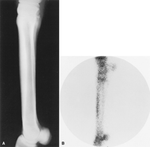
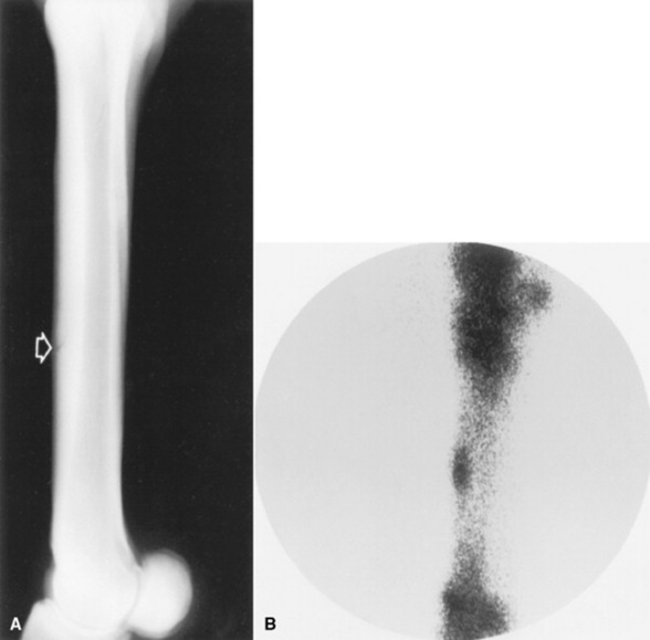
411 Stover SM. Dorsal metacarpal disease in thoroughbred horses: relationship to the development of the third metacarpal bone. [PhD Thesis]: University of California, Davis, 1987.
412 Norwood GL, Haynes PF. Dorsal metacarpal disease. In: Mansmann RA, McAllister ES, editors. Equine medicine and surgery. ed 3. Santa Barbara, Calif: American Veterinary Publications; 1982:1110.
413 Nunamaker DM. Personal communication. 1987.
414 Koblik PD, Hornof WJ, Seeherman HJ. Scintigraphic appearance of stress-induced trauma of the dorsal cortex of the third metacarpal bone in racing thoroughbred horses: 121 cases (1978-1986). J Am Vet Med Assoc. 1988;192:390.
415 Norwood GL. The bucked-shin complex in thoroughbreds. Proc Am Assoc Equine Pract. 1978;24:319.
416 Richardson DW. Dorsal cortical fractures of the equine metacarpus. Compend Cont Educ (Pract Vet). 1984;6:S248.
417 Nunamaker DM, Butterweck DM, Black J. Fatigue fractures in thoroughbred racehorses: relationship with age and strain. Trans Orthop Res Soc. 1987;33:72.
418 Nunamaker DM. The bucked shin complex. Proc Am Assoc Equine Pract. 1987;32:457.
419 Pool RR. Personal communication. 1987.
420 Arthur R. Personal communication. 1988.
421 Rubin CT, Lanyon LE. Regulation of bone mass by mechanical loading: the effect of peak strain magnitude. Calcif Tissue Int. 1985;37:411.
422 Moyer W, Spencer PA, Kallish M. Relative incidence of dorsal metacarpal disease in young thoroughbred racehorses training on two different surfaces. Equine Vet J. 1991;23:166.