CHAPTER 9 Thalamus and limbic system
Basic anatomy OF THE THALAMUS
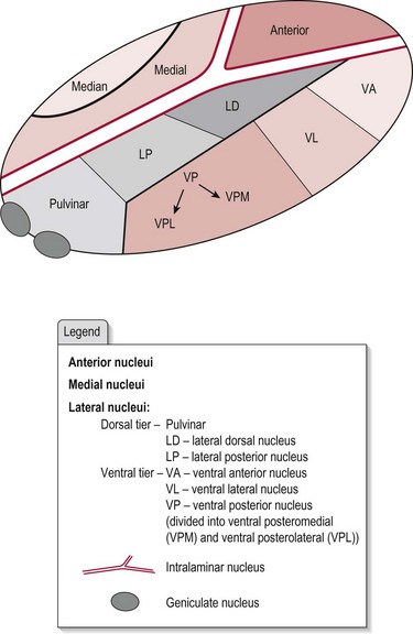
The thalamus is believed to both process and relay sensory information selectively to various parts of the cerebral cortex. The two thalami are prominent bulb-shaped masses about 5.7 cm in length, located on each side of the 3rd ventricle, and form the major part of the diencephalon. The thalamus is composed of a complex system of myelinated neurons separated by distinct clusters of neuron cell bodies, which form specific nuclei. Figure 9.1 shows the anatomical arrangement of the thalamus, with clusters of nuclei being divided into three main parts: anterior, medial and lateral, by the intralaminar nucleus. The lateral part is further sub-divided into dorsal and ventral tiers.
Function of the thalamus
Traditionally, the thalamus was considered to act as a relay station within the central nervous system subserving both sensory and motor mechanisms. However, the extent of its afferent and efferent connections with the periphery and the brain and the mass of interconnections between the thalamic nuclei implies that in addition, it has a significant role in controlling and modifying this information. More precisely, the corticothalamic system appears to synchronize the activity of thalamic and cortical neurons.
As a result of its widespread connections, damage to the thalamus may cause a range of presentations.
Function of the thalamic nuclei
The anterior part
This part is strongly connected to the limbic system (cingulate nucleus and hypothalamus) and is concerned with muscle tone related to our emotional state.
The medial part
Particularly the dorsomedial nucleus has reciprocal connections with the pre-frontal cortex in the frontal lobe, the hypothalamus and all other thalamic nuclei. Its role is the integration of all sensory, motor and visceral information entering the thalamus concerning our emotional state.
The medial and anterior parts also receive information from the spinothalamic tract (pain and temperature), which relates to the ‘medial pain system’ and projects to the anterior cingulate cortex. This area is consistently activated by noxious stimuli and has been associated with the affective–emotional aspect of pain.
The lateral part/dorsal tier
These nuclei connect with other thalamic nuclei and with the lobes of the cerebral cortex (with the exception of the frontal lobe) but little is known of their function.
The lateral part/ventral tier
The ventral lateral and ventral anterior nuclei are connected to the reticular formation, basal ganglia and the pre-motor cortex, hence influencing motor output from the cortex.
The ventral posterior nucleus (posterolateral and posteromedial) receive information related to taste, pain, temperature, and light touch, which is relayed through the posterior limb of the internal capsule to the primary somatosensory cortex. The ‘lateral pain system’ is included here and projects to the primary somatosensory cortex, related to the sensory-discriminative dimension of pain; the secondary somatosensory cortex, related to pain intensity; and to the insula for pain information processing.
Intralaminar nuclei
These nuclei receive connections from the reticular formation and are involved in regulating the level of consciousness and arousal. Neurons also project on to wide areas of the cortex and the basal ganglia.
The limbic system
The limbic system has traditionally been associated with our emotional behaviour, although areas outside the region are also implicated in this function, namely the amygdala and the medial and orbital aspects of the frontal lobe. Although some areas within the limbic system have various other functions, this section will concentrate only on the role of the limbic system in emotional behaviour.
Our emotional behaviour can be considered from two aspects, the control of a behaviour and the actual expression of that behaviour.
Basic anatomy and function of the expression of emotional behaviour
Our emotional states are various but they all possess common attributes, namely visceral and somatic motor responses and powerful subjective feelings. Figure 9.2 shows a summary of this emotional motor response in terms of the level of control and the response intiated.
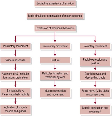
It is interesting that in different emotional states the pattern of the motor response from the autonomic system (involuntary movement) and voluntary movement is emotion specific. Recent studies indicate a strong link between the emotional state, the motor response (especially the facial expression) and the subjective experience.
Involuntary movement
Visceral motor response
This occurs as a result of changes in the autonomic nervous system (ANS), which is distributed throughout the central and peripheral nervous systems. The ANS can alter heart rate, respiratory rate, blood flow, piloerection, sweat gland activity and gastrointestinal motility. These responses occur via changes in smooth muscle, cardiac muscle and various glands throughout the body.
The ANS has two sub-divisions (sympathetic and parasympathetic) which produce the opposite affects on the body. This antagonistic action is essential in maintaining homeostasis (a stable internal environement) and is integrally linked with opposing emotional states.
Sympathetic nervous system
This initiates changes in the heart and lungs, smooth muscle of blood vessels, hair follicles, sweat glands and abdominal organs in preparation for some kind of challenge/threat. The efferent output comes from the spinal cord, the paravertebral ganglia of the sympathetic trunk (Levels T1 – L2/3), to various ganglia/nuclei and onto effector glands/organs.
Parasympathetic nervous system (including the enteric division)
This modifies the action of similar organs to the sympathetic division but its action is directed towards conserving and restoring energy. The efferent output comes from nuclei in the brain stem (cranial nerves III, VII, IX and X) and from the spinal cord grey matter (levels S2/3/4) to various ganglia close to the organs that they innervate and onto the effector glands/organs.
Voluntary movement
Movement related to facial expressions and postures associated with various emotions are clearly recognizable to most people and function as non-verbal communication about our emotional state.
Facial expression
This is achieved by a highly coordinated output from the motor specific facial nerve (VII cranial nerve) (S2.10), which innervates all 20 of the muscles involved in facial expression (e.g. occiptal frontalis, orbicularis oris, buccinator, orbicularis oculi and platysma).
Posture and distal movement
Voluntary changes in posture and fine control of distal movement also occur via voluntary control of the descending tracts (S2.14) to alpha motor neurons at all levels of the spinal cord.
Basic anatomy and function OF the control of emotional behaviour
The control of our emotional behaviour is complex and involves many different structures. The two cerebral hemispheres make different contributions to the control of emotion. First, the right hemisphere is more important for the expression of emotion via the modulation of speech patterns, e.g. when we change the tone or volume of our voice to show anger. Damage to the region would leave the voice monotone and expressionless. Second, the left hemisphere has been found to be more involved with positive moods and the right with negative moods. This has implications in unilateral brain damage, which may result in inappropriate levels of depression (left-side damage) or elation (right-side damage).
The structures involved in control of our emotional behaviour are the:
Hypothalamus
Found in the temporal lobe, the hypothalamus is the centre for coordinating the voluntary and involuntary components of emotional behaviour. The main targets for the hypothalamus lie in the reticular formation which outputs to motor and autonomic effectors in the brain stem and spinal cord.
Cingulate gyrus
The cingulate gyrus lies in the frontal and parietal lobes and is involved in motor planning and regulation of cognitive and emotional processing via its link to the basal ganglia. The selection and initiation of behaviours is especially related to gaining reward and avoiding punishment.
Amygdala
The amygdala is found in the anteromedial temporal lobe and links the cortical regions that process sensory information with the effector systems of the hypothalamus and brain stem. Although it is not part of the limbic system, it has a major role in giving emotional significance to a sensory experience, especially visual stimuli. For example, an individual understanding that a nearby wild bear is associated with fear. Of course, the significance of a sensory experience is highly subjective and therefore specific to each individual. The amygdala can also influence the expression of emotion via both somatic and visceral systems.
Medial and orbital pre-frontal lobe
These regions are linked to the amygdala and integrate a combination of information from vision, somatosensory, olfactory and taste sensations.
Mamillary body
Found in the posterior temporal lobe, the mamillary body connects hypothalamus and cingulate cortex.
Although the following structures are considered to be part of the limbic system they have very little to do with emotional behaviour:
References and Further Reading
Klit H, Finnerup NB, Jensen TS. Central post-stroke pain: clinical characteristics pathophysiology and management. The Lancet. 2009;89:857-868.
Purves D, Augustine GJ, Fitzpatrick D, et al. Neuroscience, ed 4. Sunderland: Sinauer Associates; 2008.
Snell RS. Clinical neuroanatomy, ed 6. Philadelphia: Lippincott Williams and Wilkins; 2006.