9 Acute diarrhoea
Acute diarrhoea is a frequently encountered clinical sign in emergency medicine. The majority of causes of acute diarrhoea relate primarily to the intestinal tract although small intestinal diarrhoea in particular may be associated with extraintestinal causes. There are a large number of causes of acute diarrhoea and some of the most important with respect to emergency cases are listed in Boxes 9.1 and 9.2. Clearly some of these conditions may also cause, or even be more frequently associated with, chronic diarrhoea.
Features used to distinguish small and large intestinal diarrhoea are listed in Box 9.3 but a definitive patient assessment should be made cautiously on the basis of the type of diarrhoea alone and both types may occur in the same patient (e.g. with diffuse inflammatory bowel disease). Animals with haemorrhagic watery diarrhoea are likely to be suffering from small intestinal disease (especially canine haemorrhagic gastroenteritis or parvovirus) but short transit time prevents the formation of melaena (i.e. as there is insufficient time for bacteria to break down haemoglobin).
BOX 9.3 Distinguishing features of small versus large intestinal diarrhoea
Possible iatrogenic causes of diarrhoea as a side effect of drug therapy must also be considered.
Nursing Aspect
Owners quite commonly ring for advice with respect to cats and especially dogs that have diarrhoea and the duty nurse is often the person from whom this initial advice is obtained. Acute diarrhoea is often the result of dietary indiscretion or food intolerance and may be self-limiting without the need for veterinary intervention. The recommendation to examine the animal at the clinic should be guided by the owner’s wishes but also after obtaining adequate information with respect to:
In the author’s opinion it is reasonable to recommend withholding food for a short period (e.g. 12–24 hours) with continued monitoring at home if the animal remains relatively bright, is tolerant of water, and especially where there is a suspicion of dietary indiscretion not relating to a foreign body. An easily digestible food can then be introduced, feeding small amounts frequently. Owners should be advised to contact the clinic again should the animal’s condition deteriorate, at which time examination can be arranged.
Approach to Diarrhoea
Signalment
Signalment may allow certain differential diagnoses to be viewed with a lesser or greater index of suspicion. For example, younger animals are more likely to present with dietary indiscretion, foreign body ingestion, or infectious diarrhoea including intestinal helminthiasis.
History
A thorough history is mandatory in all animals with diarrhoea, although the timing should be guided by the stability of the patient. It is important to try to determine from the owner’s description whether small or large intestinal diarrhoea is present. The history may provide useful information with respect to the possible cause of the diarrhoea (e.g. witnessed scavenging, change of diet, other animals affected), and the nature, severity and duration of the diarrhoea will help establish the likelihood of clinically significant dehydration. In addition, the presence of other concurrent signs may raise the index of suspicion for systemic diseases. Worming and vaccination history should also be established as well as details of any recent drug therapy.
Major body system examination
Cardiovascular examination may be normal in animals with diarrhoea or may be consistent with variable degrees of hypovolaemia (see Ch. 2). Hypovolaemia may be secondary to severe dehydration and/or a consequence of the primary disorder. Signs of maldistributive shock (hypoperfusion with hyperaemic mucous membranes and a rapid capillary refill time) should raise the index of suspicion for severe acute pancreatitis or a septic focus. Respiratory examination may be unremarkable in animals with diarrhoea or may reflect pain, pyrexia or abnormalities in acid–base status. Neurological examination may also be unremarkable but abnormalities may be detected that reflect the underlying cause (e.g. seizuring from intoxication).
Abdominal palpation is very important but may be more or less rewarding depending on the size and demeanour of the patient in question. Abdominal pain may be identified and potentially characterized as diffuse or localized. Abdominal pain is not likely to be present with a number of the causes of diarrhoea and its presence may raise the index of suspicion for certain differential diagnoses (e.g. intestinal obstruction, foreign body ingestion, acute infectious enteritis, acute pancreatitis). In some cases focal abnormalities such as an intussusception, foreign body or mass may be identified.
Rectal temperature may be normal, increased or decreased depending on the underlying cause and clinical status of the patient. Digital rectal examination should also be performed and may allow visual examination of faeces, collection of faeces for analysis, and identification of local rectal lesions.
Complete physical examination performed at the appropriate time may reveal other significant findings that can help establish a diagnosis.
Emergency database
The need to perform an emergency database in a diarrhoeic patient should be guided by the animal’s history and physical examination findings. The emergency database will allow the patient to be evaluated for dehydration (raised packed cell volume and serum total solids, prerenal azotaemia – see Ch. 3) as well as identify electrolyte and acid–base abnormalities. Some animals with diarrhoea will have a protein-losing enteropathy (PLE) and serum total solids may appear inappropriately low in comparison with packed cell volume in such cases. Dogs with hypoadrenocorticism (Addison’s disease) may have hyponatraemia, hyperkalaemia and mild to moderate azotaemia. A faecal ELISA test for canine parvovirus may be appropriate in young dogs.
Clinical Tip
More extensive clinicopathological testing may be required in some diarrhoeic animals, especially in those with other systemic signs, but is generally not available in the emergency setting.
Diagnostic imaging
The need to perform diagnostic imaging in a diarrhoeic animal should be guided by the animal’s history and physical examination findings. In the routine emergency setting, diagnostic imaging of the emergency patient is likely to consist of plain radiography and, if available, abdominal ultrasonography for the detection of free peritoneal fluid. Ideally orthogonal (right lateral and ventrodorsal views) abdominal radiographs should be taken and are most useful for identifying radioopaque intestinal foreign bodies and obstructions. Thoracic radiographs may be indicated to look for pulmonary metastases where neoplasia is considered a likely cause of diarrhoea.
Treatment
By collating all of the information obtained from the steps above, the clinician should be able to decide on the most appropriate management for the patient in question. This will depend not only on the underlying cause but also on the clinical status of the patient with respect to perfusion, hydration and electrolyte and acid–base abnormalities.
Acute diarrhoea is often self-limiting and may only require supportive care and short-term dietary management without a definitive diagnosis. Withholding food for a period of time (e.g. 12–24 hours) and then offering small amounts of an easily digestible diet may be all that is required. The use of specific antidiarrhoeal therapy is typically not necessary and may be contraindicated in some cases (especially diarrhoea of infectious origin).
Cases in which diarrhoea is severe or there are systemic signs such as pyrexia or neutropenia may require more aggressive therapy and investigations. Amongst the differential diagnoses for diarrhoea, the most severe clinical presentations are associated with infectious enteritides, intestinal obstruction, foreign body ingestion, canine haemorrhagic gastroenteritis, acute pancreatitis and hypoadrenocorticism. See Chapter 29 (The acute abdomen) and Chapter 34 (Endocrine emergencies) for further information on these conditions.
Clinical Tip
Haematochezia
Haematochezia refers to bright red fresh blood either on the surface of or mixed in with faeces, and this must be differentiated from melaena (see below). The source of the blood is typically the anus, rectum or descending colon (see Box 9.4). Haematochezia often appears dramatic to owners but is typically not associated with significant morbidity and is rarely associated with mortality. In many cases, it is the only clinical sign present and conservative management using a diet containing fermentable fibre may be all that is needed. Empirical deworming using a broad-spectrum preparation may be appropriate as may a course of metronidazole. More intensive management and extensive investigations may be required in animals with systemic illness or persistent haematochezia.
Melaena
Clinical Tip
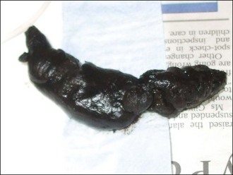
Melaena refers to dark tarry faeces that represents blood digested in the intestinal tract and is a much more serious clinical sign than haematochezia (Figure 9.1). Melaena may result from lesions in the gastrointestinal tract and is usually the result of gastric or proximal small intestinal haemorrhage. However, melaena may occur due to haemorrhage more distally in the intestinal tract. Importantly, melaena may also be the result of ingestion of blood or serious abnormalities in other organs or body systems (see Box 9.5).
Animals with melaena can suffer considerable blood loss resulting in mild to severe reduction in packed cell volume and serum total solids. Depending on the duration, anaemia due to acute melaena may be regenerative or preregenerative and peripheral blood smear examination is essential in these cases (see Ch. 3). Chronic melaena may result in hypochromic microcytic iron-deficiency anaemia. Peripheral blood smear examination is also essential to evaluate platelet numbers as melaena is relatively common in animals with sufficiently severe thrombocytopenia.
History and additional physical examination findings should help determine whether a primary gastrointestinal cause is more or less likely and therefore guide diagnostics. Treatment is ultimately aimed at the underlying cause but intensive supportive measures, including transfusion of appropriate blood products, may be required.