26 The Neck, Back, and Tail of the Ruminant
CONFORMATION AND SURFACE FEATURES
The back and loins are shaped over the framework of the thoracic and lumbar vertebrae. The loins are sharply divided from the flanks by the prominent tips of the lumbar transverse processes, but the boundaries of the back cannot be defined so precisely because the back blends smoothly with the lateral thoracic wall and incorporates the upper line of the shoulder blades with their cartilages and covering muscles. It is convenient to include in this chapter the few observations that are necessary on the dorsal sacral region, which merges with the quarters and root of the tail.
In the animal standing quietly, the dorsal contour is slightly raised over the withers, but otherwise it follows a fairly straight line from immediately behind the skull to the tail root (Figure 26–1).* The line of the neck, which is based on the funicular part of the nuchal ligament, varies of course with the carriage of the head.
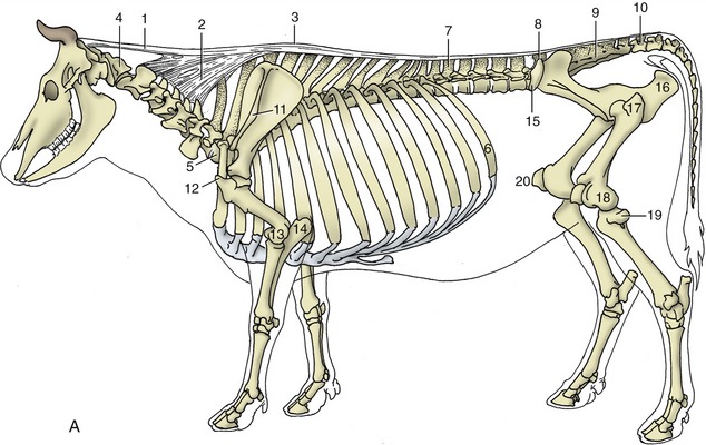
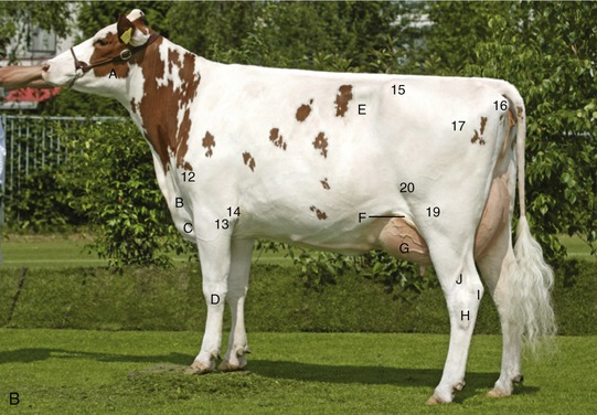
Figure 26–1 A, Skeleton with nuchal and supraspinous ligaments; most labeled parts are palpable. B, Cow in good condition. 1, 2, Nuchal ligament; 1, funiculus nuchae; 2, lamina nuchae; 3, supraspinous ligament; 4, atlas; 5, last cervical vertebra (C7); 6, thirteenth rib; 7, first lumbar vertebra (L1); 8, last lumbar vertebra (L6); 9, sacrum; 10, first caudal vertebra; 11, spine of scapula; 12, greater tubercle; 13, 14, palpable features at elbow joint; 13, lateral epicondyle; 14, olecranon; 15, coxal tuber; 16, ischial tuber; 17, greater trochanter; 18, 19, 20, palpable features of stifle joint; 18, lateral condyle of femur ; 19, lateral condyle of tibia and remnant of fibula; 20, patella. A, Masseter; B, jugular vein; C, brisket; D, carpus; E, paralumbar fossa; F, flank fold; G, udder; H, hock joint; I, calcaneus (point of the hock); J, lateral saphenous vein.
The dorsal contour of the trunk is prescribed by the summits of the spinous processes of the vertebrae, many of which can be palpated separately. Identification of individual bones is most reliable if begun at the wide space between the upright process of the last lumbar vertebra and the sloping cranial margin of the median sacral crest. The sacral crest can be followed caudally until it is succeeded by the separate projections of the spinous processes of the caudal vertebrae; any doubt about the identity of these processes may be resolved by pumping the tail up and down to discover the very mobile joint between the first and second tail bones. Certainty in identifying the first intercaudal space has a special importance because this is the site for injection of local anesthetic when producing “low” epidural anesthesia (p. 667). The tail root is sometimes elevated, especially in cows during estrus.
Working cranially from the lumbosacral space, the lumbar spinous processes are easily distinguished in lean animals. Enumeration becomes more difficult over the caudal part of the chest where several processes converge, and the count is completely lost where the vertebrae become enclosed between the scapular cartilages. The first thoracic spine lies cranial to the scapulae, where it can be felt on deep palpation even though it fails to approach close to the skin. The cervical vertebrae cannot be reached from above, but their general position is detectable on palpation from the side. The transverse processes are well developed and divided into two parts, of which the ventral one is quite large; this is very obvious at the sixth cervical vertebra. Despite this, the individual identification of these bones is difficult until the wing of the atlas provides an unmistakable landmark.
Additional features that may be picked out in the region of the hindquarters include the salient sacral tubers of the pelvis, which lie to each side of the lumbosacral space, and the strong iliac crests, which join these projections to the coxal tubers. The crests are raised above their surroundings and are crossed by cranial prolongations of the gluteal musculature.
The head is carried higher in sheep and goats; these species also slope at the croup (Figure 26–2).
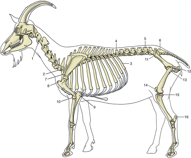
Figure 26–2 The skeleton of the goat. Most labeled parts of the skeleton are palpable. 1, Atlas; 2, last cervical vertebra (C7); 3, last rib; 4, first lumbar vertebra (L1); 5, last lumbar vertebra (L7); 6, sacrum; 7, acromion; 8, greater tubercle; 9, olecranon; 10, lateral epicondyle; 11, coxal tuber; 12, ischial tuber; 13, greater trochanter; 14, patella; 15, lateral condyle of tibia; 16, calcaneus.
THE VERTEBRAL COLUMN
The vertebral axis runs parallel to the skin line in the loins and caudal part of the back; however, more cranially it is deflected ventrally. It reaches its lowest level at the entrance to the thorax; an abrupt flexure there places it on a path that gradually returns closer to the dorsal border as it ascends the neck (see Figure 26–1).
The vertebral skeleton and articulations follow the usual pattern, and few features need be mentioned. The vertebral formula is C7, T13, L6, S5, Cd18–20 in cattle; C7, T13, L6(7), S4 in sheep or S5 in goats; and Cd16–18 in both small ruminants. The great mobility of the neck allows the animal to raise and lower its head and to reach its side with its tongue. Most cervical movements represent the summation of small changes at several joints, but the adoption of the grazing position requires a considerable straightening at the cervicothoracic joint, where the neck vertebrae are brought into line with those of the chest. Although movements of the thoracic region are limited by the presence of the rib cage, the greatest flexibility of the trunk is found cranial to the level of the diaphragm. Behind this, movement is greatly restricted, especially in the lateral direction, by the close fit of the articular processes and the tightness of the capsules that embrace them. Greater mobility is again found at the lumbosacral joint.
The generally rather limited flexibility of the spine is suggested by the relative shortness of the intervertebral disks, which in cattle contribute only 10% of the length of the column. The disks have the usual construction and are subject to the same degenerative changes as occur in other species. The lumbosacral disk is most commonly grossly damaged because of the greater stress to which it is subjected by the special mobility of the lumbosacral articulation. Disk lesions are sometimes accompanied by changes in the lumbosacral synovial articulations and by the formation of abnormal bony outgrowths (osteophytes) from the ventral margins of the vertebral bodies. Certain of these common changes have a particular importance in bulls because they may lead to an inability to serve.
The elastic nuchal ligament (Figure 26–1/1,2) consists of two parts, as in the horse. The funicular part, which runs between the occiput and the highest spines of the withers, is a paired cord that is rounded in cross section at its occipital attachment but widens as it passes caudally. It attaches to the sides of the first few thoracic spines, close to their summits; caudal to this, it approaches and fuses with its fellow to form the supraspinous ligament that caps the bone processes. The rhomboideus and trapezius muscles cover the funicular part of the ligament, in contrast to the arrangement in the horse (see Figure 25–23/1). The laminar part is divided into a cranial paired web that extends between the funicular part and the second to the fourth cervical bones and an unpaired sheet that fills the triangle between the first thoracic and last one or two cervical spinous processes. In addition to relieving the cervical muscles, the nuchal ligament has an occasional significance in determining the track followed by infection. No cranial nuchal bursa exists, but a supraspinous bursa frequently is present between the ligament and the first few thoracic spinous processes.
THE VERTEBRAL CANAL
The vertebral canal is widest within the atlas and tapers rapidly within the sacrum; in between, it is most expanded where it contains the swellings of the spinal cord that give rise to the nerves that form the limb plexuses. Access to the vertebral canal is frequently necessary to withdraw cerebrospinal fluid from the subarachnoid space or to introduce local anesthetic into the epidural space. Therapeutic agents are also occasionally injected into these spaces. Examination of the skeleton shows that although entry is theoretically possible through any of the interarcuate spaces, it will be easiest at the wider gaps between the atlas and the skull, at the lumbosacral joint, and between the first two vertebrae of the tail (see Figure 26–3). The first intercaudal space is conveniently large, measuring about 2 × 2 cm. Most other interarcuate spaces measure only a few millimeters in each direction, and because they lie at a considerable depth below the skin, they are not easily located. Epidural injections through the cranial (especially the first) interlumbar interarcuate spaces are occasionally made to obtain local anesthesia of the flank. A slightly oblique approach from a point of entry a little lateral and caudal to the target space gives the least risk of the needle impinging on bone.

Figure 26–3 Caudal part of the bovine vertebral canal and its contents, schematic. Epidural injection sites are indicated by the needles. 1, First lumbar vertebra; 1′, needle in position for flank anesthesia; 2, last lumbar vertebra (L6); 3, sacrum; 4, needle in lumbosacral space; 5, first caudal vertebra; 6, needle between first and second caudal vertebrae (tail block); 7, epidural space; 8, dura mater; 9, subarachnoid space; 10, spinal cord; 11, central canal; 12, intervertebral disk.
The cord reaches to the first sacral vertebra in adult cattle and considerably farther in young calves, perhaps into the caudal half of the sacrum. It may occupy almost the whole sacrum in the small ruminant species.
It is divided into 8 cervical, 13 thoracic, 6 lumbar, 5 sacral, and (usually) 5 caudal segments. The eight cervical segments are accommodated within the seven neck vertebrae, while each of the thoracic and cranial lumbar segments shows an almost exact correspondence with the bone of the same designation. The cranial shift of the more caudal part of the cord leaves the canal within the last lumbar vertebra available for occupation by the five short and telescoped sacral segments (Figure 26–4). The subarachnoid space extends well into the sacrum, and its dimensions are sufficiently generous to make subarachnoid puncture a relatively simple procedure at the lumbosacral level (Figure 26–3/4).
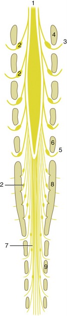
Figure 26–4 The relationship to the vertebrae of the caudal end of the spinal cord and its branches, schematic dorsal view. Note the position of the spinal ganglia (2). The schema indicates the situation in adult cattle. The cord extends to the second or even third sacral vertebra in the newborn calf and in adult sheep and goats. 1, Spinal cord; 2, spinal ganglia; 3, second lumbar spinal nerve; 4, section of arch of second lumbar vertebra; 5, sixth lumbar nerve; 6, section of arch of sixth lumbar vertebra; 7, cauda equina; 8, section of sacrum; 9, section of arch of second caudal vertebra.
The internal vertebral plexus (Figure 26–5/1) presents two features of potential interest. The first involves the possibility of the plexus conveying blood diverted from the caudal vena cava when this is narrowed or obstructed by ruminal tympany; compression of the vena cava may be direct or exerted indirectly by a shearing displacement of the liver against the diaphragm (Figure 26–6). The second significant feature involves the risk of hemorrhage in the performance of subarachnoid or epidural puncture.

Figure 26–5 Dorsal view of the venous drainage in the bovine vertebral canal. The internal vertebral plexus, with its internal connections and its lateral segmental branches, has been exposed. 1, Internal vertebral plexus; 2, intervertebral veins; 3, intervertebral disk; 4, vertebral body.
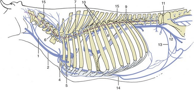
Figure 26–6 The connections of the major veins with the vertebral plexus–azygous system. Note specifically the connections between the internal vertebral plexus (15) and the intercostal veins (10) and between the plexus and the branches of the vertebral vein (6). 1, Internal jugular v.; 2, external jugular v.; 3, occipital v.; 4, axillary v.; 5, cranial vena cava; 6, vertebral v.; 7, supreme intercostal v.; 8, left azygous v.; 9, caudal vena cava; 10, intercostal vv.; 11, internal iliac v.; 12, external iliac v.; 13, deep circumflex iliac v.; 14, cranial epigastric v.; 15, internal vertebral plexus (red).
THE VESSELS OF THE TAIL
The median artery and vein of the tail require brief notice. The artery, which continues the median sacral, is ventral to the vein for most of the length of the tail and is commonly used for pulse taking; the usual site is about 18 cm from the root of the tail. The vessels lie side by side in the proximal part of the tail (Cd2 or Cd3), where both artery and vein are available for obtaining blood, although this site is an unwise choice because of the inevitable fecal contamination (Figure 26–7, B). At this level both vessels lie against the ventral aspect of the caudal vertebrae, where they are protected by the hemal processes (Figure 26–7, A) arches on the first few vertebrae (see Figure 2–12, E/9). The vessels are thus accessible only at intervertebral levels. It is usual to dock the tail of lambs.