9 The Permanent Maxillary Premolars
The maxillary premolars number four: two in the right maxilla and two in the left maxilla. They are posterior to the canines and immediately anterior to the molars.
The premolars are so named because they are anterior to the molars in the permanent dentition. In zoology, the premolars are those teeth that succeed the deciduous molars regardless of the number to be succeeded. The term bicuspid, which is widely used to describe human teeth, presupposes two cusps, a supposition that makes the term misleading, because mandibular premolars in the human subject may show a variation in the number of cusps from one to three. Among carnivores, in the study of comparative dental anatomy, premolar forms differ so greatly that a more descriptive single term than premolar is out of the question. The term premolar is used widely in dental anatomy, human and comparative; therefore it will be used here. However, its use here does not suggest that the term bicuspid should not be used when appropriate.
The maxillary premolars are developed from the same number of lobes as anterior teeth—four. The primary difference in development is the well-formed lingual cusp, developed from the lingual lobe, which is represented by the cingulum development on incisors and canines. The middle buccal lobe on the premolars, corresponding to the middle labial lobe of the canines, remains highly developed, with the maxillary premolars resembling the canines when viewed from the buccal aspect. The buccal cusp of the maxillary first premolar, especially, is long and sharp, assisting the canine as a prehensile or tearing tooth. The mandibular first premolar assists the mandibular canine in the same manner.
The second premolars, both maxillary and mandibular, have cusps less sharp than the others, and their cusps articulate with opposing teeth when the jaws are brought together; this makes them more efficient as grinding teeth, and they function much like the molars, but to a lesser degree.
The maxillary premolar crowns are shorter than those of the maxillary canines, and the roots are also shorter. The root lengths equal those of the molars. The crowns are a little longer than those of the molars.
Because of the cusp development buccally and lingually, the marginal ridges are in a more horizontal plane and are considered part of the occlusal surface of the crown rather than part of the lingual surface, as in the case of incisors and canines.
When premolars have two roots, one is placed buccally and one lingually.
Maxillary First Premolar
Figures 9-1 through 9-16 illustrate the maxillary first premolar from all aspects. The maxillary first premolar has two cusps, a buccal and a lingual, each being sharply defined. The buccal cusp is usually about 1 mm longer than the lingual cusp. The crown is angular, and the buccal line angles are prominent.
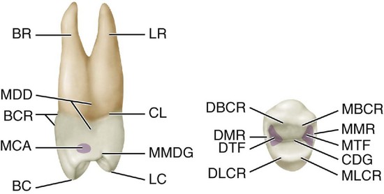
Figure 9-1 Maxillary right first premolar, mesial and occlusal aspects. LR, Lingual root; CL, cervical line; MMDG, mesial marginal developmental groove; LC, lingual cusp; BC, buccal cusp; MCA, mesial contact area; BCR, buccal cervical ridge; MDD, mesial developmental depression; BR, buccal root; MBCR, mesiobuccal cusp ridge; MMR, mesial marginal ridge; MTF, mesial triangular fossa (shaded area); CDG, central developmental groove; MLCR, mesiolingual cusp ridge; DLCR, distolingual cusp ridge; DTF, distal triangular fossa; DMR, distal marginal ridge; DBCR, distobuccal cusp ridge.
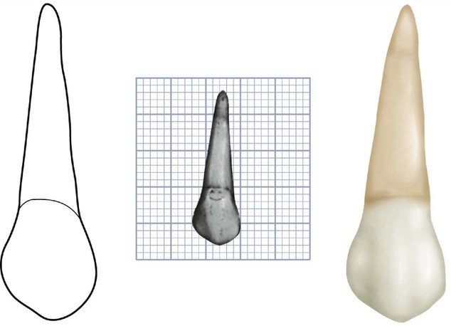
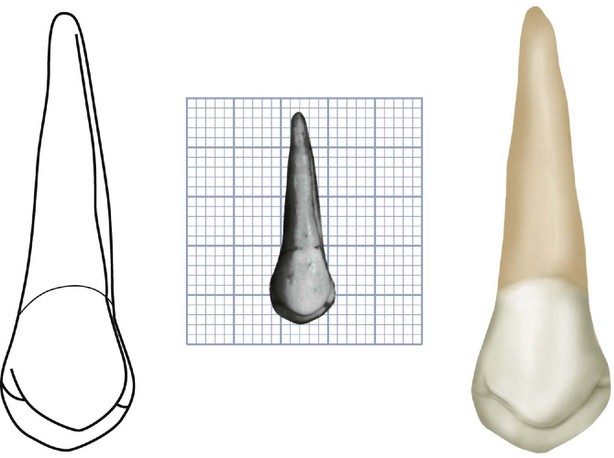
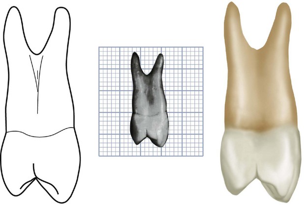
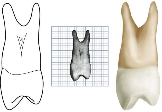
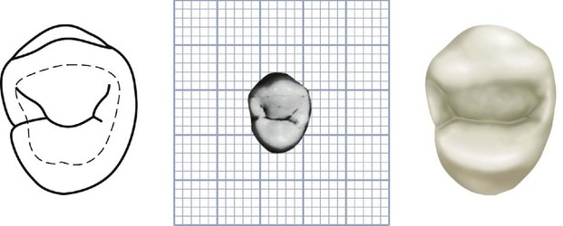
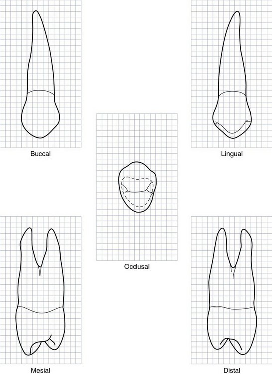
Figure 9-7 Maxillary right first premolar. Graph outlines of five aspects are shown. (Grid = 1 sq mm.)
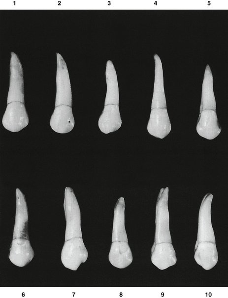
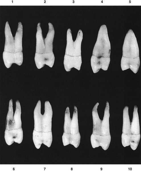
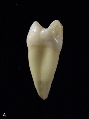
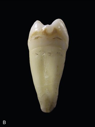
Figure 9-11 A, Mesial view of a maxillary right first premolar demonstrating single root and mesial crown concavity. B, Mesial view of a maxillary right second premolar demonstrating single root and no mesial crown concavity.
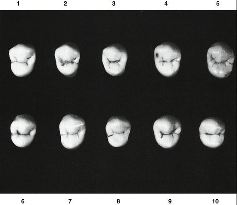
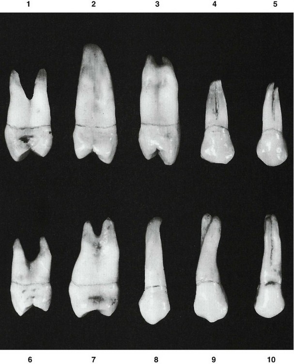
Figure 9-13 Maxillary first premolar. Ten specimens with uncommon variations are shown. 1, Constricted occlusal surface; short roots. 2, Single root of extreme length. 3, Constricted occlusal surface; mesial developmental groove indistinct on mesial surface of root. 4, Short root form, with two buccal roots fused. 5, Short root form, with two buccal roots showing bifurcation. 6, Short roots, with considerable separation. 7, Buccolingual calibration greater than usual. 8, Root extremely long; distal contact area high. 9, Twisted buccal root. 10, Three roots fused; roots are also uncommonly long.
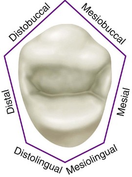
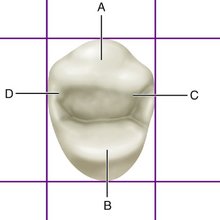
Figure 9-15 Maxillary first premolar, occlusal aspect. A, Crest of buccal ridge; B, crest of lingual ridge; C, crest of mesial contact area; D, crest of distal contact area.
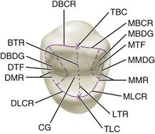
Figure 9-16 Maxillary first premolar, occlusal aspect. TBC, Tip of buccal cusp; MBCR, mesiobuccal cusp ridge; MBDG, mesiobuccal developmental groove; MTF, mesial triangular fossa; MMDG, mesial marginal developmental groove; MMR, mesial marginal ridge; MLCR, mesiolingual cusp ridge; LTR, lingual triangular ridge; TLC, tip of lingual cusp; CG, central groove; DLCR, distolingual cusp ridge; DMR, distal marginal ridge; DTF, distal triangular fossa; DBDG, distobuccal developmental groove; BTR, buccal triangular ridge; DBCR, distobuccal cusp ridge. (Compare with Figure 9-1.)
The crown is shorter than that of the canine by 1.5 to 2 mm on the average (Table 9-1). Although this tooth resembles the canine from the buccal aspect, it differs in that the contact areas mesially and distally are at about the same level. The root is shorter. If the buccal cusp form has not been changed by wear, the mesial slope of the cusp is longer than the distal slope. The opposite arrangement is true of the maxillary canine. Generally, the first premolar is not as wide in a mesiodistal direction as the canine.
Most maxillary first premolars have two roots (see Figure 9-10) and two pulp canals. When only one root is present, two pulp canals are usually found anyway.
The maxillary first premolar has some characteristics common to all posterior teeth. Briefly, those characteristics that differentiate posterior teeth from anterior teeth are as follows:
DETAILED DESCRIPTION OF THE MAXILLARY FIRST PREMOLAR FROM ALL ASPECTS
Buccal Aspect
From the buccal aspect, the crown is roughly trapezoidal (see Figure 4-16, C). The crown exhibits little curvature at the cervical line. The crest of curvature of the cervical line buccally is near the center of the root buccally (see Figure 9-2 and Figures 9-7 through 9-9).
The mesial outline of the crown is slightly concave from the cervical line to the mesial contact area. The contact area is represented by a relatively broad curvature, the crest of which lies immediately occlusal to the halfway point from the cervical line to the tip of the buccal cusp.
The mesial slope of the buccal cusp is rather straight and longer than the distal slope, which is shorter and more curved. This arrangement places the tip of the buccal cusp distal to a line bisecting the buccal surface of the crown. The mesial slope of the buccal cusp is sometimes notched; in other instances, a concave outline is noted at this point (see Figure 9-9, 7, 9, and 10).
The distal outline of the crown below the cervical line is straighter than that of the mesial, although it may be somewhat concave also. The distal contact area is represented by a broader curvature than is found mesially, and the crest of curvature of the contact area tends to be a little more occlusal when the tooth is posed with its long axis vertical. Even so, the contact areas are more nearly level with each other than those found on anterior teeth.
The width of the crown of the maxillary first premolar mesiodistally is about 2 mm less at the cervix than at its width at the points of its greatest mesiodistal measurement.
The buccal cusp is long, coming to a pointed tip and resembling the canine in this respect, although contact areas in this tooth are near the same level.
The buccal surface of the crown is convex, showing strong development of the middle buccal lobe. The continuous ridge from cusp tip to cervical margin on the buccal surface of the crown is called the buccal ridge.
Mesial and distal to the buccal ridge, at or occlusal to the middle third, developmental depressions are usually seen that serve as demarcations between the middle buccal lobe and the mesiobuccal and distobuccal lobes. Although the latter lobes show less development, they are nevertheless prominent and serve to emphasize strong mesiobuccal and distobuccal line angles on the crown.
The roots are 3 or 4 mm shorter than those of the maxillary canine, although the outline of the buccal portion of the root form bears a close resemblance.
Lingual Aspect
From the lingual aspect, the gross outline of the maxillary first premolar is the reverse of the gross outline from the buccal aspect (see Figures 9-3, 9-7, and 9-8).
The crown tapers toward the lingual because the lingual cusp is narrower mesiodistally than the buccal cusp. The lingual cusp is smooth and spheroidal from the cervical portion to the area near the cusp tip. The cusp tip is pointed, with mesial and distal slopes meeting at an angle of about 90 degrees.
Naturally, the spheroidal form of the lingual portion of the crown is convex at all points. Sometimes the crest of the smooth lingual portion that terminates at the point of the lingual cusp is called the lingual ridge.
The mesial and distal outlines of the lingual portion of the crown are convex; these outlines are continuous with the mesial and distal slopes of the lingual cusp and straighten out as they join the mesial and distal sides of the lingual root at the cervical line.
The cervical line lingually is regular, with slight curvature toward the root and the crest of curvature centered on the root. Because the lingual portion of the crown is narrower than the buccal portion, it is possible to see part of the mesial and distal surfaces of crown and root from the lingual aspect, depending on the posing of the tooth and the line of vision.
Because the lingual cusp is not as long as the buccal cusp, the tips of both cusps, with their mesial and distal slopes, may be seen from the lingual aspect.
The lingual portion of the root, or the lingual portion of the lingual root if two roots are present, is smooth and convex at all points. The apex of the lingual root of a two-root specimen tends to be more blunt than the buccal root apex.
Mesial Aspect
The mesial aspect of the crown of the maxillary first premolar is also roughly trapezoidal (see Figures 9-1, 9-4, 9-7, 9-8, and 9-10). However, the longest of the uneven sides is toward the cervical portion and the shortest is toward the occlusal portion (see Figure 4-16, E).
Another characteristic that is representative of all posterior maxillary teeth is that the tips of the cusps are well within the confines of the root trunk. (For a definition of root trunk, see Figure 11-3Figure 11-8.) In other words, the measurement from the tip of the buccal cusp to the tip of the lingual cusp is less than the buccolingual measurement of the root at its cervical portion.
Most maxillary first premolars have two roots, one buccal and one lingual; these are clearly outlined from the mesial aspect.
The cervical line may be regular in outline (see Figure 9-10, 1) or irregular (see Figure 9-10, 4). In either case, the curvature occlusally is less (about 1 mm on the average) than the cervical curvature on the mesial of any of the anterior teeth. The extent of the curvature of the cervical line mesially on these teeth is constant within a fraction of a millimeter and is similar to the average curvature of the mesial of all posterior teeth.
From the mesial aspect, the buccal outline of the crown curves outward below the cervical line. The crest of curvature is often located approximately at the junction of cervical and middle thirds; however, the crest of curvature may be located within the cervical third (see Figure 9-10, 1 and 10). From the crest of curvature, the buccal outline continues as a line of less convexity to the tip of the buccal cusp, which is directly below the center of the buccal root (when two roots are present).
The lingual outline of the crown may be described as a smoothly curved line starting at the cervical line and ending at the tip of the lingual cusp. The crest of this curvature is most often near the center of the middle third. Some specimens show a more abrupt curvature at the cervical third (see Figure 9-10, 2 and 9).
The tip of the lingual cusp is on a line, in most cases, with the lingual border of the lingual root. The lingual cusp is always shorter than the buccal cusp, the average difference being about 1 mm. This difference, however, may be greater (see Figure 9-10, 1, 4, and 10). From this aspect, it is noted that the cusps of the maxillary first premolar are long and sharp, with the mesial marginal ridge at about the level of the junction of the middle and occlusal thirds.
A distinguishing feature of this tooth is found on the mesial surface of the crown. Immediately cervical to the mesial contact area, centered on the mesial surface and bordered buccally and lingually by the mesiobuccal and mesiolingual line angles, is a marked depression called the mesial developmental depression (see Figure 9-1). This mesial concavity continues apically beyond the cervical line, joins a deep developmental depression between the roots, and ends at the root bifurcation. On single-root specimens, the concavity on the crown and root is also plainly seen, although it may not be as deeply marked. Maxillary second premolars do not have this feature (see Figure 9-11, A and B).
Another distinguishing feature of the maxillary first premolar is a well-defined developmental groove in the enamel of the mesial marginal ridge. This groove is in alignment with the developmental depression on the mesial surface of the root but is not usually connected with it. This marginal groove is continuous with the central groove of the occlusal surface of the crown, crossing the marginal ridge immediately lingual to the mesial contact area and terminating a short distance cervical to the mesial marginal ridge on the mesial surface (see Figure 9-10, 10).
The buccal outline of the buccal root, above the cervical line, is straight, with a tendency toward a lingual inclination. On those buccal roots having a buccal inclination above the root bifurcation, the outline may be relatively straight up to the apical portion of the buccal root or it may curve buccally at the middle third. Buccal roots may take a buccal or lingual inclination, apical to middle thirds.
The lingual outline of the lingual root is rather straight above the cervical line. It may not exhibit much curvature between the cervix and the apex. Many cases, however, show considerable curvature to lingual roots apical to the middle thirds. The lingual root may take a buccal or lingual inclination (see Figure 9-10, 1, 2, and 9).
The root trunk is long on this tooth, making up about half of the root length. The bifurcation on those teeth with two roots begins at a more occlusal point mesially than distally. Generally speaking, when bifurcated, the root is bifurcated for half its total length.
Except for the deep developmental groove and depression at or below the bifurcation, the mesial surface of the root portion of this tooth is smoothly convex buccally and lingually. Even when only one root is present, the developmental depression is very noticeable for most of the root length. In such cases, roots with buccal and lingual outlines ending in a blunt apex above the center of the crown are seen (see Figure 9-10, 4 and 5).
Distal Aspect
From the distal aspect (see Figures 9-5, 9-7, and 9-8), the anatomy of the crown and root of the maxillary first premolar differs from that from the mesial aspect as follows:
Occlusal Aspect
The occlusal aspect of the maxillary first premolar resembles roughly a six-sided or hexagonal figure (see Figures 9-7, 9-8, 9-12, and 9-14). The six sides are made up of the mesiobuccal, which is mesial to the buccal ridge; mesial; mesiolingual, which is mesial to the lingual ridge; distolingual; distal; and distobuccal. This hexagonal figure, however, is not equilateral. The two buccal sides are nearly equal, the mesial side is shorter than the distal side, and the mesiolingual side is shorter than the distolingual side (see Figure 9-14).
The relation and position of various anatomical points are to be considered from the occlusal aspect. A drawing of the outline of this occlusal aspect, when placed within a rectangle the dimensions of which represent the mesiodistal and buccolingual width of the crown, demonstrates the relative positions of the mesial and distal contact areas and also those of the buccal and lingual ridges (see Figures 9-6 and 9-15).
The crest of the distal contact area is somewhat buccal to that of the mesial contact area, and the crest of the buccal ridge is somewhat distal to that of the lingual ridge. The crests of curvature represent the highest points on the buccal and lingual ridges and the mesial and distal contact areas.
Close observation of the crown from this aspect reveals the following characteristics (see Figure 9-15):
The occlusal surface of the maxillary first premolar is circumscribed by the cusp ridges and marginal ridges. The mesiobuccal and distobuccal cusp ridges are in line with each other, and their alignment is in a distobuccal direction. In other words, even though they are in the same alignment, the distobuccal cusp ridge is buccal to the mesiobuccal cusp ridge (see Figure 9-16).
The angle formed by the convergence of the mesiobuccal cusp ridge and the mesial marginal ridge approaches a right angle. The angle formed by the convergence of the distobuccal cusp ridge and the distal marginal ridge is acute. The mesiolingual and distolingual cusp ridges are confluent with the mesial and distal marginal ridges; these cusp ridges are curved, following a semicircular outline from the marginal ridges to their convergence at the tip of the lingual cusp.
When one looks at the occlusal aspect of the maxillary first premolar, posing the tooth so that the line of vision is in line with the long axis, more of the buccal surface of the crown is seen than of the lingual surface. When the tooth is viewed from the mesial aspect, the tip of the buccal cusp is nearer the center of the root trunk than is the lingual cusp.
The occlusal surface of this tooth has no supplemental grooves in most cases, a fact that makes the surface relatively smooth. A well-defined central developmental groove divides the surface evenly buccolingually. It is located at the bottom of the central sulcus of the occlusal surface, extending from a point just mesial to the distal marginal ridge to the mesial marginal ridge, where it joins the mesial marginal development groove. This latter groove crosses the mesial marginal ridge and ends on the mesial surface of the crown (see Figures 9-1 and 9-4).
Two collateral developmental grooves join the central groove just inside the mesial and distal marginal ridges. These grooves are called the mesiobuccal developmental groove and the distobuccal developmental groove. The junctions of the grooves are deeply pointed and are named the mesial and distal developmental pits.
Just distal to the mesial marginal ridge, the triangular depression that harbors the mesiobuccal developmental groove is called the mesial triangular fossa. The depression in the occlusal surface, just mesial to the distal marginal ridge, is called the distal triangular fossa.
Although no supplemental grooves are present in most instances, smooth developmental depressions may be visible radiating from the central groove and giving the occlusal surface an uneven appearance.
The buccal triangular ridge of the buccal cusp is prominent, arising near the center of the central groove and converging with the tip of the buccal cusp. The lingual triangular ridge is less prominent; it also arises near the center of the central groove and converges with the tip of the lingual cusp.
The lingual cusp is pointed more sharply than the buccal cusp.
Maxillary Second Premolar
Figures 9-17 through 9-25 illustrate the maxillary second premolar from all aspects. The maxillary second premolar supplements the maxillary first premolar in function. The two teeth resemble each other so closely that only a brief description of each aspect of the second premolar is necessary. Direct comparison is made between it and the first premolar, and variations are mentioned.
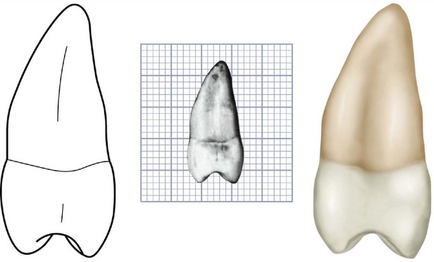
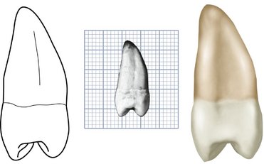
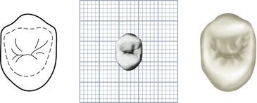
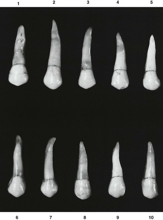
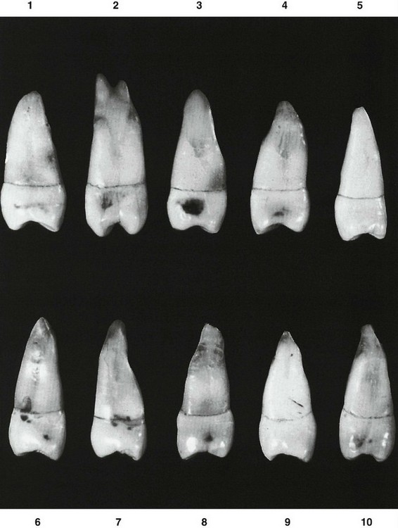
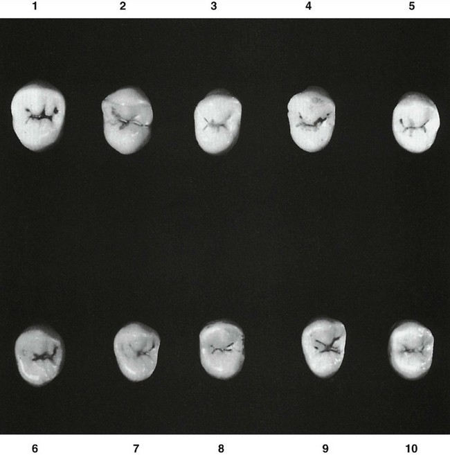
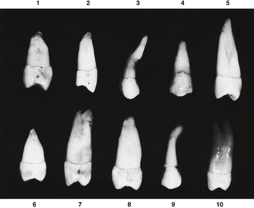
Figure 9-25 Maxillary second premolar. Ten specimens with uncommon variations are shown. 1, Root dwarfed and malformed. 2, Broad occlusal surface; lingual outline of crown straight. 3, Malformed root. 4, Crown very broad mesiodistally; root dwarfed. 5, Root extremely long. 6, Root dwarfed and very pointed at apex. 7, Root extremely long; bifurcation at root end. 8, Crown wider than usual buccolingually, curvature at cervical third extreme. 9, Root malformed, thick at apical third. 10, Root unusually long, bifurcated at apical third.
The maxillary second premolar is less angular, giving a more rounded effect to the crown from all aspects. It has a single root.
Considerable variations in the relative sizes of the two teeth may be seen, because the second premolar does not appear true to form as often as does the first premolar (Table 9-2). The maxillary second premolar may have a crown that is noticeably smaller cervico-occlusally and also mesiodistally; however, it may also be larger in those dimensions. Usually the root of the second premolar is as long as, if not a millimeter or so longer than, that of the first premolar. The two teeth have about the same dimensions on the average, except for the tendency toward greater length of the second premolar root. Ten specimens with uncommon variations are shown in Figure 9-25.
DETAILED DESCRIPTION OF THE MAXILLARY SECOND PREMOLAR FROM ALL ASPECTS
Buccal Aspect
From the buccal aspect, it may be noticed that the buccal cusp of the second premolar is not as long as that of the first premolar and it appears less pointed (see Figure 9-17). Also, the mesial slope of the buccal cusp ridge is usually shorter than the distal slope. The opposite is true of the first premolar.
In many instances, the crown and root of the second premolar are thicker at their cervical portions. This is not the rule, however (see Figure 9-22, 5, 6, 7, and 9). The buccal ridge of the crown may not be as prominent as that of the first premolar.
Lingual Aspect
From the lingual aspect, little variation may be seen except that the lingual cusp is longer, which makes the crown longer on the lingual side (see Figure 9-18).
Mesial Aspect
The mesial aspect shows the difference in cusp length between the first and second premolars (see Figures 9-4 and 9-19). The cusps of the second premolar are shorter, with the buccal and lingual cusps more nearly the same length. Greater distance between cusp tips widens the occlusal surface buccolingually.
No developmental depression is evident on the mesial surface of the crown as on the first premolar; the crown surface is convex instead. A shallow developmental groove appears on the single tapered root.
No deep developmental groove crossing the mesial marginal ridge is evident, and except for the variation in root form, no outstanding variation is noted when viewed from the distal aspect.
Distal Aspect
The distal root depression is deeper than the mesial depression on the maxillary second premolar. This characteristic of the distal root surface is the opposite of that of the maxillary first premolar, in which the depression is on the mesial surface of the root. A knowledge of where these depressions are facilitates periodontal instrumentation, that is, scaling and root planing.
Occlusal Aspect
From the occlusal aspect, some differences are to be noted between the two premolars. On the second premolar, the outline of the crown is more rounded or oval, rather than angular (see Figures 9-21 and 9-24). There are, of course, exceptions. The central developmental groove is shorter and more irregular, and there is a tendency toward multiple supplementary grooves radiating from the central groove. These supplementary grooves terminate in shallow depressions in the enamel that may extend out to the cusp ridges.
This arrangement makes for an irregular occlusal surface and gives the surface a very wrinkled appearance.
Bath-Balogh M, Fehrenbach MJ. Dental embryology, histology, and anatomy. Philadelphia: Saunders; 1997.
Carlsen O: Dental morphology, Copenhagen, 1987, Munksgaard.
Carlsen O, Hansen H. Some radicular structures observed in human premolars. Tandlægebladet. 1970;74:137.
Sowter JB, editor. Dental laboratory technology: dental anatomy. Chapel Hill: University of North Carolina, 1972.
Woefel JB, Scheid RC. Dental anatomy: its relevance to dentistry, ed 5. Baltimore: Williams & Wilkins; 1997.
 site for additional study resources.