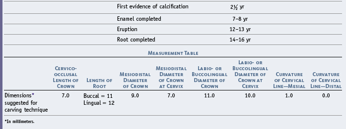
11 The Permanent Maxillary Molars
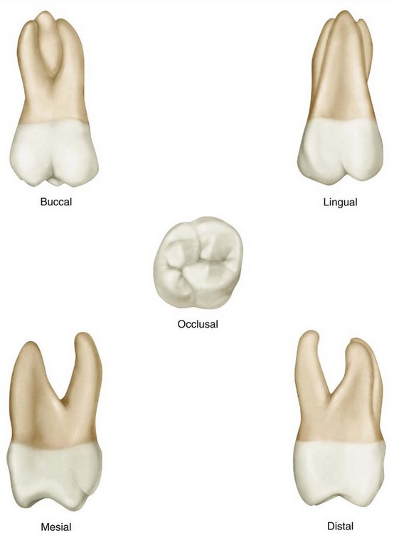
The maxillary molars differ in design from any of the teeth previously described. These teeth assist the mandibular molars in performing the major portion of the work in the mastication and comminution of food. They are the largest and strongest maxillary teeth, by virtue both of their bulk and of their anchorage in the jaws. Although the crowns on the molars may be somewhat shorter than those on the premolars, their dimensions are greater in every respect. The root portion may be no longer than that of the premolars, but instead of one root or a bifurcated root, the maxillary molar root is broader at the base in all directions and is trifurcated into three well-developed prongs that are actually three full-size roots emanating from a common broad base above the crown.
Generally speaking, the maxillary molars have large crowns with four well-formed cusps. They have three roots, two buccal and one lingual. The lingual root is the largest. The crowns have two buccal cusps and two lingual cusps. The outlines and curvatures of all the maxillary molars are similar. Developmental variations will be set forth under descriptions of the separate molars.
Before a detailed description of the maxillary first molar is begun, some statements will be made that are applicable to all first molars, mandibular and maxillary.
The permanent first molars usually appear in the oral cavity when the child is 6 years old. The mandibular molars precede the maxillary molars. The first permanent molar (maxillary or mandibular) erupts posterior to the second deciduous molar, taking up a position in contact with it. Therefore the first molar is not a succedaneous tooth because it has no predecessor. The deciduous teeth are all still in position and functioning when the first molar takes its place. Because the development of the bones of the face is downward and forward, sufficient space has been created normally at the age of 6 for the accommodation of this tooth.
The normal location of the first permanent molar is at the center of the fully developed adult jaw anteroposteriorly. As a consequence of the significance of their positions and the circumstances surrounding their eruption, the first molars may also be considered cornerstones of the dental arches. A full realization of the significance of these teeth as units in the arches and their function and positions relative to the other teeth will be gained when an opportunity comes to study the arrangement of the teeth with their occlusion and the temporomandibular articulation of the jaws. Subsequent chapters cover those phases. The mandibular molars are described in Chapter 12.
Maxillary First Molar
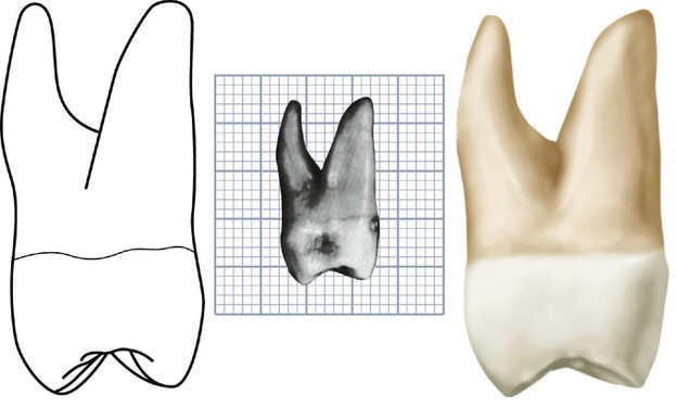
Figures 11-1 through 11-18 illustrate the maxillary first molar from all aspects. The crown of this tooth is wider buccolingually than mesiodistally. Usually the extra dimension buccolingually is about 1 mm (Table 11-1). This, however, varies in individuals (see Figure 11-17, 1, 5, 7, and 9). From the occlusal aspect, the inequality of the measurements in the two directions appears slight. Although the crown is relatively short, it is broad both mesiodistally and buccolingually, which gives the occlusal surface its generous dimensions.
The maxillary first molar is normally the largest tooth in the maxillary arch. It has four well-developed functioning cusps and one supplemental cusp of little practical use. The four large cusps of most physiological significance are the mesiobuccal, the distobuccal, the mesiolingual, and the distolingual. A supplemental cusp is called the cusp or tubercle of Carabelli. This morphological trait can take the form of a well-developed fifth cusp, or it can grade down to a series of grooves, depressions, or pits on the mesial portion of the lingual surface. This trait has been used to distinguish populations.
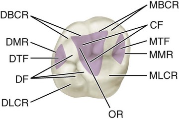
Figure 11-1 Maxillary right first molar, occlusal aspect. MBCR, Mesiobuccal cusp ridge; CF, central fossa (shaded area); MTF, mesial triangular fossa (shaded area); MMR, mesial marginal ridge; MLCR, mesiolingual cusp ridge; OR, oblique ridge; DLCR, distolingual cusp ridge; DF, distal fossa; DTF, distal triangular fossa (shaded area); DMR, distal marginal ridge; DBCR, distobuccal cusp ridge.
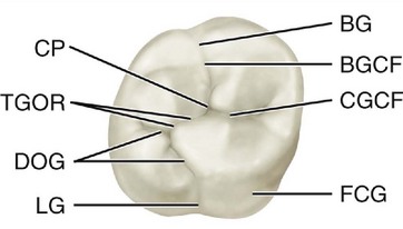
Figure 11-2 Maxillary right first molar, occlusal aspect, developmental grooves. BG, Buccal groove; BGCF, buccal groove of central fossa; CGCF, central groove of central fossa; FCG, fifth cusp groove; LG, lingual groove; DOG, distal oblique groove; TGOR, transverse groove of oblique ridge; CP, central pit.
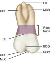
Figure 11-3 Maxillary right first molar, buccal aspect. LR, Lingual root; MBR, mesiobuccal root; BDG, buccal developmental groove; MBC, mesiobuccal cusp; MLC, mesiolingual cusp; DBC, distobuccal cusp; CL, cervical line; DBR, distobuccal root.
This supplemental cusp is found lingual to the mesiolingual cusp, which is the largest of the well-developed cusps. Usually, a developmental groove is found, leaving a record of cusp development, unless it has been erased by frictional wear. The fifth cusp or a developmental trace at its usual site serves to identify the maxillary first molar. A specimen of this tooth showing no trace of its typical characteristic would be rare.
The three roots of generous proportions are the mesiobuccal, distobuccal, and lingual. These roots are well separated and well developed, and their placement gives this tooth maximum anchorage against forces that would tend to unseat it. The roots have their greatest spread parallel to the line of greatest force brought to bear against the crown diagonally in a buccolingual direction. The lingual root is the longest root. It is tapered and smoothly rounded. The mesiobuccal root is not as long, but it is broader buccolingually and shaped (in cross section) so that its resistance to torsion is greater than that of the lingual root. The distobuccal root is the smallest of the three and smoothly rounded.
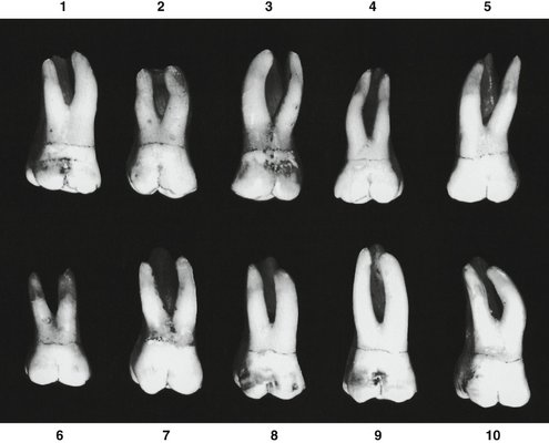
The development of maxillary first molars rarely deviates from the accepted normal. Ten specimens with uncommon variations are shown in Figure 11-18.
DETAILED DESCRIPTION OF THE MAXILLARY FIRST MOLAR FROM ALL ASPECTS
Buccal Aspect
The crown is roughly trapezoidal, with cervical and occlusal outlines representing the uneven sides (see Figures 11-4, 11-13, 11-14, and 11-15). The cervical line is the shorter of the uneven sides (see Figure 4-16, D).
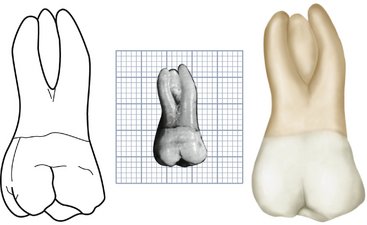
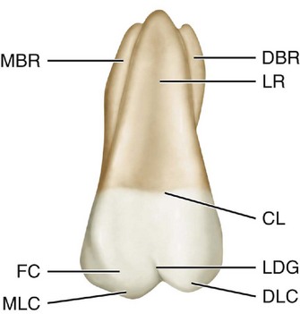
Figure 11-5 Maxillary right first molar, lingual aspect. DBR, Distobuccal root; LR, lingual root; CL, cervical line; LDG, lingual developmental groove; DLC, distolingual cusp; MLC, mesiolingual cusp; FC, fifth cusp; MBR, mesiobuccal root.
When the buccal aspect of this tooth is viewed with the line of vision at right angles to the buccal developmental groove of the crown, the distal side of the crown can be seen in perspective, which is possible because of the obtuse character of the distobuccal line angle (see Occlusal Aspect later in this chapter). Parts of four cusps are seen, the mesiobuccal, distobuccal, mesiolingual, and distolingual.
The mesiobuccal cusp is broader than the distobuccal cusp, and its mesial slope meets its distal slope at an obtuse angle. The mesial slope of the distobuccal cusp meets its distal slope at approximately a right angle. The distobuccal cusp is therefore sharper than the mesiobuccal cusp, and it is at least as long and often longer (see Figure 11-15, 4, 6, 7, 8, and 9).
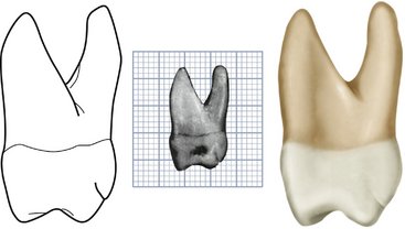
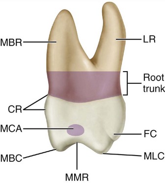
Figure 11-8 Maxillary right first molar, mesial aspect. LR, Lingual root; FC, fifth cusp; MLC, mesiolingual cusp; MMR, mesial marginal ridge; MBC, mesiobuccal ridge; MCA, mesial contact area; CR, cervical ridge; MBR, mesiobuccal root.
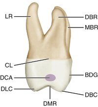
Figure 11-9 Maxillary right first molar, distal aspect. DBR, Distobuccal root; MBR, mesiobuccal root; BDG, buccal developmental groove; DBC, distobuccal cusp; DMR, distal marginal ridge; DLC, distolingual cusp; DCA, distal contact area; CL, cervical line; LR, lingual root.
The buccal developmental groove that divides the two buccal cusps is approximately equidistant between the mesiobuccal and distolingual line angles. The groove slants occlusoapically in a line of direction parallel to the long axis of the distobuccal root. It terminates at a point approximately half the distance from its origin occlusally to the cervical line of the crown. Although the groove is not deep at any point, it becomes more shallow toward its termination, gradually fading out. Lateral to its terminus is a dip in the enamel of the crown that is developmental in character and extends for some distance mesially and distally.
The cervical line of the crown does not have much curvature from mesial to distal; however, it is not as smooth and regular as that found on some of the other teeth. The line is generally convex with the convexity toward the roots.
The mesial outline of the crown from this aspect follows a nearly straight path downward and mesially, curving occlusally as it reaches the crest of contour of the mesial surface, which is the contact area. This crest is approximately two thirds the distance from cervical line to tip of mesiobuccal cusp. The mesial outline continues downward and distally and becomes congruent with the outline of the mesial slope of the mesiobuccal cusp.
The distal outline of the crown is convex; the distal surface is spheroidal. The crest of curvature on the distal side of the crown is located at a level approximately half the distance from cervical line to tip of cusp. The distal contact area is in the middle of the middle third.
Often from this aspect, a flattened area or a concave area is seen on the distal surface immediately above the distobuccal cusp at the cervical third of the crown.
All three of the roots may be seen from the buccal aspect. The axes of the roots are inclined distally. The roots are not straight, although the buccal roots show an inclination to curvature halfway between the point of bifurcation and the apices. The mesiobuccal root curves distally, starting at the middle third. Its axis usually is at right angles to the cervical line. The distal root is straighter, with its long axis at an acute angle distally with the cervical line. It has a tendency toward curvature mesially at its middle third.
The point of bifurcation of the two buccal roots is located approximately 4 mm above the cervical line. This measurement varies somewhat, of course. Nevertheless, the point is much farther removed from the cervical line than in the deciduous molars. This relation is typical when all permanent molars are compared with all deciduous molars.
A deep developmental groove buccally on the root trunk of the maxillary first molar, which starts at the bifurcation and progresses downward, becoming more shallow until it terminates in a shallow depression at the cervical line. Sometimes this depression extends slightly onto the enamel at the cervix.
The reader must keep in mind the fact that molar roots originate as a single root on the base of the crown. They then are divided into three roots, as in the maxillary molars, or two roots, as in the mandibular molars. The common root base is called the root trunk (see Figures 11-3 and 11-8).
In judging the length of the roots and the direction of their axes, we must indicate the part of the root trunk that is congruent with each root as part of it, because they [i.e., the root trunk and the root] function as an entity. Usually, the lingual root is the longest, and the two buccal roots are approximately equal in length. Considerable variance is evident, although the difference is a matter of a millimeter or so only in the average first molars with normal development.
From the buccal aspect, a measurement of the roots inclusively at their greatest extremities mesiodistally is less than a calibration of the diameter of the crown mesiodistally.
No invariable rule covers the relative length of crown and root when the upper first molar is described. On the average, the roots are about twice as long as the crown.
Lingual Aspect
From the lingual aspect, the gross outline of the maxillary first molar is the reverse of that from the buccal aspect (see Figures 11-5, 11-6, 11-13, and 11-14). Photographs or drawings show this only approximately, because all teeth have breadth and thickness; consequently, the perspective of two dimensions plus the human element, which enters into the technique of posing specimens and making drawings and photographs, are bound to result in some error in graphic interpretation.
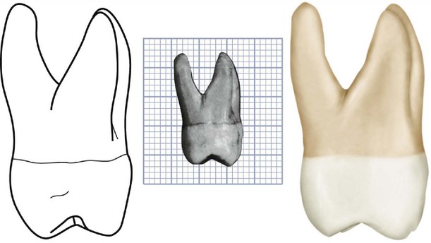

Figure 11-11 Maxillary molar primary cusp triangle. The distolingual lobe, represented by shaded areas, becomes progressively smaller on maxillary molars, starting with the first molar, which presents the greatest development of the lobe. The plain areas, roughly triangular in outline, represent the maxillary molar primary cusp triangles.
The variation between the outline of the mesial surface and that of the distal surface is apparent. Because of the roundness of the distolingual cusp, the smooth curvature of the distal outline of the crown becoming confluent with the curvature of the cusp creates an arc that is almost a semicircle. The line that describes the lingual developmental groove is also confluent with the outline of the distolingual cusp, progressing mesially and cervically and ending at a point at the approximate center of the lingual surface of the crown. A shallow depression in the surface extends from the terminus of the lingual groove to the center of the lingual surface of the lingual root at the cervical line and then continues in an apical direction on the lingual root, fading out at the middle third of the root.
The lingual cusps are the only ones to be seen from the lingual aspect. The mesiolingual cusp is much larger, and before occlusal wear, it is always the longest cusp the tooth possesses. Its mesiodistal width is about three fifths of the mesiodistal crown diameter, with the distolingual cusp making up the remaining two fifths. The angle formed by the mesial outline of the crown and the mesial slope of the mesiolingual cusp is almost 90 degrees. An obtuse angle describes the junction of the mesial and distal slopes of this cusp.
The distolingual cusp is so spheroidal and smooth that it is difficult to describe any angulation on the mesial and distal slopes.
The lingual developmental groove starts approximately in the center of the lingual surface mesiodistally, curves sharply to the distal as it crosses between the cusps, and continues on to the occlusal surface.
The fifth cusp appears attached to the mesiolingual surface of the mesiolingual cusp. It is outlined occlusally by an irregular developmental groove, which may be described as starting in a depression of the mesiolingual line angle of the crown, extending occlusally toward the point of the mesiolingual cusp, then making an obtuse-angle turn toward the terminus of the lingual groove and fading out near the lingual groove terminus. If the fifth cusp is well developed, its cusp angle will be sharper and less obtuse than that of the mesiolingual cusp. The cusp ridge of the fifth cusp is approximately 2 mm cervical to the cusp ridge of the mesiolingual cusp (see Figure 11-5).
All three of the roots are visible from the lingual aspect, with the large lingual root making up most of the foreground. The lingual portion of the root trunk is continuous with the entire cervical portion of the crown lingually. The lingual root is conical, terminating in a bluntly rounded apex.
All of the mesial outline of the mesiobuccal root and part of its apex may be seen from this angle.
The distal outline of the distobuccal root is seen above its middle third, including all of its apical outline.
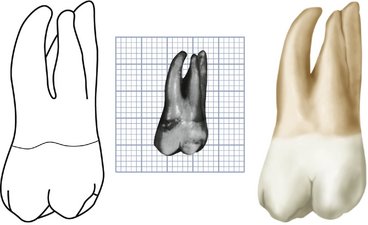
Mesial Aspect
From the mesial aspect, the increased buccolingual dimensions may be observed, as well as the outlines of the cervical curvatures of the crown at the cervical third buccally and lingually, the difference in dimensions between the crown at its greatest measurement, and the distance between the cusp tips in a buccolingual direction (see Figures 11-7, 11-8, 11-13, 11-14, and 11-16).
Starting at the cervical line buccally, the outline of the crown makes a short arc buccally to its crest of curvature within the cervical third of the crown. The extent of this curvature is about 0.5 mm (see Figure 11-13). The line of the buccal surface then describes a shallow concavity immediately occlusal to the crest of curvature (see Buccal Aspect earlier in this chapter). The outline then becomes slightly convex as it progresses downward and inward to circumscribe the mesiobuccal cusp, ending at the tip of the cusp well within projected outlines of the root base.
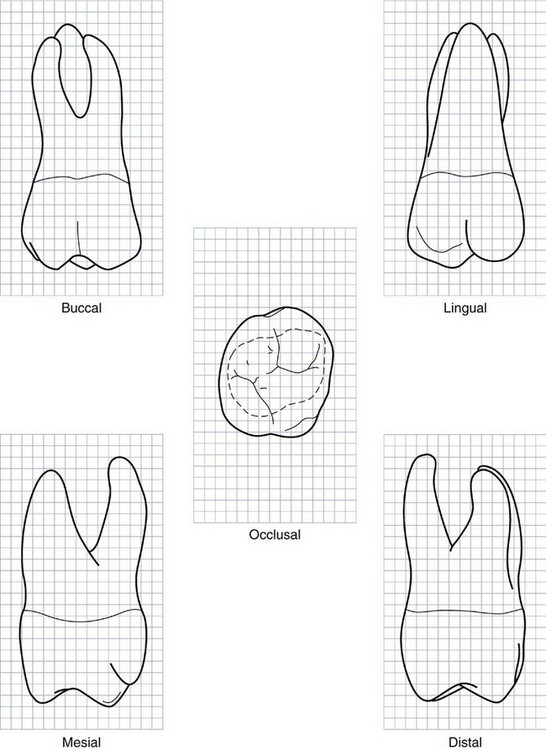
Figure 11-13 Maxillary right first molar. Graph outlines of five aspects are shown. (Grid = 1 sq mm.)
If the tooth is posed so that the line of vision is at right angles to the mesial contact area, the only cusps in sight are the mesiobuccal, the mesiolingual, and the fifth cusps. The mesiobuccal root hides the distobuccal root.
The lingual outline of the crown curves outward and lingually approximately to the same extent as on the buccal side. The level of the crest of curvature is near the middle third of the crown rather than a point within the cervical third, as it is buccally.
If the fifth cusp is well developed, the lingual outline dips inward to reveal it. If it is undeveloped, the lingual outline continues from the crest of curvature as a smoothly curved arc to the tip of the mesiolingual cusp. The point of the cusp is more clearly centered within projected outlines of the root base than the tip of the mesiobuccal cusp. The mesiolingual cusp is on a line with the long axis of the lingual root.
The mesial marginal ridge, which is confluent with the mesiobuccal and mesiolingual cusp ridges, is irregular, the outline curving cervically about one fifth the crown length and centering its curvature below the center of the crown buccolingually.
The cervical line of the crown is irregular, curving occlusally, but as a rule not more than 1 mm at any one point. If a definite curvature is present, it reaches its maximum immediately above the contact area.
The mesial contact area is apical to the marginal ridge but closer to it than the cervical line, approximately at the junction of the middle and occlusal thirds of the crown (see Figure 11-8). It is also somewhat buccal to the center of the crown buccolingually. A shallow concavity is usually found just above the contact area on the mesial surface of the maxillary first molar. This concavity may be continued to the mesial surface of the root trunk at its cervical third.
The mesiobuccal root is broad and flattened on its mesial surface; this flattened surface often exhibits smooth flutings for part of its length. The width of this root near the crown from the buccal surface to the point of bifurcation on the root trunk is approximately two thirds of the crown measurement buccolingually at the cervical line. The buccal outline of the root extends upward and outward from the crown, ending at the blunt apex. The greatest projection on this root is usually buccal to the greatest projection of the crown. The lingual outline of the root is relatively straight from the bluntly rounded apex down to the bifurcation with the lingual root.
The level of the bifurcation is a little closer to the cervical line than is found between the roots buccally. A smooth depression congruent with the bifurcation extends occlusally and lingually almost to the cervical line directly above the mesiolingual line angle of the crown.
The lingual root is longer than the mesial root but is narrower from this aspect. It is banana-shaped, extending lingually with its convex outline to the lingual and its concave outline to the buccal. At its middle and apical thirds, it is outside of the confines of the greatest crown projection. Although its apex is rounded, the root appears more pointed toward the end than the mesiobuccal root.
Distal Aspect
The gross outline of the distal aspect is similar to that of the mesial aspect (see Figures 11-9, 11-10, 11-13, and 11-14). Certain variations must be noted when the tooth is viewed from the distal aspect.
Because of the tendency of the crown to taper distally on the buccal surface, most of the buccal surface of the crown may be seen in perspective from the distal aspect. This is because the buccolingual measurement of the crown mesially is greater than the same measurement distally. All of the decrease in measurement distally is a result of the slant of the buccal side of the crown.
The distal marginal ridge dips sharply in a cervical direction, exposing triangular ridges on the distal portion of the occlusal surface of the crown.
The cervical line is almost straight across from buccal to lingual. Occasionally it curves apically 0.5 mm or so.
The distal surface of the crown is generally convex, with a smoothly rounded surface except for a small area near the distobuccal root at the cervical third. This concavity continues on to the distal surface of the distobuccal root, from the cervical line to the area of the root that is on a level with bifurcation separating the distobuccal and lingual roots.
The distobuccal root is narrower at its base than either of the others. An outline of this root, when the tooth is viewed from the distal aspect, starts buccally at a point immediately above the distobuccal cusp, follows a concave path inward for a short distance, then turns outward in a buccal direction, completing a graceful convex arc from the concavity to the rounded apex. This line lies entirely within the confines of the outline of the mesiobuccal root. The lingual outline of the root from the apex to the bifurcation is slightly concave. No concavity is evident between the bifurcation of the roots and the cervical line. However, the surface at this point on the root trunk has a tendency toward convexity.
The bifurcation here is more apical than either of the other two areas on this tooth. The area from cervical line to bifurcation is 5 mm or more in extent.
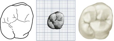
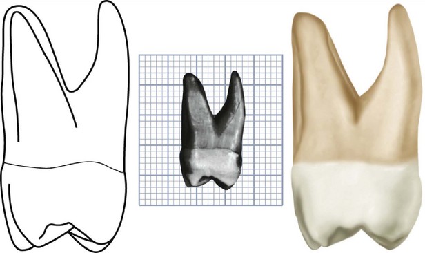
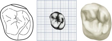
Occlusal Aspect
From the occlusal aspect, the maxillary first molar is somewhat rhomboidal. An outline following the four major cusp ridges and the marginal ridges is especially so (see Figures 11-1, 11-2, 11-12, 11-13, 11-14, and 11-17).
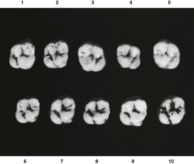
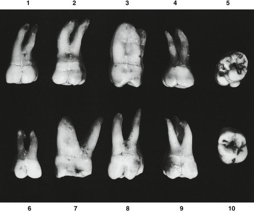
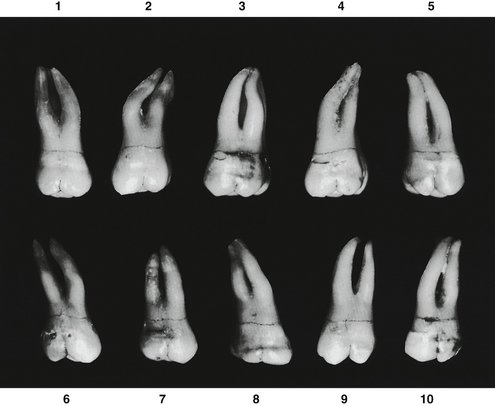
Figure 11-18 Maxillary first molar. Ten specimens with uncommon variations are shown. 1, Unusual curvature of buccal roots. 2, Roots abnormally long with extreme curvature. 3, Lingual and distobuccal roots fused. 4, Mesiodistal measurement of root trunk smaller than usual. 5, Extreme rhomboidal development of crown; fifth cusp with maximum development. 6, Tooth well developed but much smaller than usual. 7, Extreme buccolingual measurement. 8, Extreme length, especially of the distobuccal root; buccal cusps narrow mesiodistally. 9, Well-developed crown; roots poorly developed. 10, Extreme development of lingual portion of the crown compared with buccal development.
A measurement of the crown buccolingually and mesial to the buccal and lingual grooves is greater than the measurement on that portion of the crown which is distal to these developmental grooves. Also, a measurement of the crown immediately lingual to contact areas mesiodistally is greater than the measurement immediately buccal to the contact areas. Thus it is apparent that the maxillary first molar crown is wider mesially than distally and wider lingually than buccally.
The four major cusps are well developed, with the small minor, or fifth, cusp appearing on the lingual surface of the mesiolingual cusp near the mesiolingual line angle of the crown. The fifth cusp may be indistinct, or all the cusp form may be absent. At this site, however, traces of developmental lines are nearly always present in the enamel.
The mesiolingual cusp is the largest cusp; it is followed in size by the mesiobuccal, distolingual, distobuccal, and fifth cusps.
If reduced to a geometric schematic figure, the occlusal aspect of this molar locates the various angles of the rhomboidal figure as follows: acute angles, mesiobuccal and distolingual; and obtuse angles, mesiolingual and distobuccal.
An analysis of the design of the occlusal surfaces of maxillary molars may be summarized here. Developmentally, only three major cusps can be considered as primary, the mesiolingual cusp and the two buccal cusps. The distolingual cusp is common to all of the maxillary molars; any other additional one, such as the cusp of Carabelli on first molars, must be regarded as secondary.
The triangular arrangement of cusps is reflected in the outline of the root trunks of maxillary molars when the teeth are sectioned in those areas (see Root Sections in Chapter 13). The distolingual cusp becomes progressively smaller on second and third maxillary molars, often disappearing as a major cusp (see Figure 11-11). Thus the triangular arrangement of the three important molar cusps is called the maxillary molar primary cusp triangle. The characteristic triangular figure, made by tracing the cusp outlines of these cusps, the mesial marginal ridge, and the oblique ridge of the occlusal surface, is representative of all maxillary molars.
The occlusal surface, or occlusal table as it is sometimes termed, of the maxillary first molar is within the confines of the cusp ridges and marginal ridges. The morphological features are now considered.
There are two major fossae and two minor fossae. The major fossae are the central fossa, which is roughly triangular and mesial to the oblique ridge, and the distal fossa, which is roughly linear and distal to the oblique ridge.
The two minor fossae are the mesial triangular fossa, immediately distal to the mesial marginal ridge, and the distal triangular fossa, immediately mesial to the distal marginal ridge (see Figure 11-l).
The oblique ridge is a ridge that crosses the occlusal surface obliquely. The union of the triangular ridge of the distobuccal cusp and the distal ridge of the mesiolingual cusp forms it. This ridge is reduced in height in the center of the occlusal surface, being about on a level with the marginal ridges of the occlusal surface. Sometimes it is crossed by a developmental groove that partially joins the two major fossae by means of its shallow sulcate groove.
The mesial marginal ridge and the distal marginal ridge are irregular ridges confluent with the mesial and distal cusp ridges of the mesial and distal major cusps.
The central fossa of the occlusal surface is a concave area bound by the distal slope of the mesiobuccal cusp, the mesial slope of the distobuccal cusp, the crest of the oblique ridge, and the crests of the two triangular ridges of the mesiobuccal and mesiolingual cusps. The central fossa has connecting sulci within its boundaries, with developmental grooves at the deepest portions of these sulci (sulcate grooves). In addition, it contains supplemental grooves, short grooves that are disconnected, and also the central developmental pit. A worn specimen may show developmental or sulcate grooves only.
In the center of the central fossa, the central developmental pit has sulcate developmental grooves radiating from it at obtuse angles to each other. This pit is located in the approximate center of that portion of the occlusal surface that is circumscribed by cusp ridges and marginal ridges (see Figure 11-1). From this pit the buccal developmental groove radiates buccally at the bottom of the buccal sulcus of the central fossa, continuing on to the buccal surface of the crown between the buccal cusps.
Starting again at the central pit, the central developmental groove is seen to progress in a mesial direction at an obtuse angle to the buccal sulcate groove. The central groove at the bottom of the sulcus of the central fossa usually terminates at the apex of the mesial triangular fossa. Here it is joined by short, supplemental grooves that radiate from its terminus into the triangular fossa. These supplemental grooves often appear as branches of the central groove. Occasionally one or more supplemental grooves cross the mesial marginal ridge of the crown.
The mesial triangular fossa is rather indistinct in outline, but it is generally triangular in shape, with its base at the mesial marginal ridge and its apex at the point where the supplemental grooves join the central groove.
An additional short developmental groove radiates from the central pit of the central fossa at an obtuse angulation to the buccal and central developmental grooves. Usually, it is considered a projection of one of these, because it is very short and usually fades out before reaching the crest of the oblique ridge. When it crosses the oblique ridge transversely, however, as it sometimes does, joining the central and distal fossae with a shallow groove, it is called the transverse groove of the oblique ridge (see Figure 11-17, 3, 4, and 5).
The distal fossa of the maxillary first molar is roughly linear in form and is located immediately distal to the oblique ridge. An irregular developmental groove traverses its deepest portion. This developmental groove is called the distal oblique groove. It connects with the lingual developmental groove at the junction of the cusp ridges of the mesiolingual and distolingual cusps. These two grooves travel in the same oblique direction to the terminus of the lingual groove, which is centered below the lingual root at the approximate center of the crown lingually (see Figure 11-5, LDG). If the fifth cusp development is distinct, a developmental groove outlining it joins the lingual groove near its terminus. Any part of the developmental groove that outlines a fifth cusp is called the fifth cusp groove.
The distal oblique groove in most cases shows several supplemental grooves. Two terminal branches usually appear, forming two sides of the triangular depression immediately mesial to the distal marginal ridge. These two sides, in combination with the slope mesial to the distal marginal ridge, form the distal triangular fossa. The distal outline of the distal marginal ridge of the crown shows a slight concavity.
The distolingual cusp is smooth and rounded from the occlusal aspect, and an outline of it, from the distal concavity of the distal marginal ridge to the lingual groove of the crown, describes an arch of an ellipse.
The lingual outline of the distolingual cusp is straight with the lingual outline of the fifth cusp, unless the fifth cusp is unusually large. In the latter case the lingual outline of the fifth cusp is more prominent lingually (see Figure 11-17, 9). The cusp ridge of the distolingual cusp usually extends lingually farther than the cusp ridge of the mesiolingual cusp.
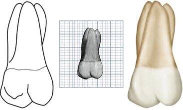
Maxillary Second Molar
Figures 11-19 through 11-27 illustrate the maxillary second molar from all aspects. The maxillary second molar supplements the first molar in function. In describing this tooth, direct comparisons will be made with the first molar both in form and development.
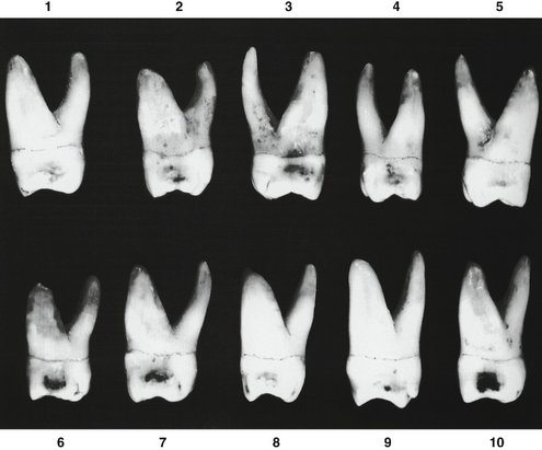
Generally speaking, the roots of this tooth are as long as, if not somewhat longer than, those of the first molar (Table 11-2). The distobuccal cusp is not as large or as well developed, and the distolingual cusp is smaller. No fifth cusp is evident.
The crown of the maxillary second molar is 0.5 mm or so shorter cervico-occlusally than that of the first molar, but the measurement of the crown buccolingually is about the same. Two types of maxillary second molars are found when the occlusal aspect is viewed: (1) The type that is seen most has an occlusal form that resembles that of the first molar, although the rhomboidal outline is more extreme. This is accentuated by the lesser measurement lingually. (2) The second type bears more resemblance to a typical third molar form. The distolingual cusp is poorly developed and makes the development of the other three cusps predominate. This results in a heart-shaped form from the occlusal aspect that is more typical of the maxillary third molar (see Figure 11-26, 1 and 7). Ten specimens with uncommon variations are shown in Figure 11-27.
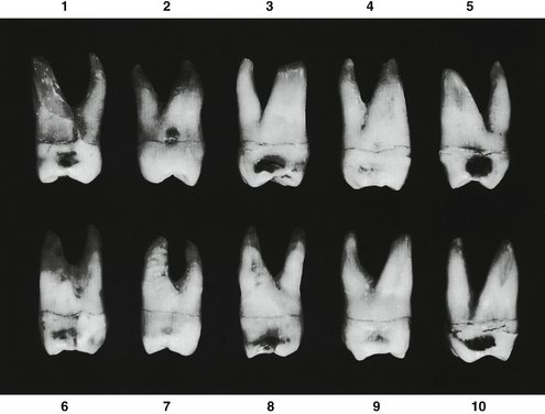
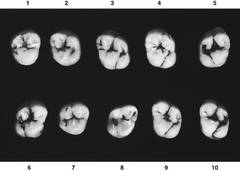
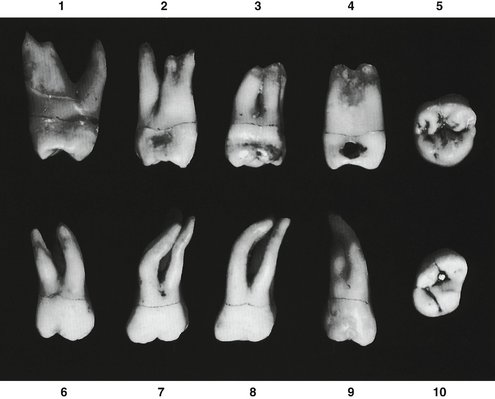
Figure 11-27 Maxillary second molar. Ten specimens with uncommon variations are shown. 1, Roots spread similar to those of first molar. 2, Bifurcated mesiobuccal root. 3, Roots very short and fused. 4, Mesiobuccal and lingual roots with complete fusion. 5, Crown similar to the typical third molar form. 6, Short roots with spread similar to that of first molar. 7, Roots extra long with abnormal curvatures. 8, Another variation similar to specimen 7. 9, Very long roots fused. 10, Crown with extreme rhomboidal form.
DETAILED DESCRIPTION OF THE MAXILLARY SECOND MOLAR FROM ALL ASPECTS
Buccal Aspect
From the buccal aspect the crown is a little shorter cervico-occlusally and narrower mesiodistally than is the maxillary first molar (see Figures 11-19 and 11-24). The distobuccal cusp is smaller and allows part of the distal marginal ridge and part of the distolingual cusp to be seen.
The buccal roots are about the same length. These roots are more nearly parallel and inclined distally more than those of the maxillary first molar, so that the end of the distobuccal root is slightly distal to the distal extremity of the crown. The apex of the mesiobuccal root is on a line with the buccal groove of the crown instead of the tip of the mesiobuccal cusp, as was found on the first molar.
Lingual Aspect
Ways in which the second molar differs from the first molar to be noted here, in addition to those mentioned earlier, are the following:
Mesial Aspect
The buccolingual dimension of the second molar is about the same as that of the first molar, but the crown length is less (see Figures 11-21 and 11-25). The roots do not spread as far buccolingually but are within the confines of the buccolingual crown outline.
Distal Aspect
Because the distobuccal cusp is smaller in the maxillary second molar than in the first molar, more of the mesiobuccal cusp may be seen from this angle (see Figure 11-22). The mesiolingual cusp cannot be seen. The apex of the lingual root is in line with the distolingual cusp.
Occlusal Aspect
The rhomboidal type of second maxillary molar is most common, although in comparison with the first molar, the acute angles of the rhomboid are less and the obtuse angles greater (see Figures 11-23 and 11-26). The buccolingual diameter of the crown is about equal, but the mesiodistal diameter is approximately 1 mm less. The mesiobuccal and mesiolingual cusps are just as large and well developed as in the first molar, but the distobuccal and distolingual cusps are smaller and less well developed. Usually, a calibration made of the crown at the greatest diameter buccally and lingually of the distal portion is considerably less than one made at the greatest diameter buccally and lingually of the mesial portion, so that more convergence distally is seen than in the maxillary first molar.
It is not uncommon to find more supplemental grooves, accidental grooves, and pits on the occlusal surface of a maxillary second molar than are usually found on that of a maxillary first molar.
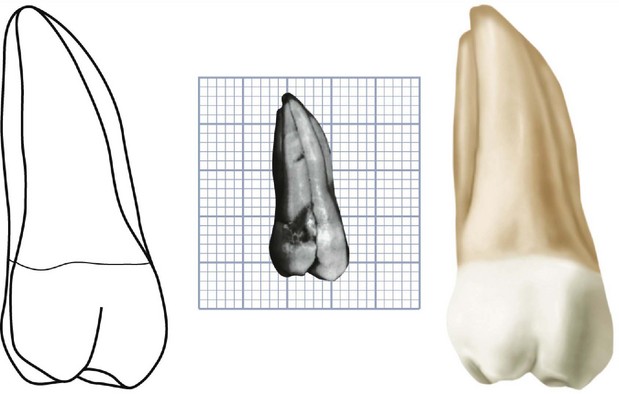
Maxillary Third Molar
Figures 11-28 through 11-36 illustrate the maxillary third molar from all aspects. The maxillary third molar often appears as a developmental anomaly. It can vary considerably in size, contour, and relative position to the other teeth (Table 11-3). It is seldom as well developed as the maxillary second molar, to which it often bears resemblance. The third molar supplements the second molar in function, and its fundamental design is similar. The crown is smaller, and the roots are shorter as a rule, with the inclination toward fusion with the resultant anchorage of one tapered root.
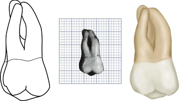
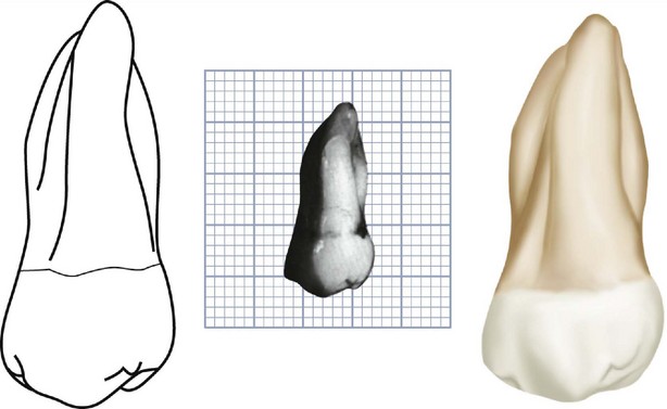
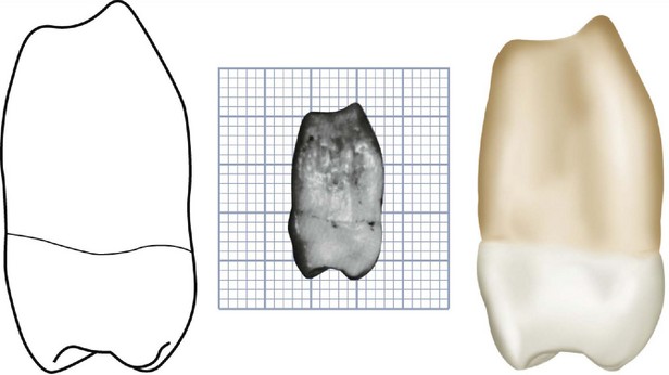
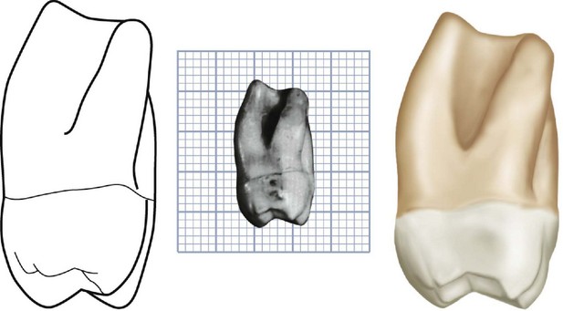
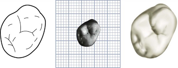
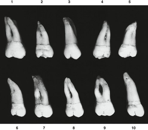
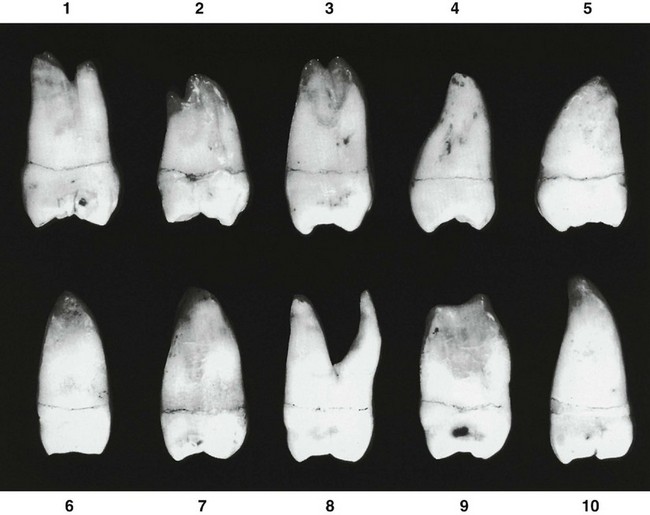
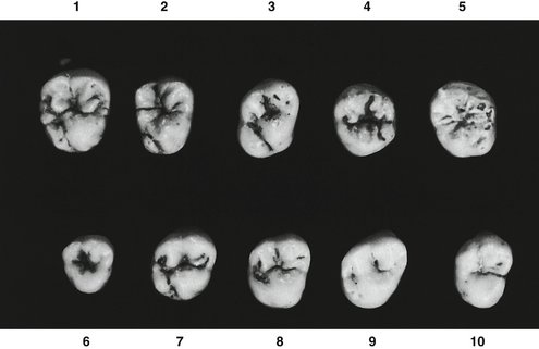
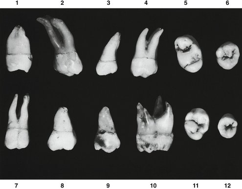
Figure 11-36 Maxillary third molar. Twelve specimens with uncommon variations are shown. 1, Very short fused root form. 2, Extremely long roots with extreme distal angulation. 3, Complete fusion of roots with extreme distal angulation. 4, Three roots well separated; crown very wide at cervix. 5, Extreme rhomboidal outline to crown, with developmental grooves oddly placed. 6, Overdeveloped mesiobuccal cusp. 7, Crown wide at cervix, with roots perpendicular. 8, Very large crown; poorly developed root form. 9, Complete absence of typical design. 10, Specimen abnormally large, with four roots well separated. 11, Five well-developed cusps, atypical in form. 12, Small specimen, atypical cusp form.
The predominating third molar design, when the occlusal surface is viewed, is that of a heart-shaped type of second molar. The distolingual cusp is very small and poorly developed in most cases, and it may be absent entirely.
All third molars, mandibular and maxillary, show more variation in development than any of the other teeth in the mouth. Occasionally they appear as anomalies bearing little or no resemblance to neighboring teeth. A few of the variations in form are shown in Figure 11-36.
It is necessary to give a short description of the third molar that is considered average in its development and one that would be in good proportion to the other maxillary molars and with an occlusal form considered normal. In describing the normal maxillary third molar, direct comparisons will be made with the maxillary second molar.
DETAILED DESCRIPTION OF THE MAXILLARY THIRD MOLAR FROM ALL ASPECTS
Buccal Aspect
From the buccal aspect, the crown of the third molar is shorter cervico-occlusally and narrower mesiodistally than that of the second molar (see Figures 11-28 and 11-33). The roots are usually fused, functioning as one large root, and they are shorter cervicoapically. The fused roots end in a taper at the apex. The roots have a distinct slant to the distal, giving the apices of the fused root a more distal relation to the center of the crown.
Lingual Aspect
In addition to the differences just mentioned, in comparison with the maxillary second molar, only one large lingual cusp is present, and therefore no lingual groove is evident (see Figure 11-29). However, in many cases, a third molar with the same essential features has a poorly developed distolingual cusp with a developmental groove lingually (see Figure 11-35, 2).
Mesial Aspect
From the mesial aspect, aside from the differences in measurement, the main feature is the taper to the fused roots and a bifurcation, usually in the region of the apical third. Figure 11-30 does not show a bifurcation. See Figure 11-34, 1, 2, and 3. The root portion is considerably shorter in relation to the crown length. Both the crown and the root portions tend to be poorly developed, with irregular outlines.
Distal Aspect
From the distal aspect, most of the buccal surface of the crown is in view (see Figure 11-31). More of the occlusal surface may be seen than can be seen on the second molar from this aspect because of the more acute angulation of the occlusal surface in relation to the long axis of the root. The measurement from the cervical line to the marginal ridge is short.
Occlusal Aspect
The occlusal aspect of a typical maxillary third molar presents a heart-shaped outline (see Figures 11-32 and 11-35). The lingual cusp is large and well developed, and little or no distolingual cusp is evident, which gives a semicircular outline to the tooth from one contact area to the other. Three functioning cusps are seen on this type of tooth: two buccal and one lingual.
The occlusal aspect of this tooth usually presents many supplemental grooves and many accidental grooves unless the tooth is very much worn.
The third molar may show four distinct cusps. This type may have a strong oblique ridge, a central fossa and a distal fossa, with a lingual developmental groove similar to that of the rhomboidal type of second molar. In most instances, the crown converges more lingually from the buccal areas than in the second molar, losing its rhomboidal outline. This is not always true, however (compare 1 and 3 in Figure 11-35).
Alexandersen V. Mandibular third molar: the root complex. II. Morphogenetic considerations. Tandlægebladet. 1985;66:53.
Ash MM. Wheeler’s atlas of tooth form, ed 5. Philadelphia: Saunders; 1984.
Black GV. Descriptive anatomy of the human teeth, ed 4. Philadelphia: S.S. White Dental Manufacturing; 1897.
Carabelli G. Anatomie des Mundes. Vienna: Braumuller and Seidel; 1842.
Carbonell VM. The tubercle of Carabelli in the Kish dentition, Mesopotamia, 3000 bc. J Dent Res. 1960;39:124.
Carlsen O. Dental morphology. Copenhagen: Munksgaard; 1987.
Diamond M. Dental anatomy. New York: Macmillan; 1929.
Hopewell-Smith A. An introduction to dental anatomy and physiology. Philadelphia: Lea and Febiger; 1913.
Kraus BS. Carabelli’s anomaly of the maxillary molar teeth. Am J Hum Genet. 1951;3:348.
Kraus BS, Jordan RE, Abrams L. Dental anatomy and occlusion. Baltimore: Williams & Wilkins; 1969.
Tomes CS. A manual of dental anatomy. London: Churchill; 1894.
Woelfel JB, Scheid RC. Dental anatomy: its relevance to dentistry. Baltimore: Williams & Wilkins; 1997.
 site for additional study resources.