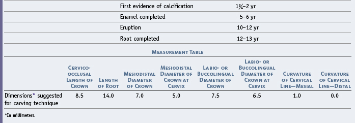
10 The Permanent Mandibular Premolars
The mandibular premolars number four: two are situated in the right side of the mandible and two in the left side. They are immediately posterior to the mandibular canines and anterior to the molars.
The mandibular first premolars are developed from four lobes, as were the maxillary premolars. The mandibular second premolars are, in most instances, developed from five lobes, three buccal and two lingual lobes.
The first premolar has a large buccal cusp, which is long and well formed, with a small, nonfunctioning lingual cusp that in some specimens is no longer than the cingulum found on some maxillary canines (see Figure 10-10, 3 and 8; and Figure 10-12, 4 and 7). The second premolar has three well-formed cusps in most cases, one large buccal cusp and two smaller lingual cusps. The form of both mandibular premolars fails to conform to the implications of the term bicuspid, a term that implies two functioning cusps.
The mandibular first premolar has many of the characteristics of a small canine, because its sharp buccal cusp is the only part of it occluding with maxillary teeth. It functions along with the mandibular canine. The mandibular second premolar has more of the characteristics of a small molar, because its lingual cusps are well developed, a fact that places both marginal ridges high and produces a more efficient occlusion with antagonists in the opposite jaw. The mandibular second molar functions by being supplementary to the mandibular first molar.
The first premolar is always the smaller of the two mandibular premolars, whereas the opposite is true, in many cases, of the maxillary premolars.
Mandibular First Premolar
Figures 10-1 through 10-12 illustrate the mandibular first premolar from all aspects. The mandibular first premolar is the fourth tooth from the median line and the first posterior tooth in the mandible. This tooth is situated between the canine and second premolar and has some characteristics common to each of them.
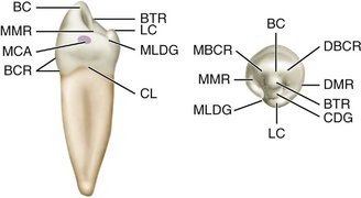
Figure 10-1 Mandibular right first premolar, mesial and occlusal aspects. BC, Buccal cusp; BTR, buccal triangular ridge; LC, lingual cusp; MLDG, mesiolingual developmental groove; CL, cervical line; BCR, buccal cervical ridge; MCA, mesial contact area; MMR, mesial marginal ridge; DBCR, distobuccal cusp ridge; DMR, distal marginal ridge; CDG, central developmental groove; MBCR, mesiobuccal cusp ridge.
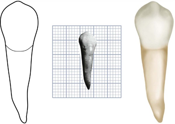
Figure 10-2 Mandibular right first premolar, buccal aspect. The specimen in this photograph shows a mesial inclination of the root. Mandibular premolars and canines have this tendency, although most of the roots of these teeth will curve, if at all, in a distal direction. (Grid = 1 sq mm.)
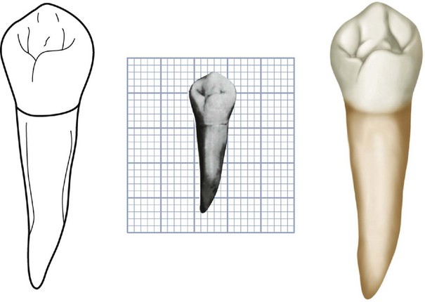
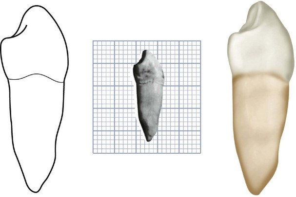
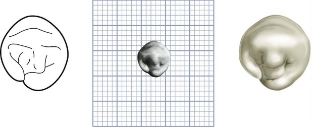
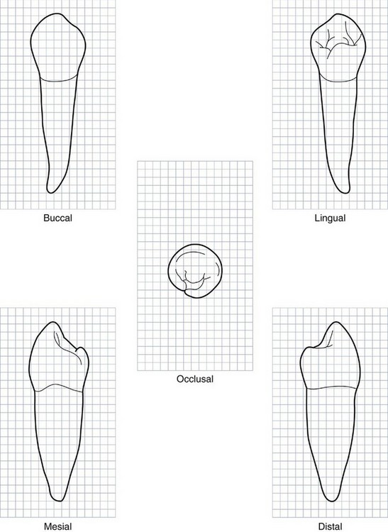
Figure 10-7 Mandibular right first premolar. Graph outlines of five aspects are shown. (Grid = 1 sq mm.)
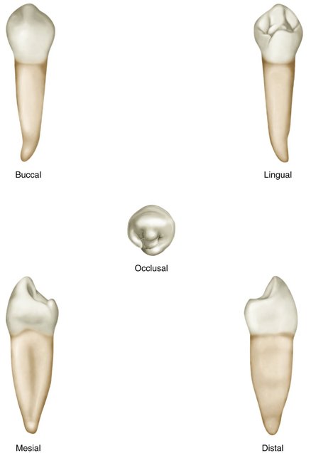
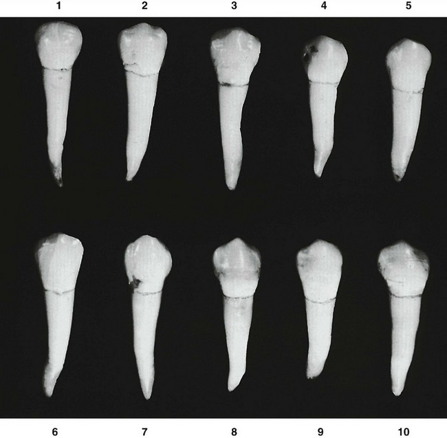
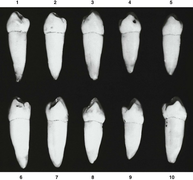
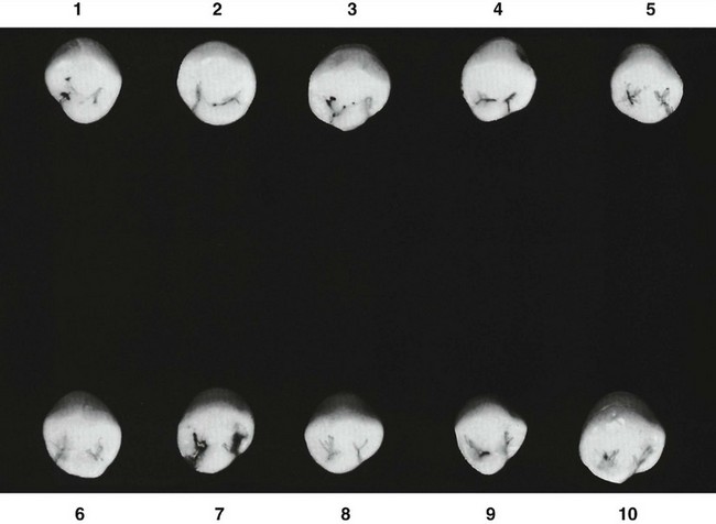
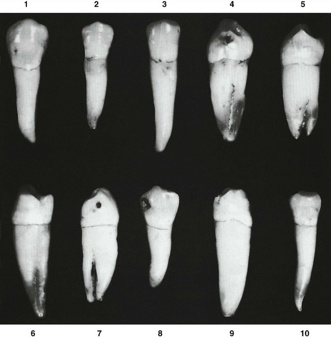
Figure 10-12 Mandibular first premolar. Ten specimens with uncommon variations are shown. 1, Crown oversized. 2, Crown and root diminutive. 3, Mesial and distal sides of crown straight; cervix wide mesiodistally; root extra long. 4, Unusual formation of lingual portion of crown; root with deep developmental groove mesially. 5, Bifurcated root. 6, Lingual cusp long; little lingual curvature; root of extra length. 7, No lingual cusp; root bifurcated. 8, Dwarfed root. 9, Crown poorly formed; root unusually long. 10, Very long curved root for crown so small.
The characteristics that resemble those of the mandibular canine are as follows:
The characteristics that resemble those of the second mandibular premolar are as follows:
Although the root of the mandibular first premolar is shorter generally than that of the mandibular second premolar, it is closer to the length of the second premolar root than it is to that of the mandibular canine (Table 10-1).
DETAILED DESCRIPTION OF THE MANDIBULAR FIRST PREMOLAR FROM ALL ASPECTS
Buccal Aspect
From the buccal aspect, the form of the mandibular first premolar crown is nearly symmetrical bilaterally (see Figures 10-2, 10-7, 10-8, and 10-9). The middle buccal lobe is well developed, which results in a large, pointed buccal cusp. The mesial cusp ridge is shorter than the distal cusp ridge.
The contact areas are broad from this aspect; they are almost at the same level mesially and distally, this level being a little more than half the distance from cervical line to cusp tip. The measurement mesiodistally at the cervical line is small compared with the measurement at the contact areas.
From the buccal aspect, the crown is roughly trapezoidal (see Figure 4-16, C). The cervical margin is represented by the shortest of the uneven sides.
The crown exhibits little curvature at the cervical line buccally, because of the slight curvature of the cervical line on the mesial and distal surfaces of the tooth. The crest of curvature of the cervical line buccally approaches the center of the root buccally.
The mesial outline of the crown is straight or slightly concave above the cervical line to a point where it joins the curvature of the mesial contact area. The center of the contact area mesially is occlusal to the cervical line, a distance equal to a little more than half the crown length. The outline of the mesial slope of the buccal cusp usually shows some concavity unless wear has obliterated the original form.
The tip of the buccal cusp is pointed and, in most cases, is located a little mesial to the center of the crown buccally (see Figure 10-9, 3, 7, 8, and 9). The mandibular canine has the same characteristic to a greater degree.
The distal outline of the crown is slightly concave above the cervical line to a point where it is confluent with the curvature describing the distal contact area. This curvature is broader than that describing the curvature of the mesial contact area. The distal slope of the buccal cusp usually exhibits some concavity.
The cervix of the mandibular first premolar crown is narrow mesiodistally when compared with the crown width at the contact areas.
The root of this tooth is 3 or 4 mm shorter than that of the mandibular canine, although the outline of the buccal portion of the root bears a close resemblance to that of the canine.
The buccal surface of the crown is more convex than in the maxillary premolars, especially at the cervical and middle thirds.
The development of the middle buccal lobe is outstanding, ending in a pointed buccal cusp. Developmental depressions are often seen between the three lobes (see Figure 10-9, 2, 3, 8, and 10).
The continuous ridge from the cervical margin to the cusp tip is called the buccal ridge.
In general, the enamel of the buccal surface of the crown is smooth and shows no developmental grooves and few developmental lines. If the latter are present, they are seen as very fine horizontal cross lines at the cervical portion.
Lingual Aspect
The crown of the mandibular first premolar tapers toward the lingual, since the lingual measurement mesiodistally is less than that buccally. The lingual cusp is always small (see Figures 10-3, 10-7, and 10-8). The major portion of the crown is made up of the middle buccal lobe (see Figure 10-11). This makes it resemble the canine.
The crown and the root taper markedly toward the lingual so that most of the mesial and distal surfaces of both may be seen from the lingual aspect.
The occlusal surface slopes greatly toward the lingual in a cervical direction down to the short lingual cusp. Most of the occlusal surface of this tooth can therefore be seen from this aspect.
The cervical portion of the crown lingually is narrow and convex, with concavities in evidence between the cervical line and the contact areas on the lingual portion of mesial and distal surfaces. The contact areas and marginal ridges are pronounced and extend out above the narrow cervical portion of the crown.
Although the lingual cusp is short and less developed (resembling a strongly developed cingulum at times), it usually shows a pointed tip. This cusp tip is in alignment with the buccal triangular ridge of the occlusal surface, which is in plain view. The mesial and distal occlusal fossae are on each side of the triangular ridge (see Figure 10-1).
A characteristic of the lingual surface of the mandibular first premolar is the mesiolingual developmental groove. This groove acts as a line of demarcation between the mesiobuccal lobe and the lingual lobe and extends into the mesial fossa of the occlusal surface.
The root of this tooth is much narrower on the lingual side, and there is a narrow ridge, smooth and convex, the full length of the root. This formation allows most of the mesial and distal surfaces of the root to be seen. Often, developmental depressions in the root may be seen with developmental grooves mesially. The root of this tooth tapers evenly from the cervix to a pointed apex.
Mesial Aspect
From the mesial aspect (see Figures 10-4, 10-7, 10-8, and 10-10), the mandibular first premolar shows an outline that is fundamental and characteristic of all mandibular posterior teeth when viewed from the mesial or distal aspect. The crown outline is roughly rhomboidal (see Figure 4-16, E), and the tip of the buccal cusp is nearly centered over the root. The convexity of the outline of the lingual lobe is lingual to the outline of the root. The surface of the crown presents an overhang above the root trunk in a lingual direction. The tip of the cusp is on a line approximately with the lingual border of the root. This differs from the situation found in maxillary posterior teeth, where both buccal and lingual cusp tips are well within the confines of the root trunks.
The mandibular first premolar, when viewed from the mesial aspect, often shows the buccal cusp centered over the root (see Figure 10-4). In other instances, the buccal cusp tip is a little buccal to the center, corresponding to the typical placement of buccal cusps on all mandibular posterior teeth.
The buccal outline of the crown from this aspect is prominently curved from the cervical line to the tip of the buccal cusp; the crest of the curvature is near the middle third of the crown. This accented convexity and the location of the crest of contour are characteristic of all mandibular posterior teeth on the buccal surfaces.
The lingual outline of the crown, representative of the lingual outline of the lingual cusp, is a curved outline of less convexity than that of the buccal surface. The crest of curvature lingually approaches the middle third of the crown, with the curvature ending at the tip of the lingual cusp, which is in line with the lingual border of the root.
The distance from the cervical line lingually to the tip of the lingual cusp is about two thirds of that from the cervical line buccally to the tip of the buccal cusp.
The mesiobuccal lobe development is prominent from this aspect; it creates by its form the mesial contact area and the mesial marginal ridge, which in turn has a sharp inclination lingually in a cervical direction. The lingual border of the mesial marginal ridge merges with the developmental depression mesiolingually; this harbors the mesiolingual developmental groove.
Some of the occlusal surface of the crown mesially may be seen with the mesial portion of the buccal triangular ridge. The slope of this ridge parallels that of the mesial marginal ridge, although the crest of the triangular ridge is above it. The sulcus formed by the convergence of buccal and lingual triangular ridges is directly above the mesiolingual groove from this aspect.
The cervical line on the mesial surface is rather regular, curving occlusally. The crest of the curvature is centered buccolingually, the average curvature being about 1 mm in extent. It may, however, be a fraction of a millimeter, or the line may be straight across buccolingually.
The surface of the crown mesially is smooth except for the mesiolingual groove. The surface is plainly convex at the mesial contact area, which is centered on a line with the tip of the buccal cusp. Immediately below the convexity of the contact area, the surface is sharply concave between that area and the cervical line. The distance between the contact area and the cervical line is very short.
The root outline from the mesial aspect is a tapered form the cervix, ending in a relatively pointed apex in line with the tip of the buccal cusp. The lingual outline may be straight, the buccal outline more curved.
The mesial surface of the root is smooth and flat from the buccal margin to the center. From this point, it too converges sharply toward the root center lingually, often displaying a deep developmental groove in this area. Shallow grooves are nearly always in evidence, and occasionally a deep developmental groove will end in a bifurcation at the apical third (see Figure 10-12, 5 and 7).
Distal Aspect
The distal aspect of the mandibular first premolar differs from the mesial aspect in some respects (see Figures 10-5, 10-7, and 10-8). The distal marginal ridge is higher above the cervix, and it does not have the extreme lingual slope of the mesial marginal ridge, being more nearly at right angles to the axis of crown and root. The marginal ridge is confluent with the lingual cusp ridge; it has no developmental groove on the distal marginal ridge. The major portion of the distal surface of the crown is smoothly convex, the spheroidal form having an unbroken curved surface.
Below this curvature and just above the cervical line, a concavity is to be noted that is linear in form and that extends buccolingually. The distal contact area is broader than the mesial, although it is centered in the same relation to the crown outlines. The center of the distal contact area is at a point midway between buccal and lingual crests of curvature and midway between the cervical line and the tip of the buccal cusp.
The curvature of the cervical line distally may be the same as that found mesially, although less curvature distally is the general rule when one is describing all posterior teeth.
The surface of the root distally exhibits more convexity than was found mesially. A shallow developmental depression is centered on the root, but rarely does it contain a deep developmental groove.
The distal surface slopes from the buccal margin toward the center of the root lingually, but the slope is more gradual than that found mesially.
Occlusal Aspect
The occlusal aspects of many specimens show considerable variation in the gross outlines of this tooth. Both mandibular premolars exhibit more variations in form occlusally than do the maxillary premolars (see Figures 10-6, 10-7, 10-8, and 10-11).
The usual outline form of the mandibular first premolar from the occlusal aspect is roughly diamond-shaped and similar to the incisal aspect of mandibular canines (see Figure 10-11, 1, 3, 4, and 7 through 10). Some of these teeth have a circular form similar to that of some mandibular second premolars (see Figure 10-11, 2); others conform to the gross outlines of the more common second premolars (see Figure 10-11, 5 and 6).
The characteristics common to all mandibular first premolars, regardless of type, when viewed from the occlusal aspect are as follows:
The occlusal surface harbors two depressions. These are called the mesial and distal fossae because of their irregular form, even though they correspond in location to the mesial and distal triangular fossae of other posterior teeth.
The most common type of mandibular first premolar shows a mesiolingual developmental depression and groove. These constrict the mesial surface of the crown and create a smaller mesial contact area that is in contact with the mandibular canine. The distal portion of the crown is described by a larger arc that creates a broader contact area in contact with the second mandibular premolar, which has a broader proximal surface than the canine (see Figures 5-7, D,Figures 5-10, B,Figures 5-17, D,).
The mesial fossa is more linear in form, being more sulcate and containing the mesial developmental groove, which extends buccolingually. This groove is confluent with its extension, which becomes the mesiolingual developmental groove as it passes over the mesiolingual surface. The distal fossa is more circular in most cases and is circumscribed by the distobuccal cusp ridge, the distal marginal ridge, the buccal triangular ridge, and the distolingual cusp ridge.
The distal fossa may contain a distal developmental groove that is crescent-shaped (see Figure 10-11, 2). It may harbor a distal developmental pit with accessory supplemental grooves radiating from it (see Figure 10-11, 10), or it may contain a linear groove running mesiodistally with an arrangement resembling the typical triangular fossa (see Figure 10-11, 4, 5, and 6).
Because of the position of this crown over the root, most of the buccal surface may be seen from the occlusal aspect, whereas very little of the lingual surface is in view.
Mandibular Second Premolar
Figures 10-13 through 10-21 illustrate the mandibular second premolar from all aspects. The mandibular second premolar resembles the mandibular first premolar from the buccal aspect only. Although the buccal cusp is not as pronounced, the mesiodistal measurement of the crown and its general outline are similar (Table 10-2). The tooth is larger and has better development in other respects. This tooth assumes two common forms. The first form, which probably occurs most often, is the three-cusp type, which appears more angular from the occlusal aspect (see Figure 10-17). The second form is the two-cusp type, which appears more rounded from the occlusal aspect (see Figure 10-20, 1, 2, 7, and 10).
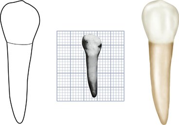
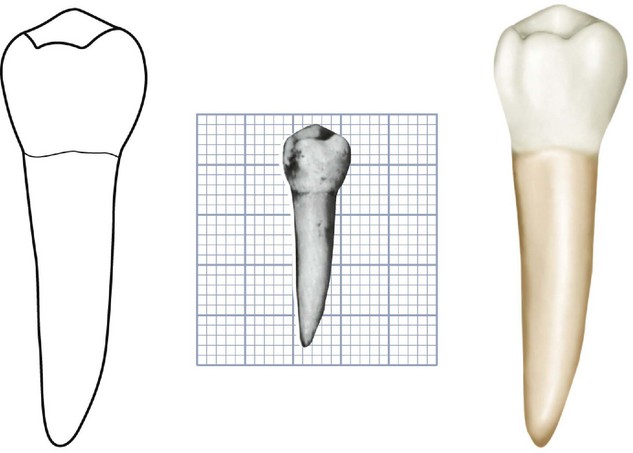
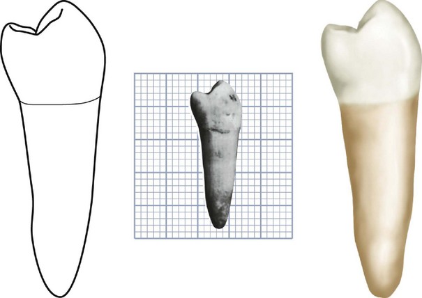
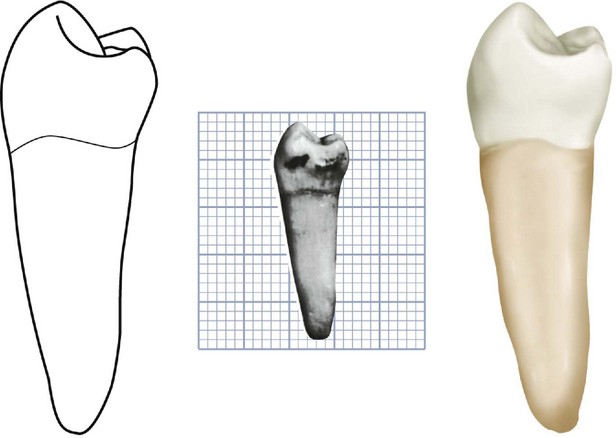
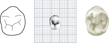
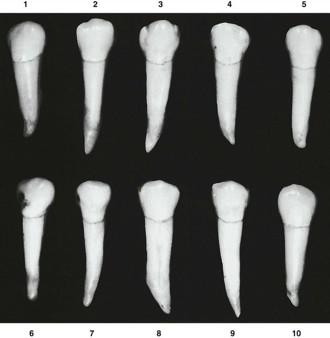
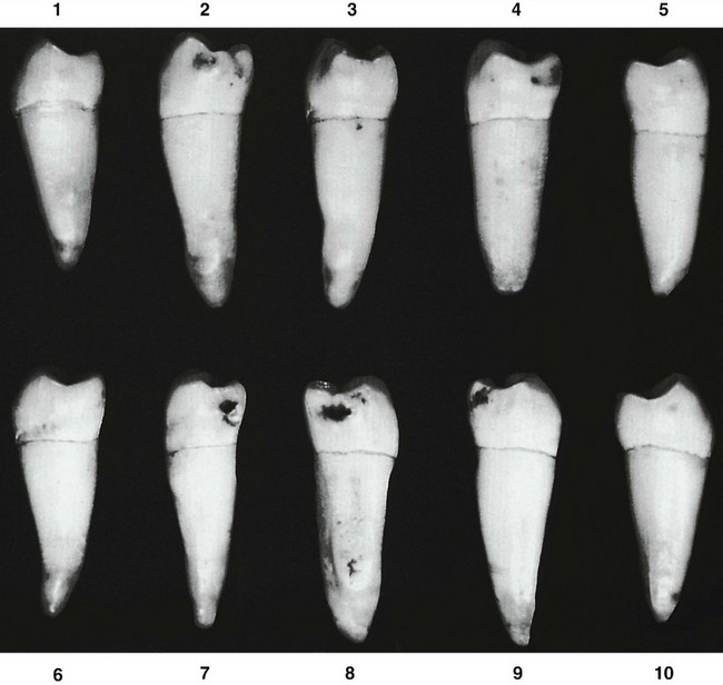
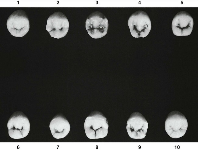
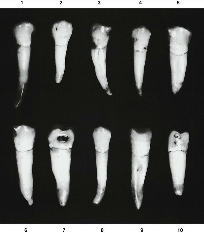
Figure 10-21 Mandibular second premolar. Ten specimens with uncommon variations are shown. 1, Root extremely long. 2, Root dwarfed. 3, Malformed root; developmental groove on buccal surface. 4, Contact areas on crown high and constricted. 5, Crown oversize; developmental groove buccally on root. 6, Root oversize. 7, Root malformed and of extra length. 8, Root very long with blunt apex; extreme curvature at apical third. 9, Crown and root oversized; developmental groove buccally on root. 10, Crown narrow buccolingually; very little curvature buccally and lingually.

The two types differ mainly in the occlusal design. The outlines and general appearance from all other aspects are similar.
The single root of the second premolar is larger and longer than that of the first premolar. The root is seldom, if ever, bifurcated, although some specimens show a deep developmental groove buccally (see Figure 10-18, 3 and 6). Often a flattened area appears in this location. Ten specimens with uncommon variations are shown in Figure 10-21.
DETAILED DESCRIPTION OF THE MANDIBULAR SECOND PREMOLAR FROM ALL ASPECTS
To describe the separate aspects of this tooth, direct comparisons are made with the mandibular first premolar except for the occlusal aspect.
Buccal Aspect
From the buccal aspect, the mandibular second premolar presents a shorter buccal cusp than the first premolar, with mesiobuccal and distobuccal cusp ridges showing angulation of less degree (see Figures 10-13 and 10-18). The contact areas, both mesial and distal, are broad. The contact areas appear to be higher because of the short buccal cusp.
The root is broader mesiodistally than that of the first premolar, the extra breadth appearing for most of its length, and the root ends in an apex that is more blunt. In other respects, the two teeth are quite similar from this aspect.
Lingual Aspect
From the lingual aspect, the second premolar crown shows considerable variation from the crown portion of the first premolar (see Figure 10-14). The variations are as follows:
In the two-cusp type, the single lingual cusp development attains equal height with the three-cusp type. The two-cusp type has no groove, but it shows a developmental depression distolingually where the lingual cusp ridge joins the distal marginal ridge (see Figure 10-20, 2 and 3).
The lingual surface of the crown of all mandibular second premolars is smooth and spheroidal, having a bulbous form above the constricted cervical portion.
The root is wide lingually, although not quite as wide as the buccal portion. Less difference in dimension is evident here than was found on the first premolar, so that much less convergence toward the lingual is seen.
Because in most instances the lingual portion of the crown converges little from the buccal portion, less of the mesial and distal sides of this tooth may be seen from this aspect than are seen from the lingual aspect of the first premolar.
The lingual portion of the root is smoothly convex for most of its length. Considered overall, the second premolar is the larger of the two mandibular premolars.
Mesial Aspect
From the mesial aspect (see Figures 10-15 and 10-19), the second premolar differs from the first premolar as follows:
Distal Aspect
The distal aspect of the mandibular second premolar is similar to the mesial aspect, except that more of the occlusal surface may be seen (see Figure 10-16). This is possible because the distal marginal ridge is at a lower level than the mesial marginal ridge when the tooth is posed vertically. The crowns of all posterior teeth are tipped distally to the long axes of the roots, so that when the specimen tooth is held vertically, more of the occlusal surface may be seen from the distal aspect than from the mesial aspect. This is a characteristic possessed by all posterior teeth, mandibular and maxillary. The angulation of occlusal surfaces to long axes of all posterior teeth is an important observation to remember, not only in the study of individual tooth forms but also later, in the study of alignment and occlusion.
Occlusal Aspect
As mentioned earlier, two common forms of this tooth are evident. The outline form of each type shows some variation from the occlusal aspect (see Figures 10-17 and 10-20). The two types are similar in that portion that is buccal to the mesiobuccal and distobuccal cusp ridges.
The three-cusp type appears square lingual to the buccal cusp ridges when highly developed (see Figure 10-20, 8). The round, or two-cusp, type appears round lingual to the buccal cusp ridges (see Figure 10-20, 3).
The square type (see Figure 10-20, 8) has three cusps that are distinct; the buccal cusp is the largest, the mesiolingual cusp is next, and the distolingual cusp is the smallest.
Each cusp has well-formed triangular ridges separated by deep developmental grooves. These grooves converge in a central pit and form a Y shape on the occlusal surface. The central pit is located midway between the buccal cusp ridge and the lingual margin of the occlusal surface and slightly distal to the central point between mesial and distal marginal ridges.
Starting at the central pit, the mesial developmental groove travels in a mesiobuccal direction and ends in the mesial triangular fossa just distal to the mesial marginal ridge. The distal developmental groove travels in a distobuccal direction, is somewhat shorter than the mesial groove, and ends in the distal triangular fossa mesial to the distal marginal side. The lingual developmental groove extends lingually between the two lingual cusps and ends on the lingual surface of the crown just below the convergence of the lingual cusp ridges. The mesiolingual cusp is wider mesiodistally than the distolingual cusp. This arrangement places the lingual developmental groove distal to center on the crown.
Supplemental grooves and depressions are often seen, radiating from the developmental grooves. Occasionally, a groove crosses one or both of the marginal ridges. On a tooth of this type, the point angles are distinct. Developmental grooves are often deep.
Figure 10-20, 8 is representative. Variations of this development may be seen in Figure 10-20, 4, 5, 6, and 9.
The round or two-cusp type (see Figure 10-20, 3) differs considerably from the three-cusp type when viewed from the occlusal aspect. It is a true typal form of the two-cusp type. Variations may be seen in Figure 10-20, 1, 2, 7, and 10.
The occlusal characteristics of the two-cusp type are as follows:
A central developmental groove on the occlusal surface travels in a mesiodistal direction. This groove may be straight (see Figure 10-20, 3), but it is most often crescent-shaped (see Figure 10-20, 1, 7, and 10). The central groove has its terminals centered in mesial and distal fossae, which are roughly circular depressions having supplemental grooves and depressions radiating from the central groove and its terminals. The enamel surface inside these fossae and around their peripheries is very irregular, acting as a contrast to the smoothness of cusp ridges, marginal ridges, and the transverse ridge from buccal cusp to lingual cusp.
Some of these teeth show mesial and distal developmental pits centered in the mesial and distal fossae instead of an unbroken central groove (see Figure 10-20, 2).
Although photographs do not demonstrate it very well, most of these two-cusp specimens show a developmental depression crossing the distolingual cusp ridge.
Carlsen O. Human lower premolars: macro-morphologic observations on the ontogenesis of the root complex. Scand J Dent Res. 1970;78:5.
Kraus B, Purr ML. Lower first premolar: a definition of discrete morphologic traits. J Dent Res. 1953;32:554.
Ludwig PJ. The mandibular second premolars: morphologic variation and inheritance. J Dent Res. 1957;36:263.
Osborn JR, editor. Dental anatomy and embryology. Oxford: Blackwell Scientific Publications, 1981.
Renner RP. An introduction to dental anatomy and esthetics. Chicago: Quintessence; 1985.
Schumacher G-H. Odontographie: Eine Oberflächenenanatomie der Zähne. Leipzig: JM Barth; 1983.
 site for additional study resources.