Diseases of cloned offspring

The successful cloning of domestic animals using somatic-cell nuclear transfer has resulted in birth of offspring with a high frequency of clinical abnormalities. Cloning of livestock and horses is achieved by transfer of nuclear material from the cell of an adult animal to the enucleated egg of an animal of the same species (somatic-cell nuclear transfer) with subsequent implantation of the resulting embryo in a surrogate dam and birth of a live, viable offspring.1 However, the use of nuclear material from somatic cells of adult animals, and from fetal cells, does not result in normal development of the embryo and placenta. The abnormal development is a consequence of altered methylation of the genome in transferred nuclear material.2 In normal reproduction, the paternal genome is demethylated during passage through the oocyte and fusion with the maternal genome. Consequently, the methylation marks of the two genomes (paternal and maternal) are different at the end of the cleavage process. Transfer of somatic nuclear material into an enucleated oocyte results in exposure of both genomes to the active demethylating process in the cytoplasm of the oocyte and uniform demethylation of both genomes.2,3 The loss of these parent-specific epigenetic markers results in widespread dysregulation of imprinted genes and subsequent abnormalities in the placenta, fetus and newborn.

A small proportion of transferred blastocysts develop in viable animals. For cattle, of 134 recipients that received blastocysts, 50 were pregnant 40 days after blastocyst transfer and 23 had full-term pregnancies.4 For all species studied, fewer than 3% of cloned embryos result in birth of viable animals.1 Abnormalities in placenta and newborn cloned animals are reported for cattle and sheep but not for pigs and equids (horses and mules).1,5 Factors influencing the risks of abnormalities in newborns have not been well defined, but include the source of the nuclear material, with frequency of birth of live animals born after somatic cell nuclear transfer from well-differentiated tissue (e.g. fibroblasts) or fetal somatic cells being lower than after nuclear transfer from embryonic cells (7%, 15% and 34%, respectively).6

The cause of placental, fetal and neonatal abnormalities is abnormal expression of imprinted genes as a consequence of transfer of nuclear material from differentiated somatic cells, conditions and media used for maintenance and culture of cytoplasts and blastocysts, and techniques used for handling cells.1,7 Candidate genes for large offspring syndrome include IGF-2 and IGFBP-2, insulin-like growth factor (IGF) concentrations in plasma of cloned calves being higher than that of normal calves7,8 although others, such as genes related to endothelin-1 production, might well be involved.9

Clinical findings in cloned calves and lambs include abortion, placental abnormalities, large birth size, poor extrauterine viability, respiratory disease, cardiovascular abnormalities and neurologic disease compatible with neonatal encephalopathy. Abortion occurs after day 90 of gestation in 30–50% of pregnancies in cattle resulting from transfer blastocysts containing transferred nuclear material.6 Abnormalities, including hydroallantois, are present in approximately 25% of advanced pregnancies.6 Placental abnormalities include hydroallantois, a reduction in the number of placentomes (from a normal of approximately 100 to as few as 26–70 in cloned calves),7,10 abnormally large placentomes (140 g in cloned calves vs 33 g in conventional calves) and edema of the placenta.6,7,11 Maternal retention of the placenta is common and occurs in most cows.11 Duration of gestation is probably longer in cloned calves, although the frequent delivery of cloned calves by cesarian section makes assessment of gestational duration difficult. Cloned calves are heavier than conventional calves, often by as much as 25%, a well-recognized part of the ‘large offspring syndrome’ that affects calves born as a result of reproductive manipulation, including in-vitro fertilization.6,12 Viability of cloned calves that are born alive (commonly by cesarian section) is less than that of conventional calves – only approximately two-thirds of cloned calves born alive survive more than 1 month,10 although others report better survival.11 Similar results are reported for horses.13

A high proportion of cloned calves have clinically detectable abnormalities at or soon after birth, including sepsis, neonatal encephalopathy, respiratory failure, umbilical abnormalities, anemia, flexure contracture, abdominal distension and renal dysfunction. Respiratory failure is a common finding and might reflect persistent fetal circulation or inadequate surfactant production, as evidenced by the high pulmonary artery pressures and signs consistent with patent ductus arteriosus. Left heart failure, which can also cause pulmonary hypertension, is reported in cloned calves.11 Umbilical abnormalities are evident as abnormal umbilical cord structure (multiple arteries and veins) and large size, with a high risk of hemorrhage from the umbilical cord after birth. Cloned calves have higher body temperatures than do conventional calves.7

Hematological abnormalities include anemia and decreased mean corpuscular volume. Biochemical abnormalities include hypoxemia, azotemia and hypoglycemia. Plasma leptin and IGF-2 concentrations are higher, and thyroxine lower, in cloned calves.7 Serum cortisol and ACTH stimulation tests do not differ between cloned and conventional calves.7

Necropsy examination reveals placentomegaly, presence of excess pleural and peritoneal fluid, hepatomegaly, interstitial pneumonia or pulmonary consolidation and alveolar proteinosis, right ventricular dilation and hepatocellular vacuolation.11

Treatment is supportive and directed toward correcting hypoxemia and providing nutritional, fluid and environmental support (see above).

There are currently no recognized methods for preventing these abnormalities, but presumably improvements in methodology and culture techniques will result in fewer cloned offspring with these abnormalities.

REFERENCES

1 Vanderwall DK, et al. J Am Vet Med Assoc. 2004;225:1694.

2 Jaenisch R. New Engl J Med. 2004;351:2787.

3 Kang YK, et al. Nat Genet. 2001;28:173.

4 Kato Y, et al. J Reprod Fertil. 2000;120:231.

5 Tsunoda Y, Kato Y. Differentiation. 2002;69:158.

6 Heyman Y, et al. Biol Reprod. 2002;66:6.

7 Chavette-Palmer P, et al. Biol Reprod. 2002;66:1596.

8 Young LE, et al. Theriogenology. 1999;51:196.

9 Wilkins PA, et al. J Vet Intern Med. 2005;19:594.

10 Hill JR, et al. Cloning. 2001;3:83.

11 Hill JR, et al. Theriogenology. 1999;51:1451.

12 Young LE, et al. Rev Reprod. 1998;3:155.

13 Galli C, et al. Nature. 2004;424:635.

Neonatal infection

Synopsis

Etiology Common infections for each animal species are listed under etiology below. Most are bacterial.

Epidemiology Commonly predisposed by management and environmental factors that increase the exposure risk and load and decrease the resistance of the neonate.

Clinical findings Septicemia or bacteremia with localization is most common but signs can be specific for the infecting agent.

Clinical pathology White blood cell and differential counts, toxic change, serum immunoglobulin concentrations, arterial oxygen concentrations, metabolic acidosis, fibrinogen levels, blood culture.

Necropsy findings Specific to disease.

Diagnostic confirmation Specific to disease.

Treatment General therapy may include antibacterial therapy, blood or plasma transfusion, correction of acid–base disturbance, fluid and electrolyte therapy, and supportive treatment.

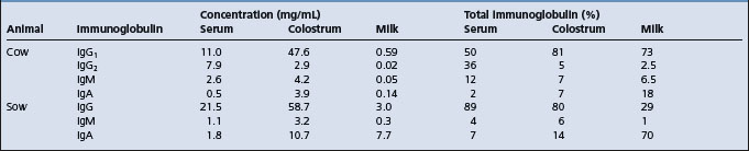
Infection is a common cause of morbidity and mortality in neonates. There are a number of specific infectious pathogens that can cause disease. Other infectious agents, normally considered to have low virulence, can also cause disease if the immunological status of the neonate is not at an optimum level. Maternal immunoglobulins are not transferred transplacentally in ungulates and the newborns are at particular risk for infectious disease during the neonatal period because they rely on the acquisition of immunoglobulins from colostrum for passive antibody protection.

ETIOLOGY

In domestic farm animals the common infections that can produce disease during the neonatal period are as follows. (Relative importance and prevalence statistics are not given, as these vary from area to area and with differing management systems.)

Calves

Bacteremia and septicemia associated with Escherichia coli, Listeria monocytogenes, Pasteurella spp., streptococci or Salmonella spp.

Enteritis associated with enterotoxigenic E. coli, Salmonella spp., rotavirus and coronavirus, Cryptosporidium parvum and Clostridium perfringens types A, B and C; and occasionally by the virus of infectious bovine rhinotracheitis and bovine virus diarrhea.

Pigs

Septicemia with or without localization in joints, endocardium and meninges associated with Streptococcus suis, Streptococcus equisimilis, Streptococcus zooepidemicus and L. monocytogenes

Bacteremia, septicemia and enteritis associated with E. coli

Transmissible gastroenteritis, Aujeszky’s disease, swine pox, enterovirus infections, and vomiting and wasting disease are associated with viruses

Enteritis associated with C. perfringens, Campylobacter spp., rotavirus and Coccidia spp.

Arthritis and septicemia associated with Erysipelothrix rhusiopathiae.

Foals

Septicemia with localization associated with E. coli, Actinobacillus equuli, Klebsiella pneumoniae, α-hemolytic streptococci, S. zooepidemicus, L. monocytogenes, Rhodococcus equi and Salmonella typhimurium

Enteritis associated with C. perfringens types A, B, and C., Clostridium difficile, R. equi, Salmonella spp., Strongyloides westeri, C. parvum and rotavirus.

Lambs

Septicemia or bacteremia with localization in joints and/or synovia and/or leptomeninges associated with E. coli, L. monocytogenes, streptococci, micrococci, E. rhusiopathiae and Chlamyodophila spp.

Enteritis associated with enterotoxigenic E. coli, Salmonella spp., rotavirus and coronavirus and C. parvum

Lamb dysentery associated with C. perfringens type B and C

Gas gangrene of the navel associated with Clostridium septicum, Clostridium novyi and Clostridium chauvoei

Pyemia associated with Staphylococcus aureus, Fusobacterium necrophorum and Arcanobacterium pyogenes

Pneumonia, polyserositis and peritonitis associated with Pasteurella multocida and Mannheimia haemolytica.

The following agents are recorded as causing neonatal infections but are less common than those listed above and not of as great importance.

Calves

Pseudomonas aeruginosa, Streptococcus pyogenes, Streptococcus faecalis, S. zooepidemicus, Pneumococcus spp.; enteritis due to Providencia stuartii, Chlamydophila spp., A. equuli.

Lambs

S. aureus (tick pyemia); enteritis due to E. coli, rotavirus; pneumonia due to Salmonella abortus–ovis.

Foals

Enterobacter cloacae, S. aureus, Pasteurella multocida, P. aeruginosa, A. pyogenes, Serratia marcescens.

All species

Nonspecific infections are associated with pyogenic organisms, including Arcanobacterium pyogenes and Fusobacterium necrophorus; S. faecalis, S. zooepidemicus, Micrococcus spp. and Pasteurella spp. occur in all species.

EPIDEMIOLOGY

The occurrence of neonatal disease is broadly influenced by two main factors: the exposure or infection pressure of the infectious agent to the neonate and the ability of the neonate to modulate the infection so that disease does not occur. With some agents the organism is sufficiently virulent in its own right that an exposure can lead to disease. With others, the majority, the defenses of the host must be compromised or the infection challenge must be very high before clinical disease occurs. Management of the neonate has a great influence on both these factors and the recognition and correction of these risks is the key to the prevention of neonatal disease in both the individual and the group.

Sources of infection

Postnatal infection

The vast majority of infections are acquired by the neonate after birth from the enteric or respiratory tract flora of the dam, from the environment or from close contact with other infected neonates. Depending upon the specific agent, the reservoir of infection may be in a carrier animal or in the environment. Details for the common neonatal diseases are given under the individual disease headings in the chapters on special medicine.

Prenatal infection

Some bacterial infections that manifest with neonatal disease are acquired in utero. The majority of these are agents that cause abortion, and neonatal septicemia is only part of the spectrum of abortion and perinatal death associated with these agents. Examples would be many of the agents producing abortion in sheep.

Some septicemic infections in foals, particularly those associated with A. equuli, S. zooepidemicus, Salmonella abortivoequina and possibly some E. coli septicemic infections, are acquired by prenatal infection. If the disease is intrauterine in origin it must gain entrance via the placenta, and probably by means of a placentitis due to a blood-borne infection or an existing endometritis. In the latter case, disinfecting the uterus before mating becomes an important hygienic precaution; disinfecting the environment may have little effect on the incidence of the disease.

Viral infections that are acquired in utero are listed in the section on congenital disease.

Routes of transmission

The portal of infection is commonly by ingestion but may occur via aerosol infection of the respiratory tract. Organisms capable of invading to produce a bacteremia and septicemia invade through the nasopharynx or through the intestinal epithelium. An alternate route of infection and invasion is via the umbilicus. Routes of excretion are via the feces in enteric disease and the nasal secretions, urine and sometimes the feces in septicemic disease to result in contamination of the neonatal environment.

Where neonates are in groups or in close contact, direct transmission by fecal, respiratory secretion and urine aerosols can also occur. Neonatal bull calves that are group-housed and that suck each other’s navels can transmit infection by this activity.

Risk factors and modulation of infection

Immunity

All newborn farm animals are more susceptible to infection than their adult counterparts. The calf, lamb, piglet and foal are born without significant levels of immunoglobulins and possess almost no resistance to certain diseases until after they have ingested colostrum and absorbed sufficient quantities of immunoglobulins from the colostrum. Failure of transfer of colostral immunoglobulins is a major determinant and is discussed under that heading later in this chapter.

Immune responsiveness

All components of the immune system are present in foals and calves at birth but the immune system of the newborn animal is less mature than its adult counterpart, at least for the first 30 days of life, and does not respond as effectively to many antigens.

Immune responsiveness is age-dependent but also varies with the antigen.1 In colostrum-fed animals part of the inefficiency of the newborn to produce humoral antibody following infection of antigen is the interference from circulating colostral antibody and the downregulation by colostrum of endogenous immunoglobulin production.2-4

Colostrum-deprived calves respond actively to injected antigens and are believed to be immunologically competent at birth with respect to most antigens. Immune competence begins during fetal life and the age of gestation at which this occurs varies according to the nature of the antigen. The bovine fetus will produce antibody to some viruses, beginning at 90–120 days, and by the third trimester of gestation it will respond to a variety of viruses and bacteria.5 The lamb will respond to some antigens beginning as early as 41 days and not until 120 days for others. The piglet at 55 days and the fetal foal also respond to injected antigens.

The presence of high levels of antibody in the precolostral serum of newborn animals suggests that an in-utero infection was present, which is useful for diagnostic purposes. The detection of immunoglobulins and specific antibodies in aborted fetuses is a useful aid in the diagnosis of abortion in cattle.

Exposure pressure

The exposure pressure is a factor of the cleanliness of the environment of the neonate. The phenomenon of a ‘buildup of infection’ in continual-throughput housing for neonatal animals has been recognized for decades and has been translated to many observations of risk for neonatal disease associated with suboptimal hygiene and stocking density in both pen and paddock birthing areas. Details for the individual species are provided in the section on perinatal disease.

Age at exposure

With several agents that produce neonatal disease, the age of the neonate at infection and the infecting dose have a significant influence on the outcome. Examples are the importance of age with respect to susceptibility to disease associated with some enteric infections. Disease associated with enteropathogenic E. coli and with C. perfringens type B and C occurs only in young animals and if infection can be avoided by hygiene in this critical period disease will not occur regardless of subsequent exposure. Colostrum-deprived calves show significant resistance to challenge at 7 days of age with strains of E. coli that invariably produce septicemic disease if challenged at the time of birth and isolation of an immunocompromised neonate is an important factor in its survival. Thus the management of the neonate and its environment is a critical determinant of its health. Age at exposure also varies with the epidemiology of the pathogen and segregated early weaning is used to reduce transmission of and infection with certain pathogens in pigs.

Animal risk factors

Animal risk factors that predispose infection include those that interfere with sucking drive and colostral intake, such as cold stress and dystocia. These are detailed in the preceding section on perinatal disease.

PATHOGENESIS

The pathogenesis varies with the neonatal infectious disease under consideration and is given for each of these in the special medicine section.

Following invasion via the nasopharynx and the gastrointestinal tract, the usual pattern of development is a bacteremia followed by septicemia with severe systemic signs; or a bacteremia with few or no systemic signs, followed by localization in various organs. If the portal of entry is the navel, local inflammation occurs – ‘navel ill’ – which can be easily overlooked if clinical examination is not thorough. From the local infection at the navel, extension may occur to the liver or via the urachus to the bladder and result in chronic ill-health. Extension systemically may produce septicemia.6

Localization is most common in the joints, producing a suppurative or nonsuppurative arthritis. Less commonly there is localization in the eye to produce a panophthalmitis, in the heart valves to cause valvular endocarditis, or in the meninges to produce a meningitis.

In some cases these secondary lesions take time to develop and signs usually appear at 1–2 weeks of age. This is especially true with some of the streptococcal infections, where bacteremia may be present for several days before localization in the joints and meninges produces clinical signs. Bacterial meningitis in newborn ungulates is preceded by a bacteremia followed by a fibrinopurulent inflammation of the leptomeninges, choroid plexuses and ventricle walls but does not affect the neuraxial parenchyma. It is proposed that the bacteria are transported in monocytes, which do not normally invade the neuraxial parenchyma.

Dehydration, acid–base and electrolyte imbalance can occur very quickly in newborn animals, whether diarrhea and vomiting (pigs) are present or not, but obviously are more severe where there is fluid loss into the gastrointestinal tract. In Gram-negative sepsis the prominent signs are those of endotoxemia.

CLINICAL FINDINGS

The clinical findings depend on the rapidity of growth of the organism, its propensity to localize and its potential to produce toxemia. With organisms that have a low propensity for toxemia there is fever, depression, anorexia and signs referable to localization. These include endocarditis with a heart murmur; panophthalmitis with pus in the anterior chamber of the eye; meningitis with rigidity, pain and convulsions; and polyarthritis with lameness and swollen joints. With more virulent organisms there are clinical signs of toxemia as well as bacteremia, including fever, severe depression, prostration, coma, petechiation of mucosae, dehydration, acidosis and rapid death.7,8

The clinical and clinicopathological characteristics of the septicemic foal have been detailed in an outbreak of septicemia in colostrum-deprived foals9 and on the clinical records of 38 septicemic foals admitted to a referral clinic,10 where the survival rate of septicemic foals, 26%, was markedly less than the rate for all other foal admissions. The major clinical findings included lethargy, unwillingness to suck, inability to stand without assistance but remaining conscious, unawareness of environment and thrashing or convulsing, diarrhea, respiratory distress, joint distension, central nervous system abnormalities, uveitis and colic. Fever was not a consistent finding.

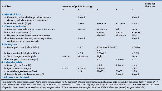
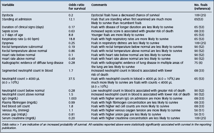
A sepsis score has been developed for foals based on 14 measures related to historical, clinical and laboratory data (Table 3.6). The score derived from the collective differential scoring of these data has been found to be more sensitive and specific for infection than any parameter taken individually.7 However, a subsequent study of 168 foals presented to a university hospital found that the sepsis score correctly predicted sepsis in 58 out of 86 foals and nonsepsis in 24 out of 45 foals resulting in a sensitivity of 67%, a specificity of 75%, a positive predictive value of 84% and a negative predictive value of 55%, and it was suggested that the score system should be used with care as the low negative predictive value limited its clinical utility.11

Table 3.6 Serum biochemical values of normal foals and calves

image

A sepsis score, based on fecal consistency, hydration, behavior, ability to stand, state of the umbilicus and degree of injection of scleral vessels, is described for calves and has reasonable predictive value.12

The clinical findings specific to individual etiological agents are given under their specific headings in the special medicine section of this book.

CLINICAL PATHOLOGY

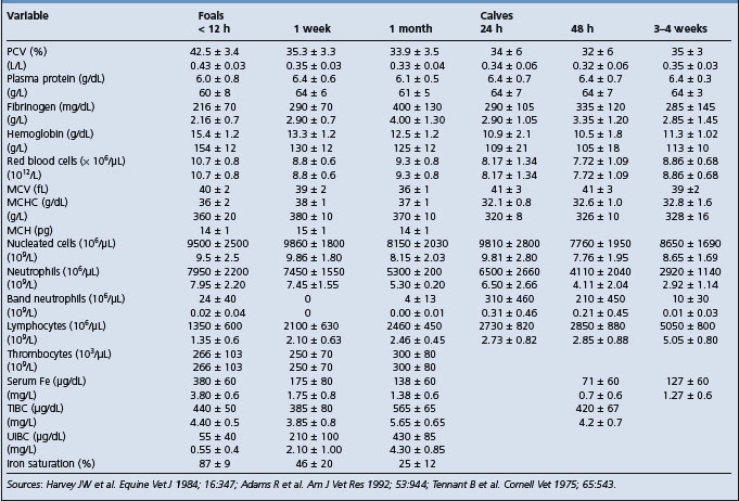
Clinical pathology is used as an integral part of the evaluation of a sick neonate and to help formulate a treatment plan. A major evaluation is to attempt to confirm the presence or absence of sepsis and this type of evaluation has been developed most successfully in the foal. Blood culture is part of this examination but the time for a positive result limits its value in the acutely ill neonate. Laboratory findings in foals with neonatal sepsis are variable and depend upon the severity, stage and site of infection.8 Serial examinations are commonly used. In examinations relating to the possible presence of septicemia, particular emphasis is placed on the results of the white blood cell and differential counts, the presence of toxic change, serum immunoglobulin concentrations, arterial oxygen concentrations, presence of metabolic acidosis and fibrinogen levels.7,8,12

DIFFERENTIAL DIAGNOSIS

The principles of diagnosis of infectious disease in newborn animals are the same as for older animals. However, in outbreaks of suspected infectious disease in young animals there is usually a need for more diagnostic microbiology and pathology

With outbreaks, owners should be encouraged to submit all dead neonates as soon as possible for a meaningful necropsy examination

In addition to postmortem examination it is necessary to identify the factors that may have contributed to an outbreak of disease in newborn calves, piglets or lambs and only detailed epidemiological investigation will reveal these

TREATMENT

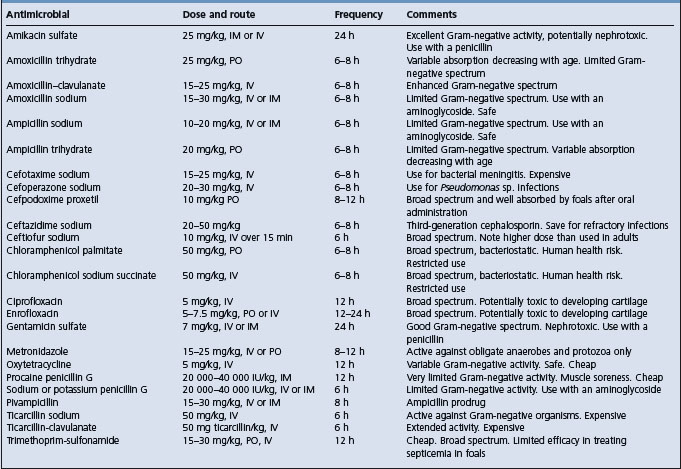
The first principle is to obtain an etiological diagnosis if possible. Ideally a drug sensitivity of the causative bacteria should be obtained before treatment is given, but this is not always possible. It may be necessary to choose an antibacterial based on the tentative diagnosis and previous experience with treatment of similar cases.

Outbreaks of infectious disease are common in litters of piglets and groups of calves and lambs, and individual treatment is often necessary to maximize survival rate. There is usually no simple method of mass-medicating the feed and water supply of sucking animals and each animal should be dosed individually as necessary. Supportive fluid and electrolyte therapy and correction of acid–base disturbances are described in detail in Chapter 2.

The provision of antibodies to sick and weak newborn animals through the use of blood transfusions or serum is often practiced, especially in newborn calves in which the immunoglobulin status is unknown. Whole blood given at the rate of 10–20 mL/kg body weight, preferably by the intravenous route, will often save a calf that appears to be in shock associated with neonatal diarrhea. The blood is usually followed by fluid therapy. Serum or plasma can also be given at half the dose rate. The blood should not be taken from a cow near parturition as the circulating immunoglobulins will be low from the transfer into the mammary gland.

Plasma is often incorporated into the therapeutic regimen in foals, both for its immunoglobulin content and for its effect on blood volume and osmotic pressure. Stored plasma can be used. A dose of 20 mL plasma/kg body weight given slowly intravenously is often used, but significantly higher doses are required to elevate circulating immunoglobulins by an appreciable amount.8 Blood may be collected, the red blood cells allowed to settle and the plasma removed and stored frozen. The donor plasma should be prescreened for compatibility. Lyophilized hyperimmune equine serum as a source of antibodies may also be fed to foals within 4 hours after birth. Good nursing care is also essential.

Further information on treatment is given in the section on critical care for the newborn later in this chapter.

CONTROL

Methods for avoidance of failure of transfer of passive immunity and the principles for prevention of infectious disease in newborn farm animals follow in this chapter. The control of individual diseases is given under specific disease headings elsewhere in this book.

REFERENCES

1 Watson DL, et al. Res Vet Sci. 1994;57:152.

2 Kitching RP, Salt JS. Aust Vet J. 1995;151:379.

3 Ellis JA, et al. J Am Vet Med Assoc. 1996;208:393.

4 Aldridge BM, et al. Vet Immunol Immunopathol. 1998;62:51.

5 Tierney TJ, et al. Vet Immunol Immunopathol. 1997;57:229.

6 Staller GS, et al. J Am Vet Med Assoc. 1995;206:77.

7 Brewer BD, Koterba AM. Equine Vet J. 1988;20:18.

8 Carter GK. Compend Contin Educ Pract Vet. 1986;8:S256.

9 Robinson JA, et al. Equine Vet J. 1993;25:214.

10 Koterba AM, et al. Equine Vet J. 1984;16:376.

11 Corley KTT, Furr MO. J Vet Emerg Crit Care. 2003;13:149.

12 Fecteau G, et al. Can Vet J. 1997;38:101.

FAILURE OF TRANSFER OF COLOSTRAL IMMUNOGLOBULINS

The acquisition and absorption of adequate amounts of colostral immunoglobulins is essential to the health of the neonate as it is born virtually devoid of circulating immunoglobulin and relies on antibody acquired from colostrum for protection against common environmental pathogens. Adequate antibody transfer is the cornerstone of all neonatal preventive health programs. This has been recognized for many years and it is discouraging that a study conducted in 2002 in the USA by the National Animal Health Monitoring System found that over 40% of dairy heifer calves sampled by the National Dairy Heifer Evaluation Project had failure of transfer of colostral immunoglobulins.1

Much of the description that follows refers to the calf because more studies on transfer of passive immunity have been conducted in calves. However, most of the information is applicable to the other species; where there are differences these are mentioned.

NORMAL TRANSFER OF IMMUNOGLOBULINS

The major immunoglobulin in colostrum is IgG, but there are also significant amounts of IgM and IgA. IgG1 is present in highest concentration and is concentrated in colostrum by an active, selective, receptor-mediated transfer of IgG1 from the blood of the dam across the mammary secretory epithelium. This transfer to colostrum begins approximately 4–6 weeks before parturition and results in colostral IgG1 concentrations in first milking colostrum that are several-fold higher than maternal serum concentrations. The transfer of the other immunoglobulin classes is believed to be nonselective and lesser concentrations in colostrum are achieved.

Following ingestion by the newborn, a significant proportion of these immunoglobulins in ingested colostrum is transferred across the epithelial cells of the small intestine during the first few hours of life and transported via the lymphatic system to the blood. Immunoglobulins in the blood are further varyingly distributed to extravascular fluids and to body secretions depending upon the immunoglobulin class.

These absorbed immunoglobulins protect against systemic invasion by microorganisms and septicemic disease during the neonatal period. Unabsorbed immunoglobulins and immunoglobulins resecreted back into the gut play an important role in protection against intestinal disease for several weeks following birth. In calves, passive immunity also influences the occurrence of respiratory disease during the first months of life and may be a determinant of lifetime productivity. In foals, failure of transfer of passive immunity presents a significant risk for the development of an illness during the first 3 months of life.

Lactogenic immunity

The IgG concentration in milk falls rapidly following parturition in all species and immunoglobulin concentrations in milk are low. In the sow, the concentration of IgA falls only slightly during the same period and it becomes a major immunoglobulin of sows’ milk. IgA is synthesized by the mammary gland of the sow throughout lactation and serves as an important defense mechanism against enteric disease in the nursing piglet. In the piglet, IgA in milk is an important mucosal defense mechanism whereas in the calf there is little IgA in milk but some enteric protection is provided by colostral and milk IgG, and IgG derived from serum that is resecreted into the intestine.2,3

FAILURE OF TRANSFER OF PASSIVE IMMUNITY

Failure of transfer of colostral immunoglobulins is the major determinant of septicemic disease in all species.4,5 It also modulates the occurrence of mortality and severity of enteric and respiratory disease in early life6 and, in some studies, performance at later ages.7-9 While an important determinant for neonatal disease and subsequent performance, it is not the sole determinant and it is not surprising that some studies have found only a minor relationship.

In terms of the modulation of disease, there can be no set cut-point for circulating immunoglobulins as the cut-point will vary according to the farm, its environment, infection pressure and also the type of disease. Figures are given as a guide. With dairy calves serum IgG1 concentrations of 500 mg/dL are associated with protection against septicemic disease and concentrations of 1000 mg/dL or more are sufficient to reduce the risk of infectious disease in most environments. With foals, the equivalent IgG1 concentrations for protection are given as 400 mg/dL and 800 mg/dL.10,11

Rates of failure of transfer of passive immunity are high in both sucking and artificially fed dairy calves but are less in beef calves.12,13 Failure rates in foals are lower and approximate 13–16%.11,14 Rates in lambs are also comparatively low.

In animals that are fed colostrum artificially, risk for failure of transfer of passive immunity is primarily dependent upon the amount or mass of immunoglobulin present in a feeding of colostrum, the time after birth that this is fed, and the efficiency of its absorption by the calf. The mass of immunoglobulin fed is determined by the concentration of immunoglobulin in the colostrum and the volume that is fed. Feeding trials with calves suggest that a mass of at least 100 g of IgG1 is required in colostrum fed to a 45 kg calf to obtain adequate (≥ 1000 mg/dL IgG1) passive blood immunoglobulin concentrations.15

In animals that suck colostrum naturally such as foals, risk for failure of transfer of passive immunity is primarily dependent upon the concentration of immunoglobulin in the colostrum, the amount that is ingested and the time of first suckling. Inadequate colostral immunoglobulin concentration and delay in ingestion of colostrum are the two important factors in failure of transfer of passive immunity in foals.16

Determinants of transfer of colostral immunoglobulins

1. Amount of immunoglobulin in colostrum fed:

a. Volume of colostrum fed
b. Concentration of immunoglobulins in colostrum

2. The amount of colostrum actually suckled or fed

3. Efficiency of absorption of immunoglobulins by neonate

4. Time after birth of suckling or feeding

Determinants of immunoglobulin concentration in colostrum

Nominal concentrations of immunoglobulin in the first milking colostrum of cows are shown in Table 3.1. There can be substantial variation in the concentration of immunoglobulin in colostrum in all species and the ingestion of a ‘normal’ amount of colostrum that has low immunoglobulin concentration may provide an insufficient amount of immunoglobulin for protection. In a study of over 900 first-milkings colostrum from American Holstein cows, only 29% of the colostrum samples contained a sufficiently high concentration of immunoglobulin to provide 100 g IgG in a 2 L volume.17 The equivalent percentages for 3 and 4 L volume feedings were 71% and 87%.

Table 3.1 Concentrations and relative percentage of immunoglobulins in serum and mammary secretions of cattle and pigs

image

A similar situation exists with horses. The mean concentrations of IgG in colostrum of mares 3–28 days before foaling is greater than 1000 mg IgG/dL, while at parturition the mean concentrations may vary from 4000–9000 mg/dL. The concentrations decrease markedly to 1000 mg/dL in 8–19 hours after parturition.18

It is apparent that variation in colostral immunoglobulin concentration can be a cause of failure of transfer of passive immunity.

Some causes of this variation are:

The concentrations of immunoglobulin in colostrum fall dramatically following parturition. Only the first milking of colostrum after calving should be considered for feeding to calves for immunoglobulin transfer. The concentrations in second-milking colostrum are approximately half those in the first milking and by the fifth postcalving milking, concentrations approach those found during the remainder of lactation

The immunoglobulin concentration of colostrum decreases after calving even when the cow is not milked. In order to facilitate early feeding of colostrum to a calf, herd policy may be to feed stored colostrum taken from a previously calved cow rather than the newborn calf’s dam. It is important that this colostrum be milked as soon as possible after parturition. Colostrum that is collected 6 hours or later after calving has a significantly lower concentration than that collected 2 hours after calving17,19

Colostrum from cows or mares that have been premilked to reduce udder edema or from dams that leak colostrum prior to parturition will have low immunoglobulin concentrations and alternate colostrum should be fed for immunoglobulin transfer as there is a higher rate of failure in their foals and calves

In cattle, dry periods of less than 30 days may result in colostrum of lower immunoglobulin concentration

Premature foaling or the induction of parturition can result in colostrum with low immunoglobulin concentration and/or low volume

In cattle, average colostral immunoglobulin concentrations are higher in cows in third or higher lactation groups compared to younger cows. However, colostrum from all lactation numbers can produce adequate immunoglobulin mass. There is no scientific basis for not feeding first-milking colostrum from first-lactation cows

Larger-volume first-milking colostrum tends to have lower immunoglobulin concentrations than smaller-volume colostrum, and colostrum weight can be used to select colostrum of higher immunoglobulin concentration for calf feeding (Table 3.2)

A recent study has shown that immunoglobulin concentrations are higher in the early temporal fractions of a single milking of first-milking colostrum.20 This might suggest that segregation of the first portion of the first-milking colostrum could provide colostrums with higher immunoglobulin concentration for feeding

There are breed differences in the concentration of immunoglobulins in first milking colostrum. In cattle, beef breeds have higher concentrations. Many dairy breeds, including American Holsteins, produce colostrum of relatively low immunoglobulin concentration, and a significant proportion of calves that suckle cows of these breeds ingest an inadequate mass of immunoglobulin. Channel Island breeds have a greater concentration of immunoglobulin in colostrum that Holsteins. Breed differences are also seen in horses, with Arabian mares having higher colostral immunoglobulin concentrations than Standardbreds, which in turn are higher than those of Thoroughbreds. Breed differences also occur in sheep, with higher concentrations in meat and wool breeds than dairy breeds21

Heat stress to cattle in the latter part of pregnancy results in lower colostral immunoglobulin concentrations22

One study found that calves from cows with mastitis have lower serum immunoglobulins.23 Colostral volume but not colostral immunoglobulin concentration is reduced in mastitic quarters and it is unlikely that mastitis is a major determinant of the high rate of failure of transfer of passive immunity in dairy calves24

There is a significant positive but weak correlation between total lactational milk and immunoglobulin concentration in that cow’s colostrum.17 Selection for production does not appear to be a negative influence on colostral immunoglobulin concentration, although dilution by high volume production in first-milking colostrum is a factor in low colostral immunoglobulin concentration25

The pooling of colostrum in theory could avoid the variation in immunoglobulin concentration of individually fed colostrum and could provide a colostrum that reflects the antigenic experience of several cattle. In practice, colostrum pools from Holsteins invariably have low immunoglobulin concentrations because high-volume, low-concentration colostrum dilutes the concentration of the other samples in the pool. If pools are used, the diluting influence of low-immunoglobulin-concentration, high-volume colostrum should be limited by restricting any individual cow’s contribution to the pool to 9 kg (20 lb) or less. However, pooling increases the risk of disease transmission, as multiple cows are represented in a pool and the pool is fed to multiple calves. This can be important in the control of Johne’s disease, bovine leukosis and Mycoplasma bovis

Bacterial contamination of colostrum can have a negative effect on transfer of passive immunity and one study26 found high bacterial counts in 85% of colostrums sampled from 40 farms in the USA. Colostrum that is to be fed or stored should be collected with appropriate preparation and sanitation of the cow and of the milking equipment used on fresh cows

Pasteurization of colostrum (both pasteurization at 63°C for 30 min and HTST 72°C for 15 s) reduces colostrum IgG concentration. A recent study27 of batch pasteurization at 63°C for 30 minutes showed that the percentage reduction in colostral IgG concentration varied with the batch size, with a 24% reduction in 57 L batch size and a 58% reduction in a larger batch size. Calves fed 2 L of pasteurized colostrum had twofold lower serum concentrations of IgG than controls

Old mares (older than 15 years) may have poor colostral immunoglobulin concentration.

Table 3.2 Immunoglobulin concentrations in the first milking of colostrum of Holstein cattle by weight of colostrum produced

image

Volume of colostrum ingested

Holstein cows

The volume of colostrum that is fed has a direct influence on the mass of immunoglobulin ingested at first feeding. The average volume of colostrum ingested by nursing Holstein calves in the first 24 hours of life is reported as 2.4 L but there is wide variation around this mean28 and a significant proportion of dairy calves fail to ingest an adequate mass of immunoglobulin in management systems that provide colostrum solely by allowing the calf to suck the dam.6,29

In natural suckling situations, calves may fail to ingest adequate colostrum volumes before onset of the closure process, and therefore absorb insufficient colostral immunoglobulin. Early assisted suckling may help avoid this. In dairy calves the volume of colostrum that is ingested can be controlled in artificial feeding systems using nipple bottle feeders or esophageal tube feeders. Bucket feeding of colostrum is not recommended, as training to feed from a bucket can be associated with erratic intakes.

The traditional recommendation for the volume of colostrum to feed at first feeding to calves is 2 L (2 quarts). However, only a small proportion of first-milking colostrum from Holsteins contains a sufficiently high concentration of immunoglobulin to provide 100 g IgG in a 2 L volume and higher volumes of colostrum are required to achieve this mass intake.17 Possibly the major cause of failure of transfer of passive immunity rests with the fact that commercial feeding bottles are made in a 2 L size and this is consequently the amount of colostrum that is fed. Some calves fed with a nipple bottle will drink volumes greater than 2 L but others will refuse to ingest even 2 L of colostrum in a reasonable period of time, and calf rearers may lack the time or patience to persist with nipple bottle feeding until the required volume has been ingested by all calves.

Larger volumes of colostrum can be fed by an esophageal feeder and single feedings of large volumes of colostrum (3.5–4.0 L per 45 kg body weight) result in the lowest percentage of calves with failure of transfer of passive immunity by allowing calves fed colostrum with relatively low immunoglobulin concentrations to receive an adequate immunoglobulin mass prior to closure.12,13,30 Feeding this volume by an esophageal feeder causes no apparent discomfort to a minimally restrained calf.

Channel Island breeds

Jersey cows produce colostrum with a higher immunoglobulin content than Holsteins and the feeding of nipple bottle 2.0 L of first milking at birth and again at 12 hours of life results in excellent circulating concentrations of immunoglobulin.31 Three L at each feeding is recommended where feeding is with an esophageal feeder.

Beef cows

With beef breeds very effective colostral immunoglobulin transfer is achieved with natural sucking. This is believed to be due to the greater vigor at birth exhibited by these calves and the higher immunoglobulin concentrations in beef colostrum, requiring a smaller volume intake to acquire an adequate mass. Natural sucking will give an adequate volume intake and there is no need to artificially feed colostrum unless the dam is observed to refuse nursing or the calf’s viability and sucking drive are compromised.32,33 The yield of colostrum and colostral immunoglobulins in beef cows can vary widely32 and range beef heifers may produce critically low volumes of colostrum. Differences in yield can be due to breed or to nutritional status, although undernutrition is not an effect unless it is very severe.34

Ewes

Colostrum yield is high in ewes in good condition at lambing but may be low in ewes with condition scores of 1.5–2.0.35

Sows

In sows there is also very effective colostral immunoglobulin transfer with natural sucking and piglets average an intake of 5–7% of body weight in the first hour of life.36 There is between-sow variation in the amount of colostrum and there can be a large variation in colostrum supply from teat to teat, which may explain variable health and performance. During farrowing and for a short period following, colostrum is available freely from the udder but thereafter it is released in ejections during mass suckling. A strong coordinated sucking stimulus is required by the piglet for maximum release of colostrum and this requires that the ambient temperature and other environmental factors be conducive to optimum vigor of the piglets. Small-birth-weight piglets, late-birth-order piglets and piglets sucking posterior teats obtain less colostrum.

All species

In all species a low-volume intake may also occur because of:

Poor mothering behavior, which may prevent the newborn from sucking, occurrence of disease or milk fever

Poor udder and/or teat conformation so the newborn cannot suck normally or teat seeking is more prolonged. Udder to floor distance is most critical and low-slung udders can account for significant delays in intake. Bottle-shaped teats (35 mm diameter) also significantly reduce intake37

Delayed and inadequate colostral intake frequently accompanies perinatal asphyxia or acidosis due to the greatly decreased vigor of the calf in the first few hours of life. Perinatal asphyxia can occur in any breed and is greatly increased by matings resulting in fetal–maternal disproportion and dystocia

The newborn may be weak, traumatized, or unable to suck for other reasons – a weak sucking drive can be a result of congenital iodine deficiency, cold stress or other factors

Failure to allow newborn animals to ingest colostrum may occur under some management systems.

Efficiency of absorption

After ingestion of colostrum by the newborn, colostral immunoglobulins are absorbed by the small intestine, by a process of pinocytosis, into the columnar cells of the epithelium. In the newborn calf this is a very rapid process and immunoglobulin can be detected in the thoracic duct lymph within 80–120 minutes of its being introduced into the duodenum. The period of absorption varies between species and with immunoglobulin class and the mechanism by which absorption ceases is not well understood but may be related to replacement of the fetal enterocyte. The region of maximum absorption is in the lower small intestine and peak serum concentrations are reached by 12–24 hours in all species. Absorption is not limited to immunoglobulins and there is a proteinuria during the first 24 hours of life associated with the renal excretion of low-molecular-weight proteins such as β-lactoglobulin.

Feeding methods, closure and immunoglobulin absorption

Under normal conditions complete loss of the ability to absorb immunoglobulin (closure) occurs by 24–36 hours after birth in all species and there is a significant reduction in absorptive ability (as much as 50% in some studies but minimal in others) by 8–12 hours following birth. The time from birth to feeding is a crucial factor affecting the absorption of colostral immunoglobulins by all species, and any delay beyond the first few hours of life, particularly after 8 hours, significantly reduces the amount of immunoglobulin absorbed.

The recommendation is that all neonates be fed colostrum within the first 2 hours of life.

Natural sucking

Natural sucking is the desired method of intake of colostrum and is the most efficient, but it is influenced by the sucking drive and vigor of the calf at birth. Calves that suck colostrum can achieve very high concentrations of colostral immunoglobulin and the efficiency of absorption is best with this feeding method. However, natural sucking of dairy calves is commonly associated with a high rate of passive transfer failure due to delays in sucking coupled with low intakes. In one study 25–34% of calves failed to suck by 6–8 hours of age and 18% of calves did not suck by 18 hours of age. There may be breed differences in sucking ability: Jersey calves have better rates of successful transfer of passive immunity with natural sucking than do Holsteins.38 Many factors influence the occurrence of delayed sucking but calf vigor and birth anoxia are the most important. Conformation of the udder is significant and the importance of this increases with parity.

Artificial feeding

In contrast, when calves are fed colostrum artificially, minimal delays from birth to the time of colostrum feeding occur and maximal colostrum immunoglobulin absorption results. In breeds like American Holsteins, where colostral immunoglobulin concentrations tend to be quite low and maximal efficiency of absorption is necessary, the logical way to minimize the effects of closure is to feed the maximum well-tolerated colostrum volume at the first feeding within the first few hours of life. The published literature consistently reports higher calf serum IgG1 concentrations and a lower rate of failures in response to larger colostrum feeding volumes.12,13

Other influences

Even with the best available on-farm colostrum selection methods, large colostrum-feeding volumes are essential to minimize failure of transfer of colostral immunoglobulins in breeds with relatively low colostral immunoglobulin concentrations. The method is particularly advantageous where time constraints of other farm activities limit the time available for calf feeding. The major detrimental influence on absorptive efficiency of immunoglobulins is delayed feeding after birth. Other factors that affect absorptive efficiency include:

Perinatal asphyxia or acidosis may have both direct and indirect effects on colostral immunoglobulin transfer. Asphyxia has a major effect on subsequent sucking drive and acidemic calves ingest far less colostrum than calves with more normal acid–base status at birth. In carefully controlled colostrum feeding studies, there was also significant negative correlation between the degree of hypercapnia and efficiency of absorption of colostral immunoglobulins, even in calves within the ‘normal’ newborn blood pH and Pco2 ranges.39 Direct oxygen deprivation of newborn calves did not cause a similar effect.40 Treatment of calves with an alkalinizing agent and a respiratory stimulant altered pH and Pco2 values towards adult normal values but did not influence immunoglobulin absorption efficiency39

In one early study, a mothering effect was reported where calves remaining with their dams absorbed colostral immunoglobulin much more efficiently than calves removed immediately to individual box stalls. However, other studies have shown much smaller or no effects of mothering using similar experimental designs. The different results of these studies have not been reconciled

There can be seasonal and geographical variations in transfer of immunoglobulins in calves although these are not always present on farms in the same area and their cause is unknown. Where seasonal variation occurs in temperate climates the mean monthly serum IgG1 concentrations are lowest in the winter and increase during the spring and early summer to reach their peak in September, after which they decrease. The cause is not known but an decrease in sucking drive is observed in colder months and may contribute. In subtropical climates, peak levels occur in the winter months, while low levels are associated with elevated temperatures during the summer months.41 Heat stress in late pregnancy will reduce colostral immunoglobulin concentration but high ambient temperature is a strong depressant of absorption and the provision of shade will help to obviate the problem

The efficiency of absorption may be decreased in premature calves that are born following induced parturition42 but the medical induction of parturition with short-acting corticosteroids in cattle does not interfere with the efficiency of absorption of immunoglobulins in calves

The absorption of small volumes (1–2 L) fed by an esophageal feeder is usually suboptimal, probably due to retention of some colostrum in the immature forestomachs for several hours. The calf will feel satiated and not inclined to suck naturally for the next few hours

A trypsin inhibitor in colostrum may serve to protect colostral IgG from intestinal degradation. It varies in concentration between colostrums. The addition of a trypsin inhibitor to colostrum improves immunoglobulin absorption43

In a study of mare-associated determinants of failure of transfer of passive immunity in foals (based on serum Ig measurements), there was a trend to increase rates of failure in foals from mares aged over 12 years but no real association with age, parity or gestational age of foals over 325 days. There was an association with season with a lower incidence in the late spring compared with foals born earlier in the year and with a foal score based on a veterinary score of foal health and ‘fitness’.44

Traditionally it has been considered that the movement of animals, either the dam just before parturition or the newborn animal during the first few days of life, is a special hazard for the health of the calf. The postulated reason is that the dam may not have been exposed to pathogens present in the new environment and thus not have circulating antibodies against these pathogens. The newborn animal may be in the same position with regard to both deficiency of antibodies and exposure to new infections. While this may obtain in some situations, the developing practice of contract-rearing of dairy heifers away from the farm to be brought back as close-up springers, and the practice of purchase of close-up heifers on to the farm, are not associated with appreciable increase in mortality in their calves.

Decline of passive immunity

Passive antibody levels fall quickly after birth and have usually disappeared by 6 months of age. In the foal, they have fallen to less than 50% of peak level by 1 month of age, and to a minimum level between 30 and 60 days. This is the point at which naturally immunodeficient foals are highly susceptible to fatal infection.

In calves, the level of IgG declines slowly and reaches minimum values by 60 days, in contrast to IgM and IgA, which decline more rapidly and reach minimum values by approximately 21 days of age. The half-lives for IgG, IgM and IgA in calves are approximately 20, 4 and 2 days respectively and half lives of IgGa, IgGb, IgG(T) and IgA in foals are approximately 18, 32, 21 and 3.5 days respectively.45

Immunological competence is present at birth but endogenous antibody production does not usually reach protective levels until 1 month, and maximum levels not until 2–3 months of age. The endogenous production of intestinal IgA in the piglet begins at about 2 weeks of age and does not reach significant levels until 5 weeks of age.

Foals that acquire low concentrations of immunoglobulins from colostrum may experience a transitory hypogammaglobulinemia at several weeks of age as the levels fall and before autogenous antibodies develop. They are, as expected, more subject to infection than normal.

OTHER BENEFITS OF COLOSTRUM

In addition to its immunoglobulin content, colostrum contains considerably more protein, fat, vitamins and minerals than milk and is especially important in the transfer of fat-soluble vitamins. It has anabolic effects and lambs that ingest colostrum have a higher summit metabolism than colostrum-deprived lambs. Colostrum also contains growth-promoting factors that stimulate DNA synthesis and cell division including high concentrations of insulin-like growth factor (IGF)-1.46,47

Colostrum contains approximately 106 leukocytes/mL and several hundred million are ingested with the first feeding of colostrum. In calves 20–30% of these are lymphocytes and cross the intestine into the circulation of the calf.48 It is postulated that they have importance in the development of neonatal resistance to disease but there is little tangible evidence. Calves fed colostrum depleted of leukocytes are claimed to be more poorly protected against neonatal disease than those fed normal colostrum.49

ASSESSMENT OF TRANSFER OF PASSIVE IMMUNITY

Because of the importance of transfer of colostral antibodies to the health of the neonate, it is common to quantitatively estimate the levels of immunoglobulins, or their surrogates, in colostrum and in serum in order to predict risk of disease and to take preventive measures in the individual or to make corrective management changes where groups of animals are at risk.

Assessment in the individual animal

Where samples are taken from an individual animal to determine the risk for infection, sampling is undertaken early so that replacement therapy can be given promptly if there has been inadequate transfer. IgG is detectable in serum 2 hours following a colostrum feeding and sampling at 812 hours after birth will give a good indication of whether early sucking has occurred and has been effective in transfer.50 This type of monitoring is commonly performed in foals. There are a number of different tests that can be used, some of which are quantitative and others semi-quantitative. For foals, these include commercially available tests such as the latex agglutination, concentration immunoassay and hemagglutination inhibition tests. These are semi-quantitative and the relative value of these tests for this type of analysis has been evaluated.51,52 A glutaraldehyde test for serum is available commercially for use in the horse and is reported to correlate well with radial immunodiffusion (RID) values.53 In calves, sampling may be undertaken for similar reasons but the cost of replacement therapy is limiting.

Monitoring assessment tests on serum

Sampling to monitor the efficacy of a farm policy for feeding colostrum, to evaluate levels in calves to be purchased or to determine the rates of failure of transfer of passive immunity in investigations of neonatal disease can be conducted at any time in the first week of life after 48 hours with most tests. This is possible because of the relatively long half-life of IgG.

Radial immunodiffusion

This test is usually used in research studies and is the gold standard. It is available commercially but is expensive and takes longer to perform than is desirable for most clinical purposes. An enzyme-linked immunosorbent assay (ELISA) for measurement of IgG concentration in horses and in porcine plasma and colostrum suffers from the same problems.

Quantitative zinc sulfate test

This is a good predictor of mortality but requires instrumentation. It has been used for many years in calves and lambs and has been validated in horses.54 Hemolyzed blood samples will give artificially high readings, and the reagent must be kept free of dissolved carbon dioxide. The suggested cut-point is 20 ZST units. Increased test solution concentrations to those traditionally used have been suggested to improve sensitivity.55

Serum γ-glutamyltransferase activity

Serum γ-glutamyltransferase (GGT) activity has been used as a surrogate for determining the efficacy of transfer of passive immunity in calves and lambs. GGT concentrations are high in the colostrum of ruminants (but not horses) and serum GGT activity in calves and lambs that have sucked or been fed colostrum are 60–160 times greater than normal adult serum activity and correlate moderately well with serum IgG concentrations.56,57 The half-life of GGT from colostrum is short and serum GGT activity falls significantly in the first week of life. Serum GGT values equivalent to a serum IgG concentration of 10 mg/mL are 200 IU/L on day 1 of life and 100 IU/L on day 4. Serum GGT concentrations less than 50 IU/L indicate failure of transfer of passive immunity.56,58

Serum total protein

Total protein, as measured by a refractometer, gives an indirect measure of the amount of immunoglobulin. Despite the indirect nature of the test, there is a reliable correlation between the refractometer reading and total immunoglobulin concentration (IgG and IgM) measured by RID. In healthy calves a serum total protein of 5.2 g/dL or greater is associated with adequate transfer of passive immunity.

Serum total protein has good predictive value for fate of the newborn, and the facile and practical nature of the test and its predictive ability commends it for survey studies in calves and lambs but not foals. Cut-points will vary with the environment59,60 and the infection pressure to the calves. The sensitivity of the test is maximal using a cut point of 5.5 g/dL and the specificity is maximal at a cut point of 5.0 g/dL.61 The refractometer can give false high values in dehydrated calves but these can be clinically identified and a cut point of 5.5 g/dL can be used.

Sodium sulfite precipitation and glutaraldehyde test

Both of these were developed as rapid field tests to evaluate the immune status of neonatal calves. An 18% test solution is used and the development of turbidity is the determinant of adequate transfer of passive immunity. The glutaraldehyde coagulation test is also available for the detection of hypogammaglobulinemia in neonatal calves but is less accurate.61 Neither test is widely used.

ELISA test

An ELISA test is commercially available and used for calf-side testing.

Monitoring colostrum

There has long been the desire for a method to select colostrums with high immunoglobulin concentration for feeding neonates.

Specific gravity

Specific gravity can be used as a measure of immunoglobulin content of colostrum. In mares the concentration of immunoglobulins in colostrum is highly correlated with the specific gravity of the colostrum, which in turn is highly correlated with the serum immunoglobulin levels achieved in foals.50,62 Temperature-corrected measurements are most accurate.63 Measurement of colostrum specific gravity provides a rapid and easy method of identifying foals likely to be at a high risk for failure of transfer of passive immunity and the need to provide them with colostrum of a higher Ig content. It is recommended that, to prevent failure of transfer of passive immunity, the colostral specific gravity should be equal to or greater than 1.060 and the colostral IgG concentration be a minimum of 3000 mg/dL.62

In cattle the relation of specific gravity of colostrum to colostral immunoglobulin concentration is linear but is better in Holsteins than in Jerseys.58 The measurement is simple but there is a correction for temperature, and air trapped in colostrum taken by a milking machine can give a false reading if the measurement is taken too quickly after milking. The cut-point recommended to distinguish moderate from excellent colostrum has been set at 1.048 and is based on the amount of immunoglobulin required for a 2 quart feeding. Specific gravity is not a perfect surrogate for immunoglobulin concentration with cattle colostrum. It has good negative prediction but it will falsely pass many Holstein colostrums that have low immunoglobulin concentration and is not accurate with Jersey colostrum.64,65 An analysis of first-milking colostrum in midwest USA dairies found that specific gravity differed among breeds and was influenced by month of calving, year of calving, lactation number and protein yield in previous lactation and that it was more closely associated with colostrum protein concentration (r = 0.76) than IgG1concentration (r = 0.53).66

Glutaraldehyde test

This test for mare colostrum is available commercially and is reported to have a high predictive value for colostrums that contain more than 38 mg/mL of IgG and have a specific gravity greater than 1.060.67,68

ELISA

Recently, a cow-side immunoassay kit has become available commercially in the US. The kit provided a positive or a negative response with the cut point being a concentration of 50 g/L of IgG in colostrum and has accuracy sufficient to recommend its use for rejection of colostrums with low immunoglobulin concentration.69

CORRECTION OF FAILURE OF TRANSFER OF PASSIVE IMMUNITY

Parenteral immunoglobulins

Blood transfusion is commonly used and the method is described elsewhere in this text. Purified immunoglobulin preparations are an alternative and are available commercially in some countries. Large amounts are required to obtain the required high serum concentrations of immunoglobulins and intravenous infusion can be accompanied by transfusion-type reactions.

AVOIDANCE OF FAILURE OF TRANSFER OF PASSIVE IMMUNITY

With all species, with the exception of dairy calves, the common practice is to allow the newborn to suck naturally. The policy for avoidance of failure of transfer of passive immunity with naturally sucking herds should be to provide supplemental colostrum by artificial feeding of those neonates with a high risk for failure, based on the risk factors detailed above. In the dairy calf, rates of failure with natural sucking are so high that many farms opt to remove that calf at birth and feed colostrum by hand to ensure adequate intakes.

Colostrum

Colostrum can be stripped from the dam and fed fresh or the neonate can be fed stored (banked) colostrum.

Colostrum for banking

With dairy cows, first-milking colostrum from a cow with a first-milking yield of less than 10 kg should be used. The temptation for the farmer is to store the leftover from the feeding of large-volume colostrum. This should not be used as it has a high probability of containing a low immunoglobulin concentration.

Colostrum from mares should have a specific gravity of 1.060 or more and 200 mL can be milked from a mare before the foal begins sucking.

Storage of colostrum

Colostrum can be kept at refrigerator temperature for approximately 1 week without significant deterioration in immunoglobulins. Storage in plastic containers also maintains the viability of cellular components.26 The addition of formaldehyde to 0.05% (wt/vol) allows maintenance at 28°C for 4 weeks without loss of immunoglobulins as detected by RID70 but information on such colostrum’s protection efficacy, when fed, is not available. The addition of 5 g of propionic or lactic acid per liter extends the storage life to 6 weeks30 but, more commonly, colostrum is frozen for storage. Frozen colostrum, at −20°C, can be stored virtually indefinitely and there is no impairment to the subsequent absorption of immunoglobulins. Frozen colostrum should be stored in flat plastic bags in the amount required for a feeding, which facilitates thawing. Thawing should be at temperatures below 55°C. Higher temperatures and microwave thawing results in the deterioration of immunoglobulins and antibody in frozen colostrum and frozen plasma.71

Cross-species colostrum

Colostrum from another species can be used to provide immunological protection where same-species colostrum is not available. Bovine colostrum can be fed to a number of different species. While absorption of immunoglobulins occurs and significant protection can be achieved,72 the use of cross-species colostrum is not without some risk and the absorbed immunoglobulins have a short half-life. Bovine colostrum has been successfully used for many years to improve the survival rate of hysterectomy-produced artificially reared pigs. It has also been used as an alternate source of colostral antibody for rearing goats free of caprine arthritis–encephalitis. Colostrum from some cows can result in the development of a hemolytic anemia, occurring at around 5–12 days of age, in lambs and kids because the IgG of some cows attaches to the red cells and their precursors in bone marrow, resulting in red cell destruction by the reticuloendothelial system.73,74 Treatment of the anemia consists of a blood transfusion. Bovine colostrum can be tested for ‘anti-sheep’ factors by a gel precipitation test on colostral whey but this test is not generally available. Bovine colostrum can provide some protection to newborn foals against neonatal infections and protection appears to be due to factors in addition to the immunoglobulins, which have a short half-life in foals.

Colostrum supplements

In recent years there has been a move to develop supplements or even replacements for colostrum to feed calves. These have been attempted using IgG concentrated from bovine colostrum, milk whey, eggs or bovine serum. The search for colostrum substitutes or colostrum replacers has been prompted by the problem of the variability of IgG concentration in natural colostrum. It has also been prompted by possible limitations of availability of high-quality colostrum on dairy farms as the result of discarding colostrum from cows that test positive to disease that can transmit through colostrum, such as Johne’s disease, bovine leukosis, Mycoplasma bovis. This problem is confounded by reports that pasteurization of colostrum can have a deleterious effect on IgG concentration in colostrum and its subsequent absorption by calves.27

Lacteal-secretion-based preparations

Colostrum supplements prepared from whey or colostrum are available commercially in many countries. Depending upon the manufacturer, they contain varying amounts of immunoglobulin but significantly less than first-milking colostrum. The amount of immunoglobulin contained varies, but the recommendations for feeding that accompany these products indicate that they will supply approximately 25% or less of the immunoglobulin required to elevate calf serum IgG concentrations above 1000 mg/dL. There is a further problem in that the immunoglobulins in products made from colostrum or whey are poorly absorbed and trials assessing their ability to increase circulating immunoglobulins when fed with colostrum have generally shown little improvement and no improvement in health-related parameters.75-80

There is evidence that their inclusion with colostrum can impair the efficiency of colostral immunoglobulin77 and if they are fed they should be fed after normal colostrum. While these milk-protein-derived products are advertised for supplementing normal colostrum feeding, they are unfortunately often promoted as replacements and used by farmers as total substitutes for colostrum. When fed as the sole source of immunoglobulin to colostrum-deprived calves, they achieve circulating concentrations of immunoglobulin that are lower than those achieved by natural colostrum containing equivalent amounts of immunoglobulin.76

Bovine-serum-based preparations

Colostrum supplements prepared from bovine serum are also available commercially but regulations governing the feeding of blood or blood products to calves (risk reduction for bovine spongiform encephalopathy) may limit their availability in some countries. The absorption of immunoglobulin from these bovine-serum-derived commercial products appears better than from milk-protein-derived products80 and consequently they are also marketed as colostrum replacers. It has been proposed that the distinction between a colostrum supplement and a colostrum replacer should be the immunoglobulin mass contained in the product, with a colostrum supplement having less than 100 g IgG per dose and a colostrum replacer having sufficient immunoglobulin mass in a dose to result in a serum IgG concentration greater than 10 mg/mL following a feeding.80

A large mass of immunoglobulin is required for acquisition of adequate circulating immunoglobulin. Calves fed a colostrum replacement containing a high mass (250 g for Holsteins) of an IgG derived from bovine serum and fed at 1.5 and again at 13.5 hours after birth, achieved equivalent serum IgG concentrations to calves fed normal colostrum and showed no difference in gain or health parameters during the first 4 weeks of life.81

The IgG in a commercially available bovine serum colostrum replacer has been shown to be effectively absorbed when fed to newborn lambs. The feeding of 200 g of IgG in the first 24 hours of life resulted in a mean plasma concentration of 18 mg/mL.82

The published literature suggests that there is little advantage to be gained from the use of milk-protein-derived colostrum supplements and their use as colostrum replacers or substitutes is not recommended, except where there is no source of natural colostrum due to factors such as the death of the dam. The feeding of colostrum-derived supplements can result in a modest elevation of circulating IgG, sufficient to protect against experimental colisepticemia. If available, the bovine-serum-derived products would be more suitable.

The use of colostrum replacers should be confined to this type of emergency and there can be little justification for more widespread use, particularly as there are limited independent health-related publications of their efficacy. Also, as mentioned above, in addition to immunoglobulins, natural colostrum contains various substances important to neonatal physiology.

Administration of colostrum

Foals

Foals should be allowed to suck naturally. The specific gravity of the mare’s colostrum can be checked at foaling and, where this is less than 1.060, supplemental colostrum may be indicated. Foals that do not suck, or that have serum IgG concentrations less than 400 mg/mL at 12 hours of age, or that require supplementation for other reasons, should be fed colostrum with a specific gravity of 1.060 or more at an amount of 200 mL at hourly feedings.50,60

Dairy calves
Assisted natural sucking

Leaving the newborn dairy calf with the cow is no guarantee that the calf will obtain sufficient colostrum and a high proportion fail either to suck early or to absorb sufficient immunoglobulins from ingested colostrum. This problem can be alleviated to some extent by assisted natural sucking but this can fail because not all calves requiring assistance are detected. An alternate approach is to milk 2 L of colostrum from the dam, bottle-feed each calf as soon after birth as possible, then leave the calf with the cow for 24 hours and allow it to suck voluntarily. While this will not be as effective as a system based entirely on artificial feeding of selected colostrum, it is an approach that is suitable for the smaller dairy farm.

Artificial feeding systems

With artificial feeding systems, the calf is removed from the dam at birth and fed colostrum by hand throughout the whole absorptive period. Nipple bottle-feeding can be used with 2 L of colostrum given every 12 hours (Holstein calves) for the first 48 hours of life. The first feeding is usually milked from the cow by hand and the remaining feedings are from the colostrum obtained from the cow after the first machine milking. If care and patience is taken with feeding, this system can result in good transfer of passive immunity in all calves except those born to dams that have very low concentrations of immunoglobulin in their colostrum. Unfortunately, with American Holsteins this can be a significant percentage. An extension from this system is to nipple bottle-feed at the same frequency but to feed stored colostrum selected for its superior immunoglobulin content. Nipple bottle-feeding of newborn calves requires considerable patience and its success is very much dependent on the calf feeder and on the availability of the feeder’s time when faced with a calf that has a slow intake.

Where the diligence of the calf feeders is poor, or where there is a time constraint on their availability, the feeding of a large volume of colostrum (4 L to a 45 kg calf) by esophageal feeder at the initial feeding immediately after birth can be a successful practice. The large-volume feeding also allows the delivery of an adequate mass of immunoglobulin with colostrum that has low immunoglobulin concentrations and the delivery of colostrum to the gut in its early absorptive period. The practice usually uses stored colostrum and the feeding can be achieved within a few minutes. It can be supplemented by bottle-feeding of a second feeding at 12 hours of life.

The practice of feeding stored colostrum as the sole source of colostrum is limited to larger dairy herds but it does allow the selection of superior colostrum for feeding with selection based on weight and specific gravity as detailed above.

Beef calves

Beef calves should be allowed to suck naturally and force-feeding of colostrum to beef breeds should not be practiced unless there is obvious failure of sucking. Where colostrum is required, as with weak beef calves, calves with edematous tongues and calves that have been subjected to a difficult birth, it can be administered with an esophageal feeder or a stomach tube.

Lambs

Lambs are allowed to suck naturally but there can be competition between siblings for colostrum and one large single lamb is capable of ingesting, within a short period of birth, all the available colostrum in the ewe’s udder. Lambs require a total of 180–210 mL colostrum/kg body weight during the first 18 hours after birth to provide sufficient energy for heat production.35 This amount will usually provide enough immunoglobulins for protection against infections. Supplemental feeding of colostrum may be advisable for lambs from multiple birth litters, lambs that lack vigor and those that have not nursed by 2 hours following birth. This can be done with a nipple bottle or an esophageal feeder.

Piglets

Colostral supplementation is not commonly practiced with piglets. An immunoglobulin dose of 10 g/kg body weight on day 1 followed by 2 g/kg on succeeding days for 10 days is sufficient to confer passive immunity on the colostrum-deprived pig.

REVIEW LITERATURE

Norcross NL. Secretion and composition of colostrum and milk. J Am Vet Med Assoc. 1982;181:1057.

Gay CC. Failure of passive transfer of colostral immunoglobulins and neonatal disease in calves: a review. In: Proceedings of the Fourth International Symposium on Neonatal Diarrhea, Saskatoon, Saskatchewan, Canada. Saskatoon: University of Saskatchewan; 1983:346-364.

Besser TE, Gay CC. Septicemic colibacillosis and failure of passive transfer of immunoglobulin in calves. Vet Clin North Am Food Anim Pract. 1985;1:445-459.

Black L, Francis ML, Nicholls MJ. Protecting young domestic animals from infectious disease. Vet Annu. 1985;25:46-61.

Sheldrake RF, Husband AJ. Immune defences at mucosal surfaces in ruminants. J Dairy Res. 1985;52:599-613.

Staley TE, Bush LJ. Receptor mechanism of the neonatal intestine and their relationship to immunoglobulin absorption and disease. J Dairy Sci. 1985;68:184-205.

Butler JE. Biochemistry and biology of ruminant immunoglobulins. Prog Vet Microbiol Immunol. 1986;2:1-53.

Carter GK. Septicemia in the neonatal foal. Compend Contin Educ Pract Vet. 1986;8:S256-S270.

Morris DD. Immunologic disease of foals. Compend Contin Educ Pract Vet. 1986;8:S139-S150.

Mellor D. Meeting colostrum needs of lambs. In Pract. 1990;12:239-244.

Brenner J. Passive lactogenic immunity in calves: a review. Israel J Vet Med. 1991;46:1-12.

Besser TE, Gay CC. Colostral transfer of immunoglobulins to the calf. Vet Annu. 1993;33:53-61.

Garry F, Aldridge B, Adams R. Role of colostral transfer in neonatal calf management: current concepts in diagnosis. Compend Contin Educ Pract Vet. 1993;15:1167-1175.

White DG. Colostral supplementation in ruminants. Compend Contin Educ Pract Vet. 1993;15:335-342.

Besser TE, Gay CC. The importance of colostrum to the health of the neonatal calf. Vet Clin North Am Food Anim Pract. 1994;10:107.

Quigley JD, Drewry JJ. Nutrient and immunity transfer from cow to calf pre- and postcalving. J Dairy Sci. 1998;81:2779-2790.

Weaver DM, Tyler JW, VanMetre D, Hoetetler DE, Barrington GM. Passive transfer of colostral immunoglobulins in calves. J Vet Intern Med. 2000;14:569-577.

Barrington GM, Parish SM. Bovine neonatal immunology. Vet Clin North Am Food Anim Pract. 2001;17:463-476.

Rooke JA, Bland IM. The acquisition of passive immunity in the newborn piglet. Livestock Product Sci. 2002;78:13-23.

McGuirk SM, Collins M. Managing the production, storage, and delivery of colostrum. Vet Clin North Am Food Anim Pract. 2004;20:593-603.

REFERENCES

1 National Animal Health Monitoring System. National Dairy Heifer Evaluation Project: Dairy herd management practices focusing on preweaned heifers. Fort Collins, CO: USDA, 2002. APHIS:VS

2 Besser TE. Vet Immunol Immunopathol. 1993;38:103.

3 Saif LJ, Smith KL. J Dairy Sci. 1985;68:206.

4 Robinson JA, et al. Equine Vet J. 1993;25:214.

5 Gay CC, et al. Gyles CL, editor. Escherichia coli in domestic animals and humans. Wallingford, Oxfordshire: CAB International. 1994:75.

6 Gay CC. Proceedings of the Fourth International Symposium on Neonatal Diarrhea, Saskatoon, Saskatchewan, Canada, 4. Saskatoon: University of Saskatchewan. 1983:346. 1983

7 Wittum TE, Perino LJ. Am J Vet Res. 1995;56:1149.

8 Wells SJ, et al. Prevent Vet Med. 1996;29:9.

9 Donovan GA, et al. Prevent Vet Med. 1998;34:31.

10 Ley WB, et al. Equine Vet Sci. 1990;10:262.

11 Tyler-McGowan JL, et al. Aust Vet J. 1997;75:56.

12 Besser TE, Gay CC. Vet Clin North Am Food Anim Pract. 1994;10:107.

13 Besser TE, Gay CC. Vet Annu. 1993;33:53-61.

14 LeBlanc MM, et al. J Am Vet Med Assoc. 1992;200:179.

15 Besser TE, et al. J Am Vet Med Assoc. 1991;198:419.

16 Chavatte-Palmer P, et al. Pferdheilkunde. 2001;17:669.

17 Pritchett L, et al. J Dairy Sci. 1991;74:2336.

18 Pearson RC, et al. Am J Vet Res. 1984;45:186.

19 Moore M, et al. J Am Vet Med Assoc. 2005;226:1375.

20 Hostetler D, et al. J Appl Res Vet Med. 2003;1:168.

21 Waelchli RO, et al. Vet Rec. 1994;135:16.

22 Nardone E, et al. J Dairy Sci. 1997;80:838.

23 Perino LJ, et al. Am J Vet Res. 1995;56:1144. 1149

24 Maunsell FP, et al. J Dairy Sci. 1998;81:1291.

25 Guy MA, et al. J Dairy Sci. 1994;77:3002.

26 Godden SM, et al. J Dairy Sci. 2003;86:1503.

27 Stott GH, et al. J Dairy Sci. 1979;62:1632. 1766, 1902, 1908

28 McGuirk SM, Collins M. Vet Clin North Am Food Anim Pract. 2004;20:593.

29 Franklin ST, et al. J Dairy Sci. 2003;86:2145.

30 White DG. Compend Contin Educ Pract Vet. 1993;15:335.

31 Jaster EH. J Dairy Sci. 2005;88:296.

32 Petrie L, et al. Can Vet J. 1984;25:273.

33 Bradley JA, Niilo L. Aust Vet J. 1984;25:121.

34 Hough RL, et al. J Anim Sci. 1990;68:2662.

35 Mellor DJ. In Pract. 1990;12:239.

36 Fraser D, Rushen J. Can J Anim Sci. 1992;72:1.

37 Ventorp M, Michanek P. J Dairy Sci. 1992;75:262.

38 Quigley JD, et al. J Dairy Sci. 1995;78:893.

39 Ayers MW, Besser TE. Am J Vet Res. 1992;53:83.

40 Tyler H, Ramsey H. J Dairy Sci. 1991;74:1953.

41 Mohammed HO, et al. Cornell Vet. 1991;81:173.

42 Johnston NE, Stewart JA. Aust Vet J. 1986;63:191.

43 Quigley JD, et al. J Dairy Sci. 1995;78:886. 1573

44 Clabough DL, et al. J Vet Intern Med. 1991;5:335.

45 Sheoran AS, et al. Am J Vet Res. 2000;61:1099.

46 Barrington GM, Parish SM. Vet Clin North Am Food Anim Pract. 2001;17:463.

47 Blum JW, Baumruckerb CR. Domest Anim Endocrinol. 2002;23:101.

48 Le Jan C. Vet Res. 1996;27:403.

49 Reidel-Caspari G. Vet Immunol Immunopathol. 1993;35:275.

50 Massey RE, et al. Proc Am Assoc Equine Pract. 1991;36:1.

51 Ley WB, et al. Equine Vet Sci. 1990;10:262.

52 Morris DD, et al. Am J Vet Res. 1985;46:S294.

53 Jones D, Brook D. Equine Vet Sci. 1994;14:85.

54 LeBlanc MM, et al. Equine Vet Sci. 1990;10:36.

55 Hudgens KA, et al. Am J Vet Res. 1996;57:1711.

56 Perino LJ, et al. Am J Vet Res. 1993;54:56.

57 Tessman RK, et al. J Am Vet Med Assoc. 1997;211:1163.

58 Parish SM, et al. J Vet Intern Med. 1997;11:344.

59 Tyler JW, et al. J Vet Intern Med. 1998;12:79.

60 Rea DE, et al. J Am Vet Med Assoc. 1996;208:2047.

61 Tyler JW, et al. J Vet Intern Med. 1996;10:304.

62 LeBlanc MM, et al. J Am Vet Med Assoc. 1986;189:57.

63 Dascanio JJ, et al. Equine Pract. 1997;19:23.

64 Quigley JD. J Dairy Sci. 1994;77:264.

65 Pritchett LC, et al. J Dairy Sci. 1994;77:1761.

66 Morin DE, et al. J Dairy Sci. 2001;84:937.

67 Jones D, Brook D. Equine Vet Sci. 1994;14:85.

68 Jones D, Brook D. Equine Vet Sci. 1995;15:269.

69 Chigerwe M, et al. J Am Vet Med Assoc. 2005;227:129.

70 Mbuthia EW, et al. Anim Feed Sci Technol. 1997;67:291.

71 Orielly JL. Aust Vet J. 1992;70:442.

72 Gomez GG, et al. J Anim Sci. 1998;76:1.

73 Winter A, Clarkson M. In Pract. 1992;14:283.

74 Perl S, et al. Israel J Vet Med. 1995;50:61.

75 Abel Francisco SF, Quigley JD. Am J Vet Res. 1993;54:1051.

76 Garry FB, et al. J Am Vet Med Assoc. 1996;208:107.

77 Morrin DE, et al. J Dairy Sci. 1997;80:747.

78 Arthington JD, et al. J Dairy Sci. 2000;83:1463.

79 Quigley JD, et al. J Dairy Sci. 2001;84:2059.

80 Quigley JD, et al. J Dairy Sci. 2002;85:1243.

81 Jones CM, et al. J Dairy Sci. 2004;87:1806.

82 Quigley JD, et al. Vet Therapeut. 2002;3:262.

PRINCIPLES OF CONTROL AND PREVENTION OF INFECTIOUS DISEASES OF NEWBORN FARM ANIMALS

The four principles are:

Reduction of risk of acquisition of infection from the environment

Removal of the newborn from the infectious environment if necessary

Increasing and maintaining the nonspecific resistance of the newborn

Increasing the specific resistance of the newborn through the use of vaccines.

The application of each of these principles will vary depending on the species, the spectrum of diseases that are common on that farm, the management system and the success achieved with any particular preventive method used previously.

REDUCTION OF RISK OF ACQUISITION OF INFECTION FROM THE ENVIRONMENT

The animal should be born in an environment that is clean, dry, sheltered and conducive for the animal to get up after birth, suck the dam and establish bonding.1,2 Calving and lambing stalls or grounds, farrowing crates and foaling stalls should be prepared in advance for parturition. No conventional animal area can be sterilized but it can be made reasonably clean to minimize the infection rate before colostrum is ingested and during the first few weeks of life when the newborn animal is very susceptible to infectious disease.

With seasonal calving or lambing there can be buildup of infection in the birth area and animals born later in the season are at greater risk of disease. In these circumstances it may be necessary to move to secondary lambing or calving areas. In northern climates snow may constrict the effective calving area and result in a significant buildup of infection. Buildup of infection pressure must be minimized by a change to a fresh calving/lambing area and by the frequent movement of feed bunks or feed areas. Any system that concentrates large numbers of cattle in a small area increases environmental contamination and close confinement of heifers and cows around calving time is a known risk factor for calf mortality.3,4 With large herds both the cow herd and heifer herd should be broken into as many subgroups as is practical. Extensive systems where cows calve out over large paddocks are optimal and with more intense systems a group size no larger than 50 has been suggested.5

Lambing sheds and calving areas for beef cattle should be kept free of animal traffic during the months preceding the period of parturition. In dairy herds, maternity pens separate from other housing functions should be provided and cleaned and freshly bedded between calvings. Certainly they should not also be used as hospital pens.

In swine herds, the practice of batch farrowing, with all-in all-out systems of management and disinfection of the farrowing rooms, is essential. Sows should be washed prior to entry to the farrowing area and the floor of the farrowing crate should be of the type that minimizes exposure of the piglet to fecal material at birth.

The swabbing of the navel with tincture of iodine to prevent entry of infection is commonly practiced by some producers and seldom by others. In a heavily contaminated environment it is recommended; ligation of umbilical vessels at the level of the abdomen using plastic clamps available for this purpose may however be more effective. The efficiency of the disinfection of the umbilicus after birth is uncertain. It is often surprising how many cases of omphalophlebitis occur in calves in herds where swabbing or ‘dunking’ of the navel in a solution of tincture of iodine is a routine practice. Severance of the umbilical cord too quickly during the birth of foals can deprive the animal of large quantities of blood, which can lead to the neonatal maladjustment syndrome.

When deemed necessary, some surveillance should be provided for pregnant animals that are expected to give birth, and assistance provided if necessary. In large herds this attention is concentrated on the heifers and because these are more susceptible to dystocia and to neonatal disease they are preferably calved in a separate group that can be easily supervised. The major objective is to avoid or minimize the adverse effects of a difficult or slow parturition on the newborn. Physical injuries, hypoxia and edema of parts of the newborn will reduce the vigor and viability of the newborn and, depending on the circumstances and the environment in which it is born, may lead to death soon after birth.

When possible, every effort should be made to minimize exposure of the neonate to extremes of temperature (heat, cold, snow). Shelter sheds should be built if necessary. Restricting feeding to between 4 pm and 6 am can reduce the number of calves born at night.

In beef herds, the practice of purchasing dairy bulls to foster on to cows whose calves have died should be discouraged. If calves are purchased they should be from a herd whose health status is known to the veterinarian and certainly never through a market. Similarly, colostrum should be obtained from cows within the herd and stored frozen for future use. Colostrum from a dairy herd is a break in herd biosecurity and may transmit the agents of leukosis and Johne’s disease. Furthermore, purchased dairy colostrum is commonly second- or third-milking colostrum and of limited immunological value. The use of a commercial colostrum supplement or replacer is possible, although they have significant limitations (see Colostrum substitutes, above).

REMOVAL OF THE NEWBORN FROM THE INFECTIOUS ENVIRONMENT

In some cases of high animal population density (e.g. a crowded dairy barn) and in the presence of known disease it may be necessary to transfer the newborn to a noninfectious environment temporarily or permanently. Adult cows shedding enteric pathogens are a risk for calf infection. Thus dairy calves are often removed from the dam at birth and placed in individual pens inside or outdoors in hutches and reared in these pens separately from the main herd. This reduces the severity of neonatal diarrhea and pneumonia and risk for mortality compared to calves allowed to remain with the dam.6,7 Individual housing in hutches is preferred because this avoids navel sucking and other methods of direct-contact transmission of disease. Humans entering these hutches should also practice interhutch hygiene. The prevalence of disease is higher in enclosed artificially heated barns than in hutches. However, despite the well-established value of individual rearing of calves, animal welfare regulations in several countries require that there be visual and tactile contact between calves. The removal of the cow–calf pair from the main calving grounds to a ‘nursery pasture’ after the cow–calf relationship (neonatal bond) is well established at 2–3 days of age, has proved to be a successful management practice in beef herds.8 This system moves the newborn calf away from the main calving ground, which may be heavily contaminated because of limited space. It necessitates that the producer plan the location of the calving grounds and nursery pastures well in advance of calving time. Calves that develop diarrhea in the calving grounds or nursery pasture are removed with their dams to a ‘hospital pasture’ during treatment and convalescence. The all-in all-out principle of successive population and depopulation of farrowing quarters and calf barns is an effective method of maintaining a low level of contamination pressure for the neonate.9

INCREASING THE NONSPECIFIC RESISTANCE OF THE NEWBORN

Following a successful birth, the next important method of preventing neonatal disease is to ensure that the newborn ingests colostrum as soon as possible. As detailed above, with natural sucking the amount which the calf ingests will depend on the amount available, the vigor of the calf, the acceptance of the calf by the dam and the management system used, which may encourage or discourage the ingestion of liberal quantities of colostrum. Beef cows that calve at a condition score lower than 4 (out of 10) are at higher risk of having calves that develop failure of transfer of passive immunity and the ideal condition score at calving is 5 to 6.4

The method of colostrum delivery that is needed to optimize transfer of passive immunity to the dairy calf will vary with the breed of cow, the management level of the farm and the priority given to calf health. Owner acceptance of alternate feeding systems to natural sucking also is a consideration. The success of the farm policy for the feeding of colostrum is easily monitored by one of the tests listed above, as is the effect of an intervention strategy.

Newborn male dairy calves are commonly assembled and transported to market or to calf-rearing units within a few days of birth. Studies have repeatedly shown high rates of failure of transfer of passive immunity in this class of calf. The high rates occur either because the original owner does not bother to feed colostrum to the calf, knowing it is to be sold, or because calves are purchased off the farm before colostrum feeding is completed. The effects of the transportation can have a further deleterious effect on the defense mechanism of the calves and they are at high risk of disease.

Calf-rearing units should preferably purchase calves directly from a farm with an established policy of feeding colostrum before the calf leaves the farm, and every effort should be made to reduce the stress of transportation by providing adequate bedding, avoiding long distances without a break and attempting to transport only calves that are healthy. In some countries there is now legislation requiring the feeding of colostrum and limiting the transport of newborn calves.

The honesty of the stated farm colostrum feeding policy can be monitored by testing the calves for immunoglobulins. Where this is not possible and market calves must be used, the entry immunoglobulin value should be tested; the incidence of infectious disease in low-testing calves will be high unless hygiene, housing, ventilation, management and nutrition are excellent. Low-testing calves should probably be culled. The entry immunoglobulin of calves entering veal or other calf-rearing units is a prime determinant of subsequent health and performance. The cull cut-point can be established for an individual farm by monitoring of individual immunoglobulin levels and subsequent calf fate.

Following the successful ingestion of colostrum and establishment of the neonatal bond, emphasis can then be given to provision, if necessary, of any special nutritional and housing requirements. Newborn piglets need supplemental heat, their eye teeth should be clipped and attention must be given to the special problems of intensive pig husbandry. Orphan and weak piglets can now be reared successfully under normal farm conditions with the use of milk replacers containing added porcine immunoglobulins. Heat is often provided to lambs for the first day in pen lambing systems.

Milk replacers for the newborn must contain high-quality ingredients. Human-grade milk products are preferred to animal-grade products because there is less heat denaturation. Calves younger than 3 weeks are less able to digest nonmilk proteins, and the fats best used by the calf are high-quality animal source fats and slightly unsaturated vegetable oils.10,11 A 22% crude protein is recommended for milk replacers comprised only of milk proteins and 24–26% in replacers that contain nonmilk protein sources. The level of fat should be at least 15%; higher fat concentration will provide additional energy which may be required in colder climates. Feeding utensils must be cleaned and disinfected between each feeding if disease transmission is to be minimized.12

With animals at pasture, the mustering and close contact associated with management procedures such as castration and docking pose a risk for disease transmission. These procedures should be performed in yards prepared for the purpose – preferably temporary yards erected for this sole purpose in a clean area.

INCREASING THE SPECIFIC RESISTANCE OF THE NEWBORN

The specific resistance of the newborn to infectious disease may be enhanced by vaccination of the dam during pregnancy to stimulate the production of specific antibodies which are concentrated in the colostrum and transferred to the newborn after birth. Vaccination of the dam can provide protection for the neonate against enteric and respiratory disease. Details are given under the specific disease headings in this text. The vaccination of the late fetus in utero stimulates the production of antibody but its practical application has yet to be determined.

REVIEW LITERATURE

Black L, Francis ML, Nicholls MJ. Protecting young domestic animals from infectious disease. Vet Annu. 1985;25:46-61.

Brenner J. Passive lactogenic immunity in calves: a review. Israel J Vet Med. 1991;46:1-12.

Vermunt JJ. Rearing and management of diarrhoea in calves to weaning. Aust Vet J. 1994;71:33-41.

Rogers GM, Capucille DJ. Colostrum management: keeping beef calves alive and performing. Compend Contin Educ Pract Vet Food Anim Pract. 2000;22(1):S6-S13.

Larson RL, Tyler JW, Schultz LG, Tessman RK, Hostetler DE. Management strategies to decrease calf death losses in beef herds. J Am Vet Med Assoc. 2004;224:42-48.

REFERENCES

1 Nowak R. Appl Anim Behav Sci. 1996;49:61.

2 Ganaba R, et al. Prevent Vet Med. 1995;24:31.

3 Sanderson MW, Dargatz DA. Prev Vet Med. 2000;44:97.

4 Larson RL, et al. J Am Vet Med Assoc. 2004;224:42.

5 Pence M, et al. Compend Contin Educ Pract Vet Food Anim Pract. 2001;23(8):S73.

6 Quigley JD, et al. J Dairy Sci. 1995;78:893.

7 Wells SJ, et al. Prevent Vet Med. 1996;29:9.

8 Radostits OM, Acres SD. Aust Vet J. 1980;21:243.

9 Edwards SA, et al. Br Vet J. 1982;138:233.

10 Heinrichs AJ. Compend Contin Educ Pract Vet. 1994;16:1605.

11 Heinrichs AJ. Compend Contin Educ Pract Vet. 1995;17:433.

12 Lance SE, et al. J Am Vet Med Assoc. 1992;201:1197.

OMPHALITIS, OMPHALOPHLEBITIS AND URACHITIS IN NEWBORN FARM ANIMALS (NAVEL-ILL)

Infection of the umbilicus and its associated structures occurs commonly in newborn farm animals and appears to be particularly common in calves. The umbilical cord consists of the amniotic membrane, the umbilical veins, the umbilical arteries and the urachus. The amniotic membrane of the umbilical cord is torn at birth and gradually the umbilical vein and the urachus close, but they remain temporarily outside the umbilicus. The umbilical arteries retract as far back as the top of the bladder.

In many countries regulations govern the minimal age at which neonatal calves can be shipped or sent to market and slaughter. Commonly this can not legally be done until the calf is in its fifth day of life. The wetness or dryness of the umbilicus is used as a surrogate measure of age in welfare regulations and the requirement is that the umbilical cord at the junction with the abdominal skin should be dry and shriveled. The drying time varies from 1 to 8 days, with variation between breeds and a longer drying period in bull calves. As might be expected, this measure is only an approximate surrogate for age but approximately 90% of calves have dry navels by 4 days of age.1

Infection of the umbilicus occurs soon after birth and may result in omphalitis, omphalophlebitis, omphaloarteritis or infection of the urachus, with possible extension to the bladder, causing cystitis. The majority of infections progress to sites beyond the umbilicus.1 There is usually a mixed bacterial flora including E. coli, Proteus spp., Staphylococcus spp., A. pyogenes, Bacteroides spp, F. necrophorum and Klebsiella spp.2,3

Bacteremia and localization with infection may occur in joints, bone, meninges, eyes, endocardium and end-arteries of the feet, ears and tail. The navel can also be the source of infection leading to septicemia, arthritis and fever of unknown origin in neonates with failure of transfer of passive immunity.4

Omphalitis

Omphalitis is inflammation of the external aspects of the umbilicus and occurs commonly in calves and other species within 2–5 days of birth and often persists for several weeks.5 The umbilicus is enlarged, painful on palpation and may be closed or draining purulent material through a small fistula. The affected umbilicus may become very large and cause subacute toxemia. The calf is moderately depressed, does not suck normally and is febrile. Treatment consists of surgical exploration and excision. A temporary drainage channel may be necessary.

Omphalophlebitis

Omphalophlebitis is inflammation of the umbilical veins. It may involve only the distal parts or extend from the umbilicus to the liver. Large abscesses may develop along the course of the umbilical vein and spread to the liver, with the development of a hepatic abscess that may occupy up to one-half of the liver. Affected calves are usually 1–3 months of age and are unthrifty because of chronic toxemia. The umbilicus is usually enlarged with purulent material; however, in some cases the external portion of the umbilicus appears normal-sized. Placing the animal in dorsal recumbency and deep palpation of the abdomen dorsal to the umbilicus in the direction of the liver may reveal a space-occupying mass.

Ultrasonography may assist in the diagnosis and can help in formulating a surgical approach.6,7 Affected calves and foals are inactive, inappetent, unthrifty and may have a mild fever. Parenteral therapy with antibiotics is usually unsuccessful. Exploratory laparotomy and surgical removal of the abscess is necessary.8,9 Large hepatic abscesses are usually incurable unless surgically removed, but the provision of a drain to the exterior and daily irrigation may be attempted if resection is not feasible.

Omphaloarteritis

In omphaloarteritis, which is less common, the abscesses occur along the course of the umbilical arteries from the umbilicus to the internal iliac arteries. The clinical findings are similar to those in omphalophlebitis: chronic toxemia, unthriftiness and failure to respond to antibiotic therapy. Treatment consists of surgical removal of the abscesses.

Urachitis

Infection of the urachus may occur anywhere along the urachus from the umbilicus to the bladder. The umbilicus is usually enlarged and draining purulent material, but can appear normal. Deep palpation of the abdomen in a dorsocaudal direction from the umbilicus may reveal a space-occupying mass. Extension of the infection to the bladder can result in cystitis and pyuria. Contrast radiography of the fistulous tract and the bladder will reveal the presence of the lesion. The treatment of choice is exploratory laparotomy and surgical removal of the abscesses. Recovery is usually uneventful.

CONTROL

The control of umbilical infection depends primarily on good sanitation and hygiene at the time of birth. The application of drying agents and residual disinfectants such as tincture of iodine is widely practiced. However, there is limited evidence that chemical disinfecting is of significant value. Chlorhexidine is more efficient in reducing the number of organisms than 2% iodine or 1% povidone iodine. High concentrations of iodine (7%) are most effective but are damaging to tissue.10

REFERENCES

1 Hides SJ, Hannah MC. Aust Vet J. 2005;83:371.

2 Hathaway SC, et al. NZ Vet J. 1993;41:166.

3 Goto Y, et al. J Jpn Vet Med Assoc. 2003;56:528.

4 Vaala WE, et al. J Am Vet Med Assoc. 1988;193:1273.

5 Virtala AM, et al. J Am Vet Med Assoc. 1996;208:2043.

6 Ataller GS, et al. J Am Vet Med Assoc. 1995;206:77.

7 Pokar J. Prakt Tierarzt. 2004;85:646.

8 Baxter GM. Compend Contin Educ Pract Vet. 1989;11:505.

9 Lewis CA, et al. J Am Vet Med Assoc. 1999;214:89.

10 Lavan RP. Proceedings of the 14th Conference of the American Association of Equine Practitioners. 1994;14:37.

Clinical assessment and care of critically ill newborns

The following discussion focuses on care and treatment of critically ill foals, although the principles are applicable to any species. The increasing availability of secondary and tertiary care for ill newborns has allowed the development of sophisticated care for newborns of sufficient emotional or financial value. This level of care, at its most intensive, requires appropriately trained individuals (both veterinarians and support staff) and dedicated facilities. True intensive care of newborns requires 24-hour monitoring. The following discussion is not a comprehensive guide to intensive care of newborns but is rather an introduction to the general aspects of advanced primary or secondary care. Sophisticated interventions, such as mechanical ventilation and cardiovascular support, are mentioned but not discussed in detail.

CLINICAL EXAMINATION

Initial assessment of an ill newborn should begin with collection of a detailed history including the length of gestation, health of the dam, parturition and behavior of the newborn after birth, including the time to stand and to commence nursing activity. Physical examination should be thorough, with particular attention to those body systems most commonly affected. A form similar to that in Figure 3.1 is useful in ensuring that all pertinent questions are addressed and that the physical examination is comprehensive.

image image

Fig. 3.1 Examples of forms used to document and record historical aspects and findings on physical examination of foals less than 1 month of age.

Examination of ill neonates should focus on detection of the common causes of disease in this age group: sepsis, either focal or systemic; prematurity or dysmaturity; metabolic abnormalities (such as hypoglycemia or hypothermia); birth trauma; diseases associated with hypoxia; and congenital abnormalities. Detailed descriptions of these conditions are provided elsewhere in this chapter.

Sepsis

Sepsis is an important cause of illness in neonates that can manifest as localized infections without apparent systemic signs, localized infections with signs of systemic illness, or systemic illness without signs of localized infection.

Localized infections without signs of systemic illness include septic synovitis or osteomyelitis, or omphalitis. Signs of these diseases are evident on examination of the area affected and include lameness, distension of the joint and pain on palpation of the affected joint in animals with synovitis or osteomyelitis, and an enlarged external umbilicus with or without purulent discharge in animals with infections of the umbilical structures. Specialized imaging, hematological and serum biochemical examinations (see below) are useful in confirming the infection.

Systemic signs of sepsis include depression, failure to nurse or reduced frequency of nursing, somnolence, recumbency, fever or hypothermia, tachypnea, tachycardia, diarrhea and colic, in addition to any signs of localized disease. Fever is a specific, but not sensitive, sign of sepsis in foals. The presence of petechia in oral, nasal, ocular or vaginal mucous membranes, the pinna or coronary bands is considered a specific indicator of sepsis, although this has not been documented by appropriate studies. A similar comment applies for injection of the scleral vessels. A scoring system (‘the sepsis score’) has been developed to aid in the identification of foals with sepsis.1

The ‘sepsis score’ was developed with the intention of aiding identification of foals with sepsis, and thereby facilitating appropriate treatment. A table for calculation of sepsis score is provided in Table 3.3. Foals with a score of 12 or greater are considered to be septic, with a sensitivity of 94%.1 However, the sepsis score, which was widely used for over a decade, was not appropriately evaluated in other clinics until very recently. These recent studies demonstrate that the sepsis score has limited sensitivity (67%, 95% confidence interval (CI) 59–75%) and specificity (76%, 95% CI 68–83%) in foals less than 10 days of age.2 The associated positive and negative likelihood ratios were 2.76 and 0.43, respectively.2 Similarly, 49% of 101 foals with positive blood cultures had a sepsis score of 11 or less.3 The low sensitivity of the sepsis score for detection of sepsis or bacteremia means that many foals with sepsis are incorrectly diagnosed. This is an important shortcoming of the test, as accurate and prompt identification of foals with sepsis is assumed to be important for both prognostication and selection of treatment. The sepsis score might be useful in some situations, but its shortcomings should be recognized when using it to guide treatment or determine prognosis.

Table 3.3 Worksheet for calculating a sepsis score for foals less than 12 days of age1

image

Prematurity and dysmaturity

Detection of prematurity is important because it is a strong risk factor for development of other diseases during the immediate postpartum period. The detection of prematurity is often based on the length of gestation. However, the duration of gestation in Thoroughbred horses varies considerably, with 95% of mares foaling after a gestation of 327–357 days4 – the generally accepted ‘average’ gestation is 340 days. Ponies have a shorter gestation (333 days, range 315–350 days).5 Therefore, a diagnosis of prematurity should be based not just on gestational age but also on the results of physical, hematological and serum biochemical examination of the newborn. Factors helping in the determination of prematurity are listed in Table 3.4. Foals that are immature (premature) at birth typically have low birth weight and small body size, a short and silky hair coat and laxity of the flexor and extensor tendons. The cranium is rounded and the pinnae lack tone (droopy ears). The foals are typically weak and have trouble standing, which is exacerbated by laxity of the flexor tendons and periarticular ligaments. Dysmature (postmature) foals are typically large, although they can be thin, and have a long hair coat and flexure tendon contracture. These signs are consistent with prolonged gestation combined with inadequate intrauterine nutrition. Examination of the placenta, either by ultrasonographic examination before birth or by direct examination, including histologic and microbiologic testing, after birth is useful in identifying abnormalities that have significance for the newborn.5-7 (See Prematurity, immaturity and dysmaturity of foals for a complete discussion of this topic.)

Table 3.4 Criteria to assess stage of maturity of the newborn foal

Criterion Premature Full term
Physical
Gestational age 320 d Normally > 330 d
Size Small Normal or large
Coat Short and silky Long
Fetlock Overextended Normal extension
Behavior
First stand > 120 min < 120 min
First suck > 3 h < 3 h
Suck reflex Poor Good
Righting reflexes Poor Good
Adrenal activity
Plasma cortisol values over first 2 h postpartum Low levels (< 30 ng/mL) Increasing levels (120–140 ng/mL) at 30–60 min postpartum
Plasma ACTH values over first 2 h postpartum Peak values (≈ 650 pg/mL) at 30 min postpartum and declining subsequently Declining values from peak (300 pg/mL at birth)
Response to synthetic ACTH1-24 (short-acting Synacthen), dose 0.125 mg IM Poor response shown by a 28% increase in plasma cortisol and no changes in neutrophil: lymphocyte ratio Good response shown by a 208% increase in plasma cortisol and widening of neutrophil:lymphocyte ratio
Hematology
Mean cell volume (fl) > 39 < 39
White blood cell count (×109/L) 6.0 8.0
Neutrophil:lymphocyte ratio < 1.0 > 2.0
Carbohydrate metabolism
Plasma glucose levels over first 2 h postpartum Low levels at birth (2–3 mmol/L), subsequently declining Higher levels at birth (4.1 mmol/L), maintained
Plasma insulin levels over first 2 h postpartum Low levels at birth (8.6 μU/mL), declining Higher levels at birth (16.1 μU/mL), maintained
Glucose tolerance test (0.5 mg/kg body weight IV) Slight response demonstrated by a 100% increase in plasma insulin at 15 min post-administration Clear response demonstrated by a 250% increase in plasma insulin at 5 min post-administration
Renin–angiotensin–aldosterone system
Plasma renin substrate Higher and/or increasing levels during 15–60 min postpartum Low (< 0.6 μg/mL) and declining levels during 15–30 min postpartum
Acid–base status (pH) < 7.25 and declining > 7.3 and maintaining or rising

IM, intramuscularly; IV, intravenously.

Hypoxia

Hypoxia during late gestation, birth or the immediate postpartum period has a variety of clinical manifestations depending on the tissue or organ most affected. Signs of central nervous system dysfunction, the so-called ‘dummy foals’ or ‘barkers and wanderers’ are often assumed to be a result of cerebral hypoxia during birth. Other signs suggestive of peripartum hypoxia include colic and anuria.

Hypoglycemia

Foals that are hypoglycemic because of inadequate intake, such as through mismothering, congenital abnormalities or concurrent illness, are initially weak with rapid progression to somnolence and coma.

DIAGNOSTIC IMAGING

Radiographic and ultrasonographic examination of neonates can be useful in determining maturity and the presence of abnormalities. Prematurity is evident as failure or inadequate ossification of cuboidal bones in the carpus and tarsus. Radiographs of the thorax should be obtained if there is any suspicion of sepsis or pneumonia, because thoracic auscultation has poor sensitivity in detecting pulmonary disease in newborns (see Table 10.2 for definition of radiographic abnormalities in foals). Severity of abnormalities in lungs of foals detected by radiographic examination is related to prognosis, with foals with more severe disease having a worse prognosis for recovery.8 Abdominal radiographs may be useful in determining the site of gastrointestinal disease (see Foal colic).

Ultrasonography is a particularly useful tool for examination of neonates, in large part because their small size permits thorough examination of all major body cavities. Ultrasonography of the umbilical structures can identify omphalitis and abscesses of umbilical remnants9 and, when available, is indicated as part of the physical examination of every sick neonate.

Examination of the umbilical structures can reveal evidence of infection, congenital abnormalities and urachal tears. Examination of the umbilicus can be achieved using a 7.5 mHz linear probe (such as that commonly used for reproductive examination of mares) although sector scanners provide a superior image. Examination of the umbilical structures should include examination of the navel and structures external to the body wall, the body wall, the umbilical stump as it enters the body wall and separates into the two umbilical arteries, the urachus and apex of the bladder, and the umbilical vein. The size and echogenicity of each of these structures should be determined. For foals less than 7 days of age the intra-abdominal umbilical stump should be less than 2.4 cm in diameter, the umbilical vein less than 1 cm and the umbilical arteries less than 1.4 cm (usually < 1 cm). Examination of these structures should be complete: the umbilical vein should be visualized in the umbilical stump and then followed as it courses along the ventral abdominal wall and into the liver; the umbilical arteries should be visualized in the umbilical stump and then as they separate from that structure and course over the lateral aspects of the bladder; the urachus should be visualized from the external umbilical stump through the body wall and as it enters the bladder.

Abnormalities observed frequently in the umbilical structures include overall swelling, consistent with omphalitis, gas shadows in the urachus or umbilical stump, which are indicative of either a patent urachus allowing entry of air or growth of gas-producing bacteria, and the presence of flocculent fluid in the urachus, vein or artery, which is consistent with pus. Urachal tears can be observed, especially in foals with uroperitoneum.

Ultrasonographic examination of the abdomen is useful in identifying abnormalities of gastrointestinal function and structure, including intestinal distension or thickening of intestinal wall. Intussusceptions are evident as ‘donut’ lesions in the small intestine. Gastric outflow obstruction should be suspected in foals with a distended stomach evident on ultrasonographic examination of the abdomen. Uroperitoneum is readily apparent as excessive accumulation of clear fluid in the abdomen. Hemorrhage into the peritoneum can be detected as accumulation of echogenic, swirling fluid. Accumulation of inflammatory fluid, such as in foals with ischemic intestine, is detected by the presence of flocculent fluid.

Ultrasonographic examination of the chest can reveal the presence of pleural abnormalities, consolidation of lung (provided that the consolidated lung is confluent with the pleura), accumulation of fluid in the pleural space (hemorrhage secondary to birth trauma and fractured ribs, inflammatory fluid in foals with pleuritis), pneumothorax (usually secondary to lung laceration by a fractured rib10) or congenital abnormalities of the heart.

Advanced imaging modalities, such as computed tomography (CT) and magnetic resonance imaging (MRI), are available at referral centers and are practical in foals and other neonates because of the small size of the animals. These modalities are useful in detection of intrathoracic and intra-abdominal abnormalities, including abscessation, gastrointestinal disease and congenital abnormalities.11,12 MRI is particularly useful for diagnosis of diseases of the brain and spinal cord.13

CLINICAL PATHOLOGY

Serum immunoglobulin concentration

Serum immunoglobulin G (IgG) concentration, or its equivalent, must be measured in every ill or at-risk newborn and should be repeated every 48–96 hours in critically ill neonates. A variety of tests are available for rapid detection of failure of transfer of passive immunity in foals14-17 and calves.18 While measurement of serum IgG concentration is ideally performed by the gold standard test, a radial immunodiffusion, this test requires at least 24 hours to run, whereas the stall side or chemistry analyzer tests can be run in a few minutes. The sensitivity and specificity of a number of these rapid tests has been determined. Overall, most tests have high sensitivity (> 80%), meaning that the few foals that have low concentrations of IgG are missed, but poor specificity (50–70%), meaning that many foals that have adequate concentrations of immunoglobulin are diagnosed as having inadequate concentrations.15-17 The exact sensitivity and specificity depends on the test used and the concentration of immunoglobulin considered adequate. The high sensitivity and low specificity of most of the available rapid tests result in a number of foals that do not need a transfusion receiving one. However, this error is of less importance than that of foals that should receive a transfusion not receiving one.

Serum or plasma concentrations of IgG should be measured after approximately 18 hours of age, and preferably before 48 hours of age – the earlier failure of transfer of passive immunity is recognized the better the prognosis for the foal. Foals that ingest colostrum within the first few hours of birth have minimal increases in serum IgG concentration over that achieved at 12 hours of age,19 suggesting that measurement of serum IgG concentration as early as 12–18 hours after birth is appropriate. This early measurement of serum IgG concentration could be especially important in high-risk foals. The oldest age at which measurement of serum IgG is useful in foals is uncertain, but depends on the clinical condition of the foal. Typically, immunoglobulin concentrations of foals that have adequate concentrations of IgG within the first 24 hours reach a nadir at about 6 weeks of age and then rise to concentrations similar to adults over the next 2–3 months.

Hematology

It is important to recognize that the hemogram of neonates differs from that of older animals (Table 3.5), as these differences can impact on the clinical assessment of the animal. The hematologic and serum biochemical values of foals and calves can vary markedly during the first days and weeks of life and it is important that these maturational changes are taken into account when assessing results of hematological or serum biochemical examination of foals. Hematological examination can reveal evidence of hemolytic disease, bacterial or viral infection, or prematurity/dysmaturity (Table 3.4). Repeated hemograms are often necessary to monitor for development of sepsis and responses to treatment.

Table 3.5 Hematological values of normal foals and calves

image

Foals with sepsis can have a leukocyte count in the blood that is low, within the reference range or high.20 Approximately 40% of foals with sepsis have blood leukocyte counts that are below the reference range. Most foals with sepsis (approximately 70%) have segmented neutrophil counts that are below the reference range, with fewer than 15% of foals having elevated blood neutrophil counts. Concentrations of band cells in blood are above the reference range in almost all foals with sepsis. Some foals born of mares with placentitis have a very pronounced mature neutrophilia without other signs of sepsis – these foals typically have a good prognosis. Lymphopenia is present in foals with equine herpervirus-1 septicemia or Arabian foals with severe combined immunodeficiency. Thrombocytopenia occurs in some foals with sepsis.21 Hyperfibrinogenemia is common in foals that have sepsis, although the concentration might not be above the reference range in foals examined early in the disease. Hyperfibrinogenemia is common in foals born of mares with placentitis, and reflects systemic activation of the inflammatory cascade even in foals that have no other evidence of sepsis. Serum amyloid A concentrations are above 100 mg/L in foals with sepsis.22 Septic foals also have blood concentrations of proinflammatory cytokines that are higher than those in healthy foals.23 Indices of coagulation are prolonged in foals with sepsis, and concentrations of antithrombin and protein C antigen in plasma are lower than in healthy foals.23 These abnormalities indicate that coagulopathies are common in septic foals.

Prematurity is associated with a low neutrophil:lymphocyte ratio (< 1.5:1) in blood and a red cell macrocytosis (Table 3.4).24 A neutrophil:lymphocyte ratio above 2:1 is considered normal. Premature foals that are not septic can have low blood neutrophil counts but rarely have immature neutrophils (band cells) or toxic changes in neutrophils.

Serum biochemistry

Care should be taken in the interpretation of the results of serum biochemical examinations because normal values for newborns are often markedly different to those of adults, and can change rapidly during the first days to weeks of life (Table 3.6). Serum biochemical examination can reveal electrolyte abnormalities associated with renal failure, diarrhea and sepsis. Elevations in serum bilirubin concentration or serum enzyme activities may be detected. As a minimum, blood glucose concentrations should be estimated using a chemical strip in depressed or recumbent newborns.

Markedly elevated serum creatinine concentrations are not uncommonly observed in foals with no other evidence of renal disease. The elevated serum creatinine in these cases is a consequence of impaired placental function during late gestation, with the consequent accumulation of creatinine (and probably other compounds). In foals with normal renal function, which most have, the serum creatinine concentration should decrease to 50% of the initial high value within 24 hours. Other causes of high serum creatinine concentration that should be ruled out are renal failure (dysplasia, hypoxic renal failure) and postrenal azotemia (uroperitoneum).

Sepsis is usually associated with hypoglycemia, although septic foals can have normal or elevated blood glucose concentrations. Hypoglycemia is attributable to failure to nurse whereas hyperglycemia indicates loss of normal sensitivity to insulin. Indicators of renal, hepatic or cardiac (troponin) damage can increase in foals with sepsis causing organ damage or failure.25 Foals with sepsis tend to have elevated concentrations of cortisol in serum.

Prematurity is associated with low concentrations of cortisol in plasma or serum and minimal increase in response to intramuscular administration of 0.125 mg of exogenous ACTH (corticotropin).26 Plasma cortisol concentration of normal, full-term, foals during the first 24 hours of life increases from a baseline value of approximately 40 ng/mL to over 100 ng/mL 60 minutes after ACTH administration, whereas plasma cortisol concentrations in premature foals do not increase from values of slightly less than 40 ng/mL.26 At 2 and 3 days of age, plasma cortisol concentrations of full-term foals increase twofold after ACTH administration, albeit from a lower resting value, but do not increase in premature foals. Blood glucose concentrations of premature foals are often low, probably because of inability to nurse.

Blood gas

Arterial blood pH, Pco2 and Po2 should be measured to determine the newborn’s acid–base status and the adequacy of respiratory function. Foals with hypoxemia are five times more likely to have pulmonary radiographic abnormalities.27 Prolonged lateral recumbency of foals compromises respiratory function, and arterial blood samples should be collected with the foal in sternal recumbency. Repeated sampling may be necessary to detect changes in respiratory function and to monitor the adequacy of oxygen supplementation or assisted ventilation.

Blood culture

Identification of causative organisms of sepsis in foals can aid in prognostication and potentially in selection of therapy, although there does not appear to be a relation between antimicrobial sensitivity of organisms isolated from blood, as determined by Kirby–Bauer testing, and survival of foals. Anaerobic and aerobic blood cultures should be performed as early in the disease process as possible, and preferably before initiation of antibiotic treatment, although antimicrobials should not be withheld from a newborn with confirmed or suspected sepsis in order to obtain a result from blood culture. Strict aseptic technique should be used when collecting blood for culture. Blood cultures should also be collected if there is a sudden deterioration in the newborn’s condition.

Gram-negative enteric bacteria are the most common isolates from blood of newborn foals, with E. coli the most common isolate.3 A. equuli is also a common isolate from foals. There are important differences in diseases produced by the various organisms, with foals with A. equuli septicemia being twice as likely to die, seven times more likely to have been sick since birth, six times more likely to have diarrhea, five times more likely to have a sepsis score of more than 11 and three times more likely to have pneumonia than foals with sepsis associated with other bacteria.3

Other body fluids

Synovial fluid should be submitted for aerobic and anaerobic culture, Gram stain and cytological examination when signs of synovitis, such as lameness, joint effusion or joint pain are present.

Analysis of cerebrospinal fluid (CSF) is indicated in newborns with signs of neurologic disease. Samples of CSF should be submitted for cytological examination, measurement of total protein concentration, Gram stain and bacterial culture.

Urinalysis may provide evidence of renal failure (casts) or urinary tract infection (white blood cells).

Abdominal fluid should be collected in foals with abdominal pain or distension and should be submitted for cytological examination and, if uroperitoneum is suspected, measurement of creatinine concentration.

TREATMENT

The principles of care of the critically ill newborn farm animal are that:

The newborn should be kept in a sanitary environment to minimize the risk of nosocomial infections

Systemic supportive care should be provided to maintain homeostasis until the newborn is capable of separate and independent existence

There should be frequent and comprehensive re-evaluations of all body systems in order to detect signs of deterioration and allow early correction

Provision should be made to ensure adequate passive immunity to reduce the risk of secondary infections or to treat existing infections. Transfer of passive immunity should be evaluated using laboratory methods that measure serum or plasma immunoglobulin G concentration.

The level of care provided depends upon the value of the animal and the available facilities, personnel and expertise. Newborns of limited financial worth are usually treated on the farm whereas valuable foals and calves can be referred for specialist care. Referral of sick neonates to institutions and practices with expertise in provision of critical care to newborns should be timely and prompt and, when necessary, should be recommended on the first visit.

Nursing care

The sophistication of care for critically ill newborns depends on the facilities and personnel available, with intensive management requiring dedicated facilities and trained personnel available 24 hours a day. The minimum requirement for providing basic care of ill newborns is a sanitary area in which the newborns can be protected from environmental stress. Often this means separating the newborn from its dam.

Excellent nursing care is essential for maximizing the likelihood of a good outcome. Critically ill animals might benefit from constant nursing care. Strict attention must be paid to maintaining the sanitary environment in order to minimize the risk of nosocomial infections. The newborn should be kept clean and dry and at an ambient temperature in its thermoneutral zone. Bedding should prevent development of decubital ulcers. Foals should be maintained in sternal recumbency, or at least turned every 2 hours, to optimize their respiratory function.

Correction of failure of transfer of passive immunity

Colostral immunoglobulin

Ideally, adequate transfer of passive immunity is achieved by the newborn nursing its dam and ingesting an adequate amount of colostrum containing optimal concentrations of immunoglobulins, principally IgG (IgGb) in foals. Foals need approximately 2 g of IgG per kilogram of body weight to achieve a plasma concentration of 2000 mg/dL (20 g/L), therefore a 45 kg foal needs approximately 90 g of IgG to attain a normal serum IgG concentration (or approximately 40 g to achieve a serum IgG concentration of 800 mg/dL (8 g/L)). Assuming that colostrum contains on average 10 000 mg/dL (100 g/L), foals must ingest at least 1 L of colostrum to obtain sufficient immunoglobulin. Because colostral IgG concentration varies considerably (from 2000–30 000 mg/dL), specific recommendations regarding the quantity of colostrum to be fed to neonatal foals cannot be made with certainty. However, colostrum with a specific gravity of more than 1.060 has an IgG concentration of more than 3000 mg/dL (30 g/L),28 suggesting that foals should ingest at least 1.5 L to achieve serum IgG concentrations above 800 mg/dL (8 g/L).

Critical plasma IgG concentrations in foals

There is some debate as to what constitutes a critical serum or plasma IgG concentration. Foals that ingest an adequate amount of colostrum typically have serum immunoglobulin concentrations during the first week of life greater than approximately 2000 mg/dL (20 g/L).29-31 Both 400 mg/dL (4 g/L) and 800 mg/dL (8 g/L) have been recommended as concentrations below which foals should be considered to have increased likelihood of contracting infectious disease. However, on a well-managed farm the serum IgG concentration was not predictive of morbidity or mortality amongst foals, suggesting that serum immunoglobulin concentration in some populations of foals is not an important risk factor for infectious disease.32 The foals in this study were from an exceptionally well-managed farm. Other researchers have found that foals with serum IgG concentration below 800 mg/dL (8 g/L) are at markedly increased risk of subsequent development of infectious disease, including sepsis, pneumonia and septic arthritis.33,34 It is likely that there is no single concentration of IgG in serum that is protective in all situations and the concentration of IgG in serum that is desirable in an individual foal depends on the risk factors for infectious disease of that foal. Our opinion is that a minimum serum IgG in foals free of disease and housed in closed bands on well-managed farms is 400 mg/dL (4 g/L). For foals at increased risk of disease, for instance those on large farms with frequent introduction of animals and foals that are transported or housed with foals with infectious disease, the minimum advisable serum IgG concentration is 800 mg/dL (8 g/L). Foals that have infectious disease should have serum IgG concentrations of at least 800 mg/dL and it might be advantageous for these foals to have even higher values, as indicated by the enhanced survival of foals with septic disease administered equine plasma regardless of their serum IgG concentration.35 This therapeutic advantage could be because of the additional IgG, or because of other factors included in the plasma. Transfusion of plasma to sick foals improves neutrophil function, an important advantage given that oxidative burst activity of neutrophils from septic foals is reduced compared to that in healthy foals.36

Plasma transfusion

The ability of foals to absorb macromolecules, including immunoglobulins, declines rapidly after birth, being 22% of that at birth by 3 hours of age, and 1% of that at birth by 24 hours of age.37 Consequently, by the time that failure of transfer of passive immunity is recognized it is no longer feasible to increase serum IgG concentrations by feeding colostrum or oral serum products. Foals should then be administered plasma or serum intravenously. The amount of plasma or serum to be administered depends on the target value for serum IgG concentration and the initial serum IgG concentration in the foal. For each gram of IgG administered per kilogram of body weight of the foal, serum IgG concentration increases by approximately 8.7 mg/dL (0.87 g/L) in healthy foals and 6.2 mg/dL (0.62 g/L) in sick foals.38 To achieve serum IgG concentrations above 800 mg/dL (8 g/L) in foals with serum IgG concentrations below 400 mg/dL (4 g/L), they should be administered 40 mL/kg of plasma containing at least 20 g/L of IgG. Similarly foals with serum IgG concentrations above 400 mg/dL (4 g/L) but below 800 mg/dL (8 g/L) should be administered 20 mL/kg of plasma. For 45 kg foals, these recommendations translate to administration of 1 or 2 L of plasma, respectively.

The ideal product for transfusion into foals with failure of transfer of passive immunity is fresh frozen plasma harvested from horses that are Aa and Qa antigen-negative and that do not have antibodies against either or both of these red blood cell antigens (see Neonatal isoerythrolysi). The donor horses should have been vaccinated against the common diseases of horses and have tested negative for equine infectious anemia. Good-quality commercial products specify the minimum concentration of IgG in the plasma. Concentrated serum products that do not need to be frozen until use are available. These are much more convenient for field use than are plasma products that must be frozen until immediately before transfusion. However, the IgG concentration of these products is often not specified, and the manufacturer’s recommendations for dosing often result in administration of inadequate amounts of immunoglobulin. Serum products can produce adequate concentrations of IgG in foals, but the dose is usually two to three times that recommended by the manufacturer. An adequate dose of concentrated serum products is approximately 1 L for some products.39 The crucial point is that it is not the volume of plasma or serum that is administered that is important, but rather the quantity of immunoglobulin delivered to the foal. A total of 20–25 g of IgG is required to raise the serum IgG concentration of a 50 kg foal by 400 mg/dL (4 g/L).39

Plasma should be administered intravenously – oral administration is likely to be wasteful, especially in foals more than a few hours old. Frozen plasma should be thawed at room temperature or by immersion in warm (< 100°F, 37°C) water. Thawing by immersion in water at temperatures higher than body temperature can cause denaturation and coagulation of proteins with loss of efficacy of transfused immunoglobulins. Plasma should never be thawed or warmed using a microwave, as this denatures the proteins.

Administration of plasma should be intravenous – intraperitoneal administration, such as used in pigs or small ruminants, has not been investigated in foals. The thawed plasma should be administered through a jugular catheter using a blood administration set containing a filter (160–270 μm mesh) to prevent infusion of particulate material. Strict asepsis should be used. The foal should be adequately restrained for the procedure, with some active foals needing moderate tranquillization. Premedication with antihistamines or nonsteroidal antiinflammatory drugs is usually not necessary. The plasma should be infused slowly at first, with the first 20–40 mL administered over 10 minutes. During this period the foal should be carefully observed for signs of transfusion reaction, which is usually evident as restlessness, tachycardia, tachypnea, respiratory distress, sweating or urticaria. If these signs are observed the transfusion should be stopped and the foal should be re-evaluated and treated if necessary. If no transfusion reactions are noted during the first 10 minutes, the infusion can then be delivered at 0.25–1.0 mL/kg/min (i.e. about 1 L/h for a 50 kg foal). Rapid infusion can result in acute excessive plasma volume expansion with the potential for cardiovascular and respiratory distress.

Serum IgG concentration should be measured after the infusion to ensure that an adequate concentration of IgG has been achieved. Serum IgG can be measured as early as 20 minutes after the end of the transfusion.39

Nutritional support

Provision of adequate nutrition is essential to the recovery of ill newborns. Newborn foals have estimated energy requirements of 500–625 (kJ/kg)/d (120–150 (kcal/kg)/d) and consume approximately 20% of their body weight as milk per day. The best food for newborns is the dam’s milk and newborns that are able to do so should be encouraged to nurse the dam. However, if the foal is unable to nurse or the dam is not available, then good-quality milk substitutes should be used. Soy and other plant-protein-based milk replacers are not suitable for newborns. Commercial products formulated for foals, calves and lambs are available. Human enteral nutrition products supplying 0.7–1 kcal/mL (2.8–4.1 kJ/mL) can also be used for short-term (several days to a week) support of foals.

It is preferable to provide enteral, rather than parenteral, nutrition to ill newborns with normal or relatively normal gastrointestinal function. Sick neonatal foals should initially be fed 10% of their body weight as mare’s milk, or a suitable replacer, every 24 hours, divided into hourly or 2-hourly feedings. If the foal does not develop diarrhea or abdominal distension, then the amount fed can be increased over a 24–48-hour period to 20–25% of the foal’s body weight (or 150 (kcal/kg)/day; 620 (kJ/kg)/day). Newborns can be fed by nursing a bottle or bucket or via an indwelling nasogastric tube such as a foal feeding tube, stallion catheter, human feeding tube or enema tube. Every attempt should be made to encourage the newborn to nurse its dam as soon as the newborn can stand. Adequacy of nutrition can be monitored by measuring blood glucose concentrations and body weight.

Parenteral nutrition (PN) can be provided to newborns that are unable to be fed by the enteral route. This can be achieved by administration of various combinations of solutions containing glucose (dextrose), amino acids and fat. A commercial product that does not include lipid has been used successfully for up to 12 days in foals. One product that has been used successfully for foals is a solution of amino acids (5%), dextrose (25%) and electrolytes (Clinimix E, Baxter Healthcare Corporation, Deerfield, IL). Lipid emulsion is not added to the preparation. Additional multivitamin supplements including calcium gluconate (provided 2.5 mmol/L), magnesium sulfate (6 mEq/L), B vitamin complex (thiamine 12.5 mg/L; riboflavin 2 mg/L; niacin 12.5 mg/L; pantothenic acid 5 mg/L; pyridoxine 5 mg/L; cyanocobalamin 5 μg/L), and trace elements (zinc 2 mg/L; copper 0.8 mg/L; manganese 0.2 mg/L; chromium 8 μg/L) are added.40 Administration is through a catheter, a single-lumen 14-gauge over-the-wire catheter (Milacath), inserted in the jugular vein with its tip placed in the cranial vena cava. A double-T extension set is used to allow concurrent constant rate infusion of isotonic crystalloid fluids and intravenous administration of medication in one line and PN solution in the other. An infusion pump is used for continuous-rate infusion of the solutions. The PN solution should be prepared under aseptic conditions just prior to administration and used for only a period of 24 hours after preparation. A 0.22 μm filter is included in the administration line to remove all bacteria, glass, rubber, cellulose fibers and other extraneous material in the PN solution. The filters and administration sets are changed with each new bag of PN solution.

The rate of PN infusion is determined based on the weight and physical and metabolic condition of the foal. The general protocol is based on the assumption that sick foals expend approximately 50 kcal/kg body weight per day (basal rate).41 The PN is started at half the basal rate for 12 hours, increasing to the basal rate over 24–48 hours, and then in some foals increased slowly to 75 (kcal/kg)/d if tolerated by the foal. The clinical condition of the foal is assessed frequently. Blood glucose concentrations should be measured every 6–8 hours during the introduction and weaning of PN until the blood glucose concentration is stabilized. Insulin can be administered during hyperglycemic crises (>>250 mg/dL) at a dose of 0.1–0.4 U/kg regular insulin intramuscularly, but this is rarely needed. When a constant rate of PN is achieved glucose concentrations should be measured every 8–12 hours, depending on the clinical condition of the foal. Foals are weaned off the PN as their clinical condition improves and enteral feeding is gradually increased. The rate of PN is halved every 4–12 hours if blood glucose concentration is stable until half the basal rate was obtained, at which time the infusion is discontinued if the foal is bright, alert and nursing well.

PN is supplemented with isotonic fluid therapy administered intravenously. The fluid rate and composition are determined based on clinical condition, packed cell volume, total protein and serum electrolyte concentrations (Na, Cl, Ca, K and HCO). The composition and rate are adjusted to maintain normal hydration, and electrolyte and acid–base status. During the period that foals receive PN, enteral feeding is initially withdrawn and the foals are muzzled or separated from the mare. Beginning 24 hours after the institution of PN, 20–40 mL of mare’s milk (‘trophic’ feeding) is administered enterally every 4 hours. The trophic feeding provides nutrition to enterocytes and stimulates production of lactase in the small intestine in preparation for resumption of enteral feeding. As the foals are weaned off the PN, enteral feedings are gradually increased from small trophic feeding every 4 hours to allowing the foal to nurse from the mare for 2–5 minutes every 2 hours and eventually unrestricted nursing from the mare.

Antimicrobial treatment

Normal newborns are at risk of acquiring life-threatening bacterial infections, and the risk increases when they do not ingest adequate colostrum in a timely fashion or are subjected to environmental stresses (see Neonatal infection). Newborns in which bacterial infection is suspected and those at high risk of developing an infection, such as sick newborns with failure of transfer of passive immunity, should be administered antimicrobials. Antimicrobial therapy should not be delayed pending the results of bacterial culture and antimicrobial sensitivity testing.

The choice of antimicrobial is determined by the likely infecting agent and clinical experience with antimicrobial susceptibility of local strains of pathogens. In general, broad-spectrum antimicrobials are chosen because it is almost impossible to predict, based on clinical signs, the nature of the infecting agent and its antimicrobial susceptibility. Although Streptococcus spp. were historically reported to be the cause of most infections in neonatal foals, currently infections of neonatal foals are usually due to Gram-negative organisms including E. coli, Klebsiella spp. and Salmonella spp.3 Because of the wide variety of infecting agents and their varying antimicrobial susceptibility, it is possible to make only general recommendations for antimicrobial therapy of neonates. A frequently used antimicrobial regimen is an aminoglycoside (gentamicin or, more commonly, amikacin) and penicillin.42 Some commonly used drugs and their doses are listed in Table 3.7. Dosage of antimicrobials in foals differs somewhat from that of adults, and the pharmacokinetics of drugs in normal foals are often different from those of the same drug in sick foals.43,44 Consequently, higher dosages administered at prolonged intervals are often indicated in sick foals, especially when concentration-dependent drugs such as the aminoglycosides are used.43,44

Table 3.7 Antimicrobials used in neonatal foals

image

The response to antimicrobial therapy should be monitored, using physical examination and clinical pathology data, on at least a daily basis. Failure to improve should prompt a reconsideration of the therapy within 48–72 hours, and a worsening of the newborn’s condition may necessitate changing the antimicrobial sooner than that. The decision to change antimicrobial therapy should be guided, but not determined, by the results of antimicrobial sensitivity testing of isolates from the affected newborn. These antimicrobial susceptibility patterns should be determined locally, as the results can vary geographically, although results of studies are published.45 The utility of antimicrobial sensitivity testing in determining optimal antimicrobial therapy for foals has not been determined, although it is likely that, as with mastitis in cows, sensitivity to antimicrobials determined by the Kirby–Bauer method will not be useful in predicting efficacy.

Fluid therapy

Fluid therapy of newborns differs from that of adult animals because of important differences in fluid and electrolyte metabolism in newborns.46,47 The following guidelines are suggested:47

Septic shock – sequential boluses of 20 mL/kg delivered over 5–20 minutes with re-evaluation after each bolus. Usually, 60–80 mL/kg is the maximum dose before use of pharmacological support of blood pressure is considered. Care should be taken to avoid fluid overload and the foal should be re-evaluated after each bolus and the need for continued fluid therapy determined. Continuous infusion of fluid is not indicated

Maintenance support – this should be determined based on the ongoing losses and the clinical status of the animal. However general recommendations are:

First 10 kg body weight – 100 (mL/kg)/d
Second 10 kg body weight – 50 (mL/kg)/d
Weight in excess of 20 kg – 25 (mL/kg)/d

Neonates with high ongoing losses, such as those with diarrhea or gastric reflux, can have higher fluid requirements.

Care should be taken to prevent administration of excess sodium to foals as they have a limited ability to excrete sodium.48 The recommended intake is 2–3 (mEq/kg)/d, and this includes sodium administered in parenteral fluids. One L of isotonic sodium chloride provides a 50 kg foal’s sodium requirements for one day.47

A suitable maintenance fluid for foals is isotonic dextrose (5%) with supplemental potassium (10–40 mEq/L).

Respiratory support

Respiratory failure, evidenced by elevated arterial Pco2 and decreased Po2, may be due to depressed central activity, weakness of respiratory muscles or lung disease. Regardless of the cause, should the hypoxemia become sufficiently severe then oxygenation must be improved by increasing respiratory drive, increasing the inspired oxygen tension, or employing mechanical ventilation. Foals should always be maintained in sternal recumbency to allow optimal respiratory function.

Provision of respiratory support should be considered when the arterial Po2 is less than 60 mmHg (8 kPa) and the arterial Pco2 is more than 60 mmHg (8 kPa) in a foal in sternal recumbency. Pharmacological respiratory stimulants have only a very short duration of action and are of limited use. Nasal insufflation of oxygen is achieved by placing a nasopharyngeal tube and providing oxygen at a rate of 5 L/min.

Mechanical ventilation is useful for maintaining oxygenation in foals with botulism, with more than 80% of foals surviving in one small study.49 However, this intervention requires considerable expertise and sophisticated equipment. The prognosis is much worse for foals with diseases of the lungs that require mechanical ventilation.

Gastroduodenal ulcer prophylaxis

Ill neonatal foals are often treated with antacid drugs in an attempt to prevent the development or progression of gastroduodenal ulcers, although the efficacy of this approach is unproven. There is a trend toward not administering antiulcer medications to foals except for those with demonstrated gastric ulceration, in part because of the recognition that critically ill foals often have gastric pH above 7.0 and administration of ranitidine does not affect this pH.50 (See Gastric ulcers in foals for further discussion.)

COMMON COMPLICATIONS

Complications of the neonate’s disease or its treatment occur frequently:

Entropion is common in critically ill foals and, although readily treated, can cause corneal ulceration if undetected

Aspiration pneumonia occurs in weak foals, often as a result of aggressive bottle feeding or regurgitation of milk around a nasogastric tube

Nosocomial infections can be severe and life-threatening and are best prevented by strict hygiene and asepsis

Septic synovitis/arthritis occurs as a consequence of bacteremia and should be treated aggressively

Omphalitis and omphalophlebitis occur and can be an undetected cause of fever and relapse. These are best detected by ultrasonographic examination of the abdomen

Patent urachus, evident as urine at the navel, usually resolves with time and local treatment

Uroperitoneum as a result of urachal rupture occurs in critically ill foals and should be suspected in any ill foal that develops abdominal distension

Angular limb deformities and excessive flexor tendon laxity occur frequently in ill neonatal foals but usually resolve with minimal symptomatic treatment as the foal recovers its strength.

PROGNOSIS

The prognosis for critically ill neonates depends on many factors, including the nature and severity of the disease, facilities available for care and the expertise of the personnel caring for the neonate. There is a consensus that the recovery rate for severely ill foals has improved over the last decade because of provision of better care. There are reports of survival rates of around 80% for foals treated at a specialized intensive care unit.51 However, the high cost of providing care for these animals has prompted studies to determine outcome, as a means of deciding whether, financially, treatment is warranted.

The increased number of foals being treated intensively has resulted in prospective studies of outcome. The prognosis for athletic activity for foals with septic arthritis is poor. Thoroughbred foals with septic arthritis have odds of 0.28 (95% CI of 0.12–0.62) (roughly one-quarter of the likelihood) for racing as compared with a cohort of healthy foals.52 Multisystemic disease, in addition to the presence of septic arthritis, decreased the likelihood of racing to 1/10th that of healthy foals (odds ratio 0.12, 95% CI 0.02–0.90).27 Affected foals that survive take almost 40% longer to race for the first time. Approximately 30–48% of affected Thoroughbred foals eventually race, compared to approximately 65% of normal foals.52,53

Attempts to determine prognostic indicators for survival of foals have been partially successful but tend to be most applicable to the intensive care unit in which they were developed.3,23,25,35,54-56 The results of these studies are summarized in Table 3.8. The common theme is that sicker foals are less likely to be discharged from hospital alive. Characteristics of foals that are more likely to survive include ability to stand when first examined, normal birth, white cell count in blood that is within or above the reference range, lack of dyspnea, normal plasma fibrinogen concentration, and short duration of disease.

Table 3.8 Variables associated with survival in sick foals

image

REVIEW LITERATURE

McKenzie HC, Furr MO. Equine neonatal sepsis: the pathophysiology of severe inflammation and infection. Compend Contin Educ Pract Vet. 2001;23:661-672.

Sanchez LC. Neonatal Medicine and Surgery. Vet Clin North Am Equine Pract. 2005;21:241-535.

REFERENCES

1 Brewer BD, Koterba AM. Equine Vet J. 1988;20:18.

2 Corley KTT, et al. J Vet Emerg Crit Care. 2003;13:149.

3 Stewart AJ, et al. J Vet Intern Med. 2002;16:464.

4 Rossdale PD, Short RV. J Reprod Fertil. 1967;13:341.

5 Rossdale PD. Equine Vet J Suppl. 1993;14:3.

6 Schlafer DH. Proc Am Assoc Equine Pract. 2004;50:144.

7 Renaudin CD, et al. Theriogenology. 1997;47:559.

8 Bedenice D, et al. J Vet Intern Med. 2003;17:876.

9 Reef VB, et al. J Am Vet Med Assoc. 1989;195:69.

10 Bellezzo F, et al. Equine Vet J. 2004;36:557.

11 Gull T. Vet Rec. 2001;149:199.

12 Wion L, et al. Equine Vet J. 2001;33:523.

13 Ferrell FA, et al. Vet Radiol Ultrasound. 2002;43:510.

14 Davis DG, et al. J Vet Intern Med. 2005;19:93.

15 Metzger N, et al. J Vet Intern Med. 2006;20:382.

16 McClure JT, et al. Proc Am Assoc Equine Pract. 2003;49:64.

17 Davis R, Giguere S. J Am Vet Med Assoc. 2005;227:1640.

18 McVicker JK, et al. Am J Vet Res. 2002;63:247.

19 Hofsaess FR. J Equine Vet Sci. 2001;21:158.

20 Koterba AM, et al. Equine Vet J. 1984;16:376.

21 Bentz AI, et al. J Vet Intern Med. 2002;16:494.

22 Stoneham SJ, et al. Equine Vet J. 2001;33:599.

23 Barton MH, et al. J Vet Intern Med. 1998;12:26.

24 Chavatte P, Rossdale PD. Equine Vet Educ. 1991;3:28.

25 Slack JA, et al. J Vet Intern Med. 2005;19:577.

26 Silver M, et al. Equine Vet J. 1984;16:278.

27 Bedenice D, et al. J Vet Intern Med. 2003;17:868.

28 LeBlanc MM, et al. J Am Vet Med Assoc. 1986;189:57.

29 Holznagel DL, et al. Equine Vet J. 2003;35:620.

30 Kohn CW, et al. J Am Vet Med Assoc. 1989;195:64.

31 Flamminio MJBF, et al. J Vet Intern Med. 1999;13:206.

32 Baldwin JL, et al. J Am Vet Med Assoc. 1991;198:423.

33 Raidal S. Aust Vet J. 1996;73:201.

34 Tyler-McGowan CM, et al. Aust Vet J. 1997;75:56.

35 Peek SF, et al. J Vet Intern Med. 2006;20:569-574.

36 McTaggart C, et al. Aust Vet J. 2005;83:499.

37 Jeffcott LB. J Comp Pathol. 1974;84:279.

38 Wilkins PA, Dewan-Mix S. Cornell Vet. 1994;84:7.

39 McClure JT, et al. Equine Vet J. 2001;33:681.

40 Buffington T, et al. Manual of veterinary dietetics. Philadelphia, PA: Elsevier, 2004.

41 Ousey JC, et al. Am J Vet Res. 1997;58:1243.

42 Furr M, Mogg TD. Compend Contin Educ Pract Vet. 2003;25:302.

43 McKenzie HC, Furr M. Compend Contin Educ Pract Vet. 2003;25:457.

44 Bucki EP, et al. J Vet Intern Med. 2004;18:728.

45 Marsh PS, Palmer JE. J Am Vet Med Assoc. 2001;218:1608.

46 Palmer J. In: Proceedings of the International Conference on Veterinary Emergency and Critical Care, San Antonio, TX. 2002;8:652.

47 Palmer J. In: Proceedings of the International Conference on Veterinary Emergency and Critical Care, San Antonio, TX. 2002;8:665.

48 Buchanan BR, et al. J Am Vet Med Assoc. 2005;227:1123.

49 Wilkins PA, Palmer JA. J Vet Intern Med. 2003;17:708.

50 Sanchez LC, et al. J Am Vet Med Assoc. 2001;218:907.

51 Axon J, et al. Proc Am Assoc Equine Pract. 1999;45:224.

52 Smith LJ, et al. Equine Vet J. 2004;36:452.

53 Steel CM, et al. J Am Vet Med Assoc. 1999;215:973.

54 Hoffman AM, et al. J Vet Intern Med. 1992;6:89.

55 Furr M, et al. J Vet Intern Med. 1997;11:183.

56 Gayle JM, et al. J Vet Intern Med. 1998;12:140.