The etiology of the most common occurrence of gastric ulcers in the horse is unknown but several risk factors have been identified, which are described under epidemiology. The disease is common in horses undertaking regular exercise and might be related to decreased stomach volume and subsequent exposure of the squamous mucosa of the proximal parts of the stomach to acid during exercise (see Pathogenesis, below).
Etiology Unknown in most cases. NSAID intoxication
Epidemiology Common in Thoroughbred, Standardbred and Quarter horses in racing or training, and horses used for endurance racing. Occasionally associated with colic
Clinical signs None in most horses. Poor appetite, failure to bloom, mild colic in some horses. Ulcers or erosions present on gastroduodenoscopy
Clinical pathology None diagnostic
Necropsy lesions Gastric ulceration. Rarely a cause of death
Diagnostic confirmation Gastroscopic demonstration of ulcers
Treatment Omeprazole 1–4 mg/kg orally once daily. Ranitidine and cimetidine are used but are less efficacious and convenient
Control Minimize risk factors, including confinement and intermittent feeding. Prolonged administration of omeprazole to symptomatic horses
Individual cases of gastric ulcers are associated with parasitic gastritis, such as in horses infested with Gasterophilus spp., and Habronema megastoma larvae. Tumors of the stomach, such as gastric squamous cell carcinoma or lymphosarcoma, may cause ulceration of the gastric mucosa. Gastric phytobezoars and persimmon seeds (D. virginiana) have been associated with gastric impaction, ulceration and perforation of the glandular portion of the stomach of a horse.1 There is no evidence that infection by Helicobacter sp. or similar organisms is associated with gastric ulcer disease in horses.
The occurrence of gastric ulceration is detected by either postmortem examination or gastroscopic examination. The frequency with which gastric ulcers are detected depends the method of examination, the group of horses examined and the reasons for examining them. Studies reporting on incidence of gastric ulceration in horses with clinical abnormalities or at necropsy examination revealed a high frequency of gastric lesions in horses with colic and in race horses.2-5 More recent studies have examined large numbers of horses without clinical signs of gastric ulcer disease but from populations at risk and have demonstrated a high prevalence in horses undertaking strenuous exercise on a regular basis.6-10
Gastric ulcer disease in horses is a recently recognized disease, with most reports originating after 1990 and coinciding with the widespread availability of endoscopes of sufficient length to permit examination of the stomach of adult horses. However, a longitudinal study of horses submitted for postmortem examination in Sweden demonstrated that horses have been affected with gastric ulcers since 1924.3
The condition is common in race horses and other breeds of horse used for athletic events and this population represents the most important occurrence of the disease.6-10
Thoroughbred and Standardbred horses in training or racing have a high prevalence of gastric lesions. Gastroscopic studies of convenience samples of clinically normal Thoroughbred race horses in training reveal a prevalence of lesions of the gastric mucosa of 82–93%.8,9 Gastric lesions are detected in 63–87% of Standardbred horses in training and actively racing.6,7 Postmortem examination of Thoroughbred race horses in Hong Kong, where many horses that retire from racing are examined post mortem, reveals a prevalence of gastric lesions of 66%, with the prevalence increasing to 80% when only horses that had raced recently were considered.5 Among race horses selected for gastroscopic examination because of clinical abnormalities, including inappetence, failure to race to expectation, poor hair coat or poor body condition, lesions of the gastric mucosa were detected in 86–90%.2,11
There were lesions of the gastric mucosa in approximately 20 of 30 endurance horses examined immediately after racing 50–80 km.10 Eight horses had lesions of the gastric glandular mucosa. Gastric lesions were present in 58% of show horses that had competed in the 30 days prior to gastroscopic examination.12
Risk factors for gastric lesions in horses include being in training for an athletic event, exercise and the amount of time exercising, and colic. Suspected risk factors include the disposition of the horse (nervous horses are at greater risk), diet, feeding practices, housing (pasture vs stall), stress (although the definition of stress is often not clear) and administration of NSAIDs such as phenylbutazone.13 While each of these risk factors can be considered separately, it is likely that many are related and act in concert to increase the risk of development of lesions of the gastric mucosa. For instance, being in training often coincides with confined housing, intermittent feeding, daily bouts of strenuous exercise and administration of NSAIDs. The combination of these factors, even without NSAID administration, reliably induces ulcers in Thoroughbred race horses.14 Young horses (2 years old) that had arrived at the track within the month before first gastroscopic examination had a marked increase in severity of lesions at the time of a second gastroscopic examination 1 month later.10
Among adult horses, age and sex are only weak risk factors, if at all, for presence of gastric lesions.6-8 Gastric lesions tend to be more severe in older horses.6,7,9 Among Standardbred race horses, trotters are twice as likely as pacers to have gastric lesions.7 Horses with a nervous disposition are considered to be at greater risk of developing gastric lesions but objective evidence is not available to support this observation.8 NSAIDs are ulcerogenic and often administered to horses in training. However, among Thoroughbred race horses there is no clear association between administration of these drugs and risk of having gastric lesions.8
Colic is associated with presence of gastric lesions, although a cause and effect relationship is often not clear in individual cases. In a series of 111 horses with clinical evidence of abdominal discomfort of varying duration and severity, 91 had endoscopic evidence of gastric ulceration.4 Other abnormalities of the gastrointestinal tract or abdominal viscera were not found in 57 of the 91 horses with gastric ulcers. Thus gastric ulceration was the primary cause of colic, based on lack of concurrent abnormalities, clinical response to treatment with H2 antagonists, and confirmation of improvement or resolution of gastric ulceration by endoscopy.4 However, 34 of the 91 horses with gastric ulceration had concurrent abnormalities of the gastrointestinal tract, demonstrating that gastric lesions can develop in horses with colic. Thus, colic can cause gastric lesions and gastric ulcers can cause colic.
Race horses in training have a higher prevalence of ulcers than do race horses that are spelling (not in active training)5,7,9 and horses that are racing regularly have a higher prevalence than resting horses or horses in training but not racing.7 Standardbred race horses in training are 2.2 times more likely to have gastric lesions, and those racing regularly are 9.3 times more likely to have gastric lesions, than are horses not training or racing.7 Although, as discussed above, many factors can contribute to the likelihood of a horse having gastric lesions, exercise is strongly associated with development of gastric lesions in horses. This is probably through the increase in intragastric pressure and decrease in pH in the proximal (nonglandular) stomach that occurs during exercise.14
Feed withholding causes gastric ulcers in horses, probably because of the lack of buffering of acid produced during periods when the stomach is empty.15 It is likely that the intermittent access to feed that occurs in many stables results in periods of time during each day when horses do not have feed within the stomach. The loss of buffering is due to lack of feed material in the stomach and to decreased production of saliva, which normally buffers gastric acid. Horses grazing at pasture eat frequently and have food in the stomach almost all the time. Diet is suggested to be a risk factor for development of gastric ulcers, but definitive studies are lacking. Horses in training for racing are usually fed diets high in concentrated rations and this is suspected to predispose these horses to gastric ulcers. Feeding of alfalfa hay and grain was associated with fewer gastric lesions in six research horses than was feeding brome grass hay.16
Confinement to stalls is associated with an increased prevalence of gastric lesions, whereas gastric lesions are uncommon to rare in horses at pasture. Horses with gastric lesions during confinement have healing of these lesions when they are pastured. Again, there is considerable confounding among the various risk factors, as housing at pasture is associated with constant access to feed, and therefore no periods of feed withholding, changes in diet from that rich in concentrates to that predominated by grasses, and, often, cessation of forced exercise.
The equine stomach is comparatively small relative to the size of the gastrointestinal tract. The stomach mucosa is divided into two parts. The proventricular part is glistening white in color, is composed of thick stratified squamous epithelium and contains no glands. It covers approximately one-third of the mucosal area and ends abruptly at the margo plicatus, a slightly raised irregular serrated border with the glandular mucosa. Most gastric lesions in horses occur in squamous mucosa.
The glandular mucosa has a velvet-like structure and is usually covered by a thick layer of viscous mucus. The mucosa contains three main gland types: mucus-secreting cardiac glands; fundic glands, which contain mucus-secreting cells, hydrochloric-acid-producing parietal cells and pepsinogen-secreting chief cells; and pyloric glands, which consist largely of mucus-secreting cells. The stratified squamous epithelial mucosa has minimal resistance to gastric acid. The glandular epithelium has elaborate mechanisms, including the mucus–bicarbonate barrier, prostaglandins, mucosal blood flow and cellular restitution, to protect itself from peptic injury. Hydrochloric acid and pepsinogens, which are converted to the proteolytic enzyme pepsin in an acidic environment, are secreted in the glandular mucosa by parietal cells and chief cells, respectively. The horse is a continuous, variable hydrochloric acid secretor, and the pH of equine gastric contents in the pylorus and antrum is often less than 2.0. Gastric pH is lowest, and acidity highest, when horses have been deprived of feed or have voluntarily stopped eating, often for as little as 2 hours. Thus there are periods during the day when gastric acidity is high. Periods of prolonged high gastric acidity (pH < 2.0) can be induced in horses by intermittent deprivation of feed, which often results in severe ulceration in the gastric squamous epithelial mucosa. Concurrent administration of the H2 antagonist ranitidine during feed deprivation substantially reduces the area of lesion in the gastric squamous epithelial mucosa.17
The pathogenesis of gastric ulcer is uncertain. Exposure of squamous mucosa to acid is probably involved in the development of ulcers in most horses. During exercise intragastric pressure increases from approximately 14 mmHg at rest to as high as 50 mmHg, stomach volume decreases and the acidity of fluid within the proximal part of the stomach declines from 5–7 to 2–4.14 The combination of reduced blood flow and exposure to low pH increases the likelihood of mucosal damage, loss of protective mechanisms and development of gastric mucosal lesions.
Other factors, including physical injury to gastric mucosa, reflux of bile acids from the duodenum18 and presence of volatile fatty acids in the stomach all may contribute to the development of gastric lesions,19 but the definitive roles, if any, of each of these factors have not been determined.
The vast majority of horses with lesions of the gastric mucosa, including ulceration, do not have clinical signs. Among race horses, signs of poor performance, feed refusal, fussy eating (not consuming all of the meal at a constant rate) and poor body condition have been associated with presence of gastric ulcers. Of these signs only poor hair coat and poor body condition have been proved to be associated with gastric ulcers.7,8 The high prevalence of both some of the clinical signs, for instance failure to perform to expectation, and gastric ulcers means that there is a high likelihood that horses with a given clinical sign will have an ulcer by chance. However, clinical experience indicates that horses with more extensive or severe lesions will have more severe clinical signs, including colic.
Colic is associated with presence of lesions of the gastric mucosa, including ulceration. Ulceration can result from lesions elsewhere in the gastrointestinal tract, probably because of feed withholding or feed refusal by horses with colic. Alternatively, gastric ulceration can cause colic. The four criteria to determine whether gastric ulceration is the primary cause of colic in horses are:
• Endoscopic confirmation of gastric ulceration
• Absence of another alimentary tract abnormality
• Clinical response to treatment that effectively suppresses or neutralizes gastric acidity
• Confirmation of improvement or complete healing of gastric lesions.4
Most gastric ulcers in horses are not associated with hemorrhage and so signs of anemia or melena are unusual in horses. Horses with severe gastric ulceration and reflux esophagitis often have bruxism and retching. Rupture of gastric ulcers, perforation and subsequent peritonitis, and exsanguination from a bleeding ulcer are rare in adult horses.
Involvement of the spleen in the horse with a perforating gastric ulcer, a rare event, results in fever, anorexia, toxemia, pain on deep palpation over the left flank and leukocytosis with a left shift.
Gastroscopic examination is the only means of demonstrating gastric lesions and assessing their extent and severity. Gastroscopic examination of the adult horse requires an endoscope of at least 2.5 m in length, although 3 m is preferable. Presence of feed material within the stomach prevents complete examination of the gastric mucosa, and in particular of the pylorus and antrum. The horse should be prepared by having feed withheld for at least 12 hours and water withheld for 4 hours before examination. If the horse is stabled on edible material such as straw or shavings, it should be muzzled to prevent it eating this material. The horse may need to be sedated before examination (xylazine hydrochloride 0.1–0.3 mg/kg intravenously) and a twitch applied. The gastric mucosa is examined in a systematic fashion. As the end of the endoscope passes through the cardia. the greater curvature and margo plicatus are examined. The endoscope is then advanced and rotated so that the lesser curvature and cardia are examined. The stomach should be inflated with air during the procedure. Excess fluid in the pylorus and antrum can be aspirated to allow better visualization of these regions. Careful attention should be paid to the margo plicatus as this is the most common site for lesions. The gastric glandular mucosa should be examined carefully for lesions as they are easily missed in this region.20 Material adherent to the mucosa should be washed away by flushing water through the endoscope. The endoscope can be passed into the duodenum to permit complete examination of the antrum. Endoscopic examination usually underestimates the number of gastric ulcers, compared to necropsy examination, and does not accurately predict the severity or depth of ulcers.20
Grading systems for description of gastric lesions in horses are:20
Score | Number of lesions |
---|---|
0 | No lesions |
1 | 1–2 localized lesions |
2 | 3–5 localized lesions |
3 | 6–10 lesions |
4 | > 10 lesions |
Score | Description |
---|---|
0 | No lesions |
1 | Appears superficial |
2 | Deeper structures involved (deeper than #1) |
3 | Multiple lesions and variable severity |
4 | Same as #2 and in addition presence of hyperemia or darkened lesion crater |
5 | Same as #4 but hemorrhage or blood clot adherent to ulcer |
A simplified scoring system recommended for use in practice is:21
Score | Description |
---|---|
0 | Intact mucosal epithelium |
1 | Intact mucosal epithelium with reddening or hyperkeratosis |
2 | Small single or small multifocal lesions |
3 | Large single or large multifocal lesions or extensive superficial lesions |
4 | Extensive often coalescing lesions with areas of apparent deep ulceration |
Most lesions in race horses are in the gastric squamous mucosa with less than 20% of lesions being in the glandular mucosa. The situation is different in hospitalized adult horses, in which lesions in the squamous and glandular mucosa occur with about the same frequency (58%).22 Most lesions in the glandular mucosa of hospitalized horses occur in the antrum or pylorus, as opposed to the glandular mucosa of the body of the stomach.22
Idiopathic gastroesophageal reflux disease occurs sporadically and rarely in adult horses.23 Affected horses have bruxism and ptyalism that can be severe. Endoscopic examination reveals ulceration and erosion of the esophagus that is more severe in the distal esophagus. Often there is no evidence of impaired gastric outflow, as is common in foals with this disease.
There are no specific laboratory tests for gastric ulceration. Horses with gastric ulcers have higher concentrations of creatinine and activity of alkaline phosphatase in serum than do unaffected horses, but these differences are not sufficient to be clinically useful.8 Horses with gastric ulcer disease are typically not anemic. A test using concentrations of sucrose greater than 0.7 mg/dL in urine after intragastric administration of 10% sucrose (1 g/kg orally after feeding) solution has a sensitivity and specificity of 83% and 90%, respectively, for detection of gastric ulceration.24 Sucrose is absorbed intact across the damaged gastric mucosa and excreted in urine, whereas that entering the small intestine is degraded to fructose and glucose.
Ulcers may be singular or multiple and are most commonly located in the squamous epithelial mucosa adjacent to the margo plicatus along the lesser curvature of the stomach. They may be linear or irregular in shape; with the exception of those in the glandular mucosa, they are rarely circular in appearance. Ulcers in the squamous mucosa often have slightly raised brown-stained keratinized borders and contain small amounts of necrotic material at their base; frank blood is uncommon. Ulcers in the glandular zone are less common and are usually circular or oval depressions surrounded by an intense zone of inflammation.
When perforation has occurred, there is an area of local peritonitis, the stomach wall is adherent to the tip of the spleen and an extensive suppurative splenitis may be present. In some cases, especially when the stomach is full at the time of perforation, a long tear develops in the wall and large quantities of ingesta spill into the peritoneal cavity. Tumor masses may be present and accompanied by several glandular ulcers.
The goals of treatment of horses with gastric ulcer disease are: healing of the ulcer, suppression of pain and prevention of ulcer recurrence. The principle underlying treatment of gastric ulcers in horses is suppression of gastric acidity (increase intragastric pH). This can be achieved by inhibiting acid production or increasing buffering of acid. Mucosal protectants are administered with the aim of preventing exposure of damaged mucosa to acid. Management changes may reduce the risk of horses developing disease.
The agents available to suppress acid production are compounds including omeprazole and lansoprazole that block the proton pump on the luminal surface of gastric parietal cells, and H2 receptor antagonists including cimetidine, ranitidine and famotidine.
Omeprazole is currently the favored treatment for gastric ulcer disease in horses. The pharmacokinetics, pharmacodynamics, safety, and efficacy of the drug have been extensively investigated in horses under a variety of conditions and management systems. Omeprazole (4 mg/kg body weight orally every 24 h) as a commercial suspension (Gastrogard®) is very effective in promoting healing of ulcers in horses that continue to train or race, a situation in which ulcers will not heal spontaneously.21,25,26 Omeprazole is safe and no adverse effects from its administration have been reported. Original studies were conducted using omeprazole at a high dose (4 mg/kg), whereas more recent evidence suggests that it may be effective at lower doses (1 mg/kg orally every 24 h), especially when administered as acid-resistant enteric-coated granules.27 A frequently used treatment regimen is omeprazole 4 mg/kg once daily for 14 days followed by maintenance therapy of 1–2 mg/kg once daily for as long as the horse is at risk of developing gastric ulcers. Omeprazole paste administered at 1 mg/kg orally once daily is effective in both preventing development of ulcers in horses entering race training and preventing recurrence of ulcers in horses in which ulcers have healed during treatment with a higher dose of omeprazole.28,29
The composition of the excipients and form of omeprazole is important in determining efficacy. Forms of omeprazole other than that in the commercial preparation are associated with reduced or nil efficacy.30,31 Omeprazole is more effective than cimetidine (20 mg/kg orally every 8 h) for treatment of gastric ulcers in race horses.32,33
Cimetidine is the prototypical H2 receptor antagonist. It acts by blocking action of histamine on the basilar membrane of the gastric parietal cells. It is used for treatment of gastric ulcer disease in horses, for which is must be administered frequently and in high doses (20–25 mg/kg orally every 6–8 h). The drug has variable absorption after oral administration to horses.34 It is usually cheaper than omeprazole, but is less effective.32,33 Cimetidine can be administered intravenously (7 mg/kg every 6 h) if rapid action is needed or the animal cannot take medication orally (e.g. a horse with colic).
Ranitidine (6.6 mg/kg orally every 8 h) effectively suppresses gastric acidity and prevents development of ulcers in horses deprived of feed.35 Commercial preparations for use in horses are marketed in some countries. It is effective in preventing ulcers induced in experimental horses, but its efficacy in field situations is not reported.
Famotidine is an H2 receptor antagonist marketed for use in humans. It is effective in suppressing gastric acidity in horses (3 mg/kg orally every 12 h or 0.3 mg/kg intravenously every 12 hours) but is expensive.
Gastric antacids given orally neutralize stomach acid to form water and a neutral salt. They are not absorbed and decrease pepsin activity, binding to bile salts in the stomach, and stimulate local prostaglandin. One oral dose of 30 g of aluminum hydroxide and 15 g magnesium hydroxide can result in a significant increase in gastric pH for up to 4 hours.36 The short duration of action, minimal and transient effect on gastric pH and need for administration of large volumes orally render these products less than optimal. Moreover, there is evidence that antacids are not effective in treatment of gastric ulcers in race horses.32
Sucralfate is an antiulcer drug with cytoprotective effect on the gastric mucosa. Sucralfate dissociates in gastric acid to sucrose octasulfate and aluminum hydroxide. The aluminum hydroxide acts as an antacid and the sucrose octasulfate polymerizes to a viscous, sticky substance that creates a protective effect by binding to ulcerated mucosa. This prevents back diffusion of hydrogen ions, inactivates pepsin and absorbs bile acid. Sucralfate is administered to horses (22 mg/kg orally every 8 h) but is not effective in promoting healing in induced disease nor associated with a lower risk of gastric ulcers in race horses administered the compound.32
Pectin–lecithin complexes are not effective in treatment of gastric ulcer disease in horses.37
Horses with gastric ulcers experience spontaneous healing when removed from training and kept at pasture. These management changes are not appropriate in most instances, and emphasis should be place on feeding diets that have a low ulcerogenic potential (such as alfalfa hay) and using feeding practices that minimize or eliminate periods when the horse does not have access to feed. Hay should be constantly available to horses, if at all possible.
The usual approach to treatment is to promote healing of the ulcer by administration of effective agents (omeprazole or possibly ranitidine) at high dose until the ulcer has healed, as demonstrated by gastroscopy. The horse is then administered omeprazole at a lower dose (1–2 mg/kg orally every 24 h) for the duration of time that it is at risk of developing gastric ulcers. Changes in management, including importantly feeding practices and diet, should be instituted at the start of treatment. While not statistically associated with risk of gastric ulceration, use of phenylbutazone or other NSAID should be minimized in horses at high risk of disease.
Prevention of gastric ulcer disease in athletic horses centers upon minimizing the effect of factors that promote ulcer development. This may involve the chronic administration of omeprazole (1 mg/kg orally once daily),29 but should include attention to dietary and feeding practices (discussed above) that minimize the time that horses have no feed in their stomach. Ideally, horses at risk would be kept at pasture, but this is not feasible under many management or husbandry systems. All horses in athletic training and confined to stalls should be considered at high risk of development of gastric ulcers and should be managed accordingly. Horses at pasture, such as brood mares, are at minimal risk of development of gastric ulcer disease and no specific control measures are indicated.
Equine Gastric Ulcer Council. Recommendations for the diagnosis and treatment of Equine gastric ulcer syndrome. Equine Vet Educ. 1999;11:262.
Various authors. Equine gastric ulceration. Equine Vet J Suppl. 1999;29:1.
Buchanan BR, Andrews FM. Treatment and prevention of Equine gastric ulcer syndrome. Vet Clin North Am Equine Pract. 2003;19:575.
1 Cummings CA, et al. J Vet Diagn Invest. 1997;9:311.
2 Johnson JH, et al. Equine Vet Educ. 2001;13:221.
3 Sandin A, et al. Equine Vet J. 2000;32:36.
4 Murray MJ. J Am Vet Med Assoc. 1992;201:117.
5 Hammond CJ, et al. Equine Vet J. 1986;18:284.
6 Rabuffo RS, et al. J Am Vet Med Assoc. 2002;221:1156.
7 Dionne RM, et al. J Vet Intern Med. 2003;17:218.
8 Vatistas NJ, et al. Equine Vet J Suppl. 1999;29:34.
9 Murray MJ, et al. Equine Vet J. 1996;28:368.
10 Nieto JE, et al. Vet J. 2004;167:33.
11 Begg LM. O’Sullivan CB Aust Vet J. 2003;81:199.
12 McClure SR, et al. J Am Vet Med Assoc. 1999;215:1130.
13 Meschter CL, et al. Vet Pathol. 1990;27:244.
14 Lorenzo-Figueras M, Merritt AM. Am J Vet Res. 2002;63:1481.
15 Murray MJ. Dig Dis Sci. 1994;12:2530.
16 Nadeau JA, et al. Am J Vet Res. 2000;61:784.
17 Murray MJ, Eichorn ES. Am J Vet Res. 1996;57:1599.
18 Berschneider HM, et al. Equine Vet J Suppl. 1999;29:24.
19 Nadeau JA, et al. Am J Vet Res. 2003;64:413.
20 Andrews FM, et al. Equine Vet J. 2002;34:475.
21 Equine Gastric Ulcer Council. Equine Vet Educ. 1999;11:262.
22 Murray MJ, et al. J Vet Intern Med. 2001;15:401.
23 Baker SJ, et al. J Am Vet Med Assoc. 2004;224:1967.
24 Hewetson M, et al. J Vet Int Med. 2006;20:388.
25 Buchanan BR, Andrews FM. Vet Clin North Am Equine Pract. 2003;19:575.
26 Murray MJ, et al. Equine Vet J. 1997;29:425.
27 Andrews FM, et al. Am J Vet Res. 1999;60:929.
28 McClure SR, et al. J Am Vet Med Assoc. 2005;226:1681.
29 McClure SR, et al. J Am Vet Med Assoc. 2005;226:1685.
30 Merrritt AM, et al. Equine Vet J. 2003;35:691.
31 Nieto JE, et al. J Am Vet Med Assoc. 2002;221:1139.
32 Orsini JA, et al. J Am Vet Med Assoc. 2003;223:336.
33 Nieto JE, et al. Equine Vet Educ. 2001;13:260.
34 Smyth GB, et al. Equine Vet J. 1990;22:48.
35 Murray MJ, Schusser GF. Equine Vet J. 1993;25:417.
Intestinal obstruction is an important cause of colic in horses, and can involve the small intestine, cecum, large (ascending) colon, or small (descending) colon. Because the clinical characteristics of obstruction of the various bowel segments are quite different, intestinal obstruction is discussed based on the site affected (small intestine, cecum, large or small colon).
Etiology Volvulus; intussusception; incarceration and strangulation in epiploic foramen, Meckel’s diverticulum, mesenteric rents, or umbilical, inguinal or diaphragmatic hernia, or by pedunculated lipoma; obstruction due to foreign bodies, intramural tumors including hematomas, neoplasms and abscesses; ileal hypertrophy; ileal impaction
Epidemiology Mostly sporadic diseases, although the age affected can vary with the disease
Clinical signs Strangulating lesions cause acute, severe disease with intense pain, tachycardia, dehydration and hemoconcentration, and usually distended loops of small intestine palpable rectally. Death occurs in untreated horses within 48 hours. Obstructive, nonstrangulating lesions cause less severe pain and clinical abnormalities and have a longer course until death
Clinical pathology None diagnostic. Hemoconcentration and azotemia are indicative of dehydration. Leukopenia and left shift are consistent with endotoxemia and peritonitis. Peritoneal fluid may be serosanguinous with infarcted intestine
Lesions Consistent with the disease
Diagnostic confirmation Surgical exploration or necropsy
Treatment Surgical correction of lesion. Analgesia. Correction of fluid, electrolyte and acid–base abnormalities
A working classification is outlined below.
Obstruction without infarction
• Impaction of Parascaris equorum17
• Impaction of the terminal ileum18
• Muscular hypertrophy of the terminal ileum19
• Intramural masses such as neoplasms (intestinal adenocarcinoma, focal lymphosarcoma, leiomyoma),20 hematomas,21 abscesses and fungal infections (intestinal pythiosis), focal eosinophilic enteritis22 and Lawsonia intracellularis proliferative enteropathy
• Compression of intestine by intra-abdominal masses including abscesses and neoplastic tumors.
• Myenteric ganglioneuritis23
• Intestinal ischemia of any cause (thromboembolic colic, mesenteric accidents, post-exertional ileus.24
The classification used above should be used only as a guide, as the actual clinical presentation may vary. For instance, intussusceptions usually result in infarction of the intussuscepted segment but, because this segment is effectively isolated from the body, the clinical signs are often not characteristic of a horse with an infarctive lesion. Similarly, horses with small intestine entrapped in the epiploic foramen often have less severe clinical signs than anticipated for the severity of the lesion.
The epidemiology of colic is covered in a previous section. There are no recognized risk factors for small-intestinal volvulus and for many small-intestinal accidents. Epidemiological information is available for some small-intestinal obstructive diseases and is presented below. Obstructive diseases of the small intestine compromise approximately 20% of colic cases referred for further evaluation and treatment.25 For small-intestinal diseases requiring surgical correction, the case fatality rate is 100% if surgery is not performed. Short-term survival of horses undergoing surgical correction of small intestinal obstruction is 34–74%.26,27 Mortality rate is greatest in the perioperative period.27 Survival rates vary depending on the nature and severity of the lesion, with long-term survival rates being lower for horses that require resection of intestine, especially for resections of more than 2 m or more than one surgery.28,29
This occurs in approximately 5% of horses with small intestinal disease requiring surgery.2 Geldings are four times more likely than mares to be affected. Thoroughbreds were over-represented in two studies, suggesting a breed predisposition, and there was no effect of age on incidence.2,30 There appears to be an increased in incidence of the disease between October and March in Britain.30 The case fatality rate for horses subjected to surgery was between 30% and 40%,2,24,30 although older reports of the disease had a much higher case fatality rate. Horses with colic that crib (a behavioral abnormality in which horses grasp a fixed object such as a fence rail or post with the incisors, flex the neck and draw air into the esophagus) are 8–34 times more likely to have herniation of the small intestine through the epiploic foramen than are horses that do not crib.30,31 The reason for this association is not known but may be related to factors that predispose horses to both cribbing and intestinal herniation through the epiploic foramen, such as diet, exercise or housing. Alternatively, cribbing might cause changes in intra-abdominal pressure that favor herniation.31 There is no age predisposition to development of this disorder.26
The prevalence of colic caused by pedunculated lipoma is 1–2.6% of horses with colic and 1–17% of all horses that have a celiotomy because of small intestinal disease.4,26 The prevalence varies depending on the population of horses studied. The proportion of horses with colic due to pedunculated lipomas increases with age, with the median age of affected horses being 19 years.26 Pedunculated lipomas cause small intestinal obstruction in older horses (> 8 years) with geldings (2 ×) and ponies (4 ×) being at increased risk.4 Pedunculated lipomas occasionally (5 of 75 cases) cause strangulating obstructive lesions of the small colon.4 The case fatality rate for horses subjected to surgery is over 60%.
Inguinal hernias occur only in males. Congenital inguinal hernias are usually self-limiting, do not require medical or surgical therapy and resolve by the time foals are 3–6 months of age. Congenital inguinal hernias rarely cause a strangulating lesion of the small intestine (see Colic in foals). Acquired inguinal hernias occur almost exclusively in stallions, the disease being rare in geldings.5 There is no apparent breed or age predilection. The case fatality rate for horses subjected to surgery is 25%.
Small-intestinal intussusception occurs more commonly in young horses and foals but also occurs in adult horses.11,12 Approximately 50% of intussusceptions in adult horses are associated with a luminal or mural mass, whereas this is not the case in younger horses and foals.11,12 The case fatality rate of horses subjected to surgery is 25–60%.
Both acute and chronic ileocecal intussusceptions occur more commonly in young (6–30 months) horses, although they are rare in foals.13,14 There is no breed or sex predilection. The disease is acute in approximately 70% of cases and chronic in the remainder.13 Ileocecal intussusceptions constitute approximately 75% of all intussusceptions involving the small intestine, and 60% of all intussusceptions.13 The case fatality rate for horses with acute ileocecal intussusception when surgery is available is approximately 70%, whereas that for chronic intussusception is less than 10%.13,14 There is strong evidence of an association between tapeworm (Anoplocephala perfoliata) infestation and ileocecal disease causing colic in horses.32,33
Foreign body obstructions occur most frequently in foals and yearlings, possibly because of their tendency to explore and eat unusual items. Impaction by Parascaris equorum occurs in foals between 3 and 18 months of age and is often associated with the administration of anthelmintics to previously untreated foals.17 Small-intestinal obstruction by feedblocks containing molasses is associated with ingestion of large quantities of the material.26
Ileal impaction occurs more commonly in mares and only in animals over 1 year of age.18 The disease represented 7% of surgical colic cases in one series.34 The case fatality rate of animals treated at a referral institution was 64%.20 The disease is attributed to the feeding of finely ground, high-fiber feed such as Bermuda hay.35 Horses with colic that have been fed coastal Bermuda hay are approximately three times more likely to have ileal impaction than are horses with colic that have not been fed this feedstuff.35 Similarly, lack of administration of a compound effective against tapeworms is associated with a three-times greater risk of ileal impaction among horses with colic,35 and tapeworm infestation is associated with an increased incidence of spasmodic colic and ileocecal impaction in Thoroughbred race horses.36
Incarceration of small intestine through mesenteric rents is a cause of colic in approximately 2% of colic patients undergoing exploratory celiotomy.1 The long-term survival rate is approximately 40%. There are no identified age, breed or sex predilections.
The effects of intestinal obstruction and the particular influence of the related endotoxemia in horses have been detailed earlier. The type of lesion is important, depending on whether the blood supply to a large section of intestine is occluded or whether effective circulation is maintained. Obstructions that do not cause widespread intestinal ischemia, such those caused by focal external pressure, such as occurs with some forms of disease caused by pedunculated lipomas, or caused by internal foreign bodies such as phytobezoars, are less acutely lethal and do not cause as severe signs as do volvulus and forms of intussusception that result in ischemia of large sections of intestine. In the latter case, endotoxins from the gut lumen pass through the devitalized tissues of the gut wall into the circulation, resulting in signs of toxemia and cardiovascular collapse.
In acute, complete obstructions of the small intestine, with intestinal ischemia due to volvulus, intussusception or strangulation, there is usually an almost immediate onset of severe abdominal pain. The pain may be minimally or only transiently responsive to administration of analgesics. During this early stage intestinal sounds may still be present and feces still passed. The pulse rate increases to 60–80/min, the respiratory rate may be as high as 80/min, and sweating begins in many horses. It may be 8–12 hours before distended loops of intestine are palpable on rectal examination and it is about the same time that clinical and laboratory evidence of hypovolemia is first apparent. Depending on the site of the obstruction there may be reflux of fluid on passage of a nasogastric tube. More proximal lesions result in distension of the stomach earlier in the course of the disease. Small-intestinal distension is readily detected by percutaneous or rectal ultrasonographic examination. The sensitivity and specificity of ultrasonographic examination for detecting small-intestinal distension (98% and 84%, respectively) is greater than that of rectal examination (50% and 98%, respectively).37
In the period 12–24 hours after obstruction commences, the pulse rate rises to 80–100/min, loops of distended intestine can be palpated per rectum, gut sounds and defecation cease, and the rectum is empty and sticky to the touch. Abdominal paracentesis yields bloodstained fluid. From 24 hours onwards, signs of hypovolemia and toxic shock become marked but the pain may not worsen. The horse will often appear depressed and poorly responsive to external stimuli. Sweating may persist. The heart rate increases to 100–120/min, intestinal loops are easily palpable, and reflux filling of the stomach occurs, with much fluid being evacuated via the stomach tube; the horse may vomit. Death due to endotoxemia or rupture of the intestine usually occurs within 48 hours. The terminal stage is one of severe endotoxic shock, with or without intestinal rupture and peracute diffuse peritonitis.
If there is no vascular involvement in the small-intestinal obstruction, the pain is less severe than for horses with infarctive lesions, it is usually responsive to analgesics and the heart rate is only mildly elevated (50–60 bpm). The pain may be low-level continuous or intermittent with moderate attacks of pain alternating with periods of uneasiness without signs of overt pain. Pain is usually responsive to administration of analgesics. The duration of colic in these cases may be several days to several weeks. Palpable intestinal distension and clinical and laboratory evidence of hypovolemia may be evident. Surgical intervention becomes an option because of the failure of the patient to improve.
This may cause a syndrome of acute, subacute or chronic colic, depending on the degree of involvement of the blood supply. Horses with acute ileocecal intussusception have an abrupt onset of moderate to severe abdominal pain, tachycardia, reflux through a nasogastric tube, complete absence of borborygmi, and tightly distended small intestine evident on rectal palpation. The course of the disease is usually less than 24 hours. Horses with chronic ileocecal intussusception have a history of chronic, intermittent colic occurring after feeding, weight loss and reduced fecal volume.13,14 The abdominal pain is mild and intermittent and the horses are not dehydrated or tachycardic. Rectal examination may reveal the presence of mildly distended small intestine, especially after a meal, and in approximately 25% of cases the intussusception can be palpated per rectum. Mild abdominal pain may be present for weeks without an abdominal crisis occurring. Ultrasonographic examination may reveal the intussusception in the right flank.
This presents a typical syndrome of acute intestinal obstruction and infarction. The onset of signs is abrupt and there is severe pain, tachycardia, sweating and a rapid deterioration in the horse’s clinical condition.
This entity is often missed in the early stages because the distension of the scrotum is easily missed unless a specific examination of that area is performed. Severe pain in an entire male, even when distended loops of small intestine are not palpable, should prompt a thorough examination of the scrotum and, per rectum, the internal inguinal rings.
When acquired after birth, this lesion may have no distinguishing characteristics and may be identified only on thoracic radiography or exploratory laparotomy. There is often a history of trauma, such as dystocia or, in adults, a fall or being hit by a car. The clinical course is characteristic of any acute, strangulating intestinal lesion. Small intestine or large colon may herniate into the thoracic cavity and be evident on radiographic or ultrasonographic examination of the thorax.7
Entrapment of small intestine in the epiploic foramen is associated with an array of clinical signs, some of which are subtle. Strangulation of small intestine through the epiploic foramen typically causes signs of acute abdominal pain with reflux of material through a nasogastric tube.2,38 However, approximately 40% of affected horses do not have signs of abdominal pain when examined at a referral center and 52% do not have nasogastric reflux.2 Horses with less severe clinical signs presumably have shorter lengths of incarcerated small intestine or incomplete obstructions to passage of luminal material or blood flow. Herniation of the parietal (antimesenteric) margin of the small intestine is sometimes associated with incomplete obstruction of the small intestine and signs of mild disease.39 Because of the anterior location of the lesion, distended small intestine cannot usually be palpated per rectum and is not identifiable without ultrasonographic examination or surgical intervention. A fatal complication of epiploic foraminal herniation is rupture of the portal vein, leading to sudden death from internal hemorrhage. Tension by the incarcerated section of gut on the portal vein causes tearing of its wall and subsequent hemorrhage.34 Hemoperitoneum in a horse with colic should prompt consideration of entrapment of small intestine in the epiploic foramen as a cause of the disease. The outcome of this combination of diseases is almost always fatal.
Functional obstructions due to anterior enteritis, intestinal ischemia or postoperative ileus can be difficult to discriminate from obstructive lesions of the small intestine that require surgical correction. Postoperative ileus is characterized by continued pain and reflux through a nasogastric tube after surgical correction of an intestinal lesion. The ileus is probably a result of the diffuse peritonitis and inflammation of the intestine that results from surgical exploration of the abdomen. If sufficient doubt exists over the cause of a horse’s signs of intestinal obstruction, then laparotomy or repeat laparotomy should be performed.
Foreign body impaction of the duodenum by agglomerations of chewed wood or cracked corn kernels cause signs of acute obstruction but without the endotoxemia caused by infarction.15
Impaction of the ileocecal valve is manifest as an initial period of 8–12 hours of subacute abdominal pain with mild increases in heart rate. Intestinal sounds are increased in frequency and intensity. Rectal examination may reveal the enlarged, impacted ileum in the upper right flank at the base of the cecum in approximately 10% of cases.35 It is easily confused with an impaction of the small colon. Reflux on nasogastric intubation occurs in approximately 50% of cases. After 24–36 hours the pain increases in severity. There is severe depression, patchy sweating and coldness of the extremities and the animal stands with its head hung down, sits on its haunches and rolls and struggles violently. The abdominal pain becomes severe and continuous, the pulse rate rises to between 80–120/min and the pulse is weak. The abdominal sounds are absent and there is reflux of sanguineous fluid through a nasogastric tube. On rectal examination the small intestine is tightly distended with gas and fluid. Death usually occurs within 36–48 hours after the onset of illness without surgical or effective medical intervention.
This causes a long-term chronic or mild intermittent colic, with reduced appetite and weight loss, which persists over a period of weeks, sometimes months, in horses more than 5 years and up to 18 years old.19 Colic pain is associated with feeding. On rectal examination the greatly thickened ileum can be palpated at the base of the cecum, and there may also be distended loops of thick-walled ileum.
Difficulty can be experienced in differentiating ileal hypertrophy from chronic intussusception, especially of the terminal ileum into the cecum. Fluid ingesta can pass the much constricted lumen of an intussusception so that mural hypertrophy occurs orally. A similar clinical picture results from stenosis of the small intestine by adhesions, usually resulting from verminous migration. In all three diseases there is increased motility of the small intestine and there is no interference with the blood supply.
Obstructive lesions of small intestine in the caudal abdomen, and therefore more likely to be palpable, include strangulation through tears in the mesentery, through a defect in the gastrosplenic ligament, entrapment behind the ventral ligament of the bladder or through a tear in the broad ligament of the uterus.
Radiography is not useful in diagnosing the cause of small-intestinal obstruction in adult horses, but ultrasonographic examination of the abdomen is rewarding and has greater sensitivity for detection of distended loops of small intestine than does rectal examination.2,37 If available, ultrasonographic examination is indicated in the initial or second examination of all horses with colic. Ultrasonographic examination can detect, in addition to distended small intestine, reductions in or absence of motility associated with ileus, thickening of the intestinal wall, intussusceptions, increased volume of peritoneal fluid and abnormalities in the echogenicity of peritoneal fluid.40
While laboratory examinations of animals with intestinal obstruction may not be used in the diagnosis of the obstruction, they are useful in assessing its severity. In general, the laboratory findings in acute intestinal obstruction include the following:
• Hemoconcentration (the PCV usually exceeds 50%)
• Increase in serum creatinine concentration (depending on severity of the decrease in circulating blood volume)
• Decreases in plasma bicarbonate and pH, with increases in lactate concentration and anion gap
• Leukopenia and neutropenia. This is due to devitalization of infarcted intestine and the development of endotoxemia and, in some cases, peritonitis
• An increase in the total number of leukocytes, erythrocytes and the protein concentration in the peritoneal fluid obtained by paracentesis. In acute intestinal obstruction with infarction, the peritoneal fluid will be bloodstained. As necrosis and gangrene develop there is an increase in the total number of leukocytes with an increase in the number of immature neutrophils. As devitalization proceeds, but prior to perforation of the gut wall, intra- and extracellular bacteria may be seen in the fluid. Peritoneal fluid from horses with intestinal infarctive lesions has a higher alkaline phosphatase activity than fluid from horses with nonstrangulating obstructions.41
The physical lesions are characteristic of the disease.
Other diseases that may mimic pain caused by gastrointestinal disease are listed under Differential diagnosis in equine colic. Gastrointestinal causes of colic that must be differentiated from small intestinal obstructive disease include:
• Enteritis and acute diarrhea
• Equine monocytic ehrlichiosis
• Gastric ulcer in foals and adults
• Disorders of the large or small colon
See also Table 5.6.
The principles of treatment of horses with small intestinal obstructive lesions are similar to those of any colic and are set out in detail under Equine colic, above.
Every attempt should be made to relieve the horse’s pain using appro-priate doses of effective analgesics (see Table 5.7). Care should be taken when using flunixin meglumine that signs of a lesion requiring surgical correction are not masked until the severity of the disease makes successful treatment unlikely.
Almost all obstructive lesions of the small intestine require surgical correction. In addition to surgery, attention should be paid to maintaining the horse’s fluid, acid–base and electrolyte status, as discussed under Equine colic and in Chapter 2. Treatment of postoperative ileus should be aggressive and include correction of acid–base, fluid and electrolyte abnormalities, continued gastric decompression through a nasogastric tube and administration of promotility drugs such as cisapride, lidocaine, erythromycin and metoclopramide (Table 5.8).
Ileal impactions can be treated medically by the administration of intravenous fluids, gastric decompression and administration of mineral oil.42 Horses treated medically should be closely monitored as prompt surgical intervention may be necessary if the horse’s condition deteriorates.
1 Gayle JM, et al. J Am Vet Med Assoc. 2000;216:1446.
2 Vachon AM, Fischer AT. Equine Vet J. 1995;27:373.
3 Hooper RN. J Am Vet Med Assoc. 1989;194:943.
4 Edwards GB, Proudman CJ. Equine Vet J. 1994;26:18.
5 Schneider RK, et al. Am J Vet Res. 1982;180:317.
6 Rijkenhuijen ABM, et al. Equine Vet Educ. 1997;9:3.
7 Santschi EM, et al. Vet Surg. 1997;26:242.
8 Helie P, et al. Can Vet J. 1999;40:657.
9 Moll HD, et al. J Am Vet Med Assoc. 1999;215:824.
10 Steenhaut M, et al. Vet Rec. 1992;129:54.
11 Gift LJ, et al. J Am Vet Med Assoc. 1993;202:110.
12 Greet TRC. Equine Vet J. 1992;24:81.
13 Ford TS, et al. J Am Vet Med Assoc. 1990;196:121.
14 Hackett MS, Hackett RP. Cornell Vet. 1989;79:353.
15 Green P, Tong JMJ. Vet Rec. 1988;123:196.
16 Mair TS. Equine Vet J. 2002;34:532.
17 Southwood L, et al. Compend Contin Educ Pract Vet. 1998;20:100.
18 Parks AH, et al. Cornell Vet J. 1989;79:83.
19 Chaffin MK, et al. Equine Vet J. 1992;24:372.
20 Kasper C, Doran R. J Am Vet Med Assoc. 1993;202:769.
21 Van Hoogmoed L, Snyder JR. J Am Vet Med Assoc. 1996;209:1453.
22 Southwood LL, et al. Vet Surg. 2000;29:415.
23 Burns GA, et al. Cornell Vet. 1990;80:53.
24 Schott HC, Charlton MR. Compend Contin Educ Pract Vet. 1996;18:559.
25 Van der Linden MA, et al. J Vet Intern Med. 2003;17:343.
26 Freeman DE, Schaeffer DJ. J Am Vet Med Assoc. 2001;219:87.
27 Van den Boom R, van der Velden MA. Vet Q. 2001;23:109.
28 Freeman DE, et al. Equine Vet J Suppl. 2000;32:42.
29 Morton AJ, Blikslager AT. Equine Vet J. 2002;34:450.
30 Archer DC, et al. Vet Rec. 2004;155:793.
31 Archer DC, et al. J Am Vet Med Assoc. 2004;224:562.
32 Proudman CJ, et al. Equine Vet J. 1998;30:194.
33 Pearson GR, et al. Vet Rec. 1993;132:179.
34 Emberston RM, et al. J Am Vet Med Assoc. 1985;186:570.
35 Little D, Blikslager AT. Equine Vet J. 2002;34:464.
36 Proudman CJ, Hodlstock NB. Equine Vet J. 2000;32:37.
37 Klohnen A, et al. J Am Vet Med Assoc. 1996;209:1597.
38 Engelbert TA, et al. Vet Surg. 1993;22:57.
39 Hammock PD, et al. J Am Vet Med Assoc. 1999;214:1354.
40 Freeman S. In Pract. 2002; May:262.
Epidemiology Sporadic disease. Case fatality rate of 6–75%
Clinical signs Colic, voluminous reflux on nasogastric intubation, mild fever, resolution of pain on gastric decompression
Clinical pathology None diagnostic
Lesions Duodenitis, proximal jejunitis. Gastric and small intestinal distension
Diagnostic confirmation None. Resolution of disease
Treatment Gastric decompression. Correction of fluid and electrolyte abnormalities
The etiology of anterior enteritis is unknown. C. difficile might be involved1 Experimental intoxication with culture media of Fusarium moniliforme produces histological, but not clinical, signs consistent with the disease.2
The disease is reported from the USA and Europe.2,3 There is no apparent effect of age, with the exception that the disease is not reported in horses less than 1 year of age and is uncommon in horses less than 2 years of age.4 There is no breed or sex predilection for the disease. There are anecdotal reports of farms with a high incidence of the disease, especially among brood mares. Similarly, some consider feeding of large amounts of concentrated feeds to horses to be a risk factor for the disease. Anterior enteritis occurs more commonly in the warmer months.4 There are no reports of the incidence, morbidity/mortality of anterior enteritis. The case fatality rate varies from 6% to 75%.4,5
The primary lesion is inflammation and edema of the duodenum and jejunum with sloughing of villus epithelium and villus atrophy.5 These lesions are probably associated with ileus and failure of small intestinal absorptive function. Fluid accumulation in the atonic small intestine causes distension and pain and reflux of alkaline small-intestinal contents into the stomach. Sequestration of fluid, electrolytes and bicarbonate in the stomach and small intestine causes a reduction in blood volume, shock and metabolic acidosis. Gastric and small intestinal distension and hypovolemia cause tachycardia. Disruption of the small intestinal mucosal barrier allows absorption of toxins, including endotoxins, that further compromise cardiovascular and metabolic function. Death in untreated cases results from acute, diffuse peritonitis secondary to gastric rupture, or shock and metabolic disturbances secondary to hypovolemia and endotoxemia.
The onset of clinical signs is usually abrupt and characterized by mild to severe colic. Affected horses are depressed, dehydrated and have prolonged capillary refill time and heart rates between 50 and 80/min. The respiratory rate is variable. The horse may sweat profusely and there are muscle fasciculations in severely affected cases. Borborygmi are absent although there may be tinkling sounds of gas bubbling in fluid-filled atonic intestine. Rectal examination usually reveals the presence of multiple loops of moderately to severely distended small intestine. Reflux of fluid through a nasogastric tube is a consistent finding, and usually results in marked relief of pain and resolution of tachycardia. The fluid is often sanguineous, malodorous, alkaline and of large (10–12 L) volume.4
Gastric decompression and administration of intravenous fluids results in marked improvement of clinical signs, although affected horses may continue to have nasogastric reflux for 24 hours to 10 days. Most cases resolve within 5 days. If untreated, horses develop severe gastric distension with subsequent rupture and death from peracute, diffuse peritonitis, or die as a result of hypovolemia and toxemia. A common sequela is the development of laminitis. Approximately 10% of horses with anterior enteritis have cardiac arrhythmias, including ventricular depolarizations and atrio-ventricular conduction disturbances.6 Arrhythmia resolves with resolution of the anterior enteritis.
There is hemoconcentration with hematocrits as high as 0.70 L/L (70%) and total serum protein as high as 96 g/L (9.6 g/dL) in severely affected horses. The leukogram is variable and not diagnostic – leukocytosis and left shift are common.5 Serum potassium concentration may be mildly low and blood bicarbonate concentration and pH are low in most cases. Horses with anterior enteritis have serum bilirubin concentrations and serum gamma-glutamyl transpeptidase, aspartate aminotransferase and alkaline phosphatase activities higher than horses with small intestinal infarctive lesions.7 However, the differences are not sufficiently large for these variables to be useful in the differentiation of horses with anterior enteritis from horses with small-intestinal infarctive lesions.
Peritoneal fluid has a normal nucleated cell count in 65% of cases; in the remaining cases it is increased.4 Peritoneal fluid protein concentration is often normal in cases sampled early in the disease but may be increased in more severe or prolonged disease and is a useful prognostic indicator.8
Gross lesions are restricted to the stomach, duodenum and jejunum in most cases. The affected stomach and small intestine are distended and the serosal surface has numerous petechial and ecchymotic hemorrhages.5 The mucosa is deep red and contains petechial hemorrhages and occasional foci of necrosis and ulceration. Histological changes include neutrophilic inflammation, edema, hyperemia, epithelial sloughing and villus atrophy. There is necrosis of mucosa, fibrin-rich edema and heavy neutrophil infiltration of the submucosa, and extensive hemorrhage in the tunica muscularis and serosa.5 A proportion of horses with anterior enteritis have biochemical and histological evidence of liver disease, including hepatocellular vacuolization and neutrophilic inflammation.7 Some horses with anterior enteritis have myocarditis.6
Horses with small-intestinal obstructive lesions require urgent surgical correction, while horses with anterior enteritis respond well to medical therapy. The differentiation of anterior enteritis and a small-intestinal obstructive lesion on clinical grounds is difficult and there is no one variable that allows the distinction to be made reliably. Horses with anterior enteritis have a lower heart rate, higher rectal temperature (fever), lower volume of gastric reflux and less turgid small intestine on rectal examination than do horses with obstructive lesions,4 although others report that horses with anterior enteritis have a higher volume of reflux at first examination and during the first 24 hours of disease.7 However, these differences are not sufficiently great to be conclusive. Horses with anterior enteritis more often have normal peritoneal fluid than do horses with small-intestinal obstructive lesions. The response to gastric decompression and intravenous fluid therapy is useful in discriminating between diseases as horses with anterior enteritis have marked resolution of abdominal pain and tachycardia within minutes of gastric decompression, whereas horses with small-intestinal obstruction have minimal or no resolution of these signs. In general, horses with a heart rate below 60/min after gastric decompression, mildly to moderately distended loops of small intestine, resolution of abdominal pain after gastric decompression and normal peritoneal fluid probably have anterior enteritis. However, horses should be examined frequently for changes in clinical condition. Worsening pain and cardiovascular status in the face of adequate fluid therapy warrant reconsideration of a diagnosis of anterior enteritis.
The principles of treatment of anterior enteritis are gastric decompression, correction of fluid, acid–base and electrolyte abnormalities and provision of maintenance fluid and electrolytes, relief of pain, and prophylaxis of laminitis.
Gastric decompression is an urgent need in affected horses and can be accomplished by nasogastric intubation. The nasogastric tube should be left in place, or replaced frequently, for as long as there is reflux of clinically significant quantities of fluid (more than 2–4 L/4 h in a 425 kg horse). Discontinuation of gastric siphonage should be approached cautiously and the should be patient monitored for any increase in heart rate or development of abdominal pain that may indicate recurrence of gastric distension. After the nasogastric tube is removed, the horse should be reintroduced cautiously to oral fluids and food. Small amounts (1–2 L) of water should be offered frequently (every 1–2 h) during the first 12–24 hours. Horses should not be given immediate access to ad libitum water as some horses in the early convalescent period from anterior enteritis will consume a large quantity of water and develop gastric dilatation and colic. Feed should be reintroduced gradually over 24–48 hours.
Complications of prolonged or repeated gastric siphonage through a nasogastric tube are pharyngitis, esophagitis, esophageal stricture and esophageal perforation with subsequent cellulitis.
Fluid, electrolyte and acid–base abnormalities should be corrected by the administration of intravenous fluid. Isotonic, polyionic fluids such as lactated Ringer’s solution are suitable. Affected horses may loss considerable chloride and potassium in reflux fluid necessitating supplementation of fluids with potassium (up to 20 mEq/L).
Analgesia can be provided by administration of any of a number of drugs, including flunixin meglumine or ketoprofen (Table 5.7). If the diagnosis of anterior enteritis is uncertain, potent analgesics such as flunixin meglumine should not be used until there is no possibility that a lesion requiring surgical correction exists.
Promotility agents such as lidocaine and cisapride (Table 5.8) and antacids such as cimetidine (Table 5.11) are sometimes administered, although their efficacy has not been determined.9
Antibiotics, such as penicillin and an aminoglycoside, are often administered to affected horses because of the presumed bacteremia associated with the disease.
Surgical treatment of the disease is described3,5 but most cases resolve without surgical intervention.4
1 Arroyo LG, et al. Proc Am Assoc Equine Pract. 2005;51:38.
2 Schumacher J, et al. Vet Hum Toxicol. 1995;37:39.
3 Huskamp B. Equine Vet J. 1985;17:314.
4 Johnson JK, Morris DD. J Am Vet Med Assoc. 1987;191:849.
5 White NA, et al. J Am Vet Med Assoc. 1987;190:311.
6 Cornick JL, Seahorn TL. J Am Vet Med Assoc. 1990;197:1054.
7 Davis JL, et al. J Vet Intern Med. 2003:896.
There is strong support for a role of Anoplocephala perfoliata infestation in cecal disease of horses.1-3 Infestation with A. perfoliata results in edema, hyperemia and hemorrhagic foci in the ileocecal valve mucosa with light parasitism through regional necrotizing enteritis, with extension of lesions to the muscularis mucosa and eosinophilic inflammation around arterioles and submucosal neural plexus with heavy parasitism.3
Etiology Cecal impaction, perforation, cecocecal and cecocolic intussusceptions, cecal torsion and cecal tympany
Epidemiology Sporadic diseases. Cecal impaction and cecal perforation are reported in horses hospitalized for unrelated conditions. Cecal rupture occurs in mares during parturition
Clinical signs Cecal impaction is evident as mild, intermittent colic that may not be noticed by a casual observer. Cecal perforation or rupture is evident as acute shock, sweating and tachycardia secondary to diffuse peritonitis. Cecocolic intussusception causes acute severe colic while cecocecal intussusception causes mild, intermittent colic
Clinical pathology None diagnostic
Lesions Gross lesions consistent with the disease
Diagnostic confirmation Physical examination, exploratory laparotomy, or necropsy examination
Treatment Cecal impaction treated medically with overhydration, fecal softeners and analgesics. No treatment for cecal rupture or perforation. Surgical correction of some cecal impactions and all cecocecal and cecocolic intussusceptions
Larval cyathostomiasis is also associated with cecocolic and cecocecal intussusception in young horses.4 Other causes include intramural and extramural masses, including cecal abscesses, and alterations in cecal and colonic motility.
Disturbed cecal motility or dehydration of cecal contents secondary to dietary changes are thought to be the cause of most cases of cecal impaction and rupture.5 Horses with recurrent cecal impaction have lower neurone densities in muscle layers of the base of the cecum and cecal body than do normal horses, supporting the hypothesis that disturbed motility secondary to neuronal abnormalities is a cause of the disease.6 Administration of drugs that interfere with cecal motility or secretory function has the potential to increase the risk of cecal disease.
Cecal disease accounts for approximately 4–10% of colic in horses examined for abdominal pain at referral centers.7,8
Cecal impaction is the cause of colic in approximately 5% of horses treated for colic in referral institutions. This estimate probably reflects a selection bias, with horses with less severe disease not being referred for further examination. Cecal impaction is therefore probably much less common as a cause of colic in field cases. Cecal impaction is the most common cause of cecal disease.7 There is no sex predisposition to the disease but Arabians, Morgans and Appaloosa breeds might be at greater risk of developing cecal impactions.9 Older horses are disproportionately affected, with horses over 15 years at increased risk compared to horses less than 7 years of age.9,10 The disease occurs sporadically but is reported in horses hospitalized for unrelated disease, and it is speculated that anesthesia, surgery and/or administration of NSAIDs are risk factors for the disease.10 Fasting, poor dentition, poor-quality feed and restricted water intake might also be risk factors for the disease. The case fatality rate is approximately 50%.10
Cecal rupture at parturition occurs in 0.1% of mares.11 Cecal rupture represents approximately 27% of cecal disease in horses, that associated with concurrent but apparently unrelated disease being the most common (13%).7 Cecal rupture or perforation is otherwise a sporadic disease that is often, but not always, a sequela to cecal impaction.12 The case fatality rate is 100%.12 Cecal rupture, often without recognized pre-existing disease, is recognized as a complication of anesthesia and phenylbutazone administration.9,12,13 As with other cecal diseases, infestation with A. perfoliata has been implicated as a cause of cecal rupture, although not all horses with cecal rupture have tapeworms.9
Cecocolic and cecocecal intussusceptions are the cause of 1% of colic cases treated surgically and approximately 3–7% of cecal disease.7,14 The case fatality rate is approximately 50–70%.14,15 There are no recognized epidemiological patterns to the occurrence of cecal or cecocolic intussusceptions, with the exception that younger horses (<3 years) and Standardbreds are disproportionately affected.14,15 Infestation with tapeworm (A. perfoliata) is suspected to increase the risk of cecal intussusceptions, although this suspicion is not universal.15,16
Cecal impaction is probably a result of impaired or altered cecal motility, with resultant reduced cecal emptying into the right ventral colon.5 Accumulation of feed material causes cecal distension and excessive tension in the wall of the cecum with ischemia, necrosis and rupture. Infestation by tapeworms, including A. perfoliata, cause disruption of the cecal mucosa and submucosa, necrosis and inflammation, changes that could contribute to cecal dysfunction.3 Death results from peracute diffuse peritonitis.
Cecal rupture at parturition is probably the result of high intra-abdominal pressures associated with expulsion of the fetus. The pathogenesis of cecal rupture without cecal impaction is unknown.
Cecal distension occurs as two clinical syndromes. Cases in which the cecum is impacted and distended with inspissated feed material usually have signs of mild to moderate abdominal pain that is often intermittent over a 1–4-day period. The signs of pain may be sufficiently mild as to be missed by a casual observer. Affected horses are usually mildly depressed and have a diminished appetite. The heart rate is 40–60/min, borborygmi are reduced and there may be mild dehydration. Nasogastric intubation yields reflux fluid only late in the course of the disease. Rectal examination reveals a doughy mass in the right caudal abdomen. The ventral, and occasionally the medial, tenia of the cecum are palpable, as is firm feed material in the base and body of the cecum. The mass extends cranially, ventrally and across the midline of the abdomen. If not treated, the cecum ruptures, causing an acute onset of tachycardia, sweating, delayed capillary refill and shock, with death occurring in hours. It is not unusual for the initial signs of the disease to be missed and the problem to be recognized only after the cecum ruptures.
Horses with chronic, recurrent cecal impaction have a mild disease characterized by recurrent subtle to moderate signs of colic, reduced food intake, weight loss and loose feces.19
Cecal distension also occurs as a syndrome in which fluid accumulates in the cecum. This disease has a much more acute course and is characterized by severe abdominal pain, tachycardia and signs consistent with toxemia. Rectal examination demonstrates a cecum tightly distended with fluid ingesta. Without surgical intervention the outcome is cecal rupture and death.
Cecal perforation may occur secondary to cecal distension or as a primary entity. There are usually only very mild premonitory signs and the disease becomes apparent when the cecum ruptures and acute diffuse peritonitis develops. Detection of serosa with a gritty feel and free gas in the abdomen on rectal examination is diagnostic of a ruptured viscus and diffuse peritonitis.
Cecocecal intussusception may present as an acute severe colic or as a mild intermittent colic, depending on the degree of involvement of the apex of the cecum. Small intussusceptions that cause little obstruction and no infarction of the invaginated section cause only mild pain. Cecocolic intussusception causes acute and severe pain and has a short course. Rectal examination may reveal a mass in the right dorsal quadrant, lack of a cecum and pain on palpation of the right dorsal quadrant. Ultrasonographic examination of the right flank reveals the presence of the cecum in the colon, apparent in cross-section as a ‘target-like’ pattern or taurus.20
Cecal impaction with feed material is usually associated with mild hemoconcentration. Cecal perforation results in severe leukopenia and left shift, hemoconcentration (hematocrit > 50%, 0.50 L/L) and azotemia.
Peritoneal fluid from horses with cecal impaction is usually normal. However, if the cecum becomes ischemic, then the fluid is sanguineous with an elevated white blood cell count (> 8000 cells/μL, 8 × 10⁹ cells/L) and protein concentration (> 2.5 g/dL, 25 g/L).10 Cecal perforation is evident as a high proportion of degenerate neutrophils, intra- and extracellular bacteria and plant material.
The distended cecum and diffuse peritonitis are readily apparent. Cases of cecal perforation without distension will have diffuse peritonitis but the cause is only apparent on close examination of the intestinal tract. There is usually no underlying disease apparent on histologic examination.
Treatment of cecal impaction involves control of pain (Table 5.7), restoration of normal fluid, acid–base and electrolyte status (see Ch. 2), and administration of fecal softeners such as sodium sulfate (Table 5.8). Mineral oil, although frequently used, may not be sufficient alone to facilitate passage of the impaction because it does not cause fecal softening.
Intravenous administration of fluid at 2–3 times maintenance needs is often used in an attempt to hasten fecal softening by increasing secretion of water into the impaction. Oral administration of large quantities of water (4 L every 2 h for 24 h) may soften the impaction.
Horses with cecal impaction should be closely monitored for signs of deterioration, and especially of cecal ischemia, by frequent physical examinations and repeated abdominocentesis. Lack of resolution within 24 hours or signs of deterioration should prompt surgical exploration with typhlotomy and evacuation of the cecum and possible partial cecal bypass.12,21
Horses with cecal perforation always die and should be euthanized without delay.
Cecocecal and cecocolic intussusceptions must be corrected surgically.
1 Proudman CJ, et al. Equine Vet J. 1998;30:194.
2 Pearson GR, et al. Vet Rec. 1993;132:179.
3 Rodriguez-Bertos A, et al. J Vet Med A. 1999;46:261.
4 Mair TS, et al. Equine Vet J Suppl. 2000;32:77.
5 Gerard MP, et al. J Am Vet Med Assoc. 1996;209:1287.
6 Schusser GF, et al. Equine Vet J Suppl. 2000;32:69.
7 Dart AJ, et al. Equine Vet Educ. 1999;11:182.
8 Van der Linden MA, et al. J Vet Intern Med. 2003;17:343.
9 Dart AJ, et al. Aust Vet J. 1997;75:552.
10 Collatos C, Romano S. Compend Contin Educ Pract Vet. 1993;15:976.
11 Littlejohn A, Ritchie JD. J S Afr Vet Med Assoc. 1975;46:87.
12 Ross MW, et al. J Am Vet Med Assoc. 1985;187:249.
13 Edwards JF, Ruoff WW. J Am Vet Med Assoc. 1991;198:1421.
14 Gaughan EM, Hackett RP. J Am Vet Med Assoc. 1990;197:1373.
15 Martin BB, et al. J Am Vet Med Assoc. 1999;214:80-84.
16 Owen RR, et al. Vet Rec. 1989;124:34-37.
17 Harrison IW. Cornell Vet. 1989;79:315.
18 Saville WA, et al. J Vet Intern Med. 1996;10:265.
19 Huskamp B, Scheidemann W. Equine Vet J Suppl. 2000;32:65-68.
20 Taintor J, et al. J Am Vet Med Assoc. 2004;225:1829-1830.
Syndromes: nephrosplenic entrapment, renosplenic entrapment, left dorsal displacement of the large colon, right dorsal displacement of the large colon.
• Left dorsal displacement of the large colon (renosplenic or nephrosplenic entrapment and entrapment of the large colon lateral to the spleen)
Etiology Unknown, probably involves disturbance of colonic motility
Epidemiology Volvulus is more common in mares during late gestation or after parturition. Left dorsal displacement (renosplenic entrapment) may be more common in large male horses
Clinical signs Left displacement of the large colon causes signs of mild to moderate colic. Rectal examination reveals large colon in the renosplenic space and ultrasonographic examination confirms the diagnosis. Right dorsal colon displacement causes mild to moderate colic. Rectal examination reveals colon lateral to the base of the cecum. Volvulus of the large colon causes mild to extremely severe abdominal pain, tachycardia, shock and abdominal distension. Rectal examination reveals the distended, displaced colon
Clinical pathology None diagnostic
Diagnostic confirmation Physical examination, laparotomy, necropsy examination
Treatment Volvulus and right dorsal displacement should be treated by surgical correction. Left dorsal displacement can be corrected by rolling the anesthetized horse or jogging the horse after administration of phenylephrine
The etiology of these conditions is unknown but presumably involves some disturbance to normal colonic motility. Other causes of obstruction of the large colon include congenital abnormalities of the right ventral colon,1 cystic duplication of the ascending colon,2 defects in the mesocolon3 and incarceration in epiploic foramen or gastrosplenic ligament.4,5 Intussusception of the large colon causes infarction and severe colic.5
The term volvulus refers to rotation of the segment of bowel about the long axis of its mesentery, while torsion refers to rotation about the long axis of the bowel. Because of the anatomical arrangement of the mesocolon, either term may be correctly used to describe displacements of the large intestine.5
Left dorsal displacement of the large colon (Fig. 5.1) is the cause of 2–10% of colic cases referred for specialist treatment.6 There is no breed, age or sex predisposition, although some authors suggest that males and large horses are more likely to be affected. The case fatality rate is approximately 5% for horses treated correctly.7-10
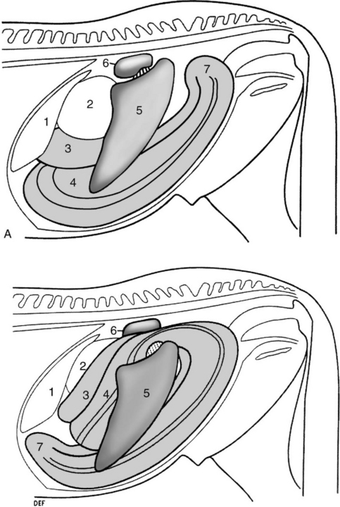
Fig. 5.1 A Left lateral view of abdomen of a normal horse. B Left dorsal displacement of the left colon, left lateral view. The left ventral and dorsal colon is displaced lateral and dorsal to the spleen and occupies the renosplenic space. 1 = liver, 2 = stomach, 3 = left dorsal colon, 4 = left ventral colon, 5 = spleen, 6 = left kidney and renosplenic ligament, 7 = pelvic flexure.
(With permission from Johnston JK, Freeman DE. Vet Clin North Am Equine Pract 1997; 13:317.)
Right dorsal displacement of the large colon (Fig. 5.2) occurs sporadically and without recognized risk factors. The case fatality rate is reported to be as high as 43%.10
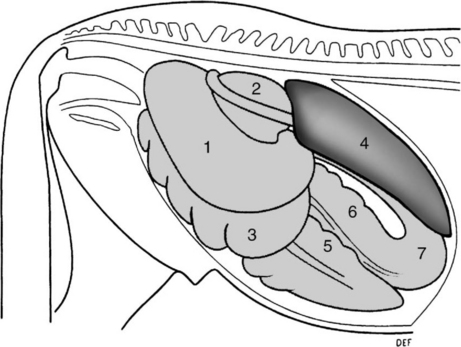
Fig. 5.2 Right dorsal displacement of the colon, right lateral view. The colon has passed lateral to the cecum, the pelvic flexure is displaced cranially and the sternal and diaphragmatic flexures are displaced caudally. 1 = right dorsal colon, 2 = base of cecum, 3 = right ventral colon, 4 = liver, 5 = cecum, 6 = left ventral colon, 7 = pelvic flexure.
(With permission from Johnston JK, Freeman DE. Vet Clin North Am Equine Pract 1997; 13:317.)
Risk factors for noninfarctive displacement of the large colon include cribbing or wind sucking (odds ratio (OR) = 90), number of hours stabled per day (OR for 24 h stabling = 35), lack of regular exercise (OR = 3.3), change in exercise program (OR = 9), lack of anthelmintic administration (OR = 13) and history of transport in the previous 24 hours (OR = 17).11
Volvulus of the large colon is the cause of colic in 11–17% of colic cases in which abdominal surgery is performed.12 The disease occurs commonly in mares, especially those late in gestation or having recently foaled.13,14 The disease has a recurrence rate of up to 15% in brood mares.15 The disease occurs in horses from 2 days of age and there does not appear to be an effect of breed on occurrence of the disease.16 The case fatality rate varies depending on the extent of the volvulus, with lesser degrees of volvulus (< 270°) having a 30% fatality rate and volvulus of 360° or more having a 65% fatality rate.13
Ingestion of large quantities of grain, such as might be fed to horses in heavy work, is associated with changes in plasma electrolyte concentrations, presence of dehydrated, foamy and homogeneous right dorsal colon contents, and fetid, less formed feces.17 These effects of a high-grain diet may be associated with colonic disease in horses.17
Proximate factors leading to volvulus or displacement are unknown, although risk factors have been identified (see above). A plausible scenario is that altered colonic motility and subsequent distension with gas or ingesta predisposes the colon to displacement, either spontaneously or as a result of the horse rolling or lying down in response to abdominal pain.
Left dorsal and right dorsal displacements of the colon rarely compromise colon blood flow and represent nonstrangulating obstructive lesions. Pathogenesis in equine colic section). The displacement of the large colon (Figs 5.1 & 5.2) impedes aboral movement of ingesta and gas and may result in colonic distension. Should the distension become sufficiently severe, colon blood flow will be impaired and cause ischemia and necrosis of the colon. The obstruction to blood flow is predominantly in venous drainage, resulting in hemorrhagic strangulating obstruction with progressive development of intramural edema, extravasation of red blood cells, microvascular thrombosis, mesothelial cell loss from the serosal surface, and mucosal necrosis with loss of colonic epithelium.18
Volvulus of the large colon of less than 270° does not compromise blood supply but does impede aboral movement of ingesta and gas.13 Volvulus of 360° or more causes ischemia through occlusion of both arterial and venous circulation of the involved large colon with rapid loss of colonic mucosal integrity and colon viability. Irreversible mucosal damage occurs after 3–4 hours of ischemia. Loss of mucosal integrity impairs normal barrier function and permits toxins and substances normally confined to the colonic lumen to enter the systemic circulation. Additionally, loss of barrier function allows leakage of vascular proteins and in severe cases red blood cells into the colonic lumen. Subsequent signs are typical of strangulating obstruction (see Equine colic) with development of toxemia, cardiovascular collapse and death within 12–18 hours.
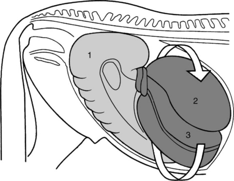
The most common displacement is medial and dorsal movement of the ventral colon to complete a 360° volvulus of the large intestine (Fig. 5.3).16 Lateral and dorsal displacement of the ventral colon is much less common. The volvulus is usually at the level of the cecocolic fold, although volvulus involving the cecum or at the diaphragmatic and sternal flexures does occur.
The disease usually has an acute onset and a duration of up to 4 days, although it can be a cause of chronic, recurrent colic.6,9 Abdominal pain in the initial stages is mild to moderate and becomes progressively more severe as distension of the large colon develops. The heart rate is usually between 50 and 70/min, but may be as low as 30/min. Rectal temperature is within normal limits. Mucous membrane color and refill time are usually normal provided that there is no ischemia of the colon. Abdominal distension is appreciable in some affected horses.6 There is more than 2 L of reflux from a nasogastric tube in approximately 28% of cases, although rarely is there profuse reflux.9 Rectal examination reveals the presence of bowel in the renosplenic space in approximately 70% of cases with the typical finding of taenia of the ventral colon being traced into that space. Distension of the large colon may impair detection of bowel in the nephrosplenic space. The spleen is usually displaced caudally, medially and ventrally from its normal position against the left body wall (Fig. 5.1).
Ultrasonographic demonstration of colon in the renosplenic space confirms the diagnosis with an accuracy of 88%.19 Gas in the displaced colon obscures the left kidney and dorsal border of the spleen normally visible on ultrasonographic examination of the left paralumbar region.19
Approximately 8% of horses with nephrosplenic entrapment have an additional lesion.9 Entrapment in which the sternal and diaphragmatic flexures are displaced cranial to the stomach and liver occurs in less than 3% of cases.9
Severity of colic varies from mild to severe in horses with right dorsal displacement of the colon. Tachycardia (50–80/min) and mild abdominal distension are characteristic provided that the entrapped bowel is not ischemic. There is usually no reflux from a nasogastric tube, although as the disease progresses gastric distension may occur. Rectal examination reveals the presence of large colon lateral to the base of the cecum, although colonic distension may make detection of the displaced bowel difficult. Right dorsal displacement is a not uncommon sequel to impaction of the pelvic flexure.
The onset of pain is abrupt and the duration of the disease ranges from hours, in horses with strangulating lesions, to days in horses with torsion of less than 270°. The pain ranges from mild to severe and intractable, with the horse violently throwing itself to the ground. Pain in horses with volvulus of 360° or greater is often unresponsive to any analgesics. Heart rate is variable and may be less than 40/min in horses with severe disease, although usually it is more than 60/min and increases with severity of the disease. Rectal temperature is within the normal range. The mucous membranes are dark red to blue and capillary refill time is more than 3 seconds in severely affected horses. Abdominal distension is marked, usually severe, and may impair respiration in horses with 360° or greater volvulus. Auscultation of the abdomen reveals a lack of borborygmi and the presence of high-pitched, tympanitic ‘pings’ on simultaneous percussion and auscultation. The pings are due to the presence of gas in tightly distended large colon or cecum. There is usually no reflux through a nasogastric tube. Rectal examination may be limited by the distended, gas-filled colon occupying the caudal abdomen. In untreated cases death occurs within 12–24 hours from cardiovascular collapse. Ultrasonographic examination reveals colon with a mural thickness of 9 mm or greater in horses with colon torsion. The test has a sensitivity of approximately 67% (i.e. correctly predicts presence of colon torsion in two-thirds of horses that have the disease) and specificity of 100% (correctly rules out the diagnosis in 100% of horses that do not have the disease).20
Changes in the hemogram, serum biochemical profile and peritoneal fluid are non-existent to mild in horses with uncomplicated left dorsal displacement, right dorsal displacement and volvulus of less than 270°. Horses with ischemic colon as a result of strangulation usually have a leukopenia with left shift, hemoconcentration and increased anion gap.16
Serum gamma glutamyl transferase (GGT) activity is elevated in approximately 50% of horses with right dorsal displacement of the colon, whereas such elevations are rare in horses with left dorsal displacement.21 The elevated GGT, and less commonly serum bilirubin concentration, in horses with right dorsal displacement is attributable to compression of the common bile duct in the hepatoduodenal ligament by the displaced colon.21
Horses with large-colon volvulus have a high prevalence of abnormalities in hemostatic variables, including thrombin– antithrombin concentration, d-dimer concentration, antithrombin activity, prothrombin time and platelet count. Nonsurviving horses have lower platelet counts, increased prothrombin time and reduced antithrombin activity.22
Peritoneal fluid often has an increased total protein concentration (> 25 g/L, 2.5 g/dL) and white blood cell count (> 8000 cells/μL, 8 × 10⁹ cells/L) in horses with compromised bowel. Examination of peritoneal fluid is often not necessary to achieve a diagnosis in horses with colon torsion, although it does have prognostic value in that horses with blood-tinged peritoneal fluid have a poor prognosis. The risk of inadvertent enterocentesis is increased in horses with severe distension of the colon and abdominocentesis should be attempted with caution in such cases. Use of a bovine teat cannula or similar blunt instrument is preferred to the use of a needle.
The colon is displaced as described above for each of the diseases. Death usually results from ischemic necrosis of the colon and the associated peritonitis, endotoxemia and shock. Histological lesions in horses dying of colon volvulus are more severe than of those that survive and are characterized by hemorrhage into the lamina propria, edema and loss of the mucosal cells and crypt architecture.13
Treatment should consist of pain control, correction of fluid, acid–base and electrolyte abnormalities, support of cardiovascular function and correction of the underlying disease (Equine colic). Decompression by trocarization of gas-distended colon or cecum may be beneficial. Correction of colon volvulus or right dorsal displacement of the colon requires surgical exploration of the abdomen and manual correction of the displacement.
Correction of left dorsal displacement can be achieved by either nonsurgical or surgical means. Nonsurgical correction is achieved by rolling the anesthetized horse in a particular sequence that causes the displaced colon to return to its normal position in the abdomen. Nonsurgical correction is successful in approximately 80% of cases,7,27 although complications are reported,28 and is recommended as the initial definitive treatment for horses with uncomplicated left dorsal displacement.27 The sequence of events following diagnosis of the condition is depicted in Figure 5.4.29 Phenylephrine (0.02–0.04 mg/kg, intravenously as a 10 min infusion) causes splenic contraction and is thought to increase the chances of the colon returning to its normal position. The horse is anesthetized within 10 minutes of phenylephrine administration and placed in right lateral recumbency. The horse is then slowly rolled into dorsal recumbency and the abdomen is vigorously massaged in an attempt to cause the colon to move ventrally and medially. If a hoist is available the horse can be lifted into dorsal recumbency. The sequence ends with the horse being rolled into left lateral recumbency and a rectal or ultrasound examination being performed to determine the position of the colon.
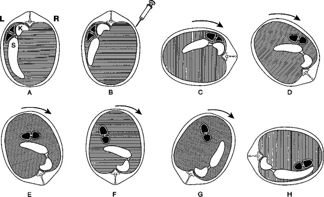
Fig. 5.4 Steps in correction of left dorsal displacement of the colon (renosplenic entrapment). A Caudal view of abdomen of horse with left dorsal displacement of the colon. Entrapped colon is shown in black; K = left kidney, S = spleen. B Injection of phenylephrine and contraction of spleen. C Horse anesthetized and placed in right lateral recumbency. D–H Horse rolled through dorsal recumbency to left lateral recumbency. Entrapped colon moves ventrally and then medially to the contracted spleen.
(Modified with permission from Kalsbeek HC. Equine Vet J 1989; 21:442.)
An alternative means of nonsurgical correction involves administration of phenylephrine (0.01 mg/kg, intravenously, slowly) and then jogging the horse.30,31 This technique was successful in correcting the displacement in 11 of 12 horses.31 It may be advantageous to relieve large-colon distension by percutaneous trocarization before jogging.9
Cases that are refractory to nonsurgical treatment require laparotomy (ventral midline or left flank) and manual correction of the displacement. Recurrence of the displacement occurs in 3–7% of cases.7 Horses with recurrent disease may benefit from surgical ablation of the nephrosplenic space.32
Burba DJ, Moore RM. Renosplenic entrapment: a review of clinical presentation and treatment. Compend Contin Educ Pract Vet. 1997;9:180-184.
Johnston JK, Freeman DE. Diseases and surgery of the large colon. Vet Clin North Am Equine Pract. 1997;13:317-340.
Gibson KT, Steel CM. Strangulating obstructions of the large colon in mature horses. Equine Vet Educ. 1999;11:234-242.
Lopes MA, Pfeiffer CJ. Functional morphology of the Equine pelvic flexure and its role in disease. A review. Histol Histopathol. 2000;15:983-991.
1 Mair TS. Vet Rec. 2002;151:152.
2 Bassage LH, et al. Equine Vet J. 2000;32:565.
3 Latimer FG, et al. Equine Vet Educ. 1999;11:229.
4 Segura D, et al. Equine Vet Educ. 1999;11:227.
5 Gibson KT, Steel CM. Equine Vet Educ. 1999;11:234.
6 Livesey MA, et al. Can Vet J. 1988;29:135.
7 Baird AN, et al. J Am Vet Med Assoc. 1991;198:1423.
8 Knottenbelt DC, Hill FWG. Vet Annu. 1989;29:161.
9 Hardy J, et al. Equine Vet J Suppl. 2000;32:95.
10 Van der Linden MA, et al. J Vet Intern Med. 2003;17:343.
11 Hillyer MH, et al. Equine Vet J. 2002;34:455.
12 Fisher AT, Meagher DM. Compend Contin Educ Pract Vet. 1985;8:S25.
13 Snyder JR, et al. J Am Vet Med Assoc. 1989;195:757.
14 Moore JN, Dreesen DW. Proc Am Assoc Equine Pract. 1993;38:99.
15 Hance SR, Emberstson RM. J Am Vet Med Assoc. 1992;201:782. 87
16 Harrison IW. Vet Surg. 1988;17:77.
17 Lopes MAF, et al. Am J Vet Res. 2004;65:687.
18 Snyder JR, et al. Am J Vet Res. 1988;49:801.
19 Santshi EM, et al. Vet Surg. 1993;22:281.
20 Pease AP, et al. Vet Radiol Ultrasound. 2004;45:220.
21 Gardner RB, et al. J Vet Intern Med. 2005;19:761.
22 Dallap BL, et al. J Vet Emerg Crit Care. 2003;13:215.
23 Steenhaut M, et al. Equine Vet J. 1993;25:550.
24 Dyson S, Orsini JA. J Am Vet Med Assoc. 1983;182:720.
25 Roy MF, et al. Equine Vet J. 2002;34:102.
26 Harvey-Micay J. Can Vet J. 1999;40:729-730.
27 Abutarbush SM, Naylor JM. J Am Vet Med Assoc. 2005;227:603.
28 Markel MD, et al. J Am Vet Med Assoc. 1985;187:1379.
29 Kalsbeek HC. Equine Vet J. 1989;21:442.
30 Johnston JK. Vet Surg. 1996;25:13.
Etiology Idiopathic, often associated with restricted exercise, poor-quality diet or restricted access to water
Epidemiology Sporadic, more common in mares. Accounts for approximately 10–15% of colics at referral institutions. Case fatality rate of 20%
Clinical signs Mild to moderate colic often of several days duration. Rectal examination reveals impacted, distended large colon
Clinical pathology No diagnostic changes
Lesions Impaction of large colon, usually pelvic flexure or right dorsal colon
Diagnostic confirmation Physical examination
Treatment Pain control. Administration of fecal softeners (sodium sulfate). Overhydration by oral or intravenous administration of isotonic fluids at 3–5 times maintenance needs
The cause of most impactions of the large colon is unknown. Known or speculated causes include:
• Poor dentition, such as occurs in older horses
• Poor feeding regimens, such as infrequent feeding of stalled horses
• Horses not fed, in preparation for surgery or racing, and then given unrestricted access to feed or allowed to eat bedding materials
• Horses fed diets too high in fiber, e.g. mature sorghum or maize plants, or even mature Bermuda grass (Cynodon spp.) meadow hay, especially if their water intake is limited;1 ingestion of large volumes of indigestible seeds, e.g. Crataegus crusgalli (cockspur hawthorn), may cause outbreaks of impaction of the right dorsal colon2
• Horses that come into loose boxes and are offered hard feed after being on soft grass on pasture are also likely to develop impaction colic
• American miniature horses develop impaction of the colon3
• Enteroliths and fiber balls may also cause obstruction of the large intestine and usually result in recurrent attacks of colic
• Amitraz, a formamidine acaricide for cattle, causes impaction colic in horses4
• Retention of the meconium in foals (see Colic in foals)
• Administration of NSAIDs, which alter colonic motility and might predispose to impaction,5 although epidemiological support of this etiology is not available
• Restricted water intake, such as during winter when watering points freeze or water is unpalatable.
The disease occurs in horses of any age and is more common in females.6 There does not appear to be a breed predisposition. The disease represented 13% of colics treated at a referral facility.6,7 An important risk factor is a change in management, especially one that involves a reduction in exercise and change in diet.6 Risk factors for nonstrangulating disease of the large colon, including pelvic flexure impaction, include cribbing or wind sucking, stabling with the risk increasing with the number of hours stabled per day, change in regular exercise program, travel within the previous 24 hours and lack of anthelmintic administration.8 The case fatality rate is approximately 1–20%.6,7
Development of impaction of the large colon is frequently attributed to abnormal colonic motility.9 Other factors, including mild dehydration as a result of limited water intake or ingestion of poorly digestible material, cause impaction in many instances. Ingestion of large quantities of grain, such as might be fed to horses in heavy work, is associated with changes in plasma electrolyte concentrations, presence of dehydrated, foamy and homogenous right dorsal colon contents, and fetid, less formed feces.10 These effects of a high grain diet may be associated with colonic disease in horses.10 The end result is accumulation of a large mass of inspissated feed material in the large colon. Material usually accumulates first at the pelvic flexure or right dorsal colon, presumably because of the reduction in lumen diameter at those points. Accumulation of inspissated material causes distension of the colon and prevents aboral passage of ingesta. Distension causes pain and changes in colonic motility that exacerbate or perpetuate the impaction. If the distension is sufficiently severe or prolonged the colon may become ischemic and necrotic with subsequent rupture, peracute diffuse peritonitis and death.
Moderate abdominal pain is the typical sign in affected horses and pulse rate and respiration are relatively normal. This often continues for 3–4 days and sometimes for as long as 2 weeks. The horse is not violent, the principal manifestation of pain being stretching out and lying down and the bouts of pain are of moderate severity occurring at intervals of up to a half-hour. There is anorexia and the feces are passed in small amounts and are hard and covered with thick, sticky mucus. Intestinal sounds are absent or much decreased in intensity. The pulse rate is usually less than 50/min.
On rectal examination impaction of the pelvic flexure of the large colon is the commonest site and the distended, solid loop of the intestine often extends to the pelvic brim or even to the right of the midline. Lying on the floor of the abdomen, it is easily palpated, the fecal mass can be indented with the fingers and the curvature and groove between the dorsal and ventral loops of the left colon can be easily discerned. Impaction of the right dorsal colon cannot usually be palpated per rectum and the only abnormality may be distension of the colon with soft ingesta that has accumulated behind the obstruction.
Hemogram, blood chemistry and peritoneal fluid are normal until the colon becomes ischemic at which time there is a leukopenia with left shift, and an increase in the white blood cell count and protein concentration in peritoneal fluid.
The large intestine is packed full of firm, dry fecal material and rupture may have occurred.
The principles of treatment are pain control, correction of fluid and electrolyte abnormalities and softening of ingesta to facilitate its passage. Pain control is discussed in Table 5.7. Fluid therapy is discussed in Chapter 2.
Softening of ingesta is achieved by rehydrating the inspissated material and providing lubrication to hasten its passage. Fecal softeners (Table 5.8) such as magnesium sulfate or sodium sulfate can be given to increase the fecal water content and soften the impacted, inspissated ingesta. Magnesium sulfate is associated with a small risk of hypermagnesemia and neurologic signs12 whereas sodium sulfate causes a mild hypernatremia and hypokalemia.13 Oral administration of a balanced, polyionic electrolyte solution is associated with the greatest increase in colonic water content and no change in serum electrolyte concentrations.13 Enteral administration of 10 L/h (to a 500 kg horse) of a balanced, isotonic, polyionic electrolyte solution is more effective than intravenous administration of the same quantity of fluid in combination with oral administration of MgO4 in hydrating colonic contents in normal horses.14 Mineral oil (Table 5.8) is a lubricant and may not penetrate the impacted ingesta sufficiently to soften the material although it is frequently given to horses with colon impaction.
Overhydration by oral administration of polyionic, isotonic fluids at 3–5 times maintenance needs (approximately 10 L/h) is the treatment of choice for colon impaction.6,13 Water can be given by nasogastric tube at a rate of 4–10 L for a 450 kg horse every 1–2 hours until the impaction softens. However, some horses develop decreased small intestinal motility or ileus with the disease, have delayed gastric emptying and have reflux of fluid through the nasogastric tube. Such horses should not be administered any medication or water through the nasogastric tube until reflux has resolved. Alternatively, isotonic fluids can be given intravenously at 10 mL/kg/h until the impaction is passed.
Promotility agents such as neostigmine are usually contraindicated because of the risk of rupture of the distended colon when vigorous contractions are induced pharmacologically.
Horses may need to be treated for 1–6 days until the impaction resolves and should not be fed during this time. When feed is again provided it should be easily digestible and initially be of limited volume. Horses recovered from impaction of the large intestine have a higher than expected rate of recurrence of colic (30%).6
Surgical treatment may be needed for refractory cases (about 15%) but is associated with a poor prognosis because of the risk of iatrogenic rupture of the colon during attempts to exteriorize it from the abdomen during surgery.6 Impaction of the right dorsal colon is more likely to require surgical treatment.15
1 Pugh DG, Thompson JT. Equine Pract. 1992;14:9.
2 Rook JS, et al. Equine Pract. 1991;13:28.
3 Ragle CL, et al. J Am Vet Med Assoc. 1992;201:329.
4 Roberts MC, Seawright AH. Aust Vet J. 1979;55:553.
5 Van Hoogmoed LM, et al. Am J Vet Res. 2000;61:1259-1263.
6 Dabrareiner RM, White NA. J Am Vet Med Assoc. 1995;206:679.
7 Van der Linden MA, et al. J Vet Intern Med. 2003;17:343.
8 Hillyer MH, et al. Equine Vet J. 2002;34:155.
9 Sellers AF, Lowe JE. Equine Vet J. 1986;18:261.
10 Lopes MAF, et al. Am J Vet Res. 2004;65:687.
11 Rose PL, et al. Vet Surg. 1991;20:260.
12 Henninger RW, Horst J. J Am Vet Med Assoc. 1997;211:82.
13 Lopes MAF, et al. Am J Vet Res. 2004;65:695.
Enteroliths are rock-like concretions, which are either spherical or tetrahedral, that form in the large colon of horses, usually around a foreign body.1 Most enteroliths in the large colon of horses are of two major types: magnesium phosphates/struvite and magnesium vivianite.1,2 There is wide variability in macrotexture and ionic concentrations between and within enteroliths of ammonium magnesium phosphate (struvite).1 Affected horses often have more than one enterolith and the enteroliths can weigh up to 12 kg.
Fecaliths are aggregations of indigestible material, such as fencing, plastic or rope, that often have an irregular shape.
Enteroliths occur sporadically in horses in most regions of the world but there is a greater than expected incidence in certain areas, such as California.3 Equids with enterolithiasis represented 15.1% of patients admitted for treatment colic, and 27.5% of patients undergoing celiotomy for treatment of colic in a study from California but less than 2% of horses with colic examined at a referral center in Texas.4,5 Arabians and Arabian crosses, Morgans, American Saddlebreds and donkeys are over-represented, and Thoroughbreds, Standardbreds, warmbloods and stallions are under-represented in some studies, suggesting a predilection of these breeds for the disease.3-5 Enteroliths rarely occur in horses less than 4 years of age and are more common in older horses (> 11 years).4,6 The disease is reported in American Miniature Horses.7 Feeding alfalfa hay and stabling for more than 12 hours a day are associated with increased risk of enterolithiasis.5,8 The mean pH of colonic contents from horses with enterolithiasis is significantly higher than for control horses and horses with enterolithiasis have a significantly lower percentage of dry matter in colonic fecal samples and higher mean mineral concentrations than controls.8 About 15% of cases examined at referral institutions that see large numbers of cases develop a ruptured viscus caused by the enterolith and die. The long-term survival rate of horses treated surgically is approximately 90%.4
Fecaliths occur sporadically and appear to be more common in younger horses, perhaps because of their propensity to dietary exploration and ingestion of foreign materials.
The mechanism underlying enterolith formation is not known. Enteroliths are formed in the large colon and, rarely, the cecum. They are clinically inapparent, even if quite large, until they cause obstruction of aboral passage of ingesta, usually by occluding the right dorsal or transverse colon. Occasional enteroliths pass into the small colon. Obstruction of the colon causes mild to moderate, often intermittent, colic, presumably when the enterolith or fecalith obstructs the colon, with the pain resolving when the enterolith moves and the obstruction clears. Complete obstruction results in obstruction of aboral movement of ingesta, accumulation of gas and ingesta proximal to the obstruction and distension of the large colon. There is no loss of integrity of the colon early in the disease but with time and distension there is ischemia and necrosis of the colon, with subsequent perforation, development of acute peritonitis, and death.
The most common historic manifestation of enterolithiasis in horses is recurrent, intermittent colic (about one-third of cases), often with passage of enteroliths in feces (about 10% of cases).4,9 Horses with acute obstruction have signs typical of obstructive, nonstrangulating disease of the large colon, including mild to moderate colic with failure to pass feces. The heart rate is 50–70/min, borborygmi are decreased but not absent, and there is mild abdominal distension. Rectal examination may reveal mildly distended large colon but the offending enterolith is never palpable, except on the rare occasion that the enterolith or fecalith is lodged in the small colon. Over a period of 6–12 hours the severity of pain increases and there is readily apparent distension of the large colon. There is usually no reflux through a nasogastric tube. The terminal phase, which may take 72 hours to occur and is due to rupture of a viscus, is marked by moderate to severe pain, abdominal distension, tachycardia (> 80/min), decreased capillary refill time and discolored mucous membranes, sweating, muscle fasciculations and death. Rupture of a viscus and acute peritonitis occurs in approximately 15% of cases.4
Radiography of the abdomen is useful in identifying enteroliths in horses with colic.10,11 The accuracy of the diagnosis is approximately 80% for enteroliths in the large colon and 40% for those in the small colon.11 The most common reason for not detecting an enterolith is poor imaging of the abdomen because of inadequate penetration by the X-ray beam,11 emphasizing the need for appropriate radiographic equipment.
There are no diagnostic changes in the hemogram, serum biochemical profile or examination of peritoneal fluid. Horses with enteroliths have higher serum bilirubin concentrations on examination at referral centers, but this change is not sufficiently large to be useful as a diagnostic aid.5 Similarly, horses with enteroliths have higher protein and white cell counts in peritoneal fluid than do horses with other forms of colic but again these differences are too small to be of diagnostic significance.5 Changes in hematological and biochemical variables during the terminal phases of the disease are characteristic of acute, diffuse peritonitis and include leukopenia with left shift, hemoconcentration and azotemia.
Enteroliths are frequent incidental findings at necropsy examination of mature horses and their presence should not be overinterpreted. Obstructive disease caused by an enterolith is characterized by colon distension, presence of an enterolith in the right dorsal, transverse or small colon and, in cases dying of the disease, acute diffuse peritonitis resulting from colon rupture or perforation at the site of the enterolith. Tetrahedral enteroliths with sharp points are believed to be more dangerous than are spherical enteroliths.6
See Table 5.6.
The main differential diagnosis is colon impaction, which may be difficult to differentiate from enterolith obstruction in the absence of radiographic examination of the abdomen.
1 Blue MG, Wittkopp RW. J Am Vet Med Assoc. 1981;179:79.
2 Hassel DM, et al. Am J Vet Res. 2001;62:350.
3 Lloyd K, et al. Cornell Vet. 1987;77:172.
4 Hassel DM, et al. J Am Vet Med Assoc. 1999;214:233.
5 Cohen ND, et al. J Am Vet Med Assoc. 2000;216:1787.
6 Evans DR, et al. Compend Contin Educ Pract Vet. 1981;3:S383.
7 Ragle CA, et al. J Am Vet Med Assoc. 1992;201:329.
8 Hassel DM, et al. J Vet Intern Med. 2004;18:346.
9 Colgan SA, et al. Aust Vet J. 1997;75:100.
10 Rose JA, Rose EM. Proc Am Assoc Equine Pract. 1987;33:95.
Sand colic is a disease of horses grazing sandy fields with short pasture, fed on sandy ground or provided with feed contaminated with sand. It is often associated with underfeeding. Horses of all ages are affected, including foals, which acquire the sand while eating dirt. The case fatality rate for horses treated by surgical removal of sand is 20–40%.1,2 The disease is attributable to sand accumulation in the right dorsal or transverse colon, or pelvic flexure, causing obstruction. Sand in the ventral colon does not cause obstruction but is associated with colon volvulus and displacement.
Clinical signs are of mild to moderate, chronic colic with diarrhea and anorexia. The colic is often very mild unless there is colon torsion or volvulus, in which case the signs are typical of that disease. The diarrhea is watery but not profuse or malodorous. Auscultation over the cranial ventral abdomen just caudal to the xiphoid reveals sounds similar to those made when a paper bag is partially filled with sand and rotated.3 This sound is diagnostic of sand accumulation in the ventral colon. Rectal palpation may reveal sand impaction in the ventral colon, but more frequently colon distension with gas is present.1,2 Rectal palpation will not detect sand accumulation in the transverse colon. Radiography will demonstrate sand in the ventral and dorsal colons and can be used to monitor the efficacy of treatment.4-6 Ultrasonography has good sensitivity (88%) and specificity (88%) compared to radiography for detection of sand in the ventral colon.7 Ultrasonography is not as effective at detecting sand in the right dorsal or transverse colon.7 Abdominal fluid is normal except when there is ischemia or necrosis of the colon or when peritonitis is present.4 Sand will settle out when feces is mixed with water in a clear plastic rectal sleeve and hung for 30 minutes.
Treatment consists of pain relief, correction of fluid and electrolyte abnormalities, prevention of continued ingestion of sand and removal of the sand. In horses with acute obstruction of the right dorsal or transverse colon by sand, surgical removal is indicated. Medical treatment to effect sand removal is indicated in less acute cases. A widely used medical treatment is administration of psyllium mucilloid (0.5–1 g/kg orally every 12 h for 4–8 weeks) administered via a nasogastric tube or as a dressing on feed. However, in an experimental model of the disease this treatment was no more effective than no specific treatment in removal of sand from the cecum and colons.8 Mineral oil (1 mL/kg) or MgSO4 (1 g/kg) orally may hasten sand removal.6 Pasturing of horses with sand accumulation housed in stables aids removal of the sand.6 Control of the disease is by preventing ingestion of sand by feeding horses hay and grain from clean feeding bins, providing adequate roughage in the diet, pasturing horses in fields with adequate grass cover, and perhaps, in areas where sand ingestion is unavoidable, daily administration of psyllium mucilloid.
1 Specht TE, Colahan PT. J Am Vet Med Assoc. 1988;193:1560.
2 Ragle CA, et al. Vet Surg. 1989;18:48.
3 Ragle CA, et al. J Vet Intern Med. 1989;3:12.
4 Bertone JJ, et al. J Am Vet Med Assoc. 1988;193:1409.
5 Rose JA, Rose EM. Proc Am Assoc Equine Pract. 1987;33:95.
6 Ruohoniem M, et al. Equine Vet J. 2001;33:59.
This is a chronic disease caused by ulcerative colitis of the right dorsal colon. The disease is associated with prolonged administration of NSAIDs, such as phenylbutazone, in most, but not all, cases.1 The case fatality rate is greater than 50%, although descriptions of large numbers of affected horses are not available.
The pathogenesis involves inhibition of mucosal prostaglandin synthesis and consequent decreases in water, chloride and bicarbonate secretion by mucosa of the right dorsal colon and apoptosis (programmed cell death) of mucosal cells.2 Loss of secretion of bicarbonate might be associated with failure of alkalinization of right dorsal colon contents and subsequent development of mucosal lesions. The right dorsal colon is the only section of the colon with net water secretion, and this unique activity may predispose this section of colon to disease. Exposure of mucosal cells to phenylbutazone can occur both from the lumen and from blood. Luminal exposure may be related to release of phenylbutazone from ingesta in the right dorsal colon.3 Ulceration of the colonic mucosa allows leakage of plasma constituents into the colonic lumen, resulting in hypoalbuminemia and loss of electrolytes, and entry of colonic substances such as endotoxin into the systemic circulation, with consequent signs of endotoxemia and systemic inflammatory response (leukopenia, hyperfibrinogenemia, fever). Chronic and extensive mucosal ulceration causes growth of granulation tissue and fibrosis of the right dorsal colon with subsequent loss of secretory function, stricture and partial obstruction.4
Clinical signs include depression, anorexia, mild fever (38.6–39.5°C, 101.5–103°F), mild intermittent colic, ventral edema, weight loss and occasionally mild diarrhea. There is almost always a history of administration of a NSAID. The disease can persist for weeks and often prompts inappropriate administration of nonsteroidal anti-inflammatory drugs. Rectal examination is unremarkable. Ultrasonography is useful in the diagnosis of right dorsal colitis by detecting the presence of a hypoechogenic submucosal layer and permitting measurement of the wall thickness of the right dorsal colon.5 The hypoechogenic layer in the wall of the right dorsal colon corresponds with edema and cellular infiltrates observed histologically. The right dorsal colon in adult horses has a maximal thickness of 6 mm while that in horses with right dorsal colitis is greater than 8 mm and can be as great as 16 mm.5 Additionally, the ratio of right dorsal colon to right ventral colon wall thickness is up to 1.6 in normal horses and greater than 2.0 in affected horses.5 Scintigraphic detection of right dorsal colitis is achieved by administration of technicium-99m hexamethylpropyleneamine-oxime-labeled white blood cells.6 Images obtained 20 hours after administration of labeled white cells demonstrate uptake of cells into the right dorsal colon (right cranioventral abdomen).
There is often mild peritonitis (neutrophilia in peritoneal fluid). Leukopenia with left shift and hypoproteinemia are characteristic. Serum biochemical abnormalities include hypoalbuminemia, hyponatremia (< 135 mEq/L), hypochloremia (<90 mEq/L) and azotemia (serum creatinine >2 mg/dL, 170 μmol/L).1,7
Necropsy examination reveals ulcerative colitis of the right dorsal colon. In chronic cases there may be stricture of the right colon with subsequent impaction of ingesta and colon rupture.1
Treatment is often unrewarding although successful treatment by feeding of a low residue diet, such as a complete pelleted ration fed 4–6 times daily, is reported.8 Psyllium (120 g once daily) for 3–6 weeks might enhance healing of the colon. Administration of misoprostol (Table 5.11) has been suggested but has no demonstrated efficacy. Surgical excision of the lesion is difficult because of its location in the abdomen but bypass of the right dorsal colon may be beneficial. Control involves minimizing the amount of NSAIDs administered to horses.
1 Karcher LF, et al. J Vet Intern Med. 1990;4:247.
2 Richter R, et al. Am J Vet Res. 2002;63:934.
3 Lees P, et al. Res Vet Sci. 1988;44:50.
4 Hough ME, et al. Aust Vet J. 1999;77:785.
5 Jones SL, et al. J Am Vet Med Assoc. 2003;222:1248.
6 East LM, et al. Vet Radiol Ultrasound. 2000;41:360.
• Obstruction by enterolith or fecalith1
• Meconium retention (see Foal colic)
• Atresia coli (see Foal colic)
• Strangulation by pedunculated lipoma,3 volvulus, intussusception,4 herniation through mesenteric rents including the mesocolon or gastrosplenic ligament5,6 or enlarged ovary7
• Rupture of mesocolon8
• Colonic lipomatosis9
Small colon disease is present in approximately 2.5–5% of horses treated for colic at referral institutions and small colon impaction represents approximately 2% of horses with colic.1,2,10 Aged female horses are most commonly affected although the conditions can occur in horses of any age.3 Arabians, ponies and Miniature horses are reported to be at increased risk of small colon disease although others have not detected this apparent predilection.3,10. Rupture of the mesocolon occurs during parturition.4 The case fatality rate depends on the condition and is 30–40% for impaction of the small colon.2,10 Small colon impaction can occur as limited outbreaks in a number of horses on a single farm over a period of days to weeks, without obvious predisposing causes or inciting events.
Obstruction of the small colon causes accumulation of ingesta and gas in the small colon aboral to the obstruction and in the large colon, with subsequent distension, pain and reduced motility. Distension of the small colon may impair blood flow with subsequent ischemia, necrosis and rupture or perforation of the small colon. Incarceration of the small colon results in ischemia of the entrapped segment and restriction of flow of ingesta. Subsequent signs are characteristic of toxemia and intestinal obstruction. The high proportion of affected horses from which Salmonella spp. are isolated suggests a role for colitis in the pathogenesis of small colon impaction.2,10
Nonstrangulating lesions manifest as mild to moderate colic that may persist without a change in severity for up to 36 hours. The heart rate depends on the severity of the colic but averages 60/min with a range of 30–110/min.2 There is mild dehydration. Abdominal distension is usually mild initially but increases as the disease progresses. Borborygmi are reduced and tympanitic sounds may develop as the large colon and cecum become distended. Rectal examination reveals the presence of distended large colon but no evidence of colon displacement.
Small colon impaction is palpable as a tubular column of material in the small colon although it may be missed if the impaction is in the cranial section of the small colon. Approximately 30% of cases have diarrhea and 13% strain to defecate.10 Complete examination per rectum may be difficult because of large colon distension and accumulation of feces in the distal small colon. There is reflux through the nasogastric tube in approximately 30% of cases.2
Strangulating lesions that interfere with small colon blood supply usually present as acute colic of moderate to severe intensity. There is tachycardia and evidence of toxemia. Abdominal distension is usually marked and there is an absence of borborygmi. Rectal examination reveals distension of the large colon and occasionally soft, compressible distension of the small colon.
Avulsion of the mesocolon occurs during parturition and is often evident as a rectal prolapse in the mare. Avulsion results in ischemia of the distal colon. Initially the mare does not display signs of pain but, as the section of the colon from which the mesocolon has avulsed becomes necrotic, signs of toxemia develop.
There are no characteristic changes in the hemogram or serum biochemical profile. Peritoneal fluid is normal until the viability of the small colon is compromised, at which time the protein concentration and white blood cell count increase. Salmonella spp. are isolated from approximately 20% of cases of small colon impaction, suggesting a role for colitis in the pathogenesis of the disease.10
Small colon impaction is evident as a tubular column of firm ingesta in the small colon with large colon distension. Small colon accidents, such as rupture of the mesocolon at parturition and intussusception, are readily apparent.4,8
The principles of treatment of small-colon impaction are relief of pain and of the impaction. Horses with signs of mild to moderate colic easily controlled with analgesics should be treated medically. Horses with intractable pain or progressively worsening pain, abdominal distension or abnormal peritoneal fluid should be treated surgically. Horses treated surgically have a worse prognosis than do horses treated medically, probably because the former group has more severe disease.2,10
Medical treatment of small-colon impaction involves administration of analgesics (see Table 5.7), correction of fluid, electrolyte and acid–base abnormalities, and administration of fecal softeners (Table 5.8). Treatments to hasten softening and passage of the impaction include overhydration, administration of sodium or magnesium sulfate and a lubricant such as mineral oil, and occasionally administration of an enema to the standing horse. Overhydration should be achieved by either intravenous or oral administration of polyionic fluids at 3–5 times maintenance (10 mL/kg/h). Administration of enemas to standing horses is controversial and should be done with care so as not to rupture the small colon. Trocarization of the large colon or cecum may be necessary in horses with severe abdominal distension.
Small-colon accidents including strangulation and intussusception require surgical correction. Surgical correction of rupture of the mesocolon is not available because of limited surgical access to the site of the lesion.
Edwards GB. A review of 32 cases of small colon obstruction in the horse. Equine Vet J Suppl. 1992;13:42-47.
Edwards GB. Diseases and surgery of the small colon. Vet Clin North Am Equine Pract. 1997;13:359-375.
Schumacher J, Mair TS. Small colon obstructions in the mature horse. Equine Vet Educ. 2002;14:19.
1 White NA, Lessard P. Proc Am Assoc Equine Pract. 1986;23:637.
2 Ruggles AJ, Ross MW. J Am Vet Med Assoc. 1991;199:1762.
3 Dart AJ, et al. J Am Vet Med Assoc. 1992;200:971.
4 Ross MW, et al. J Am Vet Med Assoc. 1988;192:372.
5 Booth TM, et al. Aust Vet J. 2000;78:603.
6 Rhoads WS, Parks AH. J Am Vet Med Assoc. 1999;214:226.
7 Mair TS. Equine Vet Educ. 2002;14:17.
8 Dart AJ, et al. J Am Vet Med Assoc. 1991;199:1612.
Spasmodic colic occurs sporadically and causative factors are not usually identified. Suggested causes include excitement, such as occurs during thunderstorms, preparations for showing or racing, and drinks of cold water when hot and sweating after work, although epidemiologic evidence of these associations is lacking. Presence of a heavy burden of tapeworms is associated with a high incidence of spasmodic (undiagnosed) colic.1 Mucosal penetration and submucosal migration of Strongylus vulgaris larvae are known to cause changes in ileal myoelectrical activity that could lead to the development of colic in horses.2 Psychogenic colic occurs rarely in horses.3
The condition is sporadic. It affects horses of all ages but is not recognized in young foals. No apparent breed or sex predisposition is noted.
The hypermotility of spasmodic colic in horses is thought to arise by an increase in parasympathetic tone under the influence of the causative factors mentioned above.
Spasmodic colic of horses is characterized by brief attacks of abdominal pain. The pain is intermittent, the horse rolling, pawing and kicking for a few minutes, then shaking itself and standing normally for a few minutes until the next bout of pain occurs. Intestinal sounds are often audible some distance from the horse and loud, rumbling borborygmi are heard on auscultation. The pulse is elevated moderately to about 60/min and there may be some patchy sweating, but rectal findings are negative and there is no diarrhea. Rectal examination is usually unremarkable. The signs usually disappear spontaneously within a few hours.
Laboratory examinations are not used in diagnosis and the disease is not fatal.
Acute hypermotility as manifested by spasmodic colic is usually transient and the use of specific spasmolytics is not necessary. Detomidine, xylazine or butorphanol are effective analgesics. Administration of hyoscine is effective. Affected horses are often administered mineral oil (1 mL/kg) by nasogastric intubation.
The cause of most cases of idiopathic intestinal tympany is unknown, although the ingestion of highly fermentable green feed is considered to be a risk factor. Feeding of rations rich in grains is associated with changes in colonic contents that might predispose to tympany.1 Intestinal tympany occurs secondary to obstructive diseases that prevent aboral passage of ingesta and gas.
The excessive production of gas or its retention in a segment of bowel causes distension and acute abdominal pain. Intestinal distension reduces intestinal motility and may contribute to the course of the disease. Severe tympany may interfere with normal respiration and cardiovascular function (see Pathogenesis of equine colic).
Abdominal distension is evident and pain is acute and severe. Peristaltic sounds are reduced but fluid may be heard moving in gas-filled intestinal loops, producing a tinkling, metallic sound. Pinging sounds consistent with tightly distended viscus may be heard on simultaneous flicking and auscultation of the abdomen. On rectal examination, gas-filled loops of intestine fill the abdominal cavity and make proper examination of its contents impossible. In primary tympany much flatus is passed. It is important to differentiate primary tympany from that occurring secondary to obstructive diseases such as enterolithiasis and displacement of the colon.
In cases of secondary tympany, the causative obstruction is evident. In primary cases, the intestines are filled with gas and the feces are usually pasty and loose.
The principles of treatment are the relief of pain and distension, maintenance of hydration and reduction of gas production. In secondary tympany the primary disease should be identified and treated.
Pain should be relieved by administration of xylazine, detomidine or butorphanol, or similar agents (Table 5.7). Distension of the bowel should be relieved by trocarization but trocarization should only be performed if there is no or minimal response to analgesic medication and no return of normal peristaltic activity. Normal hydration should be restored by intravenous administration of polyionic fluids. Intestinal gas production should be minimized by the administration of mineral oil or a similar laxative (Table 5.8).
Unknown, although it is presumed to result from thromboemboli originating at sites of verminous arteritis in the cranial mesenteric artery.
The disease is assumed to be more prevalent among horses on poor parasite control programs; however, except in extreme cases that die and have a necropsy examination or exploratory laparotomy, the diagnosis is not confirmed. Therefore accurate measures of its incidence are not available. Cases may occur in foals as young as 3–6 months.1 The incidence of the disease has decreased remarkably with the advent of effective broad-spectrum anthelmintics and almost complete prevention of Strongylus spp. infection in horses in developed countries.
Migration of the larvae of Strongylus vulgaris into the wall of the cranial mesenteric artery and its branches occurs commonly in horses and may cause thromboemboli that restrict blood supply to the intestines, with subsequent ischemia and dysfunction.2 The recurrent colic of verminous arteritis is possibly due to impairment of the vascular and nerve supply to the intestine. The disease is basically an infarction of bowel wall without displacement of the bowel. The small intestine, colon and cecum can be affected. The disease has been associated with larval cyathostomiasis.3
Signs vary depending on the severity of the disease. It is assumed that mild, intermittent colics that respond to analgesics in the short term and anthelmintics in the long term are due to verminous arteritis. Affected horses are often depressed and spend long periods recumbent. Weight loss and inappetence are features of the disease in some horses. The disease can have a course of weeks to months.
Acute, severe cases of the disease are due to infarction of parts or all of the small intestine, cecum or colon. Affected horses have an acute onset of severe abdominal pain, tachycardia (> 100/min) and sweating. Auscultation reveals decreased borborygmi. There is mild distension of small intestine or large colon, depending on the segment of bowel affected, on rectal examination. There are rarely signs of intestinal obstruction. Palpation of the cranial mesenteric artery may reveal thickening and pain but is not a useful diagnostic sign for the acute disease. Death is due to peritonitis secondary to devitalization of the intestine, usually within 24 hours of the onset of signs.
There are no diagnostic changes in the hemogram or serum biochemical profile. Peritoneal fluid in mild cases may have mild elevations in protein concentration and white blood cell count. In severe cases, peritoneal fluid protein concentration is increased (> 25 g/L, 2.5 g/dL) as is white blood cell count (9000–100000 cells/μL, 9–100 × 109 cells/L).4
Infarction of the colon and cecum is most common and evident as either gangrene of large sections of the organ or multifocal mottled lesions that are red and edematous. Histological examination rarely reveals the presence of thrombi. There may be verminous arteritis of the cranial mesenteric artery, evident as thickening of the intima and narrowing of the lumen.
Mild, recurrent cases are treated with analgesics such as flunixin meglumine (Table 5.7), laxatives such as mineral oil (Table 5.8), and anthelmintics (ivermectin 200 μg/kg orally once; or fenbendazole 50 mg/kg orally every 24 h for 3 d).
Severe cases are treated with analgesics (Table 5.7), intravenous fluids (Ch. 2) and supportive care. Usually the severity of the colic prompts surgical exploration of the abdomen with resection of small lesions. Most severe cases do not survive.