Porcine intestinal spiochaetosis is a non-fatal, colonic disease of recently weaned, grower and finisher pigs. The causative organism B. pilosicoli was first recognized in 1980.1 It is a Gram-negative, anaerobic, but oxygen-tolerant spirochete found in the colon.
It is found in a wide variety of hosts including humans,2 primates, dogs, opossums, commercial chicken production and various species of birds.3,4
Porcine colonic spirochetosis (PCS) and non-specific colitis may be two different syndromes.
PCS is certainly associated with Brachyspira pilosicoli formerly known as Serpulina pilosicoli. B. hyodysenteriae was described in 1971;5 B. pilosicoli in 19976,7 and these are the only two confirmed pathogens in the Brachyspirae group. B. innocens described in 19928 and the other two B. intermedii and murdochii in 19979 are considered non-pathogenic.
Non-specific colitis (NSC) may be associated with any of the brachyspirae or any other of the common enteric bacteria but is also more likely to be associated with dietary disturbances of the large intestine. A large postal survey of enteric disease in grower-finisher pigs in England showed that colitis in some form occurred in 345 of farms.10 Large, anaerobic weakly beta-hemolytic non-B. hyodysenteriae spirochetes have been associated with porcine colitis11 and are capable of inducing disease in gnotobiotic pigs but their role as primary or opportunistic pathogens in colitis in conventional pigs is uncertain.12 B. innocens has been considered as one member of a diverse group of non-pathogenic species which may be involved in a mild disease of the large intestine.13
A specific colitis can be part of Brachyspira, Salmonella, Lawsoniana, Trichuris or Balantidium infections.
B. pilosicoli, a new species, has now been identified as the cause of porcine colonic spirochetosis (PCS).14 Other species of weakly beta-hemolytic intestinal spirochetes are encountered less frequently and these may contribute to the non-specific colitis.15 Three strains were investigated in the UK. The genetic relationships between brachyspirae were determined by pulsed field gel electrophoresis (PFGE).16
Swedish workers grouped the intestinal spirochetes isolated from pigs into four groups based on phylogenetic studies17,18 although closely related. Groups I and II were isolated only from pigs with dysentery or diarrhea. Group II was differentiated from Group I only by weak beta-hemolysis. In Sweden, members of Group II are often isolated from young weaned pigs of up to 25 kg, in herds where a non-specific diarrhea which is clinically distinct from swine dysentery occurs frequently. These strains seem to be absent or rare in herds without such diarrheic pigs. Group III included the type strain for B. innocens. Group IV included the pathogenic, weakly beta-hemolytic strain P43 shown to cause spirochetal diarrhea in pigs. A PCR system is used for the detection and identification of Group IV spirochetes (B. pilosicoli).19 Most farms have distinct B. pilosocoli genotypes and common genotypes between and among herds are rare.20
A complex investigation involving 85 pig units in Scotland over the period from 1992–1996 has provided much needed information on the occurrence of mixed infections.21 All the pigs were within 20–40 kg (occasionally 50 kg), 8–16 weeks of age, and all had diarrhea and grew slowly over the period of 2–3 weeks. B. pilosicoli was found on 25% of the units. Atypical brachyspirae on 7%; B. hyodysenteriae on 6%; with S. typhimurium on 4%; Yersinia pseudotuberculosis on 4% and Lawsoniana intracellularis on a mere 3%. Mixed infections of B. pilosicoli with yersinia, salmonella, other combinations and B. hyodysenteriae were found on 27%. On six of the 85 units nothing pathogenic was detected.
Non-specific colitis was first seen in the UK in intensive management systems. Pigs showed a sporadic diarrhea with soft, wet feces, in 18–35 kg pigs and with a morbidity of 20–30%. Most pigs continued to thrive but some grew poorly. Pigs had enlarged colons with frothy contents and most had a reddened mucosa. Microscopically they have a mild erosive colitis. Most of these cases were probably B. pilosicoli cases but there is now some evidence that would be better called dietary colitis. They could be diet related or diet induced and most often pelleted feed was implicated as a complicating factor. It has now been shown that some diets induce a colonic acidosis and enhance colonization by spirochetes. Many of these cases have no involvement of any of the brachyspirae and the possibility exists that there may be a syndrome of colonic dysfunction without direct spirochetal involvement. It is possible that any event that leads to disturbance of colonic microflora may lead to colonic lactic acidosis and damage to the colonic mucosa. You may then get a reduction in colonic fluid absorption and as a result of this diarrhea. At the moment the role of the various possible players is not clear. It might involve feed ingredients, feeding practices, pre-disposing viral enteritis, poor management, poor hygiene, or even sudden changes in husbandry.
The condition of PCS probably has a worldwide occurrence and has certainly been reported in Korea,22 Italy,23 Finland,24 Sweden,19,25 the UK,21 Australia,26 New Zealand,27 Brazil,28 Canada29 and all the major pig producing states of the USA.30-32 Recently, 428 pens were examined in Finland; none had B. hyodysenteriae, five had B. intermedia, B. pilosocoli were found in 14 and group III brachyspirae were found in 37. Herds using Carbadox had a lower prevalence of brachyspira species that the ones using olaquindox.
It is likely that the common route of transmission is fecal/oral but there may be a role for mice and birds.
There may be a close association between this agent and other non-specific factors in the gut. Changes in colonic micro-environment may pre-dispose to colonization and damage being associated with B. pilosicoli. There is a lower incidence if antibiotics are fed compared with no antibiotics.
Consumption of a rice-based diet but not vaccination delayed and significantly reduced the onset of excretion of B. pilosicoli after experimental challenge. In a recent set of experiments five diets were used in conjunction with B. pilosicoli.33 They included pelleted feed, non-pelleted standard food, standard diet plus lactic acid, formulated liquid diet, and a diet based on cooked rice. The group that were fed rice did indeed excrete B. pilosicoli for less time in their feces and in fewer numbers than the other groups. The pigs on the pelleted diet were worse.
The initial colonization of the colon appears to be mediated by the motility-regulated mucin association34 in which there is a positive chemotaxis towards mucin.35 Galactosamine and glucosamine are important constituents of intestinal mucin and B. pilosicoli uses both of these substrates when it is grown in vitro.6,14 This is followed by the multiplication of the spirochetes in close proximity to the mucosal surface and inside the lumen of the crypts.31,36 Intimate attachment of the of B. pilosicoli to the apical membrane of the colonic enterocytes, causes destruction of the enterocyte microvilli.31 These lesions are only seen in the first 3 weeks postinoculation in the experiments that have been performed. There may be a specific spirochete ligand and host cell membrane receptor interaction.32,37,38 B. pilosicoli can invade between the enterocytes and reach the lamina propria where it may remain extracellularly or be seen in macrophages.37 B. pilosicoli can virtually eat its way through from the lumen to the lamina propria.39-41 They spread extra-cellularly in the underlying lamina propria and are phagocytosed by the macrophages and also enter the capillary blood vessels.30 They are taken up by a novel mechanism that has been called coiling phagocytosis in which the pilosicoli are localized and replicate in the endoplasmic reticulum of the infected cells which suggests intra-cellular trafficking.42
Penetration of the epithelium may involve disassociation of the intercellular junctional areas by the action of a subtilisin-like serine protease present in the outer membrane of the spirochete.43
Porcine colonic spirochetosis is characterized by mild persistent diarrhea in pigs. Growth retardation and partial anorexia occur commonly. Morbidity and mortality data are not available.
The clinical signs of porcine intestinal spirochetosis (PIS) and NSC are difficult to distinguish from one another, and are similar to those seen in other forms of colitis, as well as those in the early stages of swine dysentery. Prevalence was found to be 5–15% in affected batches and the mortality 1%.21
It occurs in pigs from 4–20 weeks of age and is characterized by diarrhea and reduced growth rates in weanlings and growing pigs.2,3,6,36 There is reduced feed conversion and more days to slaughter.
Typically, it occurs 7–14 d after weaning6 or after they have been mixed. Morbidity is in the region of 5–30% and the signs last for 2–6 weeks. It is distinct clinically and pathologically from swine dysentery. Clinical findings include a mucoid non-bloody diarrhea, often soft and wet to start forming puddles like ‘wet cement’ and then becoming watery. During recovery and in chronic cases there may be large amounts of mucus. There is also reduced feed conversion, and reduced growth rate.29 Affected pigs are usually alert and active but may become depressed, gaunt and found with stary, rough coats Affected pigs rarely die and eventually recover. Chronic infection and relapses are sometimes recorded. Mixed infections took longer to recover and had a more profound effect on growth rates and often persisted unless there was medication.21
The clinical signs of non-specific colitis are mild and are characterized by mild persistent diarrhea in pigs 5 to 14 weeks of age.29 Growth retardation and partial anorexia occur commonly. Morbidity and mortality data are not available.
Gross lesions are usually subtle or not recognized. They are restricted to the large intestine in all species. The spiral colon is flaccid, and full of watery contents with a variable amount of mucus. Mucosal lesions are most obvious in the mid region of the spiral colon followed by the proximal spiral colon. The cecal mucosa is usually not involved or only mildly. The mucosa is reddened or thickened by edema, and it may even form ridges. There are a variable number of erosions. If there are few there appears to be nothing visible but if they are many then the surface appears granular and it may be necessary to gently wash the mucosa with water to see these erosions. Fibrin may be mixed with mucus or blood and there may be variable amounts of either loose in the lumen of the colon. In mixed infections with B. pilosicoli then the lesions were more extensive and sometimes affected the cecum as well as the colon.
Microscopically, with time the surface epithelium becomes eroded and attenuated21 but these changes are not specific to pilosicoli. There is a mild to moderately severe erosive colitis which can be multifocal or diffuse. The extent and severity of this is probably a function of the colonic microflora.44,45 There is often adherent fibrino-necrotic exudates and feed particles. Goblet cell hyperplasia with distended mucus filled crypts, mucosal edema and lymphoplasmacytic infiltrates are also found.
The characteristic histological feature is a dense mat or false brush border of spirochete cells which are closely packed parallel to one another and are attached by one end to the colonic epithelium resembling a ‘brush border’. This may be a feature only in the first 2–3 weeks of infection. With time the spirochetes persist in the lumen of the colonic glands which are dilated and filled with mucus.21,36 The lesions of NSC resemble a mild form of swine dysentery.
Recovered pigs may have serum immunoglobulins to several B. pilosicoli antigens46 but in experimental infections there seems to be a lack of a systemic response.47
The laboratory diagnosis of PCS is similar to those used in swine dysentery. The identification of spirochetes in fresh wet smears of feces viewed by phase contrast microscopy may provide evidence of spirochetal infection but this method alone is not reliable and cannot differentiate between the various groups of pathogenic and non-pathogenic spirochetes. It can be combined with fluorescent labeled antibodies.48,49
Primary isolation is the technique of choice for confirmation of the disease and it is then necessary to show B. pilosicoli in the mucosa or feces by culture or PCR.50 You can then demonstrate the weakly beta-hemolytic B. pilosicoli organisms and provisional identification is by hippurate hydrolysis19 although there are organisms that are hippurate negative, but have been confirmed as pilosicoli by 16S ribosomal DNA analysis. Most have the hippurate cleaving capacity.51 It is safer to remember that biochemical analysis is not definite as they can be both hippurate negative or positive.52 For this reason it is worth checking on their reaction with beta-glucuronidase as they should be negative if they are B. pilosicoli.
Microscopic lesions are not diagnostic as they may be confused with salmonellosis or swine dysentery but you can see the organisms in HE sections and they may be confirmed in WS silver stained sections. Specific identification requires IHC staining with B. pilosicoli specific mouse monoclonal antibodies.53 Fluorescent ribosomal RNA can also be detected in ISH.54 Scanning electron microscopy shows degenerating epithelial cells and spirochetal colonization of the epithelium with B. pilosicoli but nothing with B. intermedia54 The presence of B. intermedia can then be detected by PCR using 23SrDNA genes.55
The human strains can cause colitis in pigs14 and the wide species occurrence may cause concern for their being a zoonotic risk but this has not yet been confirmed. A cause of special concern is that in some parts of the world the level of infection in humans is quite high and this may be an indicator that spread is possible from some of the other species to humans.
Treatment and control of PIS and PSC are achieved using the same principles as those used for swine dysentery. All USA isolates are susceptible to tiamulin and carbadox. Over 50% were resistant to gentamycin. With lincomycin 42% were susceptible, 15.8% resistant and 42% had an intermediate susceptibility.56
In experimental infections when given after challenge valnemulin significantly reduced diarrhea and colonization by spirochetes. More recently in-feed valnemulin has also been shown to be useful at 25 ppm for 14–27 days giving lower lesion scores and less widespread colitis.57
An effective rodent control policy and prevention of bird entry is probably essential for the control of PSC.
Treatment and control of PIS and PSC are achieved using the same principles as those used for swine dysentery. Control can produce significant savings where there is all in/all out management and multiple site production. Improving hygiene and reducing contact with feces are the essential ingredients for successful control. If there is a lot of contamination then it is always better to allow exposure for about a week before giving antibiotics as this allows at least some immunity to be produced. Since other species may be a source of infection it is necessary to control mice and birds. Rational use of antibiotics may be useful. Rotation of antibiotic usage may make the occurrence of resistance less likely. The three most likely successful treatments are vanemulin, carbadox and tiamulin, although carbadox cannot be used in many countries.
• Rations shown to contain 33 and 110 ppm of lincomycin provided an effective control
• In Finland the use of tiamulin at 200 ppm for 18–30 days combined with thorough cleaning removed PCS from a 60 farrow to finish operation
• Vanemulin at 25 ppm (1.25 mg/kg) was shown to be effective in controlling spontaneous PCS.
Vaccination seems to induce a primary and secondary serological response to B. pilosicoli but an experimental whole-cell bacterin was not protective when administered parenterally.58
Taylor DJ, Trott DJ. Porcine intestinal spirochaetosis and spirochaetal colitis. In: Hampson DJ, Stanton TB, editors. Intestinal spirochaetes in domestic animals and humans. Cambridge: CAB International, Wallingford: University Press; 1997:211-241.
Pluske JR, Pethick DW, Hopwood DE, Hampson DJ. Nutritional influences on some major enteric bacterial diseases of pigs. Nutrit Res Rev. 2002;15:333-371.
Hampson DJ, Pluske J. Role of diet in managing enteric disease in pigs. In Practice. 2004;26:438-443.
1 Taylor DJ, et al. Vet Rec. 1980;106:326.
2 Trott DJ, et al. Epid Infect. 1997;119:369.
3 Oxberry SL, et al. Epid Infect. 1998;121:219.
4 Webb DM, et al. Avian Dis. 1997;41:997.
5 Taylor DJ, Alexander TJL. Aust Vet J. 1971;127:58.
6 Trott DJ, et al. Inf Immun. 1996;63:3705.
7 Ochiai S, et al. Microbiol Immunol. 1997;41:445.
8 Stanton TB. Int J Syst Bact. 1992;42:189.
9 Stanton TB, et al. Int J Syst Bact. 1997;47:1007.
10 Pearce GP. Vet Rec. 1999;144:338.
11 Lee JI, et al. Vet Microbiol. 1993;34:273.
12 Neef NA, et al. Infect Immun. 1994;62:2395.
13 Ramanathan M, et al. Vet Microbiol. 1993;37:53.
14 Trott DJ, et al. Int J Syst Bacteriol. 1996;46:206.
15 Taylor DJ, Trott DJ. Hampson DJ, Stanton TB, editors. Intestinal spirochaetes in domestic animals and humans. Cambridge: CAB International. 1997:211-241. Wallingford: University Press
16 Atyeo RF, et al. FEMS Microbiol Lett. 1996;141:77.
17 Fellstrom C, Gunnarsson A. Res Vet Sci. 1995;59:1.
18 Fellstrom C, et al. Res Vet Sci. 1995;59:5.
19 Fellstrom C, et al. J Clin Microbiol. 1997;35:462.
20 Fossi M, et al. Inf Immun. 2003;131:967.
21 Thomson JR, et al. Vet Rec. 1998;142:235.
22 Choi C, et al. Vet Rec. 2002;150:217.
23 Bonilauri P, et al. Obietti DocumVet. 2002;23:15.
24 Heinonen M, et al. Proc 15th Int Pig Vet Soc Cong. 1998;2:57.
25 Fellstrom C, et al. Am J Vet Res. 1996;57:801.
26 Trott DJ, et al. Inf Immun. 1996;64:4648.
27 Christensen NH. Proc 15th Int Pig Vet Soc Cong. 1998;3:418.
28 Barcellos DE, et al. Vet Rec. 2000;146:398.
29 Girard C, et al. Can. Vet J. 1995;36:291.
30 Duhamel GE, et al. Pig J. 1995;35:101.
31 Duhamel GE, et al. J Vet Diag Invest. 1998;10:350.
32 Muniappa N, et al. J Vet Diag Invest. 1997;9:165.
33 Lindecrona RH, et al. Vet Rec. 2004:154. 264
34 Witters NA, Duhamel GE. Proc Conf ResWrkrs Anim Dis Abs. 1998:55.
35 Witters NA, Duhamel GE. Adv Exp Biol Med. 1999;473:199.
36 Thomson JR, et al. Infect Immun. 1997;65:3693.
37 Muniappa N, et al. Vet Path. 1996;33:542.
38 Moxley RA, Duhamel GE. Adv Exp Med Biol. 1999;473:83.
39 Muniappa N, et al. J Spir Tick Borne Dis. 1998;5:44.
40 Trivett-Moore NL. J Clin Microbiol. 1998;36:261.
41 Trott DJ, et al. Inf Immun. 1995;63:3705.
42 Cheng X, et al. Ann Meet Conf Res Work Anim Dis. 1999:105.
43 Muniappa N, Duhamel GE. FEMS Microbiol Lett. 1997;154:159.
44 Johnston T, et al. Comp Fd Anim Med Manage. 1999;21:S198-S207.
45 Johnston T, et al. Comp Fd Anim Med Manage. 1999;21:S230-S231.
46 Zhang P, et al. Adv Exp Med Biol. 1999;473:207.
47 Hampson DJ, et al. Vet Microbiol. 2000;73:75.
48 Lee BJ, et al. FEMS Microbiol Lett. 1995;131:179.
49 Tenaya IWM, et al. J Med Microbiol. 1998;47:317.
50 Atyeo RF, et al. Lett Appl Microbiol. 1998;26:126.
51 Fossi M, et al. J Clin Microbiol. 2004;42:3153.
52 Thomson JR, et al. Anim Hlth Res Rev. 2001;2:31.
53 Duhamel GE. Large Anim Pract. 1998;19:14.
54 Jensen TK, et al. Vet Path. 2000;37:22.
55 Suriyaarachchi DS, et al. Vet Microbiol. 2000;71:139.
56 Duhamel GE, et al. J Vet Diag Invest. 1998;10:350.
57 Duhamel GE. Proc 17th Int Pig Vet Soc Cong 2002; p. 327.
58 Hampson DJ, et al. Proc 15th Int Pig Vet Soc Cong. 1998;2:56.
Ulcerative granuloma is an infectious disease of pigs associated with the spirochete, Borrelia suilla1 (formerly B. suis), which is characterized by the development of chronic ulcers of the skin and subcutaneous tissues. It can be confused with necrotic ear syndrome2 and more importantly with swine vesicular disease when there are granulating lesions at the coronary groove.
It occurs most commonly under conditions of poor hygiene in Australia and New Zealand3 and is recorded in the United Kingdom.4-6
Lesions occur on the central abdomen of sows and on the mammary glands. The other important site is the face of sucking pigs, suggesting infection of cutaneous or mucosal abrasions as the portal of entry. In some instances these outbreaks have followed episodes of severe fighting.
Initially the lesions are small, hard, fibrous swellings which ulcerate in 2–3 weeks to form a persistent ulcer with raised edges and a center of excessive granulation tissue covered with sticky, gray pus. All you may see is a grayish, crusty, weeping lesion which may spread. It may follow infection with Staphylococcus hyicus or beta-hemolytic streptococci and the lesions may be contaminated by Arcanobacterium pyogenes.7 The lesions expand, often to 20–30 cm in diameter, on the belly of the sow. They are usually single or in small numbers. In young pigs, usually within 2–3 weeks of weaning and whole litters may be affected. The lesions commence about the lips and erode the cheeks, sometimes the jawbone, and often cause shedding of the teeth. The disease has also been described in weaned pigs to affect the lower margin of both ears close to the junction with the neck, with extensive tissue destruction and sloughing. The lesions may continue to enlarge particularly those found on the central abdomen of sows and on the tail of suckling pigs. The major diagnostic problem is that the initial spirochetal lesions may be secondarily infected with environmental organisms such as Fusobacterium spp. or A. pyogenes and the underlying spirochetes may be missed unless smears are viewed. The pathology usually involves edema, erythema, necrosis, ulceration and purulent lesions.
In adult animals there is considerable inconvenience if the lesions are permitted to develop. In young pigs there may be heavy losses due to severe damage to the face.
In growing pigs, the lesions need to be differentiated from necrotic lesions resulting from the vices of snout rubbing in colored pigs and ear-biting, and those resulting from excessive self-trauma with mange infestation. Necrotic ulcers on the udders of sows may continue to develop and extend deeper into areas with fistulae, and sloughing may result.
Differential diagnosis may include abscesses, foreign bodies, granulomas, and pressure necrosis. It may be mistaken for lesions of actinomycosis and swabs should be taken from the ulcers for bacteriological examination. A fresh smear of the exudates usually shows the spirochetes and if necessary they can be stained by silver stains or viewed in histological sections. A course of potassium iodide given orally (1 g/35 kg up to 3 g), or a 5-day period of injections of penicillin are the methods of treatment. Topical tetracycline spray has been used effectively with early lesions5 followed by tetracycline injection in the deeper seated and more chronic cases. Dusting with sulfanilamide, arsenic trioxide or tartar emetic has also been recommended. Removal of large granulomas surgically has also been tried. Fly repellants should be used to prevent flystrike.
The injection of 0.2 mL of a 5% solution of sodium arsenite into the substance of the lesion is reported to give good results. Improvement in hygiene particularly at the times of routine treatments and disinfection of skin wounds should reduce the incidence in affected piggeries.
1 Mullowney PC, Baldwin EW. Vet Clin North Am. 1984;6:113.
2 Richardson JA. Vet Path. 1984;21:152.
3 Hindmarsh WL. New South Wales Department of Agriculture, Veterinary Research Report. 1937;7:64.
4 Blandford TB, et al. Vet Rec. 1972;90:15.
5 Harcourt RA. Vet Rec. 1973;92:647.
6 Beaton D, et al. Vet Rec. 1974;94:611.
7 Cameron RDA. University of Sydney Post-graduate Seminar on the Skin Diseases of the Pig. Vet Rev. 1984;23:9.
The genera Mycoplasma, Acholeplasma and Ureaplasma form the family Mycoplasmataceae within the class Mollicutes. (Mollicutes: A class of Gram-negative bacteria consisting of cells bounded by plasma membrane. Its organisms differ from other bacteria in that they are deficient in cell walls. It contains a single order, Mycoplasmatales. Mollicutes: mollis = soft, cutis = skin.) More than 200 mollicutes, including 102 Mycoplasma species, have been named, although many more as yet unnamed species have been isolated.1 A summary of the major Mollicutes of farm animals and the diseases associated with them is shown in Table 20.5.2
Table 20.5 Major pathogenic Mycoplasmas of ruminants, swine, and horses
| Animal host/Mycoplasma species | Disease |
|---|---|
| Bovine | |
| M. mycoides subsp. mycoides SC | Contagious bovine pleuropneumonia, CBPP |
| Mycoplasma sp. bovine group 7 | Pneumonia and arthritis |
| M. bovis | Mastitis, pneumonia (calf), polyarthritis (calf) |
| metritis, abortion, sterility | |
| M. dispar | Pneumonia (calf) |
| M. californicum | Mastitis |
| M. canadense | Mastitis |
| M. bovocculi | Conjunctivitis |
| Ureaplasma diversens | Metritis, sterility, abortion |
| Mycoplasma (Eperythrozoon) wenyonii | Anemia |
| Sheep and goat | |
| M. capricolum subsp. capripneumonia | Contagious caprine pleuropneumonia |
| M. capricolum subsp. capricolum | Mastitis, arthritis |
| M. mycoides subsp. capri | Pneumonia, arthritis septicemia (goat) |
| M. mycoides subsp. mycoides LC | Pneumonia, mastitis, arthritis, septicemia (goat) |
| M. agalactiae | Infectious agalactia |
| M. ovipneumoniae | Pneumonia (lamb) |
| M. conjunctivae | Infectious keratoconjunctivitis (IKC) (sheep) |
| Pig | |
| M. hyopneumoniae | Enzootic pneumonia |
| M. hyorhinis | Pneumonia, arthritis |
| M. hyosynoviae | Arthritis |
| Mycoplasma (Eperythrozoon) suis | Anemia |
| Horse | |
| M. felis | Pleuritis |
| M. equirhinis | |
| M. equipharyngis | |
The Mycoplasma species that affect ruminants cause some of the most economically important diseases worldwide. Contagious bovine pleuropneumonia (CBPP) an Office International des Epizootes (OIE) List A disease is associated with Mycoplasma mycoides subspecies mycoides small colony type, and contagious carprine pleuropneumonia (CCPP), and OIE List B disease is associated with Mycoplasma capricolum subspecies capripneumoniae. The disease complex contagious agalactia, an OIE List B disease, is associated with a number of mycoplasma species, including Mycoplasma agalactia, M. capricolum subspecies capricolum, M. mycoides subspecies mycoides large colony and Mycoplasma putrefaciens.
Between 1990 and 2000, more than 1600 mycoplasmas and the related acholeplasmas were identified from ruminant animals by the Mycoplasma Group at the Weybridge Laboratories Agency, Weybridge.1 Over the period, Mycoplasma bovis was the most commonly identified pathogen, with an overall mean of 52% of the isolates, mostly from pneumonic calves but also from cattle with mastitis and arthritis. Mycoplasma canis was first isolated in Britain in 1995 from pneumonic calves and the number of isolates increased 18% of the total mycoplasmas isolated from cattle in 1999. Other species isolated include Mycoplasma dispar from lungs of cattle with respiratory disease, and Mycoplasma bovigenitalium from the reproductive tract of cows with vulvovaginitis and infertility. Mycoplasma bovirhinis and Acholeplasma species were found commonly but are considered as opportunists. In sheep and goats, the majority of Mycoplasma species isolated were Mycoplasma ovipneumonia from pneumonic sheep. Mycoplasma conjunctivae from sheep with keratoconjunctivitis, and the ubiquitous Mycoplasma arginini.1
Molecular techniques such as denaturing gradient electrophoresis (DGGE) of a 16S ribosomal DNA PCR product have been used to differentiate almost all mycoplasmas within a host animal group.3 The method can enable the rapid identification of many mycoplasma species for which there is no specific PCR available and which are currently being identified by culture and serological tests. This has demonstrated that Mycoplasma species are not necessarily host specific. Molecular epidemiological analysis of 53 Mycoplasma bovis isolates from pneumonic cattle collected in UK between 1996 and 2002, revealed two genetically distinct clusters (A and B).4 There was no clear relationship between the geographic origin or year of isolation of the isolates and the profiles produced. Group B isolates were relatively more heterogeneous than isolates of the A group.
Mycoplasma bovis is a major pathogen causing respiratory disease, arthritis, mastitis, and other diseases such as otitis in cattle.5 M. bovis is found worldwide and has spread into new areas, including Ireland and parts of South America, in the last decade. There is considerable antigenic variation in M. bovis associated with its variable surface proteins (Vsps) which differ from other mycoplasmas.6 M. bovis is highly invasive and is not confined to the initial area of colonization, the respiratory tract. Consequently, organisms rapidly gain access to multiple organ systems.
Mycoplasma sp. bovine group 7 can cause substantial economic losses due to mastitis, polyarthritis and abortion in dairy cattle.7,8 The disease may been spread from mammary gland infections, and neonatal calves were most likely infected by the consumption of milk contaminated with the mycoplasmas. Abortions probably occurred due to a systemic infection of mycoplasma.
Mycoplasmas are the smallest prokaryotes with autonomous replication. They are extracellular parasites with an affinity for mucous membranes, where they exist as commensals or pathogens. Pathogenic mycoplasma have a predilection for the respiratory system, urogenital tract, mammary gland and serous membranes. Most mycoplasmas are adapted to a main host in which they are commonly pathogenic. They may colonize other hosts without being pathogenic. As parasites of mucous membranes, they adhere firmly to epithelial cells and adhesion is a prerequisite for colonization and infection. Their mechanism of virulence is not well-understood and activation of the immune system of the host probably plays a major role in the pathogenesis of mycoplasmoses. In general, mycoplasmas are not highly virulent but rather induce chronic diseases. Most animal mycoplasmoses are herd problems with high morbidity and relatively low mortality and healthy carriers are an important part of the epidemiology of mycoplasmoses.
The most important pathogens are found in the mycoides cluster with six species, among them the pathogens of contagious bovine pleuropneumonia, contagious caprine pleuropneumonia, and the pathogens of pneumonia, arthritis and mastitis in goats and sheep. Another group of related mycoplasmas are M. agalactiae (contagious agalactia of goats and sheep) and M. bovis (mastitis, pneumonia and arthritis in cattle). M. hyopneumoniae is the agent of enzootic pneumonia of swine. Many other mycoplasma species, which occur commonly as commensals on mucous membranes, are sporadically associated with disease. They are often found along with bacteria or viruses.
Mycoplasmas lack a cell wall and are therefore resistant to γ-lactam antimicrobials but sensitive to numerous other antimicrobials. The most active are macrolides (erythromycin, spiramycin, and tylosin), the tetracyclines, quinolines and chloramphenicol. The in vitro activity of danofloxacin, tylosin and oxytetracycline against field isolates of seven Mycoplasma species from cattle and pigs from five European countries were determined.9 Danofloxacin, a fluorquinolone, has excellent in vitro activity against M. hyopneumoniae, M. dispar and M. bovigenitalium and similar activity to that of tylosin against M. bovis.9 However, response to therapy is often unsatisfactory. In general, the antimicrobial sensitivity of mycoplasmas and ureaplasmas is greatest to tiamulin, then tylosin and least to oxytetracycline, but individual sensitivities vary sufficiently for it to be necessary to carry out laboratory tests of sensitivity on each isolate.
Those diseases in which mycoplasmas have been positively identified as the causative agent are described separately: contagious bovine pleuropneumonia, bovine arthritis, bovine mastitis, caprine pleuropneumonia and enzootic pneumonia of pigs. Other diseases in which mycoplasmas play a contributory part are set out with a summary in Table 20.6. The comparative characteristics of the two most important mycoplasmas of cattle are summarized in Table 20.7.
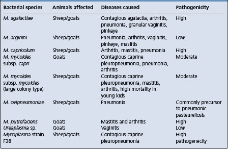
Table 20.7 Comparative properties of the two most important cattle mycoplamas
| Properties | M. mycoides subsp. | M. bovis |
|---|---|---|
| Mycoides SC | ||
| Diseases | Contagious bovine pleuropneumonia in cattle, occasionally in arthritis in calves | Calf pneumonia, mastitis, arthritis, abortion, keratoconjunctivitis |
| Distribution | Subsaharan Africa, probably in parts of Middle East, Central Asia | Worldwide |
| Hosts | Cattle, goats, (sheep) | Cattle |
| Histopathological | Fibrinous pleuropneumonima with necrosis | Interstitial pneumonia, lymphohistiocytic bronchitis, catarrhal bronchopneumonia |
| Clinical signs | Few signs, respiratory distress evident after exercise | Respiratory distress, mastitis, arthritis |
| Diagnosis | Isolation, serology, PCR, abattoir surveillance | Serology, isolation, PCR |
| Treatment | Chemotherapy not recommended because it encourages carrier status | Chemotherapy |
| Control | Vaccination, movement control, slaughter | Management, improved ventilation, reduced stocking density |
Vulvovaginitis in cattle, sheep and goats may be associated with M. agalactiae var. bovis. The same infection when introduced with semen into the uterus can cause endometritis and salpingitis, resulting in a temporary infertility and failure to conceive.
Persistent infection in the genital tract of bulls has also been produced experimentally. Ureaplasmas have been isolated from the vulva of ewes with granular vulvitis and the disease was transmitted experimentally. However, the same organisms are present in the vulva of normal ewes and cows but Ureaplasma spp. are usually limited in their distribution to the vestibule and vulva of normal cows. In some areas, Ureaplasma diversum is commonly present in the lower reproductive tract of beef and dairy cattle, both cows and bulls, and is associated with granular vulvitis, which has been associated with infertility, sporadic abortions and neonatal mortality.10 This infection adversely affects reproduction when it is either acute or chronic; it is capable of producing granular vaginitis and some strains can, if introduced to the upper reproductive tract, cause transitory endometritis and salpingitis.
Ureaplasms, M. bovis and M. bovigenitalium have been found the reproductive tract of bulls and their semen.11 Using the PCR for the detection of mycoplasma in semen, M. mycoides subsp. mycoides SC has been found in semen of yearling bulls with seminal vesiculitis while negative to the complement-fixation test for CBPP.11
A combination of lincomycin– spectinomycin–tylosin has been shown to be most effective in the treatment of Ureaplasma spp. in bull semen. Chlortetracycline at 350 mg/h/d for 30 d in the prebreeding feed of virginal heifers, most of which had vulvovaginitis and from which 44% cultured positive for U. diversum, improved pregnancy rates and decreased the vaginal colonization of the organism.12
Parenteral vaccination of heifers with killed U. diversum induced antibodies to the mycoplasma but did not prevent subsequent infection or clear the ureaplasmas.13 The use of the vaginal submucosal route for vaccination resulted in characteristic granular vulvitis in both vaccinated and control animals.14
Attempts to produce abortion in cows by the injection of mycoplasmas isolated from aborted fetuses and from weak calves has had varying success. Acholeplasma spp. have been isolated from aborted equine fetuses. M. bovigenitalium has been a frequent isolate from bovine genital tracts for many years, but its role in genital disease is still uncertain. It has been isolated from frozen bull semen and poses a threat to cows inseminated with infected semen. Mycoplasmas in semen can be transmitted through in vitro fertilization and infect embryos, and supplementation of culture media with standard antibiotics and washing embryos as recommended by the International Embryo Transfer Society are not effective in making IVF embryos free from M. bovis and M. bovigenitalium.9
Mycoplasma bovis in frozen semen can survive the antibiotic combination of gentamicin, tylosin, and lincomycin and spectinomycin.15
Mycoplasma bovis, Mycoplasma californicum, and Mycoplasma canadense are causes of outbreaks of mastitis in cattle, and occasionally pneumonia, otitis media, or arthritis in calves of those dairy herds. The literature on mycoplasma mastitis in dairy herds has been reviewed.16 The infection is highly contagious and is commonly introduced into a previously Mycoplasma-free herd by the purchase of infected heifers or cows. At least 11 other species of Mycoplasma have been isolated from milk of cows with mastitis and the disease produced by each is similar. Cows of all ages and at any stage of lactation are susceptible. Typically, acute mastitis occurs which is resistant to treatment. Usually all four quarters are affected, there is a marked drop in milk production, and abnormal udder secretions vary from being like watery milk with a few clots to colostrum-like material. The affected cow is usually systemically normal. Chronically affected animals have a tan-colored milk secretion with sandy or flaky sediments which may become purulent-like over several weeks. However, the majority of milk samples which are positive for mycoplasma appear normal. In affected cows, milk production commonly declines, with normal appearance of milk but a high somatic cell count.
Mycoplasma mastitis in cattle is highly contagious and infections are transmitted at milking by means of fomites. Diagnosis is dependent on culture of milk samples from the bulk tank milk or individual cow milk samples. To identify cows with M. bovis mastitis, an indirect ELISA is available to detect antibodies to M. bovis in milk samples from cows with recently acquired M. bovis mastitis.17 For mycoplasmal mastitis, individual cows milk sampling for culture and identification of M. bovis is time consuming and expensive. Some herds sample cows monthly with the dairy herd improvement (DHI) program but a preservative is added to the milk that kills M. bovis. A nested PCR procedure allows for rapid testing of preservative treated milk and is as sensitive as traditional culture.18
In herds where the diagnosis has been made, weekly monitoring of bulk tank milk is necessary to monitor the success of control procedures. Quality Milk Production Services, Cornell University, recommends the use of 1% iodine products to reduce the number of Mycoplasma on teat skin during mastitis outbreaks. There is no treatment for mycoplasma mastitis and vaccination has been ineffective. In affected herds, milk of all lactating animals should sampled, positive animals identified and culled or segregated from the mycoplasma-free animals. Waste milk from infected cows should not be fed to calves without pasteurization.19
Experimental vaccines against mycoplasma vaccines have been unsuccessful and may even exacerbate the mastitis.5 It is best to segregate or cull carrier cows and to implement rigid sanitation procedures to prevent transmission from infected to non-infected cows.
Excellent vigilance and rapid culling of infected cattle are critical control factors affecting the spread of mycoplasma mastitis. Herds commonly become negative within the first year of the first case of mycoplasma mastitis.20
(Additional details on Mycoplasma mastitis in cattle are available in Chapter 15.)
Several different mycoplasmas have been isolated from pneumonic and non-pneumonic lungs of cattle, sheep and goats, but attempts to reproduce respiratory tract disease with them has resulted in inconclusive findings.
In general, mycoplasmas cause a subclinical, mild pneumonia in gnotobiotic animals, but in combination with unidentified agents in lung homogenates administered intransally, ‘enzootic’ or chronic progressive pneumonia is produced in sheep. For example, M. dispar has a cytopathogenic effect and stops ciliary motility, and destroys ciliated epithelial cells in organ cultures. M. ovipneumoniae also has the capacity to colonize the sheep lung and produce mild pneumonic lesions, but in combination with pasteurellae in sheep, causes a proliferative exudative pneumonia. A similar disease in Icelandic sheep (kregda) has been identified as being associated with M. ovipneumoniae. Similar diseases are associated with M. ovipneumoniae and M. arginini.
Although contagious caprine pleuropneumonia has not been observed in Australia, a non-fatal respiratory disease of goats, characterized by coughing, fever and extensive pleurisy and pneumonia, has. A variety of mycoplasmas, including M. agalactiae and M. mycoides and M. mycoides var. capri have also been found in goats. The caprine M. mycoides var. mycoides (large colony type) is not pathogenic for cattle and has been associated with a variety of syndromes in goats including fibrinous peritonitis, pneumonia, arthritis, mastitis and abortion. It has been cultured from a goat with a subauricular abscess and mastitis.21 The most common syndrome in goats associated with mycoplasma is a chronic interstitial pneumonia with cough, unthriftiness proceeding to extreme emaciation, chronic non-painful bony enlargement of joints and chronic indurative mastitis. The pneumonia in some cases progresses to the point where cor pulmonale develops with a subsequent appearance of the signs of congestive heart failure. M. ovipneumoniae has also been credited with causing pneumonia in goats. M. adleri has been isolated from a goat with a joint abscess.22
Mycoplasma bovis is a major cause of calf pneumonia. Mycoplasma dispar, Ureaplasma diversum, Mycoplasma bovirhinis, and Mycoplasma canis have also been isolated from the lungs of pneumonic cattle but it is uncertain if they are primary causes of disease.5 M. dispar is capable of producing a pneumonia without clinical signs in gnotobiotic calves, and in conjunction with Ureaplasma spp. it has been found commonly in ‘cuffing’ pneumonia of calves. It could, therefore, be a precursor to other infections causing enzootic pneumonia in calves or with pasteurellae producing fibrinous pneumonia of calves.
Mycoplasma bovoculi (Acholeplasma oculi) has been associated with, without necessarily being the cause in the field of, outbreaks of infectious keratoconjunctivitis of cattle, sheep and goats. The mycoplasmas are capable of producing keratitis experimentally and M. bovis has been isolated from the ocular discharge of young cattle affected with conjunctivitis.24 The naturally occurring disease in goats is manifested by rapid spread and development with intense lacrimation, conjunctival hyperemia, corneal opacity and vascularization. A concurrent respiratory illness occurs in some goats. Response to treatment with oxytetracycline and polymyxin B is good. The disease is reproducible experimentally.
Mycoplasma (Eperythrozoon) suis infection occurs worldwide in pigs causing fever and anemia in young animals and, occasionally, latent anemia in older animals. E. suis, originally classified as a Rickettsia, has now been shown to belong to the mycoplasmas based on phylogenetic analysis of its rrs (16S rRNA) genes and is now proposed to be known as Candidatus Mycoplasma haemosuis.
Mycoplasma (M. hyorhinis, M. hyosynoviae) are associated with arthritis and polyarthritis in pigs and the disease is reproducible experimentally. Both organisms are carried in the respiratory tract. M. hyorhinis most commonly affects suckling and young pigs, especially after weaning or some stress, and may produce a polyserositis in addition to arthritis. The case–fatality rate is generally low, but residual fibrinous pericardial and pleural adhesions may occur and the pigs may fail to thrive.
Disease associated with M. hyosynoviae is more common in older growing pigs as arthritis. The disease is endemic in some herds and may affect up to 15% of the herd. Clinically, there is an acute episode of fever, lameness and swelling of the joint. The lesion is a serofibrinous arthritis and most cases recover, but some may be affected chronically. M. hyosynoviae was isolated from 20% of arthritic lesions of pigs from an abattoir.25 The case–fatality rate is low and does not exceed 10%. Tylosin and lincomycin are indicated and effective.
In goats and occasionally lambs, M. capricolium causes arthritis. The infection is transmissible experimentally via infected milk administered orally. There is a septicemia with subsequent pneumonia and arthritis.
M. mycoides var. mycoides (large colony type) has been isolated from goats with arthritis. It has caused acute arthritis in 3 to 8-week-old kids manifested by lameness, recumbency, diarrhea and fever. The infection may originate from infected milk from does which have had acute mastitis due to M. mycoides var. mycoides. It may also be transmitted by ear mites, Psoroptes cuniculi and goat fleas.11 A similar disease is observed in sheep. At necropsy the kids have polyarthritis, pneumonia and pleurisy. M. putrefaciens has caused major losses due to mastitis and arthritis in a large goat herd. Abortion was common in some groups.
M. capricolium and M. putrefaciens are also causes of arthritis in sheep and kids.27 The experimental inoculation of lambs with A. laidlawii, which is normally considered non-pathogenic for animals, induced arthritis and renal lesions but the organism was not cultured from the joint fluid.28
There are few records of mycoplasmosis in horses. Most descriptions relate to the isolation of mycoplasmas, usually from the respiratory and female genital tracts, and almost always without any associated lesions. M. equirhinis and M. felis are pathogens of horses.23,29,30
The moderately well-identified syndromes of these species are dealt with separately. There are many other reports of mycoplasmal diseases in these species in which the mycoplasma is not identified, and a number of diseases as set out earlier in which the etiological significance of the mycoplasmas is in doubt. For example, the two main clinical syndromes of these species are pleuropneumonia and agalactia, as described in subsequent sections. However, the signs and lesions of more than one of these ‘specific’ diseases and the infections themselves can be encountered in the one animal (see Table 20.6).
Bergonier D, Berthelot X, Frey J. Mycoplasmas of ruminants: pathogenecity, diagnostics, epidemiology and molecular genetics. Volume 4. European Commission. Directorate-General for Research. COST Action 826. Agriculture and biotechnology. European cooperation in the field of scientific and technical research. 2000:1–257
Frey J. Mycoplasmas of animals. In: Razin S, Herrman S, editors. Molecular biology and pathogenicity of mycoplasmas. New York: Kluwer Academic/Plenum; 2002:73-90.
Gonzalez RN, Wilson DJ. Mycoplasmal mastitis in dairy herds. Vet Clin Food Anim Pract. 2003;19:199-221.
Nicholas RAJ, Ayling RD. Mycoplasma bovis: disease, diagnosis, and control. Res Vet Sci. 2003;74:105-112.
1 Ayling RD, et al. Vet Rec. 2004;155:413.
2 Frey J. Mycoplasmas of animals. In: Razin S, Herrman S, editors. Molecular biology and patho-genicity of mycoplasmas. New York: Kluwer Academic/Plenum; 2002:73-90.
3 McAuliffe L, et al. J Clin Microbiol. 2003;41:4844.
4 McAuliffe L, et al. J Clin Microbiol. 2004;42:4556.
5 Nicholas RAJ, Ayling RD. Res Vet Sci. 2003;74:105.
6 Minion FC. Front Biosci. 2002;7:d1410.
7 Hum S, et al. Aust Vet J. 2000;78:744.
8 Djordjevic SP, et al. Electrophoresis. 2001;22:3551.
9 Bielanski A, et al. Theriogenology. 2000;53:1213.
10 Mulira GL, et al. Can. Vet J. 1992;33:46.
11 Stradaioli G, et al. Vet Res. 1999;30:457.
12 Rae DO, et al. Theriogenology. 1993;40:497.
13 Mulira GL, Saunders JR. Am J Vet Res. 1994;58:104.
14 Mulira GL, Saunders JR. Am J Vet Res. 1994;58:109.
15 Visser IJR, et al. Theriogenology. 1999;51:689.
16 Gonzalez RN, Wilson DJ. Vet Clin Food Anim Pract. 2003;19:199.
17 Byrne WJ, et al. Vet Rec. 2000;146:368.
18 Pinnow CC, et al. J Dairy Sci. 2001;84:1640.
19 Butler JA, et al. J Dairy Sci. 2000;83:2285.
20 Fox LK, et al. J Vet Med B. 2003;50:235.
21 Blikslager AT, Anderson KL. J Am Vet Med Assoc. 1992;201:1404.
22 Guidice RAD, et al. Int J Syst Bacteriol. 1995;45:29.
23 Wood JLN, et al. J Clin Micro. 2005;43:120.
24 Kirby FD, Nicholas RAJ. Vet Rec. 1996;138:552.
25 Fries NF, et al. Acta Vet Scand. 1992;33:205.
26 Nayak NC, Bhowmik MK. Prev Vet Med. 1990;9:259.
27 Rodriquez JL, et al. Vet Rec. 1994;135:406.
28 Concha-Bermejillo A, De La, et al. J Vet Diag Invest. 1996;8:115.
Unresponsive pneumonia, polyarthritis, otitis media/interna, mastitis in dairy herds
. Culture or detection of organism from respiratory secretions, joint fluid, milk.
Biosecurity and biocontainment procedures. Prevent entry of infected animals into herds. Purchase animals free of mycoplasma. Pasteurization of milk of cows with mycoplasma mastitis before feeding to calves. Vaccines have been unsuccessful and some vaccines in experimental stage. Mycoplasma bovis is a major pathogen causing chronic pneumonia, polyarthritis, mastitis, otitis media, and other related diseases of dairy and beef cattle of all ages.
M. bovis is a major cause of calf pneumonia, chronic pneumonia-polyarthritis syndrome in feedlot cattle, mastitis, and other diseases such as otitis media.1 Mycoplasma sp. bovine group 7 has also caused polyarthritis, mastitis and abortion in dairy cattle.2,3
Diseases associated with Mycoplasma bovis, first isolated from cattle with severe mastitis in cattle in the United States, has spread to many countries of the world via animal movements.1 Mycoplasma bovis is a primary cause of bovine pneumonia, arthritis, and mastitis and has been associated with keratoconjunctivitis, otitis, meningitis, infertility, and abortion.1,4 In Northern Ireland, M. bovis has been isolated from calves with pneumonia, and from dairy cattle with polyarthritis,5 and mastitis cases6,7 and from aborted fetuses.8 Serological surveys of suckling beef cattle in France indicated that infection was not distributed evenly across the country and that there were significant differences between certain counties.9
The prevalence of infection of M. bovis in Danish cattle appeared to increase over a period of several years. In a survey of pneumonic bovine lungs submitted to a diagnostic laboratory in Denmark, 83% were found infected with mycoplasmas.10 The predominant mycoplasmas were Ureaplasma spp. (72%), M. dispar (48%), M. bovis (24%). Multiple species mycoplasma infections were predominant.
Of 1600 isolates of Mycoplasma species recovered from ruminant animals in Britain over a 10-year period, M. bovis was the most common species, mostly from pneumonic calves, but occasionally from cattle with mastitis and arthritis.11 M. canis was first isolated in 1995 from pneumonic calves and incidence increased to 18% of the total mycoplasmas in 5 years. A serological survey of pneumonic cattle found M. bovis positive samples in 18%.
The chronic pneumonia–polyarthritis of cattle has been reported in Canada and the United States.12,13 It occurs commonly in young feedlot cattle usually affecting many animals a few weeks after arrival and mingling in the lot. The morbidity ranges from 20–85% and the case– mortality rate from 3–50%. In Canada, the disease has been seen commonly in young cattle (6–8 months of age) following shipment from western rangelands to eastern feedlots, which suggests that long transportation and mixing of cattle of different origins may be important epidemiological characteristics. Calves affected with arthritis commonly have necropsy evidence of mycoplasma pneumonia and it is proposed that the pneumonia precedes the development of the arthritis. Calves sucking cows with experimental mastitis due to this organism may develop mycoplasmal arthritis, and a high incidence is recorded in calves in dairy herds where mycoplasmal mastitis was occurring.
In a group of feedlot cattle, from Alberta, Canada, with chronic unresponsive pneumonia and polyarthritis, M. bovis was the most common pathogen demonstrated, having been detected in 82% of cases, including 71% in lungs and 45% in joints.14 All cases had been treated with antibiotics including tilmicosin, trimethoprim-sulfadoxine, ceftiofur sodium, and sulbactam and ampicillin.
In a series of cases of chronic, antibiotic-resistant pneumonia, sometimes with concurrent polyarthritis, in feedlot cattle in western Canada, M. bovis was present in the lung tissues of more than 90% of cases, and the BVDV was present in 60% of the cases suggesting a possible synergism between M. bovis and the BVDV.12 Outbreaks of pneumonia and arthritis in beef calves associated with infection due to Mycoplasma bovis and Mycoplasma californicum have been described in a mixed dairy cattle and beef cattle herd kept under extremely poor housing and hygienic conditions.15 During a 3-year period in Belgium, in calves with respiratory disease, the prevalence of M. bovis was 31.5%, M. dispar 45.5%, M. canis 10.7% and Ureaplasma diversum 14.8%, and in half the cases they occurred in association with Pasteurella and/or Mannheimia species.16
Because the clinical and pathologic findings of M. bovis pneumonia closely resemble those of contagious bovine pleuropneumonia (CBPP), it is very important to ensure the microbiological diagnosis when confronted with lesions resembling either infection. A serological and diagnostic microbiological and pathological survey of pneumonic cattle sampled on the farm followed by examination of heir lungs at slaughter in Hungary confirmed the presence of M. bovis as at causative agent of the pneumonia rather than due to Mycoplasma mycoides subspecies mycoides the cause of contagious bovine pleuropneumonia.17
Otitis media/interna in dairy and beef calves has been associated with M. bovis infection. It has been described in preweaned Holstein dairy calves in dairy herds which have expanded in size.18,19 Affected calves were 2 to 5 weeks of age, morbidity was 3 to 10% and case fatality rates estimated at 50%. In a retrospective study of Mycoplasma otitis in calves submitted for necropsy in California, affected calves were 2 weeks to 4 months of age, 92% were from dairy herds, most cases occurred during late winter and spring.20 M. bovis, M. bovirhinis, and M. alkalescens were isolated from the ears of affected calves. Outbreaks of suppurative otitis media and pneumonia associated with M. bovis have been described in calves on beef cattle farms in Japan.21 Morbidity and mortality were estimated at 8 to 40% and 30 to 100%, respectively.
The virulence factors of M. bovis and mechanisms of pathogenicity are not well-known, but the organism’s ability to vary the expression of a family of membrane surface proteins (Vsps) with high frequency is currently being investigated. The organism has 13 vsp genes involved in antigenic variation which alter the antigenic character of its surface components, and may act to enhance colonization and/or adherence or evade the host’s immune defense systems.1
M. bovis isolates from pneumonic cattle in the UK separated into two distinct groups based molecular epidemiological analysis.22 The organism produces an immunosuppressive peptide which is able in vitro to inhibit mitogen-induced proliferation of bovine lymphocytes.23 The peptide is a product of variable surface proteins which may have an immunosuppressive effect during infection of the lung.24 The organism is also able to penetrate through lung epithelial junctions and cause systemic infections.23 There is some evidence of variability of M. bovis strains to cause arthritis.13
Genetic fingerprinting of M. bovis strains isolated in Denmark over a 17-year period demonstrated remarkable genomic homogeneity which were likely epidemiologically related and have remained stable for many years.25 The technique used allows the creation of databases for inter-laboratory use and comparison, and continued surveillance to monitor the spread of the organism as a prerequisite for effective control.
Clinically normal cattle harbor M. bovis in the upper respiratory tract with no apparent adverse effect and may shed the organism through the nasal discharge for months to years.26 The methods of transmission of M. bovis as the causative agent of the pneumonia–polyarthritis syndromes, and related diseases, are unknown. It is assumed that direct contact between infected and susceptible animals is the primary mode of transmission but there is no supporting published evidence. M. bovis can be isolated from respiratory secretions such as nasal exudate which indicates at least one route of transmission. Serological surveys of feedlot cattle on arrival and 28 days later in Ontario, Canada, found an increase in titers to M. bovis and M. dispar which indicates that mixing of animals results in transmission of the organism.27
Calves fed discarded milk from cows with mycoplasma mastitis may develop pneumonia and otitis media.18 Pasteurization of discard milk can eliminate transmission of the mycoplasmas to the calves.
The intra-articular injection of M. bovis into calves causes severe fibrinosuppurative synovitis and tenosynovitis, erosion of cartilage and its replacement by polypoid granulation tissue. Erosion of the cartilage is accompanied by chronic osteomyelitis and formation of pannus tissue. Histologically, there is extensive ulceration of synovial membranes of leukocytic infiltration of the subsynovium, congestion, hyperemia and thrombosis of the subsynovial vessels. Intratracheal inoculation of the organism results in pneumonia and severe lameness, which suggests that M. bovis is involved in pneumonia–arthritis syndrome.
As with many mycoplasmas, M. bovis is both immune reactive and immunosuppressive. Upon incubation with M. bovis, alveolar macrophages are activated and produce TNF-alpha and nitric oxide, two powerful initiators of immune activity.24 M. bovis is also immunosuppressive by inhibiting neutrophil degranulation and oxidative bursts and proliferation of lymphocytes by mitogens. M. bovis also induces bovine lymphocyte death by apoptosis through the production of a protein which is different from other mycoplasmas both pathogenic and non-pathogenic.28 The protein is an immuno-inhibitory peptide which can suppress Concanavalin A (ConA)-induced proliferation of bovine lymphocytes.29 This represents a unique immunosuppressive peptide produced by the M. bovis.
Despite its deleterious effects on lymphocytes, infected cattle are able to generate measurable humoral and cellular immune responses against M. bovis. Serological analysis indicates that M. bovis stimulates increased production of antigen-specific IgG1 while very little IgG2 is produced.27 Thus experimental lung infection of cattle with M. bovis results in a Th2-skewed immune response.
There is a systemic phase of M. bovis infection, including a potential interaction of the pathogen with endothelial cells. It is one of the most invasive bovine mycoplasmas capable of invading through lung epithelial junctions and causing systemic infections such as arthritis and mastitis following pneumonia. Localized lung vasculitis and the presence of thrombi within subsynovial vessels has been observed, both suggestive of interaction of M. bovis with epithelial cells.23
In otitis media/interna of calves there is facial nerve paralysis because of proximity of CN VII to the tympanic cavity.19 Varying degrees of peripheral vestibulocochlear dysfunction occur because of the involvement of the vestibulocochlear receptors and nerve. The spontaneous regurgitation and dysphagia may be associated with lesions involving the glossopharyngeal nerve (CN IX) with or without the vagus nerve (CN X). These nerves may be affected by the inflammation associated with meningitis because both CN IX and CN X travel through the jugular foramen.19
The disease is most common in feedlot calves within a few weeks after arrival in the feedlot.13 The morbidity rate may be up to 25%. Affected calves commonly have had a history of respiratory disease but have not responded to repeated antibiotic therapy. Auscultation of the lungs reveals areas of loud bronchial tones, crackles and wheezes and areas of muffled lung sounds indicating consolidation and occlusion of the bronchi with exudate. Depression, inactivity, inappetence, coughing, nasal discharge, fever, and progressive weight loss are common.
Concurrently affected calves commonly develop lameness. There is stiffness of gait, and lameness. Swelling of the large movable limb joints and distension of tendon sheaths, associated with fibrinous synovitis and synovial fluid effusions, are characteristic. Affected calves are reluctant to move and commonly are recumbent for long periods, continue to lose weight, and develop decubitus ulcers. Mildly affected cases recover spontaneously in 10–14 d, while severe cases become progressively worse and must be culled.
Pneumonia and polyarthritis associated with M. bovis may occur alone or together in cattle of all ages, including dairy and beef calves in their original herds, in growing dairy and beef cattle heifers, and in mature dairy and beef cows.
Otitis externa is inflammation of the externa ear canal. Otitis media/interna is inflammation of the inner ear involving primarily the tympanic bullae which may extend to the meninges and the brain stem.19,30 Clinical findings depend on the extent of the inflammation involving only the external ear or middle ear causing otitis media. Varying degrees of depression, coughing, nasal discharge and inappetence are common in affected groups of calves. Otitis externa is characterized by a drooping ear and purulent exudate in the external ear which can be detected by digital palpation of the pinnae which creates a fluid squishing sound. Deep palpation of the base of the ear may be painful.
A unilateral head tilt and paralysis of the lip, eyelid, and ear muscles on the same side are common. When the eye on the affected side is threatened, the eyeball may retract but there is no palpebral fissure closure.19 An intermittent loss of balance on the affected side may be apparent when the animal attempts to walk.
Bilateral peripheral CN VII and VIII deficits (bilateral ear, lip, and eyelid paresis; bilaterally absent menace and palpebral reflexes; normal gait; balance loss to either side) are suggestive of bilateral otitis media/interna.19,30 Dysphagia, spontaneous regurgitation of milk and difficulty in sucking from a bottle or prehending feed may occur.19 Partially chewed feed may accumulate in the oral cavity along with difficult prehension and mastication. Bilateral vestibular disease (balance loss to either side) may occur. Endoscopy of the pharynx may reveal collapse of the nasopharynx, dorsal displacement of the soft palate, and a widely dilated, hypomotile esophagus.
In otitis media there may be no exudate in the external ear. Opisthotonus and nystagmus are common and ataxia, recumbency and death in several days may occur.
The mortality rate is about 50%.19
Clinical and pathological signs are not characteristic for M. bovis so laboratory diagnosis is necessary for identification.1 The organism can be detected by culture, DNA probe and PCR tests.
Culture methods for the detection of M. bovis are restricted to culture and serology but both methods are time consuming, laborious, difficult and expensive. Bronchoalveolar lavage samples and nasal swabs can be used for the culture of M. bovis from cattle with respiratory disease; the lavage samples being more representative of the infection status of the lower respiratory tract disease.31
A DNA probe and semi-nested PCR test are now available to detect the antigen of the organism in milk samples and may be applicable to mucosal samples from conjunctivae, nasal and vaginal mucosae.32 An arbitrarily primed PCR typing method provides genotypic epidemiological information to successfully characterize M. bovis from sequential sampling of outbreaks and different husbandry conditions.33
Computer tomographic imaging for otitis media/interna in calves can provide detailed information of the bony structures of the middle and inner ear. Abnormalities of the tympanic bullae and petrous temporal bone may be visible with CT imaging which are not visible with conventional radiography.19 CSF from affected calves may indicate the presence of a meningitis.
At necropsy, the fibrinous synovitis is remarkable. There is severe thickening and edema of the synovial membranes and large quantities of fibrinopurulent synovial fluid. The tendon sheaths are similarly affected. Microscopically, large numbers of lymphocytes and plasma cells are found within the hypertrophic synovial villi. M. bovis can often be recovered from pneumonic lungs and causes pulmonary foci of coagulative to caseous necrosis.14,37-39 M. bovis causes two patterns of pulmonary necrosis in cattle.34 The first pattern is characterized by large irregular areas of coagulative necrosis surrounded by a dense zone of degenerated neutrophils. M. bovis antigen is present in the center and periphery of the necrotic foci. The second pattern consists of rounded foci of caseous necrosis composed of granular eosinophilic material surrounded by a rim of granulation tissue. Large amounts of M. bovis antigen are present in the center and periphery of these necrotic foci.34 Immunohistochemical techniques are used to detect the antigens of M. bovis in the tissues of feedlot cattle with chronic unresponsive respiratory disease and/or arthritis.14 Immunohistochemical studies suggest that this organism may also briefly localize in the kidney and liver.37 Mycoplasma spp. have been identified as an important cause suppurative otitis in dairy calves, often with concurrent mycoplasmal pneumonia.20 M. bovis has also been associated with pleuritis and pericarditis and has been isolated from decubital ulcers of calves.40
The disease must be differentiated from other causes of joint swelling and lameness in feedlot cattle.13 There are usually several animals affected in a short period of time, which serves to distinguish it from other sporadic causes of arthritis in feedlot cattle. A diagnosis of infection by M. bovis should be considered when pneumonia and arthritis and synovitis occur at about the same time.
Other diseases causing lameness in feedlot cattle include:
For a definitive diagnosis, joint fluid must be placed immediately into laboratory media specially prepared for Mycoplasma spp. The failure to isolate the mycoplasma from the fluid of joints which have been affected for more than 14 d does not preclude a diagnosis of mycoplasma arthritis because the organism may have been eliminated from the joint.
Treatment is usually ineffective. Several antimicrobials, including tylosin, oxytetracycline, lincomycin and oleandomycin, have been used in naturally-occurring and experimental cases and while the isolates of mycoplasmas may be sensitive to these antibiotics in vitro, the response in affected animals has been unsatisfactory.
The published results of in vitro antimicrobial testing of isolates of M. bovis recovered from various locations are highly variable. The antimicrobial susceptibility of M. bovis strains, cultured from cases of pneumonia, arthritis, and mastitis of cattle, measured in vitro indicate that enrofloxacin was the only one which exhibited any measurable activity.41 Spectinomycin has been evaluated for the treatment of experimental M. bovis pneumonia in 3-week-old calves and only decreased the level of M. bovis and Pasteurella multocida infection in the lung but did not alter the course of the illness.42 The in vitro susceptibilities of Belgian field isolates of M. bovis to 10 antimicrobials found that tiamulin was the most active against the organism. The fluoroquinolones, danofloxacin, enrofloxacin, and marbofloxacin were effective against strains of M. bovis. Gentamicin was ineffective. Many strains were resistant to tyolsin, spectinomycin, lincomycin, tetracycline and oxytetracycline.43 In a series of British isolates of M. bovis, most isolates were susceptible to danofloxacin, less susceptible to florfenicol. Oxytetracycline and spectinomycin had only a limited effect against the majority of isolates. Approximately 20% were highly resistant to spectinomycin, and tilmicosin was ineffective. There was no evidence of resistance to danofloxacin.44
In calves with a high incidence of respiratory disease associated with M. bovis and Pasteurella spp. the use of valnemulin in the milk of the calves for four days resulted in improved weight gains and fewer cases of Mycoplasma infection, required fewer treatments with antibiotics than those in the placebo treated group.45
Effective control of the disease is not yet possible. Some vaccines have been developed but have not been sufficiently efficacious or have yielded poor results. A quadrivalent inactivated vaccine containing BRSV, PI-3 virus, and M. dispar and M. bovis provided some protection against naturally-occurring outbreaks of bovine respiratory disease.1 A vaccine containing formalin-inactivated strains of M. bovis and Mannheimia haemolytica from affected herds reduced losses from pneumonia and the cost of treatment in newly arrived feedlot calves.1
A single dose of vaccine for M. bovis pneumonia, inactivated with saponin, provided protection against experimental challenge of calves 3 to 4 weeks of age with a virulent isolate of M. bovis.46 The vaccine also reduced the spread of M. bovis to internal organs. Attempts to vaccinate against M. bovis arthritis have been unsuccessful.1 Experimental vaccines against mycoplasma vaccines have been unsuccessful and may even exacerbate the mastitis.
In dairy herds, pasteurization of mycoplasma mastitis milk at 65°C for 1 hour can kill mycoplasmas and reduce the incidence of respiratory disease in calves.47 A temperature of 65°C killed M. bovis and M. californicum after 2 min of exposure, while M. canadense remained viable for up to 10 min. Exposure to 70°C inactivated M. bovis and M. californicum after 1 min, but M. canadense samples were positive for up to 3 min.
Biosecurity and biocontainment procedures should be implemented to prevent the introduction of infection into the herd and to minimize the spread of infection in the herd.
Rosenbusch RF. Bovine mycoplasmosis. Proc Am Assoc Bov Pract. 2001;34:49-52.
Step DL, Kirkpatrick JG. Mycoplasma infection in cattle. I. Pneumonia arthritis syndrome. Bov Pract. 2001;35:149-155.
Step DL, Kirkpatrick JG. Mycoplasma infection in cattle. II. Mastitis and other diseases. Bov Pract. 2001;35:171-176.
Stokka GL, Lechtenberg K, Edwards T, et al. Lameness in feedlot cattle. Vet Clin North Am Food Anim Pract. 2001;17:189-207.
Frey J. Mycoplasmas of animals. In: Razin S, Herrman S, editors. Molecular biology and pathogenicity of mycoplasmas. New York: Kluwer Academic/Plenum; 2002:73-90.
Nicholas RAJ, Ayling RD. Mycoplasma bovis: disease, diagnosis, and control. Res Vet Sci. 2003;74:105-112.
1 Nicholas RAJ, Ayling RD. Res Vet Sci. 2003;74:105.
2 Djordjevic SP, et al. Electrophoresis. 2001;22:3551.
3 Hum S, et al. Aust Vet J. 2000;78:744.
4 Frey J. Mycoplasmas of animals. In: Razin S, Herrman S, editors. Molecular biology and pathogenicity of mycoplasmas. New York: Kluwer Academic/Plenum; 2002:73-90.
5 Henderson JP, Ball HJ. Vet Rec. 1999;145:374.
6 Brice N, et al. Vet Rec. 2000;146:643.
7 Bryne WJ, et al. Vet Rec. 2001;148:331.
8 Byrne WJ, et al. Vet Rec. 1999;144:211.
9 Le Grand D, et al. Vet Rec. 2002;150:268.
10 Kusiluka LJ, et al. Acta Vet Scand. 2000;41:139.
11 Ayling RD, et al. Vet Rec. 2004;153:413.
12 Shahriar FM, et al. Can Vet J. 2002;43:863.
13 Stokka GL, et al. Vet Clin North Am Food Anim Pract. 2001;17:189.
14 Haines DM, et al. Can Vet J. 2001;42:857.
15 Hewicker-Trautwein M, et al. Vet Rec. 2002;151:699.
16 Thomas A, et al. Vet Rec. 2002;151:472.
17 Bashiruddin JB, et al. Vet Rec. 2001;148:743.
18 Walz PH, et al. J Vet Diag Invest. 1997;9:250.
19 Van Biervliet J, et al. J Vet Int Med. 2004;18:907.
20 Lamm CG, et al. J Vet Diag Invest. 2004;16:397.
21 Maeda T, et al. J Comp Path. 2003;129:100.
22 McAuliffe L, et al. J Clin Microbiol. 2004;42:4556.
23 Lu X, Rosenbusch RF. Microb Pathogen. 2004;37:253.
24 Vanden Bush TJ, Rosenbusch RF. Vet. Immunol Immunopathol. 2003;94:23.
25 Kusiluka LJM, et al. FEMS Microbiol Lett. 2000;192:113.
26 Pfutzner H, Sachse K. Rev Sci Tech Off Int Epiz. 1996;15:1477.
27 Step DL, Kirkpatrick JG. Bov Pract. 2001;35:149. 171
28 Vanden Bush TJ, Rosenbusch RF. FEMS Immunol Med Microbiol. 2002;32:97.
29 Vanden Bush TJ, Rosenbusch RF. Biochem Biophys Res Commun. 2004;315:336.
30 Vestweber JG. Comp Cont Educ Pract Vet. 1999;21:S34-S38.
31 Thomas A, et al. Vet Res Commun. 2002;26:333.
32 Hayman B, et al. Vet Microbiol. 2003;91:91.
33 Butler JA, et al. Vet Microbiol. 2001;78:175.
34 Khodakaram-Tafti A, Lopez A. J Vet Med A. 2004;51:10.
35 Ball HJ, et al. Vet Rec. 1994;135:531.
36 Byrne WJ, et al. Vet Rec. 2000;146:368.
37 Adegboye DS, et al. J Vet Diag Invest. 1995;7:333.
38 Rodriguez F, et al. J Comp Path. 1996;115:151.
39 Khodakaram-Tafti A, Lopez A. J Vet Med A Physiol Pathol Clin Med. 2004;51:10.
40 Kinde H, et al. J Vet Diag Invest. 1993;5:194.
41 Ball HJ, et al. Irish Vet J. 1995;48:316.
42 Poumarat F, et al. Vet Microbiol. 2001;80:23.
43 Thomas A, et al. Vet Rec. 2003;153:428.
44 Ayling RD, et al. Vet Rec. 2000;146:745.
45 Stipkovits L, et al. Vet Rec. 2001;148:399.
A major plaque of cattle, endemic in eastern Europe, Asia, Africa, and has spread to Spain, France and Italy. Major concern in European Community because of relaxation of import controls and increase in international trade. Insidious nature of disease allows it to spread undetected for months
Fever, agalactia, anorexia, depression, coughing, thoracic pain, back arched, dyspnea, expiratory grunting, pleuritic friction rubs, dull areas of lung, edema of throat and dewlap
Complement fixation test. Detection of organism with polymerase chain reaction (PCR)
Mycoplasma mycoides subsp. mycoides Small Colony (SC) (MmmSC) is the cause of the disease in cattle.1 The organism belongs to the ‘mycoides’ cluster, which consists of six closely related mycoplasma. Members of the ‘mycoides’ cluster are pathogens of cattle, sheep and goats but the agent of contagious bovine pleuropneumonia (CBPP) is not communicable to other species. It is very similar culturally and antigenically to the causative organisms of caprine contagious pleuropneumonia but the two can be differentiated culturally and biochemically. Large colony types are pathogenic for sheep and goats, but not for cattle. Small colony types have been isolated from the milk of sheep with mastitis and goats with pneumonia.2
The organisms are pleomorphic and some forms are filterable. They can be maintained readily in special culture media and in embryonated hens’ eggs. MmmSC is a member of the M. mycoides cluster which includes those listed in Table 20.8. The ‘Mycoplasma mycoides cluster’ contains six important mycoplasma of ruminants. Only two of them cause disease in cattle, MmmSC which is the cause of CBPP, and Mycoplasma bovine group 7 (Bg7) which may cause arthritis and bovine mastitis. The four others, two subspecies within Mycoplasma mycoides species and two subspecies within Mycoplasma capricolum species are responsible for goat respiratory and other diseases.
Table 20.8 Members of the Mycoplasma mycoides cluster
| Name | Main disease | Main (and other) hosts |
|---|---|---|
| M. mycoides subsp. mycoides SC variant | Contagious bovine pleuropneumonia | Cattle (goats, sheep, buffalo) |
| M. mycoides subsp. mycoides LC variant | Caprine pneumonia, contagious agalactiae | Goats (sheep, cattle) |
| Mycoides subsp. capri | Caprine pneumonia | Goats (sheep) but rare |
| M. capricolum subsp. capricolum | Caprine pneumonia, contagious agalactiae | Goats (sheep, cattle) |
| M. capricolum subsp. capripneumoniae | Contagious caprine pleuropneumonia | Goats (sheep) |
| M. bovine group 7 (Bg7) | Arthritis, also mastitis, calf pneumonia | Cattle |
Under natural conditions, CBPP occurs in cattle of the species Bos and allied animals including buffalo, yak, bison and even reindeer.3 While buffaloes can be infected by artificial means, and pulmonary lesions and the organism have been found in seropositive buffaloes which have been in contact with CBPP-infected cattle in Italy, it is uncertain if they can spread the disease to cattle.
CBPP is widespread in Africa and occurs in some countries of Asia and Europe. In 1995, the Office des International Epizooties (OIE) reported that CBPP in Africa was causing greater losses in cattle than any other disease including rinderpest. In 2001, 17 countries in Africa declared the presence of the disease and a potential threat for the world.4 In Africa, it is found in an area south of the Sahara, from the Tropic of Cancer to the Tropic of Capricorn and from the Atlantic to the Indian Ocean.1 Endemic infection extends throughout the pastoral herds of much of western, central and eastern Africa, with Angola and northern Namibia in southern Africa. Newly infected areas in the 1990s include Uganda, parts of Kenya, the Ituri Region of the Democratic Republic of the Congo, and most of the United Republic of Tanzania, where recently the disease has spread alarmingly. Rwanda in 1994, Botswana in 19952 but now free, Burundi in 1997, and Zambia in 1997 were recently re-invaded, but Lesotho, Malawi, Mozamique, South Africa, Swaziland, and Zimbabwe are currently free as of 2002.1 An abattoir survey of the disease in Nigeria indicates CBPP is endemic and the campaign to control or eradicate the disease has been inadequate.5 Reasons include inadequate and irregular vaccination programs because of the high cost of mass vaccinations, and the steady illegal introduction of infected cattle into areas across control barriers, and the presence of carrier animals which may not be detected clinically or serologically. Movement of cattle between Niger and Nigeria in the north and between Cameroon and Nigeria in the northeast have also been associated with spread.
The disease was present in most sub-Saharan countries and had reinfected countries such as Uganda and Kenya, where it had been eradicated in the 1970s.6 Of more concern, countries which had been CBPP-free for many years were also reinfected. Epidemics occurred in Tanzania in 1990 and Botswana and Rwanda in 1995.7 In addition, Angola, Benin, Cameroon, Chad, Eritrea, Ivory Coast, Ghana, Nigeria, Sudan, Togo and Zaire are infected.6
CBPP was first introduced into Tanzania in 1916 and was eradicated in 1964.8 The disease re-emerged in the country in 1990 and since then it has spread widely, threatening the entire national cattle herd. Because of lack of a clear disease-control policy, uncontrolled cattle movements, lack of public awareness and commitment, ineffective legislation, attempts to control and eradicate the disease between 1990 and 2000 failed.
The reasons for increase in incidence of CBPP in Africa are relate specifically to reduced funding for vaccination, possibly linked to the success of the rinderpest campaign, changes in vaccines and vaccine usage, cost recovery for CBPP vaccination, and reduced disease surveillance.9
In Asia, CBPP has been reported recently in Assam in India, Bangladesh, and Myanmar. Sporadic outbreaks have been recognized in the Middle East, probably derived from importation of cattle from Africa.
The status of CBPP in Europe is sharp contrast with that in Africa, because as of 1999, for the first time for over 20 years, no outbreaks were reported.9 As of 2004, Europe is free from CBPP and the European Union rules prevent the importation of live animals from affected areas. CBPP is usually transmitted through movements of live animals; trade in animal products is not thought to be a significant risk. The disease was diagnosed in France and Spain in the 1960s, Portugal in 1983 and Italy in 1990. An abattoir based survey of CBPP in Turkey indicated the presence of the infection based on ELISA of blood samples and lung tissues of cattle examined in abattoirs but the organism was not identified.10
The disease was eradicated from the United States in 1892, South Africa in 1916, and Australia in 1973 after being introduced in 1858.11,12 The disease was introduced into Australia in 1858 by dairy cattle imported into the colony of Victoria from England. It spread rapidly throughout Australia by cattle being driven to take up new pastoral lands everywhere, aided by the bullock teams, which provided the only form of transportation of goods and supplies in those days. One hundred years later, in 1958, a national eradication campaign was commenced and it took only 15 years before Australia was declared free from the disease, in Darwin in 1973.11,12 An entire book, ‘Clearing a Continent: The eradication of bovine pleuropneumonia from Australia’11 documents a wonderful success story which resulted in the formation of several existing animal health agencies in Australia.12
In France, the recent epidemic was successfully eradicated. The disease is endemic in north western parts of Portugal but outbreaks have decreased significantly in recent years.6
Because of the method of spread, outbreaks tend to be more extensive in housed animals and in those in transit by train or on foot. In groups of susceptible cattle the morbidity approaches 90%, the case mortality may be as high as 50% and 25% of the infected cattle remain as recovered carriers with or without clinical signs.
The focus of infection is often provided by recovered ‘carrier’ animals in which a pulmonary sequestrum preserves a potential source of organisms for periods as long as 3 years. For many years, it was thought that conditions of stress due to starvation, exhaustion or intercurrent disease can cause the sequestrum to break down and convert the animal into an active case. Experimental evidence throws some doubt on this explanation, but droplet infection is usually associated with a donor lesion in the lungs. Renal lesions are not uncommon and large numbers of viable M. mycoides are passed in the urine of infected animals, and inhalation of urine droplets may be a route of infection. The organism has been isolated from the semen and preputial washings of two young bulls13 which were the result of frozen embryos implanted into Portuguese cows and were being considered for entry into a breeding center.
Transmission occurs from direct and repeated contacts between sick and healthy animals. The principal route of infection is by the inhalation of infective droplets from active or carrier cases of the disease. Mediate infection by contamination of inanimate objects is unlikely under natural conditions, but it has been effected experimentally, the infected hay remaining infective for up to 144 h. A separation of 6 m between animals is usually considered to be sufficient, but transmission over 45 m has been suspected to occur. Spread of the disease may also occur by discharges from local tail lesions resulting from vaccination with virulent culture. Cattle may be exposed to infection for periods of up to 8 months before the disease becomes established and this necessitates a long period of quarantine before a herd can be declared to be free of the disease. Other inanimate objects such as placenta and urine can also remain infective for long periods.
A mathematical model of the effects of chronic carriers on the within-herd spread of CBPP in an African mixed crop– livestock system indicates that chronic carriers are less infectious than clinical cases.14 Within-herd spread of CBPP occurs regardless of the measures such as isolation or the use of antibiotics.15
In 1990, a confirmed outbreak of CBPP was occurred in a dairy herd in Lombardy, Italy.16 Italy had been free of the disease since 1899. Within 3 years of the index herd, an additional 94 outbreaks occurred within an area of 59 km2. The disease was eradicated in 1993. Epidemiological investigations, especially tracebacks, during the outbreaks over a period of 3 years examined spatial segregation of infected and non-infected farms. In the high-risk area, infected and non-infected herds were spatially segregated. The high density of the cattle population within the study area and the possible intensive interactions between specialized cattle breeding farms, likely contributed to direct and indirect transmission of the infection. Both aerosol and indirect transmission of the infection could have occurred, as previously documented in Africa. It has been suggested that urine may be a mode of transmission especially in European countries with temperate climates where cattle are reared intensively in restricted geographical areas and many herds share the same watercourse.17
CBPP occurs only in cattle; rare natural cases have been observed in buffalo, yak, bison, reindeer and antelopes, and the disease has been produced experimentally in captive African buffalo and white-tailed deer. It has not been detected in other wildlife. In sheep and goats the injection of culture causes a local cellulitis without pulmonary involvement. There is no difference in the susceptibility of Bos taurus and Bos indicus cattle and both races respond equally to vaccination.
A strong immunity develops after an attack of the natural disease in cattle and vaccination plays an important part in control. The exact nature of the immunity conferred by vaccination or by naturally occurring disease is not understood, although it can be transferred by the administration of serum from an immune animal. The lack of a cell wall and endotoxins may enable mycoplasmas to colonize the animal without inducing an immune response and the predilection for the mucosal membranes may also limit the humoral response. For these reasons it is suggested that the organism is a poor immunogen, which may account for the frequent lack of good circulating antibody responses in experimentally infected cattle. There is a poor relationship between complement fixation test (CFT) antibody titer and the severity of lesions; animals with high antibody titers may have no visible lesions and those with severe lesions may have low or undetectable titers.7
The occurrence and incidence of CBPP is heavily influenced by management systems, disease control policies and regulations of a country, knowledge of the disease by farmers and veterinarians, and livestock field officers. The diagnostic capability of veterinary laboratories, disease-surveillance and monitoring systems, adequacy of vaccination programs, government budgets allocated to control programs, the effectiveness of education programs, and the desire of cattle owners and traders to control the disease are critically important management factors which influence the effectiveness of control of the disease in a country. A serious commitment from all stakeholders in the cattle industry is necessary and the government has to provide sufficient resources.
M. mycoides subsp. mycoides is sensitive to all environmental influences, including disinfectants, heat and drying, and do not ordinarily survive outside the animal body for more than a few hours. A low incidence can be anticipated in arid regions because of the rapid destruction of the organism in exhaled droplets. Restriction enzyme analysis of strains of the organism found that European strains have different patterns than African strains.6 This suggests that recent European outbreaks occurred from an established reservoir within Europe rather than as a result of importation from Africa. It also suggests that the African and Australian strains arose from strains no longer widespread in Europe.
The organism can be grouped into two major, epidemiologically distinct, clusters. One cluster contains strains isolated from different European countries since 1980 and a second cluster contains African and Australian strains collected over the last 50 years.18
The current European strains lack a substantial segment of genetic information which may have occurred by a deletion event.18 The strains found in re-emerging outbreaks of CBPP, which occurred after the eradication of the epidemic in Europe in the middle of the 20th century, represent a phylogenetically newer cluster that has been derived from a strain of the older cluster of MmmSC which is still endemic on the African continent.18 The genome of MmmSC type strain PG1T has been sequenced to map all genes and to facilitate further studies regarding the cell function of the organism.19 A variety of potential virulence factors have been identified, including genes encoding putative variable surface proteins and enzymes and transport proteins responsible for the production of hydrogen peroxide and the capsule which is thought to have toxic effects on the animal. The phylogeny of the Mycoplasma mycoides cluster according to sequencing of putative membrane protein genes has been examined.20 Molecular epidemiology of CBPP by multilocus sequence analysis of Mmm biotype SC strains found a clear distinction between European, south-western African and sub-Saharan strains.21 This indicates that the CBPP outbreaks which occurred in Europe were not due to introduction from Africa, and confirms true re-emergence. Strains of MmmSC isolated from recent outbreaks of CBPP in Africa have been compared to vaccine strains and older isolates.22 A Botswanan field isolate differed from all other strains of MmmSC tested by a variety of criteria. The new isolate may possess a set of protective antigens different from those of other strains of MmmSC including vaccine strains. Such findings have implications for the control of CBPP in Africa.
The last strains isolated from an epidemic are usually of lower virulence than the first strains.7 Strains are most virulent when first isolated and lose their virulence after subculture. Galactan is associated with pathogenicity of the organism but its mode of action is uncertain. Galactan can cause necrosis and a connective tissue response in cattle similar to the sequestra in chronically infected animals.
CBPP is the most economically important disease of cattle in Africa.5 The direct losses are from mortality, reduced milk yield, vaccination costs, disease surveillance and research programs. The indirect costs are due to the chronic nature of the disease including:
• Loss of weight and working ability
• Loss of cattle trade. In the affected countries, enormous losses are experienced each year from the deaths of animals and the loss of production during convalescence. The highly fatal nature of the disease, the ease of spread and the difficulty in detecting carriers also mean that close restriction must be placed on the movement of animals from enzootic areas. For example, in Australia many feeder cattle are reared on range country where the disease was endemic and, before the disease was eradicated, moving them into closely settled areas for fattening caused periodic outbreaks in these free areas. Losses were heavy and the costs of maintaining quarantine and eradication programs were also heavy.
Even after more than 100 years since CBPP was discovered the pathogenesis is not well understood.9 The possible role of the carbohydrate cell capsule and hydrogen peroxide production in MmmSC has been reviewed.6 The disease is an acute lobar pneumonia and pleurisy. The organism invades the lungs of cattle and causes a mycoplasmemia; this results in localization in numerous other sites including the kidneys and brain, resulting in high morbidity and mortality. An essential part of the pathogenesis of the disease is thrombosis in the pulmonary vessels, probably prior to the development of pneumonic lesions. The mechanism of development of the thrombosis is not understood, but there is no general increase in blood coagulability, and no generalized tendency to spontaneous thrombosis.
The isolation of MmmSC from the semen of yearling bulls with seminal vesiculitis has been reported.23
The production of hydrogen peroxide and other active oxygen species is widely believed to play an important role in mycoplasma pathogenicity, and has been demonstrated to result in lysis of erythrocytes, the peroxidation of lipids in M. mycoides infected fibroblasts and inhibition of ciliary movement in tracheal organ cultures infected with M. mycoides and M. ovipneumoniae.9,24 European MmmSC strains appear to be distinguished from other M. mycoides strains by their lack of glycerol phosphate oxidase activity and ability to oxidize glycerol.24
Death results from anoxia and presumably from toxemia. Under natural conditions a proportion of animals in a group do not become infected, either because of natural immunity or because they are not exposed to a sufficiently large infective dose. These animals may show a transient positive reaction to the complement fixation test. Approximately 50% of the animals that do become infected go through a mild form of the disease and are often recognized as clinical cases.
There is considerable variation in the severity of clinical disease from hyperacute to acute to chronic and subacute forms.
After an incubation period of 3–6 weeks (in occasional instances up to 6 months) there is a sudden onset of high fever (40°C; 105°F), a fall in milk yield, anorexia and cessation of rumination. There is severe depression and the animals stand apart or lag behind a traveling group. Coughing, at first only on exercise, and thoracic pain are evident; affected animals are disinclined to move, standing with the elbows out, the back arched and head extended. Respirations are shallow, rapid and accompanied by expiratory grunting. Pain is evidenced on percussion of the chest. Auscultation reveals pleuritic friction sounds in the early stages of acute inflammation, and dullness, fluid sounds and moist gurgling crackles in the later stages of effusion. Dullness of areas of the lung may be detectable on percussion. Edematous swellings of the throat and dewlap may occur and swelling of the large movable joints may be present. In calves, valvular endocarditis and myocarditis may occur. In fatal cases death occurs after a variable course of from several days to 3 weeks. In the hyperacute form, affected cattle may die within 1 week after the onset of respiratory distress.
Recovered animals may be clinically normal but in some an inactive sequestrum forms in the lung, with a necrotic center of sufficient size to produce a toxemia causing unthriftiness, a chronic cough, and mild respiratory distress on exercise. These sequestra commonly break down when the animal is exposed to environmental stress and cause an acute attack of the disease. In Europe, the disease is characterized by low morbidity and the mortality and the majority of infected cattle have chronic lesions. In Italy during the 1990s, less than 5% of cattle in an infected herd had evidence of clinical disease, which may be due to the use of antimicrobials and anti-inflammatory agents which may mask the clinical findings and allow the formation of chronic lesions. In Africa, up to one-third of acute cases that recover become potential carriers, which may be even higher in Europe where there is a more widespread use of antimicrobials.2
Isolation of the organism is essential for the diagnosis. The organism is nutritionally very fastidious and special laboratory media is required for growth and identification.6 Final identification of mycoplasmas is usually made by growth inhibition or immunofluorescence tests on agar.6 The polymerase chain reaction (PCR) has been used to identify the specific organism and differentiate it from other members of the cluster. The test can be used to detect small numbers of organisms in nasal mucous, pleural fluid and pulmonary tissue. The PCR can identify the organism in bacterial isolates or clinical material within 2 d of extraction and is highly specific.25 The organism can also be identified using the PCR on nasal filter strips placed into the nasal cavities of cattle to be tested.26
A latex agglutination test (LAT) for the diagnosis of CBPP uses latex microspheres coated with anti-MmmSC IgG and based upon the detection of MmmSC capsular polysaccharide antigen in the serum of infected animals is useful for the detection of acutely infected animals compared to the CFT which is not highly sensitive in the early stages of the disease or for animals with chronic lesions.27 In comparison to the CFT, the MmmSC IgG-coated LAT exhibited 62 and 61% correlation in diagnosis at 2 to 3 min of incubation, respectively.27 The LAT combines low cost and high specificity with ease of application in the field, without the need for any specialist training or equipment, and allows rapid and primary herd screening prior to confirmatory laboratory diagnosis (PCR or ELISA).
The complement fixation test (CFT) on serum is still the most useful method of detecting infection. It is rapid, simple to perform and easy to interpret the results. It is more specific than the ELISA tests, it lacks sensitivity for serum samples having a very low antibody level.28 ELISA tests detect late and persistent infections while CFT detects early infections. In a small proportion of animals the results may be deceptive. Early cases may give a negative reaction and some positive reactors show no lesions on necropsy. High levels of circulating capsular polysaccharide antigen can lead to false-negative diagnoses due to antibody ‘making’, and in up to 36% of CBPP positive animals may be undetectable by the CFT. The test is particularly effective in detecting carriers. Animals recovering from the disease gradually become negative and vaccinated animals give a positive reaction for about 6 weeks, although this period may be much longer if severe vaccination reactions occur. A slide flocculation test and a rapid slide agglutination test have been used but their sensitivity is lower than that of the complement fixation test and they are recommended for herd diagnosis rather than for use in individual animals. A modified complement fixation test, the ‘plate CFT’ is more accurate than the standard CFT and is much more economical of time and equipment. It has been very accurate and efficient in Australia and made eradication possible. With all of these tests there is a progressive loss of reliability if testing is delayed for very long after the clinical disease has passed.
A comparison of Western and dot blotting techniques with the CFT, indirect enzyme-linked immunosorbent assay (ELISA), mycoplasma culture and gross lung pathology to detect the organism found that the blotting techniques were more sensitive than the CF or ELISA.29
An indirect ELISA based on a recombinant protein, LppQ-NX, known as the complete ELISA kit ‘CHEKIT-CBPP’ has been developed and provides good sensitivity and specificity for the diagnosis of CBPP and is robust under harsh climatic conditions.30
The CFT, immunoblotting, indirect ELISA, and competitive ELISA for detection of antibodies to MmmSC were compared in naturally infected cattle in the 1995 outbreak in Botswana.28 The percentage of seropositive sample in the iELISA (50%), and in the c-ELISA (43%) were similar but lower than those obtained by the IBT (57%) and the CFT (61%). The percentages of positive sera in the IBT and CFT were also similar and overall the efficacy of these tests were better than that of the two ELISA tests. There was 95% agreement between the IBT and the CFT, 85% agreement between the IBT and c-ELISA, 91% agreement between the IBT and i-ELISA, 88% agreement between the i-ELISA and CFT, 80% agreement between the c-ELISA and CFT and 90% agreement between the two ELISA tests.28
No single serological test is capable of detecting all CBPP affected animals in the field, and which are useful for diagnosis at the herd level only. In the absence of a ‘gold standard’ test for the serological diagnosis of CBPP, some uncertainties remain unresolved.28 Suspicious CBPP cases identified by positive serology must be confirmed by further investigations which demonstrate the presence of antigen in the respiratory tissues of animals. In CBPP free countries like Botswana, CFT should be used in conjunction with other serological tests where possible, so that every stage of disease could be followed serologically should the disease enter the country.
Lesions are confined to the thoracic cavity and lungs and the lesions are usually unilateral.6 The pleural cavity may contain large quantities of clear, yellow-brown fluid containing pieces of fibrin. This fluid is ideal for culture of the organism. Caseous fibrinous deposits are present on the parietal and visceral surfaces of the lungs. The interlobular septae are prominently distended with amber-colored fluid surrounding distended lymphatics. This fluid distinctly outlines the lobules which vary in color with red, gray, or yellow hepatization. Consolidation of the lungs with a typically marbled appearance is characteristic. In chronic or advanced cases, a sequestrum of necrotic lung varying size from 1–10 cm in diameter is surrounded by a fibrous capsule. If these sequestrae rupture and are drained by a bronchus they can be a source of aerosol infection to cattle. Such a mechanism accounts for epidemics in closed herds. In affected calves, exudative peritonitis, arthritis, bursitis and fibrinous arthritis of carpal and tarsal joints may be present.
Histologically, in the early stages the typical lesion consists of bronchiolar necrosis and edema, progressing to exudative serofibrinous bronchiolitis with extension to the alveoli, and adjacent lymphatics.6 This process extends to the tracheobronchial lymph nodes and pleural lymphatics. The mediastinal, sternal, aortic and intercostal lymph nodes are enlarged, edematous and hemorrhagic. Lymphatics become thrombosed and fibrosed. The pulmonary lobules become consolidated with alveolar edema, fibrin and inflammatory cells. Coagulation necrosis is common and the organism can be demonstrated in these lobules by immunohistochemistry.
Perivascular organization foci or ‘organizing centers’ in the interlobular septa, are considered typical of CBPP. They consist of a center occupied by a blood vessel with proliferation of connective and inflammatory cells surrounded by a peripheral zone of necrotic cells. Type I foci contain more proliferative cells in the central zone, which is larger than the peripheral zone. In Type II foci, the proliferative cells are scarce and the peripheral zone is relatively larger. Immunoreactive antigen is visible in the central zone inside blood vessels. Immunocytochemical tests can be used to detect the organism in tissue sections and provide valuable confirmatory diagnosis after slaughter. Stained antigen is visible in the smaller bronchioles and alveoli and within the interlobular septa of the lung. Immunofluorescent staining of impression smears of lungs may be more sensitive and rapid than culture.6
Renal lesions are frequently detectable in CBPP in field and experimental cases.17 In the acute phase of the disease, multiple renal infarcts are common. In subacute and chronic cases, the infarcts progress to form large areas of fibrosis accompanied by tubular dystrophic calcification, tubular atrophy, and lymphocyte interstitial infiltrates. Immunohistochemically, the MmmSC antigen is present in several renal structures.
A PCR test has also been developed.25 Abattoir surveillance of lung samples and tracheobronchial lymph node tissues for culture and identification of MmmSC, immunohistochemistry with peroxidase anti-peroxidase system, and molecular detection by the PCR amplification of specific DNA from MmmSC, and supported by serological tests is one of the best methods for the diagnosis of CBPP.31
A diagnosis based on a history of contact with infected animals, clinical findings, a complement fixation test, necropsy findings and cultural examination is necessary.
Diseases which must be differentiated from CBPP include:
Rinderpest Erosive stomatitis, dysentery, and erosions throughout the alimentary tract
Foot and mouth disease Salivation, lameness, fever, and vesicular stomatitis
Hemorrhagic septicemia Acute disease with death in 6 to 72 hours. Edema of the neck and brisket, lung lesions similar to CBPP. Culture of Pasteurella spp.
Theileriosis (East Coast fever) Coughing, nasal and ocular discharge, diarrhea, enlargement of peripheral lymph nodes, ulceration of abomasum. No lung lesions
Ephemeral fever Ocular discharge, drooling saliva, lameness, enlarged joints, self-limiting disease of short duration; most affected cattle recover quickly; fluctuating fever; secondary pneumonia may occu.
Pulmonary abscesses Large abscesses containing foul-smelling purulent material; may have total destruction of lung
Tubercular nodules may resemble CBPP sequestra but they are degenerative cheese-like lesions, often calcified
Farcy Abscesses of lungs containing foul-smelling material and enlarged local lymph nodes
Actinobacillosis Generalized lesions of lung and other adjacent tissues
Echinococcal (hydatid cysts) Pulmonary cysts with a double wall and containing clear fluid, often calcified when old
No therapeutic treatment is effective. Antibiotics can have no role in the eradication of CBPP either at the farm level, or more importantly, nationally and internationally. Antibiotics can alleviate the clinical course of the disease enabling some improvement in condition. For the individual farmer, particularly the nomad, this prevents the loss of, often, his only form of income and livelihood. However, a treatment strategy must be balanced against the difficulty created by subclinical carrier cattle spreading the disease across international boundaries which often results in explosive outbreaks amongst susceptible populations. In reality, antibiotics are used and thus advice is necessary about which ones are most effective.
An in vitro trial of five commonly used antibiotics on recent isolates of MmmSC found that tilmicosin and danafloxacin were effective both in terms of mycoplasmastatic, and mycoplasmacidal activity.32 Florofenicol and tetracycline were intermediate, and spectinomycin was ineffective against some strains.
The major obstacles to the control and eradication of the disease are:
• Difficulty in controlling the movements of cattle, especially in sub-Sahara Africa
• Complications of applying quarantine and slaughter policies
• Lack of rapid pen-side diagnostic tests
• Insufficient funds to implement control policies
• Civil strife and drought, which have an effect on the spread of the disease in Africa.
Social and civil disturbances interfere with effective disease control. Cattle may be stolen and sold far from their point of origin. Hungry soldiers disregard movement laws. Refugees fleeing war zones sell their cattle before moving, which changes the usual pattern of livestock movement. Farmers fleeing civil unrest may move their cattle to endemic areas and then return with them when the threat is over. This explains the spread of disease into Rwanda from Uganda and Tanzania in 1994. The indiscriminate movement of cattle by cattle traders accelerates the spread of disease. The movement of cattle from sparsely populated areas to densely populated areas is a major risk factor and trade depots and feedlots which receive cattle from other parts of the country regularly experience epidemics. When outbreaks occur, village traders commonly dispose of their animals to traders. It has been suggested that nomadism and pastoralism are major means of transmission of the disease from one area to another.
Control and eradication are applied to the individual herd and to the area with the goal of eradication in the country. The possible strategies used for control in affected countries or regions are:
• Slaughter of all sick and in-contact cattle. This requires full cooperation of cattle owners and an adequate and timely compensation system. This strategy is impractical in developing countries with a pastoral economy
• Slaughter of all sick cattle and vaccination of in-contact cattle. This strategy is used frequently and usually perpetuates the disease
• Vaccination of healthy cattle with slaughter of sick cattle in an epidemic and revaccination of cattle at risk. This method depends on the ability of the authorities to detect epidemics rapidly, most effectively, by abattoir surveillance and to maintain vaccination for at least 3 years. Vaccination in endemic areas must be done annually while newly infected areas require repeat vaccinations aimed at eradication of the disease after 3, 9, 21, and 36 months. Specific therapy is prohibited.
When the disease becomes established in a herd the following measures may be adopted to prevent its spread.
Infected animals should be removed from the herd as soon as possible. The CFT is adequate to identify the infected animals and should be carried out in conjunction with clinical examination. Because animals in the incubation and early stages of the disease may test negative, it is necessary to have two negative tests 2 months apart before the herd can be classified as clean. After vaccination a positive reaction occurs; this usually disappears within 2 months but may persist for 5 months. All positive and suspicious reactors and clinical cases should be destroyed or transported under close control to abattoirs. Where this cannot be done without a chance of spread to animals along the route, destruction on the farm is necessary. Animals which eventually go to abattoirs should be kept under quarantine until slaughter, irrespective of their status.
In circumstances where a minimum of handling is desired, the herd may be blood tested and examined for clinical signs, and vaccinated all in the one visit. Animals which react to the test are then destroyed even though they have been vaccinated. The only difficulty that arises with this method is that cattle in the incubative stages of the disease may give a negative reaction to the test and, because of the serological reaction resulting from vaccination, retesting cannot be performed until 2 months later.
Any procedure which brings the animals together should be avoided. Passage through the milking shed, collecting for inspection, bleeding and vaccination all facilitate spread, especially in humid conditions, when droplet inhalation is more likely to occur. Strict quarantine of the infected and in-contact herds must be maintained until all residual infection has been eliminated – usually 12 weeks after the removal of the last reactor and/or clinical case is sufficient time. Animals in quarantine should be kept under constant surveillance so that clinical cases may be observed.
All effective CBPP vaccines have been based upon live versions of the disease-causing mycoplasma, either attenuated or not.33 Current vaccine strains (T1 44 and T1 SR) for CBPP are made from freeze dried broth cultures of live attenuated Mycoplasma mycoides subsp. mycoides SC and are generally considered to exhibit poor efficacy and stability. Protection is low (only 30 to 60% of animals are protected), and short-lived (6–12 months), and repeated vaccination is necessary.34 However, the poor efficacy of vaccines appears to be recent phenomenon because the vaccines used as early as 1852 and up until 1926 were efficacious although the procedure involved the implantation in the tail tip of serous fluid from diseased animals.34 Recent experiences with the T1 44 vaccine in Namibia showed that it was highly effective in bringing CBPP under control. The current vaccine is highly effective when administered as part of a well conducted vaccination campaign, in which high levels of coverage are achieved (in excess of 80%) and in which the vaccine is rapidly used following reconstitution (before a significant loss in titer occurred). If these conditions could be achieved over the entire continent, CBPP would probably be a disease of the past. The T1 44 vaccine which is given subcutaneously has been successfully used to control CBPP in different regions of Africa and has advantages and disadvantages. A number of postvaccinal reactions may occur. Within two to four weeks following injection, an invading edema develops known as the ‘Willems’ reaction.35,36 The incidence of these reactions varies from area to another.
The reversion to virulence of the T1 44 vaccine has also been observed when it was serially passaged by endobronchial intubation resulting in the development of lesions of CBPP in animals which were infectious to in-contact animals. This suggests animals given the currently used vaccines (T1 44 and T1 SR) subcutaneously could be reservoirs for MmmSC and infect other animals in areas previously free of CBPP. Similarly, vaccination with the V5 vaccine was abandoned in Australia when the prevalence of CBPP was sufficiently low, because the vaccine was responsible for erratic foci. In some situations, the T1 44 vaccine induces a good immunity, especially when herds are revaccinated annually in which case the level of protection exceeds 85% which compares favorably with a number of other bacterial vaccines. The T1 SR strain is completely devoid of residual virulence and the level of protection afforded seems to be similar to that provided by the T1 44 vaccine after 3 months.
Recommendations to improve the efficacy of CBPP vaccines have been reviewed and include methods of preparing the vaccines, and reconstitution procedures and solutions.34 The literature on the history of the vaccines has been reviewed.37
Inactivated CBPP vaccines have been field tested but results have been inconclusive.38 Immunostimulating complex (ISCOM) protein subunit vaccines have been developed and early results are encouraging. The capsular polysaccharide (CPS) of MmmSC is an important surface antigen and pathogenicity factor previously known as a galactan. The immune response in mice of capsular polysaccharide conjugate vaccines against CBPP indicates that protection against MmmSC mycoplasmemia in mice is cell-mediated rather than humoral immunity.38
CBPP was successfully eradicated from Australia using the V5 broth vaccine, with no problems regarding efficacy or thermostability under field conditions.11
Vaccination is an effective procedure for control but its application is usually controlled by local legislation. All the vaccines in use are living preparations and their use is always subject to the suspicion that they may spread the disease. When tail vaccination with organisms of reduced virulence is practised, the possibility of spreading the disease is remote but because the possibility exists, vaccination is usually only permitted in herds or areas where the disease is known to be present. The value of calfhood vaccination is limited because arthritis, myocarditis and valvular endocarditis occur 3–4 weeks after vaccination of calves less than 2 months old. Vaccination of calves after this age is recommended because it avoids the occasional deaths which occur after vaccination of adults.
The vaccines available include pleural exudate from natural cases (natural lymph), cultured organisms of reduced virulence, and an avianized vaccine of low virulence. Vaccination is usually carried out by injection into the tough connective tissue at the tip of the tail with a high-pressure syringe. ‘Natural lymph’ is unsatisfactory because of the possibility of spreading this and other diseases and because of the severe lesions which commonly result. Severe reactions with this type of vaccine may cause sloughing of the tail and extensive cellulitis of the hindquarters, necessitating destruction or causing death of the animal. An intranasal vaccine avoids these sequels and appears to give satisfactory results. If animals which develop a severe local lesion after vaccination are treated with a mycoplasmocidal drug, such as tylosin, the treatment will interfere with the development of immunity and animals treated in this way should be revaccinated.
In general, vaccines made from M. mycoides grown in broth culture cause less severe reactions but a correspondingly briefer immunity of about 6–10 months and require annual revaccination. The T1 strain broth culture vaccine is the one in most general use in the nomadic cattle herds of Africa. It has the virtue of long-term immunity, of at least 2 years’ duration. Avianized vaccines have been developed which overcame the brevity of the immunity, increasing its duration to 3–4 years. These vaccines are the major types in use now and are capable of great variation in their virulence. In spite of increasing the attenuation, the use of these vaccines has been followed on occasions by severe local reactions and pulmonary lesions. This led to an investigation of the KH3J strain, which is less virulent than the standard V5 strain. A vaccine attenuated by egg culture but grown in its last passage in broth eventuated and eliminated the egg proteins from the vaccine which were thought to produce some of the local reactions. However, the more virulent vaccines are still in use and, provided tylosin is available to control undesirably severe reactions, are generally preferred. All vaccines against CBPP are susceptible to light and should be kept in a dark place.
The prevention of entry of infected animals into a free area is a difficult task. Only the following classes of cattle should be permitted to enter:
1. Cattle which have not been in an infected area nor in contact with infected animals for at least 6 months. This may be relaxed to permit entry of cattle going to immediate slaughter after a clinical examination and a period of 1 month in a free area
2. Cattle which have given negative reactions to the CFT on two occasions within the preceding 2 months and have not been in contact with infected animals during this period. These animals may or may not have been vaccinated. Less rigid measures than these will permit introduction of the disease.
When the disease is already present in an area, two methods of control are possible: vaccination and eradication by test and slaughter of reactors. The method chosen will depend largely on the economy of the cattle industry in the affected area. A vaccination program may be the first step to reduce the incidence of the disease to the point where eradication becomes possible.
In areas where farms are large, fencing is poor and the collection of every animal cannot be guaranteed, eradication of the disease by test and slaughter is impractical. Vaccination with culture vaccine can be practiced whenever the cattle are brought together. Animals moving out of or into infected areas, and groups of cattle which contain active cases, must be vaccinated. Moving cattle which develop the disease should be halted, clinical cases slaughtered and the remainder vaccinated. Results are usually good provided the vaccination is carried out carefully but some further cases due to prevaccination infection are to be expected. Extensive vaccination in Australia reduced the incidence of the disease to an extremely low level and complete eradication of the disease was achieved shortly afterwards. The residual problems were largely geographical and an annoying but low proportion of false-positive reactors to the CFT. Eradication was greatly facilitated by the use of the plate agglutination test in a mobile laboratory and autopsy of reactors 24 h later. Of great help also was the appointment of special meat inspectors to local abattoirs during the eradication program.
In countries where the cattle population is nomadic, control and eradication seems impossible by the conventional means described above. Annual vaccination of as many cattle as can be found has the capacity to reduce the occurrence of the disease to negligible proportions.
When outbreaks occur in small areas where herds can be adequately controlled, complete eradication should be attempted by periodic testing and the destruction of reactors, and in-contact animals should be vaccinated. To avoid unnecessary contact between cattle, retesting is delayed until 5–6 months after the first test when vaccination reactions have usually subsided. Under most circumstances all non-reactors should be vaccinated. This practice is particularly applicable in feeder cattle which will be slaughtered subsequently and when extensive outbreaks occur in closely settled areas where the chances of spread are great. Simple test and slaughter in these latter circumstances will be too slow to control the rate of spread. In either case the herd should not be released from quarantine until two tests at an interval of more than 2 months are completely negative.
In Portugal where CBPP is endemic in certain regions, for control purposes the country is divided into three regions: infected, buffer and disease-free. In the infected areas, cattle over 6 months of age are tested twice yearly. In the buffer region, serological testing is compulsory for at least 50% of all cattle. In the disease-free regions serological testing is reduced to 10% of all cattle. Seropositive cattle in the endemic zone are slaughtered; if these cattle have lesions, the rest of the herd is slaughtered and cattle movement is prohibited within a 2 km diameter of the farm. Restrictions are lifted on neighboring farms only following three consecutive negative CFTs done at 2-month intervals on all cattle. In the disease-free zones seropositive cattle are retested. The trend from 1990 to 1995 has been a reduction in the number of outbreaks.6
In Italy, control is based on abattoir surveillance and serological testing before movement of cattle. The policy of slaughter of both affected and contact cattle appears to have been effective.
Guidelines are available for eradication of the disease initially from endemic areas and then worldwide in three stages.6 The first stage is a declaration of provisional freedom from disease, which could be made by a veterinary officer based on clinical evidence. This is accompanied by increased surveillance including meat inspection and vaccination would cease. Two years later, a country could declare the second stage, freedom from disease, which would include no clinical disease, no vaccination, an adequate surveillance and disease-reporting system, and effective measures to prevent reintroduction of disease. Following another 2 years, a country could declare freedom from infection, which would be conferred by an expert panel based on continuation of criteria in the second stage. When freedom from infection has been established, the country would make a commitment to continue monitoring. All cases of disease would be reported and vaccination would not be allowed. Countries with no recent history of disease and which do not vaccinate would be able to declare ‘freedom from infection’.
In Europe, legislation exists to prevent the spread of CBPP. Any outbreak in a previously CBPP-free country must be reported to the Commission within 24 h of confirmation of the disease; the Commission will then inform other member states.6 Unaffected regions may export only to other member states if cattle come from herds in which all animals over 12 months of age have been serologically negative in the previous 12 months. All animals for export must have been serologically tested negative 30 d before being loaded. No disease-free regions have been recognized in countries in which the disease is endemic. Cattle from restricted areas must not be exported to other member states until all herds in the area have passed three clear herd tests on all animals over 12 months of age at intervals greater than 3 weeks apart.6
The return of the disease to southern Europe in the 1960s, its endemic nature in the Iberian Peninsula, and unconfirmed reports of its existence in eastern European countries considered to be free of the disease requires an increased awareness of its spread. The illegal transit of cattle between infected and non-infected regions combined with the political and economic considerations, such as delays in implementing both slaughter and payment of compensation to farmers are factors which delay the eradication of the disease from infected countries. Rules regulating intra-community trade are designed to prevent the movement of infected cattle in Europe but the insidious nature of the disease and the limitations of the tests mean that infected cattle are undetected. Effective control will require increased surveillance at abattoirs and the judicious use of diagnostic tests.
In Africa, there is a need to improve the control of livestock movements through regional and international cooperation.
Nicholas RAJ, Bashiruddin JB. Mycoplasma mycoides ssp. mycoides small colony variant: the agent of contagious bovine pleuropneumonia and a member of the ‘Mycoplasma mycoides cluster’. J Comp Pathol. 1995;113:1-27.
Egwu GO, Nicholas RAJ, Ameh JA, Bashiruddin JB. Contagious bovine pleuropneumonia: an update. Vet Bull. 1996;66:875-888.
Newton LG, Norris R. Clearing a Continent. The eradication of bovine pleuropneumonia from Australia. Primary Industries Report Series 74. CSIRO Publishing/PISC SCARM, 2000, 295 pp.
Nicholas RAJ, Bashiruddin J, Ayling R, Miles R. Contagious bovine pleuropneumonia: a review of recent developments. Vet Bull. 2000;70:827-838.
Food and Agricultural Organization of the United Nations. Animal Production and Health Division. In: Recognizing Contagious Bovine Pleuropneumonia. Rome: FAO; 2002:24. FAO Animal Health Manual. No. 13, Rev. 1.
Kusiluka LJM, Sudi FF. Review of successes and failures of contagious bovine pleuropneumonia control strategies in Tanzania. Prev Vet Med. 2003;59:113-123.
Thiaucourt F, et al. Contagious bovine pleuropneumonia vaccines, historic highlights, present situation and hopes. Dev Biol Basel. 2003;114:147-160.
1 Food and Agricultural Organization of the United Nations. Animal Production and Health Division. In: Recognizing Contagious Bovine Pleuropneumonia. Rome: FAO; 2002:24. FAO Animal Health Manual. No. 13, Rev. 1
2 Brandao E. Vet Rec. 1995;136:98.
3 European Commission. Diagnostic tests for Contagious Bovine Pleuropneumonia (CBPP). Scientific Committee on Animal Health and Animal Welfare. 2001:1-33.
4 OIE. Handistatus. II. http://www.oie.int/hs2, 2003.
5 Aliyu MM, et al. Prev Vet Med. 2000;47:263.
6 Egwu GO, et al. Vet Bull. 1996;66:875.
7 Nicholas RAJ, Bashiruddin JB. J Comp Pathol. 1995;113:1.
8 Kusiluka LJM, Sudi FF. Prev Vet Med. 2003;59:113.
9 Nicholas RAJ, et al. Vet Bull. 2000;70:827.
10 Cetinkaya B, et al. Vet Rec. 2003;152:254.
11 Newton LG, Norris R. Clearing a continent. The eradication of bovine pleuropneumonia from Australia. Primary Industries Report Series 74. CSIRO Publishing/PISC SCARM. 2000, 295 pp.
12 Whittem JH. Aust Vet J. 2001;79:365.
13 Gonclaves MR. Vet Rec. 1994;135:308.
14 Lesnof M, et al. Prev Vet Med. 2004;62:101.
15 Lesnoff M, et al. Prev Vet Med. 2004;64:27.
16 Giovanni A, et al. Rev Sci Tech Off Int Epiz. 2000;19:764.
17 Grieco V, et al. J Comp Pathol. 2001;124:95.
18 Vilei EM, et al. Microbiology. 2000;146:477.
19 Westberg J, et al. Genome Res. 2004;14:221.
20 Thiaucourt F, et al. Vet Microbiol. 2000;72:251.
21 Lorenzon S, et al. Vet Microbiol. 2003;93:319.
22 March JB, et al. J Clin Microbiol. 2000;38:1419.
23 Stradaioli G, et al. Vet Res. 1999;30:457.
24 Rice P, et al. Vet Microbiol. 2001;78:343.
25 Bashiruddin JB, et al. Vet Rec. 1994;134:240.
26 Kaura HT, Hubschule OJB. Vet Rec. 1996;138:444.
27 March JB, et al. Clin Diag Lab Immunol. 2003;10:233.
28 Marobela-Raborokgwe C, et al. Onderst J Vet Res. 2003;70:21.
29 Nicholas RAJ, et al. Vet Rec. 1996;139:89.
30 Bruderer U, et al. Vet Microbiol. 2002;84:195.
31 Bashiruddin JB, et al. Vet Rec. 1999;145:271.
32 Ayling RD, et al. Vet Rec. 2000;146:243.
33 March JB. Vaccine. 2004;22:4358.
34 Thiaucourt F, et al. Ann New York Acad Sci. 2000;916:71-80.
35 Mbulu R-S, et al. Vet Microbiol. 2004;98:229.
36 Thiaucourt F. Vet Rec. 2002;151:156.