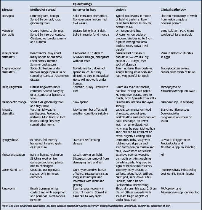
Etiology Neurovirulent strains of maedi-visna virus, a lentivirus
Epidemiology Occurs in association with maedi but endemic visna only recorded in Iceland
Clinical findings Afebrile disease with insidious onset. Progressive ataxia and wasting, long clinical course
Clinical pathology Pleocytosis and elevated protein, virus, virus proteins, and antivirus antibody in cerebrospinal fluid
Lesions Chronic demyelinating encephalomyelitis
Visna is associated with an ovine lentivirus and is the neurological manifestation of maedi-visna disease. Ovine lentivirus consists of a variety of strains that vary in their ability to infect target cells and cause disease. There are neurovirulent and non-neurovirulent strains,1 and neurovirulence can be enhanced by intracerebral passage of virus.2 Visna usually occurs in conjunction with maedi lesions in the lungs, and 10–18% of sheep with maedi have histological lesions of visna in the brain.3,4
Visna is a disease of sheep and rarely of goats. Visna was originally recorded in Iceland and was a significant cause of death in the epizootic of maedi-visna that occurred in that country. It always occurred in association with maedi and was generally of sporadic occurrence and lesser importance than the pulmonary manifestation of the infection although in some flocks it was the major manifestation of the complex.4 Visna has not been seen in Iceland since 1951 and maedi-visna has since been eradicated from Iceland.
Despite the widespread occurrence of maedi-visna or ovine progressive pneumonia in many countries visna is a very uncommon disease and a significant occurrence of clinical disease has seldom been recorded in countries other than Iceland.4-8 The reason for this is not known but might be due to an increased susceptibility of the Icelandic breed of sheep to the neurological form of the disease, or to differences in maedi-visna virus strain neuroinvasiveness and neurovirulence. Maedi-visna virus was first reported in Britain in the late 1970 and the clinical expression since that time had largely been maedi occasionally with coexistent visna. However, recently there has been the occurrence of visna with no evidence of respiratory disease suggesting that a neurotropic strain might be emerging in that country.9
Experimental transmission of the infection and the disease is effected by intracerebral inoculation and spread occurs from these to commingled sheep.10 The incubation period and the course of the disease are very long; clinical signs may not appear for 2 years after experimental inoculation.
The virus infects cells of the monocyte– macrophage lineage and replicates its RNA genome via a DNA intermediate provirus which is integrated into the chromosomal DNA of the host cell. Replication is limited and does not proceed beyond the synthesis of provirus in most cells. Persistent production of viral antigen results in lymphocytic hyperplasia. Although age has little effect on the development of disease, fetal and neonatal lambs have slightly greater permissiveness for replication of the virus. However, free infectious virus is present only in small quantities, but over very long periods. The genotype of the host is important and all breeds of sheep do not react to the same degree. Thus, Icelandic sheep react much more severely than English breeds.
There are two basic lesions, an inflammatory lesion which is not related to the occurrence of nervous signs, and a focal demyelination in the brain and spinal cord, the occurrence of which is related to the appearance of paresis.11 Experimental immunosuppression greatly reduces the severity of lesions, by suppressing the cellular proliferative response without suppressing the growth of the virus and post-infection immunization enhances the severity of experimental visna.12 Viral nucleic acid and proteins are present in oligodendrocytes, and demyelination is believed to result from the direct effect of the virus on these cells as well as being the result of an inflammatory response provoked by the presence of these antigens.13
The disease has an insidious onset and the early clinical signs include lagging behind the flock because of ataxia and body wasting. The body wasting and the hindlimb ataxia are progressive. Affected animals show hypermetria and may stumble or fall as they traverse uneven ground or when making sudden turns. There is no fever, and a normal appetite and consciousness are retained. Additional signs include severe tremor of the facial muscles, and knuckling of the distal limbs so that the animal stands on the flexed tarsi. Some animals may show a head tilt. Aimless wandering and blindness occur in some sheep.7
The clinical picture is not unlike that of scrapie without the pruritus. During the course of the disease, periods of relative normality may occur. Affected animals may show clinical signs for several months before final paralysis necessitates slaughter. The disease is always fatal. The clinical syndrome in goats is the same as that for sheep.
There is an increased number of mononuclear cells in the cerebrospinal fluid, an elevated protein, and positive Pandy test.4,8 The pleocytosis is variable during the course of the disease.4 Virus, virus antigen, and antibody are also demonstrable in cerebrospinal fluid.3 Serological tests are detailed under the section on ovine progressive pneumonia.
Muscle wasting and an interstitial pneumonia may be visible but there are no gross changes in the central nervous system. The characteristic histological lesion is patchy, demyelinating encephalomyelitis. The inflammatory infiltrate is predominantly comprised of lymphocytes and macrophages. Demyelination occurs in the white matter of the cerebrum and cerebellum, and in the spinal cord. The histological character of the lung is typical of ovine lentivirus-associated pneumonia. Isolation of the virus is difficult. Typical neural lesions and a positive serological titer usually suffice for confirmation of the diagnosis. Immunohistochemical tests and PCR-based assays have been successfully employed to confirm this lentiviral infection in lung, mammary gland,14 and even third eyelid15 but the use of these tests to confirm of the infection in CNS tissues is not well documented.
• Histology – fixed spinal cord, one-half of midsagittally-sectioned brain, lung, mammary gland, joint synovium (IHC, LM)
• Serology – heart blood serum (AGID, ELISA)
• Virology – chilled brain, spinal cord, lung, mammary gland (PCR, ISO).
1 Torsteinsdottir S, et al. Virology. 1997;229:370.
2 Craig LE, et al. J Neurovirol. 1997;3:417.
3 Brodie SJ, et al. Clin Immunol Immunopathol. 1995;77:14.
4 Petursson G, Hoff-Jorgensen R. Maedi-visna and related diseases. Boston: Kluwer Academic, 1990;185.
5 Bulgin MS. Vet Clin North Am Farm Anim Pract. 1990;6:691.
6 Biescas E, et al. J Comp Pathol. 2005;132:107.
7 Pritchard GC, et al. Vet Rec. 1995;137:443.
8 Constable PR, et al. J Am Vet Med Assoc. 1996;208:117.
9 Payne JH, et al. Vet Rec. 2004;154:94.
10 Palsson PA, et al. Acta Vet Scand. 1977;18:122.
11 Georgeson G, et al. Acta Neuropathol. 1982;57:171.
12 Cheever WP, McGuire TC. Adv Virus Res. 1988;34:189.
13 Dawson M. J Comp Pathol. 1988;99:401.
Etiology Pestivirus strains in the border disease and bovine virus diarrhea genotypes
Epidemiology Congenital disease transmitted by persistently infected sheep, rarely cattle
Clinical findings Abortions, stillbirths, barren ewes and the birth of small weak lambs some of which have an abnormally hairy birthcoat, gross tremor of skeletal muscles, inferior growth and a variable degree of skeletal deformity
Clinical pathology None specific
Lesions Hypomyelination in brain and spinal cord of lamb
Diagnostic confirmation Detection of virus and/or demonstration of serological response
Control Avoid infection of pregnant sheep. Identify and cull persistently infected animals
The causal agent, border disease virus (BDV), is a pestivirus in the family Flaviviridae. Four different species or genotypes have been identified in the pestivirus genus; bovine virus diarrhea virus (BVDV) types 1 and 2, classical swine fever virus (CSFV) and border disease virus (BDV). Isolates from border disease predominantly fall within the BDV genotype but sheep and goat isolates also fall in the BVDV genotypes.1,2 It is proposed that the BDV genotype can be divided into BDV-1, BDV-2, BDV-3 based on host origin and genetic and antigenic characteristics.3
Strains of BDV have differing pathogenicity, and variations in pathogenicity also result from interactions between the virus and different host genotypes, specifically between different breeds of sheep.1-4 Persistent infections in sheep are associated with non-cytopathic strains of virus. An isolate of BDV is thought to have caused a leukopenic enterocolitis in sheep and growing lambs in the Aveyron region of France (Aveyron disease). The disease caused high mortality in sheep in this region in 1984 but has not occurred subsequently.2
Border disease was originally described in the border country between England and Wales. It has subsequently been reported from most of the major sheep-producing countries2,4-6 and probably occurs in all. The disease occurs naturally primarily in sheep and occasionally in goats. The prevalence of infection is much higher than the incidence of clinical disease as the latter only occurs when there is infection during pregnancy.
Studies on seroprevalence vary in design but suggest that pestivirus infections in sheep and goats are less common than in cattle and that there are considerable differences in seroprevalence between different geographical areas and flocks. Flock seroprevalence in different regions or countries generally falls within the range of 5 to 50%. The prevalence of seropositive females within positive flocks is influenced by age with a lower seroprevalence in sheep 4 to 8 months of age than in older sheep. Seroprevalence is higher in flocks with persistently infected sheep but there can still be a significant proportion of seronegative sheep present in a flock containing persistently infected sheep.7
Infection can be introduced into a flock with the purchase of persistently infected replacement sheep. Persistently infected sheep excrete virus in nasal secretions, saliva, urine and feces,2,6 and provide the major source of infection. A proportion of persistently infected sheep may survive to adulthood and may breed successfully to produce further persistently infected sheep.8 However, the breeding efficiency of persistently infected sheep is poor and the probability of establishing lines of persistently infected sheep appears less than with the equivalent infection in cattle.8,9
Virus is also present in the placenta and fetal fluids at the birth of persistently infected lambs, and in the products of abortions resulting from infection with the virus in early pregnancy. In flocks where there is a long lambing period it is possible that this could provide a source for clinical disease in late lambing ewes. field observations suggest that transmission during the lambing period is limited.
Calves persistently infected with BVDV can infect sheep,10 and in countries where pregnant sheep and cattle are housed in close proximity during the winter this can be an important source of infection for outbreaks of border disease. In some countries it appears the major source and recent studies in both Northern Ireland and the Republic of Ireland suggest that cattle are the primary source of infection for sheep in those countries.11,12 In contrast BDV is the predominant ovine pestivirus in Great Britain and New Zealand.13
There is also evidence that bovine strains are important in goat infections.1,14
Free-living deer are also a potential source of infection. Outbreaks of disease are also recorded as the result of vaccinating pregnant goats with a pestivirus contaminated Orf vaccine.15
Transmission is by sheep-to-sheep contact and experimental transmission has been effected with both oral and conjunctival challenge.2,6
The speed of spread of infection in a susceptible flock will vary with the management, and will be facilitated by factors such as close contact at mating time or gathering for any purpose. There is an increased risk for explosive outbreaks of border disease where animals are housed in early pregnancy.
Border disease may occur as an outbreak or as a sporadic disease. When infection is introduced into a susceptible flock in early pregnancy, an outbreak with infertility, abortion and congenital disease in lambs from all ages of ewes is likely. Subsequently, older sheep in the flock will have immunity and disease is only common in introduced sheep and maiden ewes. Persistently infected ewes have reduced fertility but will give birth to congenitally affected lambs throughout their breeding life.8,9 The disproportional occurrence of outbreaks of clinical disease in certain breeds suggests that these breeds have higher rates of persistently infected individuals.
Border disease is readily reproduced by the experimental infection of pregnant ewes prior to 80 days’ gestation and can be effected by oral, conjunctival and parenteral challenge.6,9 Experimental disease can be produced with both BDV and BVDV strains.
The following have been produced experimentally, although there are strain differences in clinical and pathological manifestations:
• Congenital malformations, including hydrocephalus, porencephaly, cerebellar hypoplasia and dysplasia, and arthrogryposis
Experimental infections of pregnant cows with BDV results in similar defects with placentitis, mummification and abortion of fetuses, intrauterine growth retardation with abnormal osteogenesis, and hypomyelinogenesis.
The disease has also been produced experimentally in goat kids by inoculation of pregnant goats but there are no abnormalities of hair coat, and embryonic mortality and abortion are more common than in the experimental disease in ewes.16
The effect of infection varies with the immune status of the flock and whether infection occurs during pregnancy. In fully susceptible flocks, abortion and neonatal lamb loss resulting from infection can be 25–75% of the expected lamb crop depending upon the strain of the virus.9 An assessment of the economic losses due to infertility, abortion, neonatal losses, and low carcass weight indicate that an outbreak of border disease can result in a potential reduction of income in excess of 20%.17
In adolescent and adult non-pregnant sheep, infection and viremia are subclinical. The IM inoculation of immunocompetent lambs with BDV results in a mild transient disease and a subsequent reduction in growth rate, but no gross or microscopic lesions.
When BDV infects susceptible pregnant ewes the virus infects the placenta to produce an acute necrotizing placentitis and it subsequently invades the fetus. This may result in early embryonic death, abortion and stillbirth, the birth of lambs with malformations and/or neurological abnormalities, the birth of small weak lambs which are immunosuppressed, or the birth of lambs with no clinical abnormality. The ultimate outcome of the infection depends on the age of the fetus, the properties of the strain of the virus, the dose of the virus, the genotype of the host, and the ability of the fetus to respond to the virus. Fetal age at the time if infection is most important, as it is the determinant of the ability of the fetus to mount an immune response to the infection.2,6,9,18 Immune competence to the virus in sheep develops between approximately 61 and 80 days of gestation.
Fetal death occurs when there is infection of the fetus with virulent strains prior to the development of immune competence and uncontrolled viral replication. Prenatal death is more likely to follow infections in early pregnancy but is recorded with infections from 45 to 72 days of gestation.
Persistent infections occur in lambs that survive infection in early pregnancy prior to the development of immune competence and result from maternal infections between 21 and 72 days of gestation but never later.19 The virus is present in all organs, and lambs born persistently infected will remain so for their lifetime, with few exceptions; persistent infections have been recorded to at least 5 years of age.9
Most persistently infected sheep are unable to produce specific antibody to BDV but some show intermittent seropositivity with low antibody levels or occasionally undergo frank seroconversion.19 The humoral response to other pathogens and antigens is normal. However, cell-mediated immunity is compromised, with change in T-cell populations and a deficiency in lymphocyte function.20 Persistently infected lambs are more susceptible to intercurrent disease and commonly die before reaching maturity.
Hypomyelinogenesis occurs in persistently infected lambs and resolves spontaneously in lambs that survive to the age of 6 months.21 Most of these lambs exhibit neurological dysfunction at birth, varying from a continuous light tremor to tonic–clonic contraction of the skeletal muscles involving the whole body and head (shakers).
Thyroid. A deficiency of the thyroid T3 and T4 hormones has been detected in lambs affected with border disease and may be the basic cause of the amyelination. The enzyme 2,3-cyclic nucleotide- 3-phosphodiesterase is associated with normal myelination and depends upon normal amounts of thyroid hormone.20 The deficiency in thyroid hormones may also result in the reduced rate of weight gain that occurs in infected lambs. Other studies suggest a direct infection of oligodendroglia with the virus as the cause of the defective myelination.22,23
Fleece abnormality also occurs in persistently infected lambs and results from an enlargement of the primary hair follicles and a concurrent reduction in the number of secondary follicles. The resulting hairiness is due to the presence of large medullated primary fibers. BDV appears to have no effect on the skin and birthcoat of coarse-fleeced breeds of sheep or on goats.
Intrauterine growth retardation is a common feature of infection with BDV and is initiated shortly after infection.23 Deformities of the skeleton include abnormally shortened long bones, a reduction in crown–rump length and the long axis of the skull, which results in lambs appearing more compact and short-legged than normal (goat lambs). In the long bones there is evidence of growth arrest lines and disturbed osteogenesis and ossification.
Some persistently infected lambs have neither nervous signs nor abnormalities of the fleece and are phenotypically normal. This limits the value of identification of infected lambs based on the presence of clinical abnormality at birth.
When fetal infection occurs during the period of development of the ability to mount an immune response (between approximately 61 and 80 days of gestation) the effect is variable. Some fetuses infected at this stage respond with a severe inflammatory process in the CNS with nodular periarteritis, necrosis, and inflammation of the germinal layers of the brain. Resultant lesions are hydranencephaly, cerebellar dysplasia and multifocal retinal atrophy; such lambs exhibit behavioral abnormalities and more severe neurological disease than shaker lambs.9,19,21
Infection of the fetus after 80 days of gestation is likely to be controlled or eliminated by a fetal immunological response. Such lambs are born without clinical disease and are virus-negative but have precolostral circulating antibody.
In goats, fetal death is the major outcome of infection of the pregnant doe with both BDV and BVDV, and infections prior to 60 days’ gestation almost invariably result in reproductive failure.16 Persistently infected shaker kids and clinically normal kids are born with infections around 60 days’ of gestation but are a less common manifestation of the disease than occurs in sheep. The caprine fetus develops immune competence against pestiviruses between 80 and 100 days’ gestation.21
Experimental inoculation of a homologous strain of the BDV into persistently infected, but clinically recovered lambs results in a severe clinical syndrome characterized by persistent diarrhea and respiratory distress associated with an inflammatory lymphoproliferative response in the central nervous system, intestines, lungs, heart, and kidney. A similar syndrome is seen at weaning in some persistently infected sheep that survive early life.2,9 This syndrome resembles certain aspects of mucosal disease in cattle, in which it is postulated that superinfection of persistently viremic immunotolerant cattle with a homologous strain of BVDV results in fatal mucosal disease. In such animals a specific and dynamic equilibrium exists between an attenuated form of the virus and the immunotolerant host. Disturbance of this equilibrium either by injection of the homologous strain of BDV, or some other factor, results in fatal disease.9
The most obvious and characteristic features of border disease are evident at birth and relate to conformation and growth, fleece type, and neurological dysfunction.
Affected lambs may have a lower birth weight than uninfected lambs, a decreased crown–rump length, and a shorter tibia/radius length so that they have a boxy appearance. The head has a shortened longitudinal axis and the cranium may be slightly domed (goat lambs).
The fleece, when dry, appears hairy and rough due to long hairs rising above the fleece to form a halo, especially over the nape, back, flanks and rump.24 This feature is most evident in medium- and fine-wool breeds and is not observed in the coarse kempy-fleeced breeds, such as the Scottish Blackface. The halo kemp fibers are shed with time and are most evident in the first 3 weeks of life. Some lambs have abnormal pigmentation occurring as patches of pigmented fleece or hair, or a totally pigmented fleece. This can occur in white-faced sheep.
Neurological dysfunction is manifest, with rhythmic tremors of the muscles of the pelvis and upper parts of the hindlimbs, or of the whole body, resulting in a characteristic jerking movement, and of the head and neck with rhythmic bobbing of the head (shaker lambs). In some less severe cases, only fine tremors of the ears and tail are evident. Tremors are most apparent during movement, and are absent while the lamb is sleeping. The tremors usually decline in severity as the lamb matures and may seem to disappear unless the animal is stressed. More severely affected lambs have difficult in rising, and if able to stand with assistance exhibit an erratic gait especially of the hindquarters. Paralysis does not occur. Affected lambs are often unable to nurse the ewe because they cannot hold onto the teat. Affected lambs appear languid and lie around listlessly. They do not suck as they should and bloat continuously, and the ewes’ udders become engorged with milk.
Behavioral and visual defects with circling, head-pressing, nystagmus, and gross incoordination are seen in lambs with the type of infection producing hydranencephaly and cerebellar dysplasia. These lambs are of lighter birth weight but have normal birthcoats.
Growth rate is reduced, affected lambs are unthrifty, and the majority will die before or at weaning time from parasitism, pneumonia, a mucosal disease-like syndrome, or nephritis.9,20 With good nursing care they can be reared but deaths may occur at any age. Puberty may be delayed and, in males, the testes are flabby and may not develop normally.
Impaired reproductive performance of the flock occurs from low fertility, abortion, and poor viability of lambs. Abortions usually are not noticed until lambing when it is evidenced by an unexpected increase in barren ewes. In goats, where there is often closer observation, the aborted fetuses may be reasonably well-developed, small and underdeveloped, or autolyzed and unrecognizable as a fetus in expelled fetal fluid.16
There are no consistent changes in hematology or blood chemistry. Persistently infected lambs have changes in lymphocyte subpopulations, with a reduction in T-lymphocytes and an altered CD8:CD4 ratio.6
Virus can be detected by virus isolation, by antigen ELISAs, by PCR techniques and antibody by antibody ELISAs, or by serum neutralization tests.2,25 A combination of serology and virus isolation can aid in the diagnosis of border disease.
For diagnosis of border disease in newborn lambs, precolostral blood samples should be taken from both clinically normal and affected lambs. Persistently infected sheep are seronegative and BDV can be isolated from leukocytes in the blood buffy coat. Lambs infected late in gestation will be seropositive but virus-negative. Persistently infected lambs that have received colostrum from their dam will be seropositive until they lose the maternal passive immunity.
Persistently infected adolescent and adult sheep in a flock can be identified by the detection of virus in blood; however, this is expensive in large flocks and an alternative is test all sheep for antibody and then culture the buffy coat of seronegative sheep. Antigenic differences between laboratory strains and field virus can result in false-negative serologies and serological studies are best done with the homologous virus.4,26
Serological tests are of limited value as an aid to the diagnosis of abortion associated with BDV infection. The infection of the ewe that results in abortion occurs several weeks before clinical disease is apparent, and unless prospective samples can be taken there is little chance of a rise in antibody titers in paired samples. Seropositivity in ewes indicates that the flock has been exposed to pestivirus but does not incriminate it in a disease process. Seronegativity indicates that BDV is not the cause of the abortion, with the exception that aborting ewes, who themselves are persistently infected, will have no antibody titer.
Gross findings may be normal, or may include an abnormal wool coat and a reduction in the size of the brain and spinal cord. Arthrogryposis, hydranencephaly, porencephaly, and cerebellar dysplasia may also be present. Histologically, there is a deficiency of stainable central myelin, with neurochemical and histochemical evidence of demyelination or myelin dysmorphogenesis. In most sheep the myelin defect resolves substantially during the first few months of life. The brain, which has been very small, returns to normal weight, and chemical composition and degree of myelination. The histological lesions of the skin consist of primary follicle enlargement, increased primary fiber size, and an increased number of medullated primary fibers.
Virus can be demonstrated by immunofluorescent staining of cryostat sections of tissues from affected lambs or by immunohistochemical staining of formalin-fixed material. Preferred tissues for such tests include brain, thyroid gland, and skin. Virus titers reach high levels in the placentomes, so caruncles or cotyledons should be cultured for virus. Isolates are non-cytopathic and the presence of viral antigens must be demonstrated by direct or indirect immunofluorescence or immune peroxidase techniques.
Owing to the closely related character of this pestivirus and BVDV, diagnostic tests to confirm infection parallel those for BVDV. Fetal serology can be useful to confirm exposure in abortions and stillbirths. PCR and ELISA techniques may be substituted for virus isolation if available.
• Histology – formalin-fixed skin, spinal cord, one-half of midsagittally sectioned brain, skin, thyroid, distal ileum, colon, cecum, thymus, spleen, liver, heart, kidney (LM, IHC)
• Serology – heart blood serum/thoracic fluid (VN)
• Virology – placenta/caruncle, thymus, lymph node, spleen, thyroid, brain, ileum (ISO, FAT, ELISA, PCR).
There is no specific treatment. With care and nursing, many affected lambs will survive the immediate neonatal period but they grow poorly, are very susceptible to intercurrent disease during the growing period, and it is generally not economic to attempt to raise these lambs.
The principles are to attempt to engender a flock immunity and to avoid exposing sheep to infection in early pregnancy. Persistently infected sheep are a continuous source of infection and those that survive to breeding age can perpetuate the disease. They should be identified and culled.
The problem is with their identification, as some persistently infected lambs show no clinical or phenotypic abnormality. Lambs that are clinically affected at birth should be permanently identified since the tremor and fleece abnormality disappear at 1–2 months of age and the lambs may no longer be recognizable as infected. Persistently infected animals can be identified by serological screening of the ewe lambs intended for replacement stock at 6 months of age (after maternal passive immunity has waned), followed by virus isolation in seronegative animals, but this is expensive and only practical in small flocks. An alternative is to keep no replacement ewes from an affected lamb crop.
Persistently infected sheep can be run with the flock when it is not pregnant, particularly with the replacement ewes, in an attempt to produce infection and immunity prior to pregnancy. They should be removed prior to breeding. While this can result in ‘natural vaccination’ the rates of infection and seroconversion in replacement females can be low.7 In theory, cattle BVDV vaccines could be used to produce immunity but their efficacy would depend upon a significant relatedness to the BDV under consideration.
In most flocks a serious outbreak of the disease is followed by minor disease in subsequent years, the flock developing immunity in the initial outbreak.
In flocks that are free of infection, replacement ewes and rams should be screened for infection prior to purchase, or quarantined after arrival on the farm. Newly introduced sheep should be kept separate from the main flock until after lambing. Ideally, cattle should not be pastured or housed with pregnant sheep.
Terpstra C. Border disease: a congenital infection of small ruminants. Prog Vet Microbiol Immunol. 1985;1:175-198.
Moennig V. Pestiviruses: a review. Vet Microbiol. 1990;23:35-54.
Leiss B, Moennig V, Pohling J, Trautwein G. Ruminant pestivirus infections. Virology pathogenesis and perspectives of prophylaxis. Arch Virol Suppl. 1991;3:1-271.
Hussin AA, Woldehiwet Z. Border disease virus: a review. Vet Bull. 1994;64:1131-1151.
Loken T. Border disease in sheep. Vet Clin North Am Food Anim Pract. 1995;113:579-595.
Nettleton PF, Entrican G. Ruminant pestiviruses. Br Vet J. 1995;151:615-642.
1 Becher P, et al. J Gen Virol. 1997;78:1357.
2 Nettleton PF, Entrigan G. Aust Vet J. 1995;151:615.
3 Becher P, et al. Virology. 2003;311:96.
4 Kelling CL, et al. Am J Vet Res. 1990;51:2019.
5 Nettleton PF, et al. Vet Res. 1998;29:227.
6 Hussin AA, Woldehiwet Z. Vet Bull. 1994;64:1131.
7 Berriatua E, et al. Vet J. 2004;168:336.
8 Woldehiwet Z, Nettleton PF. Arch Virol Suppl. 1991;3:267.
9 Nettleton PF, et al. Comp Immunol Microbiol Infect Dis. 1992;153:179.
10 Carlsson U. Vet Rec. 1991;128:145.
11 Graham DA, et al. Vet Rec. 2001;148:69.
12 O’Neill RG, et al. Irish Vet J. 2004;57:525.
13 Vilcek S, et al. Vet Microbiol. 1999;69:227.
14 Loken T. J Comp Path. 1990;103:1.
15 Loken T, et al. J Comp Path. 1991;104:195.
16 Loken T, Bjerkas I. J Comp Path. 1991;105:123.
17 Sharp MW, Rawson BC. Vet Rec. 1986;119:128.
18 Osburn B, Castrucci G. Arch Virol. 1991;3:71.
19 Roeder PL, et al. Res Vet Sci. 1987;43:28.
20 Sawyer MM. Comp Immunol Microbiol Infect Dis. 1992;153:171.
21 Jeffery M, Roeder PL. Res Vet Sci. 1987;43:22.
22 Jeffery M, et al. Neuropathol Appl Neurobiol. 1990;16:501.
23 Caffrey JF, et al. Res Vet Sci. 1997;62:245.
24 Campbell JR, et al. Can Vet J. 1995;36:307.
Etiology Orf virus. Genus Parapoxvirus. Family Poxiviridae
Epidemiology Primarily young lambs and kids. Morbidity may reach 100% and case fatality rate 5–15%. Rapid spread in flock by contact or via inanimate objects such as feed troughs, ear tag equipment and emasculators. Scabs from lesions remain infective in the environment for a long time. Orf can cause considerable set back when young lambs are affected and also has economic importance due to restriction of movement and trade of affected sheep. May infect humans
Signs Papules, pustules, scabs covering ulceration, granulation, proliferation and inflammation. Lesions begin at oral mucocutaneous junction, oral commissures and spread to muzzle, oral cavity. Lambs cannot suck or graze. Malignant form occurs with invasion of alimentary tract. Severe systemic reaction can occur and lesions on coronets, ears, anus, and vulva. Lesions can be multifocal in goats
Clinical pathology Electron microscopy. PCR
Lesions Scabs, pustules, granulation tissue and secondary lesions. Eosinophilic cytoplasmic inclusion bodies
Diagnostic confirmation Clinical recognition and identify virus by PCR
Orf is associated with the orf virus, a type species of the genus Parapoxvirus (family Poxviridae). In addition to the orf virus (parapox ovis) the genus includes the viruses of bovine papular stomatitis (parapox bovis 1) pseudocowpox (parapoxvirus bovis 2) and a parapox virus of deer.1-3 The orf virus withstands drying and is capable of surviving at room temperature for at least 15 years. Restriction endonuclease digests of DNA shows considerable heterogeneity between different field isolates.
The disease occurs in sheep and goats and causes unthriftiness, varying degrees of pain, and some economic loss. It occurs most commonly in lambs 3–6 months of age when at pasture, although lambs 10–12 days of age and adult animals can be severely affected, and outbreaks involving the lips and face of young lambs and the udders of the ewes also are common. Outbreaks occur at any time but they are most common in dry conditions when the sheep are at pasture, or in penned sheep being fed from feed troughs. The disease has occurred in musk ox in which it causes heavy losses, reindeer, mountain goats and bighorn sheep, chamois, caribou, Dall sheep, buffalo, wild goats, and camels. The virus can be passaged in rabbits if large doses are placed on scarified skin or injected ID. Mild lesions develop on the chorioallantois of the 9 to 12-day chick embryo. Guinea-pigs and mice are not susceptible.
The disease also occurs in humans working among infected sheep. In abattoir workers it is commonest in those handling wool and skins.4
Outbreaks may occur in sheep and goats, with morbidity rates approaching 100% and case fatality rates from 5 to 15%.5,6 The deaths that occur are due to the extension of lesions in the respiratory tract, but the case fatality rate may reach 15% if severely affected lambs are not provided with adequate care and support, or if secondary infection and cutaneous myiasis are allowed to occur. In the rare outbreaks where systemic invasion occurs, the case fatality rate averages 25% and may be as high as 75%. Under field conditions recovered animals are immune for 2–3 years, but no antibodies appear to be passed in the colostrum, and newborn lambs of immune ewes are susceptible.5
Scabs that fall off from healing lesions contain virus and remain highly infective for long periods in dry conditions, but survival of the disease in a flock may be the result of chronic lesions that exist for long periods on individual animals.7 Infection can be from environmental persistence of the virus or from infected sheep. Spread in a flock is very rapid and occurs by contact with other affected animals or by contact with contaminated inanimate objects, such as feed troughs, ear-tagging pliers.8 An outbreak of lesions on the tails is recorded in association with the use of docking instruments.9
It has been assumed that natural infections on pasture are the result of invasion of the virus after skin damage induced by prickly plants or stubble; application of a viral suspension to scarified skin is the established method of inducing orf.10 However, an outbreak has occurred in a large group of lambs collected from several farms and transported in a vehicle over a period of 23 hours.11 There was no evidence of injury to their mouths.
The disease is readily reproduced by introduction of virus onto scarified areas of skin. Immunity to re-infection is relatively solid at the site of initial infection but shorter duration lesions can be reproduced by re-challenge of these sheep at other sites.10,12
The primary risk factors are the presence of the virus and the immune status of the sheep.
A simulated shipping exercise has examined these risks.13 Initially, the prevalence of scabby mouth on participating sheep farms was determined. The proportion of farms with evidence of the disease in weaner sheep was 23.6%, and on those farms with the disease the overall prevalence was 6.1%. Sheep from different farms are mixed, held at high stocking densities for approximately 3 weeks, and offered pelleted feed that can abrade the lips and mouth to promote development of the lesions. The major risk factor for the prevalence of the disease in sheep arriving in a feedlot after transportation from the farm was immunity to the disease. The arrival and mixing in the feedlot was also important for transmission of the infection to occur. Sheep vaccinated on the farm before road transportation to the feedlot had a lower prevalence at the end of the simulated shipping exercise.13
Inter-current infections may exacerbate the occurrence disease on rare occasions.
The disease has spread from clinically normal ewes to susceptible 2–4 years of age which were persistently infected with border disease virus14 and lambs experimentally infected with Ehrlichia phagocytophilia and subsequently challenged with orf virus developed more severe lesions with a longer course than those in control lambs.15
The disease produces a minor set back except when it affects young sucking lambs with associated lesions on the teats and udders of their ewes. Loss from lamb mortality and secondary mastitis in these circumstances can be significant.
The disease assumed economic importance for Australia when shipments of sheep exported from Australia in 1989–1990 were rejected at some ports in the Middle East because of the disease13 and this problem continues. Litigation is a concern when zoonotic infections occur at petting zoos or fairs.
Orf virus is readily transmitted to humans. In the agricultural environment typical lesions occur at the site of infection, usually an abrasion infected while handling diseased sheep for shearing, crutching or drenching or by accidental means when vaccinating. Lesions progress from macular to papular stages, are usually single and localized on the hands, arm or face. The lesions are self limiting and heal without scaring after 6 to 7 weeks. They are pruritic and respond poorly to local treatment. Orf is also a zoonotic consideration in ‘petting zoos’ and fairs where children allow lambs to suck their fingers or otherwise become infected from handling sheep exhibits. Historically, orf has been a risk for industrial workers handling raw wool.
Damage to the skin is essential for the establishment of orf infection and the development of typical lesions.10 Following viral challenge of mildly abraded skin, the virus does not establish in the damaged epidermis, but replicates in the cells of an underlying replacement epidermal layer derived from the walls of the wool follicles. Following scarification of ovine skin and topical application of the orf virus, antigen cannot be detected in the skin during the period when the epidermis is being renewed.16 Virus can first be detected in the center of the newly differentiated epidermis immediately below the stratum corneum, 72 hours after infection. The location of the virus during the eclipse stage is unknown. The infection spreads laterally and uniformly from the new epidermis, initially in the outer stratum spinosum and subsequently throughout the entire depth of the epidermis. The skin reaction consists of a cellular response with necrosis and sloughing of the affected epidermis and underlying stratum papillare of the dermis. The cutaneous response to infection includes a delayed-type hypersensitivity reaction and an influx of inflammatory cells involving neutrophils, basophils, and possibly mast cells.17 Class II dendritic cells are also involved and appear to form the basis of a highly integrated local dermal defense mechanism.18 The lesions evolve through the stages of macule, papule, vesicle, pustule, scab formation, and resolution. The pustules develop within a few days, and rupture resulting in ulcers and subsequently a thick overlying crust or scab forms which is shed within 3–4 weeks leaving no scar.10 Immunity is solid but will last only about 8 months. While there is an antibody immune response to the virus19 recovery is the result of cell-mediated immune mechanisms.12 Experimentally, a secondary infection, following recovery from a primary infection, is milder and accelerated.20 During the secondary challenge, pustules and scabs develop earlier, the lesions resolve more rapidly, and no vesicular stage may occur.17
Lesions develop initially as papules and then pustules, stages which are not usually initially observed, and progress to raised moderately proliferative area of granulation, and inflammation covered with a thick, tenacious scab. Time progression from the initial lesions to the formation of scabs is approximately 6 to 7 days. New lesions will develop during the first 10 days of infection. The first lesions develop at the oral mucocutaneous junction, usually at the oral commissures and are accompanied by swelling of the lips. From here they spread on to the muzzle and nostrils, the surrounding haired skin and, to a lesser extent, on to the buccal mucosa. They may appear as discrete, thick scabs 0.5 cm in diameter, or coalesce and be packed close together as a continuous plaque. fissuring occurs and the scabs are sore to the touch. They crumble easily but are difficult to remove from the underlying granulation. Affected lambs suffer a severe setback because of restricted sucking and grazing. In benign cases the scabs dry and fall off, and recovery is complete in about 3 weeks. Affected lambs sucking ewes may cause spread of the disease to the udder where a similar lesion progression is seen. Lesions on the teats predispose to mastitis and secondary infection of the skin lesions by bacteria or fly larvae occurs in some cases. In rams, lesions on the scrotum may be accompanied by fluid accumulation in the scrotal sac and associated temporary infertility. A high incidence of infection can also occur where the dominant lesions are on the feet, occurring around the coronary band, the dew claws and on the volar areas of the intervening skin.
Rarely, systemic invasion occurs and lesions appear on the coronets and ears, around the anus and vulva or prepuce, and on the nasal and buccal mucosae. There is a severe systemic reaction, and extension down the alimentary tract may lead to a severe gastroenteritis, and extension down the trachea may be followed by bronchopneumonia. Lesions may also occur in the mouth involving the tongue, gums, dental pad or a combination of those sites.21 These are more commonly seen in outbreaks affecting lambs less than 2 months of age. In the mucosa of the mouth these lesions do not scab but are papular erosive and surrounded by an elevated zone of hyperemia. Extensive painful and proliferative lesions occur on the gingival margins of the incisor teeth.
In some outbreaks the lesions on the skin are highly proliferative and present as raw raised granulating lesions without an overlying scab. This manifestation appears more common in Suffolk sheep and lesions are present on the lips, bridge of the nose and around the eyes. Cases of this proliferative form involving the feet are also recorded.22
A malignant form of the disease has also been observed in sheep. It begins with an acute episode manifested by oral vesicles, and extension of these lesions down the gastrointestinal tract, followed later by granulomatous lesions and shedding of hooves.
An atypical case of the disease in sheep after extensive cutaneous thermal injury has been described.23 The virus was present in proliferative verrucous tissue lesions at the periphery of the original thermal injury.23 The lesions consisted of tightly packed 0.5 mm-diameter papillary projections.
An unusual case of contagious ecthyma in a group of female goats has been described.24 Multifocal lesions occurred over the head, neck, thorax, and flanks of each animal. The lesions developed approximately 2 weeks after the animals returned from a local summer show at which the does were housed for 3 days in pens previously occupied by sheep. The lesions began as plaques, followed by epidermal proliferation and severe encrustation. Affected areas were discrete and approximately 2–7 cm in diameter. There were no lesions of the muzzle, lips, udder or teats. Recovery occurred uneventfully within 3–6 weeks without treatment. The skin crusts gradually dried and fell off, leaving areas of alopecia and depigmented skin. Regrowth of hair followed.
Persistent orf is recorded in a proportion of Boer goats following an outbreak. The disease in the majority of the animals in the herd was classical and ran a clinical course of 3–4 weeks but in 2% of animals it persisted for several months and lesions disseminated over the body. There were no particular distinguishing differences of the virus genome to those of other orf viruses and the persistence was possibly a result of individual host susceptibility factors.25,26
Electron microscopic identification of the virus is quick and generally reliable with multiple samples from an affected herd or flock. Virus can also be detected by PCR and restriction enzyme analysis of viral DNA and gene sequencing.26,27 Recovered animals have a high level of neutralizing antibodies in their serum and this is detectable by a gel diffusion test and other serological tests but has little clinical value.
In malignant cases there are irregularly-shaped lesions, with a hyperemic border in the oral cavity and the upper respiratory tract, with rare involvement of the mucosae of the esophagus, abomasum, and small intestine. Typical lesions are actually proliferative, with subsequent loss of centrally located cells creating an ulcer-like appearance. Microscopically, the hyperplastic epithelium contains swollen degenerate cells, some of which may house eosinophilic cytoplasmic inclusion bodies.
In most outbreaks of ecthyma, the cases are sufficiently mild to cause no real concern about losses or about diagnosis.
Dramatic outbreaks of a very severe form of the disease may occur, however, and are likely to be confused with bluetongue. Very severe cases are also commonly seen in housed experimental sheep especially colostrum-free lambs.
Ulcerative dermatosis is sufficiently similar to cause confusion in diagnosis, but this disease has not been reported in many years.
Mycotic dermatitis usually occurs on woolled skin but lesions can occur on the lips and feet (strawberry foot rot), have a thick dry asbestos-like scab and are easily differentiated by laboratory culture.
Facial eczema is distinguished by diffuse dermatitis and severe edema and damage to the ears.
Papillomatosis (warts) need also to be considered in the differential diagnosis for the proliferative manifestations of contagious ecthyma, although warts are extremely uncommon in sheep.
Bluetongue is always accompanied by a high mortality rate, a severe systemic reaction, and lesions occur on the muzzle, the coronets, and extensively on the buccal mucosa. It is more common in adults than sucking lambs. Because it is transmitted by insect vectors, the morbidity rate is usually much less than the 90% commonly seen in contagious ecthyma.
Sheeppox may present a rather similar clinical picture, but the lesions are typical, there is a severe systemic reaction and heavy mortality rate.
Foot and mouth disease. The classic developed lesions of orf are easily differentiated from foot and mouth disease but the papular and vesicular stages seen early in the course of orf, particularly lesions in the mouth, can be difficult to differentiate especially when a prompt on-farm differentiation is required. The raised firm papular erosive nature of the lesion with the surrounding zone of hyperemia is a crucial differentiating feature in the field.28
There is no specific treatment. Removal of the scabs and the application of ointments or astringent lotions are practiced but delay healing in most cases. The provision of soft, palatable food is recommended. The combined use of diathermy debridement and cryosurgery is claimed to be effective for the proliferative intraoral lesions in young lambs.29
In the early stages of an outbreak, the affected animals should be isolated and the remainder vaccinated. Vaccination is of little value when a large number of animals are already affected. Persistence of the disease in a pastured flock from year to year is common and in such circumstances the lambs should be vaccinated at 6–8 weeks of age. Vaccination when a few days old evokes a protective response, but prelambing vaccination of the ewe does not and is not recommended.30 Vaccination of housed lambs should be timed to avoid the usual occurrence of the disease that has been observed in previous years.
The vaccine is prepared from a suspension of scabs in glycerol saline and is painted onto a small area of scarified skin inside the thigh, or by pricking the ear with a needle dipped in the vaccine. Vaccination is completely effective for at least 2 years, but the lambs should be inspected 1 week after vaccination to insure that local reactions have resulted. Absence of a local reaction signifies lack of viability of the vaccine or the existence of a prior immunity. The immunity is not solid until 3 weeks after vaccination. A small proportion of vaccinated lambs may develop mild lesions about the mouth because of nibbling at the vaccination site. The efficiency of this vaccine is greater than that of the standard commercial vaccine containing live attenuated virus.31 As a further protective measure, removal of abrasive material from the environment is recommended but is not usually practicable. For live sheep being transported from Australia to the Middle East, it is suggested animals be vaccinated well in advance of shipment to allow immunity to develop, which is probably at least 3 weeks.
Because the vaccines are live virus vaccines, and shed scabs are contaminated, routine vaccination against orf in flocks that have not experienced the disease is not recommended.
1 Delhoun G, et al. J Virol. 2004;78:168.
2 Robinson AJ, Mercer AA. Arch Virol. 1988;101:255.
3 Rziha HJ, et al. J Gen Virol. 2003;84:1111.
4 Robinson AJ, Petersen GV. NZ Med J. 1983;96:81.
5 Robinson AJ, Balassu TC. Vet Bull. 1981;51:771.
6 Housawi FMT, et al. Vet Rec. 1991;128:550.
7 McKeever DJ, Reid HW. Vet Rec. 1986;118:613.
8 Allworth MB, et al. Aust Vet J. 1987;64:61.
9 Ames T, et al. J Am Vet Med Assoc. 1984;184:88.
10 McKeever DJ, et al. J Comp Path. 1988;99:317.
11 Gumbrell RC, McGregor DA. Vet Rec. 1997;141:150.
12 Anderson IE, et al. Vet Immunol Immunopathol. 2001;83:161.
13 Higgs A, et al. Aust Vet J. 1996;74:215.
14 Nettleton PF, et al. Vet Rec. 1996;139:364.
15 Gokce HI, Woldehiwet Z. J Comp Path. 1999;121:227.
16 McEwan D, et al. Vet Dermatol. 1990;1:189.
17 McEwan D, et al. Vet Dermatol. 1990;1:139.
18 McEwan D, et al. Vet Dermatol. 1991;2:1.
19 McKeever DJ, Reid HW. Vet Microbiol. 1987;14:3.
20 Yirrell D, et al. Vet Immunol Immunopathol. 1989;22:321.
21 Hawkins CD, et al. Aust Vet J. 1991;68:210.
22 Smith GW, et al. J Vet Internal Med. 2002;16:287.
23 Hooser SB, et al. J Am Vet Med Assoc. 1989;195:1255.
24 Coates JW, Hoff S. Aust Vet J. 1990;31:209.
25 Concha-Bermejillo A, et al. J Vet Diag Invest. 2003;25:423.
26 Guo J, et al. Virus Res. 2003;93:169.
27 Torfason EG, Guonadottir S. J Clin Virol. 2002;24:79.
28 Watson P. In Practice. 2004;26:182.
29 Meynink SE, et al. Vet Rec. 1987;121:594.
Etiology Papillomaviruses. Host specific, and in cattle, some types have site and lesion-type specificity
Epidemiology Occur in all countries in all species but most common in young cattle and horses. Transmission is by direct contact and fomites
Clinical findings Solid outgrowths of epidermis, may be sessile or pedunculated. Most common type in cattle occurs on head and neck and has cauliflower-like appearance, but lesion site and appearance varies with papilloma type. In the horse, lesions are on the face and lips
Clinical pathology None specific
Lesions Papilloma or fibropapilloma
Diagnostic confirmation Histology and DNA identification by PCR in biopsy or tissue scraping
Treatment Removal by surgery or cryosurgery. Vaccination with autogenous vaccine
Cutaneous warts in cattle, horses, sheep and goats are benign tumors induced by host-specific papillomaviruses. These infect epithelial cells causing hyperproliferative lesions that are benign, self-limiting and, in most cases, spontaneously regress. Differentiation of types is based on the histological features of the lesion and DNA identification by hybridization or PCR. There has been little research on the papilloma virus of horses or small ruminants, but in cattle six types have been identified: bovine papilloma virus (BPV)-1, BPV-2 and BPV-5, which cause fibropapillomas; and BPV-3, BPV-4 and BPV-6, which cause true epithelial papillomas.1,2
Cattle types show some site predilection or site specificity. Their detailed roles are:
• BPV-1 – frond fibropapillomas of teat skin and penile fibropapilloma
• BPV-1 and BPV-2 – fibropapilloma of the skin of the anteroventral part of the body including the forehead, neck and back, the common cutaneous wart
• BPV-2 – cauliflower-like fibropapillomas of the anogenital and ventral abdominal skin
• BPV-2 – associated with bladder cancer in cattle in association with the ingestion of bracken fern (Pteridium spp.)3,4
• BPV-4 – papilloma of the esophagus, esophageal groove, forestomachs and small intestine; this is capable of becoming malignant, particularly in animals fed bracken fern.4-6 BPV-4 has site specificity to the upper alimentary tract
• BPV-5 – rice grain fibropapilloma on the udder. BPV-5 has also been demonstrated in cutaneous skin warts7
• BPV-6 – frond epithelial papillomas of the bovine udder and teats.
Although a single BPV type is detected in an individual papilloma, a single animal may have papillomas at different sites associated with different BPV types.8
Other papilloma of cattle that have regional distribution and may have separate antigenic identity are:
• Oral papillomas, mostly in adult cattle and apparently reaching a high incidence, up to 16% in some areas9; these are probably BPV-48
• Papilloma of the larynx in steers
• Papillomavirus has been observed in squamous cell carcinoma of bovine eyes.10
Other skin lesions in which papillomavirus plays an etiological role are:
• Equine sarcoid which associated with BPV-1 and BPV-2
• Ear cancer of sheep.11
Papillomas in horses may be associated with a distinct equine papillomavirus.12
The method of spread is by direct contact with infected animals, infection gaining entry through cutaneous abrasions. Virus can also persist on inanimate objects in livestock buildings and infect animals rubbing against them.8
Crops of warts sometimes occur around ear tags, at branding sites or along scratches made by barbed wire, and can be spread by tattooing implements, dehorning shears, and by procedures such as tuberculin testing.13-16
An extensive outbreak of perianal warts is recorded in beef heifers, the infection having been spread by rectal examination for pregnancy. A high prevalence of papillomas on the larynx of feedlot steers is ascribed to implantation of the virus in contact ulcers, which are also entry sites for Fusobacterium nodosus (a cause of calf diphtheria), so that the two diseases may occur in the one animal. An outbreak of periorbital papillomatosis in cattle is recorded in association with a heavy periorbital infestation with Haematopinus quadripertusus.17
All species may be affected but the disease is commonest in cattle and horses. With cattle, usually several animals in an age group are affected. Outbreaks have been recorded in sheep and goats,13,18 but the disease is uncommon in sheep. It is also uncommon in pigs, usually affecting the genitalia. Amongst wildlife, it occurs in white-tailed deer (Odocoileus virginianus) and mule deer (O. hemionus).
Cutaneous papillomas of the head and neck occur predominantly in young animals, the lack of susceptibility of adults to natural infection being ascribed to immunity acquired by apparent or inapparent infection when young. The occurrence of cutaneous warts and their severity can be influenced by factors that induce immunosuppression, and latent infection has converted to clinical disease with the administration of immunosuppressive agents.8,19 Congenital infection is recorded in the foal and calf, but is rare.20
Alimentary papillomas associated with BPV-4 in cattle, rice grain teat papillomas associated with BPV-5 in cattle, and papillomas on the mammary glands of goats occur, or persist, at all production ages.
The supernatant from a suspension of wart tissue, injected ID, or applied to skin scarifications, is an effective means of experimental production of the disease.8,19 Lesions are restricted to the site of inoculation. Cutaneous and oral papillomas have been transmitted in cattle, and cutaneous papilloma transmitted in sheep and horses. The incubation period after experimental inoculation in cattle is 3–8 weeks but is usually somewhat longer after natural exposure.
Cutaneous warts are quite common in young cattle, especially when they are housed, but ordinarily they cause little harm and regress spontaneously. In purebred animals they may interfere with sales and shows because of their unsightly appearance. Animals with extensive lesions may lose condition, and secondary bacterial invasion of traumatized warts may cause concern. Warts on the teats of dairy cows often cause interference with milking. In horses, the lesions are usually small and cause little inconvenience, but they are esthetically unattractive. In all species, the development of warts on the genitalia requires immediate treatment.
The virus infects the basal keratinocytes, replicating its genome in the differentiating spinous and granular layers causing the excessive growth that is characteristic of wart formation. Expression of the late structural proteins of the virus is limited to the differentiated cells of the squamous layer where the new virus particles are encapsulated and shed into the environment as the cells die. The tumor contains epithelial and connective tissues and can be a papilloma or a fibropapilloma, depending on the relative proportions of epithelial and connective tissue present; papillomas contain little connective tissue, and fibropapillomas are mostly fibrous tissue, with very little epithelial tissue. Papillomas are the result of basal cell hyperplasia without viral antigen production. fibropapillomas are uncommon in horses, but are the common lesion in cattle, sheep, and wild ruminants.21 Latent infection in the skin and lymphocytes has been demonstrated in cattle.8
Warts are solid outgrowths of epidermis and may be sessile or pedunculated.
In cattle, warts occur on almost any part of the body, but when a number of animals in a group is affected it is common to find them all affected in the same part of the body. The most common papillomas occur in the skin of cattle under 2 years of age, most commonly on the head, especially around the eyes, and on the neck and shoulders, but may spread to other parts of the body. They vary in size from 1 cm upwards and their dry, horny, cauliflower-like appearance is characteristic. In most animals they regress spontaneously, but the warts may persist for 5–6 months, and in some cases for as long as 18 months, with serious loss of body condition.
Warts on the teats manifest with different forms depending on the papilloma virus type and may show an increasing frequency with age. The frond form have filiform projections on them and appear to have been drawn out into an elongated shape of about 1 cm in length by milking machine action. If sharp traction is used they can often be pulled out by the roots.
Other forms are a flat, round type which is usually multiple, always sessile and up to 2 cm in diameter. The third form has an elongated structure appearing like a rice grain. Teat warts may regress during the dry period and recur with the next lactation.
Perianal warts are esthetically unattractive, but do not appear to reduce activity or productivity. Genital warts on the vulva and penis make mating impracticable because the lesions are of large size, are friable, and bleed easily. They commonly become infected and flyblown. They occur on the shaft or on the glans of the penis in young bulls, may be single or multiple, are pedunculated and they frequently regress spontaneously.
Alimentary tract warts are rarely observed clinically in farm animals in most countries, but are recorded in abattoir cattle at a high incidence in some localities9 and have been reproduced experimentally.8 Papillomas occur on the lateral and dorsal aspects of the tongue, the soft palate, oropharynx, esophagus, esophageal groove, and rumen. Papillomas occurring in the esophageal groove and in the reticulum are a cause of chronic ruminal tympany.
Less common manifestations of papillomatosis in cattle include lesions in the urinary bladder, which cause no clinical signs but may predispose to enzootic hematuria. BPV-4 papillomas in the upper alimentary tract of cattle being fed bracken fern are the focus for transformation to squamous cell carcinomas. Cattle fed bracken fern are immunosuppressed, which promotes the persistence and spread of the papilloma virus, and mutagens in bracken fern cause neoplastic transformation of papilloma cells.4
Papillomas most commonly occur on the face and ears but may occur on the skin generally, especially on unpigmented skin. Most completely regress, others regress and recur, and occasional lesions progress to carcinomas. Papillomas that occur on the teats are persistent and may spread through the herd.
Warts are confined to the lower face, the muzzle, nose and lips, and are usually sessile and quite small, rarely exceeding 1 cm in diameter. They may also occur on the penis and vulva, in the mouth, and on the conjunctiva. All ages can be affected. Spontaneous recovery is usual, but the warts may persist for 5–6 months.
There are no specific changes in the hemogram or blood chemistry but cattle with papilloma have lower numbers of CD2 and CD4 lymphocyte subpopulations and higher numbers of gamma/delta+ T-lymphocytes and lymphocytes expressing IgM molecules.22
Biopsy of a lesion can be used, but is rarely necessary to confirm a diagnosis. However, it may be advisable when large growths are found on horses in order to determine if the lesion is a verrucose form of sarcoid. Microscopically, true papillomas consist of a hyperplastic epidermis with scant dermal tissue, whereas in fibropapillomas the dermal component tends to predominate. The need to identify the specific virus in a crop of warts creates a requirement for serological and histological examinations. An ELISA is available but BPV type can be determined by PCR on biopsy material or tissue scrapings7 and is more accurate where a wart problem occurs in a vaccinated population.
Clinically, there is little difficulty in making a diagnosis of papillomatosis with the possible exception of atypical papillomas of cattle, probably associated with an unidentified type of the papillomavirus. These lesions are characterized by an absence of dermal fibroplasia, and are true papillomas rather than fibropapillomas. All ages of animals can be affected and the lesions persist for long periods. They are characteristically discrete, low, flat and circular, and often coalesce to form large masses. They do not protrude like regular warts and the external fronds are much finer and more delicate.
Warts can be removed by surgery or cryosurgery. Crushing of a proportion of small warts, or the surgical removal of a few warts, has been advocated as a method of hastening regression but the tendency for spontaneous recovery makes assessment of the results of these treatments very difficult. Partial resection of a wart(s) in a horse does not always promote resolution of the residue.23 Surgical removal can be followed by vaccination with an autogenous vaccine. Surgical intervention, and even vaccination, in the early stages of wart development may increase the size of residual warts and prolong the course of the disease.
For cattle, autogenous vaccines prepared from wart tissues of the affected animal are effective in many cases. Commercially available vaccines are available for cattle but may be less efficacious; an autogenous vaccine prepared for a specific problem has the advantage of including the local virus types. The vaccine is prepared from homogenized wart tissue that is filtered and inactivated with formalin. Because of the different BPV types, care is required in the selection of the tissues. In general terms they can be selected on tumor type, location, and histological composition. The alternative is to use many types of tissue in the vaccine. Animal to animal variation in regression following vaccination of a group of calves with a vaccine prepared from a single calf in the group has been attributed to more than one BPV type producing disease in the group.24 The stage of development is also important, and virus is present in much greater concentration in the epithelial tissue of older warts than young ones.2 The vaccine can be administered SC, but better results are claimed for ID injection. Dosing regimes vary, but 2–4 injections 1–2 weeks apart are commonly recommended. Recovery in 3–6 weeks is recorded in 80–85% of cases where the warts are on the body surface or penis of cattle, but in only 33% when the warts are on the teats. The response of low, flat, sessile warts to vaccination is poor.
Other treatments no longer commonly used include the injection into the wart of proprietary preparations containing antimony and bismuth or the intralesional injection of bacille Calmette–Guérin (BCG).25
Specific control procedures are usually not instituted or warranted because of the unpredictable nature of the disease and its minor economic importance.
Vaccination has been shown experimentally to be an effective prevention and gives complete protection in cattle against stiff experimental challenge.26 The vaccine must contain all serotypes of the papillomavirus because they are very type-specific.27
Avoidance of close contact between infected and uninfected animals should be encouraged, and the use of communal equipment between affected and unaffected animals should be avoided.
1 Jarrett WFH, et al. Virology. 1984;136:255.
2 Jarrett WFH, et al. J Natl Cancer Inst. 1984;73:499.
3 Borzacchiello G, et al. J Gen Virol. 2003;84:2921.
4 Campo MS, et al. Carcinogenesis. 1994;15:1597.
5 Tsirimonaki E, et al. J Comp Path. 2003;129:93.
6 Borzacchiello G, et al. J Comp Path. 2003;128:203.
7 Bloch N, et al. Vet Res Commun. 1997;21:63.
8 Campo MS, et al. Res Vet Sci. 1994;56:151.
9 Samuel JL, et al. Zentralbl Veterinarmed B. 1985;32:706.
10 Rutten VPMG, et al. Am J Vet Res. 1992;53:1477.
11 Trenfield K, et al. Vet Microbiol. 1990;25:103.
12 Narechania A, et al. J Gen Virol. 2004;85:1243.
13 Studdert MJ. Aust Vet J. 1988;65:399.
14 Yeruham I, et al. J Comp Path. 1994;114:101.
15 Johnstone AC, et al. NZ Vet J. 1994;42:233.
16 Otter A, Leonard D. Vet Rec. 2003;153:570.
17 Yerahum I, et al. J Vet Med B. 2001;48:133.
18 Uzal FA, et al. Vet Res Commun. 2000;24:197.
19 Olson C, et al. J Am Vet Med Ass. 1992;201:56.
20 Desrochers A, et al. Can Vet J. 1994;35:646.
21 Hamada M, et al. J Comp Path. 1990;102:393.
22 Levkutova M, et al. Acta Vet Hung. 1998;46:13.
23 Sundberg JP, et al. Vet Med. 1985;80:71.
24 Ndarathi CM, Mbuthia PG. Am J Anim Sci. 1994;64:218.
25 Hill FWG, et al. Cancer Immunol Immunother. 1994;39:49.
Etiology Locally aggressive benign fibroblastic tumors of the skin associated with bovine papillomavirus BVP1 and BVP2
Epidemiology Common tumor of horses, donkeys and mules. Breed differences in prevalence. Transmission by close contact and infection of wounds
Clinical findings Single or multiple lesions in the skin of limbs, lips, eyelids, eye, penile sheath, and base of the ears. May present as warty growth or have the appearance of granulation tissue or as nodules beneath the skin. Spontaneous regression is rare
Diagnostic confirmation Histopathology
Treatment No single treatment modality has an advantage. Surgical excision, cryosurgery, immunotherapy, radiation, and local chemicals are used
The cause of sarcoid in horses, mules, and donkeys is almost certainly bovine papilloma virus (BPV) types 1 or 2.1-3 DNA of both types can be demonstrated in sarcoid tumors by PCR, as can the major transforming gene of BPV, E5, although papillomavirus has not been isolated from these tumors, nor have papillomavirus particles been demonstrated.4-6 It is assumed that this is a non-productive infection in which viral DNA exists episomally.3
However, given the demonstration of genetic susceptibility to sarcoid, the cause is almost certainly multifactorial, with virus infection being an inciting event in susceptible animals.7 There does not appear to be a role for mutation in the tumor suppressor gene, p53, in the development of sarcoid in horses.8
Equine sarcoid is the commonest neoplasm in horses, representing about 20% of all equine tumors diagnosed at necropsy. Horses, donkeys, and mules are affected. Sarcoid tumors occur in 0.7% of Swiss warmblood, and 0.4% of Freiberger horses in Switzerland.1 Of the horse breeds, Appaloosa, Arabian, and Quarter horses are at more risk than are Standardbreds or Thoroughbreds.9
Transmission can be by infection of wounds and castration is believed a risk factor, with flies as possible vectors.10 Close contact may facilitate transmission.2 BPV DNA has been detected in face flies (Musca autumnalis) associated with horses with sarcoids.3
The disease has been transmitted with sarcoid tissue and cell free supernatant from minced sarcoid tumors. The disease has also been reproduced with bovine papillomavirus although the experimentally produced tumors subsequently regressed which seldom occurs with natural sarcoid.3
There is a genetic-based susceptibility to the disease,7 and the predisposition of horses to sarcoid is associated with the type of major histocompatibility complex.11 Approximately 40% of the susceptibility to the disease in Swedish Halfbred horses is attributable to an autosomal, dominant equine leukocyte antigen (ELA)-linked gene.7 The prevalence in quarter horses is greater than that in thoroughbreds, and in both is higher than that in standardbreds.9 Sarcoids are very rare in horses less than 1 year of age, and the prevalence is highest in horses 1 to 6 years-of-age. Young males appear more at risk, possibly related to castration.10
Sarcoids are localized proliferations of epidermal and dermal tissue which may remain small and dormant for many years and then undergo a stage of rapid cancer-like growth. The lesions show moderate malignancy but do not metastasize to other sites, although there may be multiple cutaneous lesions. The virus infects fibroblasts and the infection is non-productive. It is believed that virus capsids of BVP are not found in equine sarcoids because papillomavirus are usually host specific and the expression of virus capsids of the bovine papillomavirus requires the cellular environment of keratinocytes of the host species.3 Sarcoids do not regress, in contrast to the majority of papillomavirus infections, probably because expression of BPV in equine cells elicits immune evasion mechanisms.12
Sarcoids occur as single, or more commonly, multiple lesions or clusters in the skin. The lesions occur most commonly on the lower limbs but also on the lips, eyelids, eye, penile sheath, and around the base of the ears.
Several forms of sarcoid are described:13
• Verrucous (warty) sarcoid is a dry, horny, cauliflower-like surface that is usually partially or completely hairless. It may be broad based (sessile) or pedunculated. Verrucous sarcoids occur most commonly on the face, body, groin and sheath area. It has a predilection
• Fibroblastic sarcoid has a similar appearance to that of proud flesh or excessive granulation tissue. It is often a firm, fibrous nodule in the dermis, although the surface may be ulcerated. It is found most commonly at sites of previous wounds and also the eyelid and limbs
• A combination of both of the forms described above
• Occult sarcoid is typically an area of slightly thickened skin that has a roughened surface. It is usually partially hairless. Interference with these slow-growing sarcoids, including attempts at treatment, should be avoided; as such interference can cause the tumor to proliferate. They occur most commonly around the mouth and ayes and on the neck.
Confirmation of the diagnosis requires a biopsy specimen for histologic examination. Because sarcoids are usually associated with excessive granulation tissue and pyogranulomatous debris, the preferred specimen is a transverse section of the excised tumor. If punch biopsies are to be collected, then care should be taken that they include a representative section of the tumor, and not just peripheral granulation tissue and edematous non-tumor material. Examination by a pathologist accustomed to examining equine skin sections is important, as the tumor has some features in common with papillomas and sarcomas and may be easily misdiagnosed.
PCR has been used to detect BPV DNA but the sensitivity is less than optimal.4,5
Surgical excision results in the return of the tumor in a significant proportion of animals within 6 months, often with over proliferation. BPV DNA can be detected in normal skin immediately surrounding sarcoids and the recurrence reflects activation of latent BPV in normal tissue surrounding the tumor.14
Cryotherapy is associated with a much lower recurrence rate1 but its use is limited by the anatomical location of the tumor. For instance, cryotherapy is not recommended for periocular lesions because of the risk of damaging nearby ocular tissues. The efficacy of cryotherapy may be enhanced by the use of thermocouples to monitor the temperature of the lesion to insure adequate freezing. At least two or three freeze–thaw cycles are necessary.
Radiation therapy using radon-222, gold-198, radium-226, cobolt-60 or iridium-192 has been used and is indicated for recurrent or surgically inaccessible sarcoids such as periocular sarcoid.15,16 Radiation therapy is also useful for treating sarcoids of the body and legs.17 Local hyperthermia induced by a radiofrequency current of 2 MHz is also reported to be effective.18
Immunotherapy, by injection of live organisms, killed bacilli, or cell wall extract of the bacillus of Calmette and Guerin (BCG) have been successful on occasion, but their efficacy depends on the size of the lesion, its anatomical location, and possibly its type.1 Immunotherapy may work by inducing tumor-specific immunity. Side-effects include local reactions characterized by edema and systemic anaphylactoid reactions after the second or third injections if commercial, whole-cell vaccines are used. Vaccines composed of cell-wall fractions in oil are free of such reactions and have given good results in periocular lesions, but sarcoids of the axilla did not react favorably.19 Large sarcoids or cases with multiple lesions may also respond poorly.20 Immunotherapy using mycobacterial cell wall skeleton combined with trehalose dimycolate has resulted in total tumor regression.21
Autogenous vaccines may result in the regression of existing sarcoids but have risk of inducing new tumors and are not recommended for routine therapy.16
A variety of chemical agents have been used for treatment but there are no controlled trials.16
As yet, no single treatment modality is universally successful in the treatment of sarcoid. In a study in 92 horses comparing outcome, a successful outcome was obtained in 79% of horses treated with cryosurgery, 67% of those treated with BCG vaccination, 82% of those treated with conventional excision and 71% of those treated using carbon dioxide laser.22
1 Marti E, et al. Equine Vet J. 1993;25:397.
2 Reid SWJ, et al. Vet Rec. 1994;135:430.
3 Chambers G, et al. J Gen Virol. 2003;84:1055.
4 Martens A, et al. Vet J. 2001;161:280.
5 Carr EA, et al. Am J Vet Res. 2001;62:741.
6 Chambers G, et al. Virus Res. 2003;96:141.
7 Brostrom H, et al. Vet Immunol Immunopathol. 1988;19:215.
8 Bucher K, et al. Res Vet Sci. 1996;61:114.
9 Angelos J, et al. Anim Genet. 1988;19:417.
10 Reid SWJ, Mohammed HO. Am J Vet Res. 1997;61:89.
11 Lazary S, et al. Equine Vet J. 1985;17:283.
12 O’Brien PM, Campo MS. Virus Res. 2002;88:103.
13 Tarwid JN, et al. Compend Cont Educ Pract Vet. 1985;7:S293.
14 Martens A, et al. Vet Surg. 2001;30:460.
15 Theon AP, Pascoe JR. Equine Vet J. 1994;27:117.
16 Foy JM, et al. Comp Cont Educ Pract Vet. 2002;24:242.
17 Wyn-Jones G. Equine Vet J. 1983;15:361.
18 Hoffman KD, et al. Equine Pract. 1983;5:24.
19 Owen RR, Jagger DW. Vet Rec. 1987;120:548.
20 Vanselow B, et al. Equine Vet J. 1988;20:444.
Etiology Lumpy skin disease virus, of the genus Capripoxvirus
Epidemiology Enzootic in sub-Saharan Africa and Middle East with recent incursion into Israel. Epizootics interspersed with periods of sporadic occurrence. Transmission by contact and arthropod vector
Clinical findings Fever, nodular lesions on the skin and mucous membranes and lymphadenopathy. Proportion of cattle develop generalized infection with high mortality
Clinical pathology Intracellular, eosinophilic inclusion bodies in biopsy material. Virus isolation. Fluorescent antibody and serum neutralization tests
Necropsy findings Nodules in skin, upper alimentary, respiratory tract
Diagnostic confirmation Biopsy and histology. Virus isolation
Lumpy skin disease (LSD) is a severe, systemic disease of cattle associated with the Neethling poxvirus, a capripoxvirus. It has close antigenic relationship to sheeppox and goatpox viruses which are also in the same genus. There appears to be a difference in virulence between strains.
The disease used to be confined to sub-Saharan Africa, but it is now enzootic in Egypt, and has occurred in Israel1 where it was eradicated by slaughter of infected and in-contact animals. Some field outbreaks are associated with severe and generalized infections and a high mortality, while with others there are few obviously affected animals and no deaths but in general outbreaks are more severe with the initial introduction of the infection to a region and then abate, probably associated with the development of widespread immunity. Morbidity rates reach 80% during epizootics, but are nearer 20% in enzootic areas. Morbidity rates of 31% are recorded in Egypt, and a morbidity of 10% in the recent incursion into Israel.1 In Kenya, the disease is characterized by a much lower morbidity rate and the disease is much milder. Case fatality rates average 2%, but vary with the outbreak. Israel has experienced no direct mortality from the disease. There has been a resurgence of the disease in South Africa in the past decade probably due to higher rainfall and a decrease in the use of vaccination consequent to a previously low incidence.2
Cattle can be infected by drinking water, but ingestion and direct contact transmission are not common routes, even though the virus is present in nasal and lacrimal secretions, semen, and milk of infected animals. Most cases are believed to result from transmission by an arthropod vector. Historically, LSD virus has been isolated from Stomoxys calcitrans and Musca confiscata and transmitted experimentally using S. calcitrans but other vectors are also suspect including Biomyia, Culicoides, Glossina and Musca spp. However, in a recent study, despite the detection of virus in mosquitoes (Anopheles stephensi, Culex quinquefascuatus) the stable fly and a biting midge (Culicoides nebeculosis) after they had fed on cattle with lumpy skin disease, the infection did not transmit to susceptible cattle when these arthropods were allowed to re-feed on them.3
All ages and types of cattle are susceptible to the causative virus, except animals recently recovered from an attack, in which case there is a solid immunity lasting for about 3 months. In outbreaks, very young calves, lactating and malnourished cattle develop more severe clinical disease.2
British breeds, particularly Channel Island breeds, are much more susceptible than zebu types, both in numbers affected and the severity of the disease. Wildlife species are not affected in natural outbreaks, although there is concern that they might be reservoir hosts. Typical skin lesions, without systemic disease, have been produced experimentally with Neethling virus in sheep, goat, giraffe, impala, and Grant’s gazelle, but wildebeest were resistant. Serological evidence of naturally acquired infection has been observed only in African buffalo (Syncerus caffer).4 There is only one report of the natural occurrence of LSD in a species other than cattle, in water buffalo (Bubalis), but no further such cases are recorded.
Outbreaks tend to follow waterways and extensive epizootics are associated with high rainfall and concomitant high levels of insect activity with a peak of disease in the late summer and early autumn.2
Experimental transmission can be accomplished using ground-up nodular tissue and blood, or tissue culture virus. Disease is produced following intranasal, ID or IV challenge.5 While lumpy skin disease is characterized by generalized nodular skin lesions, less than 50% of natural or experimental infections develop generalized skin nodules and the length of the viremic period does not correlate with the severity of the clinical disease.
The mortality rate is low, but the economic loss is high. In all cattle there is severe loss of milk production and the occurrence of secondary mastitis predisposed by the development of lesions on the teats. Loss also occurs from damage to hides, the loss of bodily condition during the course of the disease, and the loss of fertility in affected bulls. Cow may abort in the course of the disease. Lumpy skin disease is considered to be one of the high risk diseases for spread out of Africa to the outside world and a potential agent of agroterrorism.
In the generalized disease there is viremia accompanied by a febrile reaction, and localization in the skin occurs with development of inflammatory nodules. In the experimental disease, following ID inoculation, local lesions can develop at the site of challenge without viremia and generalization of the infection.5
An incubation period of 2–4 weeks is common in field outbreaks and 7–14 days following experimental challenge.5 In severe cases there is an initial rise of temperature, which lasts for over a week, sometimes accompanied by lacrimation, nasal discharge, salivation, and lameness. Multiple nodules appear suddenly about a week later, the first ones usually appearing in the perineum. They are round and firm, varying from 1 to 4 cm in diameter, and are flattened and the hair on them stands on end. They vary in number from a few to hundreds; they are intradermal and, in most cases, are confined to the skin area. Other manifestations that may be observed in severe cases include lesions in the nostrils and on the turbinates, causing mucopurulent nasal discharge, respiratory obstruction and snoring; plaques, later ulcers, in the mouth causing salivation; nodules on the conjunctiva, causing severe lacrimation, and on the prepuce or vulva, and spreading to nearby mucosal surfaces. The limbs may become grossly distended with edema fluid.
In most cases the nodules disappear rapidly, but they may persist as hard lumps or become moist, necrotic, and slough.
Lymph nodes draining the affected area become enlarged and cause local edema. When sloughing of the yellow center of nodules occurs there is often exposure of underlying tissues, e.g. testicles or tendons. Lesions where skin is lost may remain visible for long periods, and where lesions have coalesced, large areas of raw tissue may be exposed. The skin lesions provide an excellent point of entry for screw worms. Pneumonia is a common sequel in cases where lesions occur in the respiratory tract.
A convalescence of 4–12 weeks is usual. Pregnant cows may abort.
Antigen detection. Diagnosis is most commonly made by electron microscopic demonstration of typical capripox virions in full thickness skin biopsies or scabs coupled with the clinical findings of a generalized nodular skin disease with enlarged superficial lymph nodes. Biopsy of lesions reveals a granulomatous reaction in the dermis and hypodermis. In the earlier acute stages, there are intracellular, eosinophilic inclusion bodies.6 Virus can be cultivated from lesions. Antigen can also be detected by antigen detection ELISA with samples taken early in the course of the disease before the development of neutralizing antibodies and by fluorescent antibody tests and PCR. The AGID test can be used but the antigen will also react with parapox virus. A recent study in experimentally infected cattle found that virus in skin lesions could be detected by PCR for 90 days after infection, much longer than detection by virus isolation.7
Serology. Virus neutralization the indirect fluorescent antibody tests are commonly used. AGID tests may give false-positive reactions due to cross reaction with bovine papular stomatitis virus and pseudocowpox virus.
The cutaneous lesions are described under clinical pathology. Similar lesions are present in the mouth, pharynx, trachea, skeletal muscle, bronchi and stomachs, and there may be accompanying pneumonia. The superficial lymph nodes are usually enlarged. Respiratory distress and death are often the result of respiratory obstruction by the necrotic ulcers and surrounding inflammation in the upper respiratory tract and/or concurrent aspiration pneumonia. Histologically, a widespread vasculitis reflects the viral tropism for endothelial cells. Intracytoplasmic viral inclusion bodies may be seen in a variety of cells types.
The rapid spread of the disease and the sudden appearance of lumps in the skin after an initial fever make this disease quite unlike any other affliction of cattle.
Pseudo-lumpy skin disease (also known as Allerton virus infection and ‘general infection of cattle with bovine herpesvirus-2’), is associated with bovine herpesvirus-2, the agent of bovine mammillitis. It occurs primarily in southern Africa, but very occasional cases are recorded in the United States, Australia, and the United
Kingdom8-10 and is stated to occur more commonly in the southern United States than formally recognized.10 Multifocal lesions are distributed over the body, and are circular, up to 2 cm in diameter, with loss of hair and an intact central area and raised edges. Some lesions show a circular ring of necrosis around a central scab. The scabs fall off leaving discrete hairless lesions that may be depigmented. The disease runs a course of approximately 2 weeks and there is no mortality. Only the superficial layers of skin are involved. This is in contrast to the lesions of lumpy skin disease, which are often deep enough to expose underlying tissues. Herpesvirus can be isolated from the periphery of the lesions. Diagnosis can be made by PCR on full-thickness skin biopsy.10
No specific treatment is available, but prevention of secondary infection is essential. The use of antibiotics or sulfonamides is recommended.
Lumpy skin disease moves into new territory principally by means of the movement of infected cattle or possibly by wind-borne vectors.1 In the new territory further spread is accepted as being via an insect vector. Control of cattle movement from uninfected to infected territory is an important control measure. Further control can only be by vaccination.
A safe and effective vaccine has been produced by 60 passages of the virus through lamb kidney culture. It is administered to all animals over 6 months of age and is effective, but is associated with considerable local reaction that may persist over 1 month and may predispose fly strike. Local response to the vaccine is usually correlated with good antibody reponse.2 A freeze-dried, living attenuated virus vaccine is also available. Vaccination of cattle with sheeppox virus, also attenuated by passage through tissue culture, is effective in preventing infection with the lumpy skin disease virus and is currently the most common method of protection.11,12 A small percentage of cattle develop granulomatous local reactions but there is no spread of the sheeppox to sheep running with the cattle. Vaccination of a herd at the start of an outbreak has limited efficacy as most animals will already be incubating the disease. Poor needle hygiene in these circumstances may spread the disease.
Cattle vaccinated with a recombinant capripox-rinderpest vaccine are immune to experimental challenge with both viruses but for a different length of time with each agent.13
Slaughter of affected and in-contact animals and destruction of contaminated hides, coupled with vaccination of at-risk animals, is used when the disease gains access to a previously free country.
House JA. Lumpy skin disease. Am Anim Health Assoc Proc 93 Ann Gen Mtg. 1989:305-314.
Davies FG. Lumpy skin disease, an African capripox virus disease of cattle. Br Vet J. 1991;147:489-502.
Davies FG. Lumpy skin disease of cattle: a growing problem in Africa and the Near East. World Anim Rev. 1991;68:37-42.
Carn V. Lumpy skin disease. Manual of standards diagnostic tests. Ann Vaccines OIE, 2000.
1 Yerahum I, et al. Vet Rec. 1995;137:91.
2 Hunter P, Wallace D. J Sth Afr Vet Assoc. 2001;72:68.
3 Chihota CM, et al. Med Vet Entomol. 2003;17:294.
4 Davies FG. J Hyg Camb. 1982;88:95.
5 Carn VM, Kitching RP. Arch Virol. 1995;140:503.
6 Prozesky L, Barnard BJH. Am J Vet Res. 1982;49:167.
7 Tuppurainen ESM, et al. Onderstepoort J Vet Res. 2005;72:153.
8 St George TD, et al. Aust Vet J. 1980;56:47.
9 Woods JA, et al. Vet Rec. 1996;138:113.
10 d’Offay JM, et al. J Am Ver Med Assoc. 2003;222:1404.
11 Brenner B, et al. Israel J Vet Sci. 1982;47:17.
This disease of horses occurs in the United States, United Kingdom, and Australia. It is a contagious disease characterized by cutaneous lesions in the form of firm papules 0.5–2 cm in diameter. No vesicles or pustules are formed, but after 7–10 days a dry crust is detached, leaving small circumscribed areas of alopecia. The lesions are not itchy, there is no systemic disease and the distribution of the lesions, and the way in which they can develop simultaneously in large numbers in introduced horses, is suggestive of an insect-borne disease.
The course of the disease varies between 10 days and 6 weeks. An unidentified virus has been isolated from lesions and cultured on eggs. A febrile reaction, up to 40.2°C (104.5°F), precedes the appearance of skin lesions by about 24 hours. There is no histological description. Recovery is usually complete and uncomplicated. The disease is clinically similar to molluscum contagiosum in horses associated with poxvirus. This disease has similar papular lesions, which are hypopigmented and covered by tufts of raised hair but the disease has a long clinical course. Histologically, these show proliferation of keratinocytes containing large intracytoplasmic inclusions, known as molluscum bodies, which are composed of numerous pox virions.
Etiology Cowpox virus is a member of the genus Orthipoxvirus in the family Poxviridae
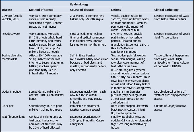
Epidemiology Endemic infection of certain rodents in Europe and east Asia. Cattle are a rare and incidental host. Spread in cattle by contact
Clinical findings Typical pox lesions on the teats and udder. Erythema, papules with a zone of hyperemia around the base, vesiculation, pustular stage and scab
Clinical pathology Electron microscopy
Diagnostic confirmation Electron microscopy and virus isolation
Cowpox virus is a member of the genus Orthipoxvirus in the family Poxviridae. Other orthipoxviruses infecting agricultural animals include horsepox, Uasin Gishu, buffalopox and camelpox.1 All orthipoxviruses are antigenically extremely similar, but they can be identified by a combination of phenotypic and genetic tests.
Cowpoxvirus could be considered to be misnamed. Infection with this virus is endemic in wild rodents such as voles (Microtus spp.) in Great Britain, Europe and western Asia, with infection in different rodent species acting as the reservoir host in different geographic areas.2,3 Domestic cats are commonly infected from hunting rodents, but cowpoxvirus infection can occur in a number of different mammalian species,2 one of which is cattle. The clinical syndrome of cowpox in cattle is now extremely rare but it occurs sporadically in Europe. Seroprevalence in British cattle is less than 1%.
The origin of infection is most probably from infected farm cats or humans. Transmission from cow to cow within a herd is effected by milkers’ hands or teat cups. Spread from herd to herd is probably effected by the introduction of infected animals, by carriage on milkers’ hands, and in the absence of either of the above methods, transport by biting insects is possible. In a herd in which the disease is enzootic, only heifers and new introductions develop lesions. Milkers recently vaccinated against smallpox may serve as a source of infection for cattle, although the vaccinia virus, the smallpox vaccine virus, is a different virus.
It is generally assumed that the virus gains access to tissues through injuries to teat skin, and extensive outbreaks of cowpox are likely to occur when the environment is conducive to teat injuries. Spread is rapid within a herd and immunity is solid, so that the disease tends to occur in sharp outbreaks of several months’ duration with subsequent immunity protecting the cattle for at least several years.
In cowpox, the five stages of a typical pox eruption can be observed. After an incubation period of 3–6 days, a roseolar erythema is followed by firm, raised papules light in color but with a zone of hyperemia around the base. Vesiculation, a yellow blister with a pitted center, follows. The subsequent pustular stage is followed by the development of a thick, red, tenacious scab.
In experimentally produced vaccinia virus mammillitis (produced by inoculation of smallpox vaccine), the lesions have three zones: a central brown crusty area of necrosis, surrounded by a gray-white zone of microvesicle formation, again surrounded by a red border due to congestion. The lesions are essentially hyperplastic.
Typical lesions may be seen at any stage of development, but are mostly observed during the scab stage, the vesicle commonly having been ruptured during milking. True cowpox scabs are 1–2 cm in diameter and are thick, tenacious, and yellow-brown to red in color. In cows being milked, scab formation is uncommon, the scab being replaced by a deep ulceration.
Distribution of the lesions is usually confined to the teats and lower part of the udder. Soreness of the teats develops and milk letdown may be interfered with; the cow usually resents being milked. Secondary mastitis occurs in a few cases. Individual lesions heal within 2 weeks, but in some animals fresh crops of lesions may cause the disease to persist for a month or more. In severe cases, lesions may spread to the insides of the thighs, and rarely to the perineum, vulva and mouth.6 Sucking calves may develop lesions about the mouth. In bulls, lesions usually appear on the scrotum.
The virus can be propagated in tissue culture, and differentiation is possible by electron microscopy.7
A number of skin diseases may be accompanied by lesions on the udder and can easily be confused with cowpox if the lesions are advanced in age. The differential points are listed in Table 22.2. Most outbreaks of teat skin disease that clinically resemble classical cowpox are associated with vaccinia virus from contact with a recently vaccinated person.
Prevention of spread is difficult, since the virus responsible for the disease is readily transmitted by direct or indirect contact. Udder cloths, milking machines and hands should be disinfected after contact with infected animals. Dipping of the teats in an alcoholic tincture of a suitable disinfectant, such as quaternary ammonium compounds, is usually satisfactory in preventing immediate spread. The prevalence and significance of the disease in cattle is too low to warrant the development of vaccines.7
1 Smith SA, Kotwal GJ. Crit Rev Microbiol. 2002;28:149.
2 Bennett M, Baxby D. J Med Microbiol. 1996;45:157.
3 Hazel SM, et al. Epidemiol Infect. 2000;124:551.
4 Baxby D, et al. Br J Dermatol. 1994;131:598.
5 Bennett M, Baxby D. Vet Ann. 1995;35:229.
6 Yerahum I, et al. Rev d’Elev Med Vet Pays Trop. 1996;49:299.
Epidemiology Primarily affects cows in early lactation. Low, but progressive, morbidity in herd. Spread during milking
Clinical findings Vesicles, pustules, formation of a thick scab elevated by granulating tissue. Horseshoe-shaped ring of small scabs surrounding granulating tissue
Clinical pathology Vesicle fluid for electron microscopy
Diagnostic confirmation Electron microscopy
Pseudocowpox virus is a member of the genus Parapoxvirus, with close similarity to the viruses of infectious papular stomatitis and contagious ecthyma. It is possible that Parapoxvirus may be a single virus adapted to different species of ruminants.
Pseudocowpox is reported from most countries. In an affected herd the rate of spread is relatively slow and may result in the disease being present in the herd for up to a year. The morbidity rate approximates 100%, but at any given time varies between 5 and 10%, and occasionally up to 50%.
The source of infection is infected cattle. The method of transmission includes physical transport by means of contaminated milkers’ hands, wash cloths, and teat cups. The virus cannot penetrate mucosa, and a pre-existing discontinuity of it is necessary for the virus to gain entry. Transmission by biting insects seems likely. The virus can be isolated from the mouths of calves sucking affected calves, and from the semen of bulls.
Freshly calved and recently introduced cattle are most susceptible, but all adult cattle in a herd, including dry cows, are likely to be affected. The disease does not appear to occur in animals less than 2 years of age unless they have calved. There is no seasonal variation in incidence. Little immunity develops and the disease is likely to recur in the herd within a short time.1
The disease can be reproduced by the introduction of the virus onto scarified areas of skin. The lesions are characterized by hyperplasia of squamous epithelium.
Acute and chronic lesions occur, and there may be up to ten lesions on one teat. Acute lesions commence as erythema followed by the development of a vesicle or pustule, which ruptures after about 48 hours, resulting in the formation of a thick scab. Pain is moderate and present only in the pre-scab stage. The scab, varying in size from 0.5 to 25 cm in diameter, becomes markedly elevated by developing granulating tissue beneath it; 7–10 days after lesions appear the scabs drop off, leaving a horseshoe-shaped ring of small scabs surrounding a small, wart-like granuloma, which may persist for months. The disease tends to disappear from a herd after 18–21 days but may recur cyclically about 1 month later. There are reports of lesions occurring occasionally in cows’ mouths.
Chronic lesions also commence as erythema, but progress to a stage in which yellow-gray, soft, scurfy scabs develop. The scabs are readily rubbed off at milking, leaving the skin corrugated and prone to chapping. There is no pain and the lesions may persist for months.
Milkers’ nodules are clinically indistinguishable from human lesions associated with ecthyma virus. The lesions vary from multiple vesicles to a single, indurated nodule.
Material for examination by tissue culture or electron microscopic examination, the latter being highly recommended as a diagnostic procedure, should include fluid from a vesicle.
Locally applied ointments of various kinds appear to have little effect on the lesions. The recommended treatment includes the removal of the scabs, which should be burned to avoid contaminating the environment, application of an astringent preparation, such as triple dye, after milking and an emollient ointment just before.
Recommended measures, such as treatment and isolation of affected cows, or milking them last, the use of disposable paper towels for udder washing, and disinfection of teat cups, appear to have little effect on the spread of the disease. An iodophor teat dip is recommended as the most effective control measure.2 An effort should be made to reduce teat trauma because infection is facilitated by discontinuity of the skin.
Etiology Bovine herpesvirus-2, rarely bovine herpesvirus-4
Epidemiology Occurs as an outbreak in cows, particularly heifers, usually within 2 weeks after calving. Commonly followed by persistent infection in the herd
Clinical findings Lesions confined to teats and udder. Vesicles leading to sloughing of skin and necrotic ulceration. Prolonged clinical course
Clinical pathology Virus isolation and electron microscopy on fresh lesions, serology
Treatment Antiseptic and emollient ointments
Control Isolation and milking hygiene, but not effective. Control of periparturient udder edema
The causative virus, bovine herpesvirus-2 (BHV-2), is an alphaherpesvirus. Infection with BHV-2 can produce two distinct syndromes in cattle, bovine herpes mammillitis where there are vesicular and erosive lesions with necrotic ulceration on the skin of the udder and teats and pseudo-lumpy skin disease (Allerton virus) manifest with generalized superficial skin lesions over the body. Pseudo-lumpy skin disease is uncommon. The difference in clinical manifestations between the two diseases may be due to the strain of the virus, or the method of infection. BHV-4 (DN599 strain), which is usually associated with respiratory disease in cattle, is also capable of causing mammary pustular dermatitis.1
Herpes mammillitis is recorded in North America, Australia, Europe and Africa, but probably has widespread occurrence. Herds infected for the first time have a high morbidity rate. Subsequently, the incidence is low and is limited to fresh heifers. The morbidity rate varies between 18 and 96%, susceptible herds recording more than 30% affected. The mortality rate is negligible.
Introduction of bovine ulcerative mammillitis into a herd may occur with the introduction of infected animals, but outbreaks have been observed in self-contained herds. Spread within the herd is probably by direct and fomite-mediated transmission, although experimentally the virus must be deposited in the deep layers of the skin, and even rubbing it into pseudocowpox lesions is not an efficient way of transmitting the disease. Milking machine liners, hands, and udder cloths may act as carriers of virus when a large amount of it is being released.
Seasonal and circumstantial evidence in the United Kingdom and Australia suggest an insect vector, but this has not been confirmed by attempts at transmission with the stable fly (Stomoxys calcitrans), and in the mid-west of the United States, disease is more common in the winter months between November and April.2
Survival of the virus for long periods in carrier animals occurs and it is thought that this may be the means of survival of the virus within a herd that has become immune.3 Infection in some cows is suspected to result in chronic infection at the teat end with the cows becoming ‘hard milkers’ and carriers of the disease.2
Lesions are most common in animals within the first 2 weeks after calving, particularly in heifers, and the disease is more severe in heifers. Heifers that have udder edema at calving are particularly prone to develop severe lesions. Occasionally, lesions may be seen on the teats of replacement heifers and calves sucking infected dams often develop mouth lesions.
The disease is usually self-limiting, persisting in a herd for 6–15 weeks, the severity of the lesions decreasing as the outbreak progresses. Immunity appears to last about a year, herds infected naturally can suffer recurrences a year later. Large herds may have persisting disease, particularly in heifers.
The virus is relatively resistant to environmental influences and can survive freezing. It is susceptible to iodophor disinfectants and less so to hypochlorites.
Forms of loss include a much higher incidence of mastitis, reduction in milk in affected herds by up to 20%, the culling of some cows because of severe mastitis and of heifers because of intractable ulcers, and a great deal of interference with normal milking procedure.
There are anecdotal reports of herpetic lesions in farmers exposed to infected cattle.4
Typical clinical lesions and histopathological changes can be produced locally by introduction of the virus into scarifications of the skin of the teat and the oral mucosa, and by ID and IV injection. There is no viremia and spread is by local extension. In contrast to the poxes, the characteristic lesion in mammillitis is destructive. The higher incidence of the disease and the greater severity of the lesions close to calving are thought to be due to the immunosuppression caused by parturition and to greater predisposition from periparturient udder edema.
There is an incubation period of 5–10 days. There is no systemic illness, and lesions are confined to the teats and udder. When the disease occurs in a herd for the first time the first case is usually in a cow that has calved during the previous 2–3 days. Rapid lateral spread then occurs to other cows.
In cows calved more than a few weeks previously, the characteristic lesions are almost entirely confined to the skin of the teats; in recently calved cows they are restricted to the skin of the teats and the udder. The severity of the disease in recently calved cattle appears to be directly proportional to the degree of postparturient edema which is present. Vesicles occur but are not commonly seen. They are characteristically thin-walled, 1–2 cm in diameter, variable in outline, and often commence at the base of the teat and spread over much of the udder surface. Rupture and confluence of the vesicles leads to weeping and extensive sloughing of the skin.
In the most severe cases, the entire teat is swollen and painful, the skin is bluish in color, exudes serum and sloughs, leaving a raw ulcer covering most of the teat. In less severe cases, there are raised, deep red to blue, circular plaques, 0.5–2 cm in diameter, which develop shallow ulcers. In most cases, scab formation follows but machine milking causes frequent disruption of them, resulting in frequent bleeding. The least severe lesions are in the form of lines of erythema, often in circles and enclosing dry skin or slightly elevated papules, which occasionally show ulceration. Mild lesions tend to heal in about 10 days but severe ulcers may persist for 2 or 3 months. The severity of lesions on the teats on longer-calved cows varies, but in all cases the lesions are sufficiently painful to make milking difficult. Lesions on the skin of the udder heal more rapidly because of the absence of trauma.
Ulcers in the mouth of affected cows have been observed rarely, and calves sucking affected cows develop lesions on their oral mucosae and muzzles. Ulcerative lesions on the vaginal mucosa have been recorded rarely. During the recovery phase there is obvious scar formation and depigmentation.
The generalized skin disease associated with BHV-2, ‘pseudo-lumpy skin disease’ is discussed in the differential diagnosis of lumpy skin disease.
Material for tissue culture, electron microscopy, or cutaneous transmission tests is best obtained by syringe from early vesicles, or as swabs from early ulcers or oral lesions. The virus may be difficult to demonstrate if the lesions are as old as 7 days, and if there has been intensive application of teat disinfectants such as iodophors.
Serology is more commonly used for diagnosis. The presence of high virus-neutralizing antibody titers in serum taken during the acute phase of the disease, and a four-fold increase or decrease in titer in paired samples, are all supportive for diagnosis.3,5 Titers of 1:16 or higher for BHV-2 and 1:20 for BHV-4 indicate exposure. Antibody to both viruses should be tested for diagnostic purposes.2
Necropsy is not commonly performed and no necropsy reports of cases of bovine ulcerative mammillitis are available.
There is no specific treatment and the aim should be to develop scabs that can withstand machine milking. This is most easily effected by the application of a water-miscible, antiseptic ointment just before putting the cups on, followed by an astringent lotion, such as triple dye, immediately after milking. Crystal violet dyes have an excellent reputation as treatments.
Isolation of affected animals and strict hygiene in the milking parlor are practiced but have little effect on the spread of the disease, nor does milking heifers first. An iodophor disinfectant is recommended for use in the dairy to prevent spread. Reducing the incidence and severity of periparturient edema in heifers may reduce the severity of herpes mammillitis. Inoculation of the natural virus away from the teats produces a local lesion and good immunity, but the method has not been tested as a control procedure.
Etiology Capripoxvirus. Strains vary in virulence and host specificity
Epidemiology Highly contagious, spread by aerosol, contact and flies. Young and non-indigenous animals more susceptible. Morbidity and case fatality rates are high
Clinical findings Fever, generalized skin and internal pox lesions, lymphadenopathy, mucopurulent nasal discharge, high mortality
Clinical pathology Fluorescent antibody and electron microscopy of biopsy material, serology, virus isolation
Necropsy findings Pox nodular lesions in alimentary and respiratory tract
Diagnostic confirmation Fluorescent antibody staining, virus isolation
Three viruses are named on the basis of their host specificity in natural outbreaks. Sheeppox virus and goatpox virus are mainly highly host specific in natural infections, but exceptions exist and Kenya sheep and goatpox virus, and Yemen and Oman sheep isolates infect both sheep and goats. All are members of the genus Capripoxvirus, are closely related on the basis of genome mapping, and many cross species barriers in experimental infections1-3 although not in natural occurrence. Recombination may occur naturally between isolates from different host species.4,5 The diseases they produce are also collectively called capripox infections.2
Sheeppox and goatpox are prevalent in North and Central Africa, the Indian subcontinent, Middle Eastern countries, China, Southwest Asia and the former Soviet Union. Sporadic outbreaks occur in southern Europe and elsewhere.6 The capripoxvirus infections of small ruminants are the most serious of all the pox diseases in animals. In susceptible flocks and herds morbidity is 75–100% with outbreaks often causing death in 10–85% of affected animals depending on the virulence of the infecting strain.2,6-9
Sheeppox and goatpox are highly contagious. The virus enters via the respiratory tract and transmission commonly is by aerosol infection associated with close contact with infected animals. The virus is present in nasal and oral secretions for several weeks after infection and can live in scabs that have fallen off the animal for several months. Spread can also occur from contact with contaminated materials and through skin abrasions produced iatrogenically or by insects. Capripox has been shown to spread via the bites of Stomoxys calcitrans and the tsetse fly.10
The disease can be transmitted by intradermal, intravenous and subcutaneous inoculation and by virus aerosols.
Both sheep pox and goat pox affect sheep and goats of all ages, breeds and sex but young and old animals and lactating females are more severely affected. In areas where sheeppox is enzootic, imported breeds such as Merinos or some European breeds may show greater susceptibility than the native stock. Young animals are more susceptible.
The virus is resistant to drying and survives freezing and thawing. It is sensitive to extremes of pH and 1% formalin. Sensitivity to heat varies between strains but most are inactivated at 60°C for 60 min.6 Isolates from most regions are host specific but isolates from Kenya, Yemen and Oman naturally infect both goats and sheep. Scabs shed by infected animals remain infective for several months.
Loss is from mortality, abortions, mastitis, loss of wool, skin condemnation and loss of exports. In ewes and does, severe losses may occur if the udder is invaded because of the secondary occurrence of acute mastitis. In some outbreaks, adult sheep are affected with the more severe form of the disease.11 Sheeppox is a potent threat to countries that have big sheep populations, and where the disease does not occur, because of its ineradicability and heavy mortality rate.
Human infections in people handling infected animals are not a consideration.6
During an initial viremia, the virus is deposited in most tissues, including the skin. The development of typical pox lesions, as in vaccinia, is characteristic of the disease. The virus is present in greatest quantities between the 7th and 14th day after inoculation. Passive protection by serum will protect against challenge. Circulating antibody limits spread of infection, but does not prevent replication of virus at the site of inoculation.
In sheeppox in sheep there is an incubation period of 12–14 days. In lambs, the malignant form is the most common type. There is marked depression and prostration, a very high fever and discharges from the eyes and nose. Affected lambs may die during this stage before typical pox lesions develop. These, when they develop, occur on unwooled skin and on the buccal, respiratory, digestive, and urogenital tract mucosae. They commence as papules, then become nodular, occasionally become vesicular, pustular and finally scab. Some progress from nodules to tumor-like masses.12 The mortality rate in this form of the disease may reach 50%. In the benign form, more common in adults, only skin lesions occur, particularly under the tail, and there is no systemic reaction and animals recover in 3–4 weeks. Abortion and secondary pneumonia are complications. Goatpox in sheep is more severe than sheeppox, and lesions occur on the lips and oral mucosa, the teats and udder.
Goatpox in goats is very similar clinically to sheeppox in sheep. Young kids suffer a systemic disease, with lesions spread generally over the skin, and on the respiratory and alimentary mucosae. Adult goats may have systemic disease and extensive lesions,7,10 but in adult goats the disease is usually mild and lesions are as described above for the benign form in sheep. A flat hemorrhagic form of capripox is seen in some European goats and this has a high case fatality.
Diagnosis is based on typical clinical signs combined with laboratory confirmation of the presence of the virus or antigen. Using electron microscopy, large numbers of characteristic ‘sheeppox cells’ containing inclusion bodies and typical capripox virions can be seen in biopsies of the skin. The virus can be cultured in tissue culture but virus isolation as a method of rapid diagnosis is limited by the time it takes for virus cytopathic effects to develop and the need, with some strains, for several blind passages before this develops. Direct fluorescent antibody test is used to detect the presence of pox virus in the edema fluid and the antigen can be detected in biopsies of lymph glands by AGID using specific immune sera. An antigen detection ELISA is also available.
In India a ‘soluble antigen fraction’, which is non infectious, has replaced infectious virus for serological tests which avoids the risk of accidental spread of the virus from diagnostic laboratories and from the postal supply of diagnostic agents.6 However, serological tests such as AGPT, serum neutralization and agar gel diffusion are confounded due to cross reaction with orf virus. A capripox-specific capture-ELISA is reported to have sufficient sensitivity, specificity and speed to have utility in rapid diagnosis using biopsy samples.13 Serological testing can be by virus neutralization, which is specific or by an indirect fluorescent antibody test or an agar gel precipitation (AGPT), however both of the latter cross react with antibody to contagious pustular dermatitis virus. Virus-specific analysis of antibody response by Western blot analysis can differentiate the infections.14
A capripox PCR for detection of antigen is used in some countries which do not have the disease and do not hold live virus.15,16
In the malignant form, pox lesions extend into the mouth, pharynx, larynx, and vagina with lymphadenopathy and a hemorrhagic spleen. Lesions may also appear in the trachea. Lesions in the lung are severe manifesting as lentil sized white pox nodules to a consolidating necrotizing pneumonia. Lesions occasionally reach the abomasum and are accompanied by a hemorrhagic enteritis.
No specific treatment is advised, but palliative treatment may be necessary in severely affected animals.
Control in free countries or regions necessitates prohibition of importation from infected areas, and if the infection is introduced, ring vaccination, the destruction of affected flocks and the quarantine of infected premises should be instituted.
Vaccination with natural lymph has been practised in some affected areas, but is capable of spreading the disease. Natural infection with one capripox strain imparts immunity to all capripox infections and vaccination with a single capripox vaccine will give protection across all species and against all capripox infections.2
A large variety of commercial vaccines is now available, and there is no easy basis for comparison.3 Killed virus vaccines elicit, at best, temporary protection but available live attenuated vaccines appear to give excellent protection for periods greater than 1 year.2,3,17,18 Colostral antibody interferes with response to vaccination before 6 months of age. A subunit capripox virus vaccine has been developed.19
Vaccination in the face of outbreak is unlikely to prevent deaths during the subsequent 2 weeks and, if needle hygiene is poor, may facilitate the spread of the disease.
1 Hosamani M, et al. Virus Genes. 2004;29:73.
2 Carn VM. Vaccine. 1993;11:1275.
3 Bhat PP. Indian J Anim Sci. 1993;63:857.
4 Gershon PD, et al. J Gen Virol. 1989;70:485.
5 Tulman ER, et al. J Virol. 2002;76:6054.
6 Rao TVS, Bandyopadhyay SK. Anim Health Rev. 2000;1:127.
7 Okaiyeto SO, et al. Israel J Vet Med. 1995;50:65.
8 Reddy YR, et al. Indian Vet J. 2003;80:1298.
9 Joshi RK, et al. Indian Vet J. 1999;76:279.
10 Kitching RP, Mellor PS. Res Vet Sci. 1986;40:255.
11 Solyom F, et al. Acta Vet Acad Sci Hungary. 1980;28:389.
12 Afshar A, et al. Can Vet J. 1986;27:301.
13 Ngichabe CK, et al. Vet Rec. 1999;145:231.
14 Chand P, et al. Epidemiol Infect. 1994;113:377.
15 Heine HG, et al. J Immunol Methods. 1999;227:187.
16 Ireland DC, Binepal YS. J Virol Methods. 1998;74:1.
17 Kitching RP, et al. Res Vet Sci. 1987;42:53.
18 Bhanuprakash BK, et al. Trop Anim Hlth Product. 2004;36:307.
Epidemiology Widespread sporadic disease that is generally benign with low morbidity and low mortality in older pigs. High case fatality in congenitally infected and very young sucking piglets. Transmitted mechanically and by the hog louse
Clinical findings Characteristic pox lesions mainly on skin of head, legs and belly
Clinical pathology Demonstration of typical lesions by histology and virus by electron microscopy
Diagnostic confirmation Demonstration of typical lesions by histology and virus by electron microscopy
The cause is swinepox virus, the sole member of the Suipoxvirus genus of Chordopoxvirinae subfamily of Poxviridae.1
Swine pox (pig pox) occurs worldwide where swine are raised and is more common in swine units where there is poor sanitation.
Transmission is by contact transmission and mechanically by the pig louse (Haematopinus suis), and possibly by flies and other insects.2 Young sucking pigs may have lesions on the face, with similar lesions on the udder of the sow, with spread by direct contact. The virus can survive in scab material for several months.
The virus infects only swine and can infect all ages but clinical disease is most commonly seen in young piglets. It is usually a sporadic disease with occasional outbreaks affecting a cluster of litters within a herd and of short duration. Some or all pigs in a litter may show clinical signs.3 The disease may appear apparently spontaneously or may occur only in pigs brought into the contaminated environment of a herd in which the indigenous pigs are immune.
The incidence in individual herds may be high. Mortality is usually low except in very young piglets and congenitally affected piglets where mortality rates can be high.2,3 Congenital infection presents with a low morbidity but high case fatality.3-6 Older animals seem to suffer little ill effect.2
In field cases, the lesions progress through the classical phases of poxvirus infections but do not usually proceed past the pustular or vesicle stage. At this time there is rupture and the formation of scabs which heal and drop off. Congenital infection is believed to occur when naïve pregnant sows become infected and develop viremia with infection of the fetal membranes. Not all fetuses are born affected and compartmentalization of placentas may restrict further uterine spread as occurs with parvovirus infections.3
Small 1 to 1.5 cm diameter papules develop first and may pass through the pustular and vesicular stage very quickly with the formation of red-brown, round scabs. In neonatal pigs, the rupture of many vesicles at one time may cause wetting and scab formation over the cheeks, and conjunctivitis and keratitis are present in many affected animals. In most cases the lesions are restricted to the belly and inside the upper limbs, but may involve the back and sides and sometimes spread to the face. Lesions may coalesce. A slight febrile reaction may occur in the early stages in young animals, and in sucking pigs deaths are observed. In adult pigs, detectable skin lesions are less well defined, restricted to the non haired softer skin areas and frequently do not progress through the developmental stages to form scabs. Congenital swinepox is characterized by striking lesions present in piglets at birth involving the skin and also commonly the tongue and hard palate.3,4-6 Affected piglets were born from healthy sows. Affected piglets may be stillborn or die within a few days after birth.
The diagnosis is confirmed by examination of skin biopsies.2 Focal superficial erosions, marked epidermal hyperplasia with acanthosis, ballooning of epidermal cells, and occasional large eosinophilic intracellular inclusion bodies are present on histological examination.2,4,5 Electron microscopy can be used to detect the viral particles and the virus can be cultivated in primary pig kidney cell tissue culture.2,4,5
The distribution of the pox-like lesions and the association of the disease with louse infestations suggest the diagnosis. Swinepox may resemble swine vesicular disease, which is characterized by vesicles on the coronary bands, lips, tongue, and snout.
Lesions associated with Tyroglyphus spp. mites are usually larger and occur anywhere on the body, and like those of sarcoptic mange, are usually accompanied by itching. The causative mites are detectable in skin scrapings. Ringworm and pityriasis rosea have characteristic lesions that do not itch, occur in older pig than typically does swine pox, and fungal spores are present in scrapings in the former disease.
A vesicular disease with necrosis resembling swine pox has been attributed to infection with parvoviris7 but there is little evidence that parvovirus is a primary skin pathogen.8
1 Afonso CL, et al. J Virol. 2001;76:783.
2 Jubb TF, et al. Aust Vet J. 1992;69:99.
3 Barlow AM, Grist C. Vet J. 2000;46:118.
4 Thibalt S, et al. Swine Health Product. 1998;6:276.
5 Paton DJ, et al. Vet Rec. 1990;127:204.
6 Borst GHA, et al. Vet Rec. 1990;127:61.
Horsepox is a benign disease characterized by the development of typical pox lesions either on the limbs, or on the lips and buccal mucosa.1 The causative ungulate poxvirus is identical to the virus of true cowpox and is transferable to cattle and to humans. Infection of a foal by cowpox was associated with streptococcal septicemia and death.2 Horsepox is rare. It is a benign disease, but badly affected horses become debilitated and occasionally young animals may die. It is spread by contact with infected grooming tools, harness, and by handling. Immunity after an attack is solid.
Typical pox lesions develop in a ‘leg form’, or in a ‘buccal form’. In the ‘leg form’ nodules, vesicles, pustules, and scabs develop, in that order, on the back of the pastern and cause pain and lameness. There may be a slight systemic reaction with elevation of temperature. In the ‘buccal form’ similar lesions appear first on the insides of the lips and spread over the entire buccal mucosa, sometimes to the pharynx and larynx and occasionally into the nostrils. In very severe cases, lesions may appear on the conjunctiva, the vulva, and sometimes over the entire body. The buccal lesions cause a painful stomatitis, with salivation and anorexia as prominent signs. Most cases recover with lesions healing in 2–4 weeks.
Cowpox was associated with severe ulcerative glossitis, stomatitis, esophagitis and gastritis in a foal.2 Other lesions included polyarthritis and nephritis, though these later lesions could have been caused by streptococcal septicemia.
Differential diagnoses include greasy heel, vesicular stomatitis; viral papular dermatitis, molluscum contagiosum3 and Uasin gishu. See Tables 22.3 and 22.4.
Table 22.3 Differential diagnosis of diseases of horses characterized by discrete lesions of the skin only
Table 22.4 Differential diagnosis of diseases of horses characterized by lesions of the skin of the lower limbs only
There is no specific treatment. Local wound care may hasten healing. Because of the contagious nature of the disease, rigid isolation and hygiene in the handling of infected horses is essential. No vaccine is available.
This is a skin disease of horses of the Uasin gishu plateau of Kenya and neighboring areas associated with a pox virus.1 Lesions on the head, neck, and flanks resemble papillomas.2,3 The source of the virus, and its method of transmission, are unknown, although a wildlife host is presumed.1 Various stages of the lesions can be present in the same horse, and lesions may develop and regress intermittently for years.4 There is no specific treatment and no control methods are reported.
Ulcerative dermatosis of sheep is an infectious disease characterized by the destruction of epidermal and subcutaneous tissues, and the development of raw, granulating ulcers on the skin of the lips, nares, feet, legs, and external genital organs. The lesions on the lips occur between the lip and the nostril, those on the feet occur in the interdigital space and above the coronet, and the genital lesions occur on the glans and the external opening of the prepuce of rams, and the vulva of ewes.
A virus, very similar but antigenically different to the ecthyma virus, is the cause of the disease, which is likely to be confused with contagious ecthyma. However, the lesions are ulcerative and destructive rather than proliferative as in ecthyma. It is not highly infectious like bluetongue or sheeppox, and the ‘lip-and-leg’ distribution of the lesions differentiates it from balanoposthitis of wethers, strawberry foot rot, foot rot, and interdigital abscess. The presence of lesions on the glans penis, and their absence from mucosae, the typical ulcerative form of the lesion, the absence of pus and the susceptibility of recovered animals to infection with ecthyma virus are diagnostic features of ulcerative dermatosis.
A morbidity rate of 15–20% is usual, but up to 60% of a flock may be affected. Mortality is low if the sheep are in good condition and the lesions are treated. Physical contact at breeding time seems to be the most probable method of spread.
The lip cutaneous form of this disease is very rare and possibly has disappeared since its original description, or is very uncommon. A clinically similar disease to the genital infection of ulcerative dermatosis, with balanoposthitis and vulvovaginitis, is associated with Mycoplasma mycoides.1
The Phylum Chlamidiae consists of obligate intracellular bacterial pathogens and contains four families of which two have members that are associated with agricultural animal disease. The family Chlamydiaceae and has recently been reclassified into two genera with Chlamydophila abortus, Chlamydophila pecorum and Chlamydophila pneumoniae and Chlamydia suis associated with animal disease. A different family within the Chlamidiae contains Waddlia chondrophila that has been isolated from aborted fetuses.
The prominent farm animal and horse diseases associated with Chlamydiaceae are chlamydial polyarthritis (Cp. pecorum) and chlamydial abortion (Cp. abortus) which are described below, and sporadic bovine encephalomyelitis (Cp. pecorum) which is described elsewhere under that heading. Cp. pecorum may also be an etiological agent in some outbreaks of ovine contagious ophthalmia which is described under that heading.
Other infections and possible diseases associated with these agents and with C. suis and C. pneumoniae are less commonly reported although it is possible that they underdiagnosed.1
The standard method of diagnosis of chlamydial disease is by isolation in embryonated eggs or in cell culture. However, some strains are difficult to grow and require specialist facilities and expertise. Also the complement fixation test, which is the standard serological test for chlamydial disease, lacks sensitivity and specificity because of cross reaction between chlamydial species.1 The organisms are obligate intracellular pathogens and depend on the host cell for energy in the form of adenosine triphosphate (ATP). Outside of the host cell they exist as metabolically inactive elementary bodies, which have a rigid cell wall and are unable to grow or divide. The elementary bodies attach to host cells and are taken in by phagocytosis. Inside the phagosome they become the metabolically active reticulate body (initial bodies), which have flexible cell walls and grow and divide to form an intravacuolar microcolony called a chlamydial inclusion. Nascent elementary bodies are formed, the host cell is lysed, and the elementary bodies are released with the cycle being accomplished in 36 to 96 hours, depending on species. The current development of new and more sensitive and specific molecular and serological tests such as PCR and the use of recombinant major outer membrane proteins as antigens can allow species specific laboratory diagnosis and should allow a better definition of the importance of C. pecorum and C. pneumoniae and C. suis to agricultural animal disease.1,2
In addition to enzootic abortion in ewes Chlamydiaceae have been associated with reproductive inefficiency in swine and cattle.
In swine, infection with Cp. pecorum and Cp. abortus have been associated with increased rates of returns to estrus, abortion, mummification, stillbirth and increased perinatal and neonatal mortality. Some studies of swine in herds with and without a problem of early embryonic death have found no significant difference in the seroprevalence of antibodies to Chlamydiaceae but have found a significant difference in the presence and proportion of Cp. abortus in cervical swabs and in the uterus and oviducts at slaughter. A dual infection with C. suis was also demonstrated in some cervical swabs in these studies but, whereas an association of Cp. abortus and reproductive inefficiency was evident, the contribution of C. suis to reproductive inefficiency was less clear.3-5
Reports of reproductive inefficiency in swine are largely from Germany and Switzerland, but in large industrial piggeries in eastern European countries, in addition to being implicated in severe outbreaks of abortion and production of weak piglets, infections with Chlamydiaceae are reported to produce reproductive disease in boars, polyarthritis and polyserositis in young pigs, arthritis in finishing pigs, and pneumonia and conjunctivitis.6 In contrast other studies suggest that chlamydial infections are endemic in pigs and that the significance of Chlamydiaceae as pathogens in swine remains to be determined.7
In cattle, Cp. abortus is a cause of bovine abortion but much less commonly than in small ruminants and not causing enzootic disease. Infertility and endometritis in cattle has been associated with the organism and has been reproduced experimentally.8 However, there is debate as to the importance of Chlamydiaceae as major reproductive pathogens in cattle as non-clinical infection of the reproductive tract is common. A recent study using PCR found a high (53%) prevalence of Cp. abortus and Cp. pecorum infection in the vagina of virgin heifers.9 A subsequent study established that infection occurred in calves at a very young age. In this study Cp. pecorum infection was fivefold more prevalent than Cp. abortus infection and was most frequently detected by vaginal swabs compared to rectal or nasal swabs.10
Waddlia chondrophila was initially isolated from an aborted calf in the USA and has subsequently been demonstrated in aborted fetuses in other countries. Serological studies suggest that it might have a role in the causation of bovine abortion. Free living amoeba may serve as hosts.11-13
C. suis has been associated with enteric disease in pigs but is also present in the intestines of clinically normal finishing and adult pigs.3,14
C. suis has also been associated with respiratory disease in swine and this has been reproduced experimentally in conventional pigs by aerogeneous challenge.15
Cp. psittaci has also been demonstrated in domestic and wild pigs (Sus scrofa).16,17 Other miscellaneous occurrences are cases of pneumonia in most animal species, including horses,18 cattle, sheep, goats and pigs, and orchitis and epididymitis in male ruminants and intestinal infections in ruminants and pigs.19,20 It is not possible to classify the Chlamydiae associated with some of these reports. The horse respiratory isolate is closely related to human isolates of Cp. pneumoniae.21 Enteritis and diarrhea in calves, from which Cp. pecorum can be isolated, are usually mixed infections and the pathogenicity of the chlamydiophila as a cause of enteritis is open to doubt.
1 Longbottom D. Vet J. 2004;168:9.
2 Hoelzle LE, et al. Vet Microbiol. 2004;103:85.
3 Camenisch U, et al. Vet Rec. 2004;155:593.
4 Eggermann G, et al. Deutsche Tieartzl Wschr. 2000;107:3.
5 Hoelzle LE, et al. Epidemiol Infect. 2000;125:427.
6 Pospischil A, et al. Pig J. 1996;37:9.
7 Vanrompay D, et al. Vet Microbiol. 2004;99:59.
8 Wittenbrink MM, et al. J Vet Med B. 1993;40:437.
9 DeGraves FJ, et al. J Clin Microbiol. 2003;41:1726.
10 JunBae J, et al. J Clin Microbiol. 2004;42:5664.
11 Rurangirwa FR, et al. Int J Syst Bacteriol. 1999;49:577.
12 Dilbeck-Robertson P, et al. J Vet Diag Invest. 2003;15:568.
13 Michel R, et al. Acta Protozool. 2004;43:37.
14 Szeredi L, et al. Vet Pathol. 1996;33:369.
15 Sasche K, et al. Comp Immunol Microbiol Infect Dis. 2004;27:7.
16 Hoetzel H, et al. Vet Microbiol. 2004;103:121.
17 Vanrompay D, et al. Vet Microbiol. 2004;99:59.
18 McChesney SL, et al. Cornell Vet. 1982;72:92.
19 Fukushi H, Hirai K. Am J System Bact. 1992;42:306.
Virulent chlamydia, including Chlamydophila pecorum, can be isolated from the joints of calves, lambs, and foals clinically affected by polyarthritis. The experimental disease in calves begins as a chlamydemia, followed by localization in the joints. In calves, the disease is uncommon but often fatal. In lambs, it is a common disease in sheep feedlots in the United States, but the mortality rate is low.1 The strain associated with arthritis is not common in the United Kingdom2 and polyarthritis in the UK has been associated with a chlamydial strain distinct from Cp. pecorum.3
In pastured sheep, the morbidity may be as high as 80% but deaths are usually less than 1%. In calves, the disease is more commonly sporadic but response to treatment is poor and affected calve are often destroyed on humane grounds. Intestinal infection with Chlamydophila pecorum is common in pastured lambs between 3 and 9 months of age, but is not associated with clinical disease.4
The clinical signs in calves and lambs include gross swelling of most limb joints but especially the larger joints, lameness, stiffness, unwillingness to move, recumbency, depression, conjunctivitis, and fever of 39–42°C (102–108°F). The navel is unaffected but there may be signs caused by localization of the infection in other organs, e.g. pneumonia, encephalomyelitis, and renal abscess. Clinically, the disease is indistinguishable from polyarthritis caused by other infections such as Mycoplasma and Haemophilus spp.
Chlamydophila abortus (previously Chlamydia psittaci bioype1/serotype1) has a tropism for ruminant placenta and causes the disease commonly referred to as ovine enzootic abortion (OEA). The organism causes a similar disease in goats, and while this organism also can produce abortion in cattle, pigs and horses, abortion associated with this organism is not common in these species.
There is considerable genetic diversity amongst strains that cause abortion.1,2
The disease is one of the commonest causes of diagnosed abortion in sheep and goats in the United Kingdom, the United States,3 and in other countries. In the UK it accounts for approximately 45% of abortions, and it is particularly common in lowland flocks that are intensively managed at lambing time. However, its importance varies from country to country. It is an uncommon cause of abortion in Northern Ireland,4 and the disease does not occur in Norway or New Zealand.5
There have been several studies of seroprevalence in Europe which show a high seroprevalence in both domestic and wild ruminants but, until recently, most surveys have used the complement fixation test which is not specific for Cp. abortus and the seroprevalence rates for Cp. abortus in different countries are not well established.6
Infection is introduced into a flock by the purchase of latently infected replacements which usually abort at the end of their first pregnancy.
Within a flock, the major source of infection is the placenta and the uterine discharge of aborting ewes. The main routes of transmission of Cp. abortus are through ingestion of organisms shed in vaginal fluids and placental membranes at the time of abortion or lambing, or through inhalation of aerosols from the environment.7 Pasture and the environment are contaminated by vaginal discharges, placenta and aborted fetuses, and infected ewes shed the organism for a week before aborting and for 2 weeks afterwards. The elementary body of Cp. abortus is resistant to both physical and chemical influences as it is metabolically inactive and the rigid cell envelope is osmotically stable and poorly permeable. As a consequence the organism is believed to survive for several days on pasture and longer in cold weather.
Infection of the ewe lamb may occur at birth, shortly following, or at subsequent lambing periods. Infection of pregnant ewes in early or mid gestation results in either abortion in the final 2–3 weeks of gestation, or the birth of stillborn or weak lambs that frequently die in the first few days of their life. Abortion always appears in the last weeks of gestation regardless of the time of infection. Infection of ewes in the last 5–6 weeks of pregnancy usually leads to the development of a latent infection, where ewes appear to be uninfected until the next lambing season, when they abort. Thus, late pregnant sheep may be infected by contact with aborting ewes but usually do not themselves abort until the next lambing season.
The common pattern of infection and disease is the a small number of abortions in year 1 usually resulting from the introduction of infected replacement ewes, followed by an epidemic abortion storm where up to 35% of ewes abort in the last 3 weeks of gestation or give premature birth to weak or dead lambs. After abortion, the ewes develop a protective immunity and, in endemically infected flocks, 5–10% of the ewes abort annually. Surviving lambs born to infected mothers may be affected by EAE in their first pregnancy.8-10
Sheep that have aborted, subsequently re-breed successfully, do not have further abortions, and the organism is not present in the placenta or vaginal discharge of subsequent pregnancies. However, levels of immunity vary and some may excrete organisms at estrus or seasonally for up to 3 years.11
In chronically infected sheep, persistent infection can be demonstrated in the endometrial cells of the reproductive tract,12 and the organism is excreted in vaginal fluids during estral periods.
Vaginal challenge of ewes at breeding time will result in infection and subsequent abortion, and venereal or passive venereal transmission is a possible route of infection,13,14 but it does not appear to be a common or important route. Chronic infection of the male genital tissues has been recorded and infection may impair fertility in both rams and bulls.8
The epidemiology of abortion with this agent in cattle is unknown but it may transmit to cattle from infected sheep on the same farm.2,15
The disease is readily reproduced experimentally.16,17 Following subcutaneous injection there are no signs of clinical disease other than a modest increase in rectal temperature for two days after infection. There is a systemic antibody response that peaks 2 weeks after infection and then decreases until just before abortion or parturition, when there is a second increase in the antibody levels to Cp. abortus. Experimental infection at 70 to 75 days pregnancy can result in abortion in the last 2 to 3 weeks of pregnancy or the birth of stillborn or live lambs. There is variation in the severity of the placental lesions in experimental infections. Abortion is associated with severe placental lesions but the reason for the variation in severity and fetal manifestations is not known.17
Enzootic abortion is the most common infectious cause of abortion in lowland flocks that are intensively managed at lambing time, and has a major economic impact on agricultural industries worldwide. There are no recent estimates of economic impact but losses in the UK were estimated in the early 1990s at £15–20 million per annum.10
There is some risk for shepherds, and those in allied fields such as abattoir workers, to contract respiratory infection with this organism but the major zoonotic risk is to pregnant women because of the ability of Cp. abortus to colonize the human placenta. Human infection in early pregnancy results in abortion whereas later infection can result in stillbirth or pre-term labor.8,18 Infection is believed by the oral route from infected hands or food following direct handling of infected sheep or goats or infected clothing. Practices at lambing such as mouth to mouth resuscitation of weak lambs or bringing weak lambs into the house to be warmed promote zoonotic spread. Infected placentas and dead lambs should be handled using gloved hands and disposed of by burning or burial.
The organism can be detected in milk both in sheep and in cattle and raw milk could also pose a risk for zoonotic infection.19,20
Following infection, it is thought that the organism resides first in the tonsil, and is then disseminated by blood and lymph to other organs although the site of latent infection is not known. The latent state is maintained under the control of the proinflammatory cytokine interferon-gamma (IFN-γ).21 Release from the latent state during pregnancy is believed due to immune modulation and leads to bacteremia and infection of the placenta.8,10
The organisms invade the trophoblast cells of the fetal cotyledon following which infection spreads to the intercotyledonary regions of the chorion to produce a suppurative necrotic placentitis with impairment of the maternal–fetal exchange of nutrients and oxygen and fetal death and abortion. An inflammatory response in the fetus may also contribute to fetal death.16,17
It is not known why, regardless of the time of infection, pathological changes in the placenta do not commence before 90 days gestation17 or even as late as 120 days.16
There are generally no premonitory indications of the impending abortions Abortion occurs in late pregnancy; ewes appear to suffer no systemic effects, but retained placenta and metritis can be sequel in goats. Additional losses are caused by stillbirths and weak-born lambs and kids which die soon after birth.
A vaginal discharge lasting up to 3 weeks following the abortion is common.
In cattle, the infection causes abortion in the last third of pregnancy. Infected calves born alive may show lethargy, depression, and may be stunted.
Infection in aborting animals can be demonstrated serologically by rising titers in paired serum samples and by culture of the organism.15
The complement fixation test is commonly used to identify flocks free of infection. It has moderately good sensitivity but is not specific for the agent of due to a common antigen shared by all members of the family and also to some Gram-negative bacteria.22 For samples testing positive, a Western blot examining for antibody against specific antigens can be used as a reference test in flock accreditation.23 ELISA tests, based on whole cell chlamydial elementary bodies or extracts of them, have better specificity than the complement fixation test but poorer sensitivity. A comparison of tests in common use in the United Kingdom in the mid 1990s found that none were both highly sensitive and specific.23 ELISA tests that are based on segments of the membrane outer protein or synthetic peptide antigens have greater sensitivity and specificity and are now used in diagnostic, epidemiologic and seroprevalence studies.7,22,24,25
Aborted fetuses typically have no gross abnormalities. Fetal fluid may contain chlamydophilal antibody and although less sensitive than either isolation in McCoy cells or detection of chlamydial LPS antigen, can be of particular use when placenta is not available. Histologically, there may be mononuclear cell infiltration of hepatic portal areas and multifocal areas of hepatitis. In both cattle and sheep, the placenta is critical for diagnosis of the condition. Placental cotyledons are necrotic and hemorrhagic, and the intercotyledonary areas are thickened, edematous, and leathery. This is in direct contrast to the targeting of cotyledons seen with toxoplasmosis. The Chlamydophila organisms can be observed in tightly-packed sheets within the cytoplasm of swollen trophoblasts in formalin-fixed tissue samples, or in direct placental smears via modified Gimenez, Koster’s, or other appropriate staining methods. Well-preserved, fresh placenta should be examined as the organisms are difficult to demonstrate in the fetus. Immunohistochemical stains perform well on formalin-fixed specimens. Most laboratories are reluctant to culture Chlamydophila spp. due to the zoonotic potential of these agents.
Note the zoonotic potential of this organism when handling carcasses or submitting specimens.
Other causes of abortion in cattle and ewes are given in (Tables 18.7 and 18.8).
Ewes that have aborted should be isolated from the rest of the flock. There should be proper hygiene of the lambing areas and in the disposal of aborted materials. Tetracycline has been used in early pregnant sheep within an aborting flock to attempt to reduce subsequent abortions but the efficacy is questionable.
Two types of vaccine are currently available but there is yet no vaccine that has excellent efficiency against this disease.
Killed vaccines, composed of egg-grown or tissue culture grown organisms of one or two strains have been used for several decades. They are variably effective and can reduce the frequency of abortion and the shedding of the organism. However, outbreaks have occurred in vaccinated sheep and strain variation is a possible cause of failure of monovalent vaccines.1
The addition of Freund’s incomplete adjuvant has provided a vaccine that provides better protection26 and it has been recently claimed that specific adjuvants can markedly improve the efficiency of killed vaccines against naturally occurring enzootic abortion.27,28
A live vaccine containing a temperature-sensitive attenuated strain of C. psittaci has shown to engender excellent, but not complete, protection against challenge; with five different abortigenic strains of Cp. abortus there was a marked reduction in the number of ewes aborting and marked increase in live-born lambs compared to controls.29 It is labeled for sheep but not for goats. Concern has been expressed for the zoonotic potential of an attenuated vaccine.27
Recombinant and DNA vaccines have shown disappointing protection in studies with experimental challenge.30,31
Perez-Martinez JA, Storz J. Chlamydial infections in animals. Modern Vet Pract. 1985;66:517-522. 603–608
Aitken I. Chlamydial abortion in sheep. In Practice. 1986;8:236-237.
Nietfeld JC. Chlamydial infections in small ruminants. Vet Clin N Am Food Anim Pract. 2001;17:301.
Longbotton D, Coulter LJ. Animal chlamydiosis and zoonotic implications. J Comp Path. 2003;128:217-244.
Kerr K, et al. Immunopathology of Chlamydophila abortus infection in sheep and mice. Res Vet Sci. 2005;78:1-7.
1 Vretou E, et al. Vet Microbiol. 1996;51:275.
2 Griffiths PC, et al. Br Vet J. 1995;151:683.
3 Kirkbride CA. J Vet Diag Invest. 1993;5:398.
4 Markey BK, et al. Vet Rec. 1993;132:389.
5 Stanislawek W, et al. Surveillance. 1995;224:14.
6 Borela N, et al. Prev Vet Med. 2004;65:205.
7 Amin JD, Wilesmore AJ. Res Vet Sci. 1995;59:136.
8 Longbotton D, Coulter LJ. J Comp Path. 2003;128:217.
9 Entrican G, et al. J R Soc Med. 2001;94:273.
10 Kerr K, et al. Res Vet Sci. 2005;78.
11 Papp JR, et al. Infect Immun. 1994;62:3786.
12 Papp JR, Shewen PE. J Infect Dis. 1996;174:1296.
13 Papp JR, Shewen PE. Infect Immun. 1996;64:1116.
14 Appleyard WT, et al. Vet Rec. 1985;116:535.
15 Holliman A, et al. Vet Rec. 1994;19:50.
16 Navarro JA. Vet Pathol. 2004;41:498.
17 Buxton D, et al. J Comp Pathol. 2002;127:133.
18 Buxton D. Vet Rec. 1986;118:510.
19 JunBae J, et al. J Clin Microbiol. 2004;42:5664.
20 Ongor H, et al. J Vet Med B. 2004;51:43.
21 Brown J, et al. Vet Immunol Immunopathol. 2001;82:107.
22 Buendia AJ, et al. Vet Microbiol. 2001;78:229.
23 Jones E, et al. Vet Rec. 1997;141:164.
24 Kaltenboeck B, et al. J Clin Microbiol. 1997;35:2293.
25 Salti-montesanto V, et al. Am J Vet Res. 1997;58:228.
26 Wilsmore HJ, et al. Br Vet J. 1990;146:341.
27 Garcia JN. Vet Microbiol. 2004;100:65.
28 Caro MR, et al. Vaccine. 2003;21:3126.
29 Clarkson MJ, Phillips HL. Vet J. 1997;153:307.