BRUCELLOSIS ASSOCIATED WITH BRUCELLA ABORTUS (BANG’S DISEASE)

Synopsis

Etiology Brucella abortus

Epidemiology Major cause of abortion in cattle in countries without a national control program. Undulant fever in humans an important zoonosis. Sexually mature animals susceptible; outbreaks occur in first-calf heifers, older cows infected but do not abort. Transmitted directly from infected animal to susceptible animal by uterine discharges. Congenital infection occurs. Infection in wildlife species but significance to domestic animals unknown. Infection introduced into herd by unknown infected carrier animal. Natural infection and vaccination result in immunity to abortion but not infection, and infected animals remain serologically positive for a long time

Signs Abortion epidemics in first-calf unvaccinated heifers after fifth month of pregnancy. Subsequent pregnancies carried to term. Orchitis and epididymitis in bulls. Synovitis (hygromas) occurs. Fistulous withers in horses

Clinical pathology Serology. Serum agglutination test is standard test. Rose Bengal test (rapid screening test). Complement fixation test. ELISA test. Milk ring test. False-positive reactors are major problem

Lesions Necrotizing placentitis, inflammatory changes in fetus

Diagnostic confirmation Culture organism from fetus. Positive serological test in unvaccinated animal

Differential diagnosis list Other causes of abortion (see Table 18.7)

Treatment No treatment

Control Test and reduce reservoir of infection. Quarantine. Depopulation. Vaccination to reduce incidence of abortion and percentage of infected animals. Eradication on herd and area basis by test and slaughter

ETIOLOGY

Brucella abortus is the causative organism and at least nine biotypes have been recognized including a number of strain variants. Approximately 5% of infections are from biotype 1. Biotype 2 was isolated in an outbreak of brucellosis in cattle in Canada in 1986. In the USA biotypes 1–4 are found.

EPIDEMIOLOGY

Occurrence and prevalence of infection

Brucellosis is an important disease of cattle and an important zoonosis worldwide. It is of major economic importance in developing countries that have not had a national brucellosis eradication program (see Introduction, above). The prevalence of infection varies considerably among herds, areas, and countries. Many countries have made considerable progress with their eradication programs and some have eradicated the disease. However, in other countries brucellosis is still a serious disease problem facing the veterinary and medical professions. In Argentina, for example, the prevalence of infection in cattle is more than 10% and it is estimated that 20 000 new cases of human brucellosis occur annually.

Cattle

Infection occurs in cattle of all ages but is most common in sexually mature animals, particularly dairy cattle. Abortions occur most commonly in outbreaks in unvaccinated heifers after the fifth month of pregnancy. Bulls are affected with orchitis, epididymitis, and seminal vesiculitis. The infection has been confirmed in cattle and water buffaloes in Trinidad.1

Wildlife species

The infection has occurs in bison (Bison bison),2,3 elk (Cervus elaphus canadensis),4 deer, coyotes, wild opossums and raccoons, moose, and other wild and domesticated ruminants, but there is no evidence that these species are a source of infection for cattle. Infection of moose with B. abortus biovar 1 is highly fatal and it is likely that the moose is a dead-end host for brucellosis. Experimental inoculation of the organism into badgers results in the development of antibodies and elimination of the organism, which indicates that the badger is relatively resistant to infection and unlikely to be a reservoir of the organism.

Bison and elk are potential reservoirs of brucellosis and, because they are the species of choice for game farming, which is a recent development in North America and elsewhere, they could serve as a source of infection for cattle. Brucellosis associated with B. abortus was first detected in bison (Bison bison) in Yellowstone National Park in 1917 and has been present ever since.5 Bison can remain latently infected with virulent B. abortus until attainment of reproductive age despite extensive use of vaccination and serological testing.6

A strain of biotype 1 isolated from bison in Canada is infectious and contagious for cattle. While bovine brucellosis is being effectively eradicated in North America, infected populations of bison, elk, and moose in Canada and the USA are of sufficient size and geographical distribution to create a real and serious threat to the livestock producers in certain areas of both countries.5 B. suis biovar 4 is the cause of rangiferine brucellosis, a disease of reindeer and caribou (Rangifer tarandus).7 Under natural conditions, rangiferine brucellosis is limited to the circumpolar Arctic but translocations to reindeer for game ranching may place traditional livestock species, game-farmed species and non-Arctic wildlife at risk. Naturally infected reindeer can transmit B. suis biovar 4 to brucellosis-free cattle.7 Exposed cattle had a serological response; the organism remained in tissues 60 days after exposure. B. suis biovar 4 does not cause clinically important disease in cattle and is not shed in sufficient numbers to maintain infection in cattle populations. Conventional serological tests can be used to detect B. suis biovar 4 infection in cattle, but their ability to detect exposed animals varies widely and they cannot discriminate between B. suis biovar 4 and B. abortus of cattle. Cattle to cattle transmission of B. suis biovar 4 is unlikely.7

In Canada, the wood bison herd in Wood Buffalo National Park has been infected with Mycobacterium bovis and B. abortus since 1925.8 The infected herd poses a potential threat to the nearby MacKenzie Bison Sanctuary, in which these infections are not present.

Horses

In horses the organism is often found in chronic bursal enlargements as a secondary invader rather than a primary pathogen. It is commonly present with Actinomyces bovis in fistulous withers and poll evil. It has also been identified as a cause of abortion in mares. A serological survey of horses over a period of 8 years revealed that 8–16% of serum samples were positive. However, experimentally infected horses do not excrete the organism in sufficient numbers to infect susceptible in-contact cattle.

Pigs and sheep

The organism can be recovered from naturally infected pigs and, although not normally pathogenic in this species, may occasionally cause abortion. The disease occurs naturally in sheep exposed to infected cattle, which has significant implications for brucellosis eradication.

Dogs

Naturally acquired B. abortus infection can occur in dogs associated with infected cattle.9 While farm dogs are not generally considered to be a major reservoir of B. abortus, the organism has been isolated from dogs on a farm where several cattle were serologically positive for brucellosis and dogs should be included in any investigation and eradication of the disease.

Methods of transmission

Parturition

The risk posed to susceptible animals following parturition of infected cattle depends on three factors:

The number of organisms excreted

The survival of these organisms under the existing environmental conditions

The probability of susceptible animals being exposed to enough organisms to establish infection.

The organism achieves its greatest numbers in the contents of the pregnant uterus, the fetus and the fetal membranes, all of which must be considered as major sources of infection. The numbers of organisms in the tissues of two naturally infected cows and their fetuses were as follows: umbilicus 2.4 × 10⁸ – 4.3 × 10⁹/g – 1.4 × 10¹³/g. This illustrates the potentially large numbers of organisms that can be shed and to which other animals and humans are potentially exposed. However, the numbers of organisms decrease when uterine discharges are cultured at sequential parturitions, and a substantial number of uterine samples from infected cows are culture-negative at the second and third parturition following challenge.

Transmission

The disease is transmitted by ingestion, penetration of the intact skin and conjunctiva, and contamination of the udder during milking. The organism does not multiply in the environment but merely persists, and the viability of the organism outside the host is influenced by the existing environmental conditions. Grazing on infected pasture, or consuming other feedstuffs and water supplies contaminated by discharges and fetal membranes from infected cows, and contact with aborted fetuses and infected newborn calves are the most common methods of spread.

Intraherd spread occurs by both vertical and horizontal transmission. Horizontal transmission is usually by direct contamination and, although the possibility of introduction of infection by flies, dogs, rats, ticks, infected boots, fodder, and other inanimate objects exists, it is not significant relative to control measures. The organism is ingested by the face fly but is rapidly eliminated and there is no evidence for a role in natural transmission. Evidence exists for horizontal, dog-to-dog, cattle-to-dog, dog-to-cattle and dog-to-human transfer of infection.9 The most likely and effective means of cattle-to-dog transfer is exposure to aborted fetuses or infected placental membranes, because dogs commonly ingest the products of parturition.

Spread between herds

Movement of an infected animal from an infected herd to a susceptible noninfected herd is a common method of transmission. The rate of spread will depend on the level of surveillance testing.10 In Great Britain, which is officially brucellosis free, 20% or more of both beef and dairy cattle more than 24 months old are tested routinely. A simulation model indicates that reducing the level of testing would have a major effect on the rate of spread of infection, should it be imported.

Spread between countries (breach of biosecurity)

A quantitative risk assessment model to determine the annual risk of importing brucellosis-infected breeding cattle into Great Britain from Northern Ireland and the Republic of Ireland, which are not brucellosis free, was developed.11 Predictions estimated that brucellosis could be imported from Northern Ireland every 2.63 years and from the Republic of Ireland every 3.23 years. Following this assessment, the Department of Environment, Food and Rural Affairs introduced postcalving testing for all imported breeding cattle. Under this system, all imported animals are issued a passport that records their age and pregnancy status. This information enables identification of animals that require testing and provides an additional safeguard in maintaining official brucellosis status.

Congenital infection

Congenital infection may occur in calves born from infected dams but its frequency is low. The infection occurs in utero and may remain latent in the calf during its early life; the animal may remain serologically negative until its first parturition, when it then begins to shed the organism. Calves born from reactor dams are serologically positive for up to 4–6 months because of colostral antibodies and later become serologically negative even though a latent infection may exist in a small proportion of these calves. The frequency of latent infections is unknown, but may range from 2.5–9%. Latent infections in serologically negative animals are of some concern because they remain unnoticed and can potentially serve as a source of infection later. However, latent infections in calves born from infected cows are infrequent. The organism could not be isolated from any of 150 calves born to infected cows, 135 of which were experiencing their first pregnancy after infection. In one report a heifer from a herd affected with widespread infection with B. abortus biotype 2 was moved to a brucellosis-free herd and remained apparently free from brucellosis until 9 years later, when the same animal produced a strongly positive serological reaction and the same biotype was isolated from its milk. Such observations have resulted in the recommendation that calves from seropositive dams should not be used for breeding. Even vaccinated heifers from seropositive dams can harbor a latent infection. There is a risk that 2.5% of heifer calves born from serologically positive dams will react in early adulthood and constitute a threat to a re-established herd.

Survival of organism

The organism can survive on grass for variable periods depending on environmental conditions. In temperate climates, infectivity may persist for 100 days in winter and 30 days in summer. The organism is susceptible to heat, sunlight, and standard disinfectants but freezing permits almost indefinite survival. The activity of several disinfectants against B. abortus has been examined, and representatives of the phenolic, halogen, quaternary ammonium, and aldehyde groups of disinfectants at 0.5% or 1.0% concentrations in the absence of serum generally inhibited a high concentration of the organism.

Uterine discharges and milk

A cow’s tail heavily contaminated with infected uterine discharges may be a source of infection if it comes in contact with the conjunctiva or the intact skin of other animals. In the same way that the more common forms of mastitis can be spread during milking, B. abortus infection can be spread from a cow whose milk contains the organism to an uninfected cow. This may have little significance in terms of causing abortion but it is of particular importance in its effects on agglutination tests on milk and the presence of the organism in milk used for human consumption.

Bulls and semen

Bulls do not usually transmit infection from infected to noninfected cows mechanically. Infected bulls may discharge semen containing organisms but are unlikely to transmit the infection. The risk of spread from the bull is much higher, however, if the semen is used for artificial insemination. Some infected bulls are negative on serum agglutination tests and their carrier status can only be detected by the isolation of organisms from the semen or agglutination tests on seminal plasma.

Carrier cows

Few infected cows ever recover from infection completely and should be considered as permanent carriers whether or not abortion occurs. Excretion of the organism in the milk is usually intermittent, is more common during late lactation and can persist for several years. In cattle vaccinated before infection the degree of excretion of B. abortus in the milk is less than in nonvaccinated animals. Embryo transfer from infected donors may be achieved without transfer of infection and superovulation is unlikely to reactivate the release of Brucella into the uterus during the period when embryos are normally collected. Thus embryo transfer is a safe procedure for salvaging genetic material from infected animals.

The herd characteristics and the results of the first herd test may be used as predictors of the potential presence or absence of B. abortus in herds with reactors to the tube agglutination test. The presence of only single suspicious reactors on the first test is a reliable predictor of lack of infection. The presence of one or more positive reactors on the first herd test is a reliable predictor of the presence of infection.

Risk factors

The risk factors that influence the initiation, spread, maintenance, and/or control of bovine brucellosis are related to the animal population, management, and the biology of disease.12 The variables that contribute significantly to seropositive animals are:

Size of farm premises

Percentage of animals on a premises that are inseminated artificially

Size of investment in livestock

Number of cows which aborted in the previous year, whether or not dairying is the major agricultural activity of the premises

Policy of the owner with regard to disposal of reactor animals.

The longer infected animals are in contact with the remainder of the herd, the greater will be the ultimate number of seropositive animals. In a defined geographical area in northern Mexico where a brucellosis control program did not exist, the greatest percentage of seropositive animals was related to larger farms, poor artificial insemination technique, and small financial investment in the farm.

From a practical viewpoint, the factors influencing the transmission of brucellosis in any given geographical region can be classified into two fundamental categories: those associated with the transmission of disease between herds and those influencing the maintenance and spread of infection within herds. Factors influencing interherd transmission include the purchase of infected replacement animals and are in turn influenced by frequency of purchase, source of purchase, and brucellosis test history of purchased animals. The proximity of infected herds to clean herds is an important risk factor. Cattle contacts at fence lines, sharing of pastures and strays of infected animals into clean herds are common methods by which transmission occurs to adjacent herds.

The risk factors associated with spread of the disease within a herd include unvaccinated animals in infected herds, herd size, population density, method of housing, and use of maternity pens. Large herd sizes are often maintained by the purchase of replacement cattle, which may be infected. It is also more difficult to manage large herds, which may lead to managerial mistakes that allow the disease to spread. There is a positive association between population density (number of cattle to land area) and disease prevalence, which is attributed to increased contact between susceptible and infected animals. The use of maternity pens at calving is associated with a decrease in the prevalence of infection, presumably due to decreasing the exposure of infected and susceptible animals.

Animal risk factors

Susceptibility of cattle to B. abortus infection is influenced by the age, sex, and reproductive status of the individual animal. Sexually mature, pregnant cattle are more susceptible to infection with the organism than sexually immature cattle of either sex. Natural exposure to field strains occurs primarily at the time of parturition of infected cows. The greater the number of infected cows that abort or calve, the greater the exposure risk to the other cattle in the herd. An important application of this observation is that infected cows need to be removed from the herd prior to parturition. Thus the stage of gestation and parturition are more important factors in herd plans than early removal of postparturient infected cows following whole-herd strain-19 vaccination. Young cattle are less susceptible to B. abortus than older, sexually mature cattle. Susceptibility appears to be more commonly associated with sexual maturity than age. Young, sexually immature cattle generally do not become infected following exposure, or recover quickly. Susceptibility increases with pregnancy and as the stage of gestation increases.

The probability of isolation of the organism at parturition increased from 0.22 to 0.90 as fetal age at exposure of nonvaccinated heifers increased from 60 to 150 gestation days. The stage of pregnancy at experimental challenge exposure of strain-19 vaccinated heifers is positively associated with the proportion of animals that become infected. Nonvaccinated young cattle are also at high risk of brucellosis if exposed to pathogenic strains of the organism. In cattle vaccinated as yearling heifers, the risk of brucellosis is also related to the number of organisms in the vaccine. The risk of brucellosis in heifers vaccinated with 108, 109, or 1010 of strain 19 was, respectively, one-third, one-seventh, or one-17th that of diluent controls or nonvaccinated heifers.

Management risk factors

The spread of the disease from one herd to another and from one area to another is almost always due to the movement of an infected animal from an infected herd into a noninfected susceptible herd. The unregulated movement of cattle from infected herds or areas to brucellosis-free herds or areas is the major cause of breakdowns in brucellosis eradication programs. A case-control study of brucellosis in Canada indicated that herds located close to other infected herds and those herds whose owners made frequent purchases of cattle had an increased risk of acquiring brucellosis. Once infected, the time required to become free of brucellosis was increased by large herd size, by active abortion and by loose housing.

Pathogen risk factors

B. abortus is a facultative intracellular parasite capable of multiplication and survival within host phagocytes.13 The organisms are phagocytosed by polymorphonuclear leukocytes in which some survive and multiply. These are then transported to lymphoid tissues and fetal placenta. The inability of the leukocytes to effectively kill virulent B. abortus at the primary site of infection is a key factor in the dissemination to regional lymph nodes, other sites such as the reticuloendothelial system, and organs such as the uterus and udder. The organism is able to survive within macrophages because it has the ability to survive phagolysosome. The bacterium possesses an unconventional non-endotoxic lipopolysaccharide, which confers resistance to antimicrobial attacks and modulates the host immune response.14 These properties make lipopolysaccharide an important virulence factor for Brucella survival and replication in the host.

Brucellas are able to survive within host leukocytes and may utilize both neutrophils and macrophages for protection from humoral and cellular bactericidal mechanisms during the periods of hematogenous spread. The placenta is a favored site for replication of the organism. Large numbers of the organism can be found in chorionic trophoblasts, which contain metabolically active cells capable of producing a variety of hormones and secretory proteins that may stimulate the growth of brucellas. The ability to survive in the host may explain both the transitory titers occurring in some animals following isolated episodes of bacteremia and the disappearance of titers in animals with latent infection.

Three major groups of outer membrane proteins have been identified in Brucella spp.15 Certain mutants of B. abortus lack a major 25 kDa outer membrane protein (Omp25), which renders them unable to replicate efficiently in bovine phagocytes and chorionic trophoblasts.

Immune mechanisms

Immunity against brucellosis is principally mediated by cellular immune responses since it is an intracellular pathogen. B. abortus is an efficient inducer of type 1 cellular immune responses and interferon-gamma is crucial for control of brucellosis.16 Infections are chronic and often lifelong. The bovine T lymphocyte in brucellosis is a critical component of host defense based on mononuclear phagocyte activation by interferon-gamma. The killing of Brucella-infected mononuclear phagocytes and interferon-gamma-mediated activation of mononuclear phagocytes are the major mechanisms of host defenses against brucellosis in cattle.17

The antibody response to B. abortus in cattle consists of an early IgM isotype response, the timing of which depends on the route of exposure, the dose of the bacteria and the health status of the animal. The IgM response is followed almost immediately by production of IgG1 antibody and later by small amounts of IgG2 and IgA. Most cross-reacting antibody from exposure to other bacteria other than Brucella spp. or environmental antigens consists mainly of IgM. Serological tests that measure IgM are therefore not desirable, as false-positive results occur. Since IgG2 and IgA antibodies accumulate later after exposure and are usually present in small and inconsistent amounts, the main isotype for serological testing is IgG1.18

Naturally infected animals and those vaccinated as adults with strain 19 remain positive to the serum, and other agglutination tests, for long periods. The serum of infected cattle contains high levels of IgM, IgG1, IgG2, and IgA isotypes of antibody. Most animals vaccinated between 4 and 8 months of age return to a negative status to the test within a year. All are considered to have a relative immunity to infection. Calves from cows that are positive reactors to the test are passively immunized via the colostrum. The half-life of colostral antibodies to B. abortus in calves that have received colostrum from either vaccinated noninfected or infected dams is about 22 days. It is possible that some calves remain immune sufficiently long to interfere with vaccination. After vaccination of cattle with strain 19 of the organism, IgM antibodies appear after about 5 days, reaching peak values after 13 days. IgG1 antibodies appear a little later or simultaneously with IgM, and peak values are reached at 28–42 days, after which they decline. The same general pattern follows experimental infection with virulent strains and also in chronic field cases, except that IgM antibody declines to low levels and residual activity resides in IgG1 and IgG2 as well as in IgA, which remain at higher levels.

Economic importance

Losses in animal production due to this disease can be of major importance, primarily because of decreased milk production in aborting cows. The common sequel of infertility increases the period between lactations, and in an infected herd the average intercalving period may be prolonged by several months. In addition to the loss of milk production, there is the loss of calves and interference with the breeding program. This is of greatest importance in beef herds, where the calves represent the sole source of income. A high incidence of temporary and permanent infertility results in heavy culling of valuable cows, and some deaths occur as a result of acute metritis following retention of the placenta.

Zoonotic implications

Brucellosis is an important zoonosis causing undulant fever in humans.19 According to the Food and Agriculture Organization, the World Health Organization and the Office International des Epizoöties, brucellosis is still one of the most important and widespread zoonoses in the world. The disease spreads mainly by the ingestion of unpasteurized dairy products.19 Officially approved methods of commercial pasteurization render naturally Brucella-contaminated raw milk safe for consumption. However, most cases in humans are occupational and occur in farmers, veterinarians, and butchers. The organism can be isolated from many organs other than the udder and uterus, and the handling of a carcass of an infected animal may represent severe exposure.

The importance of the disease in humans is an important justification for its eradication. Between the years 1965 and 1974 the incidence of brucellosis in humans in the USA increased. Most cases occur in people employed in the meat processing industry, while other sources include the domestic pig, cattle, and unpasteurized dairy products. A majority of human brucellosis cases in Texas could be prevented by prohibiting the importation of unpasteurized goat’s milk cheese. Laboratory-acquired brucellosis in humans constituted 1.7% of the cases of brucellosis reported in the USA between 1964 and 1974.

The cost-effectiveness to human health and the potential net economic benefits of a nationwide mass vaccination program for livestock over a period of 10 years has been evaluated using Mongolia as the model.20 If the costs of vaccination of livestock against brucellosis were allocated to all sectors in proportion to the benefits, the intervention would be cost-effective and would result in net economic benefits.

PATHOGENESIS

B. abortus has a predilection for the pregnant uterus, udder, testicle and accessory male sex glands, lymph nodes, joint capsules, and bursae. After the initial invasion of the body, localization occurs initially in the lymph nodes draining the area and spreads to other lymphoid tissues, including the spleen and the mammary and iliac lymph nodes.

B. abortus is phagocytized by macrophages and neutrophils in an effort by the host to eliminate the organism. However, once inside the phagocyte, B. abortus is able to survive and replicate. The phagocyte migrates via the lymphatic system to the draining lymph node, where Brucella infection causes cell lysis and eventual lymph node hemorrhage 2–3 weeks following exposure. Because of vascular injury, some of the bacteria enter the bloodstream and subsequent bacteremia occurs, which disseminates the pathogen throughout the body. If the infected animal is pregnant, B. abortus will colonize and replicate to high numbers in the chorionic trophoblasts of the developing fetus. The resulting tissue necrosis of the fetal membranes allows transmission of the bacteria to the fetus. The net effect of chorionic and fetal colonization is abortion during the last trimester of pregnancy.

Sexually immature and other nonpregnant cattle can become infected but lose their humoral antibody to the organism much more quickly than cattle infected while pregnant.

In the adult, nonpregnant cow, localization occurs in the udder, and the uterus, if it becomes gravid, is infected from periodic bacteremic phases originating in the udder. Infected udders are clinically normal but they are important as a source of reinfection of the uterus, as a source of infection for calves or humans drinking the milk, and because they are the basis for the agglutination tests on milk and whey.

Erythritol, a substance produced by the fetus and capable of stimulating the growth of B. abortus, occurs naturally in greatest concentration in the placental and fetal fluids and is responsible for localization of the infection in these tissues. Invasion of the gravid uterus results in a severe ulcerative endometritis of the intercotyledonary spaces. The allantochorion, fetal fluids, and placental cotyledons are invaded and the villi are destroyed. The organism has a marked predilection for the ruminant placenta. In acute infections of pregnant cows, up to 85% of the bacteria are in cotyledons, placental membranes, and allantoic fluid.

The successful coexistence of Brucella spp. with its preferred host is the outcome of coevolutionary relationships and selection pressures, which result in a stalemate where the pathogen has evolved to survive within the biological system of the host, and the host has evolved innate and acquired immune systems that allow controlled survival of infection by the pathogen, ultimately supporting the survival of the host–pathogen system.21 In contrast to other pathogens, genes controlling virulence, survival and persistence of Brucella spp. have been documented as the dominant feature of its genome.

The host responses at the organ and tissue levels have been described and are summarized here.21 Lymph nodes draining the sites of the early stages of infection have marked germinal center hyperplasia and hypertrophy, accompanied by acute neutrophilic and eosinophilic lymphadenitis. In the later stages of the infection, lymph nodes draining mammary gland, head, and reproductive tract develop chronic granulomatous lymphadenitis, which is usually associated with cortical and paracortical T-cell-dependent lymphoid depletion, germinal center expansion, and deep histiocytic expansion. The spleen may develop lymphoid hyperplasia and histiocytic and plasmacytic expansion in the germinal centers, and the mammary gland usually has a pronounced interstitial lymphoplasmacytic mastitis. In the uterus, there is usually an endometritis, fibrosing mural lymphocytic metritis, and caruncular necrotizing vasculitis, while the placenta is colonized with B. abortus and has extensive desquamation of fetal chorioallantoic trophoblasts with subsequent hematogenous spread to villous trophoblastic epithelium, and necrotizing fibrinopurulent cotyledonary placentitis of the placental arcades accompanied by granulation and intercotyledonary inflammation exudation. The fetal lesions consist of marked fibrinopurulent necrotizing bronchopneumonia, monocytic and neutrophilic alveolitis, thromboembolic necrotizing arteritis and lymphangitis, fibrinopurulent pleuritis, and granulomata of the liver, spleen, kidney, and lymph nodes.

Variable disease expression may occur in the male reproductive tract and musculoskeletal system of either sex. The affected joints usually develop a fibrinous and granulomatous synovitis with proliferative villous projection formation, proliferative tendovaginitis with lymphoplasmacytic nodule formation, and arthritis with articular erosions, which may be associated with suppurative, granulomatous bursitis. In the testes there are uni- or bilateral visceral to parietal tunica adhesions, interstitial lymphocyte orchitis with seminiferous tubular degeneration, necrotizing intratubular orchitis, and acute fibrinopurulent periorchitis with infarction. The ampulla may have a uni- or bilateral granulomatous epididymitis with focally necrotic purulent, calcified sperm granulomata and the seminal vesicles have uni- or bilateral necrotizing fibrinopurulent seminal vesiculitis and interstitial lymphocytic, plasmacytic seminal vesiculitis with necrosis.

In fetuses naturally and experimentally infected with B. abortus the tissue changes include lymphoid hyperplasia in multiple lymph nodes, lymphoid depletion in the thymic cortex, adrenal cortical hyperplasia and disseminated inflammatory foci composed mainly of large mononuclear leukocytes. The fetal pneumonia is due to localization of perivascular foci in the interlobular septa of the lung, indicative of hematogenous spread in the fetus rather than aspiration of contaminated fetal fluids. Fetuses inoculated with sufficient numbers of B. abortus will abort 7–19 days postinoculation. With experimental conjunctival exposure of pregnant heifers with the organism, the numbers of infected animals and the number of tissue samples positive for the organism are increased as fetal age at exposure increases from gestation days less than 127 to more than 157.

Abortion occurs principally in the last 3 months of pregnancy, the incubation period being inversely proportional to the stage of development of the fetus at the time of infection.

Congenital infection can occur in newborn calves as a result of in utero infection and the infection may persist in a small proportion of calves, which may also be serologically negative until after their first parturition or abortion.

CLINICAL FINDINGS

Abortion

The clinical findings are dependent upon the immune status of the herd. In highly susceptible nonvaccinated pregnant cattle, abortion after the 5th month of pregnancy is the typical feature of the disease in cattle. In subsequent pregnancies the fetus is usually carried to full term although second or even third abortions may occur in the same cow. Retention of the placenta and metritis are common sequelae to abortion. Mixed infections are usually the cause of the metritis which may be acute, with septicemia and death following, or chronic, leading to sterility.

The history of the disease in a susceptible herd can usually be traced to the introduction of an infected cow. Less common sources are infected bulls, or horses with fistulous withers. In a susceptible herd it is common for the infection to spread rapidly and for an abortion ‘storm’ to occur. The ‘storm’ might last for a year or more, at the end of which time most of the susceptible cows are infected and have aborted and then carry their calves to full term. Retained placentae and metritis could be expected to be common at this time. As the abortion rate subsides, the abortions are largely restricted to first-calf heifers and new additions because other animals of the herd acquire partial resistance.

In recent years, particularly in areas where vaccination is extensively practiced, an insidious form of the disease may develop, which spreads much more slowly and in which abortion is much less common.

Orchitis and epididymitis

In the bull, orchitis and epididymitis occur occasionally. One or both scrotal sacs may be affected, with acute, painful swelling to twice normal size, although the testes may not be grossly enlarged. The swelling persists for a considerable time and the testis undergoes liquefaction necrosis and is eventually destroyed. The seminal vesicles may be affected and their enlargement can be detected on rectal palpation. Affected bulls are usually sterile when the orchitis is acute but may regain normal fertility if one testicle is undamaged. Such bulls are potential spreaders of the disease if they are used for artificial insemination.

Synovitis

B. abortus can often be isolated from the tissues of nonsuppurative synovitis in cattle. Hygromatous swellings, especially of the knees, should be considered with suspicion. Progressive and erosive nonsuppurative arthritis of the stifle joints has occurred in young cattle from brucellosis-free herds that had been vaccinated with strain 19 vaccine. The calves may or may not be serologically positive, but synovial fluid and joint tissue samples contain immunological evidence of strain 19 B. abortus antigenic material. The synovitis has been reproduced by intra-articular injection of the vaccine.

Fitulous withers

In horses, the common association of B. abortus is with chronic bursal enlargements of the neck and withers, or with the navicular bursa, causing intermittent lameness, and the organism has been isolated from mares that have aborted.22 When horses are mixed with infected cattle, a relatively high proportion can become infected and develop a positive reaction to the serum agglutination test without showing clinical illness. Some horses appear to suffer a generalized infection with clinical signs including general stiffness, fluctuating temperature, and lethargy.

CLINICAL PATHOLOGY

The major objective in the laboratory diagnosis of brucellosis is to identify animals which are infected and potentially shedding the organism and spreading the disease. Most infected animals are identifiable using the standard serological tests but latent infection occurs in some animals which are serologically negative. Furthermore, vaccinated animals may be serologically positive and uninfected, and transitory titers occur sporadically in a small percentage of animals, for which there is no clear explanation. These diagnostic problems make control and eradication programs difficult to administer and difficult to explain to animal owners.

The collection and submission of samples to the laboratory must be done with care, and careful attention must be given to recording the identity of the animal and the corresponding sample which should be uniquely identified. For blood samples, it is recommended that silicone-coated evacuated glass tubes without additives be used to collect the blood sample, as they insure effective clotting and clot retraction, to provide an easy source of serum without the need for centrifugation. Clotting is also aided by maintaining the sample at 25–37°C for 1–2 hours.

Laboratory tests used in the diagnosis of brucellosis include isolation of the organism and serological tests for the presence of antibodies in blood, milk, whey, vaginal mucus, and seminal plasma. The organism may be present in the cervical mucus, uterine flushings, and udder secretions of experimentally infected cows for up to 36 days after abortion. An ELISA test is available for the detection of the organism in vaginal secretions.

Culture and detection of Brucella abortus

Culture

The ‘gold standard’ diagnostic test continues to be based on isolation and characterization of the organism from the organs and lymph nodes of the fetus, the placenta, milk, vaginal mucus, or uterine exudate. Bacteriological methods have the advantage of detecting the organism directly and thus limit the possibility of false-positive results. Isolation of the organism from the udder secretion of a cow is conclusive evidence of infection. Culture methods are reliable and usually definitive. However, disadvantages are the long time required for definitive identification, usually 2 weeks; the tests are complex and must be done by highly skilled laboratory personnel; and the zoonotic nature of the organism is a potential hazard for laboratory personnel.23

Detection by polymerase chain reaction

The PCR-based assays for Brucella have been developed and are simple.23 The PCR has been applied to tissues such as aborted fetuses and associated maternal tissues, blood nasal secretions, semen, and food products such as milk and soft cheeses.23 The detection of Brucella DNA from aborted bovine fetuses by PCR has been compared with microbiological techniques and the estimated concordance calculated by Kappa index was 0.73 which is considered satisfactory.24

Brucella spp. can be detected in the milk of naturally infected cattle, sheep, goats, and camels using a PCR assay which is more sensitive than the culture method.25 The literature on the use of the PCR-assay as a diagnostic method for brucellosis has been reviewed.23

Serological tests

In the absence of a positive culture of B. abortus a presumptive diagnosis is usually made based on the presence of antibodies in serum, milk, whey, vaginal mucus, or seminal plasma.

The antibody response following infection depends on whether or not the animal is pregnant and on the stage of gestation. On average, the agglutinins and complement fixation antibodies become positive 4 weeks following experimental infection during the fourth to sixth months of gestation and not until about 10 weeks if experimental infection occurs 2 months before or after insemination. The serological diagnosis is considered to be unreliable when applied during the period of 2–3 weeks before and after abortion or calving.

Any of the currently available serological tests or combination of tests measures the response of a single animal at one point in time and does not describe the status of the herd. When the tests are used in the recommended sequence and in combination, along with a consideration of accurate epidemiological data, the limitations of each test can be minimized. None of the tests is absolutely accurate and there are varying degrees of sensitivity. The result has been the development of a very extensive range of tests, each of which has its own special applicability. The details are available.18 The salient features are as follows.

Agglutination tests
Standard tube agglutination test

This is one of the traditional standard tests which is widely used, but its limitations include the following:

The test detects nonspecific antibodies as well as specific antibodies from B. abortus infection and vaccination

During the incubation stage of the disease the test is often the last to reach diagnostically significant levels

After abortion due to B. abortus it is often the last test to reach diagnostically significant levels

In the chronic stage of the disease, the serum agglutinins tend to wane, often becoming negative when the results of some other tests may be positive.

Rose Bengal test (buffered plate antigen or card test)

This is a simple, rapid test that detects early infection and can be used as an initial screening test. ‘Overkill’ using the test is estimated to vary from 1–3%, depending on the level of infection and vaccination history in the herd. False-positive reactions are due to residual antibody activity from vaccination, colostral antibody in calves, cross-reaction with certain bacteria, and laboratory error. False-negative reactions are observed during early incubation of disease and immediately after abortion. However, the rose Bengal test is an excellent test for the large-scale screening of sera. The application of the rose Bengal test as a screening test, followed by a confirmatory complement fixation test along with the indirect hemolysis test, can markedly increase the proportion of infected cattle which are tested positive. This combination can be useful during the latter stages of an eradication program.

For beef cattle, screening of herds can be achieved by collecting blood at abattoirs and submitting it to the rose Bengal test or tube agglutination test. Reactors are traced back to the herd of origin and the herd is tested. In heavily infected herds it is best to remove all cows positive to the rose Bengal test even though it is highly sensitive and a small percentage of false-positive cows will result. In herds where the prevalence of infection is low and where vaccination has been used, this procedure will eliminate too many false-positive cows. In this situation the sera positive to the rose Bengal test are submitted to a more definite confirmatory test such as the complement fixation test and only those animals reacting to the test are discarded.

Complement fixation test

The complement fixation test (CFT) rarely exhibits nonspecific reactions and is useful in differentiating titers of calfhood vaccination from those due to infection. The reactions to the CFT recede sooner than those to the serum agglutination test after calfhood vaccination with the strain 19 vaccine. The CFT titers do not wane as the disease becomes chronic and often the CFT reaches diagnostic levels sooner than the serum tube agglutination test following natural infection. In addition, recent technical laboratory advances have allowed much greater speed and accuracy in doing the CFT and it is now considered to be the nearest approach to a definitive test for infection.

Primary binding assays
Enzyme-linked immunosorbent assays

Two main types of immunosorbent assay have been used: the indirect and competitive formats.18 The indirect ELISA (IELISA) has been a useful test during an eradication program, after vaccination has ceased, for screening or as a supplementary test to the complement fixation test. Preliminary evaluations of the IELISA test alone, or in combination with the complement fixation test and monoclonal antibodies, indicate some comparative advantages over other serological tests. The IELISA has gained wide acceptance for serological diagnosis of bovine brucellosis because of its ability to detect antibody of all isotopes, unlike the conventional tests.18 The ELISA can be useful in conjunction with the complement fixation test during the latter stages of an eradication program when it is important to reduce the number of false-negative serological reactions which contribute to the persistence of problem herds. The sensitivity and specificity of indirect ELISA has been excellent but it could not distinguish between the antibody response induced by vaccination with B. abortus strain 19 and natural infection with the organism. The competitive ELISA (CELISA) can differentiate between the induced antibody responses. An improved competitive enzyme immunoassay (C-ELISA) has a sensitivity of 100% and specificity of 99.7% and is considered a reasonable alternative as a single assay for serological diagnosis of brucellosis.

In a comparison of the IELISA with two screening tests, the rose Bengal and the buffered plate antigen, and with two confirmatory tests, the 2-mercaptoethanol agglutination and the complement fixation tests, the overall diagnostic specificity and sensitivity of the ELISA is comparable, if not superior, to the tests used to confirm buffered plate antigen-positive reactor status.26 The indirect ELISA kits produced by the Joint Food and Agriculture Organization and the International Atomic Energy Authority (FAO/IAEA) were able to detect residual anti-B. abortus strain 19 antibodies in adult cows vaccinated with strain 19 vaccine between 3 and 8 months of age but which were negative to the rose Bengal and Rivanol tests.27 A ‘dipstick’ enzyme immunoassay is also available and being evaluated.

Fluorescence polarization assay

This test can be done outside the diagnostic laboratory, allowing for rapid and accurate diagnosis.18 The fluorescence polarization assay (FPA) can be done almost anywhere using a portable analyzer, which receives power from a laptop computer, using serum, milk, or EDTA anticoagulated blood.28 The FPA technology has been developed and validated for the serological diagnosis of brucellosis in cattle, pigs, sheep, goats, bison, and cervids. Sufficient cross-reactivity of the common epitopes of B. abortus, B. melitensis, and B. suis O-polysaccharide has allowed for the use of a single antigen for all species of smooth Brucella and animals. The FPA was initially developed for testing serum; however, the technology has been extended to testing whole blood and milk from individual animals or bulk tank samples pooled from 2000 or fewer animals. The accuracy results of the FPA equals or exceeds those obtained using other serological tests such as the buffered antigen plate agglutination test, the milk ring test, the CFT, the IELISA, and the CELISA. Validation of studies of the FPA and the CELISA for the detection of antibodies to B. abortus in cattle sera and comparison to the standard agglutination test, the complement fixation test and the indirect ELISA, found that the FPA is highly superior.29 The FPA offers clear advantage due to its ease of use. Full implementation and acceptance of FPA methods for the diagnosis of brucellosis will necessitate the use of an International Standard Serum panel containing at least a low titer positive sample and a negative.29

Sensitivity and specificity of serological tests

Serological tests must have high sensitivity to insure that all true serological reactors are detected. However, with a high sensitivity, a high rate of false-positive reactions may be expected and hence the need for the use of a confirmatory test to identify false-positive reactors. Confirmatory tests must therefore demonstrate a high level of diagnostic specificity and yet maintain an effective diagnostic sensitivity.

The sensitivity and specificity of several serological tests for the diagnosis of brucellosis in Canada have been compared.18 It is recommended that either the buffered plate antigen test or indirect enzyme immunoassay test be used as a screening test. Either the complement fixation test or the indirect enzyme immunoassay is appropriate for use as a confirmatory test in situations requiring a high specificity. In brucellosis-free herds, the specificity of tests was 98.9% for buffered plate antigen test (BPAT), 99.2% and 99.3% for the standard tube and plate agglutination tests (STAT and SPAT), respectively, and 99.8% for the 2-mercaptoethanol test (2-MET). The rose Bengal plate test (RBPT), the card test (CARD), and the CFT correctly classified all sera as negative. On a sample of culture-positive cattle, the sensitivities of the tests were complement fixation 79.0%, buffered plate antigen test 75.4%, rose Bengal plate test 74.9%, card test 74.3%, standard plate agglutination test 73.1%, standard tube agglutination test 68.9%, and 2-mercaptoethanol 59.9%. All tests combined detected only 82% of infected cattle. Analysis of the relative sensitivity of the six agglutination tests gave the following ranking from highest to lowest: BPAT, RBPT, CARD, SPAT, STAT. The 2-MET ranked between BPAT and RBPT or between the RBPT and CARD depending on the analysis used. The BPAT is recommended as a screening test, followed by the CFT.

The relationships between the quantitative serology and infection status of brucellosis in bison in Yellowstone National Park have been evaluated and found to be similar to those in chronically infected cattle.2

Antibodies in milk

The milk ring test is a satisfactory inexpensive test for the surveillance of dairy herds for brucellosis. A small sample of pooled fresh milk or cream, from no more than 25 cows, is tested and the herd is classified only as suspicious or negative. Final determination of the status of a suspicious herd and each animal in it is accomplished by blood testing. The more frequently a herd is tested with the milk ring test, the more effective the test becomes as a method to detect early infections and thereby prevent serious outbreaks in susceptible herds. At least three tests done annually are now required by some regulatory agencies. The major limitation of the test is the dilution factor which occurs in large dairy herds where large quantities of milk are stored in bulk tanks.

The Bruc ELISA test is a sensitive, specific, and inexpensive method for screening large numbers of individual or bulk milk samples for antibody to B. abortus. An ELISA using potassium chloride extract of the organism used on bulk tank milk samples of dairy herds was highly specific and is considered as a highly reliable test for monitoring brucellosis control programs which are being initiated.30 An indirect ELISA using polysaccharide as the antigen has a sensitivity of 95% and specificity of 99.95% when compared in milk samples from brucellosis-free and brucellosis-infected herds.31 The combined use of an ELISA and PCR on milk samples gives a sensitivity of 100%.32

False-positive reactors

A major problem in brucellosis eradication programs has been the false positive animals or singleton reactor which may remain persistently suspicious or positive in a herd that is otherwise considered to be free of brucellosis. It is of some concern because of the unnecessary slaughter of uninfected animals.

Cross-reacting antibodies usually result from exposure to antigen(s) that share antigenic determinants with Brucella spp. which are found in a large number of bacteria. The most prominent cross reaction is with Yersinia enterocolitica O:9, which shares the major O-polysaccharide almost completely with B. abortus. Serological cross-reactions have also been demonstrated between smooth Brucella spp. and E. coli O116:H21 and E. coli O157:H7, Francisella tularensis, Salmonella serotypes of Kauffman–White group N, Pseudomonas maltophilia, Vibrio cholerae, and Yersinia enterocolitica serotype O:9. Only rarely will naturally occurring E. coli O157H:7 infections cause false-positive reactions with standard serological tests for bovine brucellosis. The standard serological tests are unreliable in differentiating between Y. enterocolitica and Brucella-infected cows but both the lymphocyte transformation and brucellin skin tests could be used to differentiate them.

Other causes of false positives include a B. abortus-infected animal, strain 19 residual vaccination titer and naturally occurring nonspecific agglutinins, which may occur in some cattle populations. These agglutinins are EDTA-labile and can be differentiated from agglutinating antibodies by the addition of EDTA to the diluent used in the standard serum agglutination test. The serological cross-reactions are of major significance when the prevalence of infection has decreased to a very low level. At this stage it becomes much more important to correctly identify the status of animals reacting to the serological tests for brucellosis.

The incorrect attribution of such reactions to factors other than Brucella infection is likely to result in herd breakdowns and failure to control the disease. On the other hand, the misinterpretation of cross-reactions as evidence of brucellosis results in the imposition of unnecessary restrictions and waste of resources. The problem of serological cross-reactions has resulted in considerable research and an investigation to find laboratory tests, which will accurately distinguish positive, infected animals from positive, noninfected animals. Differentiation of cross-reacting antibodies can be difficult to achieve, especially in the case of Y. enterocolitica O:9 antigen, but immunodiffusion, immunoelectrophoresis and primary binding tests and cross-absorption procedures are useful. The DNA homology of B. abortus strains 19 and 2308 has been examined using restriction enzyme analysis. Strain 19 is the official USDA-attenuated Brucella vaccinal strain for cattle, and strain 2038 is a virulent laboratory-adapted strain that is pathogenic to cattle. The DNA differences between the two strains are small and will require analysis at the DNA sequence level.

The serological assay of choice for screening samples for antibody to B. abortus is the FPA.33 It is robust, very rapid and field-adaptable, without subjective results. The CELISA is a useful confirmatory assay. The sera from cattle naturally infected with B. abortus, vaccinated with B. abortus S19, or immunized with Y. enterocolitica O:9 or E. coli O157H:7 were compared for antibody content to the same bacteria by IELISA, FPA, and CELISA.33 The serological assay of choice for screening samples for antibody to B. abortus is the FPA. Between the two tests, nearly all reactivity to E. coli O157H:7 and more than one-half of the sera with antibody to Y. enterocolitica O:9 could be eliminated as Brucella reactors. These assays, perhaps in combination with a brucellergen skin test, may be capable of distinguishing virtually all reactions due to Y. enterocolitica O:9.33 A brucellosis skin test was more specific than other tests and may be useful as a herd test for brucellosis in the Office International des Epizoöties Manual of Standards for Diagnostic Tests and Vaccines and as an official test in the European Union when monitoring is made difficult by a specific brucellosis serological testing.34

NECROPSY FINDINGS

Necrotizing placentitis and disseminated inflammatory reactions in aborted fetal tissues are the characteristic changes. Adult animals are seldom necropsied. Findings in bovine fetuses infected with B. abortus usually include serohemorrhagic fluid in the body cavities and subcutis, and a pneumonia. Often granulomatous lesions and focal necrosis are noted in several fetal organs and a granulomatous leptomeningitis may also occur. Pneumonia is not a consistent finding and its character may vary. The placenta is usually edematous. There may be leathery plaques on the external surface of the chorion and there is necrosis of the cotyledons. The key microscopic feature of this inflamed chorioallantois is the presence of intracytoplasmic coccobacilli within chorionic trophoblasts. The use of modified Ziehl–Neelsen stains on impression smears from fresh placentas can provide a rapid presumptive diagnosis. The histopathological findings in experimental infections in pregnant goats are similar to those described in infected cows and fetuses.

The distribution of B. abortus in experimentally and naturally infected cattle has been examined. In experimentally infected pregnant cows, the most frequently infected specimen was the mammary lymph node; the organism could also be found in other lymph nodes, uterine caruncles, cotyledons, or fetal tissues. In naturally infected heifers the most frequently infected specimen was the mandibular lymph node. In bulls, the most frequently infected tissues were the mandibular, caudal superficial cervical, subiliac, and scrotal lymph nodes.

The lesions in Brucella-positive aborted fetuses and placentas in bison are similar to those in experimental infections of B. abortus in bison and cattle.3 Both B. abortus biovar 1 and B. abortus biovar 2 were isolated from specimens collected from aborted bison fetuses or stillborn calves and their placentas. The infection can also be associated with death in calves at least 2 weeks of age.3

The development of PCR tests for Brucella spp. antigens may permit more timely confirmation of infection and immunohistochemical tests on formalin-fixed tissues can also improve the detection of this slow-growing organism.

Samples for confirmation of diagnosis

Bacteriology – maternal caruncle; placenta, stomach content, lung (CULT – has special growth requirements; CYTO – Stamp’s or Kosters’s stain on placental smears)

Histology – fixed placenta, lung, spleen, brain, liver, kidney; maternal caruncle (LM, IHC).

Note the zoonotic potential of this organism when handling carcasses and submitting specimens.

DIFFERENTIAL DIAGNOSIS

The diagnosis of the cause of abortion in a single animal or in a group of cattle is difficult because of the multiplicity of causes that may be involved. When an abortion problem is under investigation, a systemic approach should be used. This includes a complete laboratory evaluation and follow-up inquiries into each herd. The following procedure is recommended:

Ascertain the age of the fetus by inspection and from the breeding records

Take blood samples for serological tests for brucellosis and leptospirosis

Examine uterine fluids and the contents of the fetal abomasum at the earliest opportunity for trichomonads, and subsequently by cultural methods for B. abortus, Campylobacter fetus, trichomonads, Listeria spp., and fungi

Supplement these tests by examination of urine for leptospires, and of the placenta or uterine fluid for bacteria and fungi, especially if the fetus is not available

Examine placenta fixed in formalin for evidence of placentitis.

It is most important that all examinations be done in all cases because coincident infections with more than one agent are not uncommon.

In the early stages of the investigation, the herd history may be of value in suggesting the possible etiological agent. For example, in brucellosis, abortion at 6 months or later is the major complaint, whereas in trichomoniasis and vibriosis, failure to conceive and prolongation of the diestrual period is the usual history.

A summary of the differential diagnosis of contagious abortion in cattle is provided in Table 18.7. Of special interest is epizootic bovine abortion, a major disease of rangeland cattle in the western USA. A spirochete has been isolated from the soft tick Ornithodoros coriaceus and from the blood of fetuses with lesions of epizootic bovine abortion.35 The disease occurs at a very high level of incidence but only in cattle introduced to a certain area; resident cattle are usually unaffected. Cattle returned to the area each winter are unaffected after the first abortion. The cows are unaffected systemically. Aborted fetuses show characteristic multiple petechiae in the skin, conjunctiva and mucosae, enlargement of lymph nodes, anasarca, and nodular involvement of the liver.

In most countries where brucellosis is well under control and artificial insemination limits the spread of vibriosis and trichomoniasis, leptospirosis may be the commonest cause of abortion in cattle.

However, surveys in such countries reveal that in about two-thirds of the abortions that occur no causative agent is detectable with routine laboratory techniques. In only 35% of cases was the cause determined and brucellosis accounted for less than 1% of the total. In an Australian experience the cause of abortion was determined in only 37% of cases in spite of the submission of the fetus, placenta, and maternal serum. The general procedures for submission of specimens to the laboratory and laboratory methods are available.36

Bulls

Infected bulls may be serologically positive or negative, their semen may be culturally positive or negative, but the organism may be isolated at slaughter. Clinical examination may reveal the presence of epididymitis, orchitis, seminal vesiculitis, and ampullitis. All bulls from known infected herds should therefore be considered as suspicious, regardless of their serological status, and not be used for artificial insemination.

TREATMENT

Treatment is unsuccessful because of the intracellular sequestration of the organisms in lymph nodes, the mammary gland, and reproductive organs. Brucella spp. are facultative intracellular bacteria that can survive and multiply within the cells of the macrophage system. Treatment failures are considered to be due not to the development of antimicrobial resistance but rather to the inability of the drug to penetrate the cell membrane barrier.

CONTROL AND ERADICATION

Most countries with brucellosis have programs designed to control and ultimately eradicate the infection in cattle to reduce economic losses and protect the public from the disease. These programs usually have several components, and to insure effectiveness each component needs to be scientifically sound and accepted by all concerned.37 The major components of a control and eradication program are as follows.

Test and reduction of reservoir of infection

All breeding cattle in the herd are tested and those that are positive are culled and sent for slaughter. This removes infected cows from the herd and reduces exposure and transmission within the herd. Of particular importance is the detection and removal of infected cows prior to parturition.

Quarantine

This is a period of time during which cattle movement is restricted and the cattle are tested. This will prevent interherd transmission by infected cattle, especially those that are test-negative and incubating the disease. The quarantine period should be sufficiently long that all cattle have had sufficient time to develop brucellosis and insure that the remaining cattle will not be a source for interherd transmission. The time will usually range from 120 days to 1 year, or until all breeding animals have completed a gestation without test evidence of infection.

Depopulation

Depopulation is slaughter of all cattle in a herd when all animals have been exposed and are capable of becoming infected and acting as a source of new infection.

Vaccination

The strain 19 vaccine of B. abortus provides increased resistance against field strain infection following natural exposure. Properly vaccinated cattle are less likely to be infected and, therefore, are not a source of field strains of the organism. As the number of infected cattle in the herd is reduced, the exposure potential should be reduced and, if exposure is reduced, then new cases should be reduced.

Education

All participants in a program must understand and adopt the scientific basis for the program. This includes livestock producers, veterinarians, and regulatory officials.

Guidelines

To be successful, any program needs guidelines and policies, which must be followed and modified to meet the needs of certain areas or herds. In the USA, the Uniform Methods and Rules for brucellosis eradication were developed by Veterinary Services of the US Department of Agriculture in cooperation with the US Animal Health Association. The Uniform Methods and Rules are continually updated as new scientific information is made available, and the document is used as a guide for state programs.

Bovine brucellosis can be controlled with an effective vaccination program or eradicated using a test and slaughter program. Vaccination using strain 19 will markedly reduce the incidence of abortion but the level of infection will not be reduced at a corresponding rate. Even with a widespread vaccination program there will be foci of infection, which are perpetuated indefinitely. Complete eradication is the alternative to control by vaccination; some countries have already achieved this status and others are currently engaged in eradication programs. Apart from the question of human exposure to infection, the cost and economic benefits of an eradication program must be assessed against the costs and benefits from a vaccination control program.

Certain basic considerations apply to all programs aimed at the eradication of brucellosis.

The control programs indigenous to any given area must receive primary recognition, and any plan or plans must be adapted to that area

Cooperation at all levels of government from the local to the national is essential for the success of a program. This is attained only after an intensive program of education has been carried out. The individual owner of an infected herd must recognize the problem of brucellosis and express a willingness to cooperate. Experience has shown that the owner must be impressed with the hazards of the disease for human health and with the economic losses in the herd

A reliable and uniform diagnostic procedure must be generally available

If disease is detected in a herd, established procedures should be available for handling the disease. If immunization is to be used, a standardized and effective vaccine must be readily available. The disposal of infected animals may create a serious economic threat for the owner and the possibilities of financial compensation must be explored

Finally, and of major importance, the movement of animals from one area to another must be controlled at a high level, since a rigid eradication program in one area may be nullified by neglect in a neighboring area.

Sufficient information exists about bovine brucellosis that it can be eradicated. The disease was considered to have been eradicated from Great Britain in 1981; in 1985, having met certain European Community criteria for national surveillance and with over 99.8% of the cattle herds free from brucellosis, all herds within the country not under restrictions were designated as being officially brucellosis-free for trade purposes. However, small foci of infection persisted, and following the prohibition of the use of Brucella vaccines the national herd was becoming fully susceptible to brucellosis. This was followed by outbreaks of brucellosis in south-west England during 1984–1986. The movement of cattle through premises owned by dealers who specialized in the purchase and sale of newly calved cattle was a significant epidemiological feature of these herd breakdowns.

Control by vaccination

The literature on Brucella vaccines in the past, present, and future has been reviewed.38 Because of the serious economic and medical consequences of brucellosis, efforts have been made to prevent the infection through the use of vaccines.

Brucella abortus strain 19 vaccine

B. abortus strain 19 vaccine has been most widely used to prevent bovine brucellosis. The vaccine protects uninfected animals living in a contaminated environment, enabling infected animals to be disposed of gradually. This overcomes the main disadvantage of the test and disposal method of eradication, in which infected animals must be discarded immediately to avoid spread of infection. Vaccination cannot eradicate brucellosis but can be used to lay the groundwork for eradication. Eradication requires that the infected animals be identified and eliminated from the herd as a source of infection.

Strain 19 B. abortus has a low virulence and is incapable of causing abortion except in a proportion of cows vaccinated in late pregnancy, although it can cause undulant fever in humans. Its two other weaknesses are its failure to completely prevent infection, especially infection of the udder, and the persistence of vaccinal titers in some animals.

The optimum age for vaccination is between 4 and 8 months and there is no significant difference between the immunity conferred at 4 and at 8 months of age. In calves vaccinated between these ages the serum agglutination test returns to negative by the time the animals are of breeding age, except in a small percentage (6%) of cases. The lipopolysaccharide with an O-chain on B. abortus strain 19 explains the appearance and persistence of antibodies in serum following vaccination. These antibodies are detectable in the serological assays used for the diagnosis of brucellosis and are the major problem with strain 19 vaccination, since they prevent easy differentiation of vaccinated from infected cattle. The appearance and persistence of these antibodies depends on age, dose, and route of vaccination. This situation makes the continued use of the vaccine incompatible with simultaneous application of test and slaughter procedures for the control of brucellosis.

In brucellosis-free herds where heifers are vaccinated between 4 and 9.5 months of age, positive titers may persist for up to 18 months if they are tested with screening tests such as the rose Bengal test. This supports the official policy in some countries not to test vaccinated heifers before 18 months of age and to retest positive cases with the complement fixation test.

Calves vaccinated with strain 19 at 2 months of age have resistance comparable to those vaccinated at 4–8 months of age. However, in general, calves under 75 days of age are immunologically immature in response to strain 19 vaccine. Vaccination of calves with a single dose at 3–5 weeks of age does not provide protection compared to vaccination at 5 months of age.

In most control programs, vaccination is usually permitted up to 12 months of age, but the proportion of persistent postvaccinal serum and whey reactions increases with increasing age of the vaccinates. Such persistent reactors may have to be culled in an eradication program unless the reaction can be proved to be the result of vaccination and not due to virulent infection.

Vaccination of adult cattle is usually not permitted if an eradication program is contemplated but it may be of value in reducing the effects of an abortion ‘storm’.

Vaccination of bulls is of no value in protecting them against infection and has resulted in the development of orchitis and the presence of B. abortus strain 19 in the semen. For these reasons the vaccination of bulls is discouraged.

Strain 19 has been isolated from vaccinated cattle; it is estimated that the organism can be recovered from fewer than 1:100 000, excluding hypersensitivity cases.

Efficiency of Brucella abortus strain 19 vaccine

Calfhood vaccination

This can be assessed by its effect on both the incidence of abortion and the prevalence of infection as determined by testing. Field tests show a marked reduction in the number of abortions that occur, although the increased resistance to infection, as indicated by the presence of B. abortus in milk, may be less marked. Vaccinated animals have a high degree of protection against abortion and 65–75% are resistant to most kinds of exposure. The remaining 25–35% of vaccinated animals may become infected but usually do not abort. Experimentally, 25% of cattle vaccinated with strain 19 will become infected following challenge. Vaccinated animals continually exposed to virulent infection may eventually become infected and act as carriers without showing clinical evidence of the disease.

The breed of cattle does not affect the serologic response to vaccination with strain 19. Vaccination of adult Bali cattle (Bos javanicus) with the low dose, and of calves 6–12 months of age, induced protection and the vaccine is an important aspect in the control of brucellosis in Timor, Indonesia.39

In summary, vaccination with a single 5 mL dose of B. abortus strain 19 vaccine given subcutaneously at 2–6 months of age confers adequate immunity against abortion for five or more subsequent lactations under conditions of field exposure. Multiple or late vaccinations have no appreciable advantage and increase the incidence of postvaccinal positive agglutination reactions. When breakdowns occur, they are due to excessive exposure to infection and not to enhanced virulence of the organism. In herds quarantined for brucellosis, calfhood vaccination reduces reactor rates, duration of quarantine and the number of herd tests.

Adult vaccination

Vaccination of adult cows with strain 19 vaccine is highly successful in reducing the number of infected cows in large dairy herds in which it is impossible to institute management procedures for the ideal control of brucellosis. The difficulty of eliminating brucellosis from large dairy herds by test and slaughter methods alone is well documented. Large numbers of animals are concentrated in relatively small areas, few or no replacements are raised in the herd, and the number of lactating animals is kept relatively constant by purchase of mature replacements. The infection rate is high and the acutely affected herd experiences abortion and rapidly spreading disease. In the USA this problem resulted in the evaluation and adoption of vaccination of adult cattle with strain 19 B. abortus vaccine.

The vaccination of adult cattle with a reduced dose of vaccine is efficacious.38 The use of about 1/20th of the standard subcutaneous dose of vaccine results in an agglutinin response that declines more rapidly after vaccination than when the full dose is used. The reduced dose also provides protection comparable to the standard dose. The experimental challenge of pregnant adult cattle and sexually mature nonpregnant heifers that had been vaccinated with 1/400 of the standard calf dose of strain 19 revealed that, although immunity was incomplete, the increase in resistance to infection was greater than that achieved by standard calfhood vaccination. The serological response to vaccination was greatly reduced and there was no adverse effect on pregnancy. Vaccination eliminates clinical disease and reduces exposure of infection to susceptible cattle. The reduction of infected adult cattle may vary from 60–80% in 6–9 months following vaccination. The complement fixation test becomes negative sooner than the standard tube agglutination test following vaccination and can be used to distinguish postvaccine titers from culture-positive cows. The use of reduced doses of strain 19 vaccine in adult cows will also help to eliminate the problem of postvaccine titers.

The subcutaneous and conjunctival routes of vaccination of adult cattle with strain 19 B. abortus vaccine have been compared.38 The protection provided is the same regardless of the route of administration. However, the subcutaneous route may result in a persistent serological response, which requires complement fixation testing and milk culture to identify infected animals.

The principal advantages of adult vaccination include:

An effective method of control of abortion

Reduction in the reactor losses in herds

Reduction of the number of tests required to eliminate brucellosis from infected herds.

The major disadvantages of adult vaccination are:

Residual vaccine titers

Persistent positive milk ring test

Persistent strain 19 infection in a small percentage of adult vaccinates

The stigma attached to adult vaccinates, which identifies them with infected herds, even though brucellosis has been eliminated and the herd released from quarantine.

B. abortus strain 19 has been recovered from the supramammary lymph nodes of cattle at slaughter that were vaccinated with a low dose of the vaccine 9–12 months previously and had persistent titers to the complement fixation test. The stage of gestation affects the immune responses of cattle to strain 19 vaccination. Cattle that are late in the first or early in the second trimester of gestation (84–135 days) at the time of administration of a low dose of strain 19 are at greater risk of being positive by official tests for brucellosis. Vaccination of cattle during the third trimester with a low dose of the vaccine is not as efficacious as when carried out earlier. Although reduced-dose strain 19 vaccination is a possible alternative to the total depopulation of problem herds, its use during pregnancy should be avoided because of the risk of abortion and positive serological titers and positive bulk milk ring tests. It should never be used in uninfected herds.

The results expected following adult vaccination depend on the disease situation. In herds vaccinated in the acute phase of the disease, abortion may continue for 60–90 days but the incidence begins to decline by 45–60 days. A large number of serological reactors will be present for the first 120 days following vaccination and testing is usually not done for the first 60 days. The rate of reactors declines rapidly after 120 days and with good infected herd management most adult vaccinated herds can be free of brucellosis 18–24 months following vaccination.

The prevalence of strain 19 B. abortus infection in adult vaccinated cattle is low and is often not permanent. The prevalence is lower among cattle given the reduced dose of the vaccine subcutaneously. Bacteriological examination of the milk and serological examination of the infected cattle are necessary to identify strain 19 infected cattle, which can be retained for milk production because the infections are temporary.

Adult vaccination, even with a low dose, should not be used in uninfected herds because of persistent titers, which may last for more than 12 months in up to 15% of vaccinated animals, and because of the potential for abortion. The illegal or unintentional use of the standard dose of strain 19 vaccine in adult cattle will result in a sudden steep antibody titer response in the CFT, which declines in 6–11 months. In herds where adult vaccination with a reduced dose of vaccine is used, blood samples should be collected about 4 months after vaccination and subsequently at intervals of 2 months. Those positive to the CFT should be culled. In one study of three large dairy herds in California, the CFT at 2 and 4 months after vaccination was used to identify and cull pregnant reactor cows that were at risk of aborting or calving. The prevention of parturition of infected cows is an effective management technique.

Systemic reactions to vaccination with strain 19

These occur rarely in both calves and adults, and may be more severe in Jersey calves than in other breeds. A local swelling occurs, particularly in adult cattle, and there may be a severe systemic reaction manifested by high fever (40.5–42°C, 105–108°F) lasting for 2–3 days, anorexia, listlessness and a temporary drop in milk production. An occasional animal goes completely dry. The swellings are sterile and do not rupture, but a solid, fibrous mass may persist for many months.

Deaths within 48 hours of vaccination have been recorded in calves after the use of lyophilized vaccine.

B. abortus strain 19 vaccine has been associated with lameness in young cattle with synovitis following vaccination. Experimentally, the intra-articular injection of the vaccine strain can produce synovitis similar to that which occurs following vaccination.

Septicemia due to B. abortus may cause some deaths but in most cases the reaction is anaphylactic, and vaccinated calves should be kept under close observation. Immediate treatment with epinephrine hydrochloride (1 mL of 1:1000 solution subcutaneously) or antihistamine drugs is recommended and is effective provided it can be administered in time.

Cows in advanced pregnancy may abort if vaccinated, but the abortion rate is only about 1%; although B. abortus strain 19 organisms can be recovered from the fetus and placenta, their virulence is unchanged and they do not cause further spread of infection. Vaccination with strain 19 does not have a deleterious effect on the subsequent conception rate.

Brucella abortus strain RB51 vaccine

Brucella abortus strain RB51 (SRB51) is a live, stable, rough mutant of B. abortus strain 2308 that lacks much of the lipopolysaccharide O-side chain.40,41 The O-side chains are responsible for the development of the diagnostic antibody responses of an animal to brucellosis infection.

Heifer calves vaccinated at 3, 5, and 7 months of age with the SRB51 vaccine were protected when challenged against infection and abortion during their first pregnancy.42 None of the heifers developed antibodies that reacted in the standard agglutination test, but did react in a dot blot assay using RB51 antigen. In pregnant cattle, SRB51 vaccine has a tropism for the bovine placental trophoblast but when given subcutaneously does not cause placentitis or abortion and the induced humoral and cell-mediated immune response does not interfere with the serological diagnosis of field infections.43,44 Vaccination of mature sexually intact bulls and pregnant heifers with a standard calfhood dose of SRB51 is not associated with shedding or colonization in tissues, and does not appear to cause any reproductive problems when administered to sexually mature cattle.45 One study found that B. abortus RB51 isolated from the milk of a cow was no different from the RB51 vaccine strain and it is possible that shedding of vaccine strains may be associated with the vaccine.46 Use of the vaccine in cattle already vaccinated with strain 19 vaccine will not cause positive responses on confirmation tests and does not interfere with brucellosis surveillance.37,47

Vaccination with a reduced dosage of SRB51 (reduced dose vaccination) protects adult cattle against abortion or infection caused by exposure to virulent B. abortus during the subsequent pregnancy.48 Revaccination of cows with a reduced dose of SRB51 in endemic zones does not cause abortion and protects 94% of animals against field infection but may cause an atypical response to conventional serological tests.49

The summary of studies with strain RB51 vaccine indicate that it is as efficacious as B. abortus strain 19 vaccine but is much less abortigenic in cattle. It does not produce any clinical signs of disease after vaccination, nor does it produce a local vaccination reaction at the injection site. The organism is cleared from the blood stream within 3 days and is not present in nasal secretions, saliva, or urine. Immunosuppression does not cause recrudescence and the organism is not spread from vaccinated to nonvaccinated cattle. The vaccine is safe in all cattle over 3 months of age. In case of human exposure, strain RB51 is sensitive to a range of antibiotics used in the treatment of human brucellosis but is resistant to rifampin and penicillin.

In the USA, strain RB51 vaccine was licensed by USDA’s Animal and Plant Health Inspection Service (APHIS) in 1996 for use in cattle and was approved for use in the Cooperative State-Federal Brucellosis Eradication Program. Strain RB51 vaccine must be administered by an accredited veterinarian or by a state or federal animal health official. Calves must be vaccinated with the calf dose (10–34 billion organisms) between 4 and 12 months of age. Only animals in high-risk areas should be vaccinated over 12 months of age.

Vaccinates must be identified with the standard metal vaccination eartag and a vaccination tattoo. The tattoo will be the same as with B. abortus strain 19 vaccination except the first digit for the quarter of the year will be replaced with an ‘R’ to distinguish animals vaccinated with RB51 from those vaccinated with strain 19. Recording and reporting are the same as with strain 19 vaccine. B. abortus strain 19 vaccine has not been removed from the market in the USA or from the Brucellosis Eradication Program at this time. However, APHIS has been advised that strain 19 vaccine production has ceased, and some states no longer allow vaccination with strain 19.

B. abortus strain RB51 has not yet been approved for general use in bison. Preliminary studies indicate that RB51 is safe and efficacious in bison calves. However, in order for RB51 to be conditionally licensed in bison, additional safety and efficacy trials must be completed.

Bison calves can be vaccinated with strain RB51 as part of a field safety trial evaluation prior to its being licensed. The requirements for participating in the trials are that all abnormal reactions or clinical problems associated with the vaccination be reported to a USDA, APHIS Veterinary Services veterinarian for investigation. Bison vaccinated as part of the field safety trials will be recognized as official vaccinates provided that the proper vaccination charts and identification are completed as required under the Brucellosis Eradication Program Uniform Methods and Rules.

Brucella vaccines in wildlife

The literature on the use of Brucella vaccines in wildlife has been reviewed.50 A reservoir of B. abortus-infected bison in the Greater Yellowstone Area of the USA is an obstacle in the effort to eradicate brucellosis from the USA and a source of potential reinfection for livestock in the states of Wyoming, Idaho, and Montana. The free-ranging and infected bison in the area migrate from public land on to private lands and may come into contact with cattle. Brucella-induced abortions in bison have occurred under experimental and field conditions, and infected bison can transmit brucellosis under range conditions. Wild and free-ranging bison in parts of western Canada have also been shown to be infected with bovine brucellosis. Therefore, a safe and effective vaccine suitable for delivery to free-ranging bison in the greater Yellowstone area and in Canada is considered useful in reducing the risk of transmission and an aid in the prevention and control of the disease.50

Brucella abortus strain 19 in bison

The use of strain 19 vaccine has been evaluated in pregnant bison and 10-month-old calves, and the results have been unsatisfactory.50

Brucella abortus strain RB51 in bison

The literature on the safety and efficacy of the RB51 vaccine in bison has been reviewed.50 The vaccine is safe for vaccination in herds of naive and previously exposed bison calves, young growing bison, adult males, and adult pregnant and nonpregnant females. Fetal lesions do not appear to be significant with bison cows vaccinated with RB51 in early gestation. Efficacy studies indicate that the amount of protection provided to bison from RB51 has not yet been determined precisely.50

Calfhood vaccination of bison with SRB51 is efficacious in protecting against intramammary, intrauterine, and fetal infection following exposure to a virulent strain of B. abortus during pregnancy.51 Calfhood vaccination with SRB51 would be beneficial in a program to reduce the prevalence of B. abortus field stains in American bison. To be effective, it would have to be combined with a test and slaughter policy program. As with cattle, SRB51 calfhood vaccination provides a method to prevent transmission and reduce the numbers of susceptible individuals in a bison herd without interfering with serological identification of Brucella-infected animals. The vaccine can also cause placentitis and abortion in pregnant bison.52 The vaccine can be safely used to booster vaccinate pregnant bison in a Brucella-infected herd.6

Brucellosis management programs in bison and elk are unlikely to be successful if capture and hand vaccination is necessary. The effect of hand vaccination versus ballistic vaccination for vaccination of bison and elk on the immunological responses to SRB51 has been evaluated.53 Ballistic delivery may require a greater dose of SRB51 to induce cell-mediated immune responses in bison that are comparable to those induced by hand injection.

Brucella abortus strain RB51 in elk (Cervus elaphus canadensis)

Following vaccination with SRB51, elk remain bacteremic for a prolonged period of time, rapidly develop high antibody titers and are slower to develop detectable proliferative responses in peripheral blood mononuclear cells than cattle or bison.54 The safety and efficacy of a reduced dose of SRB51 to prevent Brucella-induced abortion in elk vaccinated as calves has been evaluated.55 A single dose does not provide significant protection against B. abortus-induced abortion in elk.55 A higher dose also does not prevent abortion.55 The vaccine is safe in bull elk.12

Other wildlife species

A single oral dose of SRB51 is safe in bighorn sheep, pronghorn, mule deer, moose, and coyotes.22

Vaccination technique

The vaccine is a living agent and must be handled with care if satisfactory results are to be obtained. Lyophilized vaccine is superior to liquid vaccine because of its greater stability and greater longevity but it must be kept under refrigeration at all times, be reconstituted only when required, and unusual material must be discarded. It must be used in an aseptic manner to avoid contamination with other bacteria. The use of a common needle for vaccination on a given day in a herd of dairy cattle is not an effective means of transmission of bovine leukemia virus infection.

Control programs on a herd basis

The following recommendations are based on the need for flexibility depending on the level of infection that exists and the susceptibility of the herd and the disease regulations in effect at the time.

During an abortion storm

Test and disposal of reactors may be unsatisfactory during an outbreak because spread occurs faster than eradication is possible. Vaccination of all nonreactors is recommended in some countries or, if testing is impracticable, vaccination of all cattle. Strain K45/20A vaccine may be used and must be given in two doses at 6-month intervals. It is preferable to retest the herd before the second vaccination and to cull cows with a threefold rise in agglutination titer. However, strain-19 vaccine is a superior vaccine even for use in adult pregnant cattle, although it may cause abortion in a small percentage of animals.

Heavily infected herds in which few abortions are occurring

These do not present an urgent problem because a degree of herd resistance has been reached. All calves should be vaccinated with strain 19 immediately and positive reactors among the remainder should be culled as soon as possible. Periodic milk ring tests (preferably at 2-month, no more than 3-month, intervals) on individual cows are supplemented by complement fixation and culture tests.

Lightly infected herds

These present a special problem. If they are situated in an area where infection is likely to be introduced, the calves should be vaccinated and positive reactors immediately culled. If eradication is the goal in the area, culling of reactors will suffice, but special market demands for vaccinated cattle may require a calfhood vaccination policy. When a herd is declared free of brucellosis on the basis of serum agglutination tests, its status can be maintained by introducing only negative-reacting animals from brucellosis-free herds, and annual blood testing. In areas where dairying predominates, semi-annual testing by the milk tests may be substituted for blood testing.

In all the above programs the careful laboratory examination of all aborted fetuses is an important and necessary corollary to routine testing. There are many difficulties in the way of achieving control and eventual eradication on a herd basis. These relate mainly to the failure of owners to realize the highly infectious nature of the disease and to cooperate fully in the details of the program. Particularly, they may fail to recognize the recently calved cow as the principal source of infection. In a herd control program such cows should be isolated at calving and blood tested at 14 days, since false-negative reactions are not uncommon prior to that time.

Hygienic measures

These include the isolation or disposal of infected animals, disposal of aborted fetuses, placentas, and uterine discharges, and disinfection of contaminated areas. It is particularly important that infected cows be isolated at parturition. All cattle, horses, and pigs brought on to the farm should be tested, isolated for 30 days, and retested. Introduced cows that are in advanced pregnancy should be kept in isolation until after parturition, since occasional infected cows may not show a positive serum reaction until after calving or abortion. Chlorhexidine gluconate is an effective antiseptic against B. abortus and is recommended for washing the arms and hands of animal attendants and veterinarians who come into contact with contaminated tissues and materials.

Eradication on an area basis by test and slaughter and cessation of calfhood vaccination

Following a successful calfhood vaccination program, eradication on an area basis can be considered when the level of infection is below about 4% of the cattle population. Brucellosis control areas must be established and testing and disposal of reactors and their calves at foot is carried out. Financial compensation is paid for disposal of reactors. Infected herds are quarantined and retested at intervals until negative; in heavily infected herds complete depopulation is often necessary. Brucellosis-free areas are established when the level of infection is sufficiently low, and the movement of cattle between areas is controlled to avoid the spread of infection.

Farms with a low incidence may find it possible to engage in an eradication program immediately provided the incidence on surrounding farms is low. Breakdowns may occur if there are accidental introductions from nearby farms, and in these circumstances it is hazardous to have a herd that is not completely vaccinated. When the area incidence is low enough (about 5%) that replacements can be found within the area or adjoining free areas, and immediate culling of reactors can be carried out without crippling financial loss, compulsory eradication by testing and disposal of reactors for meat purposes can be instituted. Compensation for culled animals should be provided to encourage full participation in the program.

The work of testing can be reduced by using screening tests to select herds for more intensive epidemiological and laboratory investigation. In dairy herds the milk ring test is useful. In beef herds, the favored procedure is the collection of blood from drafts of cattle at the abattoir and use of the rose Bengal test. The same technique has also been used to screen shipments of beef destined for countries with an aversion to meat infected with B. abortus. An additional means of reducing labor costs in an eradication program is the use of automated laboratory systems such as the one available for the rose Bengal agglutination test and the one based on the agglutination and complement fixation test. An educational program to promote herd owners voluntarily submitting all aborted fetuses to a laboratory for bacteriological examination is also deemed necessary in any eradication scheme. When an area or country is declared free, testing of all or part of the population need be carried out only at intervals of 2–3 years, although regular testing of bulk milk samples and of culled beef cows in abattoirs and examination of fetuses should be maintained as checks on the eradication status. Such a program has achieved virtual eradication of the disease in Switzerland, Sweden, and Northern Ireland.

In all eradication programs some problem herds will be encountered in which testing and disposal do not eliminate the infection. Usually about 5% of such herds are encountered and are best handled by a ‘problem herd’ program. Fifty percent of these herds have difficulty because of failure to follow directions. The other half usually contain infected animals that do not respond to standard tests. Supplementary bacteriological and serological tests as set out above may occasionally help these spreader animals to be identified and the disease to be eradicated.

USA

The Cooperative State–Federal Brucellosis Eradication Program of the USA is making progress. Efforts to eradicate brucellosis associated with B. abortus in the USA began in 1934 as an economic recovery program to reduce the cattle population because of the great depression. Brucellosis was considered the most significant livestock disease at that time, with a reactor rate of 11.5%.56 In 1954, a cooperative federal and state program was launched based on calfhood vaccination and test and slaughter with compensation. Two very effective surveillance programs for detecting brucellosis were the market cattle testing and milk ring testing of dairy herds. As of December 2000, there were no brucellosis-infected herds in the USA.56 Infected animals (reactors) are traced to farms of origin. The number of human cases of brucellosis declined with the decline in number of cases in animals. As of 2002, about 100 human cases per year are reported. Most cases are associated with consumption of unpasteurized milk and milk products of goat origin infected with B. melitensis.56 A critical element in a successful surveillance program is individual animal identification. A uniform national identification system is being developed to insure the ability to trace animals to their herds of origin.

Bison and elk in the greater Yellowstone area are the last known remaining reservoir of B. abortus in the USA. Control of brucellosis in these species on public lands requires special consideration in order to preserve the largest wild, free-ranging population of bison in the USA. Vaccination trials are under way.

As of December 2004, the overall status of the Cooperative State-Federal Brucellosis Eradication Program is that 48 states are designated as Class Free for brucellosis and two states, Texas and Wyoming, are designated as Class A. Puerto Rico and the Virgin Islands maintain their Class Free status. The development of the National Animal Identification System and the National Surveillance Unit will assist in the final eradication of brucellosis.

The USA Department of Agriculture, Animal and Plant Health Inspection Service, in October 2003, published Brucellosis Eradication: Uniform Methods and Rules (UM&R), which includes the minimum standards of the Cooperative State–Federal Brucellosis Eradication Program.57 These UM&R contain minimum standards for certifying herds, classifying states and areas, and detecting, controlling and eradicating brucellosis, as well as minimum brucellosis requirements for the intrastate and interstate movement of cattle and bison.

The publication is divided into two chapters that are further subdivided into parts. Chapter 1 deals with general provisions for cattle and/or bison. Part I of Chapter 1 contains definitions. Part II covers procedures used in the Cooperative State–Federal Brucellosis Eradication Program, and part III explains participation in area plans. Chapter 2 deals with all of the classifications of herds and areas for bovine brucellosis: Certified Brucellosis-Free Herds (part I), Class Free Status (part II), Class A Status (part III), and Class B Status (part IV).

The provisions of the UM&R were approved by the USDA-APHIS Veterinary Services. They may be amended in the future by replacing pages or by adding new pages.

The USA Department of Agriculture, Animal and Plant Inspection Service, in September 2003, published Brucellosis in Cervidae: Uniform Methods and Rules (UM&R), which provides minimum program standards and procedures of the Cooperative State–Federal Cervid Brucellosis Program to eradicate and monitor brucellosis in farm or ranch-raised Cervidae.58 Content was approved by the USDA-APHIS Veterinary Services, incorporating recommendations from the state animal health authorities, industry representatives and the USA Animal Health Association. This UM&R may be amended in the future by replacing pages or by adding new pages.

The primary surveillance methods for testing eligible cattle in the US have been the market cattle testing program in the beef industry and the milk ring testing in the dairy industry. They are constant and can survey virtually all of a specific animal population.

Market cattle testing

Surveillance by this method is part of the marketing process. Testing is done at livestock markets, slaughterhouses, livestock buying stations, or dealer premises. This type of testing is very effective, especially if required at the first point of assembly of cattle after leaving the farm of origin. In the USA, 95% of more of cows and bulls 2 years of age or older are required to be tested for brucellosis at slaughter. Essential factors of this method of surveillance are animal identification and records so that infected animals (reactors) can be traced to the farms of origin.

In the Market Cattle Identification (MCI) program, cattle are identified at each point of sale by application of a paper backtag held on with glue. The type of animal eligible for backtagging and testing differs between states. In the slaughterhouses, inspectors collect blood samples from all tagged animals. Samples and tags are sent to the laboratory and reactors are traced back to the herd of origin. Epidemiological studies have shown a distinct herd size bias in the MCI surveillance system. For example, the probability of detection in a nine-cow herd is 24%, compared to 85% for a 645-cow herd 1 year after infection. This herd size bias implies that secondary testing may be efficiently used by concentrating testing in smaller herds when funds for secondary testing are limited.

Milk ring testing

Surveillance by this method involves the regular, periodic testing of milk or cream from commercial dairy herds. Milk ring testing is required twice annually in commercial dairy herds in states officially declared free of brucellosis, and four times annually in states not officially free of brucellosis. This test is very sensitive and is done on a small sample of milk from the entire herd. The milk ring test itself is simple and inexpensive. A well-managed testing program is important to public health and can reduce the exposure potential of contaminated dairy products to humans by quickly identifying affected herds.

Australia

In Australia, under range conditions, considerable progress towards eradication of brucellosis in large beef herds has been possible. Management must be motivated and confident that the disease can be permanently eradicated. All cattle should be permanently identified, security between subherds must be good, vaccination histories must be accurate, and accurate round-up (mustering) of cattle must be possible. Quarantine facilities for infected subherds must be strict and absolutely reliable, and fence lines must be impenetrable. The development of a two-herd system, based on segregation of weaned heifer calves from adult cows and maintenance of testing pressure on the adults, will reduce the chance of infection of heifers. All calves from reactor dams are discarded, which necessitates positive identification. Only bulls or semen from brucellosis-free herds should be used in clean herds. In some situations, a laboratory is established on the ranch and equipped to do the rose Bengal test and CFT. This increases the efficiency of the testing program and creates an excellent team effort between management, laboratory personnel and the field veterinarian.

New Zealand

In New Zealand, the brucellosis status of accredited herds is monitored by a triennial complement fixation test with a sensitivity of greater than 95%.50 Slaughterhouse surveillance, as carried out in Australia, has a low probability of identifying infected herds. A skin test for brucellosis is attractive because it could be used at the same time as routine tuberculin testing.50

Chile

In a region in Chile, a brucellosis eradication program was very successful in 5 years.59 There was a decrease in the incidence and prevalence of brucellosis-infected herds and in the surveillance rates monitored during the interval. The key components were: implementation of an epidemiological surveillance system that detects the occurrence of infection, an orientation of the state effort towards the cleaning of infected herds, the use of an effective vaccine that doesn’t interfere with the diagnosis of the infection, and the active participation of farmers, veterinarians, private laboratories, and the cattle industry.

Canada

In Canada, the bovine brucellosis eradication program is a success story that began in 1950 when the national prevalence of infection was about 9%. With the cooperative Federal–Provincial Calfhood Vaccination Program, the prevalence of infection was reduced to 4.5% by 1956. In 1957, a test and slaughter program was begun in which brucellosis control areas were established and mandatory testing of all cattle was done using the tube agglutination test. Reactors were identified and ordered to be slaughtered, and compensation was paid. Infected herds were quarantined and retested until negative or in some cases completely depopulated. When the infection rate was reduced to below 1% of the cattle population and 5% of the herds, the area was certified for a period of 3 years. When the infection rate was reduced to below 0.2% of the cattle in the area and 1% of the herds, the area was designated brucellosis-free and certified for a period of 5 years. In the 1960s the milk ring test and the market cattle testing programs were introduced as surveillance procedures. These are done on a continuing basis, are effective in locating infected herds and have reduced the volume of on-farm testing required to recertify areas.

When the national level of infection was reduced to below 0.2%, calfhood vaccination was de-emphasized to overcome the problem of distinguishing between persistent vaccinational titers and titers due to natural infection. Thus all seropositive animals could be disposed of and no vaccination privileges allowed. In 1973 an increase in the incidence of brucellosis occurred, which necessitated some modifications in the eradication program. The intensity of milk ring testing was increased, herds adjacent to infected herds were tested, the length of quarantine of infected herds was increased, and calves from reactor dams were ordered to be slaughtered. In heavily infected herds and in those in which it is not possible to maintain effective quarantine, it was preferable to completely depopulate a herd rather than conduct tests and successive retests. In the Canadian experience, brucellosis-free herds usually became infected when the owner unknowingly purchased an infected animal. The uncontrolled movement of infected animals from infected herds to brucellosis-free herds was a major obstacle in the final stages of the eradication.

The rate of progress in an eradication program is determined mainly by the rate at which herds that are accredited free of the infection become reinfected. The severity of reinfection (or breakdown) is dependent upon the proportion of the herd that has been vaccinated as calves. The cessation of compulsory calfhood vaccination results in a large proportion of cattle that are fully susceptible to B. abortus infection. The prevention of reinfection requires a constant surveillance system.

In Canada, three concerns followed eradication of brucellosis in 1985:

The first concern was the need for continuous surveillance to identify hidden foci of infection that might be present but not become apparent until a full generation of cattle had passed, and to detect infection that might be imported with livestock brought into the country. A related concern was the presence of infection in Canadian wildlife, particularly bison and elk, which are the species of choice in game farming

The second concern was the dilemma faced by livestock owners to use the vaccine, when normally they do not use it, to vaccinate cattle intended for export to importing states or countries that require vaccination. The continued use of the vaccine in a brucellosis-free area or country perpetuates the diagnostic problem

The third concern was the importation of cattle from countries or regions that had not eradicated brucellosis.

The importance of unknown infected or latent carriers that may be seronegative is a major concern because it can result in herd epidemics of abortion in unvaccinated cattle. The risk can be minimized by pre-entry and postentry testing combined with certification of the animals on the basis of the regional and herd health status. Limiting the importation of cattle from areas where brucellosis persists and the use of embryo transfer technology can also reduce the risk of introducing infection. Vaccination of adult cattle in Canada was not permitted.

Canada was declared free of bovine brucellosis in 1985. No cases of bovine brucellosis have been identified since an atypical biovar 5 B. abortus was isolated in 1989 from a beef cow vaccinated with strain 19 B. abortus. A field strain B. abortus has been found in one herd of farmed bison in 1988 and has never been reported in farmed Cervidae in Canada.

In 1997, a comprehensive review of Canada’s bovine brucellosis surveillance program was undertaken. As a result of the findings of this review, a number of modifications to the surveillance program were introduced in 1999. The routine serological testing of market and slaughter cattle and the routine milk ring testing of all dairy cattle were discontinued in 1999. However, auction market testing of cattle 24 months and older continues in the five markets in northern Alberta and British Columbia in response to the disease risk associated with the infected free-roaming bison herds in and around Wood Buffalo National Park.

In 1998/99, a bovine serum survey was conducted involving the collection and testing of 17 170 randomly selected sera. Twenty samples were positive to the buffered plate agglutination test and, when the competitive-ELISA was applied to these 20 sera, one weak positive sample was found. This result was considered to be insignificant. From this survey, it was concluded that the prevalence of bovine brucellosis in Canada’s cattle population is less than 0.02%.

The next bovine serum survey was postponed from 2001/02 to take advantage of the implementation of the national cattle identification program to assist in the trace-back of any suspicious findings. This survey commenced in 2003 and is nearing completion. All samples are being screened using FPA and any positive samples are subject to confirmatory testing using the competitive ELISA. To date, no evidence of bovine brucellosis has been detected.

In addition, in 2002, 80 253 cattle were tested for brucellosis in conjunction with modified market and slaughter cattle testing (27 814), export and artificial insemination center testing (43 664), investigatory testing (7130), and for other reasons (1645). Thirty-three suspect animals were detected and investigated, with negative results.

In April 2000, the vaccination of calves with reduced dosage strain 19 B. abortus vaccine was discontinued. Strain RB51 B. abortus vaccine is not licensed for use in Canada.

Bovine brucellosis in wildlife is restricted to free-roaming bison in and around Wood Buffalo National Park in northern Canada. Information on this occurrence is found in Canada’s report to the OIE Wildlife Diseases Working Group.

Porcine brucellosis (Brucella suis)

Canada continues to be free of porcine brucellosis. B. suis biovar 1 has never been found in Canadian pigs. Surveillance for porcine brucellosis is based on a national serological survey of slaughter sows that is conducted every 3 years. A survey conducted in 1997/98 concluded that the prevalence of porcine brucellosis in Canada’s pig population is less than 0.02%. B. suis biovars II and IV occur in Rangifer spp. (caribou and reindeer) in the Canadian Arctic. Movement controls within the country prevent these animals from entering the livestock-producing areas of Canada.

REVIEW LITERATURE

Tessaro SV. The existing and potential importance of brucellosis and tuberculosis in Canadian wildlife: a review. Can Vet J. 1986;27:119-124.

Nielsen K, Duncan JR. Animal brucellosis. Boca Raton, FL: CRC Press, 1990.

Cheville NF, McCullough DR, Paulsonl LR. Brucellosis in the Greater Yellowstone Area. Washington, DC: National Academy of Sciences, National Research Council, 1998.

Nielsen K, Gall D. Fluorescence polarization assay for the diagnosis of brucellosis: a review. J Immunoassay Immunochem. 2001;22:183-201.

Adams LG. The pathology of brucellosis reflects the outcome of the battle between the host genome and the Brucella genome. Vet Microbiol. 2002;90:553-561.

Cloeckaert A, Vizcaino N, Paquet J-Y, et al. Major outer membrane proteins of Brucella spp.: past, present and future. Vet Microbiol. 2002;90:229-247.

DelVecchio VG, Wagner MA, Eshenbrenner M, et al. Brucella proteomes: a review. Vet Microbiol. 2002;90:593-603.

Moreno E, Cloeckaert A, Moriyon I. Brucella evolution and taxonomy. Vet Microbiol. 2002;90:209-227.

Nicoletti P. A short history of brucellosis. Vet Microbiol. 2002;90:5-9.

Nielsen K. Diagnosis of brucellosis by serology. Vet Microbiol. 2002;90:447-459.

Ragan VE. The Animal and Plant Health Inspection Service (APHIS) brucellosis eradication program in the USA. Vet Microbiol. 2002;90:11-18.

Doganay M, Aygen B. Human brucellosis: an overview. Int J Infect Dis. 2003;7:173-182.

Lapaque N, Moriyon I, Moreno E, Gorvel J-P. Brucella lipopolysaccharide acts as a virulence factor. Curr Opin Microbiol. 2005;8:60-66.

REFERENCES

1 Fosgate GT, et al. Vet Rec. 2002;151:272.

2 Roffe TJ, et al. J Wildl Manage. 1999;63:1132.

3 Rhyan JC, et al. J Wildl Dis. 2001;37:101.

4 Ferrari MJ, Garrott RA. J Wildl Manage. 2002;66:1246.

5 Cheville NF, McCullough DR, Paulsonl LR. Brucellosis in the Greater Yellowstone Area. Washington, DC: National Academy of Sciences, National Research Council, 1998.

6 Olsen SC. J Wildl Dis. 2003;39:824.

7 Forbes LB, Tessaro SV. J Am Vet Med Assoc. 2003;222:1252.

8 Tessaro SV, et al. Can J Vet Res. 1993;57:231.

9 Palmer MV, Cheville NF. Am J Vet Res. 1997;58:851.

10 England T, et al. Prev Vet Med. 2004;63:63.

11 Jones RD, et al. Prev Vet Med. 2004;63:51.

12 Cook WE, et al. J Wildl Dis. 2000;36:484.

13 ficht TA. Vet Microbiol. 2002;90:213.

14 Lapaque N, Moriyon I, Moreno E, Gorvel J-P. Curr Opin Microbiol. 2005;8:60.

15 Edmonds MD, et al. Am J Vet Res. 2001;62:1461.

16 Baldwin CL, Parent M. Vet Microbiol. 2002;90:367.

17 Wyckoff JHIII. Vet Microbiol. 2002;90:395.

18 Nielsen K. Vet Microbiol. 2002;90:447.

19 Doganay M, Aygen B. Am J Infect Dis. 2003;7:173.

20 Roth F, et al. Bull World Health Organ. 2003;81:867.

21 Adams LG. Vet Microbiol. 2002;90:553-561.

22 Kreeger TJ, et al. J Wildl Dis. 2002;38:552.

23 Bricker BJ. Vet Microbiol. 2002;90:435.

24 Cortez A, et al. Aust Vet J. 2001;79:500.

25 Hamdy MER, Amin AS. Vet J. 2002;163:299.

26 Saravi MA, et al. Vet Immunol Immunopathol. 1995;47:93.

27 Abalos P, et al. Vet Rec. 1996;138:140.

28 Nielsen K, Gall D. J Immunoassay Immunochem. 2001;22:183-201.

29 McGiven JA, et al. J Immunol Methods. 2003;278:171.

30 Thoen CO, et al. Vet Microbiol. 1995;45:185.

31 Nielsen K, et al. Vet Microbiol. 1996;52:165.

32 Romero C, et al. J Clin Microbiol. 1995;33:3198.

33 Nielsen K, et al. Vet Microbiol. 2004;100:25.

34 Godfroid J, et al. Vet Microbiol. 2002;90:461.

35 King DP, et al. J Clin Microbiol. 2005;43:604.

36 Kirkbride CA, editor. Laboratory Diagnosis of Livestock Abortion, 3rd edn., Oxford: Blackwell, 1990.

37 Samartino LE, et al. Prev Vet Med. 2000;45:193.

38 Schurig GC, et al. Vet Microbiol. 2002;90:479.

39 Geong M, Robertson ID. Prev Vet Med. 2000;47:177.

40 Olsen SC. Vet Ther. 2000;1:183.

41 Halling SM. Vet Microbiol. 2002;90:545.

42 Cheville NF, et al. Am J Vet Res. 1996;57:1153.

43 Palmer MV, et al. Vet Pathol. 1996;33:682.

44 Palmer MV, et al. Am J Vet Res. 1997;58:472.

45 Edmonds MD, et al. Am J Vet Res. 1999;60:722.

46 Arellano-Reynoso B, et al. Vet Microbiol. 2004;98:307.

47 Olsen SC, et al. J Vet Diagn Invest. 1996;8:451.

48 Olsen SC. Res Vet Sci. 2000;69:135.

49 Leal-Hernandez M, et al. Compend Immunol Microbiol Infect Dis. 2005;28:63.

50 Davis DS, Elzer PH. Vet Microbiol. 2002;90:533.

51 Olsen SC. Res Vet Sci. 2003;74:17.

52 Palmer MV, et al. Am J Vet Res. 1996;57:1604.

53 Olsen SC. J Wildl Dis. 2002;38:738.

54 Olsen SC, et al. J Wildl Dis. 2002;38:746.

55 Cook WE, et al. J Wildl Dis. 2002;18:27.

56 Ragan VE. Vet Microbiol. 2002;90:11.

57 United States Department of Agriculture, Animal and Plant Health Inspection Service. In Brucellosis Eradication: Uniform Methods and Rules. Washington, DC: United States Department of Agriculture; 2003. October 1, 2003

58 United States Department of Agriculture, Animal and Plant Health Inspection Service. In Brucellosis in Cervidae: Uniform Methods and Rules. Washington, DC: United States Department of Agriculture; 2003. September 30, 2003

59 Rivera SA, et al. Vet Microbiol. 2002;90:45.

BRUCELLOSIS ASSOCIATED WITH BRUCELLA OVIS

Synopsis

Etiology Brucella ovis

Epidemiology Disease of sheep. Organism carried by sexually mature rams with spread by direct contact or passive venereal infection

Clinical findings Clinical or subclinical disease. Infertility in rams due to epididymitis. Epididymal abnormality can be detected by palpation in some affected rams. Abortion in ewes and neonatal mortality in lambs are also occasionally caused by the infection

Clinical pathology Semen examination. Serology of most value including complement fixation, gel diffusion, and ELISA

Diagnostic confirmation Physical palpation of the contents of the scrotum combined with cultural examination of semen (or aborted material), and serological examination

Treatment Oxytetracycline in valuable rams

Control Total segregation of normal and young rams. Initial culling of rams with palpable scrotal abnormality and subsequent repeated serological testing and culling of seropositive rams. Alternative is vaccination with live B. melitensis strain Rev. 1

ETIOLOGY

Brucella ovis has significant DNA homology with other members of the genus Brucella1,2 and also shares many antigenic and other characteristics but it is permanently rough.

EPIDEMIOLOGY

Geographical occurrence

Brucellosis of sheep associated with B. ovis has been reported in most of the major sheep-producing regions of the world and is present in Australia, New Zealand, North and South America, Central Asia, Russia, South Africa, and Europe but is not a major cause of ram wastage in Great Britain. When the disease is first diagnosed in a country, and before control procedures are established, the flock prevalence of infection can be as high as 75% and as many as 60% of rams may be infected.3,4 The prevalence of infection is generally much lower in countries and in flocks that have established control programs.

Host occurrence

In nature only sheep are affected, although the ram is more susceptible than the ewe. Infection can be established experimentally in laboratory animals only with difficulty.3 White-tailed deer and goats can be infected experimentally, leading to the development of an epididymitis, but there is no evidence of natural infection in goats, even in those that cohabit with infected sheep.

The Merino breed and Merino-derived crossbreeds show a much lower incidence of the disease than do British breeds. The disease is most important in large flocks where there is multi-sire breeding.

Source of infection

The infected ram is the source of infection and perpetuates the disease in a flock. The majority of infected rams excrete the organism in semen5 and in most rams the active excretion in semen probably persists indefinitely.

Ewes are more resistant to infection but the organism can be isolated from them in infected flocks.6 After being bred by an infected ram, the majority will not carry infection for more than one or two heat cycles.7 Infection may result in early embryonic death and occasionally abortion or the birth of weak and poorly viable lambs.3 In ewes where the infection does persist to produce abortion, the organism is present in the placenta, vaginal discharges, and milk.

Transmission

Transmission between rams occurs via passive venereal infection and by direct ram-to-ram transfer.3,7

Passive venereal infection occurs from ewes that have been bred by an infected ram in the same heat cycle. Under natural conditions, this may be the major from of transmission from ram to ram that occurs during the breeding season. Infection can also be transmitted between rams in the nonbreeding season that are housed together or grouped together on pasture. This occurs as they sniff and lick each other’s prepuce and by homosexual activity. Submissive rams may lick the prepuce of dominant rams as a trait in the dominance hierarchy. Spread of infection in a group of virgin rams is recorded.8 Lambs born from infected ewes and drinking infected milk do not become persistently infected.

The organism can survive on pasture for several months but transmission by fomites appears is believed to have no practical significance. However, transmission from infected rams to infection-free red deer stags grazed on the same pasture can occur and it is not known if this results from direct contact between the animals or indirectly via environmental and pasture contamination.9

Host risk factors

All postpubertal rams are susceptible to infection, but disease occurs more commonly in adult rams and disease prevalence increases with age, probably because of greater exposure to infection.4 Differences between flocks in the prevalence of disease suggest that environmental factors and stress may modulate susceptibility7 but the risk factors are poorly defined. When the number of affected rams in a flock is greater than about 10%, the fertility of the flock is appreciably decreased.

Experimental reproduction

Experimentally, rams can be infected by the intravenous, subcutaneous, intratesticular, oral, conjunctival, and preputial routes but the latter two are the most effective.3,10,11 The first observable abnormality is the presence of inflammatory cells in the semen, which appear at 2–8 weeks. B. ovis appears in the ejaculate at approximately 3 weeks but it is not invariably present in all examinations of an infected ram. Testicular and epididymal lesions can be palpated at about 9 weeks after infection but may occur earlier in some rams. A significant proportion of infected rams have no palpable lesions but still excrete the organism.

Ewes in early pregnancy can also be infected by the oral and intravenous routes but many of these infections are transient and do not result in abortion. Abortion due to placentitis has been produced experimentally. Intrauterine infection produced experimentally also causes lesions in, and death of, the fetus but the significance of this to natural cases is undetermined.

Economic importance

The economic effects of the disease are subtle but significant. The effect of the disease on ram fertility can influence the number of rams that are required in a flock: the required ram to ewe ratio is significantly reduced in B. ovis-free flocks. The percentage of lambs born early and within the first 3 weeks of the lambing period is also markedly increased. Lambing percentage may be reduced by 30% in flocks recently infected and by 15–20% in those where the infection is endemic.7 Additional costs are the loss of rams of high genetic potential and the cost of repeat serological testing. In the USA, an additional return of US$12 per ewe mated has been calculated as the advantage in a control program.12

Zoonotic implications

B. ovis is not a zoonosis but live Brucella vaccines used for prevention of this infection in some countries, such as Rev. 1 B. melitensis vaccine, are pathogenic to humans and should be handled and used with due caution.

PATHOGENESIS

There is an initial bacteremia, often with a mild systemic reaction, and the organism can be isolated from the internal organs of animals slaughtered after experimental infection. However, systemic disease is not a feature of the natural disease, and clinical disease results from localization and inflammation in the epididymides. Inflammation in this area results in sperm stasis and extravasation with a subsequent immunological reaction that is usually in the tail and unilateral, causing a spermatocele and therefore reduced fertility. Not all infected rams have palpable lesions in the epididymis: infection can also establish in the seminal vesicles and ampullae. In either case the organism is shed in the ejaculate.

In general, the evidence is that B. ovis has low pathogenicity for ewes: the primary effect of infection is a placentitis, which interferes with fetal nutrition, sometimes to the point of causing fetal death but more commonly producing lambs of low birth weight and poor viability.

CLINICAL FINDINGS

The first reaction in rams is a marked deterioration in the quality of the semen together with the presence of leukocytes and Brucella. Acute edema and inflammation of the scrotum may follow. A systemic reaction, including fever, depression, and increased respiratory rate, accompanies the local reaction.

Regression of the acute syndrome is followed, after a long latent period, by the development of palpable lesions in the epididymis and tunicae of one or both testicles.

The palpation of both testicles simultaneously from behind is the best method of examination. The epididymis is enlarged and hard, more commonly at the tail, the scrotal tunics are thickened and hardened, and the testicles are usually atrophic. The groove between the testis and epididymis may be obliterated.

The abnormalities are often detectable by palpation but many affected rams show no acute inflammatory stage and others may be actively secreting Brucella and poor-quality semen in the chronic stage in the absence of palpable abnormalities. Palpable abnormality of the scrotal contents may be present in less than 50% of serologically positive rams.3 Affected rams have normal libido.

There are usually no clinical signs in the ewe but in some flocks infection causes abortion or the birth of weak or stillborn lambs, associated with a macroscopic placentitis.

CLINICAL PATHOLOGY

Semen examination, including culture of the ejaculate, and serological tests are used in suspect individuals and in groups of rams. The complement fixation or other serological tests (agar gel immunodiffusion, ELISA) are by far the most important; many infected rams have palpably normal scrotal contents and microbiologically negative semen.

Semen examination

A combination of semen examination and palpation of the testicles for abnormality is stated to identify approximately 80% of infected rams.12 In affected animals the findings are a general reduction in semen quality, a reduced total sperm output, poor motility, and a high proportion of spermatozoa with secondary morphological abnormalities.

Culture

B. ovis is fastidious in its growth and requires special cultural techniques. The examination of the semen for the presence of leukocytes has been used to determine those sheep that should be cultured for B. ovis but this criterion is not highly sensitive.13,14 PCR for detection of B. ovis in semen has equivalent sensitivity to culture.15

Serology

The complement fixation test is the standard test in many countries, is the prescribed test for international trade and, when used in conjunction with genital palpation and semen culture, has allowed the eradication of B. ovis from flocks. However, a small proportion of infected rams are negative to the complement fixation test, which can compromise eradication programs. A number of tests, including ELISA tests,16-18 immunoblotting,19 and gel diffusion tests,18 are also used.

The sensitivity and specificity of the various serological tests depend mainly on the antigens used and the serological cutpoints,3,18,20 which may vary between countries and laboratories. One study20 reported the sensitivity of the ELISA, gel diffusion, and complement fixation tests as 97.6%, 96.4%, and 92.7% respectively, with all tests 100% specific. Studies in other countries support this ranking3,7,21,22 and the ELISA is becoming more commonly used. Other studies suggest that the ELISA has no advantage in specificity over the classic complement and gel diffusion tests.18 A combination of serological tests may improve sensitivity to 100%.20,21,23

Serological tests will not differentiate vaccinated from infected sheep or sheep infected with B. melitensis.

In Australia, there has been an unexpectedly high prevalence of false-positive reactors in some flocks because rams exposed to infection have developed a positive reaction to a serological test but have not developed the disease. These transient infections have not been found to be a problem in other regions.20,21

NECROPSY FINDINGS

In the acute stage, there is inflammatory edema in the loose scrotal fascia, exudate in the tunica vaginalis and early granulation tissue formation. In the chronic stage, the tunics of the testes become thickened and fibrous and develop adhesions. There are circumscribed indurations in the epididymis and these granulomata may also be present in the testicle. In advanced stages they undergo caseation necrosis. As the epididymis enlarges the testicle becomes atrophied. B. ovis can usually be isolated from the genital organs, especially the tail of the epididymis, and rarely from internal organs and lymph nodes.24

The abortus is characterized by thickening and edema, sometimes restricted to only a part of the placenta, with firm, elevated yellow-white plaques in the intercotyledonary areas and varying degrees of cotyledonary necrosis. Microscopically, organisms are visible within the cytoplasm of trophoblasts of the inflamed placenta. A vasculitis is often present. The organism can be isolated from the placenta and the stomach and lungs of the lamb.

Samples for confirmation of diagnosis

Bacteriology – epididymal granuloma, seminal vesicle, inguinal lymph node/fetal lung, stomach content, placenta (CULT – has special growth requirements, CYTO – Stamp’s or Kosters’ stain on placental smear)

Histology – formalin-fixed epididymis, testicle, inguinal lymph node/placenta, fetal lung, liver, spleen, kidney, heart, brain.

DIFFERENTIAL DIAGNOSIS

Many rams with abnormalities of intrascrotal tissues do not have brucellosis.5 Most are cases of epididymitis and need to be differentiated.

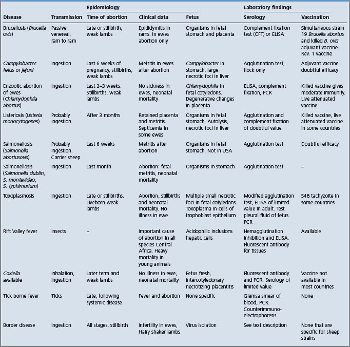
Abortion in ewes may be associated with a number of infectious diseases, summarized in Table 18.8.

Table 18.8 Diagnostic summary of infectious abortion in ewes

image

TREATMENT

Treatment of naturally occurring cases is rarely undertaken.

In experimentally infected rams, the intramuscular administration of long-acting oxytetracycline at 20 mg/kg BW, given every 3 days for 24 days, along with the daily intramuscular administration of 20 mg/kg of dihydrostreptomycin sulfate, has resulted in bacteriological cure in 11 of 12 rams.25 Oxytetracycline alone is less effective. In another study using a similar treatment, seven of nine rams had bacteriological cure.13 Treatment is economically practicable only in valuable rams and must be instituted before irreparable damage to the epididymis has occurred. The treatment of rams that are infected but without palpable lesions results in a significant improvement in breeding soundness classification on examinations subsequent to treatment.13

CONTROL

Control is by the prevention of spread of infection between rams and the detection and culling of infected rams. In small commercial flocks, culling of all rams and replacement with B. ovis-free rams may be the most economical approach. A measure of control can be achieved using scrotal palpation to detect infected rams but this must be coupled with repeated serological examinations if eradication is the goal. Vaccination may be the most economical and practical means of controlling the disease in areas with a high incidence of infection and in regions of the world where eradication by test and slaughter is impractical.14

Eradication

In a herd where the diagnosis has been confirmed all rams are palpated and those with scrotal abnormalities are culled. The remainder are tested serologically and reactors are culled. Serological tests are repeated at monthly intervals, with culling of reactors, until all rams are serologically negative. A further test, 6 months later, is used for confirmation.23

The rate of spread of infection is high during the mating season and it is not recommended that eradication should be attempted until after the breeding season. During breeding it may be wise to run two breeding flocks, with virgin rams and rams known to be free of infection separate from older or suspicious rams. Strict separation of the two ram flocks must be maintained at all times of the year, and the clean group must not mate ewe flocks that have been mated to these older rams.

The use of the ELISA test available in the USA has allowed the eradication of the disease in flocks with as few as four tests.12

Several countries have voluntary accreditation schemes.

Vaccination

A number of vaccines are in use but none is fully effective. In some countries vaccination is not permitted and eradication by test and slaughter is the only method of control.

Killed B. ovis vaccines, even when adjuvanted, have poor efficacy.7,15 The use of a killed vaccine may be inadvisable in flocks where eradication is being attempted, as it may protect against clinical disease but allow a carrier state in some rams in which there is excretion of the organism in animals that become seronegative.15 An experimental vaccine prepared from enriched outer membrane proteins and rough lipopolysaccharide of B. ovis has given equivalent protection in challenge studies to that given by B. melitensis Rev. 1 vaccine.26

A combined vaccine containing killed B. ovis in an adjuvant base and B. abortus strain 19 gives a high-level, durable immunity but the vaccine has several disadvantages. Vaccinated animals become seropositive, which hinders subsequent use of serological tests for eradication. Strain 19 itself can cause epididymitis and vaccinated rams may excrete strain 19 in their semen.3 Severe outbreaks of osteomyelitis and epiphysitis have been recorded in rams following vaccination.27

Live B. melitensis strain Rev. 1 has been found to be most effective and is generally recommended.3,28,29 Strain Rev. 1 is avirulent for rams, and subcutaneous or conjunctival vaccination provides protection against experimental and field challenge. Vaccinated animals become positive to the complement fixation test and ELISA test, but the titers produced are low and can be minimized by using the conjunctival route for vaccination.29,30

B. suis strain 2 (S2) has been used in China for vaccination of all target species against brucellosis. It is given orally in the drinking water and colonizes the cranial lymph nodes. In a comparative study of S2 vaccine and Rev. 1 vaccine, S2-vaccinated rams had less protection than Rev. 1-vaccinated rams when challenged with B. ovis and an equivalent degree of protection to the nonvaccinated controls.31

B. abortus RB51 vaccine does not protect rams against experimental challenge with B. ovis.32

Outer membrane protein (Omp) antigens are currently being examined as vaccine candidates. A detergent-extracted Omp31 from B. melitensis produced in E. coli has been shown to be a protective immunogen against B. ovis in a mouse model and to induce antibody and a cellular immune response in sheep.33 Its efficacy in protecting sheep against experimental or natural infection is not reported.

In all vaccination programs there should be a concurrent program of culling of clinically abnormal rams. All ram replacements should be yearlings vaccinated at 4–5 months.

REVIEW LITERATURE

Afzal M, Kimberling CV. How to control Brucella ovis-induced epididymitis in rams. Vet Med. 1986;81:364-370.

Rahaley RS. Ovine brucellosis; recent advances. Aust Adv Vet Sci. 1986:90-92.

Blasco JM. Brucella ovis. In: Nielsen K, Duncan JR, editors. Animal brucellosis. Boca Raton, FL: CRC Press, 1990.

Bulgin MS. Epididymitis in rams and lambs. Vet Clin North Am Food Anim Pract. 1990;6:683-690.

Genetzky RM. Epididymitis in rams. Compend Contin Educ Pract Vet. 1995;17:447-454.

Elberg S. Rev 1 Brucella melitensis vaccine. Part III 1981–1995. Vet Bull. 1996;66:1193-1200.

Ridler AL. An overview of Brucella ovis infection in New Zealand. N Z Vet J. 2002;50:S96-S98.

REFERENCES

1 Verger JM, et al. Int J Syst Bacteriol. 1985;35:292.

2 Michaux-Charachon S. J Bacteriol. 1997;179:3244.

3 Blasco JM. Nielsen K, Duncan JR, editors. Animal brucellosis. Boca Raton, FL: CRC Press. 1990:351.

4 Walker RL, et al. J Am Vet Med Assoc. 1986;188:393.

5 Genetzky RM. Compend Contin Educ Pract Vet. 1995;17:447.

6 Marco J, et al. Vet Rec. 1994;135:254.

7 Bulgin MS. Vet Clin North Am Food Anim Pract. 1990;6:683.

8 Bulgin MS. J Am Vet Med Assoc. 1990;196:1120.

9 Ridler AL, et al. N Z Vet J. 2000;48:57.

10 Worthington RW. N Z Vet J. 1983;32:58.

11 Rahaley RS, Dennis SM. Aust Vet J. 1984;61:353.

12 Kimberling CV, Schweitzer D. Agri-Practice. 1989;10:36.

13 Dargatz DA, et al. J Am Vet Med Assoc. 1990;196:605.

14 Marin CM, et al. Res Vet Sci. 1990;48:209.

15 Manterola L, et al. Vet Microbiol. 2003;92:65.

16 Kittleberger R, et al. Vet Microbiol. 1998;59:213.

17 Vigliocco AM, et al. Vet Microbiol. 1997;54:357.

18 ficipal A, et al. Vet Rec. 1995;137:145.

19 Kittleberger R, et al. N Z Vet J. 1997;45:75.

20 Marin CM, et al. Vet Rec. 1989;125:504.

21 West DM, Bruce RA. N Z Vet J. 1991;39:29.

22 Gall D, et al. Small Rumin Res. 2003;48:173.

23 West DM, et al. N Z Vet J. 1993;41:82.

24 Searson JE. Aust Vet J. 1986;63:30.

25 Marin CM, et al. Am J Vet Res. 1989;50:560.

26 Blasco JM, et al. Vet Immunol Immunopathol. 1993;37:257.

27 West DM, et al. N Z Vet J. 1978;26:133.

28 Erasmus JA, Bergh EC. J S Afr Vet Assoc. 1985;56:205.

29 Blasco JM. Prev Vet Med. 1997;31:275.

30 Marin CM, et al. Res Vet Sci. 1990;48:209.

31 Blasco JM. Vaccine. 1993;11:1291.

32 De-Baques MJP, et al. Vaccine. 1995;13:301.

33 Estein SM, et al. Vet Microbiol. 2004;102:203.

BRUCELLOSIS ASSOCIATED WITH BRUCELLA SUIS IN PIG

Synopsis

Etiology Disease in pigs is caused by Brucella suis biovars 1, 2, and 3. Biovars 1–4 cause rare disease in cattle

Epidemiology Disease in pigs is transmitted by contact, ingestion, and venereally

Clinical findings Sows: infertility, irregular estrus, small litters, and abortion. Boars: orchitis, lameness, incoordination, and posterior paralysis. Piglets: mortality

Clinical pathology Isolation of organism. Several serological tests available but none with good sensitivity

Necropsy Metritis, orchitis, osteomyelitis. Granulomatous inflammation and foci of caseous necrosis

Diagnostic confirmation Isolation of Br. suis and herd serology tests

Treatment None satisfactory

Control Serological testing and disposal of reactors. No effective vaccine. Humans, and occasionally cattle. Transmission congenital or by ingestion or contact with infected placenta, vaginal discharge, or milk

Clinical findings Abortion storms, abortions often in last 2 months of pregnancy. Weak-born lambs

Clinical pathology Culture of organism. Serological tests and skin hypersensitivity testing for herd diagnosis

Necropsy findings Placentitis

Diagnostic confirmation Only by isolation of the organism

Control Slaughter eradication. Vaccination with Rev. 1 vaccine. Rev. 1 vaccine will produce abortion in pregnant animals

ETIOLOGY

The disease is associated with Brucella suis. There are five biovars, of which biovars 1, 2, and 3 are important in pigs. Biovar 4 is a cause of rangiferine brucellosis and biovar 5 of murine brucellosis. Biovar 4 can transmit to cattle but does not appear to be a disease of pigs.

Biovar 3 has close similarity to B. melitensis biovar 2 and requires phage typing, oxidative metabolic testing or PCR for differentiation.1

EPIDEMIOLOGY

Geographic occurrence

Biovars 1 and 3

Disease associated with biovars 1 and 3 occurs in many countries and continents, including Europe, South America, particularly Venezuela,2 Africa, the Indian subcontinent, Central Asia, south-east Asia, Australia, and the Pacific islands. The infection has not been recorded in Britain, Canada is free of biovars 1 and 3, and prevalence in the USA is very low.3

Biovar 2

Domestic livestock disease associated with biovar 2 is confined to Europe and is of particular interest in the wild boar and domestic livestock in Croatia.4 It is also seen in the Czech Republic.5

Biovar 4

This occurs in mainland caribou (Rangifer tarandus) in Canada and has been isolated from a herd of commercial reindeer (Rangifer tarandus tarandus) in Canada.6

Host occurrence

Biotypes 1 and 3

Domestic, wild, or feral pigs are the host for biotypes 1 and 3 and widespread infection in feral pigs is recorded in Queensland, Australia7,8 and the southern states of the USA.3,9 Bison may remain reservoirs.

Cattle and horses may be infected, especially if they share a range with feral pigs10 and this association adversely affects the status of cattle herds undergoing brucellosis eradication programs.8 Cattle are noncontagious hosts, but an outbreak in Switzerland where the disease had not appeared since 1946 has been attributed to a spread of infection from horses.

Biovar 1 has been isolated from the semen of a ram.11

Infection in dogs, usually symptomless but occasionally producing orchitis or epididymitis, can result from eating raw pig meat.

Biovar 2

In addition to the pig, the European hare (Lepus capensis) is also a major host for biovar 2 and this biovar is common in central Europe.12

Biovar 4

This can transmit to cattle in contact with infected reindeer.6 Wild canids can also be naturally infected with biovar 4, presumably by ingestion.12

Source of infection

The introduction of infected pigs or the communal use of an infected boar is the common means of introduction into a pig unit. Artificial insemination using noncertified or untreated semen can also spread the disease.

The feeding of kitchen waste containing raw pig meat also presents a risk. Domestic herds are also at risk when they are kept under extensive husbandry methods in areas where there is a high prevalence of infection in feral pigs. Cattle infected with biovar 1 are noncontagious to other livestock and can have normal pregnancies and give birth to uninfected calves.10

Wild animals, including hares and rats, may provide a source of infection with biovar 2 and ticks are also suspected of transmitting the disease.

Transmission

Within a piggery the disease is spread by ingestion and by coitus. The ingestion of food contaminated by infected semen and urine and discharges from infected sows are also important methods of spread. Dried secretions, if frozen, may remain infective. Most disinfectants and sunshine kill the virus.

Host and pathogen risk factors

The fact that it survives so well in raw meat, e.g. 128 days in sausage meat, means that prepared pork products are always a source of infection.

B. suis is more resistant to adverse environmental conditions than B. abortus, although its longevity outside the body has not been fully examined. It is known to survive in feces, urine, and water for 4–6 weeks.

Amongst pigs, susceptibility may vary with age. The prevalence of infection is much higher in adults than in young pigs, although this may represent an exposure risk rather than an age-related risk.12 Susceptibility is much greater in the postweaning periods and is the same for both sexes, but there may also be genetically determined differences in susceptibility. Some piglets acquire infection from the sow, either from the ingestion of infected milk or by congenital infection.

Lateral spread through a herd is rapid because of the conditions under which pigs are kept. No durable herd immunity develops and, although a stage of herd resistance is apparent after an acute outbreak, the herd is again susceptible within a short time and a further outbreak may occur if infection is reintroduced.

In an enzootic area, the proportion of herds infected is usually high (30–60%). The prevalence of seropositivity in an infected herd varies but can be as high as 66%.13 Seroprevalence in feral pigs is also high, is higher in adult pigs than pigs under 6 months of age, and varies between populations of feral pigs.9

Economic importance

The disease owes its economic importance to the infertility and reduction in numbers of pigs weaned per litter. Mortality in liveborn piglets, which occurs during the first month of life, may be as high as 80%. The mortality rate is negligible in mature animals but sows and boars may have to be culled because of sterility, and occasional pigs because of posterior paralysis. In addition, eradication involves much financial loss if complete disposal of a registered herd is undertaken.

Zoonotic implications

Biovar 2 is not a zoonosis but biovars 1 and 3 have considerable significance for public health and are very pathogenic to humans. In countries where pigs are a significant part of animal farming and the human diet, B. suis is the major cause of human brucellosis. A recent report describes the death of a retired pig farmer at least 20 years after her last exposure to livestock.14

B. suis presents an occupational hazard, particularly to abattoir workers, and to a lesser extent to farmers and veterinarians. B. abortus and B. melitensis may also be found in pig carcasses and present similar hazards. B. suis can be widespread in the carcass of infected pigs, and undercooked meat can be a source of human infection.12 This is particularly true for wild boar and feral pigmeat. A recent experiment described infection with biovar type 1 and its transmission to negative pigs after 4–6 weeks. Antibody was detected in blood samples from farmers and abattoir workers.15

In infected cattle B. suis localizes in the mammary gland without causing clinical abnormality and, where cattle and pigs are run together, the hazard to humans drinking unpasteurized milk may be significant.10 Biovar 4 causes human disease associated with consumption of caribou.16

PATHOGENESIS

As in brucellosis associated with B. abortus, there is initial systemic invasion, the organism appearing in the bloodstream, usually within 1–7 weeks and persisting for up to 34 weeks. However, infection with B. suis differs from that associated with B. abortus in that localization occurs in several organs in addition to the uterus and udder, the organism being found in all body tissues and producing a disease similar to undulant fever in humans. The organisms persist in lymph nodes, joints, bone marrow, and the genital tract. The more common manifestations of localization are abortion and infertility due to localization in the uterus; lymphadenitis, especially of the cervical lymph nodes; arthritis and lameness due to bone and joint localization; and posterior paralysis due to osteomyelitis. In boars, involvement of the testicles often leads to clinical orchitis.12 Widespread infection makes handling of the freshly killed carcass hazardous and creates a risk for brucellosis in humans eating improperly cooked pork.

CLINICAL FINDINGS

Do not forget that clinical signs in pigs may also be produced by B. abortus and B. melitensis.

The clinical findings in swine brucellosis vary widely, depending upon the site of localization. The signs are not diagnostic and in many herds a high incidence of reactors is observed with little clinical evidence of disease. Reproductive inefficiency is the common manifestation.

Sows

Infertility, irregular estrus, small litters, and abortion occur. Mummification and stillbirths do occur. The incidence of abortion varies widely between herds but is usually low and is usually early on. Infection of the fetus may lead to abortion. As a rule, sows abort only once in a lifetime and this is most common during the third month of pregnancy. Affected sows usually breed normally thereafter. Sows may remain carriers and may shed organisms in milk and uterine discharges. These discharges may be extremely bloody. They may be accompanied by an endometritis and retained placenta.

Boars

Orchitis with swelling and necrosis of one or both testicles is followed by sterility. Lameness, incoordination, and posterior paralysis occur reasonably commonly. The onset is gradual, and signs may be caused by arthritis or, more commonly, osteomyelitis of lumbar and sacral vertebral bodies. Testicular atrophy may result. Boars have a low rate of recovery (less than 50%). After infection, enough animals remain infected to perpetuate the disease.

Piglets

A heavy mortality in piglets during the first month of life is sometimes encountered but most piglet loss results from stillbirths and the death of weak piglets within a few hours of birth. Up to 10% may contract infection when they are young and retain the infection until adulthood.

CLINICAL PATHOLOGY

Culture

Laboratory identification of the disease is difficult. Isolation of the organism should be attempted if suitable material is available. Such material for culture includes aborted fetuses, testicular lesions, abscesses, lymph nodes, and blood. The organism is a small, slender, aerobic, Gram-negative organism that produces 1–2 mm colonies on blood agar after 2–4 days.

Serology

There is no satisfactory serological test. Some animals remain seronegative to all tests. Recently two ELISAs have been developed.17,18 An ELISA compared with complement fixation was found to be just as sensitive and as specific a test for both pigs and hares for B. suis infections.19 A meat juice ELISA has also been shown to be a valuable method for testing both hares and wild boars.20 There is considerable individual variation in the antibody response of pigs following infection – some may be culture-positive but have negative or indefinite titers to the common tests.12 Pigs under 3 months of age have a poor antibody response to infection.12

Serological tests in common use include the rose Bengal plate agglutination test, Rivanol test, rose Bengal card test, complement fixation, agar gel immunodiffusion, and tube agglutination. The preferred test varies between countries but most use the rose Bengal plate or card test. B. abortus antigens are used for diagnosis as B. suis has the same surface lipopolysaccharide antigens. Estimates of the sensitivities of the complement fixation and tube agglutination tests range from 40–51%, and from 62–79% for the rose Bengal plate test.12,21 The immunodiffusion test has poorer sensitivity than the standard serological tests.2 The sensitivity and specificity of all the tests have been shown to vary with the stage of infection in the experimental disease and it has been recommended that more than one test should be used for diagnosis.21,22 A recent study23 showed a range of sensitivity from 84–100% with the CFT low at 84% and the serum agglutination test high at 100%. The sensitivities ranged from 79.7–100%, with the serum agglutination test low at 79.7% and indirect ELISA and competitive ELISA high at 100%. A recent validation of the polarization assay as a serological test for the presumptive diagnosis of porcine brucellosis has shown promise.24,25

NECROPSY FINDINGS

Many organs may be involved in chronic cases. Chronic metritis manifested by nodular, white, inflammatory thickening, 2–5 mm in diameter, and abscessation of the uterine wall is characteristic. Arthritis may be purulent and necrosis of vertebral bodies in the lumbar region may be found in lame and paralyzed pigs. The clinical orchitis of boars is revealed as testicular necrosis, often accompanied by lesions in the epididymis and seminal vesicles. Splenic enlargement and pronounced lymphadenopathy, due to hyperplasia of mononuclear phagocytes, occur in some cases. Typical histological changes consist of granulomatous inflammation and foci of caseous necrosis in liver, kidney, spleen, and reproductive tract.

Samples for confirmation of diagnosis

Bacteriology – adults: culture swab from joint, lymph nodes, spleen, uterus, epididymis, or other site of localization; fetus: lung, stomach content, placenta (has special growth requirements)

Histology – formalin-fixed samples of above tissues (LM).

Note the zoonotic potential of this organism when handling carcasses or submitting specimens.

DIFFERENTIAL DIAGNOSIS

The protean character of this disease makes it difficult to differentiate. Syndromes that need differentiation include:

Abortion and infertility in sows (Table 20.2)

Posterior paresis diseases of spinal cord

Mortality in young pigs is also caused by many agents and the important entities are listed under disease of the newborn (Chapter 3).

TREATMENT

Treatment with a combination of streptomycin parenterally and sulfadiazine orally, or with tetracycline, is ineffective. It is unlikely that treatment will ever be attempted on a commercial scale.

CONTROL

Vaccination

No suitable vaccine is available. Strain 19 B. abortus, B. abortus ‘M’ vaccine, living attenuated B. suis vaccines and phenol and other extracts of B. suis are all ineffective.12

Test and disposal

In herds where the incidence of reactors is high, complete disposal of all stock as they reach marketing age is by far the best procedure because of the difficulty in detecting individual infected animals. This is most practicable in commercial pork-producing herds. Restocking the farm should be delayed for 6 months. The existing serological tests can be used for certifying herds free of infection that can then provide replacement stock. Repopulation programs can also use specific-pathogen-free pigs.

The alternative is to commence a two-herd segregation program, and this is recommended for purebred herds that supply pigs for breeding purposes. Total disposal is not usually economical in these herds. Once a herd diagnosis has been established, all the breeding animals must be considered to be infected; all piglets at weaning are submitted to the serum agglutination, Rivanol or other test and, if negative, go into new quarters to start the nucleus of a free herd. It is probably safer to wean the pigs as young as possible and test again before mating. If complete protection is desired, these gilts should be allowed to farrow only in isolation, should then be retested, and their piglets used to start the clean herd. A modified scheme based on the above method of weaning and isolating the young pigs as soon as possible but without submitting them to the serum agglutination test has been proposed, but its weakness is that infections may occur and persist in young pigs.

After eradication is completed, breakdowns are most likely to occur when infected animals are introduced. All Introductions should be from accredited free herds, should be clinically healthy and be negative to the serum agglutination test twice at intervals of 3 weeks before introduction.

Eradication of swine brucellosis from an area can only be achieved by developing a nucleus of accredited free herds and using these as a source of replacements for herds that eradicate by total disposal. Sale of pigs for breeding purposes from infected herds must be prevented.

REVIEW LITERATURE

Macmillan AP, Stack J, Angus RD. Brucellosis. In: Manual of standards for diagnostic tests and viruses. List A and B diseases of mammals, birds and bees. Paris: Office International des Epizoöties; 2000:623-629.

Godfroid J, Kasbohrer A. Brucellosis in the European Union and Norway at the turn of the 21st century. Vet Microbiol. 2002;90:135.

Moreno E. Brucellosis in Central America. Vet Microbiol. 2002;90:31.

Gresham A. Infectious reproductive disease in pigs. In Pract. 2003;25:466-473.

REFERENCES

1 Bricker BJ, Halling SM. J Clin Microbiol. 1994;32:2660.

2 Lord VR, et al. J Clin Microbiol. 1997;35:295.

3 Frye GH, Lenard DM. Proc US Anim Health Assoc. 1991;95:81.

4 Cvetnic Z, et al. Acta Vet Hung. 2003;51:465.

5 Hubalek Z, et al. Vet Med (Prague). 2002;47:60.

6 Forbes LB, et al. J Am Vet Med Assoc. 1993;203:289.

7 Cook DR, Kingston GC. Aust Vet J. 1988;65:162.

8 Rogers RJ, et al. Aust Vet J. 1989;66:77.

9 Van der Leek ML, et al. J Wildl Dis. 1993;29:410.

10 Ewalt DR, et al. J Vet Diagn Invest. 1997;9:417.

11 Paolicchi FA, et al. Vet Rec. 1993;132:67.

12 Alton GG. Nielsen K, Duncan JR, editors. Animal brucellosis. Boca Raton, FL: CRC Press. 1990:379.

13 Cornell WD, et al. J Vet Diagn Invest. 1989;1:20.

14 Paton NI. Clin Infect Dis. 2001;32:1248.

15 Sudibyo A. J Ilmu Dan Vet (Indonesia). 1998;3:257.

16 Forbes LB. Can Vet J. 1991;32:686.

17 Teson M, et al. Vet Argent. 1998;15:32.

18 Sarga M, Neculescu M. Stud Res Vet Med. 1996;4:29.

19 Szulowski K. Pol J Vet Sci. 1999;2:65.

20 Szulowski K. Bull Vet Inst Pulawy. 2000;44:45.

21 Payeur JB, et al. Proc US Anim Health Assoc. 1990;94:147.

22 Ferris RA, et al. J Am Vet Med Assoc. 1995;207:1332.

23 Jauregui LH. In: Proceedings of the 15th International Pig Veterinary Society Congress 1998:171.

24 Nielsen K. Vet Microbiol. 1999;68:245.

25 Nielsen K, Gall D. J Immunoassay Immunochem. 2001;22:183.