Etiology Ischemic necrosis of large muscles of pelvic limbs secondary to prolonged recumbency associated with milk fever. Other causes of recumbency can also result in downer cow syndrome.
Epidemiology Most common in dairy cows which have had milk fever but are unable to stand following treatment with calcium. Delay of more than 4 h in treatment for recumbent milk fever cows. Hypophosphatemia and/or hypokalemia may be risk factors.
Signs Unable to stand following treatment for milk fever. Sternal recumbency. Normal mental status, vital signs and alimentary tract. Appetite and thirst normal. Most will stand in few days if provided good clinical care and secondary muscle necrosis minimized. Some cases have bizarre behavior of lateral recumbency, abnormal position of legs, groaning, anorexia, and die in several days.
Clinical pathology Increased serum levels of creatine phosphokinase (CPK) and aminotransferase (AST); serum phosphorus and potassium levels may be subnormal.
Necropsy findings Ischemic necrosis, edema and hemorrhage of large medial thigh muscles.
Diagnostic confirmation Increased serum levels of CPK, AST, proteinuria; necropsy lesions in cow unable to rise with no other lesions.
• See differential diagnosis of milk fever and Table 29.5.
• Common causes of recumbency in dairy cows around time of parturition include:
• Maternal obstetrical paralysis
• Physical injuries of pelvic limbs (dislocation of hip joints, rupture of gastrocnemius, femoral fracture)
• Acute diffuse peritonitis (ruptured uterus, other causes).
Treatment Provide feed and water and excellent bedding or ground surface like sand or dirt pack. Roll animal from side to side every few hours. Fluid and electrolyte therapy as necessary.
Control All recently calved dairy cows which are at high risk for milk fever must be observed closely 12– 24 h before and after calving for evidence of milk fever and while still standing; if recumbent do not delay treatment for more than 1 h. Can treat all high-risk cows with calcium chloride gel orally to prevent clinical milk fever.
Most commonly, the downer cow is a complication of milk fever.1 Ischemic necrosis of the large muscles of the pelvic limbs and injuries to the tissues around the hip joint and of the obturator muscles are common in cows which do not fully recover and stand but remain recumbent following treatment for milk fever. Injuries to the musculoskeletal system are also common as a result of cows ‘spread-eagling’ their hindlimbs if they are unsteady during parturition or forced to stand or walk on a slippery floor immediately before or following parturition. Dystocia due to an oversized calf may result in extensive edema of the pelvic tissues and vulva, and failure of the cow to stand following parturition. If these cows develop milk fever, it is unlikely they will be able stand following treatment with calcium.
The disease occurs most commonly within the first 2 or 3 days after calving in high-producing dairy cows immediately following milk fever. Cattle may also become persistently recumbent for many reasons other than complications of milk fever such as peracute coliform mastitis and carbohydrate engorgement.
Downer cows can be divided generally into non-ambulatory cows with non-progressive neurological findings and non-ambulatory with progressive neurological findings indicative of the presence of lesions in the nervous system as the cause of the recumbency.2
A survey of bovine veterinarians in the USA in 1998 collected data on non-ambulatory dairy and beef cows with non-progressive neurological findings, and non-ambulatory with progressive neurological findings.2 Of the population studied, 0.25% developed a non-ambulatory non-progressive neurological disorder for a case rate of 066 animals/106 cow years or 106 cows/year. Of these, 74.3% failed to recover. In non-ambulatory dairy cows with non-progressive neurological findings, 85% of cases fit into three categories: injury/trauma, septicemia/toxemia, or non-responsive milk fevers. Of the total, 3.3% remain undiagnosed. In non-ambulatory beef cows with non-progressive neurological findings, trauma/injury, septicemia, and other neurological cases accounted for 78% of cases. Of the population studied, 0.12% of cows developed non-ambulatory non-progressive neurological findings syndrome for a case rate of 2476/106cows. Of the total, 73% failed to recover. Of the beef cows, 0.0053% remain undiagnosed, which implies that 4.3% of all beef cows with non-ambulatory non-progressive CNS findings were unknown and undiagnosed and 96% were diagnosed.
Of the total number of non-ambulatory progressive CNS cases reported for dairy cattle, the percentage was 0.027% for a case rate of 554/106. Of these, 17.7% were unknown or undiagnosed indicating that 82% of progressive CNS dairy cattle cases were diagnosed. The progressive CNS cases of unknown cause represented 0.0047% of the study’s cattle population, or a rate of 94 cases/106 dairy cows. Seventy-one of the cases failed to recover for a mortality of 375 cases per million dairy cows in the study.
The profile of a progressive CNS dairy cow case found that other known CNS diseases, septicemia/toxemia, unknown CNS diseases, and non-responsive milk fever to be the most frequently reported cases.
Of the total number of non-ambulatory progressive CNS cases reported in beef cattle, the percentage was 0.018%. Of these, 9.0% were of unknown etiology indicating that 91% of progressive CNS beef cow cases were diagnosed by veterinarians. The unknown progressive CNS case rate was 32 cases/106 beef cattle. The total deaths in progressive CNS cases accounted for a mortality rate of 282/106.
The profile of progressive CNS beef cow cases found that four categories accounted for 83.4% of the total causes: injury/trauma, known infectious agent, septicemia, toxemia, and known CNS disease.
The incidence as a complication of milk fever is high because many affected animals are high producers and of high economic value. Accurate data on the incidence are not available because of variations in the nomenclature used and the accuracy of diagnoses. For example, some observations report that all cases are caused by nerve injury.3 Cases included in this classification are classified by others as maternal obstetric paralysis, obturator paralysis, or hypophosphatemia. Because it is a syndrome lacking in clinical definition and includes all those ‘other cases’ which cannot be otherwise classified, the incidence varies depending on the clinical acuity of the individual veterinarian, and on various environmental factors in different areas. However, the incidence seems to be increasing, particularly in intensive dairy farming areas, although this impression could arise from the increased necessity to effect a cure in valuable animals.
A mail survey of 723 dairy herds in Minnesota found a downer cow incidence of 21.4/1000 cow years at risk.3 The overall outcome was that 33% recovered, 23% were slaughtered and 44% died. The owners perceived that downer cows were high producers (48%) or average producers (46%), with only 6% being low producers. Approximately 58% occurred within 1 day of parturition and 37% occurred during the first 100 days of lactation. The incidence was highest (39%) during the three coldest months: December, January, and February. In New Zealand, the prevalence ranges from 3 to 5% of all dairy cows at calving time.4
In a clinical and laboratory survey of 433 periparturient recumbent cows in New Zealand, 39% recovered, 30% died, and 32% were destroyed.4 The case-fatality rate was 11% higher in pre-calving recumbent cows than post-calving cows.
An audit of 21 slaughter plants in the USA in 1993 found 1.1% of arriving dairy cows were non-ambulatory, and in 1999, 1.5%.5 In 1993, 1.0% of beef cows arriving at slaughter plants were non-ambulatory and in 1999, 0.7%.5
Prolonged recumbency after a long delay in the treatment of milk fever is a major risk factor. Prolonged recumbency before treatment for milk fever (more than 4–6 h) results in ischemic necrosis due to obstruction of the blood supply, especially in a heavy cow if she lies on one leg for a long period.6 Cows which develop milk fever while in a standing tie-stall may slide backwards into the gutter behind the stall, resulting in extreme pressure to their pelvic limbs and leading to ischemic necrosis.
A case-control study to identify risk factors for the development of downer cow syndrome within 30 days post partum in 12 dairy herds over 2705 lactations found that clinical hypocalcemia and stillbirth increased the risk of the disease five-fold.7 Cows with retained placenta and dystocia were also more likely to develop downer cow syndrome than cows without either problem.
A marked increase in the CPK levels in cows with milk fever and failure to stand after repeated treatments is supporting evidence for ischemic necrosis associated with prolonged recumbency as a major cause of downer cow syndrome.1 The CPK levels increase markedly between the first and second treatments, which indicates that muscle damage has occurred and the levels are highest in cows which do not recover.
Experimentally, enforced recumbency of cattle for 6, 9, or 12 h with one hindlimb positioned under the body results in downer cow syndrome. Affected cows are unable to stand and the affected limb is swollen and held rigid similar to the injured limbs of human patients with compartmental/crush syndrome.
Surveys have shown that downer dairy cattle have 3.3-fold-higher prevalence of E. coli 0157:H7 than healthy cattle within a certain time frame and geographic area.8 Culled dairy cows account for approximately 17% of the ground beef produced in the US and thus downer cattle harboring E. coli 0157:H7 may be an important source of contamination of ground beef which is commonly processed from downer cattle.
Traumatic injuries to pelvis and pelvic limbs.
Traumatic injuries to the nerves of the pelvis and hindlimbs are present in 25% or more of downer cows.1 The sciatic and obturator nerves are vulnerable to injury by pressure from the calf moving through the pelvic canal during parturition. Pressure injuries on the superficial nerves (radial and peroneal) of the extremities also occur in recumbent cows.
Serum electrolyte imbalances or deficits may be associated with prolonged recumbency following treatment for parturient paresis.
A persistent hypocalcemia following treatment for milk fever may exist in a downer cow but is unlikely to be the principal cause because treatment with calcium salts does not resolve the signs, even temporarily. However, the use of an insufficient amount of calcium for the initial treatment of milk fever in large, heavy cows may result in an incomplete response and failure of the cow to stand. If these cows are not retreated soon enough with an adequate amount of calcium, ischemic necrosis of the limb muscles occurs and leads to prolonged recumbency. In many cases, even after the cow is given a sufficient amount of calcium, prolonged recumbency occurs due to the ischemic necrosis.
The serum levels of inorganic phosphorus decline to below normal along with a hypocalcemia in cases of milk fever. Following treatment of milk fever with calcium borogluconate, the levels of serum calcium and phosphorus return to normal if the animal responds favorably and stands normally. Following treatment for milk fever, some cows do not or are unable to stand and their serum phosphorous levels are subnormal. This persistent hypophosphatemia has been regarded as a cause of downer cow syndrome associated with milk fever. Many veterinarians claim that these cows respond to treatment with phosphorus. However, persistent recumbency is associated with subnormal levels of serum phosphorus which increase to normal if the cow stands regardless of treatment with or without phosphorus. Mature dairy cows may become recumbent in early lactation and subnormal levels of serum phosphorus may be present.9 Other cows in the herd may be lame due to demineralization of bones associated with a dietary deficiency of phosphorus.
A long-term low-level hypomagnesemia has been associated with the downer cow, especially when it accompanies hypocalcemia. But it is usually manifested by a tetanic hyperesthetic state which is not part of downer cow syndrome. Hypokalemia is, with hypophosphatemia, the most commonly quote cause, especially in the so-called ‘creeper’ cows, which are bright and alert and crawl about, but are unable to rise.10
Ischemia due to prolonged recumbency associated with milk fever, may increase the cell membrane permeability of muscle fibers and allow the loss of potassium from the cell; this in turn causes the myotonia, which appears to be the basis of downer cow syndrome. This view is supported by the low serum and muscle potassium levels in downer cows. Claims are made that potassium salts are successful in treatment but these have been difficult to evaluate.4
Hypokalemia occurs in dairy cows which have been treated with isoflupredone acetate for ketosis.11 Affected animals are weak, recumbent and severely hypokalemic with serum potassium levels ranging from 1.4 to 2.3 mEq/L.
A slippery ground surface is a major risk factor. Cattle which must walk across slippery floors, especially at the time of calving, may slip and fall and injure the large muscles of the pelvic limbs, resulting in an inability to stand. Prolonged recumbency results in ischemic necrosis and downer cow syndrome.
Downer cow syndrome is a complication of the recumbency associated with milk fever. A delay of 4 h or more in that treatment of cows with milk fever may result in ischemic necrosis of the muscles of the pelvic limbs. Traumatic injury to leg muscles at the time of parturition or when the cow is unsteady and falls during the first stage of milk fever will also result in the inability of the cow to stand following treatment of milk fever.
Several different primary factors or diseases can result in recumbency.
A long delay in the treatment of milk fever can result in pressure damage and the subsequent inability to stand after treatment for the primary disease. Prolonged recumbency results in pressure damage, which occurs secondarily and is a factor common to all cases.3
Regardless of the cause, the prolonged recumbency results in varying degrees of ischemic necrosis of major muscles of the hindlimbs, particularly the semitendinous muscle and muscles caudal to the stifle. Prolonged compression of the muscle leads to tissue anoxia, cell damage and inflammation which causes swelling; the swelling causes a further increase in pressure which limits tissue perfusion and leads to a detrimental cascade of events. The thick fascial boundaries of the semitendinous muscle prevent expansion which results in pressure-induced compartmental syndrome. Sciatic nerve damage due to pressure also occurs and may contribute to downer cow syndrome. Experimental external compression of the pelvic limb of the goat, to simulate limb compression in recumbent cows, resulted in a marked reduction in nerve condition velocity of the peroneal nerve which was associated with clinically evident limb dysfunction. Damage to the peroneal nerve will result in hyperflexion of the fetlock if and when the cow is able to stand.
Traumatic injury to limb muscles and nerves immediately prior to parturition or at the time of parturition can also result in prolonged recumbency and subsequent pressure damage.1
Experimentally induced sternal recumbency with one hindlimb positioned under the body to simulate prolonged recumbency will result in a swollen rigid limb within 6–9 h.12 Following injury to the muscle cells, the serum levels of CPK are markedly elevated at about 12 h after the onset of recumbency. Proteinuria and in some severe cases myoglobinuria occur between 12 and 36 h after the onset of prolonged recumbency, due to the release of myoglobin from damaged muscles. In cows which make efforts to stand but cannot do so, continued struggling results in rupture of muscle fibers and hemorrhage which increases the severity.
Acute focal myocarditis may occur in about 10% of cases resulting in tachycardia, arrhythmia, and the unfavorable response to IV calcium salts observed in some cases. The cause of the myocardial lesion is unknown but repeated administration of calcium salts has been suggested.1 Downer cows with a poor prognosis also have greatly enhanced adrenocortical function.10
The prolonged recumbency can result in additional complications such as acute mastitis, decubitus ulcers, and traumatic injuries of the limbs.
The pathogenesis of the non-alert downer cow is not understood.13 Most have had an initial episode of milk fever but do not respond satisfactorily. Within 1 or 2 days, affected cows have a preference for lateral recumbency and exhibit expiratory moaning and groaning. They represent about 2% of all cases of milk fever.
Experimental prolonged hypocalcemia may provide some clues about the pathogenesis of downer cow syndrome as a complication of milk fever. The prolonged infusion of ethylenediamine tetra-acetic acid (EDTA) in sheep over 18 h at a rate to induce hypocalcemia and maintain recumbency results in prolonged periods of recumbency, ranging from 36 to 64 h before the animals are able to stand.14 There are also decreases in plasma sodium, plasma potassium, and erythrocyte potassium and prolonged increases in packed cell volumes, which suggests that fluid replacement therapy may be indicated in cattle with prolonged recumbency associated with hypocalcemia.15 A 4-h IV infusion of EDTA in high erythrocyte potassium and low erythrocyte potassium dairy cows causes decreases in plasma inorganic phosphorus and plasma potassium which are still below normal 24 h later.16 The AST, CPK, and PCVs and WBC counts are also elevated 24 h later. Plasma magnesium and erythrocyte sodium and potassium were decreased but this was delayed. The increase in PCV was most pronounced in the low erythrocyte potassium cows, which may provide some clues about the pathogenesis of downer cow syndrome. Some cows may have a more precipitate increase in PCVs due to loss of plasma volume and an inability to mobilize calcium. As a basis for studying the effects of hypertonic solutions to correct these abnormalities in downer animals, a 200 mL solution of 10 g of sodium chloride and 0.5 g of potassium chloride can be given IV to sheep safely over a period of 4–8 min.17
The downer cow syndrome may occur independently, or follow apparent recovery after treatment for milk fever, except for the prolonged recumbency. In the typical case, affected cows either make no effort or are unable to stand following treatment for parturient paresis. About 30% of cows treated for milk fever will not stand for up to 24 h following treatment. Those which are unable to stand after 24 h and after two treatments are classified as downers. They are usually bright and alert and, although the appetite is reduced, the cow eats and drinks moderately well. The temperature is normal and the heart rate may be normal or elevated to 80–100 bpm. Tachycardia and arrhythmia occur in some cows, especially immediately following the administration of calcium IV and sudden death has occurred. Respirations are usually unaffected. Defecation and urination are normal but proteinuria is common and if marked may indicate extensive muscle damage.
Some affected cows may make no effort to stand. Others will make frequent attempts to stand but are unable to fully extend their pelvic limbs and lift their hindquarters more than 20–30 cm from the ground. These frequent attempts to stand result in ‘crawling’ or ‘creeping’ along the ground with both hindlegs in a partially flexed position and displaced posteriorly – the frogleg attitude. On a non-slippery surface (bare ground, sand pack, or deep bedding) some cows are able to stand with some assistance by lifting on the tail head or with the use of hip slings. Those cows which do not make an effort to stand usually cannot stand even with assistance and if supported with hip slings will usually make no effort to bear weight with either the hindlimbs or the forelimbs. Their limbs appear stiff, painful, or numb and they are unable or reluctant to bear weight. Damage to the peroneal nerve is usually present when there is hyperflexion of the fetlock joints, which is evident if and when the cow is able to stand and bear weight on the hindlimbs.
In some cases, the hindlimbs are extended on each side of the cow and reach up to the elbows on each side. In this position, the cow is bearing considerable weight on the medial thigh musculature and causing ischemic necrosis. This abnormal position of the legs may also be due to dislocation of one or both hip joints or associated with traumatic injuries surrounding the hip joints with or without rupture of the ligamentum teres. Regardless of the cause, the cow prefers this leg position and invariably will shift the legs back to the abnormal position if they are placed in their normal position.
In some cows, the signs may be more marked and bizarre, including a tendency to lie in lateral recumbency with the head drawn back. When placed and propped up in sternal recumbency, these cows appear almost normal but, when they are left alone, within a short period of time they revert to the position of lateral recumbency. Still more severe cases are hyperesthetic and the limbs may be slightly stiff but only when the cow is lying in lateral recumbency. These severe cases do not usually eat or drink, have been described as ‘non-alert downers’, and are thought to have brain damage which has not been documented.13
Complications in the downer cow syndrome are common and often result in death or the need for euthanasia. Coliform mastitis, decubitus ulceration, especially over the prominences of the hock and elbow joint, and traumatic injuries around the tuber coxae caused by the hip slings are common. When these complications occur in the early stages of the disease, they commonly interfere with any progress being made and become the focus of clinical attention.
The course of the disease is variable and dependent on the nature and extent of the lesions and the quality of the care and comfort which is provided for the cow during the first few days. About 50% of downer cows will stand within 4 days or less if cared for properly. The prognosis is poor for those which are still recumbent after 7 days, although some affected cows have been down for 10–14 days and subsequently stood up and recovered. Death may occur in 48–72 h following the onset and is usually associated with myocarditis.
Clinical examination of the downer cow can be very difficult and challenging depending on the environmental circumstances and the physical size of the animal. Many different metabolic, nutritional, musculoskeletal, toxic, neurological, neoplastic, inflammatory, and infectious diseases can cause recumbency in cattle.18-20 It is very important to obtain an adequate history of the case on the first visit to the animal. Key aspects of the history include age of the animal, duration of recumbency, any previous clinical abnormalities before the recumbent stage such neurological in the case of bovine spongiform encephalopathy, or spinal cord lymphomatosis, any previous treatments with particular attention to mineralocorticoids which may cause hypokalemia, the anatomical location of any parenteral injections, time since recent parturition, diet and accidental access to new feeds, sudden unaccustomed exercise, and an assessment of the management provided.
The environment and the ground surface surrounding the recumbent animal may provide clues about the possibility that the animal slipped, fell, and was injured.
A systematic physical examination of all accessible body systems is necessary. The animal should be examined visually from a distance for evidence of abnormalities of the carriage of the head and neck, the position of the limbs, observe any attempts of the animal to stand or creep along the ground surface.
The details of the clinical examination are presented in Chapter 1. The standard close clinical examination is necessary to determine body temperature, heart rate and pulse, respiratory rate, and the state of the major body systems such as the respiratory tract, cardiovascular system, central nervous system for mental state, and gastrointestinal tract, mammary gland, reproductive tract, any of which may indicate the presence of abnormalities associated with shock which results in recumbency.
In the recently calved cow, particular emphasis must be given to adequate examination of the udder for mastitis, the uterus for metritis, and the gastrointestinal tract for diseases associated with toxemia and dehydration and shock (acute diffuse peritonitis, carbohydrate engorgement), which results in recumbency. A urine sample must always be obtained and tested for ketones, and the presence of myoglobinuria. A vaginal examination of the uterus should always be done along with a rectal examination.
Careful systematic examination of the musculoskeletal system includes palpating the muscles, bones, joints, and feet of each limb, including passive flexion and extension of each limb is necessary. The coxofemoral joints are examined for evidence of dislocation. The vertebral column is examined for evidence of painful sites or displacement of vertebrae. It is important to examine both sides of the animal which means rolling the cow over from side to side; often the animal may have to be rolled over more than once to repeat a particular examination.
A neurological examination includes examination of the withdrawal reflexes and sensation of all four limbs, reflex arcs of the spinal cord, careful examination of lumbar and sacral areas including sensation and tone in the tail, and examination of the cranial nerves.
The examination can be extended by lifting the downer cow with appropriate lifters and observing if the animal extends its limbs and attempts to bear weight. While the animal is being assisted to stand, additional examinations of other parts of the body can be made.
The calcium, phosphorus, magnesium and glucose levels of the blood are within the normal range and the results of hematological examinations are usually consistent with those found in normal cows which have recently calved. The CPK and AST levels are usually markedly elevated by 18–24 h after the onset of recumbency and continue to elevate within the next few days. Continued elevation of CPK levels indicates continued muscle damage. In experimentally induced recumbency in cows, the CPK levels remained within normal limits for the first 6 h. However, by 12 h there was a marked increase to mean values of 12 000 U/L rising to 40 000 U/L by 24 h. There may be moderate ketonuria. A marked proteinuria is usually evident by 18–24 h after the onset of recumbency. The proteinuria may persist for several days or be absent within a few days. In severe cases, the urine may be brown and turbid because of severe myoglobinuria. Low arterial blood pressures and abnormal electrocardiograms (ECGS) have been observed in some animals.
Elevations of serum urea, muscle enzymes, and laboratory evidence of inflammation are considered the best prognostic indicators of an unfavorable recovery.4 The recovery rate was lower in cows with a total protein:fibrinogen ratio less than 10:1, and evidence of neutropenia and/or left shift.4 Cows with a serum urea level above 25 mmol/L and serum creatinine levels above 130 mmol/L had a poor prognosis.
The CPK levels need to be interpreted in relation to the days of recumbency when the sample was taken. Critical levels may be highest initially (up to 50 times the upper normal reference range) and reduce to 10 times normal range at 7 days of recumbency.
In a series of 262 recumbent dairy cows serum samples were analyzed for creatine phosphokinase (CPK), lactate dehydrogenase (LDH) and aspartate aminotransferase (AST) to evaluate the value of serum enzyme activities for predicting a failure to recover.21 The most common diagnosis was milk fever 61.1%, dystocia 11.1%, mastitis 8.4%, and trauma 6.9%. The prior probability of the cow not recovering was 0.24, 0.69, and 0.81 on days 1, 2, 3, and 4 of recumbency. The optimal cut-off points maximizing the sensitivity and specificity of the tests were 2330, 2225, and 171 U/L for CPK, LDH, and AST, respectively. The predictive value of AST was significantly better with optimal cut-off points of 128 and 189 U/L, respectively. AST provided the best predictive indicator of whether a recumbent cow would not recover, the best results being obtained with serum samples taken on the first day of recumbency.
Hemorrhages and edema of the skin of traumatic origin are common. The major pathological changes consist of hemorrhages and degeneration of the medial thigh muscles. Hemorrhages around the hip joint with or without rupture of the ligamentum teres are also common. Local areas of ischemic necrosis of the musculature (gracilis, pectineus, and adductor muscles) occur at the anterior edge of the pelvic symphysis.1 Eosinophilic infiltration of ruptured necrotic thigh muscles of downer cows has been described.22 Hemorrhages and edema of the nerves of the limbs (obturator, ischiatic, peroneal, radial) are also common and usually associated with severe muscle damage. The heart is dilated and flabby and histologically, there is focal myocarditis. There is fatty degeneration of the liver and the adrenal glands are enlarged. Histologically, there are also degenerative changes in the glomerular and tubular epithelium of the kidneys.
The diagnosis of downer cow syndrome is made after all other known causes of recumbency have been eliminated in a cow which had milk fever and failed to stand within 24 h following two successive courses of treatment. The other common causes of prolonged recumbency are described under the differential diagnosis of milk fever (Table 29.5). It is difficult and time consuming to examine a downer cow thoroughly to eliminate all other causes of recumbency. Only by repeated careful clinical examination will the clinician avoid the embarrassment of failing to detect the presence of coliform mastitis, a fractured leg or a dislocated hip.
The prognosis of a downer cow depends on the cause of the recumbency and whether or not treatment is indicated or if euthanasia should be recommended because of the presence of abnormalities which are unlikely to respond favorably to treatment and also be economical. If the prognosis is poor, euthanasia on the farm should be recommended.
If the prognosis is favorable, the clinician should inform the owner about the nature of the treatment which will be necessary and its duration which may be several days of supportive care and therapy, and should outline the costs which will be incurred.
Many treatments including the injections of magnesium salts, phosphates, corticosteroids, stimulant tonics, and vitamin E and selenium have been used without consistent success. The use of parenteral solutions containing potassium, calcium, magnesium, and phosphorus has been recommended10 but there is no scientific evidence that these electrolytes, in addition to what was probably given to the cow already, are indicated or are of any beneficial value. Large quantities of fluid and multiple electrolyte therapy by the oral or parenteral route is indicated for cows which may not be drinking normal quantities of water. Multiple electrolytes can be added to the drinking water if the cow is drinking normally.
The most important aspect of treatment is to provide the most comfortable bedding possible and to roll the cow from side to side several times daily to minimize the extent of ischemic necrosis and para-analgesia which results from prolonged recumbency. With conscientious care and the provision of good bedding, palatable feed and liberal quantities of water, most cows will attempt to stand with some difficulty and assistance within 24 h, and will stand unassisted and normally 1 or 2 days later. A sand or dirt pack is the ideal ground surface which facilitates standing when downer cows attempt to stand.12 If affected cows are left on a slippery ground surface, they will not make an effort to stand and will become progressively worse. Cows should be milked normally and the udder kept clean by washing with germicide soap before milking, and post-milking teat dips applied.
The clinician and farmer are commonly faced with the questions of whether or not to lift a recumbent cow which has not attempted to stand within a few hours after treatment for milk fever. The guiding principle should be the behavior of the cow. If the cow makes an effort to stand on her own or by some coaxing such as a gentle nudge in the ribs, she should be assisted to stand by insuring a good non-slip ground surface, deep bedding, and lifting up on the tailhead when she attempts to stand. The cow should be rolled from side to side every few hours and encouraged to stand a few times daily. With good clinical care, most cows with the uncomplicated form of downer cow syndrome secondary to milk fever will stand in 12–24 h.
Several different kinds of cow-lifting devices have been used to assist downer cows to stand. Hip lifters, which fit and tighten over the tuber coxae, and body slings like harnesses are designed to fit around the abdomen and thorax of the animal. These devices can assist a downer cow to stand if she makes some effort on her own and it appears that ‘if she were given some help she could stand’. For those cows which make some effort to stand, the hip lifters or slings can be applied and the animal lifted to the standing position. If the animal bears weight on all four legs, she should be allowed to stand with the aid of the devices for 20–30 min and then lowered down. This procedure should be repeated several times daily. In most cases, such downer cows will stand on their own within a few days. While the cow is in the standing position, she can be milked and other clinical examinations can be carried out.
The hip lifters can result in traumatic injuries to the tissues surrounding the tuber coxae if not used judiciously. Animals which make no effort to stand and bear weight on their own must not be left suspended in the lifter for more than a few minutes but lowered immediately. If the hip lifters are not applied carefully, the animal may slip out of the device while she is being lifted, which commonly results in tissue injury around the tuber coxae; fractures of the coxae have even occurred. These injuries are often unnoticed clinically, contribute to persistent recumbency and the true extent of the lesions are evident at necropsy. Lifting devices must be used carefully by experienced personnel.
Body slings which fit around the abdomen and thorax of the animal appear to be the ideal ‘animal lifter’ because they distribute the weight over several sites in contrast to the hip lifters, which concentrate the weight over the tuber coxae. However, the body slings are cumbersome to apply to a recumbent animal, and require more time and experienced personnel to insure proper application. When the slings are applied properly, they do appear to allow the lifted animal to stand comfortably for 30 min or more and promote recovery.
Lifting cows which make no effort to stand on their own is usually unsuccessful. When lifted, they usually do not bear any significant weight.
A water flotation tank has been designed for the management of downer cows.23 A prototype consists of a metal tub with inside dimensions of 92 in long, 43 in wide, and 51 in deep. The system is affordable, portable, durable, effective, and simple to use. The downer cow is pulled into the tub on a mat and the ends of the tub closed to make a water-tight container with an open top like a bath tub. With the cow’s head held up by a halter, the tub is filled with water at 100–102°F as quickly as possible. Cows in lateral recumbency will roll into sternal recumbency when 12–24 in of water are in the container and will usually attempt to stand when the tub is one-half to two-thirds full. Cows are allowed to stand in the water for 6–8 h. If the water temperature falls below 95°F, more hot water is added. When the decision is made to remove the cow, the water is drained and the end of the tub opened, which allows the cow to walk out preferably onto a ground or grass surface. A success rate of 46% has been reported.23 However, the success rate could be higher if the selection of cases for flotation are more rigorous. Cows with ruptured tendons, fractures, luxated coxofemoral joints, septic polyarthritis, and other physical injuries of the musculoskeletal system are not good candidates for flotation. The most suitable case for flotation would appear to be the downer cow as a sequel to milk fever.
There has been considerable controversy and disparity among veterinarians and livestock producers about the handling, transportation, and disposition of non-ambulatory cattle.24 Economics has a major influence on decision making in these cases. There has been no common understanding of whether or not they are fit for transportation and which ones are fit for slaughter for salvage. When the owner and veterinarian are faced with a downer cow which is valuable, and the cause of the recumbency is uncertain, the tendency is to either attempt to provide treatment for several days and assess the progress, or consider slaughter for salvage. In the case of valuable breeding animals which are recumbent as a complication of milk fever, or a disease such as acute carbohydrate engorgement, peracute mastitis, supportive, and specific therapy are commonly selected. In the case of downer cattle of commercial value, slaughter for salvage has been a common option. Cattle producers would like to obtain as much financial return as possible by slaughter for salvage. Cattle affected with complications of milk fever (ischemic necrosis of the pelvic limbs), traumatic injuries of the musculoskeletal system and other diseases not associated with toxemia or septicemia were commonly submitted to slaughter for salvage. Transportation of these compromised animals has always been an animal welfare issue because of the difficulty of loading them humanely because of their size. The mere act of lifting, pulling, dragging, and by other means of forcefully loading an animal weighing 500–800 kg onto a truck cannot be done without considerable pain and discomfort to the animal. However, beginning in the 1990s worldwide, concern emerged from the public about the handling and disposition of non-ambulatory animals particularly downer cows regardless of the cause of their recumbency. Government animal health regulatory agencies, livestock associations, and veterinary associations began drafting regulations on the care and handling of non-ambulatory recumbent animals like the downer cow.24
The downer cow syndrome is an animal welfare issue and the veterinarian should be proactive about the problem. Society is concerned about how downer animals are cared for and handled and the methods used for their disposition.25 If recovery does not occur within a few days the prognosis is uncertain and the owner and veterinarian must decide whether to continue providing clinical care to the downer cow or if the animal should be euthanized. In the USA in 1990–1992, 117 301 recumbent cattle were slaughtered at federally inspected abattoirs. Many consumers believe that meat derived from any non-ambulatory animal is unwholesome.
In December 2003, the US Department of Agriculture issued a ban on the slaughter and sale of non-ambulatory cattle for food. The ban applies to all states and at all federally-inspected slaughter plants.
In Canada, the Health of Animals Regulations states ‘no person shall load or cause to be loaded on any railway car, motor vehicle, aircraft, or vessel and no one shall transport or cause to be transported an animal (a) that by reason of infirmity, illness, injury, fatigue or any other cause cannot be transported without undue suffering during the expected journey’.
The Canadian Veterinary Medical Association’s position statement regarding non-ambulatory livestock states: ‘If the animal is to be moved to a suitable processing facility, a veterinary inspection of the non-ambulatory animal must be performed on the premises of origin. The animal must be accompanied by an antemortem veterinary certificate declaring whether the animal can or cannot be humanely loaded, that the animal is fit for slaughter and that the owner has observed all applicable withdrawal times for drugs used. The loading and transportation of non-ambulatory animals must be performed in a manner to avoid pain, suffering and distress to the animal and upon arrival at the processing facility, the animal must be humanely stunned or euthanized on the vehicle prior to unloading. Equipment currently being used includes slide boards and mats, forklifts, front-end loaders, hand carts, slings, ‘cow caddys’ and stone boats or sleds. In those situations where the non-ambulatory animal is passed for slaughter, but where the veterinarian deems loading and transportation inhumane, the Canadian Veterinary Medical Association recommends on-farm slaughter. Non-ambulatory animals deemed unfit for slaughter should be humanely euthanized on-farm and the carcass disposed of in accordance with local regulations’.24
In order to quantify the frequency of non-ambulatory cattle being transported to federally inspected slaughter plants and auction markets, the Canadian Food Inspection Agency conducted a national, non-statistical survey, focusing on inspection sites at 19 slaughter facilities and 3 auction markets across Canada.24 These represent only a portion of all such federally inspected facilities. During the year 2001, 7382 non-ambulatory cattle were observed to arrive at these sites. Of this total, 89.8% were classified as dairy carcasses, while 10.2% were beef carcasses. The data strongly suggested that the vast majority of non-ambulatory animals originate on-farm, with less than 1% becoming non-ambulatory in transit or accidentally. Inspection led to carcass condemnation in 37% of non-ambulatory dairy animals.
In 2003, the Canadian Food Inspection Agency (CFIA) conducted stakeholder consultations on the evaluation of non-ambulatory livestock for fitness for transport. Stakeholder comments indicated that the small potential salvage value does not justify the animal suffering, human health hazards, reduced meat quality and negative impact on the image of the Canadian livestock industry that are associated with the loading of non-ambulatory livestock.
Most often, producers ship non-ambulatory livestock because they see no alternative – be it due to provincial restrictions, lack of inspectors, or missing infrastructure. Veterinarians have a professional responsibility to educate producers in the prevention, proper care, handling, and humane disposition of the non-ambulatory animal.
On 18 December, 2004 a proposal to amend the Health of Animals Regulations was published in Canada Gazette I. The proposed amendment would define a non-ambulatory animal as ‘an animal of the bovine, caprine, cervid, camelid, equine, porcine, or ratite species that is unable to stand without assistance or to move without being dragged or carried’. It would also clarify that:
• no person shall load or cause to be loaded on a conveyance or unload or cause to be unloaded a non-ambulatory animal for any purpose other than for transport for veterinary treatment or diagnosis on the advice of a veterinarian, and that
• non-ambulatory animal may be loaded on a conveyance or unloaded for purposes other than for veterinary treatment or diagnosis if the animal has first been rendered unconscious.
The Canadian Food Inspection Agency declared its Compromised Animals Policy, which is accessible at http://www.inspection.gc.ca/english/anima/heasan/transport/polie.shtml
An animal with reduced capacity to withstand the stress of transportation, due to injury, fatigue, infirmity, poor health, distress, very young or old age, impending birth, or any other cause. Some animals can be transported under certain conditions without being exposed to additional suffering. Others, such as non-ambulatory animals, animals with a body condition score indicating emaciation or weakness, or animals with severe lameness, would endure additional suffering during the transportation process and must not be transported except for veterinary treatment or diagnosis. This is true of any condition associated with pain that will be aggravated by transport.
A proposed Council Regulation on the protection of animals during transport and related operations would define fitness for transport and ban the transport of animals deemed unfit. Animals that are injured or that present physiological weaknesses or pathological processes shall not be considered fit for transport. This includes animals that are unable to move independently without pain or to walk unassisted, and animals with severe open wounds. All member states of the EU would have to comply with the New Council Regulation. Access at: www.europa.eu.int/eur-lex/en/com/pdf/2003/com2003_0425en03.pdf
The early detection and treatment of milk fever will reduce the incidence and severity of downer cow syndrome. Under ideal conditions, cows should be treated during the first stage of milk fever before they become recumbent. Once recumbent, cows should be treated as soon as possible and not delayed for more than 1 h. Cows with milk fever should be well-bedded with liberal quantities of straw, or moved to a soft-ground surface. Recumbent cows should be coaxed and assisted to stand if possible after treatment for milk fever. If they are unable to stand, they should be rolled from one side to the other every few hours if possible. It is usually difficult to get owners to comply with this recommendation but frequent rolling from side to side is necessary to minimize the ischemic necrosis. Dairy cows should be placed in a comfortable well-bedded box stall prior to calving and should be left in that box stall until at least 48 h after partition in the event that milk fever develops.
Grandin T. Welfare of cattle during slaughter and the prevention of non-ambulatory (downer) cattle. J Am Vet Med Assoc. 2001;219:1377-1382.
Van Metre DC, Callan RJ, Garry FB. Examination of the musculoskeletal system in recumbent cattle. Compendium of continuing education for the practicing veterinarian. 2001;23:S5-S24.
Van Metre DC, et al. Downer cows — diagnosis and assessment. Melbourne Conf Proc Aust Assoc Cattle Vet. 2001:14-21.
Van Metre DC. Downer cows — prognostic indicators and treatment options. Melbourne Conf Proc Aust Assoc Cattle Vet. 2001:42-48.
Poulton PJ, Steinfort JJ. Examination techniques to improve the diagnosis of and determine the prognosis for ‘downer cows’. Melbourne Conf Proc Aust Assoc Cattle Vet. 2001.
Cox VS. Nonsystemic causes of the downer cow syndrome. Vet Clin North Am: Food Anim Pract. 1988;4:413-433.
1 Cox VS. Vet Clin North Am: Food Anim Pract. 1988;4:413.
2 Hansen D, Bridges V. Bov Pract. 1999;33:179.
3 Cox VS, et al. Prev Vet Med. 1986;4:249.
4 Clark RG, et al. N Z Vet J. 1991;35:126.
5 Grandin T. J Am Vet Med Assoc. 2001;219:1377.
6 Fenwick DC. Aust Vet J. 1969;45:184.
7 Correa MT, et al. J Dairy Sci. 1993;76:3460.
8 Byrne CM, et al. Appl Environ Microbiol. 2003;69:4683.
9 Gerloff BJ, Swenson EP. J Am Vet Med Assoc. 1996;208:716.
10 Nakao T, Grunnert E. J Vet Med Series A. 1990;37:610.
11 Sielman ES, et al. J Am Vet Med Assoc. 1997;210:240.
12 Cox VS, Marion RS. Vet Rec. 1992;130:74.
13 Fenwick DC, et al. Vet Rec. 1986;118:124.
14 Fenwick DC, Daniel RCW. Aust Vet J. 1992;148:425.
15 Fenwick DC, Daniel RCW. Aust Vet J. 1992;148:301.
16 Fenwick DC, Daniel RCW. Aust Vet J. 1992;148:283.
17 Fenwick DC. Br Vet J. 1992;148:413.
18 Van Metre DC, et al. Melbourne Conf Proc Aust Assoc Cattle Vet. 2001:14.
19 Van Metre DC. Melbourne Conf Proc Aust Assoc Cattle Vet. 2001:42-48.
20 Poulton PJ, Steinfort JJ. Melbourne Conf Proc Aust Assoc Cattle Vet. 2001.
21 Shpigel NY, et al. Vet Rec. 2003;152:773.
22 Bindseil E. Vet Rec. 1987;120:183.
23 Smith BP, et al. Proc Am Assoc Bov Pract. 1997;30:43.
Hypokalemia in cattle may occur secondary to:
In most cases, the hypokalemia is not severe enough to cause weakness and recumbency.
Hypokalemia resulting in severe weakness and recumbency has occurred in dairy cattle treated with isoflupredone acetate for ketosis.1 Serum potassium levels were below 2.3 mEq. Cows ranged in age from 2 to 7 years, all had a history of moderate to severe ketosis and had calved within the previous 30 days. Most had been also treated with insulin, IV glucose, and oral propylene glycol for the ketosis. However, not all cases have been treated with corticosteroids.2,3 The disease has occurred in cattle of all age groups and a common history was the occurrence of a fever or infectious disease. Potential contributory factors to the development of significant hypokalemia in the chronically ketotic cow include reduced potassium intake subsequent to metabolic alkalosis and hyperglycemia, kaluresis resulting from hyperglycemic osmotic diuresis, and increased potassium loss from the mineralocorticoid effects of exogenously administered corticosteroids. Excessive use of corticosteroids with mineralocorticoid activity in cows with mastitis may also lead to hypokalemia.
Affected cows are recumbent, profoundly weak, appeared flaccid and lay in sternal or lateral recumbency. They are unable to support the weight of their heads off the ground and commonly hold them in their flanks. Profound weakness of the lateral cervical muscles may occur.4 Anorexia is common. Cardiac arrhythmias are detectable on auscultation and atrial fibrillation is present on electrocardiography.
Treatment includes IV and oral administration of potassium chloride and fluid therapy but the response is commonly ineffective. Addition of potassium chloride to a 0.9% saline solution given as a continuous IV infusion at rates of up to 300 mmol of potassium per hour (approximately 0.4 mmol/kg per h) has been used in a 3-year-old cow. Oral supplementation with potassium chloride salt at 230 g, two to three times daily for 3 days was associated with recovery.4 Palatable hay and propylene glycol orally are recommended. In a series of 14 cases, treatment consisted of potassium chloride given IV and orally at an average total daily dose of 42 g/100 kg BW (26 g orally and 16 g IV) for an average of 5 days, resulting in recovery in 11 cases after an average of 3 days.3 During recumbency, affected cattle require special attention to minimize ischemic necrosis of muscles of the pelvic limbs.
At necropsy, muscle necrosis is present in the pelvic limbs and histological examination of non-weight bearing muscle reveal multifocal myonecrosis with microphage infiltration and myofiber vacuolation, which is characteristic of hypokalemic myopathy in man and dogs. It is important to note that myopathy is also present in muscles not subject to ischemia of recumbency.
Potassium excretion by the kidneys is via secretion by the distal tubular cells. Aldosterone or other steroids with mineralocorticoid activity enhance distal tubular secretion of potassium by increasing permeability of the tubular luminal membranes to potassium and increasing losses of potassium in the urine.
Glucocorticoids are often used to treat ketosis and the most commonly used are dexamethasone and isoflupredone acetate. Dexamethasone has little mineralocorticoid activity compared with prednisone and prednisolone, which are related chemically to isoflupredone. Dexamethasone is recommended for the treatment of ketosis in dairy cattle at a single dose of 10–20 mg IM, and repeated if necessary, 12–24 h later. Field observations indicate that repeated doses of isoflupredone acetate decrease plasma concentrations of potassium by 70–80%, which suggests a strong mineralocorticoid activity. It is recommended that isoflupredone be used judiciously and animals be monitored for plasma potassium and any evidence of weakness and recumbency. Treatment with oral potassium chloride may be required but treatment may be ineffective.
Transit recumbency (tetany) occurs after prolonged transport, usually in cows and ewes in late pregnancy. It is also recorded in lambs transported to feedlots,1 and in cows,2 and sheep3 delivered to abattoirs. It is characterized by recumbency, alimentary tract stasis, and coma, and is highly fatal. It occurs in most countries. Large losses are encountered when cows and ewes in late pregnancy are moved long distances by rail, truck, or on foot.
Although cows of any age in late pregnancy are most commonly affected, the disease has also been recorded in cows recently calved, bullocks, steers, dry cows, and lambs. Risk factors include:
• Heavy feeding before shipment
• Deprivation of feed and water for more than 24 h during transit
There is an increased incidence of the disease during hot weather. The cause is unknown, although physical stress is an obvious factor. In lambs there is:
Death may occur quickly, or after 2–3 days of recumbency. There is a mild hypocalcemia (7–7.5 mg/dL;1.75–1.87 mmol/L). The recovery rate even with treatment is only fair.
Clinical signs may occur while the cattle are still on the transportation vehicle or up to 48 h after unloading. In the early stages, animals may exhibit excitement and restlessness, trismus, and grinding of the teeth. A staggering gait with paddling of the hindlegs and recumbency occur, and are accompanied by stasis of the alimentary tract and complete anorexia. Animals that do not recover gradually become comatose and die in 3–4 days. There may be a moderate hypocalcemia and hypophosphatemia in cattle. In sheep of various ages, some are hypocalcemic and hypomagnesemic and some are hypoglycemic, but some have no detectable biochemical abnormality.3 There are no lesions at necropsy other than those related to prolonged recumbency. Ischemic muscle necrosis is the most obvious of these lesions. The relationship of the disease to transport or forced exercise is diagnostic.
Some cases respond to treatment with combined calcium, magnesium, and glucose injections. Repeated parenteral injections of large volumes of electrolyte solutions are recommended. In lambs, the SC injection of a solution of calcium and magnesium salts is recommended but the response is usually only 50%, due probably to an intercurrent myonecrosis.4
If prolonged transport of cows or ewes in advanced pregnancy is unavoidable, they should be fed on a moderately restricted diet for several days beforehand and provided with adequate food, water, and rest periods during the trip. The administration of an ataractic before loading is highly recommended, especially for nervous animals.5 On unloading, they should be allowed only limited access to water for 24 h and should be allowed a minimum of exercise for 2–3 days.
Lactation tetany of mares is caused by hypocalcemia and is characterized by abnormal behavior progressing to incoordination and tetany. The precise cause of the hypocalcemia has not been determined, but the cause of the clinical signs is a marked reduction in serum concentration of ionized calcium. The effect of feeding diets high in calcium, such as alfalfa hay, during late pregnancy, and of abrupt changes in diet after parturition, have not been investigated in horses as they have in cattle (see Milk fever).
The disease was most common when draft horse breeding was widely practiced but is uncommon now. The mortality rate is high in untreated animals. Most cases occur in lactating mares, either at about the 10th day after foaling or 1–2 days after weaning. High-producing mares grazing on lush pasture are most susceptible and in many instances are engaged in hard physical work. The housing of wild ponies, or prolonged transport may precipitate an episode. The latter has been a particularly important factor in the etiology of the disease in Britain and has been credited with precipitating it even in stallions and dry mares. Occasional cases occur without there being any apparent cause.1 The disease has occurred in a 20-year-old gelding pony.2 Hypocalcemia with clinical signs also occurs in horses used for prolonged exercise, such as endurance racing or 3 day eventing.3
Many mild cases which recover spontaneously occur after transport but the mortality rate in some shipments may be greater than 60%. Mares that develop the disease at the foal heat or at weaning are usually more seriously affected and the mortality rate is high if mares are not treated in a timely fashion.
Severely affected animals sweat profusely and have difficulty in moving because of tetany of the limbs and incoordination. The gait is stiff and the tail is slightly raised. Rapid, labored respirations and wide dilatation of the nostrils are often accompanied by synchronous diaphragmatic flutter (‘thumps’) evident as a distinct thumping sound from the thorax. Muscular fibrillation, particularly of the masseter and shoulder region, and trismus are evident but there is no prolapse of the membrana nictitans.4 Affected animals are not hypersensitive to sound but handling may precipitate increased tetany. The temperature is normal or slightly elevated, and although the pulse is normal in the early stages, it later becomes rapid and irregular. The mare may make many attempts to eat and drink but appears to be unable to swallow and passage of a stomach tube can be difficult. Urination and defecation are in abeyance, and peristalsis is reduced.
Within about 24 h the untreated mare becomes recumbent, tetanic convulsions develop and become more or less continuous; the mare dies about 48 h after the onset of illness. The tetany and excitement in the early stages may suggest tetanus but there is no prolapse of the third eyelid and there is the usual relationship to recent foaling or weaning and physical exertion. The anxiety and muscle tremor of laminitis may also be confused with those of lactation tetany, especially as it may occur in mares which have foaled and retained the placenta. Pain in the feet is the diagnostic feature of this latter disease.
Hypocalcemia occurs with serum levels in the range of 4–6 mg/dL (1–1.50 mmol/L) and the degree of hypocalcemia has been related to the clinical signs.5 When serum calcium levels are higher than 8 mg/dL (2 mmol/L) the only sign is increased excitability. At levels of 5–8 mg/dL (1.25–2 mmol/L) there are tetanic spasms and slight incoordination. At levels of less than 5 mg/dL (1.25 mmol/L), there is recumbency and stupor. It is the concentration of ionized calcium that is important and some animals, such as horses used for 3 day eventing, can have normal total calcium concentrations but abnormally low ionized calcium concentrations as a result of changes in acid:base status. If possible, serum concentrations of ionized calcium should be measured in horses with clinical signs suggestive of hypocalcemia. Hypomagnesemia with serum magnesium levels of 0.9 mg/dL (0.37 mmol/L) has been observed in some cases but only in association with recent transport. Hypermagnesemia has been reported in other cases.
Treatment by IV administration of calcium borogluconate as recommended in the treatment of parturient paresis in cattle results in rapid, complete recovery. The dose for a 500 kg mare is 300–500 mL of a 25% solution of calcium borogluconate or gluconate administered slowly (over 15–30 min) intravenously. One of the earliest signs of recovery is the voiding of a large volume of urine. Occasional cases which persist for some days are recorded.
Tetany associated with depression of serum magnesium levels is a common occurrence in ruminants. The syndrome associated with hypomagnesemia is relatively constant, irrespective of the cause, but the group of diseases in which it occurs has been divided into hypomagnesemic tetany of calves, which appears to be due specifically to a deficiency of magnesium in the diet, and a group of hypomagnesemias in ruminants characterized by lactation tetany, in which there may be a partial dietary deficiency of magnesium but in which nutritional or metabolic factors reduce the availability, or increase the body’s loss, of the element so that serum magnesium levels fall below a critical point. In general, the occurrence of hypomagnesemic tetany is related to three sets of circumstances. Most common is the occurrence in lactating cows turned out on to lush, grass-dominant pasture in the spring after wintering in closed housing – the classic lactation or grass tetany of Holland. Wheat pasture poisoning may occur when any type of cattle or sheep is grazed on young, green cereal crops. The third occurrence is in beef or dry dairy cattle running at pasture in the winter, usually when nutrition is inadequate and where no shelter is provided in changeable weather rather than in severe, prolonged cold. Less common forms occur in housed animals on poor feed. Hypomagnesemia of sheep, although it is less common, occurs in the same general groups of circumstances as the disease in cattle. A chronic hypomagnesemia, without manifestations of tetany, can be a cause of suboptimal production efficiency and may predispose to hypocalcemia.
Etiology The etiology is multifactorial, related to magnesium concentration in the diet and the presence of competing cations such as potassium and sodium that affect either herbage magnesium status or magnesium absorption.
Epidemiology Disease of all classes of ruminants but reaches its highest incidence in older lactating cows exposed to bad weather or grazing green cereal crops or lush grass-dominant pasture.
Clinical findings Incoordination, hyperesthesia and tetany, tonic–clonic muscular spasms and convulsions. High case fatality without treatment.
Clinical pathology Serum, urine, or cerebrospinal fluid (CSF) magnesium concentrations. Hypomagnesemia, and in some circumstances hypocalcemia.
Necropsy findings None specific.
Diagnostic confirmation Response to treatment, serum or urinary magnesium concentrations.
Treatment Magnesium or combined calcium/magnesium solutions administered IV and/or SC.
Control Magnesium supplementation but a palatable and practical delivery method is a problem. Magnesium applied to pastures. Avoidance of movement and food deprivation at risk periods.
Magnesium is the major intracellular divalent cation, and is an essential element in a large number of enzymic activities in the body. For this reason it might be expected that hypomagnesemia would be rare. However, because of the peculiarities of absorption of magnesium in the ruminant forestomachs, and the use of animal and pasture management systems that can lead to marginal magnesium uptake, ruminants are at risk of hypomagnesemia.
There is no feedback regulatory mechanism to control concentrations of magnesium in the body of ruminants. As a consequence, magnesium concentrations in blood and extracellular fluid are essentially determined by the balance between dietary intake of magnesium, loss in feces and milk, and the modulating effect of magnesium homeostasis by the kidney.1
In normal circumstances, magnesium absorbed from the diet is sufficient to meet the requirements of the body and excess amounts are excreted in the urine.
The kidney is the major organ of homeostasis and can act to conserve magnesium. Magnesium is freely filtered across the renal glomerulus and is reabsorbed within the renal tubules, the degree of reabsorption acting in homeostasis. When the dietary intake of magnesium is decreased, blood and interstitial fluid magnesium concentrations fall; excretion of magnesium in the urine will cease when serum concentrations fall below 1.8 mg/dL. The renal threshold for magnesium excretion is partially under the control of parathyroid hormone and increased levels of parathyroid hormone will act to conserve magnesium.
There are large stores of magnesium in the body, especially in bone. These are available to the young calf but mobilization decreases with age and in the adult ruminant there is little mobilization in response to short-term deficits of magnesium.1 In ruminants, this control mechanism for magnesium can maintain adequate concentrations of magnesium in bodily fluids in most production circumstances but it can fail where there is a high requirement for magnesium coupled with a decreased intake. This combination leads to hypomagnesemia and hypomagnesemic tetany is a possible outcome.
Increased requirement for magnesium is almost always associated with the loss of magnesium in the milk during lactation. Whereas the amount of magnesium in milk is not high (12 mg/kg) the loss of magnesium to milk represents a significant proportion of the dietary intake of magnesium. As a consequence of this drain, most instances of hypomagnesemia occur in lactating animals around the period of peak milk production, although in some circumstances the demands of late pregnancy are the cause of the increased requirement. The decreased intake of magnesium can result from an absolute deficiency of magnesium in the diet or because the availability or absorption of magnesium from the diet is impaired. These factors determine the circumstances of occurrence of the disease and are the factors that can be manipulated for control.
In the adult ruminant, magnesium absorption occurs in the forestomach with little absorption in the abomasum and small intestine. Some absorption occurs in the large intestine, particularly in sheep,2 however it cannot compensate for malabsorption in the forestomach.
Magnesium is transported across the epithelium of the forestomachs by an active sodium-linked ATPase-dependent transport system. Absorption, and the serum magnesium concentration, is influenced by the Na:K ratio in the rumen, which is determined by the dietary and salivary concentrations of sodium and potassium.3,4 Absorption of magnesium increases with an increasing Na:K ratio to plateau at a ratio of 5:1. Absorption is significantly impaired if the Na:K ratio is less than 3:1.
Young rapidly growing grass is low in sodium and high in potassium, can result in sodium deficiency in ruminants that graze it, and can significantly depress the Na:K ratio in the rumen fluid, causing impairment of magnesium absorption. Depression is observed at dietary potassium concentrations of greater than 22 g/kg dry matter.
Saliva normally has a high Na:K ratio but where there is a deficit of sodium in the diet, a proportion of sodium in saliva may be replaced with potassium under the influence of aldosterone, which further negatively influences the uptake of magnesium.
Approximately 40% of the total magnesium available in extracellular fluid is secreted daily in saliva and 20% of this is reabsorbed in the forestomach. When animals are on tetany-prone grass, this absorption is impaired, which accounts for the susceptibility of ruminants to hypomagnesemia compared with monogastric animals.3
Young grass fertilized with nitrogenous fertilizers has an increased crude protein which is readily fermentable and leads to increased ammonia concentrations. A sudden rise in ruminal concentrations of ammonia impairs magnesium absorption in the rumen.5,6 The uptake of magnesium is also influenced by the carbohydrate content of the diet, magnesium absorption is improved with increasing amounts of readily degradable carbohydrates.7 The mechanism of this action is not known but low concentrations of readily degradable carbohydrate in tetany-prone pastures in combination with high concentrations of protein may be important to the occurrence of the syndrome.5 Volatile fatty acids provide the energy for the active transport of magnesium across the rumen wall and increase magnesium absorption.
Other dietary substances have been proposed to influence the absorption of magnesium including calcium and phosphorus, organic acids such as citric acid and transaconitate, fatty acids, and aluminum, but the significance of their role is controversial.5
The dietary intake of magnesium in grazing animals is directly related to the magnesium concentration in pastures but other elements in pastures also influence magnesium absorption by the ruminant as detailed earlier.
Hypomagnesemia can result from the ingestion of pastures that have insufficient magnesium to meet dietary requirements. The estimated magnesium concentration in pasture required to meet the dietary requirement for pregnant or lactating cattle varies from 1.0 to 1.3 g/kg dry matter (DM) for pregnant cattle, depending upon the stage of pregnancy, and 1.8–2.2 g/kg DM for lactating cattle with both estimates assuming minimal interference of absorption by other elements in the pasture.8
The recommended minimal ‘safe’ concentration of magnesium in pastures is 2 g/kg DM for lactating and pregnant cattle with a preference for a concentration of 2.5 g/kg DM.
Hypomagnesemia can also occur in animals grazing pastures with adequate concentrations of magnesium but that contain high concentrations of potassium and nitrogen, which as detailed earlier, impair absorption of magnesium in the rumen. Pastures with concentrations of potassium of greater than 30 g K/kg DM and nitrogen greater than 40 g N/kg dry matter are considered hazardous.
An alternate method for estimating the potential hazard of a pasture is to calculate the K/(Ca + Mg) ratio using milliequivalent (mEq) values for this estimate. Pastures with ratios above 2.2 are considered a risk.9
The occurrence of hypomagnesemia is not restricted to cattle grazing lush pastures and it also occurs in the winter periods. In housed lactating dairy cattle being fed conserved feeds, hypomagnesemia probably has the same genesis as that in grazing cattle being associated with a high lactational drain of magnesium in combination with the feeding of conserved feeds prepared from pastures with marginal magnesium concentrations. It also occurs in cattle outwintered on poor quality feed.
In some outbreaks of hypomagnesemic tetany, there is also hypocalcemia and, although it is of less severe degree than in parturient paresis, there is increasing evidence that the actual onset of clinical tetany may be associated with a rapid fall in serum calcium levels superimposed on a pre-existing hypomagnesemia. This is particularly true for wheat pasture poisoning but can also apply to outbreaks with different predisposing factors.
Chronic hypomagnesemia can have a profound effect on calcium homeostasis. Hypomagnesemia reduces the production and secretion of parathyroid hormone, reduces hydroxylation of vitamin D in the liver, and also causes target organ insensitivity to the physiological effects of parathyroid hormone and 1,25-dihydroxyvitamin D3.6,10,11 Chronic subclinical hypomagnesemia can increase susceptibility to milk fever and can predispose to episodes of milk fever and downer cows in lactating dairy cows during the period of peak lactation.
In summary, it appears that a number of factors are capable of causing hypomagnesemia in ruminants and that under particular circumstances one or other of them may be of major importance.
In lactation tetany of cows and ewes turned on to lush pasture in the spring, a primary dietary deficiency of magnesium or the presence of high relative concentrations of potassium and nitrogen in the diet reduces the absorption of magnesium and possibly calcium.
In wheat (cereal) pasture poisoning, the ingestion of abnormally large amounts of potassium and low levels of calcium in the diet leads to hypomagnesemia and also hypocalcemia.
Hypomagnesemic tetany in cattle wintered at pasture and exposed to inclement weather is associated with low magnesium intake and inadequate caloric intake, and possibly to the resultant hyperactivity of the thyroid gland.
Although the above suggestions as to the most important etiological factors in each set of circumstances in which lactation tetany occurs may be valid, undoubtedly combinations of these and other factors have etiological significance in individual outbreaks of the disease. The worst combination of causative factors, and the most common circumstances in which the disease occurs, is inadequate energy intake with a low dietary content of magnesium (grass pasture) in recently calved cows during a spell of cold, wet, and especially windy weather.
One other important factor is the variation between individual animals in susceptibility to hypomagnesemia and to the clinical disease. These variations are quite marked in cattle and in intensively managed, high-producing herds it is probably worthwhile to identify susceptible animals and give them special treatment.12
Lactation tetany in dairy and beef cattle turned out to graze on lush, grass dominant pasture after winter housing is common in northern Europe, the UK, and the northern parts of North America. Grass tetany also occurs in Australia and New Zealand, where the cows are not housed in winter but have access to a phenomenal flush of pasture growth in the spring.13 This also commonly occurs in beef cattle in all countries.
With housed cattle, or cattle fed conserved feed during the winter, most cases occur during the first 2 weeks after the cattle are turned out to spring pasture. Pasture which has been heavily topdressed with fertilizers rich in nitrogen and potash is potentially the most dangerous. The disease may also occur on this type of pasture even when the cattle have wintered on pasture in temperate regions. In regions where there is an autumn flush of pasture, a high incidence of hypomagnesemic tetany may occur in the autumn or early winter.
Cattle in the first 2 months of lactation and 4–7 years of age are most susceptible, which probably reflects an increased risk due to a higher loss of magnesium in milk. Friesian cows have lower magnesium concentrations than Jerseys grazed under the same conditions.14
In the northern parts of the USA, outbreaks commonly occur during periods of low barometric pressure when the ambient temperature ranges between 7°C (45°F) and 15.5°C (60°F) and soil temperatures are below 7°C (45°F). Outbreaks may be precipitated by inclement weather. In beef cattle there is commonly a history of poor nutrition and falling body condition in the past few weeks due to diminishing hay supplies.
Wheat pasture poisoning is a misnomer as it can occur with grazing of any small-grain cereal pasture. It has been recorded in many countries but is most prevalent where young cereal crops are utilized for ‘winter grazing’. The southwestern USA has experienced heavy losses of cattle caused by this disease. This pasture can induce hypomagnesemia in pregnant and lactating cattle and sheep. The risk is with young rapidly growing pasture, either in the spring, or in the autumn and winter with pastures planted in late summer. The pasture is usually dangerous for only a few weeks but heavy losses may occur in all classes of sheep and cattle. Bos taurus breeds are more susceptible to the development of hypomagnesemia than Bos indicus.15
Hypomagnesemic tetany in cattle wintered in the open causes some losses in the UK, New Zealand, southern Australia, and the east-central states and Pacific slope of the USA. It occurs in cattle grazed on pasture in the winter with minimal supplemental hay and in cattle grazed on aftermath crops and corn stover. The disease occurs in regions with temperate climates, and risk is increased by exposure to bad weather, which is exacerbated by absence of trees or other shelter in fields and by failure to supply supplementary feed during these cold spells. The disease does not seem to occur in cattle kept outside in prolonged winters where environmental temperature is consistently very low and there is adequate feed. Hypomagnesemia, commonly presenting as chronic hypomagnesemia and sudden death, has been recognized as occurring in housed cattle in the winter in Europe for many years and recently has also been reported in the USA.
In all of these forms of the disease, the morbidity rate is highly variable, reaching as high as 12% in individual herds, and up to 2% in particular areas. The incidence varies from year to year depending largely on climatic conditions and management practices, and the disease is often limited in its occurrence to particular farms and even to individual fields.
Although an effective treatment is available, the case-fatality rate is high because of the short course. Since animals die before they are observed to be ill, there are not accurate figures on case fatality, but it is probably of the order of 30% in dairy cattle and considerably higher in beef cattle.
There have been few epidemiological studies specifically addressing the importance of the syndrome. In Finland, a lactational incidence rate varying between 0.1% and 0.3% is recorded, with an increase in parity to at least six for lactation tetany occurring on pasture but not for indoor tetany.15 No association with other diseases was found other than for milk fever. In Northern Ireland, approximately 10% of dairy cows and 30% of beef cows have subnormal or deficient blood magnesium concentrations during the grazing season and hypomagnesemia is considered the cause of 20% of the ‘sudden death’ mortality in beef cattle.16,17 Surveys of beef cattle owners of the relative importance of different diseases invariably rate hypomagnesemia high in importance.
In most areas of the world, there is a strong association between risk for hypomagnesemia and systems of pasture improvement and pasture fertilization to increase forage yield. There are a number of influences on the concentration of magnesium and other elements in pasture.
Hypomagnesemia is a problem on grass-dominant pastures. Concentrations of calcium and magnesium are higher in legumes and forbs than in grasses. Within the grasses, different genotypes of the same species can differ markedly in calcium and magnesium concentrations and most cool season grasses have the potential to produce hypomagnesemia. However, there are some differences and grasses with a high ratio of potassium to calcium and magnesium (e.g. Dactylis glomerata, Lolium perenne, Phalaris arundinacea) are more likely to cause grass tetany than those with low ratios (e.g. Bromus inermis, Poa pratensis, Agrostis spp.).9 On soil types where the disease is common, cool-season grass pastures top-dressed with nitrogenous fertilizers are dangerous and their toxicity may be increased by the application of potash. Warm-season grasses do not have the same risk and grass tetany is not a problem in cattle grazing tropical grasses.
The greater tendency of cereal grazing to cause hypomagnesemia, is related to a high content of potassium as well as a low content of magnesium. Tetany hazard, in order of decreasing hazard, is wheat, oats, barley, rye.14
High concentrations of potassium and nitrogen and low concentrations of sodium and soluble carbohydrates occur in pastures during the early growing season and during rapid growth following cold, wet periods. Pasture magnesium concentrations may not be depressed but the K/(Ca + Mg) ratio is increased.18
Application of potash and nitrogenous fertilizers to pastures will decrease the concentration of calcium and magnesium in plants and will also increase the concentration of potassium and nitrogen. There is some evidence that nitrate sources of nitrogen depress magnesium less than ammonium sources of nitrogen.
The availability of magnesium to the plant is influenced by soil type and some deficiencies in plant magnesium can be corrected by soil fertilization with magnesium.19 There is no strong association with any one soil type but high potassium concentrations are consistently associated with increased risk for tetany.
Highly leached, acid, sandy soils are particularly magnesium deficient and the most likely to respond to liming and magnesium fertilization.8 In very acidic soils, high aluminum concentrations may depress magnesium uptake by plants.
A local knowledge of soil type and its influence on magnesium, potassium, calcium, and nitrogen uptake by pastures can allow the judicious selection or avoidance of the use of pastures for at-risk groups during periods of risk for hypomagnesemia.12
The dry matter and energy intake of ruminants can influence susceptibility to hypomagnesemia.20 A reduction in dry matter intake must reduce the magnesium intake and, in situations where hypomagnesemia is already present, a further depression of serum magnesium levels can be anticipated when complete or partial starvation occurs. An insufficient intake of fiber in the winter months can precipitate hypomagnesemia in pastured cows and ewes and lipolysis is accompanied by a fall in serum magnesium.
Many outbreaks of hypomagnesemia are preceded by an episode of stress or temporary starvation. Whether chronic hypomagnesemia pre-exists or not, a period of starvation in lactating cows and ewes is sufficient to produce a marked hypomagnesemia and the fall may be sufficiently great to cause clinical tetany. A period of bad weather, yarding, transport, or movement to new pastures or the introduction to unpalatable pastures may provide such a period of partial starvation.
Diarrhea is commonly associated with lactation tetany on spring pasture and by decreasing the alimentary sojourn may also reduce magnesium absorption.
A close association between climatic conditions and serum magnesium levels has also been observed. Reduced levels occur in adult cattle and sheep exposed to cold, wet, windy weather with little sunshine and no access to shelter or supplementary feed. Supplementary feeding appears to reduce the effect of inclement weather on serum magnesium levels and it is possible that failure to eat, or depression of appetite, and a negative energy balance during bad weather may be a basic contributing cause to hypomagnesemia in these circumstances.
Epinephrine release will result in a precipitous fall in serum magnesium and this may explain the common observation that clinical cases are often precipitated by excitement or movement of the herd.
Hypomagnesemia occurs in sheep, particularly in Australia and the United Kingdom. The disease is not common but appears to be increasingly associated with pasture improvement practices, and can cause heavy losses in individual flocks. It is more common in ewes bred for milk and lamb production. In outbreaks, ewes with twins are more liable to develop clinical disease than those with singles and the main occurrence is in ewes 1–4 weeks after lambing with cases up to 8 weeks after lambing.
Disease is often precipitated by a management procedure involving movement and temporary food deprivation and cases will occur within the first 24 h following this and for a few days afterwards. As in cattle, disease occurs when ewes are placed on lush grass pastures but it is especially common where ewes in early lactation are placed on young cereal pastures. Losses usually cease when the flock is moved onto rough, unimproved pasture.
Cases also occur in sheep which are exposed to inclement weather when on low nutritive intake. Simultaneous hypomagnesemia and ketosis can occur in ewes after lambing if they are exposed to low feed availability. These cases do not respond well to treatment. Hypomagnesemia in ewes is predisposed by prior pregnancy toxemia in the flock.
Most evidence points to hypomagnesemia as the cause of the tetanic signs observed but the concurrent hypocalcemia may have a contributory effect and in many instances may even be the dominant factor. Most clinical cases of the disease have serum magnesium levels below 1 mg/dL (0.41 mmol/L) compared with the normal levels in cattle of 1.7–3 mg/dL (0.70–1.23 mmol/L) and there is a striking relationship between the incidence of the clinical disease and the occurrence of a seasonal hypomagnesemia.
The reduction in serum levels of magnesium is concurrent with a marked fall in the excretion of magnesium in the urine. In affected herds and flocks, many clinically normal cows and sheep have low serum magnesium levels. In some of these circumstances a concurrent hypocalcemia may be the precipitating cause.
Magnesium has many influences on impulse transmission at the neuromuscular system, including effects on the release of acetylcholine, on the sensitivity of the motor end plate, on the threshold of the muscle membrane and on activation of the cholinesterase system. These offer an attractive hypothesis for the muscular irritability seen with the disease. However, it has also been established that magnesium concentrations in the cerebrospinal fluid are more predictive of clinical disease than those in serum, which would indicate that alterations in CNS function are more important than alterations in peripheral nerve function. It is also evident that CSF levels of magnesium in hypomagnesemic animals rise significantly after treatment with a magnesium salt.21 The need for this to happen would explain the delay of about 30 min after an IV injection before recovery occurs.
For convenience, lactation tetany is described in acute, subacute and chronic forms.
The animal may be grazing at the time and suddenly cease to graze, adopt a posture of unusual alertness and appear uncomfortable; twitching of the muscles and ears is also evident. There is severe hyperesthesia and slight disturbances precipitate attacks of continuous bellowing, frenzied galloping, and occasionally aggression. The gait becomes staggering and the animal falls with obvious tetany of the limbs, which is rapidly followed by clonic convulsions lasting for about a minute. During the convulsive episodes there is:
Between episodes, the animal lies quietly but a sudden noise or touch may precipitate another attack.
The temperature rises to 40–40.5°C (104–105°F) after severe muscle exertion; the pulse and respiratory rates are also high. The absolute intensity of the heart sounds is increased so that they can be heard some distance away from the cow. Death usually occurs within 5–1 h and the mortality rate is high because many die before treatment can be provided. The response to treatment is generally good if the animal is treated early.
In this form of the disease, the onset is more gradual. Over a period of 3–4 days, there is slight inappetence, wildness of the facial expression and exaggerated limb movements. The cow often resists being driven and throws her head about as though expecting a blow. Spasmodic urination and frequent defecation are characteristic. The appetite and milk yield are diminished and ruminal movements decrease. Muscle tremor and mild tetany of the hindlegs and tail with an unsteady, straddling gait may be accompanied by retraction of the head and trismus. Sudden movement, noise, the application of restraint or insertion of a needle may precipitate a violent convulsion.
Animals with this form of the disease may recover spontaneously within a few days or progress to a stage of recumbency with a similar but rather milder syndrome than in the acute form. Treatment is usually effective but there is a marked tendency to relapse.
Many animals in affected herds have low serum magnesium levels but do not show clinical signs. There may be sudden death.22 A few animals do evidence a rather vague syndrome including dullness, unthriftiness and indifferent appetite and may subsequently develop one of the more obvious syndromes. In lactating cows, this may be the development of paresis and a milk fever-like syndrome that is poorly responsive to calcium treatment. Depressed milk production has also been attributed to chronic hypomagnesemia in dairy herds in New Zealand.23 The chronic type may also occur in animals which recover from the subacute form of the disease.
Serum or urinary magnesium concentrations can be used for clinical cases. Where an animal is dead and hypomagnesemia is suspect, a presumptive diagnosis can be made from samples taken from other at-risk animals in the group, or from the vitreous humor of the dead animal. An acute phase inflammatory response with leukocytosis and increased numbers of neutrophils and monocytes has been recorded in ruminants and laboratory animals fed magnesium deficient diets.24
Normal serum magnesium concentrations are 1.7–3 mg/dL (0.70–1.23 mmol/L). These levels in cattle are often reduced in seasonal subclinical hypomagnesemia to between 1 and 2 mg/dL (0.41 and 0.82 mmol/L) but risk for tetany is not present until the level falls to below 1.2 mg/dL (0.49 mmol/L).
The average level at which signs occur is about 0.5 mg/dL (0.21 mmol/L) and in sheep it is suggested that clinical tetany does not occur until the serum magnesium level is below 0.5 mg/dL (0.21 mmol/L).
Serum magnesium in some animals may fall to as low as 0.4 mg/dL (0.16 mmol/L) without clinical illness. This may be due to individual animal variation in the degree of ionization of the serum magnesium and in the difference between serum and CSF concentrations. It is also possible that a transitory elevation of serum concentrations occurs after violent muscular exercise.
Total serum calcium levels are often reduced to 5–8 mg/dL (1.25–2.00 mmol/L) and this may have an important bearing on the development of clinical signs. Serum inorganic phosphate levels may or may not be low.
In wheat pasture poisoning of cattle there is hypocalcemia, hypomagnesemia, and hyperkalemia. In acute tetany, serum potassium levels are usually dangerously high and may contribute to the high death rate.
Magnesium concentrations in CSF can be used as a diagnostic procedure but CSF is not easily or safely collected in tetany cases. Fluid collected up to 12 h after death can be used diagnostically.
Levels in CSF of 1.25 mg/dL (0.51 mmol/L) magnesium were found in tetanic cows with hypomagnesemia (serum magnesium levels of 0.54 ± 0.41 mg/dL; 0.22 ± 0.17 mmol/L). In clinically normal cows with hypomagnesemia comparable levels in CSF were 1.84 mg/dL (0.74 mmol/L) and in serum 0.4 mg/dL (0.16 mmol/L). In normal animals CSF levels are the same as in plasma, i.e. 2.0 mg/dL (0.82 mmol/L) and up. The magnesium content of ventricular CSF may be quite different to that of lumbar CSF. It is also more responsive to changes in magnesium levels of the blood and is preferred for diagnosis at necropsy.25
The occurrence of low urine magnesium levels is good presumptive evidence of hypomagnesemia.26
The kidney is the major organ of homeostasis and it has been argued that analysis of urine magnesium status is a more accurate method of assessing herd magnesium status than serum magnesium concentrations.27 The magnesium status of a herd, and the need to supplement the diet to prevent lactation tetany, can be established from:
• urinary magnesium fractional clearance ratios
Laboratory charges for urinary magnesium fractional clearance ratios are expensive. The determination of the creatinine-corrected urinary magnesium concentration from 10 cows in a herd has been found to be a more sensitive indicator of magnesium status of the herd than estimates from serum, and a better predictor of response to supplementation. Values of less than 1.0 mmol/L indicate that a positive response to supplementation is likely.27 Urine magnesium concentrations below 1.0 mg/dL (0.4 mmol/L) indicate a danger for tetany.
There are no specific findings. Extravasations of blood may be observed in SC tissues and under the pericardium, endocardium, pleura, peritoneum, and intestinal mucosa. Agonal emphysema may also be present.
The magnesium content of the bovine vitreous humor is considered to be an accurate estimate of magnesium status for 72 h after death, provided the environmental temperature does not exceed 23°C (73°F) and there is not growth of bacterial contamination after sampling which can result in a false low magnesium concentration.25,28 The addition of a small amount of 4% formaldehyde (3% of the vitreous humor volume) will allow accurate analysis for periods up to 72 h after sampling.28
Concentrations in the aqueous humor are not stable after death.27
IV administration of preparations containing magnesium or magnesium and calcium are used. The efficiency of the various treatments appears to vary from area to area, and even within areas under different conditions of management and climate. Response rates and recovery rates are much higher in cases treated early in the clinical course. IV chloral hydrate may be administered to reduce the severity of convulsions during treatment with magnesium. Case fatality, even with therapy, can be high, especially in advanced cases.
The safest general recommendation is to use a combined calcium–magnesium preparation (e.g. 500 mL of a solution containing 25% calcium borogluconate and 5% magnesium hypophosphite for cattle, 50 mL for sheep) IV followed by a SC injection of a concentrated solution of a magnesium salt. The details and risks of administration of the type of solution is given in the section on parturient paresis. A combination of 12% magnesium adipate and 5% calcium gluconate at a dose rate of 500 mL is also used.
When magnesium solutions are used 200–300 mL of a 20% solution of magnesium sulfate may be injected IV; this is followed by a rapid rise in serum magnesium concentration which returns to preinjection levels within 3–6 h. A much slower rise and fall occurs after SC injection and for optimum results the SC injection of 200 mL of a 50% solution of magnesium sulfate has been recommended. A rise in serum magnesium of 0.5 mg/dL (0.21 mmol/L) occurs within a few minutes and subsequent levels do not go above 5 mg/dL (2.06 mmol/L). In cases where serum magnesium levels are low because of a seasonal hypomagnesemia, the injection of magnesium salts is followed by a rise and then a return to the subnormal preinjection levels.
The IV injection of magnesium salts is not without danger. It may induce cardiac dysrhythmia, or medullary depression may be severe enough to cause respiratory failure. If signs of respiratory distress or excessive slowing or increase in heart rate are noticed, the injection should be stopped immediately and, if necessary, a calcium solution injected.
The substitution of magnesium lactate for magnesium sulfate has been recommended to provide a more prolonged elevation of serum magnesium levels. A dilute solution (3.3%) causes minimal tissue injury and can be administered IV or SC. Magnesium gluconate has also been used as a 15% solution at dose rates of 200–400 mL. High serum magnesium levels are obtained more slowly and are maintained longer than with magnesium sulfate.
The feeding of magnesium-rich supplements, as described under control later, is recommended after parenteral treatment.
The predisposing factors that lead to a case of hypomagnesemia apply to the herd as a whole and it is probable that further clinical cases will occur before the effects of corrective strategies are in effect. In extensive range situations, it is advisable to instruct the owner on how to treat cases as a delay in treatment can markedly increase the rate of treatment failures. SC treatment is within the realm of most, but successful therapy is also recorded by the rectal infusion of 30 g of magnesium chloride in a 100 mL solution; serum concentrations of magnesium return to normal levels within 10 min of administration.29
Where possible, animals at high risk should be moved to low-risk pastures during the grass tetany season. High-risk pastures can be grazed by low-risk animals, steers or yearling heifers for example, during this period.
The occurrence of hypomagnesemia can be corrected by the provision of adequate or increased amounts of magnesium in the diet. A requirement as high as 3.0 g/kg DM diet may be required for lactating cows on spring pasture. The problem is in determining an adequate delivery system and this will vary according to the management system. Thus blocked minerals containing magnesium or foliar dressing of magnesium may be adequate delivery systems where there is a high stocking density of cattle, but they are totally inadequate or economically unfeasible on range with one cow per 20 acres.
Magnesium oxide is commonly used for supplementation but other magnesium salts can be used and they have an approximate equivalent availability.30 The biological availability of magnesium from magnesium carbonate, magnesium oxide, and magnesium sulfate for sheep is influenced by particle size but has been determined as 43.8%, 50.9%, and 57.6%, respectively.
The preventive measure which is now universally adopted is the feeding of magnesium supplements to cows during the danger period. The feeding of magnesite (containing not less than 87% magnesium oxide), or other sources of magnesium oxide, prevents the seasonal fall in serum magnesium levels. Daily administration by drenching, or in the feed, of at least 60 g of magnesium oxide per day is recommended to prevent the disease. This is not always completely effective and in some circumstances large doses may be necessary. Daily feeding of 120 g is safe and effective but 180 g daily may cause diarrhea. The dose for sheep is 7 g daily or 14 g every second day. Magnesium phosphate (53 g/d) is also a safe and effective way of insuring a good intake of magnesium. The protection afforded develops within several days of commencing administration and terminates abruptly after administration ceases.
The problem with magnesium supplements is with getting the stock to eat the required amount as they are unpalatable. This can be partially countered by mixing the supplement with molasses in equal parts and allowing free access to the mixture, or feeding it in ball feeders, but uniform intake by all animals does not occur and at-risk animals may still develop hypomagnesemic tetany. Similarly, magnesium blocks may have limited efficacy in preventing hypomagnesemia.16,17 Salt blocks can help repair the sodium deficiency associated with young spring grasses and improve the Na:K ratio in the rumen. If they also contain Mg they can be an aid in prevention but usually, by themselves, do not guarantee freedom from risk for tetany.
One method of attempting to insure an adequate intake of magnesium is to spray it on hay and to feed this hay as a supplement during periods of grass tetany risk. The common practice is to:
1. Mix magnesite with molasses
3. Spray mixture onto hay in the windrows when it is being made
4. Inject mixture into the bales before feeding or spray onto the hay at feeding
5. Determine the level of application by the amount of hay intended to be fed.
Depending upon local circumstances, this method may or may not be effective, as cattle and sheep will frequently not eat hay when on spring pasture unless they are confined for that purpose.
Magnesium-rich pellets suggest themselves as a means of supplementation when the additional cost can be borne. Palatability is again a problem and care needs to be taken to include palatable material in the pellets; alternatively they may be mixed with other grain or molasses for feeding. Calves should be restricted from access as magnesium oxide at high levels of intake (2% and 4% of the ration) is toxic to calves and causes diarrhea with much mucus in the feces.
In some high-risk situations it may be advisable to provide magnesium in several forms to insure adequate intake.
A once-daily oral administration of magnesium oxide or magnesium chloride to lactating dairy cows (to provide 10 g magnesium per cow), administered with a drenching gun just before the cows leave the milking parlor, is used in New Zealand to insure adequate supplemental magnesium during periods of high risk. The cows become used to the procedure (and the farmers adept at carrying it out) and it causes minimal disruption of management.
The use of heavy ‘bullets’ of magnesium to prevent hypomagnesemia has been effective in laboratory trials and they are available commercially in some countries. The objective is to place a heavy ‘bullet’ of magnesium in the reticulum from which site it constantly liberates small amounts of magnesium – about 1 g/d. This objective is achieved and the occurrence of the clinical disease is usually greatly reduced but not eliminated. In dangerous situations, it is customary to administer up to four bullets at a time. As with all bullets, there is a proportion lost by regurgitation and by passage through the gut. A special sheep-sized ‘bullet’ is used in ewes with similar results.
This, together with magnesium-rich fertilizers, raises the level of magnesium in the pasture and decreases the susceptibility of cattle to hypomagnesemia. For top dressing, calcined magnesite (1125 kg/ha) or magnesic limestone (5600 kg/ha) are satisfactory, the former resulting in a greater increase in pasture magnesium.
Other magnesium-containing fertilizers can be used depending on cost. The duration of the improved magnesium status varies with the type of soil: greatest on light sandy loams on which a dressing of 560 kg/ha of calcined magnesium can provide protection for 3 years. On heavy soils protection for only 1 year is to be expected. To avoid unnecessary expense, it may be possible to top dress one field with the magnesium fertilizer and keep this field in reserve for spring grazing. Fertilization with magnesium is expensive and the response of pastures varies markedly with the soil type. It is advisable to seek agronomic advice.
The magnesium content of pastures can be raised much more quickly by spraying with a 2% solution of magnesium sulfate at fortnightly intervals or by application of very finely ground magnesium oxide to the pasture (30 kg/ha) before grazing commences. The technique is referred to as ‘foliar dusting or spraying’ and has the advantage over feed supplementation that the intake is standard. It is very effective in cattle in maintaining serum magnesium levels and preventing the occurrence of the clinical disease.
Dusting is with 20–50 kg MgO/ha can provide protection for up to 3 weeks but the duration is adversely influenced by wind and rain. A MgO-bentonite-water slurry sprayed onto pastures (26 kg MgO and 2.6 kg bentonite/ha) is effective in providing protection in high rainfall periods.
The problem with water medication is that the water intake of the group to be treated is not known but may be minimal on rapidly growing pastures. However, water medication may provide a delivery system for magnesium on management systems such as extensive range pastures where other methods may have limited success. Water sources other than the medicated supply need to be fenced off or otherwise restricted. The addition of magnesium sulfate (500 g/100 L) or magnesium chloride hexahydrate (420 g/100 L) to the water supply during the risk period for hypomagnesemia has proved effective.
The economics of daily farming make it necessary to produce maximum pasture growth, and the development of tetany-prone pastures is unavoidable in many circumstances. In some areas it may be possible to reduce the danger of such pastures by encouraging the development of legumes. In other areas the period of legume growth does not coincide with the period of maximum risk for grass tetany.
Restricting the amount of potash added to pastures, especially in the period immediately preceding the risk period for tetany, or using potash fertilizers in the autumn or late spring after the period of risk, can reduce risk of the disease. The grazing of low-risk animals on high-risk pastures is another strategy. Insuring that ample salt is available during the danger period to counteract the high intake of potassium can also reduce risk of the disease.
Plant geneticists are developing cultivars of cool-season grasses with high magnesium content that could be used for grazing during the tetany season. Lactating sheep grazing a high magnesium cultivar of perennial rye grass (Lolium perenne cv Radmore) in the spring have shown higher blood magnesium concentrations than sheep grazing control cultivar31 and cultivars of tall fescue (Festuca arundinacea) with high Mg and Ca concentrations and low tetany potential are also available.32
In areas where winter pasturing is practiced, the observation that serum magnesium levels fall during the winter and in association with inclement weather suggests that cattle and sheep should be provided with shelter at such times. If complete housing is impractical, it may be advisable to erect open access shelters in those fields that have no tree cover or protection from prevailing winds. Fields in which lactating cows are kept should receive special attention in this regard. Unfortunately, the disease is most common on highly improved farms, where most natural shelter has been removed and it is desired to keep the cows on the highly improved pasture to maintain milk production or fatten calves rapidly.
In areas where the incidence of the disease is high, it may be advisable to avoid having the cows calve during the cold winter months when seasonal hypomagnesemia is most likely to develop. Unfortunately it is often important to have cows calve in late winter to take advantage of the flush of spring growth when the cows are at the peak of their lactation.
Because of the probable importance of lush, improved, grass pasture in producing the disease, the provision of some grain, hay or rough grazing may reduce its incidence. It is most important that the periods of fasting, such as occur when cattle or sheep are yarded or moved or during bad weather, should be avoided, especially in lactating animals and when seasonal hypomagnesemia is likely to be present.
Rendig VV, Grunes DL. Grass tetany. Wisconsin: American Society of Agronomy, 1979. Special Publication No. 35
Rogers PAM. Hypomagnesemia and its clinical syndromes in cattle: a review. Irish Vet J. 1979;33:115.
Littledike ET, Young JW, Beitz DC. Common metabolic diseases of cattle: ketosis, milk fever, grass tetany, and the downer cow complex. J Dairy Sci. 1981;64:1465-1482.
Roberson DL, Koppel LC, Boling JA. Management practices to overcome the incidence of grass tetany. J Anim Sci. 1989;67:3470-3484.
Fontenot JP, et al. Factors influencing magnesium absorption and metabolism in ruminants. J Anim Sci. 1989;67:3445-3455.
Hoffsis GE, Saint-Jean G, Rings DM. Hypomagnesemia in ruminants Comp Cont Educ Pract Vet. 1989;11:519-526.
McCaughan CJ. Treatment of mineral disorders in cattle. Vet Clin North Am: Food Anim Pract. 1992;8:107-145.
Dua K, Care AD. Impaired absorption of magnesium in the aetiology of grass tetany. Br Vet J. 1995;151:413-426.
Oetel GR. Management of dry cows for the prevention of milk fever and other mineral disorders. Vet Clin North Am: Food Anim Pract. 2000;16:369-386.
Edmeades DC. The magnesium requirements of pastures in New Zealand: a review. N Z J Agric Res. 2004;47:363-380.
1 Martens H, et al. Vet Clin North Am Food Anim Pract. 2002;16:369.
2 Robson AB, et al. Br J Nutr. 1997;78:975.
3 Mann GE, Lamming GE. Aust Vet J. 1995;151:427.
4 Dalley DE, et al. J Agric Sci. 1997;129:99.
5 Fontenot JR, et al. J Anim Sci. 1989;67:3445.
6 Schnieder KM, et al. Aust Vet J. 1985;62:82.
7 Giduch SP, et al. J Anim Sci. 1988;66:532.
8 Robinson DL, et al. J Anim Sci. 1989;67:3470.
9 Wilkerson SR, et al. Magnesium. 1987;6:74.
10 Van de Braak AE, et al. Res Vet Sci. 1986;42:101.
11 Van Mosel M, et al. Res Vet Sci. 1990;48:280.
12 McCaughan CJ. Vet Clin North Am Food Anim Pract. 1992;8:107.
13 Morris CA. N Z J Agric Res. 1994;37:59.
14 Greene LW, et al. J Anim Sci. 1989;67:3463.
15 Erb HN, Grohn VT. J Dairy Sci. 1988;71:2557.
16 McCoy MA, et al. Vet Rec. 1996;138:41.
17 Menzies FD, et al. Vet Rec. 1995;137:531.
18 Cheyney DJR, Robinson DL. Vet J. 1985;77:827.
19 Mayland HF, Wilkerson SR. J Anim Sci. 1989;67:3437.
20 Feyter C, et al. N Z J Exp Agric. 1986;14:183.
21 Meyer H. Vet Sci Commun. 1977;1:43.
22 Donovan GA, et al. J Am Vet Med Assoc. 2004;224:96.
23 Caple IW, Halpin CG. Sydney Part Grad Course Dairy Production. 1985;76:307.
24 McCoy PB, et al. Vet Rec. 2002;150:176.
25 McCoy MA, et al. Vet Rec. 2001;148:268.
26 Allsop TF, Pauli JV. Res Vet Sci. 1985;38:61.
27 Sutherland RJ, et al. N Z Vet J. 1986;34:133.
28 McCoy MA, et al. Vet Rec. 2001;148:313.
29 Bacon JA, et al. J Dairy Sci. 1990;73:470.
30 Davenport GM, et al. J Anim Sci. 1990;68:3765.
Etiology Hypomagnesemia, resulting from inadequate magnesium in the diet.
Epidemiology Most commonly calves 2–4 months of age, on whole milk or milk replacer diets and poor or no roughage. Diarrhea and chewing bedding or other coarse fiber may exacerbate the deficiency.
Clinical findings Apprehension, agitation, hypersensitivity to all external stimuli, fine muscle tremors progressing to spasticity and violent convulsions. Rapid course and high case-fatality rate.
Clinical pathology Serum magnesium levels below 0.8 mg/dL, bone calcium:magnesium ratio above 90:1.
Necropsy findings Calcification of the spleen, diaphragm and endothelium of the aorta and endocardium. Enzootic muscular dystrophy is often concurrent.
Diagnostic confirmation Blood magnesium and response to treatment. Bone calcium:magnesium ratios.
Treatment and control Magnesium injection and dietary supplementation with magnesium compounds.
The disease results when the dietary intake of magnesium is inadequate for the requirements of the calf. Affected animals may have concurrent hypocalcemia.
Milk has low concentrations of magnesium. A milk diet provides adequate magnesium for the requirements of a growing calf up to a body weight of approximately 50 kg, but if milk is the sole diet, the intake of magnesium will be inadequate for requirements once his body weight is reached.1 The deficit will perpetuate if the other feeds that are fed are also low in magnesium.
In the young calf, magnesium is absorbed in the intestine; however, the efficiency of magnesium absorption decreases markedly up to about 3 months of age, when maximum susceptibility to the disease occurs. The efficiency of absorption is decreased by a reduction in transit time in the intestine caused by diarrhea.
In contrast to adult cattle, young calves can mobilize body stores of magnesium, which are principally located in the skeleton. Approximately 40% of the magnesium stored in the skeleton can be mobilized, which will protect against a short-term deficit.1
Hypomagnesemic tetany in calves is often complicated in field cases by the coexistence of other diseases, especially enzootic muscular dystrophy.
The disease is not common. Cases may occur sporadically or a number of deaths may occur on the one farm within a short period of time.
The disease can occur under a number of different circumstances.
Most commonly, hypomagnesemic tetany occurs in calves 2–4 months of age or older which are fed solely on a diet of whole milk, and calves receiving the greatest quantity of milk and growing most rapidly are more likely to be affected because of their greater need for magnesium for incorporation into developing soft tissues. It is most likely to occur in calves being fattened for veal. Those cases which occur on milk replacer appear to be related to chronic scours and low magnesium content of the replacer. This problem is less common than it once was because most modern commercial milk replacers have added adequate magnesium.
A significant loss of magnesium in the feces also occurs in calves allowed to chew fibrous material such as bedding; the chewing stimulates profuse salivation and creates greater loss of endogenous magnesium. Peat and wood shavings are bedding materials known to have this effect.
Cases have also been reported in calves fed milk-replacer diets or milk, concentrates, and hay, and in calves running at pasture with their dams. Deaths due to hypomagnesemic tetany have also occurred in 3–4-month-old calves whose hay and silage rations were low in magnesium content.2
Hypomagnesemia also occurs in young cattle, about 6 months of age, which are being fattened intensively indoors for the baby beef market. The phosphorus content of their diet is high and a lack of vitamin D is probable. The situation is exacerbated by a shortage of roughage. The hypomagnesemia is accompanied by a hypocalcemia.
A condition closely resembling the field syndrome has been produced experimentally by feeding an artificial diet with a very low content of magnesium; a high calcium content and biochemical hypomagnesemia is readily produced in calves with a diet based on skim milk and barley straw.3 Hypomagnesemia has also been produced experimentally in very young foals by feeding a diet with a very low magnesium content. The clinical signs are similar to those in calves, and the calcification found in the walls of vessels of calves also occurs in foals.
On affected farms, calves are born with normal serum magnesium levels of 2–2.5 mg/dL (0.82–1.03 mmol/L) but the levels fall gradually in the succeeding 2–3 months, often to below 0.8 mg/dL (0.33 mmol/L). Tetany does not occur until the serum magnesium falls below this concentration and is most severe at concentrations below 0.6 mg/dL (0.25 mmol/L), although some calves in a group may have concentrations even lower than this and show few clinical signs.
Magnesium deficiency inhibits the release and action of parathyroid hormone and this is believed to be the genesis of the concurrent hypocalcemia.4 It is probable that depression of the serum calcium level precipitates tetany in animals rendered tetany prone by low serum magnesium levels. Tetanic convulsions can occur in hypocalcemic calves in the absence of hypomagnesemia.
Hypomagnesemic tetany is not related in any way to enzootic muscular dystrophy, although the diseases may occur concurrently.
The first sign in the experimental disease is constant movement of the ears. The temperature is normal and the pulse rate accelerated. Hyperesthesia to touch, and grossly exaggerated tendon reflexes with clonus, are present. Shaking of the head, opisthotonos, ataxia without circling and a droopy, backward carriage of the ears are constant. There is difficulty in drinking due to the animal’s inability to get to the bucket.
Initially, the calves are apprehensive, show agitation and retraction of the eyelids when approached, and are hypersensitive to all external stimuli but show no tetany. Later, fine muscle tremors appear, followed by kicking at the belly, frothing at the mouth and spasticity of the limbs. Convulsions follow, beginning with stamping of the feet, head retraction, champing of the jaws and falling.
During the convulsions the following signs are present:
• There are tonic and clonic movements of the limbs
• There is involuntary passage of urine and feces
• There are cycles of protrusion and retraction of the eyeballs.
The pulse rate rises to 200–250/min and the convulsions disappear terminally. The pulse becomes impalpable and cyanosis appears before death.
In field cases the signs are almost identical but are rarely observed until the terminal tetanic stage. Older calves usually die within 20–30 min of the onset of convulsions but young calves may recover temporarily only to succumb to subsequent attacks. Cases which occur in young calves with scours, usually at about 2–4 weeks of age, show ataxia, hyperesthesia, opisthotonos and convulsions as the presenting signs.5 The convulsion is usually continuous and the calves die within 1 h.
Serum magnesium levels below 0.8 mg/dL (0.33 mmol/L) indicate severe hypomagnesemia and clinical signs occur with levels of 0.3–0.7 mg/dL (0.12–0.29 mmol/L). Normal values are 2.2–2.7 mg/dL (0.9–1.11 mmol/L). Erythrocyte magnesium concentrations are also low, indicating a chronic deficiency. Serum calcium levels tend to fall when serum magnesium levels become very low and are below normal in most clinical cases.
The estimation of the magnesium in bone (particularly ribs and vertebrae) is a reliable confirmatory test at necropsy. Values below a ratio of 70:1 for calcium:magnesium may be regarded as normal and above 90:1 are indicative of severe magnesium depletion. In the normal calf the ratio is about 55:1. Absolute bone calcium values are not decreased and are often slightly elevated. An incidental change is the marked increase in serum creatinine phosphokinase levels observed in calves after an acute attack of hypomagnesemic tetany.
There is a marked difference between the necropsy lesions of some natural cases and those in the experimental disease. In field cases, there is often calcification of the spleen and diaphragm, and calcified plaques are present in the aorta and endocardium, together with hyaline degeneration and musculature. In other cases necropsy lesions similar to those in enzootic muscular dystrophy occur.
In experimentally produced cases these lesions are not evident but there is extensive congestion in all organs, and hemorrhages in unsupported organs, including the:
The lesions are obviously terminal and are associated with a terminal venous necrosis. Some field cases present a picture identical to this.
Response to magnesium injections (100 mL of a 10% solution of magnesium sulfate) is only transitory because of the severe depletion of bone reserves of magnesium. This dose provides only a single day’s requirements. Follow-up supplementation of the diet with magnesium oxide or carbonate as described later is advisable. Chloral narcosis or tranquilization with an ataractic drug may be essential to avoid death due to respiratory paralysis during convulsions.
The provision of a hay that is high in magnesium, such as alfalfa, helps to prevent the disease as will well-formulated concentrates.
If begun during the first 10 days of life, supplementary magnesium feeding will prevent excessive falls of serum magnesium, but if begun after the calf is 7 weeks old, may not prevent further depression of the levels. Supplementation should continue until at least 10 weeks of age. Daily feeding of the magnesium compound and fairly accurate dosing are necessary to avoid scouring or inefficient protection. For calves of average growth rate appropriate dose rates are 1 g/d for calves to 5 weeks of age, 2 g/d for calves 5–10 weeks of age and 3 g/d for calves 10–15 weeks of age of magnesium oxide or twice this dose of carbonate. Supplementation of the diet with magnesium restores serum calcium levels to normal as well as correcting the hypomagnesemia.
Two bullets of the sheep size (together releasing approximately 1 g/d of magnesium) per calf, have shown high efficiency in preventing the clinical disease and also the hypomagnesemia which precedes it. Calves kept indoors and fed largely on milk should get adequate mineral supplement and vitamin D (70 000 IU vitamin D3/d). Magnesium utilization will not be affected but calcium absorption, which is often sufficiently reduced to cause a concurrent hypocalcemia, will be improved.
Etiology A multifactorial disorder of energy metabolism. Negative energy results in hypoglycemia and ketonemia (the accumulation in blood of acetoacetate, β-hydroxybutyrate and their decarboxylation products acetone and isopropanol).
Epidemiology Primary ketosis and subclinical ketosis occurs predominantly in well-conditioned cows with high lactation potential, principally in the first month of lactation with a higher prevalence in cows with a higher lactation number. Loss of body condition in the dry period and immediately post partum. Secondary ketosis occurs where other disease reduces feed intake.
Clinical findings Cattle show wasting with decrease in appetite, fall in body condition and milk production. Some have short periods of bizarre neurological and behavioral abnormality. Response to treatment is good. Subclinical ketosis is detected by tests for ketones, usually in milk or urine.
Clinical pathology Hypoglycemia, ketonemia, ketonuria, or elevated ketones in milk.
Necropsy findings None specific.
Diagnostic confirmation Ketonemia, ketonuria or elevated ketones in milk.
Treatment In cattle, parenteral glucose with corticosteroid and oral glucose precursors such as propylene glycol, occasionally insulin. In cattle, the disease responds readily to treatment and is self-limiting.
Control Correction of energy imbalance. Herd biochemical monitoring coupled with condition scoring.
The maintenance of adequate concentrations of glucose in the blood is critical to the regulation of energy metabolism. The ruminant absorbs very little dietary carbohydrate as hexose sugar because dietary carbohydrates are fermented in the rumen to short chain fatty acids, principally acetate (70%), propionate (20%) and butyrate (10%). Consequently, glucose needs in ruminants must largely be met by gluconeogenesis. Propionate and amino acids are the major precursors for gluconeogenesis with glycerol and lactate of lesser importance.1
Propionate is produced in the rumen from starch, fiber, and proteins. It enters the portal circulation and is efficiently removed by the liver, which is the primary glucose-producing organ. Propionate is the most important glucose precursor; an increased availability can spare the hepatic utilization of other glucose precursors,2 and production of propionate is favored by a high grain inclusion in the diet.3
The majority of amino acids are glucogenic and are also important precursors for gluconeogenesis. Dietary protein is the most important quantitative source but the labile pool of body protein is also an important source; together they contribute to energy synthesis and milk lactose synthesis as well as milk protein synthesis.1
Dietary acetate is transported to peripheral tissues and to the mammary gland and metabolized to long chain fatty acids for storage as lipids or secretion as milk fat.
In high-producing dairy cows there is often a negative energy balance in the first few weeks of lactation. The highest dry matter intake does not occur until 8–10 weeks after calving but peak milk production is at 4–6 weeks and energy intake may not keep up with demand. In response to a negative energy balance and low serum concentrations of glucose and insulin, cows will mobilize adipose tissue with consequent increases in serum concentrations of non-esterified fatty acids (NEFA) and subsequently BHBA. The hepatic mitochondrial metabolism of fatty acids promotes both gluconeogenesis and ketogenesis. Cows partition nutrients during pregnancy and lactation and are in a lipolytic stage in early lactation and at risk for ketosis during this period.
Hepatic insufficiency has been shown to occur in bovine ketosis but it does not occur in all cases.3,4 It has been suggested that ketosis can be divided into two types.3,5 In Type I, or ‘spontaneous’ ketosis, it is proposed that the gluconeogenic pathways are maximally stimulated and ketosis occurs when the demand for glucose outstrips the capacity of the liver for gluconeogenesis because of an insufficient supply of glucose precursors. Rapid entry of non-esterified fatty acids (NEFA) into hepatic mitochondria occurs and results in high rates of ketogenesis and high blood ketones. There is little conversion of NEFA to triglycerides resulting in little fat accumulation in the liver. In Type II ketosis, manifest with fatty liver, gluconeogenic pathways are not maximally stimulated and consequently mitochondrial uptake of NEFA is not as active and NEFA become esterified in the cytosol, forming triglyceride. The capacity of cattle to transport triglyceride from the liver is low, resulting in accumulation and fatty liver.3 The occurrence of a fatty liver can further suppress hepatic gluconeogenic capacity. Hepatic insufficiency may occur more commonly in those cows predisposed to ketosis by overfeeding in the dry period.5
Ketones arise from two major sources: butyrate in the rumen and mobilization of fat. A large proportion of butyrate produced by rumen fermentation of the diet is converted to β-hydroxybutyrate (BHBA) in the rumen epithelium and is absorbed as such. Free fatty acids produced from the mobilization of fat are transported to the liver and oxidized to produce acetyl-CoA and NADH.
Acetyl-CoA may be oxidized via the TCA cycle or metabolized to acetoacetyl-CoA. Its oxidation via the TCA cycle depends upon adequate supply of oxaloacetate from the precursor propionate. If propionate, and consequently oxaloacetate, is deficient, oxidation of acetyl-CoA via the TCA cycle is limited and it is metabolized to acetoacetyl CoA and subsequently to acetoacetate and BHBA.1
The ketones BHBA and acetoacetate can be utilized as an energy source. They are normally present in blood and their concentration is a result of the balance between production in the liver and utilization by the peripheral tissues.
The regulation of energy metabolism in ruminants is primarily governed by insulin and glucagon. Insulin acts as a glucoregulatory hormone stimulating glucose use by tissues and decreasing hepatic gluconeogenesis. Blood insulin concentrations decrease with decreasing blood concentrations of glucose and propionic acid. Insulin also acts as a liporegulatory hormone stimulating lipogenesis and inhibiting lipolysis. Glucagon is the primary counter-regulatory hormone to insulin. Their counteracting effects play a central role in the homeostatic control of glucose. A low insulin: glucagon ratio stimulates lipolysis in adipose tissue and ketogenesis in the liver. Cows in early lactation have low insulin: glucagon ratios because of low blood insulin and are in a catabolic state.5 Elevated ketones may stimulate insulin production and may act as a negative feedback.5,6 Regulation is also indirectly governed by somatotropin, which is the most important determinant of milk yield in cattle and is also lipolytic. Factors that decrease the energy supply to ruminants, that increase the demand for glucose, or that increase the utilization of body fat as an energy source are likely to increase ketone production and ketonemia. There is however considerable cow-to-cow variation in risk for clinical ketosis.
It is not unreasonable to view clinical ketosis as the top end of a spectrum of a metabolic state that is common in heavily producing cows in the post-calving period. This is because high-yielding cows in early lactation are in negative energy balance and are subclinically ketotic as a result. This can be predisposed by nutrition inadequacies during the dry period.
Ruminants are particularly vulnerable to ketosis because, although very little carbohydrate is absorbed as such, a direct supply of glucose is essential for tissue metabolism, particularly the formation of lactose. The utilization of volatile fatty acids for energy purposes is also dependent upon a supply of available glucose. This vulnerability is further exacerbated, particularly in the cow, by the tremendous rate of turnover of glucose.
In the period between calving and peak lactation, the demand for glucose is increased and cannot be completely restrained. Cows will reduce milk production in response to a reduction of energy intake, but this does not follow automatically nor proportionately in early lactation because hormonal stimuli for milk production overcome the effects of reduced food intake. Under these circumstances, lowered blood glucose levels result in a lowered blood insulin. Long chain fatty acids are released from fat stores under the influence of both a low blood insulin:glucagon ratio and the influence of high somatotropin concentration, and this leads to increased ketogenesis.
The rate of occurrence of negative energy status, and therefore the frequency of clinical cases, has undoubtedly increased sharply in the recent past because of the steep increase in the lactation potential of the modern dairy cow. Because of the mammary gland’s metabolic precedence in the partitioning of nutrients, especially glucose, milk production continues at a high rate, causing an energy drain. In many individual cows, the need for energy is beyond their capacity for dry matter intake but there is between-cow variation in risk under similar nutritional stress.1-35 Clinical ketosis has been produced in recently calved dairy cows by reducing the daily feed intake by 15–20% ad libitum and supplementing it with 1,3-butanediol, a ketogenic substrate. The biochemical characteristics of ketosis including depletion of hepatic glycogen and major increases in hepatic stores of triglycerides and ketone bodies were produced but ketosis was only produced in those cows that had a predisposition to the disease.7,8
There are many theories on the cause, biochemical and hormonal pathogenesis of ketosis, and the importance of predisposing factors. Reviews of these studies are cited at the end of this disease section. In general, it can be stated that clinical ketosis occurs in ruminants when they are subjected to demands on their resources of glucose and glycogen that cannot be met by their digestive and metabolic activity.
Lean1 has presented a classification of the disease based on its natural presentation in intensively and extensively managed dairy herds, and one that accounts for the early lactational demand for glucose, a limited supply of propionate precursors and preformed ketones or mobilized lipids in the pathogenesis. Such a classification includes the following geneses of ketosis, which will be discussed in turn:
This is the ketosis of most herds, the so-called estate acetonemia. It occurs in cows in good to excessive body condition that have high lactation potential and are being fed good-quality rations but that are in a negative energy balance. There is a tendency for the disease to recur in individual animals, which is probably a reflection of variation between cows in digestive capacity or metabolic efficiency. A proportion of cases appear as clinical ketosis but a much greater proportion occur as cases of subclinical ketosis in which there are increased levels of circulating ketone bodies but no overt clinical signs.
This occurs where other disease results in a decreased food intake. The cause of the reduction in food intake is commonly the result of abomasal displacement, traumatic reticulitis, metritis, mastitis, or other diseases common to the postparturient period. A high incidence of ketosis has also been observed in herds affected with fluorosis. An unusual occurrence reported was an outbreak of acetonemia in a dairy herd fed on a ration contaminated by a low level (9.5 ppm) of lincomycin, which caused ruminal microbial dysfunction.9 The proportion of cases of acetonemia which are secondary, and their diagnosis as such, are both matters of great interest as a significant proportion of cases of ketosis are secondary to other disease.
This form is due to excessive amounts of butyrate in silage and possibly also due to decreased food intake resulting from poor palatability of high butyrate silage. Silage made from succulent material may be more highly ketogenic than other types of ensilage because of its higher content of preformed butyric acid.10 Spoiled silage is also a cause and toxic biogenic amines in silage, such as putrescine, may also contribute.11 This type of ketosis is commonly subclinical but it may predispose to the development of production or primary ketosis.
This occurs in cattle that are in poor body condition and that are fed poor-quality feedstuffs. There is a deficiency of propionate and protein from the diet and a limited capacity of gluconeogenesis from body reserves. Affected cattle recover with correct feeding.
Specific dietary deficiencies of cobalt and possibly phosphorus may also lead to a high incidence of ketosis. This may be due in part to a reduction in the intake of total digestible nutrients (TDN), but in cobalt deficiency, the essential defect is a failure to metabolize propionic acid into the tricarboxylic acid (TCA) cycle. The problem is restricted to the cobalt deficient areas of the world, although the occurrence of cobalt deficiency in high-producing dairy cows in non-deficient areas has been described.12
There is a marked nadir in food intake around calving, followed by a gradual increase. This increase is quite variable between cows, but in the great majority of cases does not keep pace with milk yield. The net result is that high-yielding dairy cows are almost certain to be in negative energy balance for the first 2 months of lactation.13
Ketosis is a disease of dairy cattle and is prevalent in most countries where intensive farming is practiced. It occurs mainly in animals housed during the winter and spring months and is rare in cows that calve on pasture. In housed or free-stalled cattle it occurs year around. The occurrence of the disease is very much dependent upon management and nutrition and varies between herds. As might be expected, lactational incidence rates vary between herds and a recent review of eleven epidemiological studies showed a lactation incidence rate for ketosis that varied from 0.2–10.0%.14
Rates of subclinical ketosis are influenced by the cut-point of plasma BHBA used for definition but are much higher, especially in undernourished herds, and can approach 40%.2,15-19
There are conflicting reports on the significance of risk factors for ketosis and subclinical ketosis which probably reflect that the disease can be a cause or effect of interacting factors. The disease occurs in the immediate postparturient period with 90% of cases occurring in the first 60 days of lactation.15-20 Regardless of specific etiology, it occurs most commonly during the first month of lactation, less commonly in the second month, and only occasionally in late pregnancy. In different studies, the median time to onset following calving has varied from 10 to 28 days,20,21 with some recent studies showing a peak prevalence of subclinical ketosis in the first 2 weeks post-calving.2,15 A prolonged previous inter-calving interval increases risk.2
Cows of any age may be affected but the disease increases from a low prevalence at the first calving to a peak at the fourth. Lactational incidence rates of clinical ketosis of 1.5% and 9%, respectively were found in a study of 2415 primiparous and 4360 multiparous cows.22 Clinical ketosis can also recur in the same lactation.
Herd differences in prevalence are very evident in clinical practice, and in the literature, with some herds having negligible occurrence. Although apparent differences in breed incidence are reported, evidence for an heritable predisposition within breeds is minimal.17,20,23 Feeding frequency has an effect with the prevalence much lower in herds that feed TMR ad libitum compared with herds that fed roughage and concentrate separately of that feed twice a day.
There are conflicting reports on the relation between BCS at calving and ketosis but it is suggested that studies that have found no relationship have not had many fat cows in the herds examined.24,25 Fat body condition post partum was observed to be associated with a higher first test day milk yield, milk fat to protein ratio of >1.5, increased body condition loss and a higher risk for ketosis.25 In another study, cows with a BCS >3.25 at parturition and that lost 0.75 BCS in the first 2 months of lactation developed subclinical ketosis.26 Body condition loss during the dry period also increases risk for ketosis in the following lactation.2,27,28
There is no clear association with season. In some but not all summer grazing areas, a higher risk is generally observed in cattle during the winter housing period.2,29 Higher prevalence has been observed in the late summer and early winter in Scandinavian countries.30
There is a greater risk for the development of ketosis in cows that have an extended long dry period, that develop milk fever, retained placenta, lameness or hypomagnesemia.21,25,28,31-35 Cows with twins are also at risk for ketosis in the terminal stages of pregnancy.36,37 There is a bidirectional relation between risk for displaced abomasum and risk for ketosis, but in a field study of 1000 cows in 25 herds, cows that had a serum BHBA greater than 1400 μmol/L in the first 2 weeks of lactation had odds of 4:1 that displaced abomasums would be diagnosed 1–3 weeks later.38 In another study of 1010 cows a serum concentration of 1500 μmol/L or greater in the first 2 weeks of lactation was found to be associated with a threefold increase in ketosis or displaced abomasums.2
Clinical and subclinical ketosis are major causes of loss to the dairy farmer.2,19,39 In rare instances the disease is irreversible and the affected animal dies but the main economic loss is due to the loss of production while the disease is present, the possible failure to return to full production after recovery and the increased occurrence of periparturient disease.1,2 Both clinical and subclinical ketosis are accompanied by decreased milk yields and lower milk protein and milk lactose1,2,16,35,40 and increased risk for delayed estrus and lower first service conception rates, increased inter-calving intervals10,41 and increased risk of cystic ovarian disease, metritis and mastitis and increased involuntary culling.11,35,42 A year 2001 report has estimated the loss from a single case of subclinical ketosis at US$145.19
The principal metabolic disturbances observed, hypoglycemia and ketonemia, may both exert an effect on the clinical syndrome. However, in the experimental disease in cattle, it is not always clear what determines the development of the clinical signs in cases that convert from subclinical to clinical ketosis.43 In many cases, the severity of the clinical syndrome is proportional to the degree of hypoglycemia and this, together with the rapid response to parenterally administered glucose in cattle, suggests hypoglycemia as the predominant factor. This hypothesis is supported by the development of prolonged hypoglycemia and a similar clinical syndrome to that of ketosis, after the experimental, IV or SC injection of insulin (2 units/kg BW).
However, in most field cases the severity of the clinical syndrome is also roughly proportional to the degree of ketonemia. This is an understandable relationship as ketone bodies are produced in larger quantities as the deficiency of glucose increases. However, the ketone bodies may exert an additional influence on the signs observed. Acetoacetic acid is known to be toxic and probably contributes to the terminal coma in diabetes mellitus in man.
The nervous signs which occur in some cases of bovine ketosis are thought to be caused by the production of isopropyl alcohol, a breakdown product of acetoacetic acid in the rumen, although the requirement of nervous tissue for glucose to maintain normal function may also be a factor in these cases.
Spontaneous ketosis in cattle is usually readily reversible by treatment; incomplete or temporary response is usually due to the existence of a primary disease with ketosis present only as a secondary development, although fatty degeneration of the liver in protracted cases may prolong the recovery period. Changes in ruminal flora after a long period of anorexia may also cause continued impairment of digestion.
Immunosuppression has been demonstrated with energy deficiency and ketosis.44,45 The higher susceptibility of ketotic postpartum cows to local and systemic infections may be related to impairment of the respiratory burst of neutrophils which occurs with elevated levels of BHBA.46
Two major clinical forms of bovine ketosis are described – wasting and nervous – but these are the two extremes of a range of syndromes in which wasting and nervous signs are present in varying degrees of prominence.
The wasting form is the most common of the two and is manifest with a gradual but moderate decrease in appetite and milk yield over 2–4 days. In herds that feed components separately, the pattern of appetite loss is often unusual in that the cow first refuses to eat grain, then ensilage but may continue to eat hay. The appetite may also be depraved.
Body weight is lost rapidly, usually at a greater rate than one would expect from the decrease in appetite. Farmers usually describe affected cows as having a ‘woody’ appearance due to the apparent wasting and loss of cutaneous elasticity due presumably to disappearance of subcutaneous fat. The feces are firm and dry but serious constipation does not occur. The cow is moderately depressed and the hangdog appearance and disinclination to move and to eat may suggest the presence of mild abdominal pain.
The temperature and the pulse and respiratory rates are normal and although the ruminal movements may be decreased in amplitude and number, they are within the normal range unless the course is of long duration when they may virtually disappear. A characteristic odor of ketones is detectable on the breath and often in the milk.
Very few affected animals die, but without treatment the milk yield falls and although spontaneous recovery usually occurs over about a month, as equilibrium between the drain of lactation and food intake is established, the milk yield is never fully regained. The fall in milk yield in the wasting form may be as much as 25% and there is an accompanying sharp drop in the solids-not-fat content of the milk. In the wasting form, nervous signs may occur in a few cases but rarely comprise more than transient bouts of staggering and partial blindness.
Signs are usually bizarre and begin quite suddenly. The syndrome is suggestive of delirium rather than of frenzy and the characteristic signs include:
• Straddling or crossing of the legs
• Head pushing or leaning into the stanchion
• Aimless movements and wandering
Hyperesthesia may be evident, the animal bellowing on being pinched or stroked. Moderate tremor and tetany may be present and there is usually an incoordinate gait. The nervous signs usually occur in short episodes which last for 1 or 2 h and may recur at intervals of about 8–12 h. Affected cows may injure themselves during the nervous episodes.
Many cows that are in negative energy balance in early pregnancy will have ketonuria without showing clinical signs, but will have diminished productivity including depression of milk yield and a reduction in fertility. Clinical diagnosis is not effective and in one study,22 diagnosis by routine urine testing at 5–12 days post partum was considerably more efficient (15.6% detected) than diagnosis by the herdsman (4.35% detected). In a British study of 219 herds the annual mean rate of reported clinical ketosis was 0.5 per 100 adult cows but the rate of subclinical ketosis, as defined by high blood concentrations of BHBA and non-esterified fatty acids, was substantially higher.47,48
Potential milk production is reduced by 1–9%.17,20 Surveys of large populations show a declining prevalence of ketosis-positive cows after a peak in the period immediately after calving, and a positive relationship between hyperketonemia and high milk yield.15,49 Infertility may appear as an ovarian abnormality, delayed onset of estrus or as endometritis resulting in an increase in calving to conception interval and reduced conception rate at first insemination.
Hypoglycemia, ketonemia and ketonuria are characteristic of the disease.
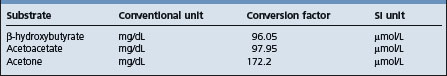
Blood glucose levels are reduced from the normal of approximately 50 mg/dL to 20–40 mg/dL. Ketosis secondary to other diseases is usually accompanied by blood glucose levels above 40 mg/dL and often above normal. Conversion factors are shown in Table 29.7.
Most commonly, plasma or serum β-hydroxybutyrate (BHBA) measured in SI units is used for analysis of ketonemia. BHBA is the predominant circulating ketone body. Plasma concentrations of BHBA significantly correlate with plasma concentrations of acetoacetate but acetoacetate is unstable in samples whereas BHBA is relatively stable.2 Normal cows have plasma BHBA concentrations less than 1000 μmol/L, cows with sub-clinical ketosis have concentrations greater than 1400 μmol/L, and cows with clinical ketosis have concentrations often in excess of 2500 μmol/L. Plasma BHBA shows some diurnal variation in cows fed twice daily with peak concentrations occurring approximately 4 h after feeding and higher concentrations in the morning than in the afternoon. This is not seen in cows fed a total mixed ration ad libitum.50,51
Plasma BHBA is not a cost effective or convenient analysis for routine analysis and cow side monitoring and the content of acetoacetate or BHBA in urine and milk are used for these purposes. Concentrations of BHBA and acetoacetate in urine and milk are less than those in blood and the correlation coefficients for blood and milk BHBA and blood and milk acetoacetate are 0.66 and 0.62, respectively.52
Cowside tests have the advantage of being inexpensive, giving immediate results, and they can be used as frequently as necessary. A minor source of error is that the concentration of ketone bodies in these fluids will depend not only on the ketone level of the blood but also on the amount of urine excreted or on the milk yield. Milk is less variable, easier to collect and may give fewer false negatives with subclinical ketosis.
Milk and urine ketone levels have been traditionally detected by the reaction of acetone and acetoacetate with sodium nitroprusside and can be interpreted in a semi-quantitative manner based on the intensity of the reaction. Several products are available commercially as test powders or strips are commonly accompanied by a color chart that allows a classification in grades such as negative, trace, small, moderate, large, based on the intensity of the color of the reaction.
Conventional wisdom is that milk powder tests are not sensitive for detection of subclinical ketosis (report too many false negatives) and urine tests are not sufficiently specific (report too many false positives).53
The sensitivity and specificity of the nitroprusside powder test with milk in various studies is reported as 28–90% and 96–100%, respectively.16,53,54 More recently, a milk strip test detecting the presence of BHBA in milk is available and is graded on the concentration of BHBA in μmol/L. In different studies it has a reported sensitivity and specificity of 73–96% and 69–96%, respectively.53-57 These variations are, in part, due to different plasma BHBA reference values (1200 and 1400 μmol/L) for designation of subclinical ketosis and different cut points used in urine BHBA. Somatic cell counts greater than 1 million cells/mL will cause an elevation in reading of both the BHBA strip test and the nitroprusside tests.
A nitroprusside tablet has a reported sensitivity and specificity of 100% and 59%, respectively, compared with serum BHBA concentrations above 1400 μmol/L16 and a nitroprusside strip test a reported sensitivity and specificity of 78% and 96% with a urine cut point corresponding to ‘small’ on the color chart or 49% and 99% with a urine cut point corresponding to ‘moderate’ on the color chart.53 BHBA test strips when used with urine has a reported sensitivity and specificity of 73% and 96%, respectively at a urine cut point of 100 μmol/L BHBA and 27% and 99% at a urine cut point of 200 μmol/L BHBA.53
One author has suggested that the nitroprusside urine strip test or the BHBA milk strip test are best for screening individual cows for ketosis in herds with average prevalence but that the nitroprusside powder test would have limited application.53
Milk fat concentration tends to increase and milk protein concentration tends to decrease during postpartum negative energy balance. A fat to protein ratio >1.5 in first day teat milk is indicative of a lack of energy supply in the feed and of risk for ketosis.25
Clinical chemistry and hematology.
White and differential cell counts are variable and not of diagnostic value for ketosis.
There are usually elevations of liver enzymes but liver function tests are within the normal range. Liver biopsy is the only accurate method to determine the degree of liver damage.58 Plasma concentrations of non-esterified fatty acids are elevated as are cholesterol concentrations and bilirubin. Bilirubin is not a sufficiently sensitive indicator to asses the extent of fat mobilization and liver function.26,27 Liver glycogen levels are low and the glucose tolerance curve may be normal. Volatile fatty acid levels in the rumen are much higher in ketotic than in normal cows and the ruminal levels of butyric acid are markedly increased relative to acetic and propionic acids. There is a small but significant fall in serum calcium levels (down to about 9 mg/dL (2.25 mmol/L)), due probably to increased loss of base in the urine to compensate for the acidosis.
The disease is not usually fatal in cattle but fatty degeneration of the liver and secondary changes in the anterior pituitary gland and adrenal cortex may be present.
The clinical picture is usually too indefinite, especially in cattle, to enable a diagnosis to be made solely on clinical grounds. General consideration of the history, with particular reference to the time of calving, the duration of pregnancy in ewes and the feeding program, and biochemical examination to detect the presence of hypoglycemia, ketonemia, and ketonuria are necessary to establish a diagnosis.
In cattle, a number of effective treatments are available but in some affected animals, the response is only transient; in rare cases, the disease may persist and cause death or necessitate slaughter of the animals. Most of these cases are secondary and failure to respond satisfactorily to treatment is due to the primary disease.
The rational treatment in ketosis is to relieve the need for glucose formation from tissues and allow ketone body utilization to continue normally. Theoretically, the simplest means of doing this is by the administration of glucose replacement therapy. The effect of the administration of glucose is complex but it allows the reversal of ketogenesis and the establishment of normal patterns of energy metabolism.12 Ideally, treatment should be at an early stage of the disease to minimize loss and with subclinical ketosis this requires biochemical testing.52
The IV injection of 500 mL of a 50% solution of glucose results in transient hyperglycemia, increased insulin and decreased glucagon secretion, and reduced plasma concentration of non-esterified fatty acids. It effects a marked improvement in most cows but relapses occur commonly unless repeated treatments are used. This is probably due to the transience of the hyperglycemia or insufficient dosing – the dose required varies directly with the amount of lactose being lost in the milk. A significant proportion of the administered glucose is lost to urinary excretion. SC injections prolong the response but are not recommended as they cause discomfort, and large unsightly swellings, which often become infected, may result. IP injections of 20% solution of dextrose may be used alternatively but are also accompanied by risk of infection.
Other sugars, especially fructose, either alone or as a mixture of glucose and fructose (invert sugar), and xylitol, have been used in an effort to prolong the response but idiosyncrasies to some preparations, in the form of polypnea, muscle tremor, weakness and collapse, can occur while the injection is being given.
To overcome the necessity for repeated injections, propylene glycol can be administered as a drench. The traditional does is 225 g twice daily for 2 days, followed by 110 g daily for 2 days to cattle, but higher volumes are also used. Propylene glycol (200–700 g daily), or salts of propionic acid, can be administered in the feed and give good results. Administration in feed is preferred by some because this method avoids dangers of aspiration with drenching; however, cows not used to its inclusion in the feed may show feed refusal. It is recommended that for best results, dosing with these preparations be preceded by an IV injection of glucose.
Parenteral infusions of glucose solutions and the feeding of glycerol depress the fat content of milk, and the net saving in energy may favorably influence response to these drugs. Glycerol and propylene glycol are not as efficient as glucose because conversion to glucose does utilize oxaloacetate. Propylene glycol is absorbed directly from the rumen and acts to reduce ketogenesis by increasing mitochondrial citrate concentrations; its metabolism to glucose occurs via conversion to pyruvate with subsequent production of oxaloacetate via pyruvate carboxylase.12
Because of its glucogenic effect, sodium propionate is theoretically a suitable treatment but when administered in 110–225 g doses daily, the response in cattle is often very slow. Lactates are also highly glucogenic but both calcium and sodium lactate (1 kg initially, followed by 0.5 kg for 7 days) and sodium acetate (110–500 g/d) have given less satisfactory results than those obtained with sodium propionate. Ammonium lactate (200 g for 5 days) has however, been used extensively with reported good results.
Lactose, in whey, or in granular form in the diet, can increase dry matter intake but increases ruminal butyrate and plasma BBHA concentrations.59
The efficiency of glucocorticoids in the treatment of bovine ketosis has been demonstrated in both experimental and field cases. Hyperglycemia occurs within 24 h of administration and appears to result from a repartitioning of glucose in the body rather than from gluconeogenesis.7 Historically, many preparations have been used successfully but current drugs are more potent, require lower dosage, and have fewer side-effects. A hyperglycemic state is produced for 4–6 days in ketotic cows given 10 mg of dexamethasone 21-isonicotinate and other preparations such as dexamethasone sodium phosphate (40 mg) and flumethasone (5 mg) are also used. Label regulations vary between countries and in general, the recommendations of the manufacturer with regard to use and dosage should be followed. Profound hypokalemia with high case fatality is a potential sequel to prolonged repeated therapy of ketosis with isoflupredone acetate.60 Response of cows with primary ketosis to treatment with corticosteroids and IV glucose is superior, with fewer relapses, than therapy with corticosteroids or glucose alone.61
Insulin facilitates cellular uptake of glucose, suppresses fatty acid metabolism and stimulates hepatic gluconeogenesis. It is administered in conjunction with either glucose or a glucocorticoid and may be of particular value in early-onset cases of ketosis that are unresponsive to glucose or corticosteroid therapy6 but is not commonly used. The dose of protamine zinc insulin is 200–300 IU per animal administered SC every 24–48 h as required.
have also been used for treatment of lactational ketosis and ketosis in late pregnant cows that are overfat, stressed, or have twin fetuses. Experimentally, 60 mg and 120 mg of trenbolone acetate are effective as single injections but no extensive field trials are recorded and the drug is banned for use in food animals in most countries.
Vitamin B12 and cobalt are indicated in regions where cobalt deficiency is a risk factor for ketosis. They are sometimes administered to cattle with ketosis in regions where cobalt deficiency does not occur but their therapeutic value is not proven. Cysteamine (a biological precursor of coenzyme A) and also sodium fumarate have been used to treat cases of the disease. Reported results were initially good but the treatment has not been generally adopted. The recommended dose rate of cysteamine is 750 mg IV for three doses at 1–3 day intervals.
Glucagon although ketogenic is strongly gluconeogenic and glycogenolytic and glucagon concentrations are decreased in the blood of fat cows at calving and cows with ketonemia. It could be of value in prevention and therapy but it would require a prolonged delivery system as it has a very short physiologic half life and its effects following a single injection are short-lived.62
The control of clinical ketosis is integrally related to the adequate nutrition of the cow in the dry and lactating period. This encompasses details such as:
It is difficult to make general recommendations for the control of the disease because of the many conditions under which it occurs, its probable multiple etiology, and feeding systems that vary from those that feed components separately to those that feed total mixed rations. Cows should neither have been starved nor be overfat at calving. Careful estimation of diets by reference to feed value tables is recommended and detailed recommendations on diet and management are available with the caveat that planned rations can deviate from feed bunk rations and feed bunk dry matter and actual dry matter intake may not be the same. Too low a feeding frequency and the feeding of concentrates separate from roughage rather than as a total mixed ration can lead to an increase in rates of ketosis.
In the USA, dry cows are typically divided into two groups; ‘far off’ and ‘close up’ cows. ‘Far off’ cows are generally fed to National Research Council (NRC) dry cow feeding guidelines and ‘close up’ cows are given a ration that is halfway between the dry cow and early lactation ration starting 3 weeks before estimated calving and aiming to maximize dry matter intake and provide adequate energy.2,63-65 Practical recommendations based on British feeding standards and units are also available.66,67
In high-producing cows being fed stored feeds, poor quality roughage commonly leads to acetonemia. Wet ensilage containing much butyrate, and moldy or old and dusty hay, are the main offenders. In concentrates, it is the change of source which creates off-feed effects and precipitates attacks of acetonemia.
Cows that are housed should get some exercise each day and in herds where the disease is a particular problem during the stabling period, the cattle should be turned out to pasture as soon as possible in the spring.
The ration should contain adequate amounts of cobalt, phosphorus and iodine.
If there is a high incidence in a herd receiving large quantities of ensilage, reduction of the amount fed for a trial period is indicated.
Propylene glycol is used for the prevention of clinical and subclinical ketosis. Traditionally, propylene glycol has been drenched to cattle in early lactation at doses varying from 350 to 1000 mL daily for 10 days after calving. There is a linear effect of dose on plasma glucose.68 Propylene glycol can also added to feed and is frequently present in commercial feed product but a bolus dose of propylene glycol is more effective in raising blood glucose than incorporation in feed.2 A dose of 1 L per day given as an oral drench for 9 days prior to parturition has also been shown efficacious.69 At doses above 500 mL administered by drench or present in feed some cows may develop rapid and shallow respiration, ataxia, salivation, and somnolence.
Glycerol can be substituted for propylene glycol at equivalent dose rates. A preliminary report of a small experimental study with larger doses of glycerol showed that glycerol given orally at a dose of 1, 2, or 3 L elevated blood glucose concentrations to 16, 20, and 25% of pre-treatment values at 0.5 h after treatment and that these concentrations remained elevated for 8 h. Staggering, depression and diuresis were observed in some cows given the 2 or 3 L dose but this could be prevented by administering the glycerol in a large (37 L) volume of water. It concluded that a dose of 1 L was effective in increasing milk production and reducing urinary letones.70 Glycerol, fed as a constant component in the transition dairy cow diet is not effective, and possibly may be ketogenic when fed continually.71 Glycerol should only be used as drench in hypoglycemic cows and not fed as a component of the diet.
Propionic acid absorbed across the rumen wall is transported to the liver where it is converted to glucose via gluconeogenesis to result in an increase in serum blood glucose levels. Older literature reports that 110 g/d fed daily for 6 weeks, commencing at calving, has given good results in reducing the incidence of clinical bovine ketosis and improving production, but is not palatable and has the risk of reducing feed intake. In controlled trials, feeding energy supplements containing propionic acid and/or its salts for 3 weeks prepartum and 3 weeks post partum had a beneficial effect on milk production but a variable effect on reducing subclinical ketosis.72,73
Ionophores alter bacterial flora of the rumen, leading to decreases in Gram-positive bacteria, protozoa, and fungi and increases in Gram-negative bacteria. The net effect of these changes in bacterial flora is increased propionate production and a decrease in acetate and butyrate production providing increased gluconeogenic precursors. Field trials with monensin have demonstrated a reduction in plasma BHBA and a reduced prevalence of clinical ketosis.29,74,75 It can be administered as a slow release capsule to cattle 2–4 weeks before calving. The capsule contains 32 g of monensin and releases approximately 335 mg monensin a day for 95 days. Ionophores are not labeled for inclusion in lactating cow rations in some countries.
Niacin is antilipolytic and induces increases in blood glucose and insulin but there is conflicting evidence that niacin given in the feed has a beneficial effect on subclinical ketosis in cattle.1,20,76 It has been suggested that it should be supplemented from 2 weeks prior to parturition to 12 weeks post partum.77
Biochemical monitoring of herds for subclinical ketosis and adequacy of periparturient feeding can be conducted using blood glucose estimations on a sample of cows in their second week of lactation.55 Blood glucose levels of below 35 mg/dL (1.9 mmol/L) suggest subclinical ketosis. For individual cows, blood glucose estimations should be done at about 14 days after calving. This method of monitoring is expensive.
More commonly, testing for ketones in urine or milk of cows in their first or second week of lactation is recommended for early detection of ketosis and early treatment to prevent milk loss and ketosis-associated diseases. One recommendation is to routinely test such cows on a specific day each week.19 This should be coupled with body condition scoring to monitor the efficacy of the nutritional program. Condition scoring at dry off, mid dry period, calving, calving plus 20–50 days, and two to three subsequent periods in lactation have been suggested.28,67 Plasma glucose coupled with plasma BHBA are the best predictive model for monitoring energy balance of cattle on a pasture diet with milk acetone the best ‘on-farm’ predictor. However, the variation in milk acetone is high and frequent sampling is required for accurate estimation.78
Automated monitoring by in-line measurements of ketone bodies in milk have been studied and may be of particular value in large dairies. BHBA is proposed as the candidate as it is the more robust in milk, and where cows are fed a TMR, is not subject to significant diurnal variation. It can be measured with a fluorometric method that requires no pretreatment of the milk.79,80
Littledike ET, Young JW, Beitz DC. Common metabolic diseases of cattle: ketosis, milk fever, grass tetany and downer cow complex. J Dairy Sci. 1981;64:1465-1482.
Kronfield DS. Major metabolic determinants of milk volume, mammary efficiency, and spontaneous ketosis in dairy cows. J Dairy Sci. 1982;65:2204-2212.
Baird GD. Primary ketosis in the high-producing dairy cow: clinical and subclinical disorders, treatment, prevention and outlook. J Dairy Sci. 1982;65:1-10.
Kelly JM, Whitaker DA. Subclinical ketosis and dairy cows. Vet Ann. 1984;24:83-93.
Brockman RP, Loorveld B. Hormonal regulation of metabolism in ruminants; a review. Livestock Prod Sci. 1986;14:313-334.
Herdt TH, Emery RS. Therapy of diseases of ruminant intermediary metabolism. Vet Clin North Am Food Anim Pract. 1991;8:91-106.
Lean IJ, et al. Bovine ketosis: a review. I Epidemiology and pathogenesis. Vet Bull. 1991;61:1209-1218.
Lean, et al. Bovine ketosis: a review. II Biochemistry and prevention. Vet Bull. 1992;62:1-13.
Gerloff BJ. Dry cow management for the prevention of ketosis and fatty liver in dairy cows. Vet Clin North Am Food Anim Pract. 2000;16:283-292.
Duffield T. Subclinical ketosis in lactating dairy cattle. Vet Clin North Am Food Anim Pract. 2000;16:231-253.
Herdt TH. Ruminant adaption to negative energy balance influences on the etiology of ketosis and fatty liver. Vet Clin North Am Food Anim Pract. 2000;16:215-230.
Geishauser T, et al. Monitoring for subclinical ketosis in dairy herds. Comp Cont Educ Pract Vet: Food Anim Pract. 2001;23:S65-S71.
Nielsen NI, Ingvartsen KL. Propylene glycol for dairy cows. A review of the metabolism of propylene glycol and its effects on physiological parameters, feed intake, milk production and risk of ketosis. Anim Feed Sci Technol. 2004;115:191-213.
Huxley J. Optimising health, productivity and welfare of dairy cattle: on-farm nutrition. In Practice. 2004;26:466-475.
Hayirli A, Grummer RR. Factors affecting the dry matter intake prepartum in relationship to etiology of peripartum lipid-related metabolic disorders: a review. Can J Anim Sci. 2004;84:337-347.
Oerzel GR. Monitoring and testing herds for metabolic disease Vet Clin North Am Food Anim Pract. 2004;20:651-674.
1 Lean IJ, et al. Vet Bull. 1992;62:1.
2 Duffield T. Vet Clin North Am Food Anim Pract. 2000;16:231.
3 Herdt TH. Vet Clin North Am Food Anim Pract. 2000;16:215.
4 Steen A, et al. J Vet Med Assoc. 1997;44:521.
5 Holtenuis P, Holtenuis K. J Vet Med Assoc. 1996;43:579.
6 Herdt TH, Emery RS. Vet Clin North Am Food Anim Pract. 1992;81:91.
7 Mills SE, et al. J Dairy Sci. 1986;69:362.
8 Drackley JK, et al. J Dairy Sci. 1992;75:1622.
9 Rice DA, et al. Vet Rec. 1983;113:495.
10 Andersson L, Lyndstrom K. Zentralbl Veterinarmed. 1985;32:15.
11 Tveil B, et al. J Dairy Sci. 1992;75:2421-2433.
12 Sanders DE. Comp Cont Educ Pract Vet. 1989;11:757.
13 Knight CH. Proc Nutr Soc. 2001;60:527-537.
14 Ingvartsen KL, et al. Livestock Product Sci. 2003;83:277.
15 Duffield TF, et al. Can Vet J. 1997;38:713.
16 Nielen M, et al. Can Vet J. 1994;35:229.
17 Dohoo IR, Martin SW. Am J Comp Med. 1984;48:1.
18 Carrier J, et al. J Dairy Sci. 2004;87:3725.
19 Geishauser T, et al. Comp Cont Educ Pract Vet: Food Anim Pract. 2001;23:S65.
20 Lean IJ, et al. Vet Bull. 1991;61:1209.
21 Bigras-Poulin M, et al. Prev Vet Med. 1992;10:79.
22 Nir O. Acta Vet Scand Suppl. 2003;98:21.
23 Uribe HA, et al. J Dairy Sci. 1995;78:421.
24 Gillund P, et al. J Dairy Sci. 2001;84:1390.
25 Heuer C, et al. J Dairy Sci. 1999;82:295.
26 Busatol A, et al. J Vet Med A. 2002;49:455.
27 Ill-Hwa K, Gook-Hyun S. Theriogenol. 2003;60:1445.
28 Markusfeld O, et al. Vet Rec. 1997;141:67.
29 Duffield TF, et al. J Dairy Sci. 1998;81:2866.
30 Tweit B, et al. J Dairy Sci. 1992;75:2421.
31 Dohoo IR, et al. Prev Vet Med. 1984;2:665-671.
32 Eriksson JA, Wretler E. World Rev Anim Prod. 1990;25:29.
33 Grohn YT, et al. J Dairy Sci. 1989;72:1876.
34 Smith TR, et al. J Dairy Sci. 1997;80:1569.
35 Lean IJ, et al. Res Vet Sci. 1994;57:200.
36 Morris CA, et al. Proc N Z Soc Anim Prod. 1992;52:21.
37 Tyler JW, et al. J Am Vet Med Assoc. 1994;204:1665.
38 Geishauser T, et al. Vet Clin North Am Food Anim Pract. 2000;16:255.
39 Vagirholm I, et al. Prev Vet Med. 1991;10:195.
40 Miettinen PVA, Setala JJ. Prev Vet Med. 1993;17:1.
41 Harman JL, et al. Am J Vet Res. 1996;57:640.
42 Miettinen PVA. J Vet Med Assoc. 1994;41:102.
43 Veenhuizen JJ, et al. J Dairy Sci. 1991;74:4238.
44 Goff JP, et al. J Dairy Sci. 1997;80:1260.
45 Lacetera N, et al. Am J Vet Res. 2002;63:958.
46 Hoeben D, et al. Vet Immunol Immunopath. 1997;58:165.
47 Whitaker DA, et al. Vet Rec. 2004;155:43.
48 Laven R, Howe M. Vet Rec. 2004;155:63.
49 Anderson L, Emanuelson LI. Prev Vet Med. 1985;3:449.
50 Eicher R, et al. Acta Vet Scand. 1999;60:1493.
51 Nielsen N, et al. Acta Vet Scand. 2003;98:305.
52 Enjalbert FMC, et al. J Dairy Sci. 2001;84:583.
53 Carrier J, et al. J Dairy Sci. 2004;87:3725.
54 Geishauser TK, et al. J Dairy Sci. 1988;81:438.
55 Geishauser TK, et al. J Dairy Sci. 2000;83:296.
56 Osborne TM, et al. Proc Am Assoc Bov Practit. 2002;35:188.
57 Dirksen G, Breitner WJ. Vet Med Assoc. 1993;40:779.
58 Grohn Y, et al. J Dairy Sci. 1983;66:2320.
59 DeFrain JM, et al. J Dairy Sci. 2004;87:2486.
60 Sielman ES, et al. J Am Vet Med Assoc. 1997;210:240.
61 Shpigel NY. J Am Vet Med Assoc. 1996;208:1702.
62 Hippen AR. Vet Clin North Am Food Anim Pract. 2000;16:267.
63 Oetzel GR. Comp Cont Educ Pract Vet: Food Anim Pract. 1998;20:391.
64 Gerloff B. J Vet Clin North Am Food Anim Pract. 2000;16:283.
65 Sniffen CJ, Herdt TH. Vet Clin North Am Food Anim Pract. 1992;81:1.
66 Baird GD, et al. Br Vet J. 1974;130:214-318.
67 Huxley J. In Practice. 2004;26:466.
68 Nielsen NI, Ingvartsen KL. Anim Feed Sci Technol. 2004;115:191.
69 Studer VA, et al. J Dairy Sci. 1993;76:2931.
70 Goff JP, Horst RL. Acta Vet Scand Suppl. 2003;98:214.
71 DeFrain JM, et al. J Dairy Sci. 2004;87:4195.
72 Ballard C, et al. Anim Feed Sci Technol. 2001;93:55.
73 Mandebvu P, et al. Anim Feed Sci Technol. 2003;105:81.
74 Heuer YH, et al. J Dairy Sci. 2001;84:1085.
75 Duffield TF, Bagg RN. Aust Vet J. 2000;41:388.
76 Neilsen N, Ingvartsen KL. Acta Vet Scand Suppl. 2003;98:306.
77 Hutjens MF. Vet Clin North Am Food Anim Pract. 1992;81:525.
78 Clark CE, et al. Livestock Prod Sci. 2005;94:199.