As mentioned, input conveyed through spinal afferent fibers is concerned with visceral sensation, especially visceral nociception. The sensations that reach consciousness are poorly localized and, in the case of pain, may be referred and may produce hyperalgesia (see Appendix II for definitions of terms related to pain), as well. The cell bodies of these fibers are located in the dorsal root ganglia of the thoracic and upper lumbar dorsal roots. These visceral afferent fibers travel from the peripheral receptors in the cardiac, pulmonary, and splanchnic nerves; continue through the sympathetic trunk, white rami communicantes, and dorsal roots; and finally terminate in the spinal cord.
As mentioned, pain of visceral origin differs from somatic pain, both in the neurophysiologic characteristics associated with the production of the pain and more obviously in the clinical presentation to the painful stimuli. Numerous conditions help to define visceral pain. Overdistension of the hollow muscular walled organs, ischemia and the resulting release of chemicals such as bradykinin, and inflammation of the viscera are stimuli that induce pain. However, the severity of the condition resulting in pain is not always a reflection of the severity of the pain experienced. For example, mild pain may be experienced with early appendicitis, whereas severe pain may be present with the fairly normal presence of gas in the GI tract. The least sensitive of the viscera tend to be the solid organs, whereas the serosae of the hollow organs appear to be the most sensitive. Also, a mildly painful or even nonpainful stimulus can result in severe pain (Al-Chaer & Traub, 2002) if an organ is already inflamed or altered pathologically (e.g., irritable bowel syndrome [IBS]). Visceral pain often is described as being either true or referred. True visceral pain is diffuse pain that is perceived as originating from deep, midline structures in the thorax or abdomen. It is often accompanied by the sense of nausea and ill-being. An example of this is during the early stage of appendicitis when the pain is initially felt in the midline. Referred pain also is diffuse but is localized to a distant cutaneous site or to muscles. The following is a list of important clinical characteristics associated with visceral pain:
1. Not all viscera (e.g., liver, kidney) are sensitive to painful stimuli. This is because of functional variances of the peripheral receptors and the phenomenon that some receptors of visceral afferents, when stimulated, do not evoke a conscious perception (Cervero & Laird, 1999).
2. Visceral pain is not always linked to an injurious event. For example, distension of the bladder, although painful, does not damage the tissue, whereas cutting the gut wall is not painful.
3. Visceral pain is referred to distant locations based on the convergence of visceral afferent fibers and somatic afferent fibers on viscerosomatic neurons in the spinal cord.
4. Visceral pain is poorly localized and diffuse. This is a result of such factors as the following:
a. The viscera are less densely innervated compared with somatic structures.
b. In the gut there are few (7%) visceral afferent fiber cell bodies in the dorsal root ganglia (DRG) compared to the number of somatic afferent cell bodies in DRG (Beyak et al., 2006).
c. In the gut the sensory innervation of an organ arises from a range of DRG although there are peak distributions. This results in a generalized, overlapping, and viscerotropic distribution of visceral afferent fibers (Knowles & Aziz, 2009).
d. The distribution of visceral afferent fibers terminating in cord segments is broader than that of somatic fibers, spanning as many as five segments (Chandler, Zhang, & Foreman, 1996; Jänig, 1996).
e. A specific visceral sensory pathway is lacking.
f. Viscerovisceral convergence occurs in the cord (Al-Chaer & Traub, 2002).
5. Visceral pain is accompanied by autonomic reflex and motor responses such as vomiting, changes in heart rate, or hypertonicity in skeletal muscles.
Peripheral receptors are located in the blood vessels, walls of hollow organs, parenchyma of visceral organs, and serosae (outer covering of certain organs). The receptors conveying nociception are sensitive to chemical, mechanical, and thermal stimuli and are classified physiologically as being either high- or low-threshold receptors. The high-threshold receptors respond to noxious mechanical stimuli and are the lone receptors found in organs in which pain is the only conscious sensation (e.g., ureter). There is a paucity of these in organs that respond to both innocuous and noxious sensations. Low-threshold receptors respond primarily to innocuous mechanical stimuli but some encode a wide range of stimuli that extends into the noxious range. These are located, for example, in the gut and bladder (Cervero & Laird, 1999; Al-Chaer & Traub, 2002; Knowles & Aziz, 2009). The distribution of these receptors varies among the visceral organs. A third type of receptor, called the silent nociceptor, also is located in the viscera. The silent receptors are minimally responsive or unresponsive to normally occurring stimuli, but become activated by inflammation and various chemical insults.
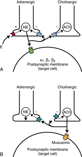
Because spinal visceral afferents are similar to somatic afferents in their basic morphology and in that their first synapse is in the dorsal horn, it is likely that the mechanism of visceral pain transmission is similar to the mechanism of somatic pain transmission. In the context of this chapter, the discussion of visceral pain transmission will be based on studies of pain mechanisms relative to the GI system. (See Chapter 11 and Appendix II for a discussion of terms related to pain and pain referral and a discussion of peripheral and central hyperalgesia [sensitization] of nociception from somatic tissues related to the spine.) Both high and low threshold afferent fibers are activated in brief acute visceral pain events. In this case a noxious stimulus initiates the opening of voltage-gated sodium channels, which produces an action potential. The activation of the sodium channels can occur directly or secondarily by the activation of transducer channels that respond to noxious mechanical, chemical, and thermal stimuli (Fig. 10-26, A). Examples of these channels, transient receptor potential (TRP), purinoceptors (P2X) and acid-sensing ion channels (ASICs), have been identified on neurons in the gut. If the stimulation is prolonged or repetitive (e.g., in hypoxia or inflammation), a change in the chemical environment allows the nociceptors (including the now activated silent nociceptors) to fire at lower thresholds than they would have in an acute noxious event. This causes decreased pain thresholds at the location of the injury, resulting in the phenomenon called primary hyperalgesia. This form of stimulus-evoked nociceptor plasticity is referred to as peripheral sensitization. The peripheral sensitization of these nociceptors is caused by the release of chemical mediators from the injured or inflamed tissue cells, which subsequently lower the firing threshold of the nociceptors. These events result in nociceptors being sensitive to and activated by normal innocuous stimuli. There is evidence suggesting that some GI disorders such as gastroesophageal reflux disease demonstrate peripheral sensitization. Using GI disorders as an example, it has been suggested (Knowles & Aziz, 2009) that the following three membrane events result in lowering of the threshold (Fig. 10-26, B): (1) G protein–coupled receptors are activated by sensitizers such as kinins, biogenic amines (histamine and 5-HT), prostanoids (prostaglandin E2 [PGE2] and growth factors), proteases, chemokines, and cytokines; decreased pH; and increased levels of ATP. The binding to the receptors activates intracellular signaling pathways, which in turn reduce the transduction thresholds in cation channels. (2) Neuroimmune interactions occur with neighboring epithelial cells and inflammatory cells such as mast cells and lymphocytes. This interaction causes the release of bioactive amines, which stimulate the release of neuropeptides such as substance P and CGRP (neurogenic inflammation). These in turn stimulate nearby target cells, which release mediators including nerve growth factor (NGF). (3) NGF activates a phenotypic switch through retrograde signaling in the dorsal root ganglion cell bodies and up-regulates neuropeptide expression, especially cations. These proteins can be transported centrally and peripherally, causing an extended peripheral and central sensitized phenotype (Knowles & Aziz, 2009).
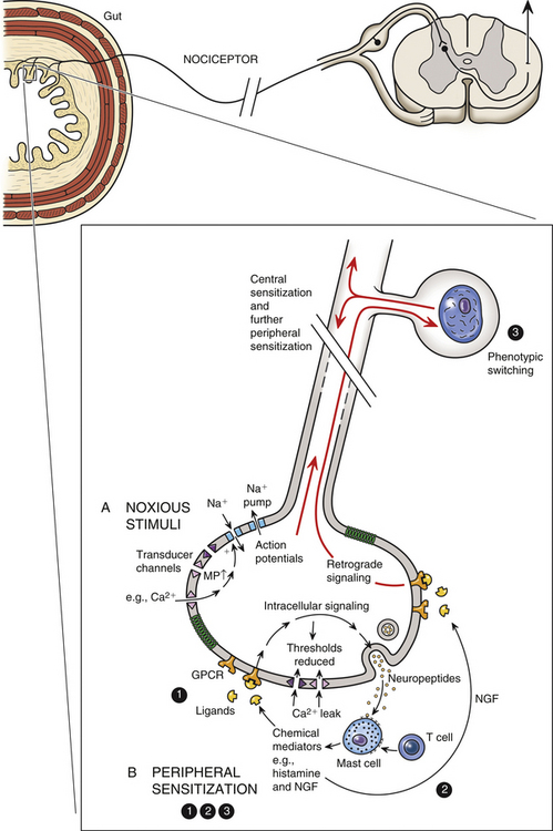
FIG. 10-26 Visceral nociception. A, Noxious stimuli (mechanical, chemical, thermal) activate Na+ channels directly or indirectly through the utilization of transducer channels, resulting in an action potential. B, Peripheral sensitization. The following three events may cause the threshold for nociceptor firing to be lowered: (1) Sensitizing mediators such as histamine and kinins bind to G protein–coupled receptors, which initiate intracellular signaling pathways. This results in lowering the thresholds for activating transducer channels and ultimately decreasing pain thresholds. (2) Biogenic amines released by nearby immune cells (neurogenic inflammation) stimulate the release of neuropeptides such as substance P, which in turn activates the release of inflammatory mediators such as nerve growth factor (NGF). (3) Through retrograde signaling, NGF acts on the nociceptor cell body, promoting a switch in the phenotype and leading to the up-regulation of neuropeptide and cation channels. These proteins can be transported to peripheral or to central terminal sites on the nociceptor, allowing for continued sensitization peripherally and sensitization centrally in the dorsal horn (Knowles & Aziz, 2009).
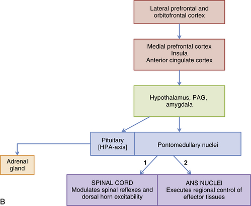
The repetitive firing of these afferent fibers produces a barrage of sensory input to dorsal horn viscerosomatic neurons, which results in hyperexcitability and hyperactivity of these neurons. The continuous firing of the afferent fibers and their release of neurotransmitter causes a progressive increase in the excitability of the dorsal horn neurons referred to as “wind-up.” All of this activity leads to visceral pain, which may be referred, and often is accompanied by hyperalgesia and allodynia. Hyperalgesia is an increase in sensitivity, resulting in enhanced pain perception. Allodynia is the perception of pain from a stimulus that is normally innocuous. The term hyperalgesia is used in this section to refer to both hyperalgesia and allodynia. These dorsal horn neuronal changes are highly organized and specific, corresponding only to those neurons receiving input directly from the injured tissue (Cervero, 2000b). The dorsal horn neurons are described as forming an “irritable focus,” and their collective alterations in excitability are called central sensitization (Cervero & Laird, 1999; Basbaum & Jessell, 2000; Cervero, 2000a,b; Al-Chaer & Traub, 2002). Afferent nociceptive fibers release glutamate (a neurotransmitter common to somatic afferent fibers) as well as neuropeptides (substance P, serotonin) and trophic factors (brain-derived neurotrophic factor), which act as neurotransmitters and modulators. Experimental studies show that receptors for these mediators are located on the postsynaptic membranes of dorsal horn neurons (Knowles & Aziz, 2009). It is speculated that central sensitization may be mediated by the phosphorylation of the N-methyl-d-aspartate (NMDA) glutamate receptors (through intracellular signaling) with a change in their receptor kinetics resulting in an increased responsiveness to glutamate. It is also possible that neurokinin-1 substance P receptors are involved (Basbaum & Jessell, 2000; Cervero, 2000b; Al-Chaer & Traub, 2002; Knowles & Aziz, 2009). In addition, the neuronal hyperexcitability may be facilitated by positive feedback loops established between the dorsal horn neurons and higher centers. The presence of these feedback loops may explain the motor and autonomic reflex responses (e.g., nausea and hypertonicity in abdominal muscles) that accompany visceral pain experiences (Cervero & Laird, 1999). As long as peripheral sensitization of the nociceptors continues during the inflammatory process, normal physiologic (innocuous) stimuli activate afferent fibers and perpetuate the increased volley of sensory input to the “irritable focus” that was initiated by the acute injury. The increased excitability of these neurons in turn facilitates and sustains the effects of the sensory input from the viscera, resulting in an increase in pain intensity and duration. In addition, the normal function of the affected viscera may be altered because of the injury and inflammation, resulting in further activation of the sensitized nociceptors and even the recruitment of more distant nociceptors. Thus the presence of the central sensitization phenomenon in conjunction with normal physiologic visceral afferent activity results in the persistence of visceral pain. Central sensitization may be the mechanism that produces hyperalgesia and allodynia both in sites to which pain is referred (referred hyperalgesia) (see section Central Projections and the Referral of Pain) and in the same or nearby viscera (visceral hyperalgesia). The processing of nociceptive input in the dorsal horn can be modulated by descending pathways from supraspinal regions. The PAG receives input from the anterior cingulate cortex and is able to modulate nociceptive input by activating neurons in the rostral ventromedial medulla and dorsolateral pons. These in turn project to the dorsal horn. Also, PAG activation of brain stem regions such as the locus ceruleus (noradrenaline region), raphe nuclei, and the rostral ventrolateral medulla results in the initiation of a gating effect on nociceptive input in the dorsal horn (Knowles & Aziz, 2009).
Certain abdominal and pelvic disorders are characterized by persistent abnormal visceral sensations (including allodynia and hyperalgesia). These sensations are commonly organized under the term of visceral hypersensitivity (Vergnolle, 2010). As mentioned previously, the initial inflammatory process or infection results in drastic changes in neuronal functions and membrane receptor expression that appear to be caused by the release of inflammatory mediators. These chemicals cause a hyperexcitabiltiy in the membranes of afferent fibers innervating the diseased organ. Once the acute inflammatory or infectious state is resolved, instead of returning to normal function, visceral afferents remain hypersensitized, producing chronic pain symptoms. In this postinflammatory stage, it is thought that the chemical mediators may not be directly responsible for the hyperexcitability of the visceral afferent fibers. Instead, the mediators may influence molecular structures on the membrane. Studies of the P2X ligand-gated ionotropic receptors have shown that they may be important for normal mechanosensitivity in the colon and that they may also play a role in postinflammatory hypersensitivity (Vernolle, 2010). In addition, members of the transient receptor potential vanilloid (TRPV) family of ion channels (specifically TRPV1 and TRPV4) and TRPA1 (an ion channel similar and functionally related to TRPV1) have been identified as being involved in nociception and in the development of visceral afferent sensitization and colonic hypersensitivity such as IBS. In fact, it has been suggested that the TRPV4 receptor may function as a central mediator. In this role, the release of chemical mediators involved in the pathophysiology of IBS, such as histamine, serotonin, and protease-activated receptor 2 (PAR2), activate TRPV4, which then initiates colonic hypersensitivity (Christianson et al., 2009; Vernolle, 2010). Knowing the functional importance of these receptors, treatment regimens may be designed to target them rather than target the inflammatory mediators, which have not yet been specifically identified in the postinflammatory tissues.
It has been suggested that conditions of the GI tract that present as chronic unexplained symptoms, including abdominal pain in the absence of any clear pathology, can be divided into two overlapping groups. One group includes gastrointestinal neuromuscular diseases (GINMDs) and the other includes the functional gastrointestinal disorders (FGIDs) (Knowles & Aziz, 2009). The underlying pathology of GINMD appears to be abnormalities in the components involved with normal GI sensory and motor functions, which include the interstitial cells of Cajal, smooth muscle, and the enteric nervous system and its extrinsic neurons. Symptoms of impaired motor activity manifested as slow or blocked transit are common in this group of diseases. Most GINMDs appear later in life and include enteric dysmotility, intestinal pseudoobstruction, and slow-transit constipation but some may appear as congenital defects such as Hirschsprung’s disease. In these disorders the mechanisms underlying the pain may be neuropathic. In FGIDs the predominant and defining symptom is chronic abdominal pain. This group of conditions includes irritable bowel syndrome, functional dyspepsia, functional heartburn, and functional abdominal pain syndrome. The distinguishing pathophysiologic characteristic of FGID is visceral hypersensitivity. The main mechanisms that are believed to underlie visceral hypersensitivity are “peripheral sensitization, central sensitization, altered descending excitatory or inhibitory influences (neural and humoral), and misinterpretation of non-noxious sensation as noxious due to cognitive and emotional biasing” (Knowles & Aziz, 2009). As mentioned earlier, treating these conditions may include using agents that act on voltage-gated ion channels, cation channels, and G protein–coupled receptors in the periphery (i.e., the serotonin receptors) and dorsal horn (i.e., NMDA receptors), thus reducing peripheral and central sensitization.
Visceral afferent fibers entering the spinal cord may initiate reflex responses or synapse on tract neurons. Although they comprise a minority of the total number of afferent fibers entering the spinal cord, they synapse in numerous laminae, including I, II, V, VII, and X (Beyak et al., 2006; Wood, 2007; Knowles & Aziz, 2009; Mayer, 2011).
These predominantly A-delta and C fibers, including those transmitting visceral nociception, enter the dorsolateral tract of Lissauer. They immediately enter the dorsal horn, as well as send collateral branches up and down as many as five segments within Lissauer’s tract before they enter the dorsal horn and synapse. The axons of the second-order neurons project to the brain through numerous ascending pathways including the spinothalamic, dorsal column (see below), spinoreticular, spinomesencephalic/parabrachial, spinohypothalamic, and spinosolitary tracts. The spinothalamic tract projects to the thalamus and from here to the somatosensory cortex (SI and SII) where the localization and intensity of the stimulus are provided. The other tracts project to brain stem nuclei (e.g., the reticular formation) and higher centers such as the amygdala, anterior cingulate cortex, and insula of Reil, which are involved with the unconscious and automatic responses that alter one’s emotional state and behavior. The activation of the cortex by all of these tracts allows a conscious perception of the nociception as pain and also allows the individual to respond to the pain. The spinothalamic tract helps to localize pain, although pain from the viscera is localized much less accurately than pain of somatic origin. Activating the reticular formation permits some localization and a conscious attentiveness to the pain. The widespread distribution to cortical areas beyond the somatosensory cortex and the interconnections subsequently established may account for the often accompanying discomfort and unpleasantness that can produce a particular affective mental state and subsequent behavioral patterns. The anterior cingulate cortex is an integrative center that may be responsible for linking pain and emotional states, and through connections with other neural structures it is an important source of descending pain modulation. The insula appears to have an important function in the integration of visceral, emotional, and cognitive functions. Through its connections with other areas such as the prefrontal cortex it is involved with the higher control of autonomic visceromotor and behavioral responses (Beyak et al., 2006; Wood, 2007; Knowles & Aziz, 2009; Mayer, 2011).
Evidence gained from experimental studies and midline myelotomies (intentionally placed midline lesions of the spinal cord) performed to treat pain from pelvic cancer indicates that visceral information also ascends in the dorsal column of the cord (Cervero & Laird, 1999; Willis et al., 1999; Joshi & Gebhart, 2000; Nauta et al., 2000; Ness, 2000; Westlund, 2000; Al-Chaer & Traub, 2002; Palecek, Paleckova, & Willis, 2002). Postsynaptic dorsal column cells (PSDCs) are located around the central canal of the spinal cord. These cells are responsive to noxious and innocuous cutaneous stimuli, as well as to mechanical and chemical visceral noxious stimuli. Axons from these cells in the sacral cord segments conveying pelvic visceral nociception ascend ipsilaterally in the midline of the dorsal column in an area distinct from mechanoreceptive ascending fibers and synapse in the gracile nucleus. Fibers from thoracic PSDCs carrying thoracic visceral nociception ascend at the lateral edge of the fasciculus gracilis and synapse in the gracile and cuneate nuclei. From these nuclei, axons ascend in the medial lemniscus and terminate in the ventral posterior thalamic nucleus. In addition to the fibers coursing in the medial lemniscus, some fibers synapse in the medullary reticular formation, periaqueductal gray, hypothalamus, amygdala, and medial areas of the thalamus. These dorsal column fibers transmitting visceral nociception to higher centers have been suggested as being as important as or more important than the spinothalamic tract (Beyak et al., 2006; Wood, 2007).
The data that have demonstrated the termination of visceral afferent fibers in the spinal cord gray matter also lend credence to the convergence-projection theory of referred pain (Ruch, 1946). This theory maintains that referred pain occurs because of the high degree of convergence of visceral and somatic afferent fibers on the same pool of viscerosomatic neurons in the cord (see Fig. 10-25). Because somatic pain is more common than visceral pain, the higher centers misread the visceral input as originating from somatic afferent fibers. Therefore pain is referred to the area of skin and deep structures (e.g., muscle, bone) supplied by the somatic afferent fibers that have entered the same cord segments as the visceral afferent fibers. The viscerosomatic neurons, located in laminae I, V, and X (Jänig, 1996) of the cord, are either tract neurons or local interneurons. The site of referral may be to the skin, muscles (or other deep somatic tissues), or both. In addition, referred pain often is accompanied by hyperalgesia. The hyperalgesia, which is an increased sensitivity to pain produced by noxious and innocuous (allodynia—see previous discussion) stimuli, is the result of peripheral sensitization of nociceptors and, more importantly, central sensitization. Although hyperalgesia is evident at the onset of the painful experience, it often persists longer than the pain associated with the initial visceral insult. This may occur for several reasons: (a) the persistence of the previously described plastic changes in the dorsal horn neurons make this area of the spinal cord function independently of peripheral nociceptive activation; (b) changes in the viscera persist longer than the initial focal lesion and send afferent input to the dorsal horn, maintaining the hyperactivity of the neurons; (c) viscerosomatic reflexes are activated back toward the peripheral sites, causing increased sensitivity of nociceptors (including muscle nociceptors) and therefore hyperalgesia; and (d) trophic changes occur in hyperalgesic somatic tissue. Studies in rat models of hyperalgesic muscle have shown morphofunctional changes (essentially characteristics associated with muscle atrophy) indicative of muscles undergoing sustained contractions, which often occur as a consequence of visceral referred pain.
Examples of visceral pain referral to muscle are the referral to abdominal oblique and quadratus lumborum muscles seen in patients experiencing pain from renal calculus, referred pain to the rectus abdominis muscle as a result of biliary calculus, and referred pain to the inferior aspect of the rectus abdominis and pelvic muscles as a result of dysmenorrhea (Vecchiet, Vecchiet, & Giamberardino, 1999; Giamberardino, 2003).
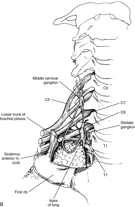
A common example of pain referral to cutaneous sites occurs after a myocardial infarction or angina pectoris episode. The relationship between visceral afferent fibers and the spinothalamic tract for pain originating from the heart was determined from data obtained from experiments on primates. These investigations were designed to demonstrate that cardiac ischemia stimulates cardiac afferent fibers, which in turn synapse on spinothalamic tract cells. Bradykinin, a peptide released from ischemic cells, was injected into cardiac tissue and resulted in stimulation of afferent fibers. By measuring tract neuron discharge rates, it was shown that 15 seconds after bradykinin injection (the time needed for receptor activation), 75% of the spinothalamic tract cells increased their firing rate (Cervero & Foreman, 1990). These data support the theory that visceral afferent fibers converge on the same tract cells on which somatic afferent fibers terminate. The peripheral distribution of these same somatic afferent fibers becomes the general location of the pain referral. The afferent fibers subserving nociception from the heart course primarily in the middle and inferior cardiac nerves and left thoracic cardiac branches (Kiernan, 2009) and eventually enter the first five thoracic cord segments. Pain is most frequently referred superficially to the left side of the chest and left inner arm; however, pain often is referred to the neck and jaw also. It is speculated that the mechanism for this referral pattern is the convergence of cardiac afferent fibers with the upper cervical cord segments. Cardiac afferent fibers travel in sympathetic nerves and the vagus nerve. The vagal fibers enter the medulla and synapse in the nucleus of the tractus solitarius (NTS) and also send branches inferiorly into the C1-3 cord segments (see Relationship between the Dorsal Horn and Trigeminal Nerve in Chapter 9). Fibers from the NTS also may send relay neurons to the C1-2 segments (Chandler, Zhang, & Foreman, 1996). These segments are the site of convergence of trigeminal afferent fibers serving the head, and somatic afferent fibers conveying sensory information from the area served by the C1-3 cord segments. This region of the dorsal horn provides the anatomic substrate for convergence of the trigeminal, somatic, and vagal afferent fibers on tract neurons (Chandler, Zhang, & Foreman, 1996; Chandler et al., 1999; Foreman, 1999) and subsequent misinterpretation of the origin by higher centers. Another theory explaining this referral pattern is that afferent information synapsing in thoracic spinal cord segments may ascend to upper cervical segments via propriospinal fibers. Also, visceral afferent fibers that enter the zone of Lissauer may ascend or descend as many as five cord segments before synapsing in the dorsal horn. These connections may provide a route for cardiac afferent fibers to directly terminate in the upper cervical segments (Chandler, Zhang, & Foreman, 1996), thus referring pain to the neck and even the head (via the connections to the spinal nucleus of the trigeminal nerve).
In order for clinicians to be able to describe the location of a patient’s pain, an abdominal viscus, or a mass in the abdominal cavity, the anterior abdominal surface has been divided into nine arbitrary regions. From superior to inferior, these are as follows: right side—right hypochondrium, right lumbar, right iliac fossa; middle—epigastric, central/umbilical, suprapubic/hypogastrium; left side—left hypochondrium, left lumbar, left iliac fossa. The specfic surface of the body to which visceral pain is referred is related to the embryologic origin of the organ, which is segmentally linked to the somatic innervation. Relative to the abdominal viscera, pain in general is referred to the three middle abdominal regions; that is, organs derived from the foregut (e.g., stomach [although the gastroesophageal junction refers to the subxiphoid and retrosternal areas], gallbladder, pancreas) will refer visceral pain to the central epigastrium region; organs derived from the midgut (e.g., appendix, jejunum, ileum) will refer pain to the umbilical region; and organs derived from the hindgut (e.g., descending colon, anal canal) will refer pain to the suprapubic region. Knowledge of the innervation of the peritoneum is also clinically important. The visceral peritoneum (as well as the pleura), supplied by branches of the afferent fibers (predominantly vagal) innervating the underlying viscera, is sensitive to stretch. When this occurs it causes a poorly localized sensation of discomfort and often initiates cardiovascular reflex responses likely mediated through connections among brain stem nuclei. The parietal peritoneum (and pleura) as well as the overlying skin and muscles is innervated by branches of somatic afferent and efferent nerve fibers. [This difference in innervation is important to note relative to certain surgical procedures involving incisions in the parietal peritoneum. Spinal anesthesia will anesthetize local regions of the parietal peritoneum. However, because the vagus nerve bypasses the cord and synapses in the brain stem, stretching of the visceral peritoneum that it supplies may have serious effects and initiate reflexes that may result in acute vascular instability in the underlying viscera causing ischemia and abdominal pain that is poorly localized (Standring et al., 2008).] The parietal peritoneum, pleura, or pericardium near the diseased organ includes nociceptors that may be activated by the spreading of the disease process that has caused the visceral pain referral. (The capsules of some viscera such as the liver and spleen are also innervated by branches of nerves that innervate the parietal peritoneum.) The somatic afferent fibers of these nociceptors are segmentally related to muscles and skin in the body wall overlying the viscus. The pain pattern, different than the visceral referred pain pattern, is well-localized, lateralized, and often limited to one or two dermatomes for each somatic afferent fiber’s area of distribution. (The involvement of the parietal serous membranes lining the inside wall of the abdominal and thoracic cavities has been described as viscerosomatic pain [FitzGerald & Folan-Curran, 2002].) In addition, these somatic afferent fibers terminate in corresponding segments that innervate muscles. The activation of these afferent fibers may cause reflex muscle contraction and, in the case of the abdominal wall musculature, hypercontractility (guarding) or rigidity. A good example of the change in the nature and location of pain patterns is a diseased appendix. Initially, visceral afferent fibers that course in the splanchnic nerves are stimulated and terminate in the T10 to T11 cord segments on viscerosomatic neurons. The result is poorly localized aching pain and cramping that is referred to the periumbilical region in the T10 to T11 skin surface area of the abdominal wall. The parietal peritoneum adjacent to the appendix is innervated by somatic afferent fibers that terminate approximately in the T12 to L1 cord segments on the right side. Therefore when the parietal peritoneum also becomes inflamed the pain now becomes sharp and localized to the right lower abdominal quadrant (Standring et al., 2008).
Another example of pain referral is that which occurs as a result of biliary calculus (gallstones). Sensory afferents from the gallbladder and bile ducts course in the right greater splanchnic nerve and into the T7 and T8 cord segments. The referral site is to the central epigastrium and, because of the involvement of the overlying somatic peritoneum, to the right upper quadrant of the abdomen and right infrascapular region, corresponding to the T7 and T8 dermatomal patterns. As the disease progresses, inflammation of the peritoneum associated with the diaphragm occurs. The chemical mediators involved with the inflammatory process activate the peritoneal sensory fibers that travel in the phrenic nerve. These afferent fibers terminate in the C3-5 cord segments and thus can refer pain to the top of the shoulder.
Because pain is the most important clinical visceral sensation, knowledge of visceral pain referral patterns and the spinal cord segments to which visceral afferent fibers project (which is the same location as the sympathetic preganglionic cell bodies) is extremely important. This knowledge allows a clinician to more effectively diagnose pathologic conditions occurring in the viscera (Fig. 10-27 and Table 10-2).
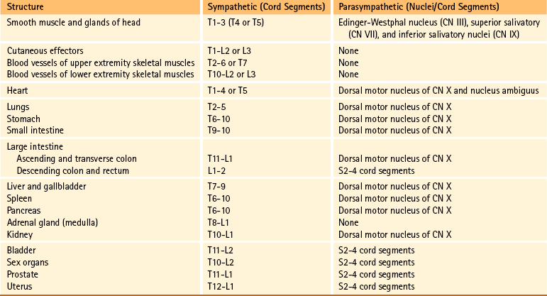
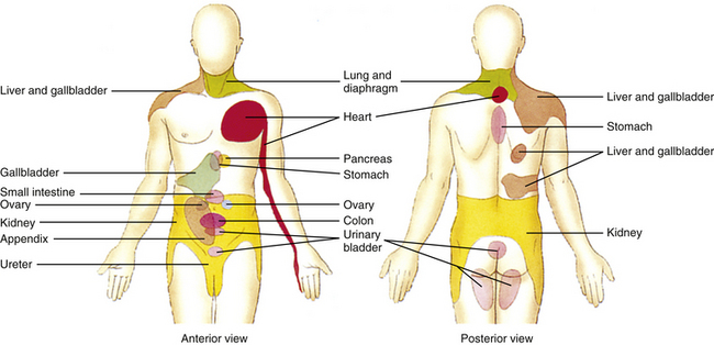
FIG. 10-27 Referral patterns. The colored regions show cutaneous areas to which visceral pain is referred (see text for discussion). (From Tortora GJ & Grabowski SR. [2003]. Principles of anatomy and physiology [10th ed.]. New York: John Wiley & Sons.)
There is evidence suggesting there is viscerovisceral referral and sensitization (called cross-organ sensitization) among different organs as well (Al-Chaer & Traub, 2002; Giamberardino, 2003; Brumovsky & Gebhart, 2010). Based on data from experiments on animals, both central and peripheral mechanisms have been proposed to explain this phenomenon. In the spinal cord there is viscerovisceral convergence on neurons similar to the segmental neuronal arrangement seen in viscerosomatic convergence. Afferent fibers from two different organs converge on the same second-order spinal neurons such that when one organ becomes diseased, the afferent fibers from that organ sensitize the spinal neurons. This results in an increased excitability to afferent fibers transmitting normal stimuli from the other organ producing the signs and symptoms as if the “normal” organ was diseased. For example, a pathologic condition of the esophagus can produce the same symptomology and referral pattern commonly seen in patients with myocardial ischemia. In addition to hyperexcitability of second-order spinal neurons, interneurons in the dorsal horn may also be involved. Afferent fibers from a diseased organ may synapse on interneurons that excite afferent fibers from another organ, resulting in an antidromic axon response leading to neurogenic inflammation in the healthy organ. There are also some data that suggest that there may be viscerovisceral convergence at the level of the brain stem and possibly the thalamus (Brumovsky & Gebhart, 2010).
There are also a number of possibilities that may explain a peripheral mechanism for cross-organ sensitization. One is that there are some dichotomizing primary afferent neurons that innervate viscera—that is, two different organs are innervated by the same primary afferent neuron, thus contributing to cross-tissue/organ sensitization. Another theory is that there may be chemical and electrical coupling between neuron cell bodies in close proximity to each other in the dorsal root ganglion or between lightly myelinated primary afferent fibers found in the same nerve fascicle. Through this coupling between the cell body or fiber from a diseased viscus with the cell body or fiber from a healthy organ, cross-organ sensitization could occur. Another possibility is through the connection visceral afferent fibers (including intestinofugal afferent fibers) have with the autonomic ganglia. Collateral branches of an afferent fiber from a diseased organ may excite sensory or motor neurons in an autonomic ganglion that innervates a healthy organ and could result in cross-organ sensitization and neurogenic inflammation in that nondiseased viscus. Although peripheral and central mechanisms have been studied as processes that work independently, it has also been suggested (Brumovsky & Gebhart, 2010) that the initiation and persistence of cross-organ sensitization may be the result of peripheral and central mechanisms working simultaneously. In this scenario, a disease process such as inflammation causes peripheral excitation/sensitization to occur and subsequently intensify through positive feedback loops. This results in an increased amount of convergence on spinal neurons, causing amplified transmission of input to higher centers. Consideration has also been given to glial (e.g., microglia) interactions with the neurons that receive converging input at the spinal cord level and higher as a mechanism that may affect neuronal excitability and contribute to cross-organ sensitization.
Based on experimental and clinical research it appears that cross-organ sensitization occurs between organs found within certain organ groups. One group includes organs in the thoraco–upper abdominal region, which are the esophagus, heart, lower airways, gallbladder, stomach, and duodenum. Clinical data and data from animal studies indicate that cross-organ sensitization may occur between the heart and esophagus, heart and stomach or gallbladder, and duodenum and esophagus. The other group includes the colon, rectum, ureter, bladder, pelvic urethra, prostate, and uterus, which are located in the pelvic–lower abdominal region. In this group, the most common cross-organ sensitization occurs between the colon and bladder, which have a close functional relationship under normal physiologic conditions. Clinical studies have shown that patients with IBS may present with signs and symptoms of bladder hypersensitivity (e.g., back pain, frequency and urgency of micturition, and nocturia), and data from animal studies also indicate there is sensitization between these two organs. Other studies show that cross-organ sensitization occurs between the bladder and uterus, uterus and pelvic urethra, pelvic urethra and female reproductive organs, uterus and vagina, and prostate and pelvic viscera such as the lower urinary tract. Clinical studies show that cross-organ sensitization between lower abdominal organs and pelvic urinary or reproductive organs is a common occurrence. Because of this phenomenon, diagnosing and initiating proper treatment for the pathologic organ becomes problematic (Brumovsky & Gebhart, 2010).
Reflexes are common events mediated by the nervous system. A reflex can be described simply as an involuntary action that occurs fairly quickly, regulates some effector function, and has no direct involvement with the cerebral cortex. The components of a reflex arc include a peripheral receptor and its afferent fiber, which form the sensory limb; an efferent fiber that forms the motor limb; and an effector. Depending on whether the reflex arc is monosynaptic or polysynaptic, the presence of interneurons connecting the afferent and efferent fibers is variable. Both types of afferents (somatic and visceral) and efferents (somatic and visceral) may be involved, thus creating four major kinds of reflex arcs. These are somatosomatic, viscerosomatic, viscerovisceral, and somatovisceral.
Somatosomatic reflexes consist of somatic afferent fibers that influence somatic effectors, that is, skeletal muscle. Chapter 9 discusses examples of this type of reflex, which included the muscle stretch reflex and superficial reflexes (cremasteric and abdominal). The flexor (withdrawal) reflex and the crossed extensor reflex also are examples of somatosomatic reflexes.
The existence of polysynaptic viscerovisceral and viscerosomatic reflexes implies that visceral afferent fibers are involved not only with the mediation of visceral functions but also with the functions of somatic effectors (i.e., skeletal muscles). Physiologic activities that exemplify viscerosomatic reflex responses concern respiratory function and GI activity. Regulation of respiratory rhythmicity is under the control of respiratory centers located in the medulla. These neurons receive input from lung receptors that inhibit inspiration and facilitate expiration. The Hering-Breuer reflex is activated to prevent overinflation of the lung in the hyperinflated state and when the tidal volume increases to greater than 1.5 L. Stretch receptors that lie in the bronchi and bronchioles of the lungs increase their firing rate as the lungs inflate. This information is conveyed via visceral afferent fibers in the vagus nerve to the nucleus of the tractus solitarius of the brain stem medulla. From this nucleus, neurons project into the respiratory center that inhibits inspiration. From here, descending fibers inhibit the motor neurons that innervate the skeletal muscles of respiration and subsequently terminate the inspiration phase. Other visceral afferent fibers that reflexively influence respiratory skeletal muscles course in the glossopharyngeal and vagus nerves from chemoreceptors located in the carotid and aortic bodies. A change in the carbon dioxide concentration causes a reflex change in the rate and depth of respiration. Abnormal stimuli such as visceral nociception also can produce skeletal muscle contractions. An example of this type of viscerosomatic reflex is the contraction of the abdominal skeletal musculature after excessive distension of a viscus or the inflammation of peritonitis (see Central Projections and the Referral of Pain).
Experiments on rabbits have shown that stimulation of organs such as the renal pelvis and small intestine causes reflex paravertebral muscle contractions. In addition, some pathologic conditions (e.g., coronary artery disease) cause stimulation of afferent fibers that produce not only skeletal muscle contractions but also concurrent activation of autonomic effectors in somatic tissue that results in cutaneous vasomotor and sudomotor changes (Beal, 1985).
Visceral afferent fibers also mediate visceral reflex responses. Viscerovisceral reflex responses are common occurrences and are best exemplified in the functioning of the cardiovascular and GI systems. Changes in blood pressure are monitored by baroreceptors of the carotid sinus and aortic arch. For example, an increase in blood pressure stimulates the baroreceptors. The visceral afferent fibers from these course in the glossopharyngeal and vagus nerves to the brain stem, causing a reflex slowing of the heart rate via visceral efferent fibers in the vagus nerve and peripheral vasodilation via inhibition of sympathetic efferent fibers. Visceral afferent fibers from the GI tract and bladder convey information allowing for the normal functioning of digestion, elimination, and voiding. Sensory input such as distension produces reflex responses, including contraction of smooth muscle (in the wall and sphincters) and mucosal secretion.
The enteric nervous system is intimately involved with viscerovisceral reflex responses. For example, a toxic microbial organism may stimulate the intrinsic sensory neurons of the submucosal plexus that innervate the epithelium of the gut. Although the circuitry is not completely understood, these neurons cause reflex secretion of water and ions, a decrease in absorption, and, by means of the myenteric plexus, an increase in motility of the gut (Loewy, 1990a) (see section Enteric Nervous System).
The existence of somatovisceral reflexes indicates that visceral afferent fibers are not the sole initiators of visceral responses; somatic afferent fibers also can reflexively stimulate autonomic efferent fibers. This usually occurs when changes of skin temperature result in cutaneous vasomotor and sudomotor responses. Although evidence exists that stimulating the receptors of somatic afferent fibers produces changes in visceral activity, the exact neural circuitry for somatovisceral reflexes is not clearly understood.
Sato and colleagues (Sato, Sato, & Schmidt, 1984; Sato & Swenson, 1984; Sato, 1992a,b) have provided much evidence supporting the presence of somatovisceral reflexes. Using anesthetized rats, they stimulated the receptors of somatic afferent fibers from the skin, muscle, and knee joint and measured the reflex changes in heart rate, gut motility, bladder contractility, adrenal medullary nerve activity, and secretion of the adrenal medulla. Reflex responses to cutaneous stimuli produced the following varied responses depending on the type of stimuli and organ involved:
1. Noxious and innocuous mechanical stimuli and thermal stimuli produced an increase in heart rate.
2. Noxious pinching of the abdominal skin resulted in inhibited gastric motility, although motility sometimes was facilitated when the hindpaw was pinched.
3. Stimulation of the perianal area caused increased efferent firing to and reflex contractions in a quiescent (slightly expanded) bladder, but this caused the inhibition of bladder contractions in an expanded bladder.
4. Noxious pinching of the skin and noxious thermal stimuli resulted in an increase in the secretory activity of and neural activity to (via the greater splanchnic nerve) the medulla of the adrenal gland, whereas innocuous stimuli had the opposite effect.
Type III and IV muscle afferent fibers, stimulated by intraarterial injections of potassium chloride (KCl) and bradykinin (both of which are analgesic agents), produced the following effects on heart rate and smooth muscle of the bladder:
1. “Injection of KCl regularly accelerates heart rate. With bradykinin, both accelerations and decelerations can be observed” (Sato, 1992a).
2. Both substances had effects on the bladder similar to those initiated by cutaneous stimuli (i.e., excitation to the quiescent bladder and inhibition to the contractions of an expanded bladder).
Joint receptors from both a normal and an inflamed knee joint were stimulated by movements both within and beyond the joint’s normal range of motion. Results showed that heart rate and secretory and nerve activity of the adrenal medulla increased when the normal knee joint was moved beyond its normal range and when the inflamed knee joint was moved within and beyond its normal range, with a greater increase occurring during the latter. These data indicated the variability that can occur in different effectors in response to various stimuli of somatic afferent fibers. These experiments showed that effectors could be mediated through both sympathetic and parasympathetic efferent fibers and that the response could be excitatory or inhibitory. Further, reflex responses may be integrated at the segmental level (spinal cord) or at the supraspinal level, and the data indicated that both paths were used. For example, segmental integration occurred for the cutaneovesical reflex of the quiescent bladder, cutaneoadrenal reflex, and cutaneogastric reflex, and supraspinal integration was necessary for the cutaneocardiac reflex and cutaneovesical reflex of the expanded bladder.
In other experiments exploring somatovisceral reflexes, different forces were applied to the lateral aspect of two regions of immobilized spines of anesthetized rats (Fig. 10-28) to study the effect on heart rate, blood pressure, and activity in the adrenal nerve to the adrenal medulla and the renal nerve to the kidney (Sato & Swenson, 1984; Sato, 1992a). Lateral flexion resulting from applied mechanical force stimulated afferent fibers supplying the vertebral column and produced the following results:
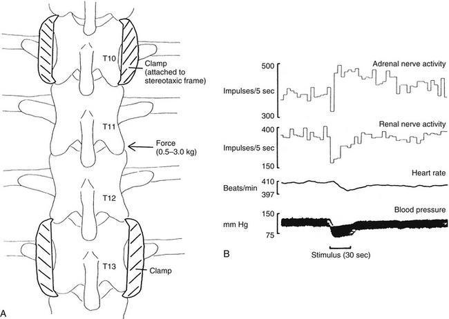
FIG. 10-28 A, Illustration of the stimulation procedure (thoracic spine shown). Segments isolated from skin and muscle, upper and lower segments fixed in a spinal stereotaxic device, and force exerted (0.5 to 3 kg) on mobile segments. B, Sample record from a central nervous system–intact animal with thoracic spine stimulation. Force (3 kg) delivered during the period marked by the dark bar below the blood pressure trace. (From Sato A & Swenson RS. [1984]. Sympathetic nervous system response to mechanical stress of the spinal column in rats. J Manipulative Physiol Ther, 7(3), 141-147.)
1. A consistently large decrease in blood pressure that returned to normal after the stimulus was removed
2. An inconsistently small decrease in heart rate
3. A decrease in blood flow to the gastrocnemius and biceps femoris muscles, with a concomitant decrease in systemic arterial blood pressure
4. An initial decrease in activity in the renal nerve and subsequent recovery, both during the period of stimulation
5. An initial decrease in activity in the adrenal nerve with a gradual return to baseline activity, which was followed by an additional increase in activity (likely caused by a baroreceptor-mediated reflex response)
A study in 2006 (Bolton, Budgell, & Kimpton, 2006) investigated the effect of innocuous mechanical stimuli of cervical vertebrae on the activity in the sympathetic nerve to the adrenal gland. Rats were used to see if localized cervical intervertebral displacement resulted in a change in sympathetic efferent activity. To prevent activation of the vestibular system and any resulting vestibulosympathetic reflex responses, the heads of the rats were fixed in place. Sympathetic nerve responsiveness was first tested by innocuous mechanical cutaneous stimuli (brushing of the skin of the neck) and noxious stimuli (pinching the forepaw). The results showed an increase in activity to noxious stimuli and showed no response to innocuous stimuli, which were consistent with data of other studies. Low amplitude, low velocity displacement of the C2 vertebra at various ranges within the normal physiologic range was performed on the rats. In general, the results from the study suggest that sympathetic nerve activity to the adrenal gland was not reflexively altered by innocuous mechanical stimulation induced by low amplitude and low velocity movements of the C2 vertebra. However, it was noted in a few rats that C2 vertebral displacements at greater than 20-degree rotation (beyond the normal physiologic range) did result in modulation of adrenal nerve activity and a change in blood pressure. These data, which are typical of a sympathetic response to an injurious event, may indicate that a noxious event occurred as a result of the mechanical displacement of C2 at those higher degrees of rotation that occur beyond the normal limits of rotation.
More studies have confirmed the fact that noxious and innocuous stimuli affect cardiovascular and other autonomic responses. Kurosawa and colleagues (2006) used anesthetized rats and studied the effects of innocuous mechanical (brushing) stimuli on the blood flow to the dorsal spinal cord. Blood flow rate was measured when the forepaw, forelimb, upper and lower back, hindlimb, and hindpaw were stimulated. The data indicated that an ipsilateral increase in blood flow occurred with no change in blood pressure and that it was segmentally organized by neuronal excitation in the cord. Kurosawa and colleagues suggested that sympathetic nerves and α-adrenoceptors may be involved in this somato-autonomic reflex response but that metabolic effects (e.g., release of local vasodilators) induced by neuronal excitation may contribute as well. Toda et al. (2008) studied anesthetized rats and the effects of noxious mechanical (pinching) cutaneous stimuli on blood flow of the dorsal spinal cord. In this study, spinal cord blood flow (SCBF), which was measured in the spinal cord region associated with the hindlimb, was increased when all four paws were stimulated although blood flow increased the greatest to the ipsilateral hindpaw stimulation. Mean arterial pressure (MAP) also increased when stimuli were applied to the paws. To gain greater insight into the mechanism of the increase in SCBF, experiments were performed in which baroreceptors were denervated, phenoxybenzamine (α-adrenoceptor antagonist) and phenylephrine (α-adrenoceptor agonist) were administered intravenously, and rats were spinalized. Based on the data, the authors suggest that somato-autonomic (sympathetic) reflexes and autoregulatory responses (both possibly through the activation of α-adrenoceptors) as well as systemic arterial pressure provide mechanisms that generally regulate SCBF. However, because an increase in SCBF in spinalized rats and in rats treated with phenoxybenzamine and phenylephrine occurred when the ipsilateral hindpaw was stimulated, it was proposed that a mechanism also existed that was specific for that local region. Organized ipsilaterally and segmentally, sensory input would result in neuronal activation inducing metabolic effects on spinal vasomotor tone that may already have been altered in response to increased systemic arterial pressure and/or noxious mechanical cutaneous stimulation. Anatomic and experimental evidence has shown that numerous mechanoreceptors are found in structures of the neck (e.g., skin, muscles, tendons, ligaments, periosteum, intervertebral discs, zygapophysial joints) that work in conjunction with the vestibular system to provide reflex responses to postural changes (Bolton, 1998). Reflexes associated with postural adjustments include the cervicocollic reflex, tonic neck reflex, and cervico-ocular reflex. In addition, there is evidence that activation of neck receptors elicits reflexes that produce autonomic responses. Experimental studies on cats (Bolton et al., 1998) suggested that stimulation of afferents of cervical neck muscles produced cervicosympathetic and cervicorespiratory reflex responses. This was evident by the changes seen in nerve activity in the greater splanchnic and abdominal nerves (respiratory motoneurons to the abdominal wall musculature), as well as the hypoglossal nerve. It has been established that input from neck afferents is relayed through brain stem vestibular nuclei to sympathetic and respiratory neurons. In addition, brain stem transections through the caudal medulla performed to separate the vestibular nuclei connections from the cord have shown that afferent fibers from the neck also stimulate sympathetic and respiratory nerves without relaying through the vestibular nuclei. However, transections through the middle of the medulla, which possibly interrupt descending fibers from the rostral ventrolateral (VL) medulla (part of the central autonomic control network), have been found to alter the activity of the sympathetic and respiratory neurons. Therefore based on the data from these experiments, a complex mechanism appears to exist to produce the physiologically normal cervicosympathetic and cervicorespiratory reflexes. This complex mechanism involves caudal brain stem and spinal cord structures that must be intact for these reflexes to take place. In addition, numerous studies on anesthetized animals also indicate that autonomic reflex responses are seen in visceral organs in response to somatic stimuli. Some of the effector organs studied that demonstrated changes are the gastrointestinal tissues, heart, urinary bladder, vasculature of the peripheral nerves, vasculature of the cerebrum, medulla of the adrenal gland, and spleen (see overview article by Sato [1997]).
Other animal studies also confirm that stimulation of somatic structures has an effect on autonomic responses. In one study, the effects of somatic stimuli were observed on the motility of the quiescent bladder. The results showed that there was an increase in bladder muscle tone in response to noxious chemical stimuli of the interspinous tissue but little change in bladder pressure in response to innocuous somatic stimuli (Budgell, Hotta, & Sato, 1998). Another animal study examined the effects of noxious chemical stimulation of interspinous tissues on gastric motility. The results showed that motility was strongly inhibited and that the reflex arc responsible for this action was segmentally located in the middle to lower thoracic region. Although both vagus and sympathetic nerve supply to the stomach was found to be involved in the reflex, the sympathetic innervation appeared to be more important (Budgell & Suzuki, 2000).
It also has been demonstrated that cardiac function can be altered by the innocuous stimulation of mechanoreceptors through spinal manipulation. These reflexes are sometimes referred to as spinovisceral reflexes. Budgell and Igarashi (2001) reported one case study of a young man with bradycardia and an arrhythmia. While being monitored continuously by electrocardiography, one cervical (C2) spinal manipulation was performed on the patient, which resulted in the coincidental disappearance of the arrhythmia. Although the result was clearly apparent, no distinct mechanism has yet to be proposed to explain the event. Another study investigated the effects of spinal manipulation (C1 and C2 levels) of healthy young adults on cardiac function. Results from this study, which used sham and authentic manipulations (non-noxious stimuli within normal physiologic ranges of motion), demonstrated significant changes in heart rate and heart rate variability (HRV) when only the authentic manipulations were performed (Budgell & Hirano, 2001).
In summary, many experiments show that somatic afferent stimulation, especially noxious stimulation, can modulate autonomic output, resulting in changes in heart rate, blood pressure, and activity in sympathetic efferents to the kidney and medulla of the adrenal gland. This includes studies that show that various stimuli applied to spinal structures can initiate somatovisceral reflex arcs. For example, studies show that noxious chemical stimulation results in modulation of sympathetic output related to cardiovascular and adrenal medullary activity (Budgell, Hotta, & Sato, 1995; Budgell, Sato, & Suzuki, 1997) and that mechanical stimulation causes varied responses depending on the location and type of stimulus applied (Sato & Swenson, 1984; Bolton, Budgell, & Kimpton, 2006). Pathologic processes affecting the spine also may result in reflex changes in visceral activity (Sato, 1992a). Based on this evidence, the neural components for this type of somatovisceral reflex do exist. In addition, spinal manipulation performed within the normal physiologic range of motion may stimulate somatic afferent fibers to create somatovisceral reflex responses.
The ANS is regarded as the division of the nervous system that modulates and regulates the physiologic mechanisms of the organ systems of the body in order to maintain homeostasis. However, it is now known that the ANS is also involved with the immune system and it helps to regulate the inflammatory response (i.e., the physiologic response to injury and infection manifested clinically by heat, pain, redness, and edema). In general, the immune response can be divided into an innate response (the naturally occurring defense system of all animals) and an acquired response (adaptive) although in many circumstances they work in tandem. The body’s innate immune system is continuously vigilant for invaders that have breached physical barriers (such as the skin and mucous membranes), which provide the first line of defense. If that invasion causes tissue damage, the innate immune system initiates a discrete and localized inflammatory response to impede further injurious insults and restore tissue homeostasis and wound healing. This response, which is a tightly controlled and well-organized process (Tracey, 2005), begins with the synthesis and release of cytokines from activated immune cells, such as macrophages. Cytokines are proteins that bind to receptors on immune cells affecting differentiation and proliferation, and that direct the inflammatory responders to the site of infection or injury. Some are pro-inflammatory and function to destroy the pathogen and restore homeostasis (i.e., tissue repair and normal health). Others are anti-inflammatory and help to contain the cytokine response along with humoral and neural mechanisms. The extent of the inflammatory response and cytokine production must be controlled such that the invading element is managed sufficiently. Too little of a response may lead to an immunodeficiency resulting in infection and cancer. An exaggerated or persistent response may lead to cytokines escaping into the systemic circulation, resulting in widespread activation and the systemic inflammatory response syndrome (Johnston & Webster, 2009). This may result in inflammatory diseases such as rheumatoid arthritis, atherosclerosis, inflammatory bowel disease, type 2 diabetes, Alzheimer’s disease, multiple sclerosis (MS), hemorrhagic shock, ischemia/reperfusion injury, and sepsis (Tracey, 2002; Oke & Tracey, 2008). Because of the potential deleterious and lethal effects of excessive pro-inflammatory cytokines, it is easy to understand why physiologic mechanisms have evolved to regulate and keep the process ‘in check’ (see section Efferent Response and the Cholinergic Anti-inflammatory Pathway). The immune system is a very complex system and has an integral role in homeostasis in general and cellular survival specifically and it is becoming more evident that it does not function autonomously within the body. It is known that the complex mechanisms of the immune system (including the inflammatory response), like other physiologic systems in mammals, are coordinated and regulated through pathways mediated by humoral mechanisms, including the neuroendocrine system (hypothalamic-pituitary-adrenal axis or HPA axis) and neural circuitry (Tracey, 2002). Now there is evidence supporting a neural network that provides the ‘hard-wiring’ and use of the vagus nerve for an inflammatory reflex arc allowing the nervous system to monitor and adjust the inflammatory response rapidly and in a localized manner.
Evidence exists that the immune system communicates with the brain, allowing the brain to monitor the status of invading organisms and react to the immune system as it functions to detect pathogens and other harmful substances. One method is through the blood vascular system. Once pro-inflammatory cytokines (such as tumor necrosis factor [TNF] and interleukin-6 [IL-6]) are in the circulation, they are able to signal the brain humorally by being transported across the blood-brain barrier by saturatable transport systems and by accessing and activating circumventricular organs devoid of the blood-brain barrier such as the area postrema. This important area is located in the region of the floor of the fourth ventricle and is in close proximity to the dorsal motor nucleus of the vagus, the nucleus tractus solitarius (NTS), and the rostral ventrolateral medulla (RVLM). When activated, the area postrema produces and releases prostaglandin E2. The release of prostaglandins as well as the presence of neural circuits formed by the area postrema, NTS, and RVLM allows for the activation of the HPA axis, mediation of fever, and the activation of the sympathetic system to occur. Cytokines binding to cerebral vascular endothelium also may alter cell function, resulting in the release of neuroactive substances such as prostaglandins into the brain parenchyma (Pavlov et al., 2003; Johnston & Webster, 2009). In addition, other clinical symptoms including anorexia, somnolence, weight loss, and depression (the clinical signs of ‘sickness behaviour’) appear as a result of the brain’s humoral activation by cytokines (Tracey, 2002; Hopkins, 2007; Tracey, 2009).
The second method by which the immune system communicates with the CNS is by the nervous system. Inflammatory mediators released in connective tissues associated with the vagus nerve activate vagal afferent fibers and initiate an anti-inflammatory response. Vagal afferent fibers in the viscera have been shown to be activated by low doses of endotoxin or interleukin-1 (IL-1). Electrophysiologic studies show that vagal afferents can also be activated by TNF and other cytokines released from dendritic cells, macrophages, or other immune cells present at the site of inflammation (Tracey, 2002). Endogenous and exogenous factors associated with inflammation can interact with immune cells such that there is an increase in pro-inflammatory cytokine release, which can activate vagal afferents. This mechanism of activation may occur directly or it may be mediated through glomus (chemoreceptive) cells of numerous paraganglia scattered along the vagus nerve and its branches. The glomus cells are located near fenestrated capillaries and are similar in appearance to those in the carotid body. They are able to detect cytokines in the blood and respond by releasing mediators that in turn activate the vagal afferent fibers (Goehler et al., 1997; Goehler et al., 1999; Goehler et al., 2000; Pavlov et al., 2003; Tracey, 2009). Evidence suggests that information concerning inflammation that courses in vagal afferent fibers may be dependent upon the extent and degree of the response. It has been suggested that vagal input to the CNS is more involved with mild to moderate peripheral inflammatory responses and that the humoral pathway mediates the more acute and vigorous responses (Pavlov et al., 2003). The activation of nociceptors by pro-inflammatory cytokines also leads to the induction and maintenance of pain (Üceyler, Schäfers, & Sommer, 2009). Neural pathways are stimulated, providing another route for the CNS to receive information pertaining to the activity of the immune system (i.e., the inflammatory response). The activation of neural circuitry at the CNS level and the resulting perception of pain will result in the activation of the stress response mediated by the sympathetic division and the HPA axis.
Within the CNS, the vagal afferent fibers terminate primarily in the NTS but also in the dorsal motor nucleus of the vagus and area postrema. Collectively these three areas form the dorsal vagal complex. The NTS, acting as an integration center, sends axons to numerous sites (see section Control of Autonomic Efferents: Central Autonomic Network) including the paraventricular nucleus of the hypothalamus, allowing for the activation of the neuroendocrine arm of the anti-inflammatory pathway (i.e., the HPA axis). NTS axons also synapse in the RVLM, which projects to the locus ceruleus, a brain stem nucleus that is a major source of noradrenergic input to higher centers including the hypothalamus. Also, both the RVLM and locus ceruleus project to sympathetic preganglionic neurons, thus activating the neural connections for an anti-inflammatory response (Pavlov et al., 2003). In addition, and most commonly recognized, is the NTS connection with the nearby dorsal motor nucleus of the vagus.
When an inflammatory response occurs, anti-inflammatory mechanisms are initiated to modulate the immune response. This limits the acute inflammatory response and prevents widespread inflammation throughout the body, which may cause serious damage to tissues. These mechanisms include both humoral and neural components. The humoral mechanism utilizes diffusible anti-inflammatory mediators that produce a concentration gradient–dependent, slow, and nonintegrated response. The mediators include those that are released at the local site of inflammation such as prostaglandin E2, spermine, and heat shock proteins and anti-inflammatory cytokines delivered to the local inflammatory site by the bloodstream or by diffusion through tissues. Also, once pro-inflammatory cytokines have interacted with the brain (see text above) hormones such as α-melanocyte-stimulating hormone (α-MSH) and glucocorticoids are released into the bloodstream (Tracey, 2002; Johnston & Webster, 2009). Glucocorticoid (cortisol in humans) released from the cortex of the adrenal gland is the result of the neuroendocrine regulatory mechanism, referred to as the HPA axis. The details of the mechanisms by which these mediators act are beyond the scope of this chapter.
There is also a neural component that is involved with the anti-inflammatory response. Activation of sympathetic fibers produces a powerful anti-inflammatory action upon the innate immune system. In general, immunomodulation of the immune system is mediated by sympathetic innervation of lymphoid tissue. Norepinephrine (NE) released from postganglionic neurons exerts an immunosuppressive response by binding to adrenoceptors expressed on lymphocytes and macrophages such that pro-inflammatory cytokine synthesis is inhibited and anti-inflammatory cytokine synthesis is stimulated. (See following section on Sympathetic Innervation of Lymphoid Tissue). In addition, preganglionic sympathetic fibers coursing in the greater splanchnic nerve innervate the chromaffin cells of the medulla of the adrenal gland with subsequent release of epinephrine (E) and some NE. These catecholamines act as hormones and have a widespread effect on effector tissues. The increase of E and NE levels can inhibit macrophage activation and the pro-inflammatory cytokine TNF synthesis. The activation of the β-adrenergic receptor has been shown to not only suppress TNF production but also up-regulate the production of the anti-inflammatory cytokine IL-10 (Tracey, 2002; Pavlov et al., 2003; Nance & Sanders, 2007; Johnston & Webster, 2009).
Evidence that has accumulated over the last decade indicates that there appears to be a cholinergic component to the anti-inflammatory mechanisms utilized by the central nervous system. This component is mediated through efferent fibers coursing within the vagus nerve. Vagal afferent fibers provide input into the CNS and the vagal efferent fibers serve as the efferent arc of an inflammatory reflex. This provides a direct, localized, and rapid response to inflammatory effects on the body—an advantage that allows for containing immune activation at the critical period of the initiation of the response. This theory is the conclusion of experiments showing that the vagal efferent fibers appear to have an immunosuppressive effect on pro-inflammatory cytokines, such as the potent cytokine TNF, produced by innate immune cells found in tissues innervated by the vagus nerve such as the spleen, liver, GI tract, and heart. Evidence supporting the presence of an anti-inflammatory pathway mediated by vagal efferent fibers includes data concerning the utilization of the tetravalent guanylhydrazone CNI-1493 drug, which activates the vagus nerve; the use of cholinergic agonists and antagonists as well as antisense and gene knockout methods in mice; and the investigation of the anti-inflammatory pathway in experimental models of disease including sepsis, endotoxemic shock, hemorrhagic shock, ischemia/reperfusion injury, subcutaneous inflammation, postoperative ileus, pancreatitis, and inflammatory bowel disease (Pavlov et al., 2003; Tracey, 2007; Gallowitsch-Puerta & Pavlov, 2007; Tracey, 2009). Because the major neurotransmitter used by parasympathetic/vagal efferents is acetylcholine (ACh), the pathway was named the ‘cholinergic anti-inflammatory pathway’. It was also determined that the receptor that was activated by ACh in the cholinergic anti-inflammatory pathway, resulting in the inhibition of cytokine synthesis, was the nicotinic acetylcholine receptor α7 subunit (α7nAChR) (Borovikova et al., 2000; Wang et al., 2003). The receptor is expressed on macrophages, monocytes, B and T cells, keratinocytes, endothelial cells, and dendritic cells. It has been determined that the signaling transduction mechanisms used by these receptors on immune cells and on neurons are different (Gallowitsch-Puerta & Pavlov, 2007; Tracey, 2009). Also, it has been shown that although ACh binds to muscarinic receptors, some of which are expressed on cytokine-producing cells, these receptors are not required for modulating cytokine production via the cholinergic anti-inflammatory pathway (Johnston & Webster, 2009). This mechanism is different than the one used by parasympathetic fibers to modulate effector tissues in other physiologic systems. However, muscarinic receptors are found in the CNS (NTS and dorsal motor nucleus) and may be involved in cholinergic activation. Through their activation, they have been shown to inhibit systemic inflammation in endotoxemic rats and also to significantly increase the activity of vagal efferent neurons, implicating a possible function, at least in the rat model, of modulating the cholinergic anti-inflammatory pathway (Gallowitsch-Puerta & Pavlov, 2007).
Macrophages and monocytes, important cells of the immune system and components of the reticuloendothelial system, target foreign pathogens. Their actions are essential during the early innate immune response. They reside in the spleen, liver, lungs, and other tissues. Microorganisms and their products, such as bacterial endotoxins, localize to macrophages primarily in the spleen and liver and when the macrophages are activated they are capable of secreting pro-inflammatory cytokines. The spleen is the major source of hepatic and systemic TNF during endotoxemia and its relationship with the vagus nerve has been studied. Data (Huston et al., 2006) show that TNF production in the liver and spleen, but not in the lung, was significantly decreased when the vagus nerve was stimulated. It was also shown that the spleen is a major source of the pro-inflammatory cytokine TNF and that the suppression of its production after stimulation of vagal fibers requires the α7nAChR. Therefore it appears that the cholinergic anti-inflammatory pathway may be hardwired through the spleen as a route by which it regulates suppression of TNF production and limits the magnitude of the inflammatory response (Huston et al., 2006; Rosas-Ballina et al., 2008). The spleen is a major source of ACh and it has been shown that large quantities of ACh are released into the tissue and splenic vein as a result of splenic nerve activation. It is possible that this process may allow the spleen to control the movement of immune cells to distant sites (Saeed et al., 2005; Huston et al., 2006; Tracey, 2009). However, it has also been established that there are no cholinergic fibers terminating in the spleen and in fact the innervation of the spleen is by catecholaminergic fibers (splenic nerve) originating in the celiac-superior mesenteric ganglion. It becomes problematic because the receptors that are activated in order to suppress TNF production via the cholinergic anti-inflammatory pathway have been shown to be nicotinic receptors specific for ACh. The vagus nerve does play a role in the innervation of the spleen in that the right vagus nerve gives off a large celiac branch that ends within the celiac plexus and it also sends small branches into the splenic plexus (Standring et al., 2008). Research studies indicate that the celiac branches of the vagus nerve terminate in synaptic-like structures around main cells in the celiac-superior mesenteric ganglia (Berthoud & Powley, 1993; Berthoud & Powley, 1996; Szurszewski & Miller, 2006; Rosas-Ballina et al., 2008), which is the location of the sympathetic postganglionic catecholaminergic fibers of the splenic nerve. ACh can be synthesized, stored, and released from nonneuronal cells. These cells include vascular endothelial cells and lymphocytes (primarily T cells). It has been suggested (Kawashima & Fujii, 2003; Kawashima & Fujii, 2004) that a resident population of these interacting lymphocytes in lymphoid tissue may be involved in a local cholinergic system that regulates immune function. The spleen includes populations of these cells, which also have been shown to be innervated by catecholaminergic fibers. The synthesis and release of ACh from these cells and subsequent binding to the α7nAChR expressed on immune cells such as macrophages suppress TNF production (Mignini, Streccioni, & Amenta, 2003; Wang et al., 2003; Rosas-Ballina et al., 2008; Tracey, 2009). From these studies it can be hypothesized that the wiring of the cholinergic anti-inflammatory pathway consists of the vagus nerve activating postganglionic noradrenergic fibers coursing in the splenic nerve. These fibers cause the release of ACh from immune cells such as lymphocytes in the spleen; the ACh binds to the α7nAChR on macrophages and down-regulates pro-inflammatory cytokine release (Fig. 10-29).
FIG. 10-29 Cholinergic anti-inflammatory pathway. Vagal afferent fibers (afferent arc) provide input to the nucleus tractus solitarius (NTS) regarding the presence of pro-inflammatory cytokines. The NTS sends input to the dorsal motor nucleus of X (DMNuc) and stimulates vagal efferent fibers (efferent arc), which synapse in the celiac ganglion on cell bodies of noradrenergic fibers coursing in the splenic nerve. These fibers cause ACh to be released from splenic immune cells. ACh binds to α7nACh receptors and down-regulates the release of pro-inflammatory cytokines. The NTS also sends input to the hypothalamus initiating the neuroendocrine component of an anti-inflammatory pathway (HPA-axis). Sympathetic efferents activate NE release from postganglionic neurons and E release from the adrenal medulla (see text and section Sympathetic Innervation of Lymphoid Tissue).
It is interesting and somewhat surprising that the vagus nerve has been implicated as having this uncharacteristic role of providing the ‘hard-wiring’ for an inflammatory reflex arc and suppressing the inflammatory response. Classically, the function of the vagus has been associated with the regulation of physiologic systems such as the cardiovascular, respiratory, and GI systems through its sensory and motor innervations of the viscera. Research continues to produce data elucidating in more molecular detail the types of receptors located on various cell populations and their mechanisms of action. In light of this information, it perhaps makes sense that the vagus nerve is involved because it is well-positioned anatomically throughout the body to interact with cells that are involved with providing an immediate immune response (and producing pro-inflammatory cytokines) to pathogens, such as the nonneuronal cells residing in the organs (e.g., spleen, liver, and lung) of the reticuloendothelial system. In fact, it has been proposed (Tracey, 2007), based on evidence of endotoxemia studies in mice, that the vagus nerve, through its ability to suppress cytokine production, may provide the means by which the CNS is able to continuously and precisely modulate and control the extent of the inflammatory response, that is, “…maintain homeostasis by limiting the inflammatory responses within the healthy, protective, and non-toxic range” (Tracey, 2009). Studies of mice (van Maanen et al., 2009) subjected to collagen-induced arthritis indicated that the cholinergic anti-inflammatory pathway has a suppressive effect on arthritis. Although the vagus nerve does not directly innervate peripheral sites (e.g., joints of the extremities) that might be involved with the anti-inflammatory responses, there is evidence of vagus nerve involvement in animal models of induced peripheral inflammation (Borovikova et al., 2000). It is theorized that the suppressed inflammatory response seen in these models is the result of a decrease in cytokine production and a redirection of immune cells away from the site of peripheral inflammation possibly through the association of the vagus nerve with the spleen and the obstruction of endothelial cell activation (see earlier text) (Saeed et al., 2005; Huston et al., 2006; Tracey, 2007).
The parasympathetic and sympathetic divisions of the ANS have distinct anatomic features and specific interactions with the effector tissues such that traditionally they are often described as being separate and as functioning antagonistically. However, this classical description is not entirely true. Anatomically, it has been shown that the splenic nerve, consisting of catecholaminergic fibers, can be stimulated either by sympathetic preganglionic neurons or, in the case of the cholinergic anti-inflammatory pathway, by vagal preganglionic neurons. Also, peripheral effector cells modulated by vagus nerve activation express nicotinic receptors and not the traditional muscarinic receptor. Homeostatic control mechanisms that act daily in an individual are not functioning in isolation but are the result of the composite activity of many neural networks including the sympathetic and parasympathetic divisions. For example, activation of only sympathetic fibers to the heart results in an increase in heart rate and an increase in cardiac output. Parasympathetic activity in an isolated setting results in decreased heart rate and decreased cardiac output. In an intact organism, when both divisions are activated simultaneously, the net increase in cardiac output is greater than when the sympathetic fibers are firing in isolation because the decreased heart rate allows the blood to fill the heart more efficiently (Tracey, 2009). Thus, when the two divisions work synergistically the overall result is a more beneficial response. This is also the case regarding the modulation and monitoring of the body’s defense system against pathogens such that the traditional ‘rules’ are changed both anatomically and physiologically. As mentioned earlier, the cholinergic anti-inflammatory pathway is neither sympathetic nor parasympathetic because it contains neural components of each division. Both sympathetic (see Sympathetic Innervation of Lymphoid Tissue) and parasympathetic (via the vagus nerve) divisions are activated by immunogenic stimuli. Stimulation of vagal afferent fibers results in activation of the NTS, which has the capability to signal not only vagal efferent fibers in the dorsal motor nucleus but also the RVLM and locus ceruleus with subsequent firing of sympathetic neurons in the cord, as well as activating the HPA axis. Both function synergistically through the innervation they provide to the thymus, spleen, heart, liver, lungs, GI tract, pancreas, and kidneys, allowing for the co-regulation of the production of cytokines by immune and nonimmune cells (Pavlov et al., 2003).
The discovery of the cholinergic anti-inflammatory pathway opens up the avenue for designing therapeutic protocols to treat inflammatory diseases by altering vagus nerve activity or targeting the components of the pathway. Also, understanding the mechanism of the cholinergic anti-inflammatory pathway may explain why current treatments and activities that result in anti-inflammatory responses occur. Examples of the clinical relevancy of the pathway are briefly described next.
• Heart rate variability (HRV). One method of determining vagal activity is by measuring time variations between heartbeats and calculating instantaneous heart rate variability (HRV). It has been shown that in resting conditions vagus nerve activity provides an inhibitory influence on instantaneous heart rate. Epidemiologic evidence suggests there is an association between basal heart rate and an increased risk and mortality of systemic inflammatory diseases such as diabetes, cardiovascular disease, sepsis, and arthritis. Studies have shown that HRV (indicating vagal nerve activity) is decreased in patients showing signs/symptoms of inflammatory diseases such as rheumatoid arthritis and sepsis, and that there is an association between decreased HRV and the risk of atherosclerosis. This may imply that during resting conditions the vagus nerve provides a tonic modulatory effect (inhibition) on the innate immune response and controls the extent of the inflammatory response. In other words, the inflammatory reflex mediated by vagal neurons helps to establish a physiologic ‘set point.’ If the activity of the vagus nerve is decreased or eliminated, the set point increases and the inflammatory response becomes unrestrained, leading to an increased risk of systemic (possibly even lethal) inflammation. Clinical studies show that patients with inflammatory diseases that produce increased levels of pro-inflammatory mediators and have functionally decreased instantaneous HRV have functionally increased basal set points. Increased indices of vagal nerve activity and decreased levels of pro-inflammatory mediators in healthy individuals are associated with decreased set points. HRV data may be a tool that could be used in the future to measure vagal activity and predict patients who may have an increased risk for developing inflammatory diseases (Tracey, 2005; Tracey, 2009).
• Vagus nerve stimulation. Studies show that electrically stimulating the vagus nerve decreases TNF-α production in experimental models of endotoxemia and hemorrhagic shock (Tracey, 2007; Johnston & Webster, 2009). Vagus nerve stimulation in rats has been shown to inhibit the development of collagen-induced arthritis and in humans it can provide positive effects in treating depression and drug-resistant epilepsy (Koopman et al., 2011). Also, alternative therapies such as acupuncture, meditation, relaxation therapy, hypnosis, and biofeedback have been shown to modulate vagus nerve activity. These may have the ability to activate the cholinergic anti-inflammatory pathway and suppress cytokine release (Pavlov et al., 2003; Tracey, 2007; Johnson & Webster, 2009; Tracey, 2009).
• Pharmacologic agents that target receptors involved in the cholinergic anti-inflammatory pathway. Studies show that the pathway may be modulated by the binding of drugs to CNS muscarinic receptors, by the binding of nicotine-like agonists to the α7nACh receptor, and by the binding of cholinergic and anticholinergic drugs that are used to treat brain degenerative disorders and to treat sepsis and organ failure (Pavlov et al., 2003; Tracey, 2007; Johnston & Webster, 2009).
• Dietary supplementation with fats (fish oil, soy oil, and olive oil). Studies suggest that the cholinergic anti-inflammatory pathway may be activated by a high-fat diet through the release of cholecystokinin and its subsequent activation of vagal afferent fibers. Stimulating the pathway may afford some protection to dietary antigens and bacterial toxins in the gut by suppressing cytokine release (Luyer et al., 2005; Tracey, 2005; Tracey, 2007).
As mentioned previously, the sympathetic division has a very important role in mediating immune system functions. Research shows that sympathetic efferent fibers innervate primary (bone marrow and thymus) and secondary (lymph nodes, spleen, and peripheral) lymphoid tissue. Two adrenergic receptor subtypes are shown to be expressed on immune cells regulated by sympathetic neurons and NE: α (1 and 2) and β (1 and 2). These belong to a G protein–coupled receptor superfamily and are further classified into subgroups (Pavlov et al., 2003). In humans and rodents the β2 receptor is the primary subtype. Cell activation, cytokines, hormones, and neurotransmitters can influence and regulate the number of these receptors expressed. Cells involved in the innate immune response express the β2 and the α1 and α2 receptors. The β2 receptor is exclusively expressed on cells involved in the adaptive immune response (Nance & Sanders, 2007). NE has an anti-inflammatory effect by binding to adrenoceptors on lymphocytes and macrophages. Relative to the inflammatory response, a decreased production and release of inflammatory mediators such as TNF-α, IL-1β, interferon γ, and NO and an elevation in the levels of anti-inflammatory cytokines IL-6 and IL-10 via β2 receptors were seen in stress-induced situations such as endotoxemia and hemorrhagic shock in which the sympathetic division was activated (Pavlov et al., 2003). In vivo activation of the sympathetic division by stress or by central inflammatory stimuli results in a potent anti-inflammatory response in the spleen inhibiting the production and release of TNF-α from macrophages (Nance & Sanders, 2007). It appears that the mechanism by which both NE and E (released from the adrenal medulla) inhibit pro-inflammatory cytokines and stimulate anti-inflammatory cytokines is through the activation of the β2-adrenoceptor–cAMP–protein kinase A pathway. This activation may occur at synapse-like junctions similar to those in the spleen or at a distant site subsequent to the diffusion of NE through the tissue. The latter type of activation plays the dominant physiologic role. The release of NE can be presynaptically regulated through the effects of chemical mediators such as neuropeptide Y, ACh, dopamine, prostaglandins, and other microenvironmental factors (Pavlov et al., 2003). Although activated sympathetic fibers have an overall anti-inflammatory effect relative to the innate immune system response, in the beginning of some inflammatory responses, sympathetic firing may activate a local response including the accumulation of neutrophils. Also, during the acquired immune response, activated sympathetic fibers may enhance or inhibit cellular activity possibly by altering gene expression for cytokines and antibodies (Nance & Sanders, 2007). The dual role of sympathetic modulation on cellular activity may be dependent upon the peripheral receptor expressed on the effector cell and its affinity for NE, the tissue in which it is located, and its local concentration in the synaptic cleft (Koopman et al., 2011).
The thymus has a rich sympathetic innervation. Preganglionic neuron cell bodies are located in the intermediolateral cell column of T1 to T7 cord segments and postganglionic cells bodies are located in the cervical ganglia and the first two or three thoracic ganglia. Fibers entering the thymus along blood vessels travel through the capsule and septa to enter the cortex and medulla and terminate near immune cells such as thymic epithelial cells and thymocytes (Mignini, Streccioni, & Amenta, 2003; Nance & Sanders, 2007). Both of these cells have been shown to express both muscarinic and nicotinic receptors and to synthesize and release ACh. It is possible that this population of cells forms a cholinergic system that regulates and modulates thymic function (Kawashima & Fujii, 2004). Sympathetic fibers innervating bone marrow course as perivascular plexuses that enter nutrient foramina and terminate in the vicinity of hemopoietic and lymphopoietic marrow cells. This may indicate a sympathetic function of modulating hemopoiesis (Mignini, Streccioni, & Amenta, 2003).
Secondary lymphoid tissue includes lymph nodes, spleen, and other regions that contain lymphatic tissue. These tissues are the sites where immune responses occur. Sympathetic fibers course along blood vessels and innervate lymph nodes located in the specific area of distribution of the nerve fibers. Studies indicate the fibers travel to the parenchyma in T-cell–rich regions such as the paracortical and cortical areas but not in B-cell–containing areas such as the nodular regions and germinal centers. Postcapillary venules have also been found to have nerve fibers associated with them, possibly for regulating the movement of lymphocytes into the lymph node (Panuncio et al., 1998; Mignini, Streccioni, & Amenta, 2003).
The spleen, another secondary lymphoid organ, and its sympathetic innervation have already been discussed relative to the inflammatory response. The preganglionic neuron cell bodies of this pathway are located in the T1 to T12 cord segments whereas the postganglionic neuron cell bodies are located in the celiac-superior mesenteric ganglion. The postganglionic efferent fibers form the splenic nerve and course with blood vessels to the hilum. Studies of the feline spleen have shown that it is innervated by approximately 12,000 sympathetic fibers. When the weight of the spleen is compared with the weight of the kidney, the number of splenic sympathetic fibers is three times the number supplying the kidney. The fibers to the spleen appear to be different from sympathetic renal fibers, which function as vasoconstrictors. Although splenic fibers have the functions of capsular contraction and vasoconstriction, some splenic fibers appear to function independently from arterial baroreceptor input and are activated reflexively to sensory input from the spleen, as well as the GI tract. These splenic fibers may be involved with immune system regulation. Studies alluded to in previous sections indicate the important functional connection the spleen has with the immune system. Studies in rats show the alteration of various immune functions in the spleen with sympathetic nerve stimulation. These alterations include changes of cellular elements involved in immune responses after sympathectomy; decreased number of immune responses after splenic nerve stimulation; and activation of certain other immune responses (e.g., stimulation of a specific hypothalamic region results in splenic nerve activity that correlates with the suppression of cytotoxic activity of splenic natural killer cells) (Jänig & Häbler, 2000). Sato (1997) used anesthetized rats and looked at the effect of pinching the hindpaws and brushing and pinching the skin on sympathetic activity in the spleen. The results showed a reflex response that included vasoconstriction and a decrease in cytotoxic activity of the natural killer cells. Evidence from studying the cellular characteristics of splenic innervation shows that when the fibers traverse the capsule and enter the white pulp, they terminate in proximity of immune cells such as macrophages, T and B lymphocytes, and plasma cells that are located in specific regions of the white pulp (see Mignini, Streccioni, & Amenta [2003]). Scattered fibers along the trabeculae in red pulp also are present. Because of the various immune cells involved, their variety of receptors, and a possible local lymphocytic cholinergic regulating system (Kawashima & Fujii, 2004), modulation of aspects of both the innate and the adaptive immune systems, such as lymphocyte proliferation, antibody secretion, and cytokine production, may occur (Mignini, Streccioni, & Amenta, 2003).
There is also an important relationship between the immune system of the GI tract and the enteric division of the ANS, which regulates GI motility, blood flow, and secretion (see Enteric Nervous System). The immune system of the gut is comprised of a diverse population of immune cells and inflammatory cells such as lymphocytes, macrophages, dendritic cells, and mast cells. These cells provide a security system in case the mucosal lining is breached by any one of a number of potentially harmful substances such as dietary antigens, parasites, viruses, and bacteria that appear in the intestinal lumen. The immune/inflammatory cells release chemical mediators and signal enteric neurons as well as vagal and spinal afferent fibers. Of these cells the mast cell has been studied the most. Antigenic activation of the mast cells results in the release of chemical mediators such as histamine, IL-6, serotonin, leukotrienes, platelet activating factor, adenosine, IL-1β, and mast cell proteases. These mediators are involved in the activation of the enteric neural circuitry that runs the defense program. This program is designed to eliminate the injurious agent from the lumen by the coordination of increased secretion and blood flow followed by powered propulsive motor activity. Because receptors for inflammatory mediators such as mast cell proteases, bradykinin, and prostaglandins are expressed on spinal afferent fibers, it has been suggested that activation of these receptors may result in the sensitization of nociceptors and the recruitment of silent nociceptors. The clinical manifestations of these neuronal processes, which are often seen in individuals with IBS, are cramping abdominal pain, fecal urgency, and watery diarrhea. It is also known that symptoms seen in functional gut disorders such as IBS can be exacerbated by psychological stress. Because of the similarity in symptomology to the gut’s response seen in inflammatory conditions such as infectious enteritis, it is thought that there may be a connection between the brain and mast cells. Psychological states in the brain could activate a neural pathway that stimulates the release of chemical mediators from mast cells, resulting in the activation of the same neural circuitry used in response to a breach in the mucosal lining (Wood, 2007).
The neurotransmission that occurs at the autonomic ganglion and neuroeffector junction is important physiologically and pharmacologically. The classical description of chemical transmission at these two locations was simply to explain the actions of the two major neurotransmitters: norepinephrine (also called noradrenaline) and acetylcholine. Fibers releasing those chemicals thus were called adrenergic and cholinergic, respectively. However, recent information has demonstrated that the autonomic ganglion and neuroeffector junction are regions of complex interactions involving not only acetylcholine and norepinephrine, but also cotransmitters and neuromodulators.
As stated, the ganglion is the location of postganglionic cell bodies, and therefore the site in which preganglionic neurons synapse. Functionally it is the location in which information from the CNS (via preganglionic neurons) and the periphery (via collateral branches of afferent neurons, releasing substance P and CGRP [Iversen, Iversen, & Saper, 2000]) is integrated and then distributed to the peripheral effectors via postganglionic neurons. Each preganglionic neuron diverges onto many postganglionic neurons (divergence) and each postganglionic neuron in turn receives input from more than one preganglionic neuron (convergence) (see Fig. 10-4). The divergence occurring in the sympathetic system traditionally is thought to be greater compared with the divergence in the parasympathetic system so that the sympathetic system can produce widespread responses. This concept has been used to delineate the two systems. However, some authors suggest that the difference in divergence ratios should not be used to characterize the sympathetic and parasympathetic systems, but rather should be noted as the means by which small targets with distinct functions are regulated (limited divergence) and extensive effectors (targets) acting in unison are regulated (vast divergence) (Wang, Holst, & Powley, 1995; Jänig & Häbler, 2000). Each postganglionic cell is inhibited via dendrodendritic synapses with other postganglionic cells and interneurons, the latter being excited by preganglionic fibers (see Fig. 10-4) (Kiernan, 2009). The primary neurotransmitter released by preganglionic neurons at their synaptic boutons is ACh (Fig. 10-30). This binds to and activates nicotinic receptors (so-named because the effect can be reproduced using the drug nicotine) found on the postganglionic neurons. In addition to postganglionic neurons, nicotinic receptors also are found on skeletal muscle cells and CNS neurons. The binding causes depolarization and the generation of a fast excitatory postsynaptic potential (EPSP). Use of the nicotine receptors is regarded as the main mechanism for transmission in both sympathetic and parasympathetic divisions of the ANS (Iversen, Iversen, & Saper, 2000). However, if the preganglionic neuron is stimulated repeatedly, ganglionic ACh also binds to muscarinic receptors on postganglionic cells. These are G protein–coupled receptors that evoke slow EPSPs and inhibitory postsynaptic potentials (IPSPs). Neuropeptides such as enkephalins, neurotensin, somatostatin, substance P, VIP, and neuropeptide Y (Iversen, Iversen, & Saper, 2000; Bear, Conners, & Paradiso, 2001) are colocalized with (contained in) cholinergic preganglionic neurons. These molecules are important in helping to determine the overall activity of the postganglionic neuron. They act by modulating the postsynaptic membrane such that the membrane will reach threshold more easily when activated via a fast EPSP (Iversen, Iversen, & Saper, 2000; Bear, Conners, & Paradiso, 2001). Animal studies on sympathetic ganglia also show variability among postganglionic neurons in paravertebral (sympathetic chain) ganglia versus those in prevertebral ganglia. Paravertebral ganglia cells have consistent properties (i.e., each preganglionic neuron evokes an EPSP via nicotinic receptor channels and an action potential always is produced). On the other hand, prevertebral ganglia cells do not have uniform properties, but instead can be divided into three groups based on their electrophysiologic, anatomic, and neurochemical differences (Jänig & Häbler, 2000). The transmission of information coursing through this site is complex and likely modulated and controlled based on the structure of the ganglion and the chemical diversity at the level of the preganglionic and postganglionic synapse and at the synapse of the interneuron pool.
FIG. 10-30 Major neurotransmitters released by parasympathetic and sympathetic neurons. Note the receptors (nicotinic, green; muscarinic, purple; alpha, blue) to which the neurotransmitters bind. Preganglionic neurons of both systems and postganglionic parasympathetic neurons release acetylcholine (ACh). Postganglionic sympathetic neurons typically release norepinephrine (NE). (Those that innervate sweat glands release ACh.) Often, the fibers of the two divisions are described by their length. In general, the parasympathetic division consists of long preganglionic fibers and short postganglionic fibers whereas the sympathetic division consists of short preganglionic fibers and long postganglionic fibers.
Postganglionic neurons innervate the smooth muscle, cardiac muscle, and glands of the body. The axons are approximately 1 µm in diameter and typically are unmyelinated. Studies on the terminal endings of primarily sympathetic postganglionic axons show that they are different from somatic efferent fibers. The motor neurons that innervate skeletal muscle fibers do so at the neuromuscular junction at specialized regions on the postsynaptic membrane called the motor end plate. The neurotransmitter is released at this one site and binds to the receptors. On the other hand, postganglionic autonomic fibers branch extensively near the effector muscle cells, thus regulating the function of numerous muscle fibers. These fibers are arranged in bundles and some smooth muscle fibers are directly innervated, whereas the rest are electrically linked to the directly innervated cells by gap junctions. There are no well-defined presynaptic and postsynaptic specializations, nor just one transmission site. Instead the ends of the terminal branches of the postganglionic neurons are varicosed (i.e., they have a beaded appearance) (Fig. 10-31). Each of these beads or swellings (numbering from 10,000 to more than 2 million/mm3) (Hamill, 1996) is called a varicosity, and it contains mitochondria and vesicles of stored neurotransmitters (100 to 1000 per varicosity in adrenergic fibers). Each varicosity is approximately 0.3 to 1 µm in diameter and is located approximately 4 µm from its neighboring varicosity (Hirst et al., 1996; Bennett, 1998). The membrane of each varicosity is devoid of its Schwann cell neurilemma. Because there are no postsynaptic specializations, the varicosities are dynamic and may move along the axons (Standring et al., 2008). Because these are strung out along the terminal fiber ending, the receptors are also spread over the surface of the effector cell membrane and each varicosity is a possible site for neurotransmission onto muscular (or glandular) tissue. The neurotransmitter, which is released typically during passage as the action potential travels along the axon, crosses the synaptic cleft and may diffuse a distance (as many as several hundred nanometers) before reaching its target receptors (Iversen, Iversen, & Saper, 2000). The synaptic cleft varies considerably in size among tissues, ranging from as little as 20 nm in densely innervated effector tissues (e.g., ductus deferens) to 1 to 2 µm in large elastic arteries (Standring et al., 2008).
FIG. 10-31 A, Varicosities located on the distal portion of an autonomic neuron. B, Scanning electron micrograph of one terminal autonomic nerve fiber showing its varicosities (enlarged view of area outlined in A). This fiber is lying over the smooth muscle in the rat small intestine. C, Transmission electron micrograph of a section through a varicosity. (From Williams PL et al. [Eds.]. [1995]. Gray’s anatomy [38th ed.]. Edinburgh: Churchill Livingstone.)
The traditional description of chemical transmission at the neuroeffector junction has been based on the release of the “conventional” transmitters ACh, released by the parasympathetic postganglionic fibers, and NE, released by sympathetic postganglionic fibers (see Fig. 10-30). In general, these neurotransmitters are released and then bind to and activate their specific receptors, which in turn release intracellular chemical messengers. In turn, these messengers initiate a cascade of cellular reactions; however, other chemical substances have been found to be involved with neurotransmission. For example, ATP is a cotransmitter with NE in adrenergic neurons, although its concentration relative to NE varies in different fibers. Neuropeptides are common substances that colocalize with both NE- and ACh-containing neurons and function to modulate neurotransmission. Neuropeptide Y is found in approximately 90% of postganglionic adrenergic fibers. It facilitates responses by acting postsynaptically on effector cells located more than 60 nm away. On densely innervated target cells that are 20 nm away, neuropeptide Y acts on the presynaptic membrane and dampens effector activity by inhibiting the release of ATP and NE. Neuropeptide Y often is associated with other neuropeptides such as galanin and dynorphin. The few sympathetic neurons that are cholinergic (e.g., to sweat glands) have been shown to often contain CGRP and VIP. Adenosine also modulates transmission of sympathetic neurons at both the presynaptic and postsynaptic junctions (Iversen, Iversen, & Saper, 2000).
Parasympathetic postganglionic neurons have been found to colocalize VIP with ACh. VIP is a vasodilator that may function to assist effector activity by increasing blood flow, which is necessary in the process of glandular secretion (Iversen, Iversen, & Saper, 2000). Other populations of neurons use ATP or nitric oxide (or both) as cotransmitters. In addition, nitric oxide, which has been implicated in the mediation of smooth muscle relaxation, and ATP have been recognized as chemical substances associated with nonadrenergic, noncholinergic (NANC) fibers (Standring et al., 2008).
The actual physiologic response of effector tissues to postganglionic neuron activation is dependent on the neurotransmitter (and possible cotransmitters and comodulators) released. More importantly, the response also is dependent on the presence and distribution of ligand receptors at the neuroeffector junction to which the neurotransmitter will bind. Depending on the physiologic status of the target cell, the concentration of receptors found on its postsynaptic membrane can be modified by the cell inserting or removing receptors into its cell membrane (up-regulation or down-regulation, respectively), thus affecting the binding capability of neurotransmitter and the resulting activity of the target cell.
NE is the primary neurotransmitter that sympathetic postganglionic fibers release, although fibers that innervate merocrine sweat glands release ACh (apocrine glands associated with hair follicles are innervated by adrenergic fibers) (Standring et al., 2008). NE binds to adrenergic receptors, of which there are two major types, α (alpha) and β (beta). The α type is further subdivided into an α1 type, which is subdivided into three subtypes (A, B, D), and an α2 type, which is also subdivided into three subtypes (A, B, C) (Insel, 1996). The β receptor is divided into β1, β2, and β3 types. Each type of adrenergic receptor is linked preferentially to specific subclasses of G proteins located in the cell membrane. Each G protein in turn is linked to specific effector molecules within the cell. For example, phospholipase Cβ and adenylyl cyclase are examples of such effector molecules. Activation of the effector molecules in turn leads to changes in intracellular concentrations of second messengers (e.g., cyclic adenosine monophosphate [cAMP]), which results in the modulation of cellular activities (Insel, 1996).
The primary neurotransmitter that parasympathetic postganglionic fibers release is ACh. At the neuroeffector junction, ACh binds to muscarinic receptors. These receptors are so-named because administration of the alkaloid muscarine (derived from the Amanita mushroom) was found to mimic specific actions of the parasympathetic system. Muscarinic receptors found at the neuroeffector junction are subdivided into three major classes: M1, which is excitatory to postganglionic neurons and when activated modulates NE release; M2, which decreases heart rate and contractility; and M3, which causes smooth muscle contraction and increased glandular secretion (Hamill, 1996). Two other classes, M4 and M5, have been identified but their functions outside of nervous tissue are unclear (Eglen, 2005). Table 10-3 summarizes major functions of the adrenergic and cholinergic receptors.
Table 10-3
∗G protein–coupled receptor.
†Subclasses have been identified.
From Eglen RM. (2005). Muscarinic receptor subtype pharmacology and physiology. Prog Med Chem, 43, 105-136; Albuquerque EX et al. (2009). Mammalian nicotinic acetylcholine receptors: from structure to function. Physiol Rev, 89(1), 73-120.See Table 10-1 for references and for other structures and their specific adrenergic receptors.
Adrenergic and muscarinic receptors are located on both presynaptic (postganglionic) and postsynaptic (target cell) membranes (Fig. 10-32). More specifically, M1, M2, and M3 receptors are located on the postsynaptic membranes. Some muscarinic receptors are located on sympathetic neuron presynaptic membranes and these receptors inhibit the release of NE. Others are located on parasympathetic presynaptic membranes. When stimulated, these inhibit the further release of ACh. α1-Adrenergic receptors are located on postsynaptic membranes and when activated mediate smooth muscle contraction in peripheral small arteries and large arterioles, dilator pupillae muscle, GI sphincters, the bladder neck, and the vas deferens. α2 receptors are located on sympathetic presynaptic membranes (as autoreceptors involved with an auto-feedback loop regulating NE release) and on neighboring parasympathetic presynaptic membranes. In both cases, when activated, neurotransmitter release is inhibited. β1 receptors found on postsynaptic membranes cause an increase in heart rate and force of contraction, and those found on cells in the kidney cause the secretion of renin. Renin is part of the pathway that forms the vasoconstrictor peptide angiotensin II. β2 receptors are activated by epinephrine (released from the medulla of the adrenal gland) and by locally released NE. These receptors are postsynaptic and cause relaxation of tracheal and bronchial smooth muscle and the ciliary muscle of the eye, and they initiate glycogenolysis and gluconeogenesis in the hepatocytes of the liver. β2 receptors also are found on presynaptic adrenergic fiber membranes acting as autoreceptors and causing the release of NE (FitzGerald & Folan-Curran, 2002). β3 receptors are located in brown adipose tissue. When activated, these receptors are thought to promote lipolysis and heat production in fat (Insel, 1996). Table 10-1 lists the effector tissues and their adrenergic receptor type.
FIG. 10-32 Neuroeffector junction illustrating the multiple receptors on the presynaptic neuron and the postsynaptic (target) cell and their influence on the release of acetylcholine (ACh) and norepinephrine (NE). A, NE release is inhibited by presynaptic α2 receptors (blue), which also inhibit the release of ACh from adjacent parasympathetic fibers. NE release is promoted by β2 receptors (red), which bind epinephrine (E). α1 (excitatory), β1 (excitatory), and β2 (excitatory and inhibitory) receptors (green) are located on the postsynaptic cell membrane. B, Presynaptic muscarinic receptors (teal) inhibit the release of excess ACh and inhibit neurotransmitter release from adjacent sympathetic fibers. Muscarinic receptors (dark yellow) are located on the postsynaptic cell membrane.
In addition to the adrenergic and cholinergic receptor categories, there also are receptors for nitric oxide. These receptors function to relax vasculature smooth muscle. In addition, there are receptors that bind adenosine and ATP (called purinergic P1 and purinergic P2, respectively). Adenosine modulates sympathetic activity by activating presynaptic receptors that prevent further release of ATP and NE after intense sympathetic activation. It also inhibits cardiac and smooth muscle activity that is in direct opposition to NE-produced excitation. ATP is a cotransmitter with NE and is involved with the fast and slow responses observed in smooth muscle (Iversen, Iversen, & Saper, 2000).
Notice that controlling the activity of the target cell is dependent on the concentration and availability of neurotransmitter present in the synaptic cleft. This is linked to the release and removal of that neurotransmitter. The release is determined by the frequency of action potentials and the influx of calcium ions, which in turn controls the release of neurotransmitter. In addition, noradrenergic presynaptic membranes include receptors that modulate the release of NE. These are the β2- and α2-adrenergic receptors (see the preceding text) and receptors for histamine, prostaglandin E1, 5-hydroxytryptamine, ACh (all of which may decrease NE release), and angiotensin II (which promotes release of NE). The removal of NE at the synaptic cleft (and therefore the cessation of its action) occurs primarily by inactivation through either diffusion or uptake. Neuronal reuptake of NE results largely in the restorage of NE into vesicles, although some NE is degraded by monoamine oxidase (MAO). Uptake by the effector cell (extraneuronal) results in the degradation of NE, by MAO and catechol O-methyltransferase (COMT), into inactive metabolites. The inactive metabolites subsequently diffuse out of the cell and into the capillaries. Because of the large synaptic cleft, NE also is able to diffuse away from the junction and into capillaries. Removal by neuronal uptake occurs, the majority of the time (70%), in densely innervated tissues compared with removal by extraneuronal uptake (20%). However, diffusion and extraneuronal uptake may be more important in sparsely innervated tissues (Bray et al., 1994; Weisbrodt, 1998).
The control of ACh concentration is similar to that of NE in that action potentials trigger the release of ACh into the synaptic cleft. However, the removal of ACh is via degradation of ACh by acetylcholinesterase into choline and acetate. Subsequent reuptake returns choline to the nerve terminal. Also, ACh is removed by its diffusing away from the junction (Bray et al., 1994; Weisbrodt, 1998).
In summary, the mechanism of neurotransmission that occurs in the ANS is more complex than the simple traditional description of a sympathetic division, which uses NE, and a parasympathetic division, which uses ACh, to control the activities of visceral tissues of the body. Research has resulted in information that reveals concepts illustrating the complexity and versatility of ANS neurotransmission. Some of these advances, which have been discussed in the previous section, indicate that nerve fibers can release more than one neurotransmitter (cotransmission); that neuromodulation of neurotransmitter release and action can occur at either the presynaptic or the postsynaptic membrane; and that the neuroeffector junction has structural and functional attributes that suit the overall functioning of the ANS and differ from the skeletal neuromuscular junction (Standring et al., 2008). Other factors contributing to the complexity of autonomic neuronal transmission are the presence of NANC fibers, which use peptides and modulate the function of the major neurotransmitters (FitzGerald & Folan-Curran, 2002); the presence of afferent neurons that release neurotransmitter from their peripheral endings (called sensorimotor nerves) and function in the autonomic control of the GI system, heart, lungs, ganglia, and vasculature; the discovery of integrative circuitry found not only in the enteric nervous system but also in peripheral intrinsic ganglia associated with the heart, bladder, and respiratory structures; and the plasticity (the ability to change) of autonomic neurons (demonstrated by variations in the expression of neurotransmitters, cotransmitters, and receptors) that occurs not only during normal development and aging processes but also in response to hormones and nerve growth factors released as a result of injury, surgery, and disease states (Williams et al., 1999).
The synapse between preganglionic and postganglionic neurons, and between postganglionic fibers and effectors, is of interest pharmacologically. Various agents, some of which are produced synthetically, can produce numerous effects. Some mimic the actions of sympathetic (sympathomimetic) stimulation, and others mimic the actions of parasympathetic (parasympathomimetic) stimulation. Many agents block receptor sites or alter the deactivation mechanism of the neurotransmitter. Examples of blocking agents are high concentrations of nicotine, which act at the ganglion level and sustain postganglionic depolarization; atropine, which binds to muscarinic receptors (used to dilate the pupils and increase heart rate by blocking the effects of postganglionic parasympathetic stimulation); phenoxybenzamine, which blocks α-adrenergic receptors; propranolol, which blocks β-adrenergic receptors (used to treat hypertension); and reserpine, which inhibits NE synthesis and storage (Snell, 2001).
Some pharmacologic agents also inhibit or inactivate acetylcholinesterase. Because ACh is not deactivated, it continues to stimulate cholinergic fibers. Examples of reversible anticholinesterase drugs are physostigmine and neostigmine, used in treating glaucoma and myasthenia gravis. Irreversible anticholinesterase drugs are also produced. Some of these are toxic, such as “nerve gas” (Carpenter & Sutin, 1983).
Pharmacologic agents can also mimic autonomic function by stimulating receptors. Phenylephrine (Neo-Synephrine; used to decrease nasal secretions) and isoproterenol (Isuprel; used as a bronchodilator during attacks of asthma) activate α and β beta receptors, respectively. Pilocarpine can mimic parasympathetic activity and also is used in the treatment of glaucoma (constriction of the sphincter pupillae muscle allows drainage of the anterior chamber of the eye by opening the canals of Schlemm).
The results of much investigation have clarified the components, neurotransmitters, and functions of the sympathetic and parasympathetic divisions of the ANS. However, not until relatively recently have the results of research begun to elucidate the complex neural circuitry that integrates and regulates autonomic efferents. This circuitry, called the central autonomic network, integrates the input it receives and subsequently activates numerous structures that are responsible for widespread autonomic, endocrine, and behavioral effects. For the visceral effectors to produce a response, the central autonomic network sends input to the preganglionic parasympathetic (primarily the vagal system) and sympathetic neurons. The central autonomic network is located in the brain stem and in forebrain structures. It includes many reciprocally connected nuclear areas such as the hypothalamus, NTS (the nucleus of the tractus solitarius), ventrolateral medulla (VLM), parabrachial nucleus, thalamus, and various regions of the cerebral cortex (Fig. 10-33).
FIG. 10-33 Central autonomic connections. A, The distribution of visceral afferent information throughout the brain. The nucleus of the tractus solitarius (nucleus of the solitary tract) projects to preganglionic neurons, the ventrolateral medulla for reflex responses, and higher centers. The parabrachial nucleus also provides indirect input (dotted lines) to higher centers. B, The direct and indirect (dotted lines) output of the central autonomic network on parasympathetic and sympathetic preganglionic neurons. (From Kandel ER, Schwartz JH, & Jessell TM. [Eds.]. [2000]. Principles of neural science [4th ed.]. New York: McGraw-Hill.)
One integral component of this circuitry is the NTS (Loewy, 1990c; Barron & Chokroverty, 1993). This nucleus is bilateral and lies adjacent to the dorsal motor nucleus of the vagus in the dorsomedial aspect of the brain stem medulla rostral to the obex (Fig. 10-34 and see Fig. 9-12). Along with the area postrema and dorsal motor nucleus it forms the dorsal vagal complex. Studies performed on rats show that the NTS is the major brain stem integrator of visceral afferent fibers, including those conveying cardiovascular, respiratory, GI, and taste information.
FIG. 10-34 Nucleus of the tractus solitarius. This nucleus is an integrative center and a major component of the central autonomic network of the brain. The nucleus receives various afferent input, such as taste, cardiovascular, respiratory, and gastrointestinal. The output (arrows) is dispersed to numerous areas, including brain stem nuclei and forebrain nuclei (e.g., the thalamus and hypothalamus), resulting in reflex responses, as well as autonomic, endocrine, and behavioral responses.
The NTS has been described as being divided into three parts. The rostral part receives input from the oral cavity, pharynx, and larynx. The intermediate portion receives input from the esophagus, stomach, and intestines. The caudal part receives cardiac and respiratory input (Benarroch, 2001; Gamboa-Esteves et al., 2001). Therefore the NTS is viscerotopically organized. Some afferent fibers terminate in organ-specific subnuclei, whereas other afferent fibers synapse on a common region of the caudal NTS called the commissural nucleus (Loewy, 1990c). This nucleus is reciprocally connected to many areas of the CNS, including the spinal cord. Evidence suggests that the NTS receives afferent input from the spinal cord via the spinosolitary tract (Gamboa-Esteves et al., 2001). Afferent fibers from superficial dorsal horn laminae (I to III) ascend in the dorsal column and synapse bilaterally in the caudal NTS. Ascending fibers from deeper laminae (IV to V) course in the dorsolateral white matter and synapse ipsilaterally in the lateral and caudal NTS. Thus somatic sensory input from spinal nerves may be integrated with visceral afferent input from cranial nerves and be used to initiate reflex responses in the cardiovascular system and respiratory system. In addition, trigeminal afferent fibers have been shown to terminate in the caudal NTS, which may explain cutaneous reflex responses (e.g., sweating) occurring in the face in response to temperature changes (Amonoo-Kuofi, 1999). The connections of the cord and trigeminal system with the NTS provide the foundation for integration between somatic and autonomic components of the nervous system and possibly for somatovisceral and viscerovisceral reflex responses (Menetrey & Basbaum, 1987).
The NTS projects locally to the dorsal motor nucleus and other medullary nuclei in order to generate autonomic reflex responses. The NTS also sends fibers to hypothalamic nuclei such as the medial, lateral, and paraventricular nuclei and to nuclei associated with initiating emotional and behavioral responses to sensory input from the viscera. These include the periaqueductal gray (PAG), paraventricular thalamic nucleus, and the amygdala. The PAG is a nuclear region surrounding the cerebral aqueduct of the midbrain. It receives input not only from the NTS but also from other regions including the dorsal horn of the spinal cord (nociception). It has a major role in integrating behavioral, somatic, autonomic, and antinociceptive responses to stress and it is involved with the micturition response (see Innervation of the Lower Urinary Tract). Through its projections to pontine and medullary reticular formation neurons, the PAG also initiates typical “fight-or-flight” autonomic responses (Iversen, Iversen, & Saper, 2000; Benarroch, 2001). The amygdala is a nuclear region located in the anterior temporal lobe of the cerebrum. It functions in the integration and regulation of autonomic responses associated with emotions, including conditioned behaviors such as fear.
The major projection from the NTS is to the parabrachial nucleus. These fibers are viscerotopically organized (Amonoo-Kuofi, 1999; Iversen, Iversen, & Saper, 2000; Benarroch, 2001). The parabrachial nucleus is located in the dorsolateral rostral pons. It receives input from spinal afferents as well as the NTS and therefore is able to send integrated information to behavioral control regions such as the hypothalamus, PAG, and amygdala. It also provides the major visceral input to the thalamus. The viscerosensory thalamic nucleus, which receives information primarily from the parabrachial nucleus, is the ventroposterior parvocellular nucleus. This nucleus is located adjacent to the ventral posterior thalamic nucleus, which is the nucleus that receives nociceptive and thermoreceptive information via the spinothalamic tracts (as well as information via the medial lemniscus). This thalamic region is an area in which visceral information is integrated with somatic pain and temperature sensations, providing a more comprehensive analysis of the integrity of the tissues of the body. The ventroposterior parvocellular nucleus projects viscerotopically to the anterior insular cortex (visceral sensory cortex), such that gustatory input terminates most anteriorly, cardiorespiratory input terminates most posteriorly, and GI input terminates in the middle. The hypothalamus, once considered to be the major controller of the ANS, is now viewed as an integrator of autonomic activity and endocrine function with behavior associated with the basic physiologic processes essential to homeostasis regulation (Iversen, Iversen, & Saper, 2000). The hypothalamus is able to maintain homeostasis by having access to sensory information from essentially all areas of the body. This occurs through neuronal input from numerous regions of the CNS and through information conveyed by the blood that is detected by hypothalamic neurons. Such information includes temperature, osmolarity, and glucose and hormone concentrations. The hypothalamus compares this sensory input with the biologic set points for numerous physiologic processes and then initiates the necessary endocrine, behavioral, and autonomic responses to restore homeostasis. The autonomic responses are mediated through descending fibers from hypothalamic nuclei such as the paraventricular nucleus to brain stem nuclei and the spinal cord. The hypothalamus is also intimately involved with the limbic system, which is a phylogenetically old system concerned with behaviors and visceral responses necessary for survival.
Cortical processing of integrated somatic and visceral (vagal and spinal) input occurs in numerous regions. The insula (of Reil) appears to have an important function in the integration of visceral, emotional, and cognitive functions. The anterior cingulate cortex (ACC) is an integrative center that may be responsible for linking pain and emotional states and possibly memory recall of emotional events. The loss of conscious appreciation of visceral sensations, such as taste, can be the result of a lesion in the insular cortex. A loss of emotional responses to external stimuli (abulia) can be the result of a lesion in the anterior cingulate cortex (Iversen, Iversen, & Saper, 2000).
A hierarchy of reflexes and reflex loops was discussed earlier (see Fig. 10-22) in the context of the ENS. In Figure 10-35, A, the hierarchical organization has been modified to apply to visceral regulation in general and expanded to include structures mentioned previously. The highest reflex loop involves the interconnection between the ACC and the insula. These two areas are modulated by the prefrontal cortex, which can exert cognitive control over behaviors such as ingestion. Interconnections among the ACC, insula, and prefrontal (lateral and medial) and orbitofrontal cortices allow for the integration of multisensory input concerning complex homeostatic physiologic states (Beyak et al., 2006; Wood, 2007; Knowles & Aziz, 2009; Mayer, 2011).
FIG. 10-35 Flowcharts showing the supraspinal areas involved with processing visceral information and their top-down modulation of regions controlling output to visceral effector tissues. A, Reflex circuits are established through sensory input to the spinal cord and nucleus tractus solitarius and motor output to effectors via sympathetic efferents and the vagus nerve. Information concerning the internal environment is also conveyed via brain stem nuclei to higher interconnected cortical areas. In these cortical regions, somatic and visceral input concerning complex physiologic states can be integrated and modulated (overlaid with affective and motivational aspects) in order to maintain homeostasis. ACC, Anterior cingulate cortex; DMNuc, dorsal motor nucleus of X; NTS, nucleus tractus solitarius; PAG, periaqueductal gray. B, After the integration of multisensory input, higher cortical centers project to an interconnected region or network comprised of the hypothalamus, PAG, and amygdala. Output from here stimulates pontomedullary nuclei, which leads to the activation of (1) pain modulatory mechanisms through descending tracts to the dorsal horn and (2) the activation of ANS nuclei and autonomic efferent fibers. Output also stimulates the pituitary gland, thus activating the neuroendocrine system (HPA-axis) and releasing glucocorticoids from the adrenal gland.
Figure 10-35, B, summarizes top-down modulation of the HPA axis and ANS output through parasympathetic and sympathetic fibers. This neural system can be activated in response to ascending visceral signals, descending cognitive or emotional influences, or external or internal demands of the body. The regulation may be in response to an environmental stimulus resulting in the overriding of local reflexes when homeostasis is threatened (hemorrhagic shock), when severe environmental stressors are present, or during a strong emotional response such as anger, sadness, or fear. The prefrontal and orbitofrontal cortex, ACC, and insula project to an integrated network comprised of the paraventricular nucleus of the hypothalamus, the amygdala, and the PAG, which have also received somatic and visceral input. The output from this network (1) signals the pituitary to release hormones and activate the HPA axis and (2) terminates on pontomedullary neurons such as the raphe nuclei, locus ceruleus (noradrenergic neurons), clusters of noradrenergic and adrenergic neurons, the superficial rostral ventrolateral reticular area of the medulla (RVLM), and the dorsal vagal complex. The dorsal vagal complex includes the dorsal motor nucleus (preganglionic parasympathetic neurons), area postrema, and the NTS. The NTS not only is an integrative site but also has been implicated as being part of a descending pain control system. Electrical and chemical stimulation studies show that the NTS influences nociceptive transmission from the cord and that other known nuclei of the pain control system, such as the periaqueductal gray (PAG) and raphe nuclei, project to the NTS. In addition, data show that the NTS projects via the solitariospinal tract to the superficial and deep laminae of the dorsal horn (Amonoo-Kuofi, 1999; Gamboa-Esteves et al., 2001). The VLM reticular formation area consists of groups of catecholaminergic neurons located near the ventrolateral surface of the medulla. It receives its major input from the NTS and it functions as a relay center for cardiovascular, baroreceptor, chemoreceptor, and respiratory sympathetic reflexes (Hamill, 1996; Amonoo-Kuofi, 1999; Koganezawa, Shimomura, & Terui, 2008; Standring et al., 2008). The VLM is divided into a rostral part (RVLM) and a caudal part (CVLM). A major projection of the RVLM is bilaterally to the intermediolateral cell column of the thoracic spinal cord. The CVLM inhibits sympathetic activity indirectly via inhibitory neurons to the RVLM. In addition to RVLM output to the sympathetic neurons, pontomedullary reticular formation adrenergic fibers and noradrenergic fibers, including those from the locus ceruleus, activate sympathetic spinal cord neurons (Standring et al., 2008).
In addition to the activation of the endocrine system by way of the pituitary gland, the projections of all these pontomedullary nuclei result in the modulation of spinal reflexes, the excitability of dorsal horn neurons and pain modulation when necessary, and the activation of parasympathetic and sympathetic neurons for regional control of effectors. The complex circuitry established among the forebrain and brain stem neurons modulates stress responses through an effector system that is called the ‘emotional motor system.’ It consists of integrated neuroendocrine, autonomic, and pain modulatory components that provide output through the HPA axis, parasympathetic and sympathetic fibers, and descending spinal pathways (Knowles & Azis, 2009; Mayer, 2011).
Because the majority of this information was gathered from animal studies, more research needs to be done to clarify the central autonomic circuitry of the human brain. It is clear that the control of sympathetic and parasympathetic efferents is through a very complex neural circuit that involves numerous areas in the brain. This network is linked in such a way that the autonomic and endocrine systems are integrated and modulated to control (and when necessary correct) physiologic states of the body and affect behavioral responses.
A discussion of the vast number of lesions that can alter autonomic activity is beyond the scope of this chapter. Therefore the following are just a few examples of various pathologic processes that may cause autonomic dysfunction.
Although total disruption of the innervation to skeletal muscles prohibits contraction from occurring, many viscera are autoregulated, and lesions of preganglionic and postganglionic fibers to these autonomic effectors may not cause total cessation of function. However, the autonomic effector may not function in the most efficient manner under these circumstances. Depending on its location, a lesion would likely eliminate the release of neurotransmitters either between the preganglionic and postganglionic neurons or between the postganglionic neuron and effector. When this occurs, the denervated structures show an increase in sensitivity to their neurotransmitters, which at times may be found in the circulation. This hypersensitivity is possibly caused by an increase in the number of cell membrane adrenergic receptor sites or by alterations in the reuptake mechanism of certain neurotransmitters (e.g., epinephrine) (Carpenter & Sutin, 1983; Snell, 2001). The effectors show greater sensitization as a result of sectioning postganglionic fibers rather than sectioning preganglionic fibers (Carpenter & Sutin, 1983).
An example of the effects of denervation hypersensitivity may be observed in the pupils of individuals who have Horner’s syndrome, which is caused by a disruption of sympathetic fibers (see the following discussion). If the individual’s sympathetic nervous system is stimulated (e.g., overexcitement), epinephrine and NE are released from the medulla of the adrenal gland into the blood and cause the pupil to dilate (paradoxic pupillary response), even though the sympathetic innervation to the iris has been interrupted (Noback, Strominger, & Demarest, 1991). Administration of sympatheticomimetic agents to individuals with Horner’s syndrome also produces this same pupillary response (Cross, 1993a).
Horner’s syndrome is primarily an acquired pathologic condition but rarely may occur as a congenital condition. This syndrome is caused by the interruption of the sympathetic innervation to effectors located in the head. Characteristic signs seen in Horner’s syndrome are those associated with the ipsilateral loss of sympathetic innervation to the following structures: smooth muscle of the dilator pupillae muscle of the iris, producing pupillary constriction (miosis), which is more apparent in dim light; smooth muscle (Müller’s or superior tarsal muscle) of the upper eyelid, producing ptosis; sweat glands of the face, causing anhidrosis; and smooth muscle of the blood vessels, resulting in vasodilation (this makes the skin flushed and warm to the touch). The patient also may appear to have enophthalmos (“sunken eye”). This feature actually is caused by the narrowed palpebral fissure after denervation of the upper eyelids. Because of its denervation, the dilator pupillae muscle also is hypersensitive to circulating adrenergic neurotransmitters (see previous discussion).
The neuronal pathway that supplies the effector tissues involved in Horner’s syndrome includes a central pathway, preganglionic neurons, and postganglionic neurons. An interruption of any part of these components may result in Horner’s syndrome. The central pathway is ipsilateral and polysynaptic and includes interconnected fibers from the insular cortex, amygdala, hypothalamus (hypothalamospinal fibers), and brain stem nuclei. Although the exact location is unclear, the hypothalamospinal fibers have been shown to descend in the lateral aspect of the brain stem and lateral funiculus of the cord and terminate in the region of the intermediolateral cell column (Fig. 10-36, A). Interruption of these fibers can be caused by tumors, multiple sclerosis, trauma, or vascular insufficiency, such as that seen in lateral medullary (Wallenberg’s) syndrome. Preganglionic and postganglionic sympathetic fibers also can be disrupted, resulting in Horner’s syndrome. Preganglionic fibers originating from the upper three thoracic segments enter the sympathetic chain, ascend, and synapse in the superior cervical ganglion. Postganglionic fibers to the effectors travel with the external and internal carotid arteries. Therefore an interruption of preganglionic fibers (along their pathway in the ventral roots or in the cervical sympathetic chain) or postganglionic fibers may also result in Horner’s syndrome (see Fig. 10-36, A).
FIG. 10-36 Sites of lesions that may cause Horner’s syndrome. A, An interruption of descending hypothalamospinal fibers within the brain stem or lateral white of the cervical cord, sympathetic preganglionic fibers, or sympathetic postganglionic fibers may produce the symptoms associated with Horner’s syndrome. Postganglionic fibers course on branches of the internal and external carotid arteries. B, Because of the location of the lung apex, stellate ganglion, and lowest trunk of the brachial plexus, a tumor in the lung apex may result in Horner’s syndrome and lesion signs in the upper extremity (as a result of pressure on the stellate ganglion and lowest trunk of the brachial plexus, respectively).
The preganglionic neuron axons are anatomically related to the spine, apex of the lung, cervical pleura, subclavian artery, common carotid artery, internal jugular vein, thyroid gland, and upper ribs. Lesions of any of these structures could affect the preganglionic fibers, resulting in Horner’s syndrome (Amonoo-Kuofi, 1999). Examples of these lesions include an apical lung (Pancoast) tumor pressing on the stellate ganglion (Fig. 10-36, B), surgical trauma to the thorax or neck (e.g., during the anterior surgical approach to the lower cervical vertebrae [Ebraheim et al., 2000]), severe whiplash, and a cervical spine fracture or dislocation (Cross, 1993a). Postganglionic fibers may be disrupted in the neck or within the cranium. A lesion distal to the superior cervical ganglion may produce a variation in the clinical signs and symptoms presented by the patient, because the postganglionic fibers use several different arteries to travel to their effectors. Therefore the signs and symptoms depend on which postganglionic fibers have been damaged. The majority of sudomotor fibers to the face travel along the external carotid artery and its branches. The fibers to the eyelid, eyeball, and orbit course with the internal carotid artery and its plexus. Because this artery may be the site of an aneurysm or dissecting lesion, the postganglionic fibers become susceptible to disruption. Also, as the fibers pass by the trigeminal ganglion to enter the ophthalmic division of the trigeminal nerve, they may be lesioned as a result of irritation of the trigeminal nerve. This syndrome, known as Raeder’s (paratrigeminal) syndrome, presents with ptosis, miosis, and enophthalmos. It can be differentiated from Horner’s syndrome by the presence of ipsilateral facial pain (from irritation of the trigeminal ganglion) and the preservation of facial sweating. Raeder’s syndrome also can be caused by aneurysms or dissections of the internal carotid artery, head trauma, parasellar masses, hypertension, vasculitis, and migraines (Amonoo-Kuofi, 1999).
Raynaud’s disease is the result of vasospasms in the digital arteries and arterioles of the fingers (most frequently). Although rarely affected alone, the toes may become involved in conjunction with the fingers. Induced by cold, this painful episodic condition presents bilaterally as changes in skin color caused by vasoconstriction and later a reactive hyperemia. This phenomenon also may be present secondary to other disorders, such as thoracic outlet syndrome, carpal tunnel syndrome, connective tissue disorders, and occupational trauma (e.g., operating air hammers or chain saws). Although conservative treatment should be attempted first, the administration of sympathetic pharmacologic blockers (e.g., reserpine) or even sympathectomy may be necessary to treat serious cases (Carpenter & Sutin, 1983; Khurana, 1993; Snell, 2001).
Hirschsprung’s disease (megacolon) is a congenital condition affecting the enteric nervous system. It occurs as a result of neural and glial stem cells not migrating from the neural crest appropriately, which results either in a reduction or in the absence of ganglionic neurons populating the myenteric plexus. The amount of involvement of the large intestine varies but the most common site is in the lower and middle rectum. The rectum, sigmoid colon, descending colon, and often the proximal colon can be involved in severe cases. Because of the absence of the myenteric plexus, peristalsis is unable to occur and the affected segment of colon is left in a state of constriction. This subsequently blocks evacuation of the bowel and causes the region proximal to the constriction (which is normal) to become immensely dilated, thus giving the condition its name, megacolon. In infants, this condition results in vomiting, a distended abdomen, constipation, and a slowing in the passage of meconium. In a minority of cases, a very short region of the rectum just proximal to the anorectal junction is affected (‘ultra-short segment Hirschsprung’s disease’). In this case the amount of functional obstruction is very small and clinical signs and symptoms present later in life (Standring et al., 2008).
Complex regional pain syndrome (CRPS) is a neurogenic pain condition that most commonly occurs in either an upper or a lower extremity, and that may be either localized or include the entire limb. This disorder is divided into two types: CRPS I, which was formerly known as reflex sympathetic dystrophy; and CRPS II, which was known as causalgia. CRPS I is the result of a minor injury (e.g., sprain, bruising), bone fracture (the most common precipitating event), or surgery of the affected limb. In CRPS I there is typically no detectable nerve damage and the symptoms are not confined to any specific nerve distribution. CRPS II develops after a major peripheral nerve, or one of its branches, is damaged. The classic example for this type of injury is a bullet wound to a major nerve, but any similar trauma may produce CRPS II. CRPS should be regarded as a disorder in which psychological, behavioral, and pathophysiologic factors are intricately and complexly interrelated. It may be mild and short lived or chronic, in which case the disorder results in long-term suffering and has a severe impact on the patient’s quality of life. Statistics show the disorder interferes with ability to work (about 62% are disabled), mobility (about 86%), self-care (about 57%), and sleep (96%). Although CRPS may develop in children, the majority of patients are between 50 and 70 years of age, mainly Caucasian and Japanese, and more often (two to three times) women (Shipton, 2009).
Diagnosis of CRPS is based mainly on history, clinical examination, and laboratory findings. Changes in the criteria have resulted in four diagnostic criteria to be met, allowing the disorder to be more easily identified (Harden et al., 2007). It is characterized initially by signs and symptoms localized to the affected extremity. However, the signs and symptoms typically increase in severity and number over time, and the disease ultimately may spread to other ipsilateral areas or even to the contralateral limb. The signs and symptoms, in order of general occurrence and progression, are as follows:
1. Pain that is spontaneous and severe and that may be associated with mechanical, thermal, and deep somatic hyperalgesia and allodynia
2. Motor disturbances such as weakness, increased physiologic tremors, joint movement restrictions, difficulty in performing complex movement patterns, dystonia, and myoclonic jerks (the latter two have been reported in 30% of patients; in chronic cases, the prevalence of those with motor disorders may reach as high as 50%) (Shipton, 2009)
3. Vasomotor and autonomic changes such as changes in skin color, temperature change in the affected limb, swelling, and sweating abnormalities
4. Trophic changes of the skin and subcutaneous tissues such as abnormal nail and hair growth
These clinical features of CRPS may be devastating. In addition to these signs and symptoms, a distinguishing feature of this condition is that its symptoms are disproportionate to the severity of the original insult (Walker & Cousins, 1997; Wasner, Backonja, & Baron, 1998; Schwartzman & Popescu, 2002; Turner-Stokes, 2002; Wasner et al., 2003). In addition, the clinical presentation and severity of CRPS vary. The pathophysiologic mechanism underlying the disorder is still uncertain and under investigation, but it appears that there is abnormal processing of noxious stimuli at a number of sites (Shipton, 2009). One of these is in the periphery, where the body’s response to peripheral trauma is a local neurogenic inflammatory response typically occurring when primary afferent nociceptors have been activated. These unmyelinated fibers are capable of releasing inflammatory mediators peripherally in response to tissue damage. In CRPS patients, the concentrations of the inflammatory neuropeptide mediators substance P, calcitonin gene–related peptide (CGRP), and bradykinin have been shown to be elevated in the blood. It is thought that the neurogenic inflammation is amplified by the sensitization of nociceptors by cytokines released as an immune response and by growth factors released to repair the damaged tissue. White blood cells and nonspecific antibodies also migrate to the inflamed region. Another location is in the spinal cord, where central sensitization occurs. Continued activation of polymodal nociceptors and the firing of primary afferent fibers result in a state of hyperexcitability and disinhibition (sensitization) of wide dynamic range dorsal horn neurons. These sensitized neurons then can be activated by innocuous cutaneous mechanoreceptors, resulting eventually in touch being perceived as pain (allodynia).
A third site where processing of nociception is altered is in the cortex, where cortical reorganization or plasticity occurs (Shipton, 2009). In CRPS patients, the cortical sensitization and reorganization of sensory and motor units may occur as the result of continuous firing of primary afferent fibers conveying nociception. It appears that a number of cortical areas are reorganized, including the parietal cortex, prefrontal cortex, and motor cortex. In some cases of CRPS, patients over a long period of time actively attempt to suppress motor and sensory activity in the affected painful limb, which results in a deficit in the attention to and awareness of the limb. That is, sensory input from the limb does not become incorporated into the body schema, resulting in a type of neglect syndrome. This continued attempt to suppress normal neuronal activity may explain the data from studies of cortical areas including those involved in the processing of emotional, autonomic, and pain perception that show the relationship between the volume of whole-brain gray matter and white matter anisotropy is disrupted (Shipton, 2009).
In CRPS patients showing abnormal or altered activity to real or imagined motor tasks and tactile stimuli, studies such as fMR imaging indicate that the posterior parietal cortex, which integrates visual and proprioceptive input, may be altered and central motor circuits may be rewired. fMR imaging studies also show a change in the somatotopic mapping in the somatosensory and motor cortices. In upper limb CRPS, the area of the somatosensory cortex designated for the affected hand has been shown to be remapped and decreased. The area of primary motor cortex for the affected upper limb has been shown to be increased on the contralateral side compared to the ipsilateral primary motor cortex. It is thought this difference in the remapping of sensory and motor cortices is because more cognitive processing is likely necessary for initiating and maintaining required motor activity and therefore more motor cortical units are stimulated. Also, the level of pain experienced by CRPS patients and the amount of cortical plasticity have been shown to be directly correlated. When the somatotopic remapping of the somatosensory cortex begins to reverse, the perceived pain is reduced.
A dysfunctioning sympathetic system can also be involved in CRPS. This results in an imbalance in regulating the blood vascular system and sweat gland activity, leading to hyperhidrosis, trophic changes, and vasoconstriction-related coldness. The hyperactivity of the skin reflexes may be the result of new sympathetic fibers sprouting in the dermis or in the dorsal horn. Although the sympathetic innervation to the sweat glands and blood vessels is abnormal, the density of adrenoreceptors is greater in the affected skin area. It has been suggested that a primary cause for the adrenoreceptor up-regulation and sensitization is due to a temporary decrease in sympathetic activation. Sympathetically maintained pain (SMP) may also be present. In SMP, pain is provoked by sympathetic output via sympathetic-afferent coupling. In this case, after acute tissue damage the primary afferent nerve endings of already sensitized nociceptors also become sensitive to catecholamines and up-regulate adrenergic receptors. The release of NE from sympathetic fibers or circulation of NE in the blood can activate these receptors located in the dorsal root ganglion, the periphery, and possibly the lesioned nerve. Based on this scenario, two mechanisms may be involved in the occurrence of painful sensations in SMP: (1) hyperactivity or increased sympathetic output and (2) normal sympathetic output but abnormally sensitive adrenergic receptors (Shipton, 2009). Sympathetic afferent coupling occurs not only in cutaneous locations but also in deep somatic tissues, resulting in patients experiencing painful bone, muscle, or joint tissues. The mechanism appears to be an indirect coupling involving the capillary bed and nonneuronal elements such as mast cells and macrophages (Jänig & Baron, 2003). SMP and sympathetic dysregulation are considered to be components of the clinical presentation in a significant number of patients with CRPS but they are not always present. Sympathetic blockade is commonly administered for the treatment of CRPS and generally relieves SMP. Because both vasomotor and thermoregulatory instability respond to sympathetic blockade, relief of these associated conditions is considered to be diagnostic for SMP. However, in placebo-controlled studies, data show that the responses to sympathetic blockades and placebo blocks are about the same (Pontell, 2008). In addition to SMP, some patients also present with non–sympathetically-mediated or sympathetic-independent pain (SIP). Often the two exist simultaneously; however, in other cases SMP is present initially and converted to SIP over the progression of the disease (Albazaz et al., 2008; Pontell, 2008). Although it is theorized that SMP is mediated by the sympathetic system, the actual pathophysiologic mechanism of CRPS is still unclear. Campero and colleagues (2010) performed microneurography on 24 CRPS I and II patients, looking for interactions between sympathetic efferent fibers and C nociceptors. Their results showed no evidence that activation of C fiber nociceptors was related to activated sympathetic efferent fibers. They also concluded that “the pain in CRPS I is not in general maintained by a peripheral nociceptive input, whether involving sympathetic outflow or not, so the source must lie elsewhere.”
CRPS is a complex disorder that appears to be a chronic neuropathic condition that involves both PNS and CNS components. The exact mechanisms explaining its pathophysiology and unpredictable clinical course are still not fully understood. There is consensus that CRPS patients should be managed by a multidisciplinary approach that includes psychological, pharmacologic, interventional, and rehabilitation components (Shipton, 2009). The treatment of CRPS is primarily to relieve pain and to rehabilitate the affected limb. Based on clinical studies of a patient population in the Netherlands, treatment protocols included pharmacotherapy (90%), noninvasive therapy (physiotherapy) (89%), intravenous therapy (45%), and nerve blocks (18%) (De Mos et al., 2009). Some research suggests that CRPS patients may benefit from surgical or chemical sympathectomy, a procedure that is used to interrupt the sympathetic nervous system. Chemical sympathectomies use alcohol or phenol injections to destroy nervous tissue of the sympathetic chain. Surgical sympathectomy can also be performed and involves open removal or electrocoagulation of the sympathetic chain. Less invasive procedures exist in which thermal or laser interruption is utilized. Nerve regeneration is a common occurrence in either chemical or surgical ablation; however, regeneration may take longer following surgical ablation. Unfortunately, the long-term effectiveness of sympathectomy has not yet been demonstrated; this, compounded with the reality that sympathectomy also carries the risk of potentially serious complications, has led current researchers to suggest that sympathectomy be used with great caution outside of a research setting, if at all (Straube et al., 2010). Because there has been inadequate research on the treatment of CRPS, protocols remain unclear. However, when a sympathectomy procedure is used it appears that the earlier the treatment begins the better the prognosis.
As mentioned in Chapter 9, an injury to the spinal cord may cause dysfunction in somatic motor activity and impair somatic sensory input. This same type of lesion also may have widespread and disastrous effects on the ANS. These effects may occur by destroying the preganglionic neuron cell bodies or removing the descending influence on preganglionic neurons from higher centers.The latter scenario results in loss of input from the hypothalamus, medullary centers, and other centers on the preganglionic neurons below the level of the lesion.
The specific level of the lesion and the amount of neuronal loss determine which functions of the ANS are lost and which are retained. High lesions of upper thoracic or cervical segments are especially detrimental because they can eliminate all brain control on essential homeostatic mechanisms. These mechanisms are extremely important in permitting the body to respond to such events as environmental changes (e.g., temperature) or emotional stresses. For example, with complete lesions of the lower cervical spinal cord, all integration between segmental autonomic reflexes and descending influences is eliminated. This leaves any remaining control of bladder and bowel function, sexual function, cardiovascular regulation, and thermoregulation to the uninhibited reflex arcs formed by visceral afferent fibers and preganglionic sympathetic and sacral parasympathetic efferent fibers. This can be life threatening in the case of regulation of vasomotor tone and thermoregulation. The dysfunctions resulting from spinal cord injuries usually are most apparent in effectors that normally function automatically, usually are taken for granted (e.g., bladder and bowel function), but are an extremely important part of daily living.
The initial reaction to a complete transection of the spinal cord is spinal shock, which affects somatic (see Chapter 9) and autonomic functions. Removing supraspinal input causes loss of all autonomic reflexes below the level of the lesion. This results in paralytic ileus and an areflexic, atonic bladder that is characterized by acute retention (Abdel-Azim, Sullivan, & Yalla, 1991) with overflow incontinence (Hanak & Scott, 1983; Adams & Victor, 1989; Snell, 2001). High thoracic or cervical transections also can result in profound hypotension from loss of vasomotor tone, loss of thermoregulation caused by impaired vasomotor tone, and possible loss of sweating and piloerection.
After the effects of spinal shock have dissipated, autonomic functions and homeostatic control rely on the integrity of the afferent and efferent neurons below the level of the lesion, and a stage of heightened reflex activity ensues. This stage is characterized by hyperactivity in deep tendon reflexes, a spastic bladder (see Effects on Bladder Function), and heightened vasoconstrictor and sweating responses to epinephrine (see Denervation Hypersensitivity).
In addition to the disruption of bladder, bowel, and sexual functions, lesions causing quadriplegia continue to produce serious impairment of other ANS functions, because descending input to sympathetic efferents destined for the heart, peripheral blood vessels, sweat glands, and arrector pili muscles is severed. A specific example of a difficulty that arises from the loss of descending input to preganglionic sympathetic neurons is a marked decrease in the ability to regulate blood pressure. Normally a decrease in cerebral blood pressure, such as would occur when sitting up from a supine position, is corrected easily. However, in patients with cervical cord injuries, the decrease in blood pressure stimulates baroreceptors, but the stimulation does not result in reflex sympathetic vasoconstriction. Therefore these patients are prone to orthostatic hypotension, and consciousness may be lost if the drop in blood pressure is severe enough. Also, the regulation of vasomotor tone in response to temperature changes or emotional stress (which usually acts as a sympathetic stimulus) is absent in dermatomes below the level of the lesion. For example, with an injury of the upper thoracic spinal cord (near T3) the face and neck may demonstrate flushing and sweating in response to a rise in temperature, but reflex vasodilation of the rest of the body does not occur (Appenzeller, 1986; Adams & Victor, 1989). These patients have difficulty controlling their body temperature.
The normal functioning of the bladder is regulated by numerous areas in the CNS (see Fig. 10-23). The involvement of higher centers with sacral afferent and efferent neurons generates a sense of fullness and the need to void. These connections allow the suppression of voiding until an appropriate time, and provide the ability to start and stop voiding and evacuate the bladder completely. Lesions in the spinal cord disrupt this type of voluntary control. A disruption of the PNS or CNS components that control bladder function results in a neurogenic bladder (Benarroch et al., 1999). Neurogenic bladder is classified as reflex (upper motor neuron type) or nonreflex (lower motor neuron type). The reflex bladder consists of uninhibited and automatic (or spastic) types. Suprapontine lesions such as in the medial frontal cortex result in an uninhibited bladder (i.e., one that has decreased capacity and detrusor overactivity). It has been suggested that this is due to the removal of tonic inhibitory control of the pontine micturition center. Facilitation of sacral cord neurons causes hyperexcitability so that a minimal amount of sensory stimuli results in uncontrollable and frequent micturition reflex responses (Guyton & Hall, 2006; Fowler et al., 2008).
A complete lesion above the lumbosacral cord segments severs ascending sensory input to the brain and descending information from the brain and results in an automatic (spastic) bladder. The bladder is hyperreflexic, and stretch receptors initiate reflex contraction on filling. However, because descending fibers have been disrupted, the detrusor muscle and external urethral sphincter contract simultaneously and voiding is inefficient. This lack of coordination between the two muscles is called detrusor-striated sphincter dyssynergia (de Groat et al., 1990; Abdel-Azim, Sullivan, & Yalla, 1991; Bradley, 1993; Fowler et al., 2008). It is thought that capsaicin-sensitive C afferent fibers provide the sensory input that drives these segmental sacral spinal reflexes. Several mechanisms mediate these reflexes such as changes in central synaptic circuitry and morphologic and physiologic changes of the peripheral afferent receptors. The latter may be mediated by neurotrophic factors and leads to sensitization of the silent C fibers, which subsequently can be stimulated by mechanical stimuli (Fowler et al., 2008). A lesion in sacral cord segments (conus medullaris) or the cauda equina destroys the innervation to the bladder and produces a nonreflex, flaccid (autonomous) bladder. The detrusor muscle is areflexic and atonic, and the bladder fills and overflows (overflow incontinence). Also, there is no perianal sensation or anal and bulbocavernosus reflexes, which are preserved in the uninhibited and spastic bladder conditions (Benarroch et al., 1999). Patients often can manage bladder function by learning and maintaining a routine of consistent fluid intake and by performing catheterization (preferably intermittent). In some instances, pharmacologic therapy may also be necessary. In patients with supraspinal lesions, surgical enlargement of the bladder (augmentation cystoplasty) also may become an option. In individuals with sacral cord lesions, micturition is possible by using the Credé (applying manual pressure to the suprapubic region) and Valsalva maneuvers (Abdel-Azim, Sullivan, & Yalla, 1991).
Lesions that result in bladder dysfunction also affect bowel activity. The types of effectors involved with normal bowel function (i.e., smooth muscle and striated sphincters) are similar to those involved with bladder functions. The pattern of innervation of the bowel is similar to that of the bladder as well. Thus similarities exist between the effects of spinal cord lesions on bowel function and the effects on bladder function. Loss of descending input from the brain eliminates the voluntary control of defecation, the awareness of the sensation to defecate, and the knowledge that defecation is occurring. Instead, the bowel is automatic, which means that it contracts in response to local reflexes. These reflexes are initiated by distension, irritation (e.g., suppositories), and in some instances digital anal stimulation. Management of bowel, as well as bladder, function is of great concern for the patient, and helping the patient become as independent as possible is a psychological advantage. Setting aside a routine time for reflex bowel action is important for bowel training. Proper diet, fluid intake, positioning, and medication also can help to maximize the success of such training (Sutton, 1973).
The extent to which their sexual functions are impaired is of great importance to many patients with spinal cord injuries. As with the urinary system, the genitalia receive parasympathetic, sympathetic, and somatic innervation (see Fig. 10-24). Male sexual dysfunction has been studied extensively, more so than female sexual dysfunction, most likely because fewer females experience spinal cord injuries and their functional loss is less detrimental. Similar to the effects seen in other organs, the degree of dysfunction depends on the completeness of the lesion and the level of the injury.
Erections can be psychogenic or reflexogenic (see previous discussion). The former are initiated by supraspinal input channeled through the hypothalamus and limbic systems to descend ultimately to parasympathetic and sympathetic efferents (de Groat & Steers, 1990). Reflexogenic erections are elicited by exteroceptive stimuli and are mediated by a reflex arc that uses sacral cord segments. Both supraspinal and reflex connections probably work in concert in healthy individuals.
Patients with spinal cord injuries usually are still capable of having erections (Seftel, Oates, & Krane, 1991). Patients with complete lower motor neuron lesions in the sacral cord (conus medullaris) or cauda equina may still retain psychogenic erections via the sympathetic innervation of the penis, although reflexogenic erections are absent (de Groat & Steers, 1990; Seftel, Oates, & Krane, 1991). However, patients with complete upper motor neuron lesions above the T12 cord segment are incapable of psychogenic erections, although reflexogenic erections are usually present (Seftel, Oates, & Krane, 1991). Tactile stimulation of the genital area is the initiator of this reflex response. Incomplete lower or upper motor neuron lesions increase the chances of being able to have psychogenic erections.
Although erections occur frequently in spinal cord–injured patients, ejaculation is uncommon in patients with complete upper motor neuron lesions. The CNS mediation of ejaculation is highly complex, involving an ejaculatory center in the lower thoracolumbar spinal cord segments and in supraspinal areas, such as the cerebral cortex. Although the circuitry is not completely understood, these centers are thought to be vulnerable to injury. Infertility is a major problem among patients with spinal cord lesions because of the failure to ejaculate (Seftel, Oates, & Krane, 1991).
As mentioned previously, the pattern of innervation to the bladder, bowel, and genitalia is similar and involves sympathetic, parasympathetic, and somatic neurons. Lesions in the sacral segments produce a conus medullaris syndrome affecting the bladder, bowel, and genitalia in the manner described previously. Conus medullaris syndrome also results in anesthesia in the perianal region. Of diagnostic value is that this syndrome causes perianal sensory loss and autonomic disturbances, but the lower extremities retain their normal sensory and motor functions (Carpenter & Sutin, 1983).
Spinal cord lesions of midthoracic and cervical segments also produce a condition called autonomic dysreflexia, also known as autonomic hyperreflexia. This syndrome is a widespread autonomic reflex reaction to afferent stimuli that is normally modulated by descending supraspinal input. The initiation of autonomic dysreflexia occurs when a noxious stimulus causes afferent fibers to fire and send input into the spinal cord below the level of the lesion. Common stimuli are distension of the bladder, infection of the urinary tract, blockage or insertion of a catheter, distension of the rectum, and occasionally cutaneous stimulation and flexion contractures (Appenzeller, 1986; Benarroch, 1993). Without normal supraspinal inhibition, sympathetic efferents cause widespread reflex vasoconstriction in areas innervated by cord segments below the lesion, which results in hypertension. Baroreceptors monitoring the increase in blood pressure send information to vasomotor centers, which in turn attempt to correct this threatening situation. This results in bradycardia, and above the level of the lesion (usually in the face and neck), vasodilation, flushing, and profuse sweating occur.
However, because of the lesion, no corrective message reaches the sympathetic fibers below the spinal cord blockage; therefore vasoconstriction continues and is accompanied by piloerection and skin pallor (Naftchi et al., 1982b). Patients monitored during a hypertensive crisis exhibit an increase of their mean arterial pressure from 95 to 154 mm Hg (Naftchi et al., 1982a). Others demonstrate a systolic pressure that may exceed 200 mm Hg (Ropper, 1993). This hypertension usually produces a throbbing headache and is extremely serious because it can result in seizures, localized neurologic deficits, myocardial infarction, visual defects, and cerebral hemorrhage (Hanak & Scott, 1983; Adams & Victor, 1989; Benarroch, 1993). Immediate alleviation of this condition by identifying and removing the cause of the stimulus, which can produce a decrease in blood pressure within 2 to 10 minutes (Ropper, 1993), is imperative.
Lesions of the spinal cord producing dysfunction of normal sexual, bowel, and bladder activities cause not only considerable physical impairment but also significant psychological concern for the patient. Rehabilitation of the patient to assume as much independent control as possible with as little reliance on others as possible is extremely important. The location of the lesion determines the amount and type of function that remain, and the amount of remaining function determines the methods that may be used to achieve self-reliance.
As can be seen from the previous discussion, the effects of spinal cord injury to the somatic (see Chapter 9) and autonomic nervous systems can be considerable and in some cases life threatening. The resulting loss of sensory and motor functions serves as a continual reminder of the importance of the intricate and complex neural circuitry that is necessary for the normal functioning of the human body.
Abdel-Azim, M., Sullivan, M., Yalla, S.V. Disorders of bladder function in spinal cord disease. Neurol Clin. 1991;9(3):727–740.
Adams, R.D., Victor, M. Principles of neurology, 4th ed. St Louis: McGraw-Hill; 1989.
Albazaz, R., et al. Complex regional pain syndrome: a review. Ann Vasc Surg. 2008;22:297–306.
Albuquerque, E.X., et al. Mammalian nicotinic acetylcholine receptors: from structure to function. Physiol Rev. 2009;89(1):73–120.
Al-Chaer, E.D., Traub, R.J. Biological basis of visceral pain: recent developments. Pain. 2002;96:221–225.
Amonoo-Kuofi, H.S. Horner's syndrome revisited: with an update of the central pathway. Clin Anat. 1999;12:345–361.
Appenzeller, O. Clinical autonomic failure. New York: Elsevier; 1986.
Barron, K.D., Chokroverty, S. Anatomy of the autonomic nervous system: brain and brain stem. In: Low P.A., ed. Clinical autonomic disorders. Boston: Little, Brown, 1993.
Basbaum, A.I., Jessell, T.M. The perception of pain. In Kandel E.R., Schwartz J.H., Jessell T.M., eds.: Principles of neural science, 4th ed., New York: McGraw-Hill, 2000.
Bayliss, W.M., Starling, E.H. The movements and innervation of the small intestine. J Physiol (Lond). 1899;24:99–143.
Beal, M.C. Viscerosomatic reflexes: a review. J Am Osteopath Assoc. 1985;85(12):53–68.
Bear, M.F., Conners, B.W., Paradiso, M.A. Neuroscience, 2nd ed. Baltimore: Lippincott Williams & Wilkins; 2001.
Benarroch, E.E. Central autonomic disorders. In: Low P.A., ed. Clinical autonomic disorders. Boston: Little, Brown, 1993.
Benarroch, E.E. Pain-autonomic interactions: a selective review. Clin Auto Res. 2001;11:343–349.
Benarroch, E.E., et al. Medical neurosciences, 4th ed. New York: Lippincott Williams & Wilkins; 1999.
Bennett, M.R. Transmission at sympathetic varicosities. News Physiol Sci. 1998;13:79–84.
Berthoud, H.R., Powley, T.L. Characterization of vagal innervation to the rat celiac, suprarenal and mesenteric ganglia. J Auton Nerv Syst. 1993;42:153–169.
Berthoud, H.R., Powley, T.L. Interaction between parasympathetic and sympathetic nerves in prevertebral ganglia: morphological evidence for vagal efferent innervation of ganglion cells in the rat. Microsc Res Tech. 1996;35:80–86.
Bertrand, P.P., Bertrand, R.L. Serotonin release and uptake in the gastrointestinal tract. Autonom Neurosci. 2010;153:47–57.
Beyak, M.J., et al. Extrinsic sensory afferent nerves innervating the gastrointestinal tract. In Johnson L.R., et al, eds.: Physiology of the gastrointestinal tract, 4th ed., San Diego: Academic Press, 2006.
Birder, L., et al. Neural control of the lower urinary tract: peripheral and spinal mechanisms. Neurourol Urodyn. 2010;29:128–139.
Blok, B.F.M., Holstege, G. The central nervous system control of micturition in cats and humans. Behav Brain Res. 1998;92:119–125.
Bolton, P.S. The somatosensory system of the neck and its effects on the central nervous system. JMPT. 1998;21:553–563.
Bolton, P., Budgell, B., Kimpton, A. Influence of innocuous cervical vertebral movement on the efferent innervation of the adrenal gland in the rat. Autonom Neurosci. 2006;124:103–111.
Bolton, P.S., et al. Influences of neck afferents on sympathetic and respiratory nerve activity. Brain Res Bull. 1998;47:413–419.
Borovikova, L.V., et al. Vagus nerve stimulation attenuates the systemic inflammatory response to endotoxin. Nature. 2000;405:458–462.
Bradley, W.E. Autonomic regulation of the urinary bladder. In: Low P.A., ed. Clinical autonomic disorders. Boston: Little, Brown, 1993.
Bray, J.J., et al. Lecture notes on human physiology, 3rd ed. Cambridge, UK: Blackwell Science; 1994.
Brookes, S.J.H., Costa, M. Functional histoanatomy of the enteric nervous system. In Johnson L.R., et al, eds.: Physiology of the gastrointestinal tract, 4th ed., San Diego: Academic Press, 2006.
Brumovsky, P.R., Gebhart, G.F. Visceral organ cross-sensitization—an integrated perspective. Autonom Neurosci Basic Clin. 2010;153:106–115.
Budgell, B., Hirano, F. Innocuous mechanical stimulation of the neck and alterations in heart-rate variability in healthy young adults. Autonom Neurosci Basic Clin. 2001;91:96–99.
Budgell, B., Hotta, H., Sato, A. Spinovisceral reflexes evoked by noxious and innocuous stimulation of the lumbar spine. JNMS. 1995;3:122–133.
Budgell, B., Sato, A., Suzuki, A. Responses of adrenal function to stimulation of lumbar and thoracic interspinous tissues in the rat. Neurosci Res. 1997;28:33–34.
Budgell, B., Suzuki, A. Inhibition of gastric motility by noxious chemical stimulation of interspinous tissues in the rat. J ANS. 2000;80:162–168.
Budgell, B.S., Hotta, J., Sato, A. Reflex responses of bladder motility after stimulation of interspinous tissues in the anesthetized rat. JMPT. 1998;21:593–599.
Budgell, B.S., Igarashi, Y. Response of arrhythmia to spinal manipulation: monitoring by ECG with analysis of heart-rate variability. JNMS. 2001;9:97–102.
Cabot, J.B. Sympathetic preganglionic neurons: cytoarchitecture, ultrastructure, and biophysical properties. In: Loewy A.D., Spyer K.M., eds. Central regulation of autonomic functions. New York: Oxford University Press, 1990.
Camilleri, M. Autonomic regulation of gastrointestinal motility. In: Low P.A., ed. Clinical autonomic disorders. Boston: Little, Brown, 1993.
Campero, M., et al. A search for activation of C nociceptors by sympathetic fibers in complex regional pain syndrome. Clin Neurophysiol. 2010;121:1072–1079.
Carpenter, M.B., Sutin, J. Human neuroanatomy, 8th ed. Baltimore: Williams & Wilkins; 1983.
Cervero, F. Visceral hyperalgesia revisited. Lancet. 2000;356:1127–1128.
Cervero, F. Visceral pain: central sensitization. Gut. 2000;47(suppl IV):iv56–iv57.
Cervero, F., Foreman, R.D. Sensory innervation of the viscera. In: Loewy A.D., Spyer K.M., eds. Central regulation of autonomic functions. New York: Oxford University Press, 1990.
Cervero, F., Laird, J.M.A. Visceral pain. Lancet. 1999;353:2145–2148.
Chandler, M.J., et al. Convergence of trigeminal input with visceral and phrenic inputs on primate C1-C2 spinothalamic tract neurons. Brain Res. 1999;829:204–208.
Chandler, M.J., Zhang, J., Foreman, R.D. Vagal, sympathetic and somatic sensory inputs to upper cervical (C1-C3) spinothalamic tract neurons in monkeys. J Neurophysiol. 1996;76(4):2555–2567.
Cho, H.M., et al. Anatomical variations of rami communicantes in the upper thoracic sympathetic trunk. Eur J Cardiothorac Surg. 2005;27:320–324.
Christianson, J.A., et al. Development, plasticity, and modulation of visceral afferents. Brain Res Rev. 2009;60:171–186.
Correia, J.A., et al. The developmental anatomy of the human superior hypogastric plexus: a morphometrical investigation with clinical and surgical correlations. Clin Anat. 2010;23:962–970.
Cross, S.A. Autonomic disorders of the pupil, ciliary body, and lacrimal apparatus. In: Low P.A., ed. Clinical autonomic disorders. Boston: Little, Brown, 1993.
Cross, S.A. Autonomic innervation of the eye. In: Low P.A., ed. Clinical autonomic disorders. Boston: Little, Brown, 1993.
de Groat, W.C. Integrative control of the lower urinary tract: preclinical perspective. Br J Pharmacol. 2006;147:S25–S40.
de Groat, W.C., Steers, W.D. Autonomic regulation of the urinary bladder and sexual organs. In: Loewy A.D., Spyer K.M., eds. Central regulation of autonomic functions. New York: Oxford University Press, 1990.
de Groat, W.C., et al. Mechanisms underlying the recovery of urinary bladder function following spinal cord injury. J Auton Nerv Syst. 1990;30:S71–S78.
De Mos, M., et al. Referral and treatment patterns for complex regional pain syndrome in the Netherlands. Acta Anaesthesiol Scand. 2009;53:816–825.
Drake, M.J., et al. Neural control of the lower urinary and gastrointestinal tracts: supraspinal CNS mechanisms. Neurourol Urodyn. 2010;29:119–127.
Ebbesson, S.O. Quantitative studies of superior cervical sympathetic ganglia in a variety of primates including man. I. The ratio of preganglionic fibers to ganglionic neurons. J Morphol. 1968;124(1):117–132.
Ebraheim, N.A., et al. Vulnerability of the sympathetic trunk during the anterior approach to the lower cervical spine. Spine. 2000;25:1603–1606.
Eglen, R.M. Muscarinic receptor subtype pharmacology and physiology. Prog Med Chem. 2005;43:105–136.
FitzGerald, M.J.T., Folan-Curran, J. Clinical neuroanatomy and related neuroscience, 4th ed. Edinburgh: WB Saunders; 2002.
Foreman, R.D. Mechanisms of cardiac pain. Annu Rev Physiol. 1999;61:143–167.
Fowler, C.J., et al. The neural control of micturition. Nat Rev Neurosci. 2008;9:453–456.
Furness, J.B. Types of neurons in the enteric nervous system. J Auton Nerv Sys. 2000;81:87–96.
Furness, J.B. Intestinofugal neurons and sympathetic reflexes that bypass the central nervous system. J Comp Neurol. 2003;455:281–284.
Furness, J.B. Novel gut afferents: intrinsic afferent neurons and intestinofugal neurons. Autonom Neurosci. 2006;125:81–85.
Furness, J.B. The enteric nervous system: normal functions and enteric neuropathies. Neurogastroenterol Motil. 2008;20:32–38.
Furness, J.B., Enteric nervous system: neural circuits and chemical coding. Squire, L.R., eds.. Encyclopedia of neuroscience. Oxford: Academic Press; 2009;Vol 3:1089–1095.
Gallowitsch-Puerta, M., Pavlov, V.A. Neuro-immune interactions via the cholinergic anti-inflammatory pathway. Life Sci. 2007;80:2325–2329.
Gamboa-Esteves, F.O., et al. Projection sites of superficial and deep spinal dorsal horn cells in the nucleus tractus solitarii of the rat. Brain Res. 2001;921:195–205.
Gershon, M.D. Nerves, reflexes, and the enteric nervous system. J Clin Gastroenterol. 2005;39:S184–S193.
Gest, T.R., Hildebrandt, S. The pattern of the thoracic splanchnic nerves as they pass through the diaphragm. Clin Anat. 2009;22:809–814.
Giamberardino, M.A. Referred muscle pain/hyperalgesia and central sensitisation. J Rehabil Med. 2003;41:85–88.
Goehler, L., et al. Vagal paraganglia bind biotinylated interleukin-1 receptor antagonist: a possible mechanism for immune-to-brain communication. Brain Res Bull. 1997;43:357–364.
Goehler, L., et al. Interleukin-1 beta in immune cells of the abdominal vagus nerve: a link between the immune and nervous systems? J Neurosci. 1999;19:2799–2806.
Goehler, L., et al. Vagal immune-to-brain communication: a visceral chemosensory pathway. Auton Neurosci. 2000;85:49–59.
Grundy, D. Neuroanatomy of visceral nociception: vagal and splanchnic afferent. Gut. 2002;51:i2–i5.
Guyton, A.C. Textbook of medical physiology, 8th ed. Philadelphia: WB Saunders; 1991.
Guyton, A.C., Hall, J.E. Textbook of medical physiology, 11th ed. Philadelphia: Saunders; 2006.
Haam, S., et al. An anatomical study of the relationship between the sympathetic trunk and intercostal veins of the third and fourth intercostal spaces during thoracoscopy. Clin Anat. 2010;23:702–706.
Hamill, R.W. Peripheral autonomic nervous system. In: Robertson D., Low P.A., Polinsky R.J., eds. Primer on the autonomic nervous system. New York: Academic Press, 1996.
Hanak, M., Scott, A. Spinal cord injury. New York: Springer; 1983.
Hansen, M.B. The enteric nervous system I: organization and classification. Pharmacol Toxicol. 2003;92:105–113.
Harati, Y. Anatomy of the spinal and peripheral autonomic nervous system. In: Low P.A., ed. Clinical autonomic disorders. Boston: Little, Brown, 1993.
Harden, R.N., et al. Proposed new diagnostic criteria for complex regional pain syndrome. Pain Med. 2007;8:326–331.
Hirst, G.D.S., et al. Transmission by post-ganglionic axons of the autonomic nervous system: the importance of the specialized neuroeffector junction. Neuroscience. 1996;73:7–23.
Hopkins, S. Central nervous system recognition of peripheral inflammation: a neural, hormonal collaboration. Acta Biomed. 2007;78:231–247.
Huston, J.M., et al. Splenectomy inactivates the cholinergic antiinflammatory pathway during lethal endotoxemia and polymicrobial sepsis. J Exp Med. 2006;203:1623–1628.
Insel, P.A. Adrenergic receptors: evolving concepts and clinical implications. N Engl J Med. 1996;334:580–585.
Iversen, S., Iversen, L., Saper, C.B. The autonomic nervous system and the hypothalamus. In Kandel E.R., Schwartz J.H., Jessell T.M., eds.: Principles of neural science, 4th ed., New York: McGraw-Hill, 2000.
Jänig, W. Integration of gut function by sympathetic reflexes. Baillières Clin Gastroenterol. 1988;2(1):45–62.
Jänig, W. Functions of the sympathetic innervation of the skin. In: Loewy A.D., Spyer K.M., eds. Central regulation of autonomic functions. New York: Oxford University Press, 1990.
Jänig, W. Neurobiology of visceral afferent neurons: neuroanatomy, functions, organ regulations and sensations. Biol Psychol. 1996;42:29–51.
Jänig, W., Baron, R. Complex regional pain syndrome: mystery explained? Lancet Neurol. 2003;2:687–697.
Jänig, W., Häbler, H. Specificity in the organization of the autonomic nervous system: a basis for precise neural regulation of homeostatic and protective body functions. Prog Brain Res. 2000;122:351–367.
Johnston, G.R., Webster, N.R. Cytokines and the immunomodulatory function of the vagus nerve. Br J Anaesth. 2009;102:453–462.
Joshi, S.K., Gebhart, G.F. Visceral pain. Curr Rev Pain. 2000;4:499–506.
Kawashima, K., Fujii, T. The lymphocytic cholinergic system and its contribution to the regulation of immune activity. Life Sci. 2003;74:675–696.
Kawashima, K., Fujii, T. Expression of non-neuronal acetylcholine in lymphocytes and its contribution to the regulation of immune function. Front Biosci. 2004;9:2063–2085.
Khurana, R.K. Acral sympathetic dysfunctions and hyperhidrosis. In: Low P.A., ed. Clinical autonomic disorders. Boston: Little, Brown, 1993.
Kiernan, J.A. The human nervous system, 7th ed. Philadelphia: JB Lippincott; 2009.
Kiray, A., et al. Surgical anatomy of the cervical sympathetic trunk. Clin Anat. 2005;18:179–185.
Knowles, C.H., Aziz, Q. Basic and clinical aspects of gastrointestinal pain. Pain. 2009;141:191–209.
Kollarik, M., Ru, F., Brozmanova, M. Vagal afferent nerves with the properties of nociceptors. Autonom Neurosci. 2010;153:12–20.
Koganezawa, T., Shimomura, Y., Terui, N. The role of the RVLM neurons in the viscero-sympathetic reflex: a mini review. Autonom Neurosci. 2008;142:17–19.
Komuro, T. Structure and organization of interstitial cells of Cajal in the gastrointestinal tract. J Physiol. 2006;576:653–658.
Koopman, F.A., et al. Restoring the balance of the autonomic nervous system as an innovative approach to the treatment of rheumatoid arthritis. Mol Med. 2011;17:937–948.
Kurosawa, M., et al. Responses of dorsal spinal cord blood flow in innocuous cutaneous stimulation in anesthetized rats. Autonom Neurosci. 2006;126:185–192.
Langley, J.N. The autonomic nervous system. Brain. 1903;26:1–26.
Langley, J.N., Magnus, R. Some observations of the movements of the intestine before and after degenerative section of the mesenteric nerves. J. Physiol (Lond.). 1905;33:34–51.
Loewy, A.D. Anatomy of the autonomic nervous system: an overview. In: Loewy A.D., Spyer K.M., eds. Central regulation of autonomic functions. New York: Oxford University Press, 1990.
Loewy, A.D. Autonomic control of the eye. In: Loewy A.D., Spyer K.M., eds. Central regulation of autonomic functions. New York: Oxford University Press, 1990.
Loewy, A.D. Central autonomic pathways. In: Loewy A.D., Spyer K.M., eds. Central regulation of autonomic functions. New York: Oxford University Press, 1990.
Loewy, A.D., Spyer, K.M. Vagal preganglionic neurons. In: Loewy A.D., Spyer K.M., eds. Central regulation of autonomic functions. New York: Oxford University Press, 1990.
Loukas, M., et al. A review of the thoracic splanchnic nerves and celiac ganglia. Clin Anat. 2010;23:512–522.
Luyer, M.D., et al. Nutritional stimulation of cholecystokinin receptors inhibits inflammation via the vagus nerve. J Exp Med. 2005;202:1023–1029.
Mayer, E.A. Gut feelings: the emerging biology of gut-brain communication. Nat Rev Neurosci. 2011;12:453–466.
McEwen, B.S. Physiology and neurobiology of stress and adaptation: central role of the brain. Physiol Rev. 2007;87:873–904.
McLean, P.G., Borman, R.A., Lee, K. 5-HT in the enteric nervous system: gut function and neuropharmacology. Trends Neurosci. 2007;30:9–13.
Menetrey, D., Basbaum, A.I. Spinal and trigeminal projections to the nucleus of the solitary tract: a possible substrate for somatovisceral and viscerovisceral reflex activation. J Comp Neurol. 1987;255:439–450.
Mignini, F., Streccioni, V., Amenta, F. Autonomic innervation of immune organs and neuroimmune modulation. Auton Autacoid Pharmacol. 2003;23:1–25.
Mitchell, G.A.G. Anatomy of the autonomic nervous system. Edinburgh: E&S Livingstone; 1953. [pp 209–269].
Mostafa, R.M., Moustafa, Y.M., Hamdy, H. Interstitial cells of Cajal, the maestro in health and disease. World J Gastroenterol. 2010;16:3239–3248.
Murata, Y., et al. Sensory innervation of the sacroiliac joint in rats. Spine. 2000;25:2015–2019.
Murata, Y., et al. Variations in the number and position of human lumbar sympathetic ganglia and rami communicantes. Clin Anat. 2003;16:108–113.
Naftchi, N.E., et al. Autonomic hyperreflexia: hemodynamics, blood volume, serum dopamine-hydroxylase activity, and arterial prostaglandin PGE2. In: Naftchi N.E., ed. Spinal cord injury. New York: Spectrum, 1982.
Naftchi, N.E., et al. Relationship between serum dopamine-hydroxylase activity, catecholamine metabolism, and hemodynamic changes during paroxysmal hypertension in quadriplegia. In: Naftchi N.E., ed. Spinal cord injury. New York: Spectrum, 1982.
Nance, D., Sanders, V. Autonomic innervation and regulation of the immune system. Brain Behav Immun. 2007;21:736–745.
Nauta, H.J.W., et al. Punctate midline myelotomy for the relief of visceral cancer pain. J Neurosurg: Spine. 2000;92:125–130.
Ness, T.J. Evidence for ascending visceral nociceptive information in the dorsal midline and lateral spinal cord. Pain. 2000;87:83–88.
Noback, C.R., Strominger, N.L., Demarest, R.J. The human nervous system, 4th ed. Philadelphia: Lea & Febiger; 1991.
Nolte, J. The human brain, 5th ed. St Louis: Mosby; 2002.
Ogawa, T., Low, P.A. Autonomic regulation of temperature and sweating. In: Low P.A., ed. Clinical autonomic disorders. Boston: Little, Brown, 1993.
Oke, S., Tracey, K. From CNI-1493 to the immunological homunculus: physiology of the inflammatory reflex. J Leukoc Biol. 2008;83:512–517.
Palecek, J., Paleckova, V., Willis, W.D. The roles of pathways in the spinal cord lateral and dorsal funiculi in signaling nociceptive somatic and visceral stimuli in rats. Pain. 2002;96:297–307.
Panuncio, A.L., et al. Adrenergic innervation in reactive human lymph nodes. J Anat. 1998;194:143–146.
Pather, N., et al. Cervico-thoracic ganglion: its clinical implications. Clin Anat. 2006;19:323–326.
Pavlov, V.A., et al. The cholinergic anti-inflammatory pathway: a missing link in neuroimmunomodulation. Mol Med. 2003;9:125–134.
Pontell, D. A clinical approach to complex regional pain syndrome. Clin Podiatr Med Surg. 2008;25:361–380.
Proctor, G.B., Carpenter, G.H. Regulation of salivary gland function by autonomic nerves. Autonom Neurosci. 2007;133:3–18.
Raybould, H.E. Nutrient tasting and signaling mechanisms in the gut. I. Sensing of lipid by the intestinal mucosa. Am J Physiol. 1999;277:G751–G755.
Raybould, H.E. Sensing of glucose in the gastrointestinal tract. Autonom Neurosci. 2007;133:86–90.
Raybould, H.E. Gut chemosensing: interactions between gut endocrine cells and visceral afferents. Autonom Neurosci. 2010;153:41–46.
Rocco, A.G., Palombi, D., Raeke, D. Anatomy of the lumbar sympathetic chain. Reg Anesth. 1995;20:13–19.
Ropper, A.H. Acute autonomic emergencies in autonomic storm. In: Low P.A., ed. Clinical autonomic disorders. Boston: Little, Brown, 1993.
Rosas-Ballina, et al. Splenic nerve is required for cholinergic anti-inflammatory pathway control of TNF in edotoxemia. PNAS. 2008;105:11008–11013.
Ruch, T.C. Visceral sensation and referred pain. In Fulton J.F., ed.: Howell's textbook of physiology, 15th ed., Philadelphia: WB Saunders, 1946.
Saeed, R.W., et al. Cholinergic stimulation blocks endothelial cell activation and leukocyte recruitment during inflammation. J Exp Med. 2005;201:1113–1123.
Salvesen, R. Innervation of sweat glands in the forehead. A study in patients with Horner's syndrome. J Neurol Sci. 2001;183:39–42.
Sato, A. The reflex effects of spinal somatic nerve stimulation on visceral function. J Manipulative Physiol Ther. 1992;15(1):57–61.
Sato, A. Spinal reflex physiology. In Haldeman S., ed.: Principles and practice of chiropractic, 2nd ed., East Norwalk, Conn: Appleton & Lange, 1992.
Sato, A. Neural mechanisms of autonomic responses elicited by somatic sensory stimulation. Neurosci Behav Physiol. 1997;27:610–621.
Sato, A., Sato, Y., Schmidt, R.F. Changes in blood pressure and heart rate induced by movements of normal and inflamed knee joints. Neurosci Lett. 1984;52:55–60.
Sato, A., Swenson, R.S. Sympathetic nervous system response to mechanical stress of the spinal column in rats. J Manipulative Physiol Ther. 1984;7(3):141–147.
Saylam, C., et al. Neuroanatomy of cervical sympathetic trunk: a cadaveric study. Clin Anat. 2009;22:324–330.
Schemann, M., Mazzuoli, G. Multifunctional mechanosensitive neurons in the enteric nervous system. Autonom Neurosci. 2010;153:21–25.
Schwartzman, R.J., Popescu, A. Curr Rheumatol Repts. 2002;4:165–169.
Seftel, A.D., Oates, R.D., Krane, R.J. Disturbed sexual function in patients with spinal cord disease. Neurol Clin. 1991;9(3):757–778.
Shefchyk, S. Sacral spinal interneurons and the control of urinary bladder and urethral striated sphincter muscle function. J Physiol. 2001;533(1):57–63.
Shefchyk, S. Spinal cord neural organization controlling the urinary bladder and striated sphincter. Prog Brain Res. 2002;137:71–82.
Shipton, E.A. Complex regional pain syndrome—mechanisms, diagnosis, and management. Curr Anaesth Crit Care. 2009;20:209–214.
Singh, B., Ramsaroop, L. Upper limb sympathectomy: a historical reappraisal of the surgical anatomy. Clin Anat. 2007;20:863–867.
Snell, R.S. Clinical neuroanatomy for medical students, 3rd ed. Boston: Little, Brown; 2001.
Snell, R.S. Clinical anatomy by regions, 8th ed. Philadelphia: Lippincott Williams & Wilkins; 2008.
Song, Z., et al. Anatomical study and clinical significance of the rami communicantes between cervicothoracic ganglion and brachial plexus. Clin Anat. 2010;23:811–814.
Standring, S., et al. Gray's anatomy: the anatomical basis of clinical practice, 40th ed. Edinburgh: Churchill Livingstone; 2008.
Stewart, J.D. Autonomic regulation of sexual function. In: Low P.A., ed. Clinical autonomic disorders. Boston: Little, Brown, 1993.
Straube, S., et al. Cervico-thoracic or lumbar sympathectomy for neuropathic pain and complex regional pain syndrome. Cochrane Database Syst Rev. 2010. [CD002918].
Sutton, N.G. Injuries of the spinal cord. Toronto: Butterworth; 1973.
Szurszewski, J.H., Miller, S.M. Physiology of prevertebral sympathetic ganglia. In Johnson L.R., et al, eds.: Physiology of the gastrointestinal tract, 4th ed., San Diego: Academic Press, 2006.
Toda, H., et al. Responses of dorsal spinal cord blood flow to noxious mechanical stimulation of the skin in anesthetized rats. J. Physiol Sci. 2008;58:263–270.
Tortora, G.J., Derrickson, B. Principles of anatomy and physiology, 12th ed. Hoboken, NJ: John Wiley & Sons; 2009.
Tortora, G.J., Grabowski, S.R. Principles of anatomy and physiology, 10th ed. New York: John Wiley & Sons; 2003.
Tracey, K. The inflammatory reflex. Nature. 2002;420:853–859.
Tracey, K. Fat meets the cholinergic anti-inflammatory pathway. J Exp Med. 2005;202:1017–1021.
Tracey, K. Physiology and immunology of the cholinergic anti-inflammatory pathway. J Clin Invest. 2007;117:289–296.
Tracey, K. Reflex control of immunity. Nat Rev Immunol. 2009;9:418–428.
Tubbs, R., et al. The vertebral nerve revisited. Clin Anat. 2007;20:644–647.
Turner-Stokes, L. Reflex sympathetic dystrophy: a complex regional pain syndrome. Disab Rehab. 2002;24:939–947.
Üceyler, N., Schäfers, M., Sommer, C. Mode of action of cytokines on nociceptive neurons. Exp Brain Res. 2009;196:67–78.
Ulrich-Lai, Y.M., Herman, J.P. Neural regulation of endocrine and autonomic stress responses. Nat Rev Neurosci. 2009;10:397–409.
van Maanen, M.A., et al. Stimulation of nicotinic acetylcholine receptors attenuates collagen-induced arthritis in mice. Arthritis Rheum. 2009;60:114–122.
Vecchiet, L., Vecchiet, J., Giamberardino, M.A. Referred muscle pain: clinical and pathophysiologic aspects. Curr Rev Pain. 1999;3:489–498.
Vergnolle, N. Visceral afferents: what role in post-inflammatory pain? Autonom Neurosci. 2010;153:79–83.
Victor, M., Ropper, A.H. Adams and Victor's principles of neurology, 7th ed. New York: McGraw-Hill; 2001.
Walker, S.M., Cousins, M.J. Complex regional pain syndromes: including “reflex sympathetic dystrophy and causalgia.”. Anaesth Int Care. 1997;25:113–125.
Wang, F.B., Holst, M., Powley, T.L. The ratio of pre- to postganglionic neurons and related issues in the autonomic nervous system. Brain Res Rev. 1995;21:93–115.
Wang, H., et al. Nicotinic acetylcholine receptor alpha7 subunit is an essential regulator of inflammation. Nature. 2003;421:384–388.
Ward, S.M., Sanders, K.M. Involvement of intramuscular interstitial cells of Cajal in neuroeffector transmission in the gastrointestinal tract. J Physiol. 2006;576:675–682.
Wasner, G., Backonja, M., Baron, R. Complex regional pain syndromes (reflex sympathetic dystrophy and causalgia): clinical characteristics, pathophysiologic mechanisms and therapy. Neurol Clin. 1998;16:851–868.
Wasner, G., et al. Complex regional pain syndrome: diagnostic, mechanisms, CNS involvement and therapy. Spinal Cord. 2003;41:61–75.
Watson, C., Vijayan, N. The sympathetic innervation of the eyes and face: a clinicoanatomic review. Clin Anat. 1995;8:262–272.
Waxman, S.G. Clinical neuroanatomy, 25th ed. Chicago: McGraw-Hill; 2003.
Weisbrodt, N.W. Autonomic nervous system. In Johnson L.R., ed.: Essential medical physiology, 2nd ed., Philadelphia: Lippincott-Raven, 1998.
Westlund, K.N. Visceral nociception. Curr Rev Pain. 2000;4:478–487.
Williams, P.L., et al. Gray's anatomy, 38th ed. Edinburgh: Churchill Livingstone; 1999.
Willis, W.D., et al. A visceral pain pathway in the dorsal column of the spinal cord. Proc Natl Acad Sci USA. 1999;96(14):7675–7679.
Wood, J.D., Autonomic nervous system. Johnson, L.R., eds.. Encyclopedia of gastroenterology. New York: Elsevier Academic Press; 2004;Vol 1:126–130.
Wood, J.D. Neuropathophysiology of functional gastrointestinal disorders. World J Gastroenterol. 2007;13:1313–1332.
Wood, J.D., Enteric nervous system: physiology. Squire, L.R., eds.. Encyclopedia of neuroscience. Oxford: Academic Press; 2009;Vol 3:1103–1113.
Xue, J., et al. Autonomic nervous system and secretion across the intestinal mucosal surface. Autonom Neurosci. 2007;133:55–63.
Zagorodnyuk, V.P., Brookes, S.J.H., Spencer, N.J. Structure-function relationship of sensory endings in the gut and bladder. Autonom Neurosci. 2010;153:3–11.
Zhang, B., et al. Anatomical variations of the upper thoracic sympathetic chain. Clin Anat. 2009;22:595–600.