Tumors of the Endocrine System
Endocrine tumors may be classified as nonneoplastic hyperplasias, benign adenomas, or malignant carcinomas. These conditions represent points on a continuous spectrum of endocrine function-dysfunction. Nonneoplastic hyperplasia is the consequence of aberrant secretion of trophic hormones resulting in growth and increased function. Hyperplasia is often focal but not necessarily reversible when the inciting trophic factor is removed, illustrating the overlap between nonneoplastic and neoplastic endocrine disease.1 Adenomas and carcinomas grow autonomously in the absence of trophic stimulation. Distinguishing between benign and malignant lesions can be challenging. In particular, the cellular morphologic features of adenomas and carcinomas are often similar, making cytologic samples, small biopsies, and even entire tumors difficult to accurately classify.1 The clinical manifestations of endocrine tumors may be the result of growth, expansion, and metastasis of the tumor, producing traditional sequelae due to compression of normal tissue (adenoma), invasion and destruction of regional or systemic normal tissue function (carcinoma), secretion of hormones or hormone-like substances that produce signs of disease specific to the downstream tissue impact, and often a combination of these effects.
In this chapter, we will review the common endocrine tumors of dogs and cats. A discussion of endocrine neoplasia in ferrets is beyond the scope of this chapter; however, a thorough review is available for the interested reader.2 Multiple endocrine neoplasia is not discussed here because it remains rarely reported in companion species.
Pathogenesis of Endocrine Tumors
Endocrine tissue is distinct from nonendocrine tissue in several ways that influence the pathogenesis of cancer. Endocrine glands normally expand in secretory capacity by an increased number of cells, increase in size and productivity of individual cells, or both, when the necessary stimulus is applied. They then return to a relatively quiescent state when the trophic substance is removed. Growth (cellular enlargement and division) and function (production of hormone) are therefore tightly linked and controlled by the same physiologic stimulus. Cells within endocrine organs are relatively stable. For example, the thyroid gland is expected to have only six to eight renewals in the lifespan of an adult human, dog, or mouse.3 This low turnover rate (2.3 years in dogs) is associated with longer intermitotic intervals compared to many other cell types in nonendocrine tissues. Such quiescence may result in a lower mutation rate, although any mutations acquired may be retained for longer periods.4 Prolonged stimulation of mutation-bearing endocrine tissue by a trophic substance may result in transformation. Clinically, this may be observed when longstanding hyperplasia ultimately progresses to neoplasia. In this situation, growth and function within the endocrine organ ultimately become independent of trophic hormone stimulation.
The orchestrated accumulation of somatic mutations leading to dedifferentiation from mature to anaplastic cells is considered the classic theory of carcinogenesis in thyroid tissue, and this area is where the majority of research has occurred.5 Loss of tumor suppressor gene function, or oncogene activation, may lead to neoplasia. Only recently has a cancer stem cell theory of carcinogenesis emerged for endocrine neoplasia, in particular for thyroid neoplasia.6-9 Although a cancer stem cell has not yet been isolated from human thyroid cancer tissue, the strong appearance of an epithelial-to-mesenchymal transition in the morphology of thyroid anaplastic carcinoma suggests the potential of cancer stem cells. Additional well-known pathways of carcinogenesis include the germline alterations predisposing to tumor development and conventional agents such as exposure to ionizing radiation or environmental exposures to endocrine disruptors and thyroid mimics.
Growth factors such as growth hormone (GH), insulin-like growth factors (IGFs), and epidermal growth factor (EGF) also play an integral role in the pathogenesis of endocrine neoplasia.10 However, normally differentiated and neoplastic endocrine cells respond similarly to growth factor stimulation, and it is unlikely that any single growth factor can cause transformation of a cell. The role of growth factors within endocrine oncogenesis therefore is thought to be that of a tumor promoter, whereby growth factor–stimulated hyperplasia increases the probability of mutational events that may eventually release the cell from growth control.
Pituitary Tumors
Primary tumors of the pituitary gland can arise from several different cell types, including corticotrophs, somatotrophs, thyrotrophs, gonadotrophs, and lactotrophs. The clinical signs associated with these tumors depend on their size and their secretory properties. The most clinically important pituitary tumor in the dog is the corticotroph adenoma. This tumor produces chronically excessive amounts of adrenocorticotrophic hormone (ACTH) and is associated with clinical signs of hypercortisolism. In the cat, the most clinically significant pituitary tumor is the GH-secreting somatotroph adenoma, causing acromegaly and insulin-resistant diabetes mellitus.
Nonfunctional pituitary tumors become clinically significant when they are large enough to cause neurologic signs, including obtundation, stupor, behavioral changes, decreased appetite, gait abnormalities, seizures, blindness, and other cranial nerve abnormalities.11-14 In one case series, pituitary tumors were the second most common type of secondary brain tumor, accounting for 25% of 177 cases.15
The pituitary gland may also be affected by secondary tumors, either through direct extension or by metastatic spread from a distant site.1 Locally invasive or compressive primary or secondary pituitary tumors also have the potential to cause loss of pituitary function, resulting in hypothyroidism, hypocortisolism, gonadal atrophy, or central diabetes insipidus.16
Pituitary Corticotroph Tumors: Hypercortisolism (Hyperadrenocorticism or Cushing’s Syndrome)
Hypercortisolism (HC), also termed hyperadrenocorticism or Cushing’s syndrome, is a common endocrine disease of middle-aged and older dogs.17 It is uncommon in cats. This clinical syndrome results from chronic exposure to excessive blood levels of glucocorticoids. Naturally occurring canine and feline HC are almost always either pituitary-dependent or a result of excessive glucocorticoid secretion from an adrenocortical tumor.17,18
HC in human medicine is differentiated into ACTH-dependent and ACTH-independent disease.19 ACTH-dependent disease most commonly results from excessive ACTH secretion from a pituitary tumor but may also be caused by ectopic secretion of ACTH from an extrapituitary tumor or very rarely by ectopic secretion of corticotrophin-releasing hormone (CRH). There is only one clearly documented report of ectopic ACTH secretion in the dog,20 which may reflect the fact that it is very difficult to prove this diagnosis.
Pituitary-dependent hypercortisolism (PDH) is the most common form of spontaneous HC in dogs and cats, accounting for 80% to 85% of cases in these species.17,18,21,22 This disorder is a consequence of autonomous synthesis and secretion of ACTH from a pituitary tumor. The secretion of ACTH from the pituitary tumor is chronically excessive, leading to bilateral adrenal cortical hyperplasia and hypercortisolemia. The pituitary tumor is relatively insensitive to negative feedback by cortisol and there is also a loss of hypothalamic control over ACTH release because CRH secretion is suppressed by the chronic hypercortisolemia.18
Pituitary tumors that secrete ACTH are derived from pituitary corticotroph cells. Expression of several proteins and their receptors thought to be involved in the etiology of human pituitary tumors responsible for ACTH hypersecretion has been studied. Leukemia inhibitory factor and its receptor are expressed in canine pituitary adenoma samples, although no mutations were identified in the receptor that might account for autonomous ACTH production.23,24 Dopamine and somatostatin receptor subtype expressions were evaluated in normal canine pituitary tissue and compared to adenomatous tissue. The type 2 dopamine receptor and somatostatin type 2 (SST2) receptor were most prevalent in canine pituitary tumors but expressed at low levels.
Pituitary tumors may be described as macrotumors or microtumors.18 The latter distinction is derived from human medicine: microtumors are less than 1 cm in diameter and macrotumors are 1 cm or larger in diameter. The use of this size-based classification is controversial in veterinary medicine, at least partly due to variability in patient size and conformation.12 Pituitary tumors may also be classified as noninvasive adenomas, invasive adenomas, or adenocarcinomas. The latter term is reserved for tumors in which there is demonstrated evidence of metastatic disease. Canine pituitary adenocarcinomas are uncommon. A recent study of 33 dogs with pituitary tumors, all of which underwent necropsy evaluation after brain imaging, revealed that 61% had a pituitary adenoma, 33% had an invasive adenoma, and 6% had an adenocarcinoma.11
ACTH-independent HC refers to disease of the adrenal cortex, including neoplasia, dysplasia, or hyperplasia. This will be referred to as adrenal-dependent hypercortisolism (ADH) in this chapter and will be discussed in the section on adrenal gland tumors.
Clinical Findings and Diagnostic Evaluation in Dogs
The majority of dogs with PDH are older than 9 years of age, and female dogs are slightly overrepresented. Breed predispositions have been noted in Dachshunds, terrier breeds, German shepherd dogs, and poodle breeds.17,18 The onset of canine Cushing’s syndrome is often slow, and the signs may progress slowly. Affected dogs are often not considered by their owners to be sick; they have a good appetite and do not show signs such as vomiting, diarrhea, coughing, or weight loss. Because spontaneous HC typically affects elderly dogs, the signs may initially be attributed to normal aging. The progress of the disorder is generally insidious, but eventually the owners of affected dogs seek veterinary care due to the frustration associated with signs such as polyuria, polydipsia, panting, and exercise intolerance.
The most common signs of canine HC are polyuria, polydipsia, polyphagia, abdominal enlargement, lethargy, panting, exercise intolerance, muscle weakness, alopecia, calcinosis cutis, thinning of the skin, and reproductive abnormalities.17,18 These clinical signs are the result of the gluconeogenic, catabolic, immunosuppressive, and antiinflammatory effects of excessive circulating glucocorticoids. Glucose intolerance and insulin resistance have been shown to be common in dogs with HC,25 and overt diabetes mellitus may develop in as many as 10% of these patients.26 The catabolic effects of glucocorticoids result in thinning of the skin, poor wound healing, muscle wasting, and decreased bone density.27 The antiinflammatory and immunosuppressive properties of glucocorticoids are responsible for the increased susceptibility to infections in dogs with hypercortisolemia.17 This most often manifests as an increased incidence of urinary tract infections in canine patients. In one study, 46% of dogs with HC were found to have a urinary tract infection.28
More serious disorders associated with canine HC include hypertension and proteinuria.17,29 These are particularly insidious because they may not initially be associated with overt clinical signs. If untreated, hypertension can damage end organs such as the eye, brain, and kidneys, leading to complications that may include blindness or glomerular disease. Over 80% of dogs with canine Cushing’s syndrome were reported to be hypertensive in one case series.30 Although uncommon, pulmonary thromboembolism is another potentially life-threatening complication of HC.26,31
Spontaneous HC is suspected when typical clinical signs are detected in a middle-aged or older dog that is not receiving exogenous glucocorticoid therapy. The diagnosis is further investigated when initial laboratory tests reveal classic abnormalities, including neutrophilia, monocytosis, lymphopenia, eosinopenia, thrombocytosis, elevated serum alkaline phosphatase levels, a mild increase in alanine aminotransferase, and elevated cholesterol. Urinalysis may demonstrate minimally concentrated, isosthenuric, or hyposthenuric urine. Ancillary tests recommended in dogs suspected to have HC include urine culture, blood pressure measurement, thoracic radiographs, and abdominal ultrasonography.17,18,32
The tests that are most commonly used to screen for the presence of spontaneous HC are the urine cortisol : creatinine ratio, the ACTH stimulation test, and the low-dose dexamethasone suppression test (LDDST). These tests should be reserved for patients in which there is clinical suspicion of HC. Due to space considerations, readers are directed to several excellent references for more in-depth discussion of the comparative aspects of these tests.17,18,32,33 The urine cortisol : creatinine ratio is very sensitive, but specificity is low and it should not be used in patients with concurrent illnesses. The ACTH stimulation test has a lower sensitivity and higher specificity in comparison to the LDDST. The LDDST is highly sensitive and therefore less likely to give false-negative results. An additional advantage of this test is that it is capable of distinguishing between PDH and ADH in some cases.17 For patients with typical clinical signs of HC and positive results on the ACTH stimulation test, further testing is generally performed to differentiate between pituitary and adrenal-dependent disease. These further tests are also necessary after positive results on the LDDST for those patients in which this test does not additionally confirm the presence of PDH. Differentiation tests that are commonly used include the high-dose dexamethasone suppression test (HDDST) and the measurement of endogenous ACTH levels.17,32 In dogs with ADH, it is expected that endogenous ACTH levels will be low due to feedback inhibition of pituitary ACTH release by chronic hypercortisolemia. Patients with PDH would be expected to have elevated endogenous ACTH levels.34-36
The results of imaging studies, including ultrasonography, computed tomography (CT), or magnetic resonance imaging (MRI), may assist in differentiating between PDH and ADH.17,18 Abdominal ultrasonography should not be used as a screening test for HC, and it should also not be used as the sole mechanism for discriminating between PDH and ADH; however, it can provide useful information.35,37-40 The adrenals of patients with PDH are often bilaterally enlarged with increased thickness, and they typically maintain a normal shape and are homogeneous in echogenicity.37 However, there can be overlap between adrenal gland measurements in normal dogs, dogs with nonadrenal disease, and dogs with HC. Adrenal gland asymmetry may also be detected in dogs with PDH due to nodular hyperplasia. In some cases, this appearance can be confused with adrenal neoplasia. To further complicate the diagnostic accuracy of abdominal ultrasonography, a small percentage of patients with HC may have concurrent PDH and ADH.41 Bilateral adrenal tumors may also occur, including both functional or nonfunctional adrenocortical tumors and pheochromocytomas.39,42-45 Thus ultrasound findings must always be interpreted concurrently with clinical findings and endocrine test results. Abdominal CT is used less commonly than ultrasonography to evaluate the adrenals, but CT findings may also assist in the discrimination between PDH and ADH.46,47 This technique also demonstrates overlap between adrenal volume in dogs with PDH and dogs with nonadrenal disease and also confirms that dogs with PDH can have nodular adrenal lesions.47
Although 80% to 85% of dogs with spontaneous HC have PDH and almost all cases of PDH are due to the presence of a pituitary tumor, canine patients do not often show clinical signs directly referable to the local effects of the tumor.17 The vast majority of cases are initially presented for veterinary care due to the typical clinical signs of HC, as described previously. Pituitary tumors may be detected by CT,12,14 dynamic CT,48,49 MRI,12,14,50-53 or dynamic MRI54; however, these techniques are not routinely performed in all dogs that are diagnosed with PDH.12 In most cases, the diagnosis is based on the presence of typical clinical signs and clinicopathologic changes of hypercortisolemia, together with the results of endocrine testing.17
As noted previously, brain imaging is not performed in the great majority of dogs with PDH, and most receive treatment to address adrenal hyperfunction rather than the pituitary tumor itself. This is most likely due to the fact that brain imaging and pituitary surgery or radiation therapy (RT) are not affordable or accessible to many clients. Although medical therapy for PDH (see later) has a long history of successful use, it is important to note that the pituitary lesion in dogs with PDH will progress over time. In a study of 13 dogs that underwent MRI evaluation of the brain at the time of diagnosis of PDH and before medical therapy was instituted, 8 of the dogs had a visible pituitary mass and none of the dogs had clinical signs of neurologic disease.53 Four of the dogs showed enlargement of the pituitary tumor on MRI 1 year later, and a pituitary tumor was also detected in two dogs that did not have a visible mass on the initial MRI. Two of the 13 dogs had developed neurologic signs at the time of the 1-year follow-up MRI. A recent study evaluated diagnostic imaging findings in 157 dogs with PDH with and without neurologic signs.14 Central nervous system (CNS)-specific signs such as circling, seizures, or ataxia were neither sensitive nor specific for predicting the presence of a pituitary macrotumor. However, signs such as lethargy, mental dullness, and decreased appetite were highly specific for detection of a pituitary macrotumor but not highly sensitive. Other studies have also documented that mentation and appetite changes are the most common signs associated with pituitary tumors.13,55
When considering brain imaging in dogs with PDH, several factors should be taken into account: 40% to 50% of dogs with PDH have tumors that are not visible on CT or MRI, and these dogs are unlikely to develop neurologic signs associated with the tumor; 15% to 25% of dogs with PDH are at risk for the development of neurologic signs due to the presence of an enlarging tumor, and these signs typically develop within 6 to 18 months of the diagnosis of PDH; brain imaging may be helpful in predicting dogs likely to develop neurologic signs in patients with PDH that initially have no signs directly attributable to the tumor17; and if RT is being considered, early treatment will likely improve prognosis.13,56 It is also possible that measurement of plasma ACTH precursor concentrations could help in the selection of patients for brain imaging because it has been shown that pro-opiomelanocortin/pro-ACTH levels in plasma are correlated with pituitary tumor size in dogs with PDH.57,58 However, plasma cortisol concentrations at baseline and 4 or 8 hours after administration of a low dose of dexamethasone do not appear to correlate with the development of neurologic signs.14
Treatment of Canine Pituitary-Dependent Hypercortisolism
Options for management of canine PDH include medical therapies that address adrenocortical hyperfunction, as well as direct treatment of the pituitary lesion through surgery or RT. The decision to treat patients with PDH should be guided by the patient’s quality of life, the owner’s wishes, and consideration of the risks of serious or life-threatening complications of the disease, such as neurologic signs, hypertension, recurrent infections, or thromboembolic disease. Some dogs with PDH have mild clinical signs and a good quality of life when initially diagnosed. There is no clear evidence that early treatment of these patients is necessary to improve long-term survival. Treatment of PDH is rarely an emergency, and more serious concurrent illnesses should be addressed first.
Surgery
Hypophysectomy is the treatment of choice for PDH in humans and can be successful in dogs.59-61 A recent prospective study of 150 dogs that underwent transsphenoidal hypophysectomy for PDH gave 1-, 2-, 3-, and 4-year estimated survival rates of 84%, 76%, 72%, and 68%, respectively. Twelve dogs died postoperatively and 127 went into remission, with 32 of those dogs later experiencing a recurrence of disease. Complications included central diabetes insipidus in 53% of dogs undergoing remission and incomplete hypophysectomy in nine dogs. The overall success rate of transsphenoidal hypophysectomy was determined to be 65% in this study.61 Unfortunately, this approach requires a high degree of specialized surgical skill and experience, and it is not readily available outside of one center in Europe.
Radiation Therapy
There are several reports of the successful use of radiation in the treatment of canine PDH.13,56,62-64 Dow and colleagues treated six dogs with functional pituitary macrotumors with 40 Gy given in 10 equal fractions. Median survival was reported to be 743 days, neurologic signs resolved in all dogs, and ACTH levels remained high for at least 1 year after therapy.62 Goossens et al used cobalt 60 RT to treat six dogs with PDH caused by a pituitary tumor that was detectable on MRI, and tumor size was significantly reduced in all cases. However, clinical signs of PDH were only adequately controlled in one dog.63 Théon and Feldman evaluated the effects of megavoltage irradiation on pituitary tumors in 24 dogs with neurologic signs. Ten dogs experienced complete remission of neurologic signs, and another 10 dogs achieved partial remission. As in previous studies, these authors noted a correlation between relative tumor size and the severity of neurologic signs in dogs with pituitary tumors. They also noted a correlation between tumor size and remission of neurologic signs after pituitary irradiation, suggesting that early treatment of these tumors should improve prognosis, although control of ACTH secretion was unlikely.56 A recent retrospective study of RT for the treatment of pituitary masses demonstrated significantly improved survival times and control of neurologic signs in 19 dogs that received RT, compared to 27 untreated control dogs with pituitary masses. Mean survival time in the treated group was 1405 days, compared to 551 days in the nonirradiated group. The 1-, 2-, and 3-year estimated survival rates were 93%, 87%, and 55% for the irradiated and 45%, 32%, and 25% for the nonirradiated dogs, respectively. Treated dogs with smaller tumors lived longer than those with larger tumors, again suggesting that early diagnosis and treatment of pituitary tumors are beneficial. Five of 14 dogs with PDH in this study were reported to show resolution of clinical signs of HC together with at least one normal ACTH stimulation test result after completion of RT.13
Most of the reports that document the use of RT for the treatment of pituitary tumors in dogs provide little detailed information regarding the progress of the clinical syndrome of HC in these patients.13,56,62-64 Thus, although RT appears effective in controlling neurologic signs and increasing survival,13 it is difficult to predict the endocrinologic outcome of RT for dogs with PDH.
Medical Therapy
A detailed review of medical therapy for PDH is beyond the scope of this chapter, and the information here should be regarded as a brief introduction. Readers are strongly encouraged to consult any of several excellent discussions of this subject before initiating medical therapy in any patient.17,18 The two medications that are most commonly used to treat PDH in dogs are mitotane and trilostane. Other drugs such as ketoconazole, l-deprenyl, metyrapone, bromocriptine, and retinoic acid are used rarely and will not be discussed further in this chapter.
Mitotane (o,p′-DDD, Lysodren) is a potent adrenocorticolytic agent that is cytotoxic to the adrenal cortex, particularly the zona fasciculata and zona reticularis. Mitotane therapy is most commonly divided into a loading or induction phase that typically lasts between 5 and 9 days, followed by maintenance therapy. Patients should be monitored very closely during loading with mitotane, and this phase is regarded as being complete when the post-ACTH cortisol is less than 5 (or 4) µg/dL on an ACTH stimulation test. This usually coincides with a decrease in appetite or in polyuria/polydipsia in the patient, but not always. Thus an ACTH stimulation test should be performed as soon as the patient shows any change in clinical signs or within 5 to 7 days of starting mitotane, whichever occurs first. A typical induction dose of mitotane is 30 to 50 mg/kg day, and maintenance therapy with mitotane typically requires giving the initial induction dose weekly, although the exact dose depends on how the patient responded to the induction course. Thus a patient requiring 500 mg per day for induction will often be started on 500 mg weekly for maintenance, with the weekly dose typically divided between 2 to 4 days. The mitotane dose should always be divided when possible and given with food, and clients should be instructed to wear gloves when handling the medication. An ACTH stimulation test is typically performed 1 month after initiation of maintenance therapy and then several times a year thereafter. The frequency of testing will depend on the dog’s clinical signs and the frequency with which dose adjustments are made. An alternate mitotane protocol has also been described for dogs with PDH. In contrast to the selective adrenocorticolysis protocol summarized earlier, which is designed to spare the zona glomerulosa, mitotane can also be used to achieve nonselective complete adrenocorticolysis. In this protocol, the medication is given at a higher dose for a fixed time period, and glucocorticoid and mineralocorticoid supplementation are started shortly after initiation of mitotane therapy and continued thereafter.65 This approach may be preferred in dogs that relapse frequently on the selective protocol and in dogs with concurrent diabetes mellitus. It may also be less expensive in some patients. However, this protocol was reported to cause adverse effects requiring temporary cessation of therapy in 29% of dogs, and relapse occurred in 39% of dogs that underwent remission.65 Thus the use of this protocol does not eliminate the need for careful long-term follow-up.
Trilostane (Vetoryl) is an orally active synthetic corticosteroid analog that competitively inhibits 3-β-hydroxysteroid dehydrogenase.66 This enzyme is essential for synthesis of cortisol and other steroids such as corticosterone, androstenedione, and aldosterone. Trilostane has been used in Europe for several years for the management of canine PDH and recently was approved for use in the United States. Initial publications suggested a starting dose of trilostane of around 6 mg/kg once daily, with the first follow-up ACTH stimulation test performed at 10 to 14 days.67 As more experience has been gained with this medication, it has become apparent that much lower starting doses may be effective and potentially associated with fewer adverse effects.68,69 It is also appears that trilostane should be used twice daily in most dogs,70 with a recent study suggesting a starting dose in the range of 0.2-1.1 mg/kg every 12 hours.68 The response to trilostane is also monitored with ACTH stimulation testing, with the aim of achieving a post-ACTH cortisol value in the range of 1.5 to 5 µg/dL. Values slightly above this range are acceptable if the patient’s clinical signs are well controlled.66 It is recommended that the ACTH stimulation test be started 3 to 4 hours after trilostane is given for dogs receiving twice daily therapy. Although most authors suggest performing the first ACTH stimulation test at 10 to 14 days, the reason for this is unclear because the results of this test generally continue to improve beyond 2 weeks, even when the dose is not changed.
Both mitotane and trilostane are highly effective for the treatment of canine PDH. A retrospective comparison of mitotane and trilostane in dogs with PDH demonstrated a median survival time (MST) of 662 days for trilostane and 708 days for mitotane.71 It is important to note that both mitotane and trilostane can have adverse effects in dogs. Both medications can be associated with anorexia, vomiting, or diarrhea, and both can cause hypoadrenocorticism.17,18,66 Trilostane therapy has also been associated with adrenal necrosis.72,73 It should also be noted that the adrenal glands of dogs receiving mitotane therapy for PDH will become smaller over time, whereas the adrenal glands of dogs on trilostane therapy have been shown to increase in size by 6 weeks after initiation of therapy and may develop an irregular nodular ultrasonographic appearance after several months of therapy.74
In deciding whether to use trilostane or mitotane in a patient with PDH, the clinician should consider his or her own comfort level and experience with each medication and also the wishes of the client. A cost analysis should also be performed, including the cost of follow-up office visits and ACTH stimulation testing, as well as the cost of the medication itself. It is recommended that clinicians exercise caution in using compounded trilostane as any potential cost savings may be offset by the highly variable drug concentrations that have been detected within and between batches of medications obtained from compounding pharmacies.75
Feline Pituitary-Dependent Hypercortisolism
As noted previously, approximately 80% to 85% of cases of HC in the cat are due to pituitary disease.17,21,22 However, Cushing’s syndrome is considerably less common in cats than in dogs. The mean age of cats with PDH is reported to be around 10 years.21 Feline HC is often associated with insulin-resistant diabetes mellitus, with signs of polyuria, polydipsia, polyphagia, and weight loss. Cats with HC often have a potbellied appearance due to hepatomegaly and muscle weakness, and they frequently have thin fragile skin that tears and bruises easily. Additional signs include lethargy, generalized muscle atrophy, weakness, alopecia, and an unkempt haircoat (Figure 25-1). On routine laboratory testing, elevated alkaline phosphatase is much less frequently detected in cats with HC compared to dogs. Cats may have elevated alanine aminotransferase, hypercholesterolemia, azotemia, and minimally concentrated urine. Hyperglycemia and glycosuria are expected in cats with concurrent diabetes mellitus. No consistent complete blood count (CBC) changes have been reported in cats with HC.21,22
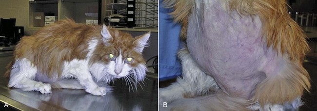
Figure 25-1 A 14-year-old male neutered domestic longhair cat with pituitary-dependent hypercortisolism. A, The patient appears weak, with muscle atrophy and an unkempt haircoat. B, The abdomen has a potbellied appearance with thinning of the skin.
Tests used to screen for spontaneous HC in cats include the urine cortisol : creatinine ratio, the ACTH stimulation test, and the LDDST. It is important to note that the details of these protocols differ between dogs and cats, and readers are directed to more complete references for further information.21,22 The HDDST, endogenous ACTH levels, and abdominal ultrasound examination may be used to assist in differentiation of PDH from ADH in the cat.
Because PDH is relatively uncommon in the cat, there are few case series and case reports on which to base treatment recommendations.76,77 Direct treatment of the pituitary tumor has been reported, with either surgical hypophysectomy or RT.59,78-80 Surgical bilateral adrenalectomy has also been described and was considered the treatment of choice for some time.81 However, these cats are often poor surgical candidates, they have poor healing ability, and complications are common. Laparoscopic adrenalectomy is potentially a better option for these patients because the incisions are much smaller and more likely to heal; however, this has not yet been reported in cats with PDH. Medical therapy with trilostane may be a reasonable option for these cats, based on the authors’ experience and a small number of published cases.82,83 This drug could be used as the sole therapy or to prepare cats for surgery or RT.
Pituitary Somatotroph Tumors (Feline Acromegaly)
Feline acromegaly is a disease of older cats resulting from chronic excessive GH secretion, usually from a functional somatotroph adenoma of the pars distalis of the pituitary gland.84 Feline acromegaly has historically been regarded as a rare condition; however, recent findings suggest that it may be significantly underdiagnosed. In a study of 184 diabetic cats, 59 had markedly increased IGF-1 concentrations, and acromegaly was confirmed in 17 of 18 cats that were examined by CT, MRI, or necropsy.85 These findings have prompted the suggestion that any cat with clinical features of acromegaly, including insulin resistance, should be screened for this disorder.86
Acromegaly is reported to be more common in male cats, with no apparent breed predilection, and most affected cats are middle-aged or older.87 The typical history is of insulin-resistant diabetes mellitus, with affected cats requiring 10 to 20 units of insulin per dose or more, often with inadequate control of the diabetes. This insulin resistance is due to a GH-induced postreceptor defect in the action of insulin on target cells. Affected cats remain polyuric, polydipsic, and polyphagic and continue to gain weight. Most cats with poorly regulated diabetes mellitus will lose weight, and therefore weight gain in this situation is highly suggestive of feline acromegaly. The physical changes of acromegaly develop slowly and are often not noted by the owner until they are advanced. These changes may include enlarged feet, broadening of the face, protrusion of the mandible (Figure 25-2), increased spacing between the teeth, and abdominal enlargement.85,87-90 Owners of affected cats also frequently note noisy or stertorous breathing or respiratory stridor.85,89-91 Physical examination may reveal additional abnormalities, such as enlarged abdominal organs and cardiac murmurs, arrhythmias, or a gallop rhythm.85,87,90
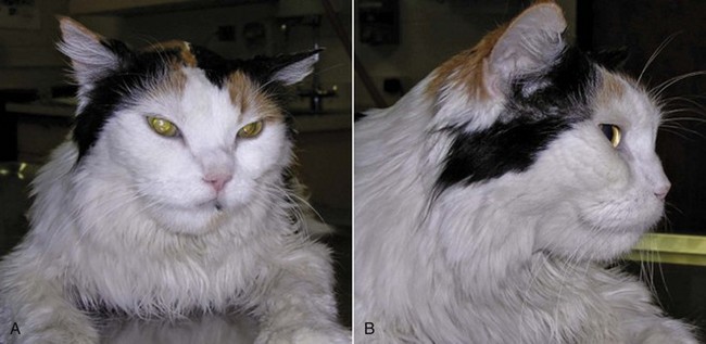
Figure 25-2 A and B, A 12-year-old female spayed domestic longhair cat with acromegaly due to a pituitary tumor. Note the broad forehead and large mandible.
Neurologic signs associated with the pituitary tumor appear to be generally uncommon but may be underrecognized or underreported. Lethargy, mental dullness, or impaired vision may occur but can often be subtle. Affected cats may also demonstrate signs of diabetic neuropathy or lameness, possibly due to acromegaly-associated arthopathy.85,87 Additional complications of acromegaly may include cardiac disease and protein-losing nephropathy.87
Acromegaly is the result of excessive GH secretion from a pituitary gland tumor, and elevated GH levels have been reported in several cats with acromegaly.* Overlap between GH values in diabetic cats with and without acromegaly has also been noted.93 An ovine GH assay has been validated for the diagnosis of feline acromegaly and is available in Europe.94 This assay clearly differentiated between normal cats and cats with acromegaly. Unfortunately, at the time of writing, a feline GH assay was not reliably available in the United States.
The physical changes in patients with acromegaly are due to the anabolic effects of GH, which are mediated by peripherally synthesized IGF-1.84 This hormone is predominantly produced in the liver, and levels of IGF-1 increase in the presence of chronically increased GH production. Because GH secretion may be pulsatile, even in some acromegalics, and because it has a short half-life, increased serum IGF-1 has been suggested to be a more sensitive test for acromegaly because it may reflect GH levels over the preceding 24 hours.84,86 Serum IGF-1 values are widely reported in acromegalic cats.* A recent study confirmed that IGF-1 measurement is a useful screening test for feline acromegaly, with sensitivity and specificity of 84% and 92%, respectively.89 There was no difference in serum IGF-1 concentrations among well-controlled diabetic cats, poorly controlled diabetic cats, and healthy cats.89 The highest IGF-1 noted in a diabetic cat was 153 nM, with a normal reference range of 12-92 nM; thus there is some overlap between the IGF-1 values found in acromegalic cats and those found in poorly regulated diabetic cats. However, it is these authors’ experience that IGF-1 levels in cats with acromegaly are usually at least twice the value of the high end of the reference range. Feline IGF-1 measurement is currently readily available to veterinarians in the United States, and this test should be considered in cats with diabetes mellitus that appear to be insulin resistant.86
The presence of a pituitary tumor can be demonstrated by either CT or MRI in cats with acromegaly, and both have been reported in the literature.† MRI is likely more sensitive than CT,85 but both imaging modalities may reveal a normal pituitary in a cat with acromegaly, if the size of the mass is below the limit of detection.
Treatment options for acromegaly in human medicine include surgery, conventional external-beam RT, stereotactic RT (SRT), and medical therapy.103-105 Some of these therapies are also used in feline patients, but not all are available and many have not been adequately evaluated in cats.
In humans, transsphenoidal surgery to remove the pituitary tumor is generally regarded as the treatment of choice106; however, there are only rare reports of the use of surgery for treatment of feline acromegaly.79,96,99
RT is rarely used as a first-line therapy in human medicine.105 In contrast, RT is the treatment for feline acromegaly that is most widely reported in the veterinary literature.* Conventional fractionated RT is administered in multiple fractions typically spread over a period of weeks. Protocols range from 5 fractions given weekly to as many as 20 fractions given over a period of 4 weeks.80,98,108 A recent study showed that RT improved diabetic control in 13 of 14 cats, although IGF-1 concentrations did not correlate with this improved control.100 Improved control of diabetes mellitus has also been noted in other studies.†
There are few reports of long-term follow-up of acromegalic cats receiving RT; thus it is difficult to assess the risk of complications of this modality in this species. Published case reports and case series of cats receiving conventional RT suggest that short- and long-term adverse effects of this therapy are relatively uncommon.80,100,107,108
Disadvantages of RT as a treatment for feline acromegaly include cost, availability, and the necessity for repeated anesthetic events. The latter disadvantage can be lessened by the use of SRT (see Chapter 12). SRT is widely used in the treatment of pituitary tumors in human patients.109 There is one case series in veterinary medicine in which cats with pituitary tumors received treatment with a linear accelerator-based modified radiosurgical approach.101 Cats received a single large dose of radiation, but it was delivered in a nonconformal fashion. The technique was reported to be safe and effective. At Colorado State University (CSU), SRT is routinely used in the treatment of feline acromegaly, with promising results.110 Cats typically receive 2 to 4 fractions of radiation administered over a period of up to 5 days. This offers considerable advantages in terms of owner time commitment and the risks of anesthesia and hospitalization in elderly diabetic cats.
Medical therapy for acromegaly is commonly used in humans, either as a first-line treatment or as an adjunct to surgery or RT. The classes of drugs used are somatostatin analogs, GH-receptor antagonists, and dopamine agonists.103-106 GH-receptor antagonists have not been evaluated in cats, and dopamine agonists do not appear to be useful in this species.111
Somatostatin analogs, also termed somatostatin receptor ligands (SRLs), bind to somatostatin receptors, suppressing the release of GH from the pituitary gland. The medications are available as long- or short-acting preparations, and response to SRLs is assessed by measurement of IGF-1 and GH levels and tumor size and evaluation of clinical signs.103 Octreotide has been evaluated in a small number of cats with acromegaly. In five cats, short-acting octreotide was used for up to 4 weeks, with no apparent improvement in GH levels.87,92 However, a more recent study showed that GH levels were significantly decreased for up to 120 minutes postinjection in five cats with acromegaly that received a single dose of octreotide.95 These studies used the short-acting form of octreotide and were performed over a very short time period without assessment of clinical response. A study is currently underway at CSU to evaluate the use of a long-acting octreotide preparation for the treatment of feline acromegaly.
For many cats with acromegaly, insulin therapy is the only treatment that is available or acceptable to the owner. In general, these patients should receive the amount of insulin that is necessary to control their diabetes, although adequate blood glucose regulation can be difficult to achieve in many cases. The use of home blood glucose monitoring, with close cooperation between the owner and veterinarian, is strongly recommended. The feeding of a low-carbohydrate diet may also be beneficial.111 It should be expected that these patients will receive insulin doses in the range of 10 to 20 units per dose or more. Concurrent illnesses and complications of acromegaly and diabetes mellitus should also be addressed.
The short-term prognosis for cats diagnosed with acromegaly is generally fair to good, but the long-term prognosis is poor. Patients may succumb to cardiac or renal failure, neurologic disease, or complications of poorly regulated diabetes mellitus.84 The median survival was reported to be 20.5 months in one case series.87
Adrenal Gland Neoplasia
The prevalence of primary adrenal gland tumors in the dog and cat is difficult to discern from the literature. A search of the Veterinary Medical Database from 1985 to 1996 revealed that primary adrenal tumors were reported in approximately 0.17% to 0.76% of pet dogs (representing 1% to 2% of all canine tumors) and 0.03% of cats (representing 0.2% of feline tumors).112 However, this likely underestimates the true prevalence because gross pathologic or histopathologic records were not available for the majority of these patients. For dogs and cats undergoing necropsy or adrenal surgery, it appears that tumors of the adrenal cortex are more common than those of the medulla. A retrospective study of patients with adrenal tumors identified from surgical biopsies or necropsy during a 20-year period at the University of California, Davis (UC Davis), demonstrated that 195 (41%) of 472 neoplastic canine adrenal lesions were adrenocortical tumors (154 adenomas; 41 carcinomas), 151 (32%) were pheochromocytomas (84 benign; 67 malignant), and 126 (27%) were metastatic lesions.113 Of 20 feline adrenal neoplastic lesions, 6 (30%) were adrenocortical tumors (3 adenoma; 3 carcinoma), 2 (10%) were pheochromocytomas (1 benign; 1 malignant), and 12 (60%) were metastatic lesions.113 Less than half of these metastatic lesions were grossly visible at necropsy. Lymphoma was the most common cancer to spread to the adrenal glands in both species. Other metastatic tumors commonly identified in the dog included hemangiosarcoma, mammary carcinoma, histiocytic sarcoma, pulmonary carcinoma, and melanoma. Right and left adrenal glands were affected equally, as were the cortex and medulla. The only notable exception was that all metastatic melanomas were restricted to the adrenal medulla.113
A number of case series in the last decade have documented the outcome of adrenal surgery in dogs.43,45,114,115 When the data from these cases are combined, a histopathologic diagnosis was reported for a total of 191 adrenal tumors, with 153 (80%) arising from the adrenal cortex and 33 (17%) from the medulla. The remaining tumors included two myelolipomas and one each of fibrosarcoma, lymphoma, and leiomyosarcoma. For the adrenocortical tumors that were further classified, 63 of 125 (50%) were carcinomas, 54 of 125 (43%) were adenomas, and 8 of 125 (6%) were described as hyperplastic lesions. It is important to note the bias inherent in these data because only dogs that underwent surgery are included.
Advanced imaging techniques such as ultrasonography, CT, and MRI have now greatly enhanced our ability to identify both clinical and subclinical adrenal abnormalities,38,43,116,117 and it appears that the adrenal gland is affected with neoplasia more commonly than was previously suspected. The ability to detect these adrenal lesions also leads to diagnostic dilemmas as the clinician attempts to elucidate whether the lesions arise from the cortex or medulla, whether they are functional or nonfunctional, and whether they are benign or malignant. Functional adrenal tumors may secrete cortisol, catecholamines, aldosterone, sex hormones, or steroid hormone precursors, and these may be associated with specific clinical and laboratory findings. Hormonal testing and imaging techniques are central to the diagnostic evaluation of these patients so that the most appropriate course of therapy can be pursued. Large adrenal masses may be detected on abdominal radiographs,34,118,119 and the presence of mineralization suggests an adrenal tumor; however, this finding is not highly specific, and it cannot be used to differentiate between benign and malignant masses. The normal ultrasonographic appearance of canine adrenal glands has been described,116,120 and there are many reports of the ultrasonographic appearance of adrenal lesions in dogs. Abdominal ultrasound examination is frequently used to detect metastatic disease and determine the local invasiveness of adrenal tumors. Ultrasonography has been reported to be 80% to 100% sensitive and approximately 90% specific for the detection of adrenal tumor invasion into the caudal vena cava.43,45
The CT appearance of both normal and abnormal canine adrenal glands has been described.46,119,121-124 Contrast-enhanced CT has been shown to provide accurate preoperative evaluation of canine adrenal masses, with 92% sensitivity and 100% specificity for the detection of vascular invasion by adrenal tumors.125 The MRI appearance of presumed normal canine adrenal glands has also been described,126 but as yet there are few reports that document the systematic use of MRI for evaluation of adrenal lesions in dogs and cats. The characterization of adrenal lesions by imaging techniques has undergone recent significant advancement in human medicine. Malignant and benign lesions can frequently be differentiated using modalities such as contrast-enhanced ultrasound, CT densitometry, CT washout characteristics, chemical shift MRI, positron emission tomography (PET), and PET/CT.127-130 Most of these techniques have yet to be explored in veterinary medicine.
Adrenocortical Tumors
Functional cortisol-secreting tumors of the adrenal cortex are responsible for 15% to 20% of canine and feline cases of naturally occurring HC, with PDH accounting for 80% to 85%.17,18,21,22 In the necropsy and surgical biopsy data from UC Davis, adrenocortical adenomas were almost 4 times more common than carcinomas113; however, the functionality of these tumors was not assessed. A review of case reports of functional adrenocortical tumors in dogs suggests that approximately 60% of surgically removed tumors are carcinomas.* Adenomas are typically smaller, with tumors larger than 2 cm more likely to be carcinomas.132 However, adenomas up to 6 cm have been reported.17 On histopathologic examination, adenocarcinomas appear more likely to exhibit a trabecular growth pattern, peripheral fibrosis, capsular invasion, necrosis, and/or hemorrhage.132 They are less likely to exhibit cytoplasmic vacuolization, extramedullary hematopoiesis, or fibrin thrombi. Approximately 20% of adrenocortical carcinomas locally invade into the phrenicoabdominal vein, with extension into the renal vein and/or caudal vena cava.42,131 Intravascular invasion has the potential to cause severe and life-threatening intraabdominal or retroperitoneal hemorrhage.45,133 Metastasis was identified in approximately 50% of dogs with adrenocortical carcinomas.131,132 Although involvement of the liver and lungs is most common, other organs reported to be affected with metastases include the kidney, ovary, mesenteric lymph nodes, peritoneal cavity, and thyroid gland. In the absence of evidence of tumor invasion or metastasis, there are no consistent clinical, biochemical, or imaging findings that reliably distinguish between functional adrenocortical adenomas and carcinomas in dogs and cats. The cellular and molecular events underlying the development of canine adrenocortical tumors are unknown. Recent studies have demonstrated downregulation of ACTH receptors in cortisol-secreting adrenocortical carcinomas and ectopic expression of gastric-inhibitory polypeptide and vasopressin (2) receptor proteins in neoplastic zona fasciculata tissue from canine adrenocortical tumors.134,135 The significance of these findings in tumorigenesis remains to be elucidated.
Dogs with PDH and dogs with ADH are similar in age, but almost 50% of dogs with ADH weigh more than 20 kg, compared to approximately 25% of dogs with PDH.34 The historic features, physical changes, clinical signs, and basic laboratory findings in canine Cushing’s syndrome are essentially the same in dogs with PDH and dogs with ADH and are described in the earlier section on Pituitary Corticotroph Tumors. Similar screening tests are used to confirm the diagnosis of HC; however, the sensitivity of the ACTH stimulation test for the diagnosis of ADH is only around 60%.32 Thus the LDDST is a better screening test when ADH is suspected. Dogs with ADH fail to show suppression on the LDDST or the HDDST, and differentiation from PDH is generally determined by imaging studies, particularly abdominal ultrasound examination,33 and measurement of endogenous ACTH levels.34 Excessive secretion of glucocorticoids by a functional adrenocortical adenoma or adenocarcinoma occurs independent of pituitary control, with secondary atrophy of the normal adrenocortical cells in both the affected and contralateral adrenal glands. Unfortunately the functional atrophy of the contralateral adrenal gland is not always easily detected on abdominal ultrasonography.39 This finding, termed equivocal adrenal asymmetry, is also observed in some dogs with PDH, associated with asymmetric hyperplasia of the glands.37 The results of a recent ultrasound study of dogs with equivocal adrenal asymmetry suggested that a maximal dorsoventral thickness of the smaller gland of less than 5.00 mm was consistent with a diagnosis of ADH.40
Surgical adrenalectomy is the treatment of choice for dogs with ADH,* and surgical management is further addressed later. An early case series of dogs undergoing surgical removal of adrenocortical tumors revealed that 60% of patients were euthanized during surgery or died within 2 weeks.131 In a more recent case series, the perioperative mortality has ranged from 19% to 28%.17,42,43,136 In one series of 144 dogs undergoing surgical removal of a functional adrenocortical tumor, the prognosis was described as excellent for patients that survived 4 weeks postoperatively, with an average life expectancy of 3 years. Nine of 144 dogs were euthanized at the time of surgery, and 29 dogs died during surgery or immediately postoperatively.17 Median survival times of 230 to 778 days have been reported for dogs undergoing adrenalectomy for adrenal carcinomas,42,114,115 compared to a MST of 687.5 days for dogs with adenomas.114 Laparoscopic adrenalectomy has been described for noninvasive adrenocortical carcinomas in a small number of dogs; further studies are needed to determine if this technique can reduce the postoperative complications associated with conventional surgery.137
Medical therapy for ADH should be used when surgery is not a good option for the patient or client, or it may be used prior to adrenalectomy in patients that are significantly debilitated by HC. The primary options for medical therapy are mitotane and trilostane. Treatment with mitotane as an alternative to surgical adrenalectomy utilizes the drug as a true cytotoxic agent. Detailed protocols are readily available,138 and clinicians should be aware that this approach typically requires higher doses of mitotane than those used in PDH139 and that relapses are common. However, this treatment can be effective, with a mean survival of 16.4 months reported in a series of 32 dogs. Dogs without evidence of metastatic disease may show a better response to mitotane therapy.140 Trilostane is not a cytotoxic drug, but it has been used to successfully manage patients with ADH,68,69,141 including a small number of dogs with metastatic disease.142 A recent retrospective study comparing trilostane and mitotane in dogs with ADH reported a median survival of 353 days with trilostane therapy compared to 102 days for mitotane. These survival times were not significantly different; however, this study did further confirm that survival times are significantly decreased in the presence of metastatic disease.143
Functional adrenocortical tumors in dogs and cats can also secrete one or more sex hormones, including androstenedione, progesterone, 17-hydroxyprogesterone, testosterone, and estradiol. These tumors may or may not secrete glucocorticoids, and some patients have been reported to show signs of HC in the absence of elevated cortisol levels on typical screening tests.144-149 Signs of sex hormone excess appear uncommon in dogs with sex-hormone–secreting adrenal tumors but have been reported in a small number of cats.150,151
Aldosterone-secreting adrenocortical tumors have rarely been reported in dogs,152-154 but there is increasing evidence that primary hyperaldosteronism (also termed primary aldosteronism or Conn’s syndrome) may be an underrecognized condition in cats. In fact, it has been suggested to be the most common adrenocortical disorder in this species.155 Affected cats are middle-aged or older, and the most common clinical sign is muscle weakness due to hypokalemia. Arterial hypertension is frequently detected in these patients and may be associated with ocular changes. Routine laboratory testing often reveals hypokalemia, but hypernatremia is uncommon, presumably due to intact water balance mechanisms in these patients. Some cats may also have evidence of concurrent renal disease. Plasma aldosterone can be measured in cats, and normal or elevated levels in the face of hypokalemia would be regarded as inappropriate. However, definitive diagnosis using aldosterone levels is difficult without the measurement of plasma renin activity and the calculation of an aldosterone : renin ratio.156 Unfortunately, a plasma renin activity assay is not readily available to most clinicians. An oral fludrocortisone suppression test has recently been suggested to be a useful diagnostic test for feline hyperaldosteronism,157 but the use of this tool has not yet been widely reported. Imaging of the adrenal glands is often performed in the evaluation of these patients,158,159 and this may distinguish between unilateral and bilateral lesions and also reveal the presence of vascular invasion or metastatic disease. Most cats with hyperaldosteronism have an adrenal adenoma or carcinoma.160 Bilateral adenomas have been reported,160 and some cats have adrenal hyperplasia.156 Adrenalectomy is the treatment of choice for cats with unilateral disease, and good outcomes have been reported for both adenomas and carcinomas, as well as for tumors associated with vena cava thrombosis.160-163 Medical management with potassium supplementation, antihypertensive drugs, and the aldosterone antagonist spironolactone can give reasonable survival times in patients that are not surgical candidates.156,160,161
Adrenal Medullary Tumors
Chromaffin cells are part of the sympathetic nervous system and are present in the adrenal medulla and other locations throughout the body. Neoplastic chromaffin cells in the adrenal medulla give rise to pheochromocytomas, which are tumors that predominantly secrete catecholamines. Chromaffin cell tumors (termed paragangliomas or extraadrenal pheochromocytomas) can arise in other parts of the body, but these are rare in veterinary medicine. Pheochromocytomas are uncommon in dogs and rare in cats.164,165 In past decades, the diagnosis of pheochromocytoma was most often made incidentally at necropsy,166,167 but these tumors are now likely to be detected antemortem because advanced abdominal imaging techniques are routinely used in small animal patients. Pheochromocytomas are generally considered to be malignant tumors in dogs.164 Metastasis is reported in up to 40% of affected dogs; sites include liver, spleen, lung, regional lymph nodes, bone, and CNS.166-168 Vascular invasion by the tumor has been reported in as many as 82% of cases.43,45,169 This finding is not specific for pheochromocytoma because vascular invasion can also occur with adrenocortical tumors.
Pheochromocytoma is usually diagnosed in older dogs,164,167 and males may be overrepresented.169 Catecholamine release by pheochromocytomas is typically episodic, and thus clinical signs may be intermittent and often absent at the time of physical examination. Signs may include weakness, episodic collapse, panting, anxiety, restlessness, exercise intolerance, decreased appetite, weight loss, polyuria, and polydipsia. Physical examination of dogs with pheochromocytoma may be normal due to the episodic nature of catecholamine release or may reveal tachypnea, panting, tachycardia, weakness, pallor, cardiac arrhythmias, or hypertension.164,166,167 Some dogs have signs referable to an abdominal mass, and acute collapse may occur secondary to tumor rupture with abdominal or retroperitoneal bleeding.133 There are no consistent abnormalities on the CBC, serum biochemistry profile, or urinalysis in dogs with pheochromocytomas.
Diagnostic imaging, particularly abdominal ultrasound examination, is central to the evaluation of patients with pheochromocytoma. In many dogs, evaluation for pheochromocytoma occurs after an adrenal mass is found when abdominal ultrasonography is performed for other reasons. In addition to revealing the presence of an adrenal tumor, abdominal ultrasonography may reveal metastatic disease and is sensitive and specific for detecting vascular invasion by adrenal tumors.43,45 CT and MRI are the imaging modalities of choice for humans with pheochromocytomas, and early experience in canine patients has been encouraging.170-172 Abdominal radiographs may reveal the presence of a large adrenal mass166,170 but are generally less informative than ultrasound examination. Thoracic radiographs are recommended to evaluate the cardiovascular system and for detection of pulmonary metastases in any patient with a suspected adrenal tumor. There are rare reports of PET or nuclear scintigraphy imaging in dogs with pheochromocytomas.173,174 Immunohistochemical staining for chromogranin A can distinguish pheochromocytomas from adrenocortical tumors on tissue obtained at surgery or necropsy.175
Plasma and urinary concentrations of catecholamines and their metabolites are routinely measured in humans for the diagnosis of pheochromocytoma.164 Recent reports have suggested that urinary catecholamine and metanephrine to creatinine ratios hold promise as a diagnostic tool in dogs.176-178 Plasma-free metanephrine and normetanephrines have also been evaluated in cats and dogs, with encouraging preliminary results.165,179
Surgery is the only definitive treatment for pheochromocytoma.43,164,166,167,169 This should be performed by an experienced surgical and anesthesiology team because potentially life-threatening complications, including hypertension, hypotension, cardiac arrhythmias and hemorrage,169 may occur during anesthetic induction and handling of the tumor. It has been shown that dogs receiving phenoxybenzamine, a noncompetitive α-adrenergic antagonist, prior to surgery are significantly more likely to survive adrenalectomy. Specifically, dogs that received this medication at doses ranging from 0.1 to 2.5 mg/kg every 12 hours, for a median period of 20 days, had a 13% mortality rate, compared to a mortality rate of 48% in dogs that did not receive this therapy.169 Chemotherapy and RT have not been evaluated in dogs with pheochromocytoma. RT using 131I-metaiodobenzylguanidine (131I-MIBG) was recently reported in one case.180
The prognosis for dogs with pheochromocytoma is affected by tumor size, presence of metastases, and local invasion. A MST of 374 days has been reported after surgical treatment of pheochromocytoma,114 and some dogs may survive for as long as 2 to 3 years.167,181 Dogs without metastatic disease that survive the perioperative period appear to have a good prognosis.164
Surgical Management of Adrenal Tumors
Prior to adrenalectomy, every attempt should be made to determine whether an adrenal tumor is functional, whether there is evidence of metastatic disease, and whether there is vascular invasion. Patients suspected to have a pheochromocytoma should be treated preoperatively with phenoxybenzamine. If tachyarrhythmias are present, a β-blocker such as propranolol or atenolol may also be administered but should only be started after α-adrenergic blockade has been initiated to prevent unopposed α-adrenergic stimulation and severe hypertension.164 Patients with HC due to ADH may be medically managed with trilostane or mitotane prior to surgery if they are significantly debilitated by their disease, although this is rarely necessary. The potential for intraoperative and postoperative complications associated with adrenalectomy is significant182; these cases are best managed by an experienced team, including a surgeon, anesthesiologist, internist, and critical care specialist. Particular concerns with pheochromocytomas include cardiovascular complications and hemorrhage, as noted previously. Patients with functional adrenocortical tumors are at risk for adrenocortical insufficiency, pulmonary thromboembolism, pancreatitis, renal failure, and wound dehiscence.* Protocols are available to guide the perioperative management of adrenalectomy patients.17 At CSU, patients with pheochromocytoma are treated preoperatively with phenoxybenzamine as described earlier. Patients with functional adrenocortical tumors receive heparin and corticosteroid therapy during and after surgery. Postoperatively, an ACTH stimulation test is performed 24 to 48 hours after surgery, and electrolytes and blood glucose are measured frequently. The duration of prednisolone therapy and the necessity for mineralocorticoid supplementation are each determined on an individual case basis. For patients with adrenal tumors of unknown origin, preoperative phenoxybenzamine is recommended, and the protocol for functional adrenocortical tumors is followed, until postoperative ACTH stimulation test results and histopathology results are available to guide further management.
The overall perioperative mortality rate for dogs undergoing adrenalectomy for all adrenal tumors is around 10% to 20%,43,45,114,115 and MSTs of 690 to 953 days have been reported.114,115 A number of investigators have evaluated prognostic factors and predictors of outcome in these patients. In two studies, the presence of caval tumor thrombus did not affect perioperative morbidity and mortality, although the long-term prognosis for dogs with an adrenocortical tumor may be poorer in the presence of a thrombus.43 In contrast, a more recent study suggested that vein thrombosis was associated with a poorer prognosis.115 In the latter study, vein thrombosis was associated with tumors with major axis length of 5 cm or larger, and the presence of metastases or tumor size of 5 cm or larger were both associated with a poorer prognosis.115 Large tumors were also associated with increased perioperative mortality in a series of dogs undergoing elective or emergency adrenalectomy. The dogs in this report that had emergency surgery for acute adrenal bleeding experienced a 50% perioperative mortality rate.45
Incidental Adrenal Masses
Advances in abdominal imaging have led to the diagnostic dilemma of the incidental adrenal mass (“incidentaloma”) in both human and veterinary medicine. When an incidental adrenal mass is identified in a dog or cat, a thorough history and physical examination, including blood pressure measurement and fundic examination, are indicated. Endocrinologic testing should be pursued to rule out a functional adrenocortical tumor. Given the high incidence of metastatic lesions in canine and feline adrenal glands, imaging of the thorax and abdomen should be performed to rule out another primary tumor. Aspiration cytology and ultrasound- or CT-guided biopsies are not routinely recommended for incidentalomas because of the high risk of complications and the inability to reliably differentiate benign and malignant lesions.38,112 Adrenalectomy should be considered for masses that are functional, locally invasive, or larger than 2.5 cm in maximum dimension. Masses smaller than 2 cm with no evidence of hormonal activity should be monitored with abdominal ultrasonography. A suggested interval is to repeat the sonogram 1 month after the initial study and then after 2, 4, and 6 months, with further intervals determined by the appearance of the mass and the clinical status of the patient.164
Thyroid Gland Neoplasia in Dogs
Thyroid tumors account for 1.1% to 3.8% of all tumors in dogs.183-185 In necropsy studies, it is estimated that 30% to 50% of canine thyroid tumors are benign adenomas.183,186 A recent Veterinary Medical Database review reported that 90% of 545 canine thyroid cancers submitted to the database from veterinary teaching hospitals were carcinomas or adenocarcinomas, with 9.3% being adenomas.185 Most adenomas are small, noninvasive, and clinically silent. Consequently, almost all canine thyroid masses associated with clinical signs are malignant.184,187,188 Thyroid tumors of follicular cell origin are subclassified as papillary, follicular, compact (solid), or anaplastic. All subgroups stain positive for thyroglobulin and thyroid transcription factor-1.189-191 Papillary carcinomas are most common in humans,10,192 whereas follicular and compact forms are most common in dogs.1,184,189,193 Medullary thyroid carcinomas, also called parafollicular or C-cell carcinomas, are relatively uncommon in both humans and dogs.191 Positive immunohistochemical staining for calcitonin is the most accurate way to identify these tumors, but they also often stain positive for calcitonin gene-related peptide, thyroid transcription factor-1, chromogranin A, and neuron-specific enolase.175,189-191,194
The etiology of thyroid neoplasia in dogs is largely unknown. The molecular pathogenesis of thyroid neoplasia is best defined in humans.5,7,10 The classic hypothesis involves a discrete series of mutations. Activation of receptor tyrosine kinases such as RET and TRK are common in papillary carcinomas, activating mutations in RAS are frequently identified in follicular carcinomas, and inactivation of p53 is commonly seen in anaplastic carcinomas. Thyroid-stimulating hormone (TSH) or the TSH receptor may play a contributing role in carcinogenesis.195 The TSH receptor in humans with thyroid neoplasia is frequently affected with either hyperfunctioning or silencing mutations. Canine thyroid tumors retain TSH receptors, and hypothyroid beagles that did not receive thyroid hormone supplementation had an increased incidence of thyroid tumors, presumably due to TSH trophic effects without feedback in the context of potential mutations.196,197 Thyroid irradiation is associated with an increased incidence of thyroid tumors in all species, including humans, rodents, and dogs.1,10,186,192 In dogs, one report identified a p53 mutation in 1 of 23 primary thyroid carcinomas.198 Another report confirmed trisomy 18 in a canine thyroid adenoma.199
Thyroid tumors typically arise in older dogs, with a median reported age of 9 to 11 years.184,193,200-202 A sex predilection has not been reported. Predilection of breeds for thyroid tumors includes golden retrievers, beagles, Boxers, and Siberian huskies.184,185 Familial medullary thyroid carcinoma has also been described in a family of dogs with an Alaskan malamute influence.203 The right and left lobes are affected with equal frequency in canine thyroid tumors, and as many as 60% of patients will have bilateral involvement.200,204 On rare occasions, ectopic thyroid tissue can give rise to tumors at the base of the tongue, cervical ventral neck, cranial mediastinum, and heart.204-210 Up to 35% to 40% of dogs have visible metastatic disease at initial presentation, and as many as 80% will ultimately develop metastasis.184,191,204,211 Metastatic potential is reported to increase when the primary tumor volume exceeds 23 cm3 and approaches 100% when tumor volume exceeds 100 cm3.186 Bilateral tumors are 16 times more likely to metastasize than unilateral tumors.200 The lungs and regional lymph nodes, including the retropharyngeal, cranial cervical, and mandibular lymph nodes, are affected most commonly, but a wide variety of tissues can be affected. Medullary carcinomas may have a lower metastatic potential than follicular and solid carcinomas.191
The majority of canine thyroid carcinomas are nonfunctional. Based on clinical signs and serum T4 concentrations, approximately 60% of patients are euthyroid, 30% are hypothyroid secondary to destruction of the normal thyroid parenchyma, and 10% are hyperthyroid.188,202,212-214 Most dogs are presented for a palpable ventral cervical mass.184,191,193 Less common abnormalities include coughing, rapid breathing, dyspnea, dysphagia, dysphonia (change in bark), laryngeal paralysis, Horner’s syndrome, and facial edema. Acute severe hemorrhage can occur secondary to invasion into the cervical vasculature.215 In addition to clinical signs referable to the physical thyroid mass, dogs with hyperthyroidism frequently exhibit polyphagia, weight loss, muscle wasting, polyuria, and polydipsia.188,212-214
The differential diagnosis for a mass in the region of the thyroid gland in dogs includes abscesses or granulomas, salivary mucoceles, lymphatic metastasis from tonsillar squamous cell carcinoma, lymphoma, carotid body tumor, and sarcomas. In humans, thyroid cytology is very accurate for identifying thyroid tumors and distinguishing whether they are benign or malignant.10,192 Accuracy of cytology in dogs with thyroid masses is reported to be problematic. In several reports, cytology confirms the mass to be of thyroid origin in only half of affected dogs, and definitive recognition of malignancy occurs less often.184,188 Use of a needle without physical aspiration and thorough examination of the feathered edge may improve diagnostic accuracy. Malignant thyroid tumors have a higher vascular density than normal thyroid tissue and benign tumors,202 and hemodilution is a common problem (see Chapter 6). This increased vascularity also adds significant risk to large core needle biopsy procedures.
Routine staging for dogs with thyroid carcinoma includes general health assessment with laboratory evaluation (CBC, serum biochemistry profile, and urinalysis), three-view thoracic radiographs, and cytologic or histologic evaluation of the mandibular lymph nodes. Cervical ultrasonography can be used to confirm if a mass is of thyroid origin and to assess invasiveness and vascularity.216,217 The retropharyngeal and cranial cervical lymph nodes can also be examined for evidence of metastasis. The MRI and CT appearances of the normal canine thyroid have been described,218,219 and these modalities are useful in the investigation of cervical masses in the dog and in the staging of thyroid carcinomas.215,218,219 Scintigraphy using 99mTc-pertechnetate or, less commonly, radioactive iodine (123I or 131I) is performed primarily to identify local residual disease after surgery, ectopic tumors, or metastatic disease.* Most primary tumors are visualized, although the pattern of uptake is often heterogeneous. Metastatic disease is identified less consistently. To be visualized with scintigraphy, a thyroid tumor must be capable of trapping 99mTc-pertechnetate or trapping and organifying 123I or 131I. It may or may not be able to complete the remaining steps necessary for synthesis and secretion of functional thyroid hormone.
Treatment of canine thyroid carcinomas is dictated by the size of the mass, extent of invasion, presence or absence of gross metastatic disease, and any concurrent symptoms of thyrotoxicosis. Surgical excision provides the best outcome with the least morbidity when tumors are freely movable without extensive deep tissue invasion.191,193,222 Thyroidectomy is not recommended when the tumor is not freely movable in all directions or extensively invades adjacent structures, including major vasculature, recurrent laryngeal nerves, the vagosympathetic trunk, the larynx, the trachea, or the esophagus. Extensive hemorrhage can result from the vascularity of the tumor, invasion into adjacent blood vessels, and local coagulopathies.202 Other potential complications of thyroidectomy include hypocalcemia due to hypoparathyroidism if the parathyroid glands are removed, damage to the recurrent laryngeal nerve(s), and hypothyroidism after bilateral thyroidectomy.193,223 According to limited, retrospective data sets, it was estimated that only 25% to 50% of thyroid carcinomas were mobile and amenable to surgery at the time of initial diagnosis.191,193 Palpation under anesthesia and preoperative imaging are current general recommendations for surgical evaluation at the authors’ institution, with some extracapsular extension and invasion considered acceptable. Median survival after thyroidectomy is around 3 years if the tumor is freely movable and 6 to 12 months if the tumor is more invasive.191,193 Recently, a report describing resection of mobile, discrete bilateral thyroid tumors in a limited number of dogs suggested reasonable overall survival with persistent postoperative management of thyroid and parathyroid endocrinopathies.224
Nonresectable thyroid carcinomas may be managed with radiation as a primary therapy or as a means to achieve a surgical option. External-beam RT is used most commonly for dogs. One study evaluated definitive RT (48 Gy delivered in 4 Gy fractions on an alternate-day schedule) in 25 dogs with unresectable thyroid carcinomas and no visible metastasis.200 Tumors either stabilized or decreased in size. The time to maximal tumor reduction ranged from 8 to 22 months in dogs whose tumors did respond. The progression-free survival rates were 80% at 1 year and 72% at 3 years. The first cause of treatment failure was local progression in three dogs, metastasis in four dogs, and concurrent local progression and metastasis in three dogs. Limited information exists regarding the use of definitive radiation in the adjuvant or neoadjuvant settings.201,225 This same RT protocol described was evaluated in an additional eight dogs, seven of which had undergone incomplete thyroidectomy prior to irradiation.201 Median survival was just over 2 years (range 1 to 3 years). None developed local recurrence, although four died from metastatic disease. Radiation-induced toxicoses to the larynx, trachea, and esophagus are usually well tolerated. Hypothyroidism may develop months to years after treatment.200,225
For dogs that present with gross metastatic disease, hypofractionated RT may still provide effective palliation of the primary tumor, due to a generally slow rate of progression in both the primary and metastatic lesions. In a study evaluating palliative RT as the sole treatment modality, 13 dogs received 36 Gy in four weekly 9 Gy fractions.211 Complete or partial reduction of the primary tumor occurred in one and nine dogs, respectively. Local progression occurred in five dogs, 11 to 24 months after irradiation. Gross metastatic disease was present in five dogs at initial presentation and developed in an additional two dogs during the study. Overall median survival was around 22 months, with local and metastatic progression occurring equally in all dogs.
In humans with well-differentiated thyroid carcinoma, 131I is routinely administered postoperatively to destroy occult microscopic local or metastatic carcinoma.10,192 Experience with 131I thyroid ablation in dogs is substantial, with two recent, relatively large clinical studies providing evidence of efficacy of 131I for advanced unresectable, metastatic, or residual thyroid neoplasia.226,227 Collectively, from all reports, over 80 dogs with stage II (2 to 5 cm diameter; fixed or unfixed), stage III (>5 cm diameter, fixed or unfixed), or stage IV metastatic thyroid carcinoma have received 131I for the intent of managing tumor burden and clinical signs. MSTs for stage II and III patients exceed 2 years, and dogs with metastatic carcinoma experienced survival times of approximately 1 year. Such results are comparable to external-beam RT. Interestingly, similar survival times were noted in dogs that were hypothyroid, euthyroid, or hyperthyroid. Recommendations for 131I dosimetry remain unresolved. Fatal myelosuppression was observed in three dogs in one report, although no specific dose-effect relationship was defined.227 The biologic effect both on tumor and normal tissue is a complex function of 131I uptake that depends on extent of the tumor burden, degree of organification and excretion of 131I, bone marrow sensitivity, and administered dose of radiation. Dosing regimens are currently empiric in dogs but could be more carefully defined by administering a 131I tracer for calculation of definitive dosing, as in humans. Until such time as dosing is individualized, the maximum dose administered should be 0.2 GBq/kg (5 mCi/kg), and bone marrow monitoring posttreatment is recommended. Additional doses may be administered if necessary, as determined by persistent hyperthyroidism or activity on posttreatment nuclear scans. Dogs receiving 131I will require thyroid hormone supplementation.
Chemotherapy has been evaluated in dogs with thyroid tumors. Of those dogs treated with either doxorubicin or cisplatin, 30% to 50% demonstrated a partial response (>50% reduction in volume).228,229 Individual responses have also been reported using mitoxantrone or actinomycin D.230,231 Chemotherapy may be considered for dogs with large nonresectable primary tumors and/or gross metastatic disease.
Thyroid Gland Neoplasia in Cats
Hyperthyroidism (thyrotoxicosis) is the most common endocrine disorder in cats.232,233 It is almost always caused by a primary thyroid abnormality that results in the production and secretion of excessive thyroxine (T4) and triiodothyronine (T3). Multinodular adenomatous hyperplasia is identified histologically in the majority of thyrotoxic cats.1,232-234 Both thyroid lobes are affected in 70% to 90% of cases,233-235 although they may be asymmetrically enlarged at the time of diagnosis. Malignant carcinomas are the least common cause of hyperthyroidism, occurring in only 1% to 3% of thyrotoxic cats.1,232-234 Nonfunctional thyroid carcinomas are uncommon.236,237 Feline thyroid carcinomas are more locally invasive than their benign counterparts, and their metastatic rate is up to 70%, with regional lymph nodes and lungs being affected most commonly.238,239
TSH from the pituitary regulates both the secretion of thyroid hormones and the proliferation of thyroid cells. The interaction of TSH with receptors on the surface of thyroid cells activates G protein-mediated cyclic adenosine monophosphate (cAMP) signal transduction pathways.240 Some of the components of the TSH receptor-G protein-cAMP system have therefore been evaluated for changes that could result in feline hyperthyroidism. Decreased expression of the inhibitory subunit Giα has been identified in adenomatous feline thyroid glands,241 and a further study demonstrated that Gi2 was decreased.242 Mutations in the Gsα gene were also reported in 4 of 10 hyperthyroid cats evaluated.243 Both of these changes could lead to stimulation of adenyl cyclase with the potential for increased levels of cAMP. However, a more recent study showed that ligand-stimulated activation of G proteins was the same in thyroid cell membranes obtained from hyperthyroid and euthyroid cats.244 Therefore the role of alterations in inherent G(s) or G(i) activities in the pathogenesis of hyperthyroidism in cats remains unresolved. Mutations in the TSH receptor in hyperthyroid cats have been investigated and were not documented in early studies.243,245 However, a more recent study that targeted hyperplastic or adenomatous thyroid nodules from 50 hyperthyroid cats detected a total of 11 different mutations in the TSH receptor gene.246 Oncogene expression has also been evaluated in thyroid tissue from hyperthyroid cats. Overexpression of one or more RAS oncogenes was identified in adenomas and hyperplasias from 18 of 18 cats diagnosed with hyperthyroidism but not in adjacent normal thyroid tissue or in thyroid tissue from 14 unaffected cats.247 In the same tumors that overexpressed RAS, Bcl-2 and p53 proteins were undetectable using immunohistochemistry.
Hyperthyroidism was not recognized as a clinical disorder in cats until 1979, yet currently it is estimated to affect as many as 1 out of every 300 cats.232 This may reflect a true increase in incidence, heightened awareness and testing by veterinarians, or both. If there has been a true increased incidence, environmental factors may have contributed. Several risk factors have been associated with hyperthyroidism, including the consumption of commercially prepared canned cat food, indoor residence, use of cat litter, exposure to brominated flame retardants, and use of flea-control products, but none have been incriminated as a primary inciting cause.248-253 The iodine content of cat food has also been suggested to play a role in the development of hyperthyroidism, but this relationship is unproven.254
Due to space constraints, only a brief overview of the clinical features, diagnosis, and treatment of feline hyperthyroidism will be presented here, with an emphasis on malignant thyroid tumors. The reader is directed to several excellent and detailed reviews of feline hyperthyroidism for additional information.232,233
Most hyperthyroid cats are older, with mean and median ages of 12 to 15 years.234,255-257 The disease is rare in cats under 8 years of age.232 There is no gender predilection, but Siamese and Himalayan breeds were found in one study to be at decreased risk.248 The most common signs reported by owners include weight loss, polyphagia, polydipsia, polyuria, gastrointestinal signs (vomiting, diarrhea, increased stool production), and increased activity.232,233,235,255 Additional abnormalities commonly identified on physical examination include the presence of a palpable thyroid nodule, cardiovascular abnormalities (tachycardia, heart murmur, gallop rhythm, premature beats), and poor hair coat. Hyperthyroidism may also contribute to hypertension in elderly cats.258 Definitive diagnosis of hyperthyroidism is routinely based on an elevated serum total T4 level.235,255,259 Less than 10% of hyperthyroid cats will have a total T4 level within the reference range. This is usually due to normal fluctuations in serum thyroid hormone concentrations and/or the presence of concurrent nonthyroidal illness.259,260 If hyperthyroidism is suspected in a cat with a normal total T4 level, total T4 should be measured again in 1 to 2 weeks, particularly if the total T4 is in the upper half of the reference range. Free T4 measurement may also aid in the diagnosis of hyperthyroidism when total T4 is within the reference range. However, this test should only be used in cats with clinical signs of hyperthyroidism in which the total T4 is high-normal. Free T4 concentrations can be high in cats with nonthyroidal illness,259,261,262 and these patients would be expected to have low total T4 values. Thus free T4 should never be used as a screening test for hyperthyroidism. For those patients in which hyperthyroidism is suspected, but not confirmed by measurement of total or free T4, additional tests have been used to confirm the diagnosis. These include thyroid scintigraphy (see later), the TSH stimulation test or the thyrotropin-releasing hormone (TRH) stimulation test (both of which are of limited utility263,264), or the triiodothyronine suppression test. The latter test can provide useful information but relies on significant owner and patient compliance.265,266 Thyroid function tests cannot be used to differentiate benign and malignant tumors.
Staging for cats with hyperthyroidism should minimally include baseline CBC, serum biochemistry profile, urinalysis, and blood pressure measurement. Additional diagnostic tests that may be recommended include thoracic radiography, electrocardiography, and echocardiography.235,267,268 99mTc-pertechnetate scintigraphy is very useful for determining the anatomic extent of functional thyroid tissue and planning therapy, as well as for confirming the diagnosis.257,269 Unilateral uptake occurs in cats with a solitary adenoma and atrophy of the normal contralateral gland. Bilateral uptake, even if asymmetric, is indicative of adenomatous hyperplasia. Thyroid scintigraphy is particularly useful for revealing the presence of ectopic thyroid tissue or multiple areas of hyperfunctioning thyroid tissue. In one study, these were present in 20% to 25% of hyperthyroid cats undergoing thyroid scintigraphy.257 Metastatic disease due to thyroid carcinoma may be detected by scintigraphy,235 and the pattern of uptake of radionuclide may be suggestive of the presence of malignant disease; however, two recent studies demonstrated that there are no scintigraphic findings that can definitively distinguish between benign and malignant thyroid disease in all hyperthyroid cats.237,257
Treatment options for feline hyperthyroidism include antithyroid drugs, surgical thyroidectomy, and radioactive iodine therapy. Methimazole is the most widely used antithyroid drug in North America.270 Carbimazole is more widely used in Europe.271 These are thioureylene drugs that inhibit thyroid hormone synthesis by interfering with oxidation of iodide, iodination of tyrosyl residues in thyroglobulin, and the coupling of iodotyrosines to iodothyronines.233,270,271 These drugs are often used for assessing the effect of resolution of hyperthyroidism on renal function (see later) and preparing a cat for anesthesia and thyroidectomy. They are also frequently used as a long-term treatment modality, but it is important to note that they have no antitumor activity and no cytotoxic effect on thyroid follicular cells. Methimazole and carbimazole are both highly effective in lowering serum thyroid hormone concentrations and controlling hyperthyroidism. Carbimazole is converted to methimazole in the body, and a dose of 5 mg of carbimazole is considered to be equivalent to 3 mg of methimazole.233 Methimazole is usually administered at a starting dose of 2.5 mg orally twice daily for 2 weeks. Based on clinical signs and serum T4 levels, the dosage can be adjusted incrementally, with monitoring of serum T4 concentrations. Once-daily administration of methimazole has been reported to be less effective,272 but this approach can be successful in some cats, particularly those that need very low doses to control their disease. Carbimazole is usually administered 2 to 3 times daily, but a controlled-release formulation has recently been shown to be effective when administered once daily.273 For cats that are difficult to medicate orally or that have gastrointestinal side-effects, methimazole compounded in pluronic lecithin organogel (PLO) can be applied topically to the pinna.274-276 Transdermal carbimazole has also been shown to be effective.277 Approximately 10% to 15% of patients treated with methimazole develop adverse effects, including lethargy, anorexia, vomiting, facial excoriations, hepatotoxicity, and blood dyscrasias.232,233,270,278 Gastrointestinal side effects are often self-limiting or can be avoided by transdermal drug delivery. Blood dyscrasias are rare but most likely to occur within the first 3 months of treatment; therefore CBCs should be monitored most closely during that time. Medication should be discontinued in patients that experience facial excoriations, blood dyscrasias, or hepatotoxicity. Carbimazole or transdermal methimazole are likely to have the same effects and should therefore not be used in patients experiencing these adverse effects with oral methimazole. Other medical therapies that have been used to treat feline hyperthyroidism include ipodate and iopanoic acid279,280; however, these are unlikely to be effective for long-term therapy and are rarely used. A potentially interesting new development in the management of feline hyperthyroidism is the use of an iodine-restricted diet to control the disease.281-283 At the time of writing, a feline prescription diet for the management of hyperthyroidism had been recently released, but no peer-reviewed manuscript was available for evaluation.
Definitive therapy for feline hyperthyroidism currently consists of surgical thyroidectomy or radioactive iodine. Surgical excision of the affected thyroid lobe(s) is an effective treatment.234,284,285 Although the majority of cats have bilateral disease, this may be asymmetric and not apparent on palpation or surgical exploration. Thus thyroid scintigraphy is recommended before surgery in order to determine whether unilateral or bilateral thyroidectomy is necessary.234 Intracapsular and extracapsular thyroidectomy techniques have been described.284,286 Intracapsular techniques better preserve adjacent parathyroid tissue, whereas extracapsular ones more consistently remove all hyperplastic or neoplastic thyroid tissue. Hyperthyroid cats are often poor anesthetic candidates, and preoperative stabilization with oral antithyroid medications or β-adrenergic blockers should be considered. The most significant intraoperative complication of thyroidectomy in hyperthyroid cats may be cardiac dysrhythmias.285 Otherwise, the surgery is not considered to be technically demanding.234,285 Hypocalcemia due to transient hypoparathyroidism is the most commonly reported postoperative complication, with rates ranging from 6% to 15%.234,285 Other potential complications include hypothyroidism and rarely, Horner’s syndrome or laryngeal paralysis. All surgically excised tissue should be submitted for histopathology to rule out the presence of a thyroid carcinoma. Cats with thyroid carcinoma that undergo thyroidectomy usually experience improvement in their clinical signs, but most remain hyperthyroid or develop recurrent hyperthyroidism within a few months of surgery.236,238 Cats with ectopic hyperplastic thyroid tissue are also at risk for postoperative recurrence of hyperthyroidism.234 Radioactive iodine therapy is recommended for patients with thyroid carcinoma or ectopic hyperplastic thyroid tissue.
Radioactive iodine, or 131I therapy, is often regarded as the treatment of choice for cats with hyperthyroidism, particularly those with bilateral thyroid hyperplasia, ectopic thyroid tissue, or thyroid carcinoma.236-238,287,288 131I has a half-life of 8 days and emits both beta and gamma radiation.233 Beta particles, which account for 80% of the tissue damage, travel a maximum of 2 mm in tissue and have an average path length of 400 µm. They therefore cause local destruction while sparing adjacent hypoplastic thyroid tissue, parathyroid glands, and other cervical structures. The dose of 131I can be calculated from tracer kinetic studies,289,290 but these are rarely performed. The administration of a fixed dose of 131I is reported by some authors,288,291 whereas others use doses that take into account variables, such as the number or size of thyroid nodules, patient body weight, severity of clinical signs, or magnitude of elevation in serum total T4.232,287,292,293 131I is usually administered by the subcutaneous route because it is effective, less stressful for the patient, and safer for personnel.292 For cats with benign thyroid disease, 131I doses typically range from 2.0 to 6.0 mCi based on clinical signs, serum T4 concentration, and thyroid nodule size.287 Using this dosing strategy, less than 5% of cats remain hyperthyroid or experience relapse of clinical signs. When this occurs, a second treatment is usually curative. The proportion of cats that develop persistent hypothyroidism requiring thyroid hormone supplementation varies among studies, and the risk of this has been suggested to be higher in cats with scintigraphic evidence of bilateral disease.294 Cats with thyroid carcinomas usually have larger tumor burdens, and malignant cells trap and retain iodine less efficiently.233,236 These cats are therefore treated with higher ablative doses of 131I, in the range of 20 to 30 mCi.232,233,236-238 In one large study of hyperthyroid cats treated with 131I, median survival was reported to be 2 years, with survival rates at 1, 2, and 3 years of 89%, 72%, and 52%, respectively.287 The most common causes of death or euthanasia were cancer or renal disease,287 which is perhaps not surprising in this population of older cats. A more recent study reported a MST of 4 years for cats treated with 131I, compared to 2 years for cats that were treated with methimazole.256
Ultrasound-guided percutaneous ethanol injection has also been evaluated as a treatment for feline hyperthyroidism. Cats with solitary adenomas have a good response, with resolution of clinical signs persisting for over 12 months.295 This technique is not recommended for bilateral hyperplasia.296 Ultrasound-guided percutaneous radiofrequency heat ablation has been shown to be ineffective for long-term control of hyperthyroidism.297 Given the ready availability of permanent effective treatments for unilateral or bilateral disease, these alternative treatments are unlikely to be widely used.
Chronic kidney disease (CKD) is a relatively common problem in older cats, and therefore concurrent CKD and hyperthyroidism frequently occur in this population. The hyperthyroid state increases glomerular filtration rate (GFR)298,299 and therefore decreases serum creatinine values. The implications of this are that hyperthyroid cats with normal serum creatinine values may in fact have concurrent “masked” CKD and that decline in renal function is a risk of all effective forms of treatment of feline hyperthyroidism, with some nonazotemic cats becoming azotemic, or the potential for worsening of preexisting azotemia.293,298,300,301 This decline in renal function occurs within 1 month after treatment and appears to remain stable thereafter.293,302 Measurement of pretreatment GFR may help to predict which cats will become azotemic after resolution of hyperthyroidism293,301; however, this is impractical for most patients. Unfortunately, no readily available clinical data can predict the effects of therapy on renal function in an individual cat.303 For this reason, many clinicians recommend a therapeutic trial with methimazole prior to definitive therapy for feline hyperthyroidism.303 This may have value in providing owners with information about the likely consequence of therapy for these cats, but regardless of the detected change in renal function, effective therapy for hyperthyroidism is still required in these patients. One recent study showed that the development of azotemia was not significantly associated with survival of cats treated for hyperthyroidism,304 but the same group also demonstrated a significantly shorter survival time in cats with iatrogenic hypothyroidism that became azotemic after treatment compared with those that remained nonazotemic.305
Parathyroid Tumors
Parathyroid tumors are uncommon in dogs and rare in cats. These tumors arise from the chief cells and autonomously secrete parathyroid hormone (PTH), leading to hypercalcemia due to primary hyperparathyroidism. Hypercalcemia is the result of direct effects of PTH on bone and the kidneys and indirect effects on the intestine, mediated by vitamin D. Approximately 90% of dogs and cats with primary hyperparathyroidism have a single parathyroid mass,306-312 with adenomas being most commonly diagnosed and cystadenoma, carcinoma, and hyperplasia diagnosed less frequently.306-309,311-314 Two or more parathyroid masses may be found in some canine and feline patients, and they may not necessarily all be of the same histologic type. The presence of four hyperplastic parathyroid masses should prompt careful evaluation for causes of secondary hyperparathyroidism.
Primary hyperparathyroidism is most common in older dogs and cats, with reported mean ages of approximately 11 years in dogs309,311 and 13 years in cats.306 A breed predisposition has been reported in Keeshonden dogs307,309,314,315 in which the disease appears to follow an autosomal dominant mode of inheritance, although the affected gene has not yet been identified in this breed.314,315 It is not clear if there is a breed predilection in cats. The clinical signs of hyperparathyroidism result from hypercalcemia and include polyuria/polydipsia, weakness, lethargy, decreased appetite, weight loss, muscle wasting, vomiting, and trembling. It is not uncommon for owners to detect no clinical signs in affected dogs or cats, with hypercalcemia being diagnosed when blood is drawn for a routine health check or for investigation of an unrelated problem. However, it is also the case that signs can be subtle and are only recognized in retrospect after the hyperparathyroidism has been treated and the hypercalcemia has resolved. In a large case series, the most common clinical problems reported in dogs with hyperparathyroidism were related to the lower urinary tract, usually associated with urolithiasis or urinary tract infection.309 Specific physical examination abnormalities are rare in dogs and cats, although a palpable parathyroid mass has been reported in a proportion of cats with hyperparathyroidism; the latter is an extremely rare finding in dogs.308,309
Hyperparathyroidism is usually diagnosed after finding hypercalcemia on a serum biochemistry profile, either as an incidental finding or when investigating a problem such as calcium oxalate urolithiasis, polyuria/polydipsia, or weakness. The presence of hypercalcemia should be verified by measuring serum ionized calcium, with appropriate careful sample handling.316,317 There are many causes of hypercalcemia in dogs and cats,318,319 and diagnostic tests may be performed to investigate several possible causes simultaneously. The reader is directed to Chapter 5 for a further discussion of the causes of hypercalcemia in dogs and cats. Hypercalcemia due to primary hyperparathyroidism is often accompanied by hypophosphatemia or serum inorganic phosphorus at the low end of the reference range. This finding is not pathognomonic for hyperparathyroidism and can be associated with humoral hypercalcemia of malignancy, but it can assist in ranking the differential diagnoses because vitamin D toxicosis and renal failure would both be expected to cause hyperphosphatemia. The diagnosis of hyperparathyroidism is confirmed by documenting an inappropriately high serum PTH level in the presence of ionized hypercalcemia. It is important to note that PTH is frequently within the reference range in patients with hyperparathyroidism, with 73% of cases reported to have a normal PTH in one large series.309 A normal PTH in the face of hypercalcemia is an abnormal finding because PTH should be suppressed as calcium increases. The lack of suppression of PTH is indicative of loss of the normal negative feedback effects of calcium due to autonomous hormone secretion by hyperplastic or neoplastic parathyroid tissue. Ultrasound examination of the neck is commonly used in the diagnosis of hyperparathyroidism in dogs and cats309,310,320,321 and is particularly useful for localizing parathyroid mass(es) prior to surgery or other ablative procedures. The normal sonographic appearance of canine parathyroid glands has been described,322 and parathyroid masses as small as 3 mm in greatest diameter have been identified ultrasonographically.309 Parathyroid scintigraphy and selective venous sampling to assess local PTH concentrations do not appear to be helpful in localizing hyperplastic or neoplastic parathyroid tissue.320,323,324
The management of hypercalcemia is further addressed in Chapter 5. Primary hyperparathyroidism in dogs and cats is usually associated with slowly progressing hypercalcemia, and the calcium elevation itself rarely requires emergency treatment. Hypercalcemia is a risk factor for renal failure. Mechanisms include altered glomerular capillary permeability, decreased renal blood flow, and mineralization of the kidneys. The risk of mineralization is increased when the calcium × phosphorus product exceeds 70. As noted previously, decreased or low-normal phosphorus often occurs in patients with hyperparathyroidism, thus decreasing the risk of renal mineralization. In fact, it appears that renal failure occurs rarely in dogs with primary hyperparathyroidism. In a large canine case series, it was found that mean blood urea nitrogen (BUN) and serum creatinine were both significantly lower in 210 dogs with primary hyperparathyroidism, compared with 200 control dogs.309 In addition, 95% of the hyperparathyroid dogs had BUN and serum creatinine values within or below the reference range. This may partly be a result of the secondary nephrogenic diabetes insipidus that causes polyuria/polydipsia in these patients.
Definitive therapy for primary hyperparathyroidism requires removal of the hyperfunctioning gland(s). This is most commonly achieved by surgery in both dogs and cats; however, percutaneous ultrasound-guided ethanol ablation and percutaneous ultrasound-guided heat ablation are also well described in the dog.311,325,326 The latter techniques may not be as widely available as surgery, and success likely depends on the experience of the operator.311 In a retrospective comparison of surgery, ethanol ablation, and heat ablation, all three techniques performed well, but surgical parathyroidectomy had the highest success rate and lowest rate of complications.311 Ultrasound examination of the neck is strongly recommended prior to surgery, and the surgeon should carefully evaluate all the parathyroid glands because approximately 10% of patients have masses in more than one gland. Up to three of the four parathyroid glands can be removed without risk of permanent hypoparathyroidism. Patients with involvement of all four glands present a dilemma, and it is important to ensure that hyperplasia in these cases is not secondary. If the hyperparathyroidism is believed to be primary, removal of all parathyroid tissue is required to control hypercalcemia, but this will necessitate life-long supplemental therapy. In human medicine, total parathyroidectomy may be followed by autotransplantation of one of the glands or may be accompanied by cryopreservation of parathyroid tissue.327 These approaches have not been explored in veterinary medicine.
Hypocalcemia is a potential complication of surgical parathyroidectomy, ethanol ablation, or heat ablation. This happens because chronic hypercalcemia inhibits PTH secretion by the normal parathyroid glands and leads to parathyroid atrophy. It is logical to assume that the risk of this posttreatment complication increases with duration and severity of hypercalcemia; however, it is still difficult to predict whether an individual patient will become hypocalcemic. Thus it is recommended that serum ionized calcium levels be monitored at least twice daily for as long as 5 to 7 days after surgery or other ablative procedures. Hypocalcemia should be treated if the ionized calcium falls below 0.8 to 0.9 mmol/L, the total calcium is less than 8 to 9 mg/dL, or the patient has signs of tetany. Intravenous (IV) calcium salts are used for acute therapy for hypocalcemia; subcutaneous (SQ) administration should be avoided. Vitamin D and oral calcium are used for subacute and chronic therapy. Several excellent references are available regarding treatment of hypoparathyroidism.312,328 In summary, 1,25-dihydroxyvitamin D3 (calcitriol) is recommended for vitamin D supplementation because it has a rapid onset of action and a short half-life. This facilitates dose adjustments and reduces the risk of hypercalcemia. Oral calcium supplementation alone is not sufficient to treat hypoparathyroidism, and in fact this therapy can be gradually withdrawn once the calcium is stable because most maintenance diets contain an adequate amount of calcium. Calcitriol therapy can be started up to 24 hours prior to surgery or parathyroid ablation, if the risk of hypocalcemia is thought to be high. This allows the medication to reach therapeutic levels more quickly, but it will not necessarily prevent the development of hypocalcemia. When adjusting the dose of calcitriol, the goal is to maintain the calcium barely below the normal reference range rather than within normal. This reduces the risk of hypercalcemia and provides the stimulus for recovery of function of the remaining normal parathyroid glands. Once the serum calcium has been stable for at least 1 to 2 weeks in an outpatient, the dose of calcitriol can be gradually reduced, with careful monitoring. The time for return of normal parathyroid function is unpredictable, and therefore clients should expect frequent rechecks of the patient’s calcium levels for several weeks to months after treatment of hyperparathyroidism.
The long-term prognosis after surgical or ablative treatment for hyperparathyroidism is very good. Metastatic disease is extremely rare, and the complication of hypocalcemia is generally amenable to medical therapy. A small number of patients may appear resistant to the postoperative management of hypocalcemia, and this may be the result of the “hungry bone syndrome” in which there is aggressive unregulated uptake of calcium by the bones. In human medicine, this syndrome has been managed with preoperative bisphosphonate administration329 or the use of recombinant PTH.330 Neither of these approaches has been used in veterinary medicine, and most patients will eventually respond to high doses of calcitriol and calcium supplementation. Less than 10% of dogs and cats treated for hyperparathyroidism will experience a recurrence of the disease.308,312 If this occurs, a second surgery or ablative procedure should be performed. The short-term prognosis for dogs and cats that do not undergo definitive surgical or ablative therapy for hyperparathyroidism may still be favorable because the disease tends to be slowly progressive, clinical signs may be mild, and renal failure may be a less common outcome than previously suspected.309
Pancreatic Beta-Cell Tumors (Insulinomas)
Pancreatic beta-cell tumors are rare in humans and cats and uncommon in dogs.331-333 These tumors are often functional, but the neoplastic beta-cells fail to appropriately inhibit insulin secretion at low blood glucose concentrations. Thus the hallmark of insulinoma is a normal or elevated blood insulin concentration in the presence of low blood glucose levels. A feline insulinoma was recently shown to demonstrate abnormal glucokinase and hexokinase expression.332 These changes may contribute to enhanced glucose sensitivity and hence an abnormal insulin secretory response in insulinoma cells. Although the clinical signs of insulinoma result from hypoglycemia associated with unregulated insulin secretion, immunocytochemical analysis reveals that these tumors often produce many additional hormones, including glucagon, somatostatin, pancreatic polypeptide, GH, IGF-1, and gastrin.332,334-338
In humans, 90% of insulinomas are solitary and benign, and 5% to 10% are associated with multiple endocrine neoplasia type 1 (MEN1). Insulinomas in dogs are much more likely to be malignant, although morphologic classification into adenoma or adenocarcinoma does not consistently reflect the biologic behavior of these tumors.331,336,339 Metastatic lesions are detected in approximately 50% of canine insulinomas, with the regional lymph nodes and liver most commonly affected. Pulmonary metastases are very rare in dogs.337,340-344
Beta-Cell Tumors in Dogs
The cellular and molecular events causing beta-cell tumors in dogs are unknown. Canine insulinomas have been shown to express somatostatin receptors, which may have implications for both diagnosis and therapy (see later).345 Local production of GH and IGF-1 have also been demonstrated in canine insulinomas, with a higher level of expression of GH and IGF-1 mRNA in metastases compared to primary tumors.338,346 It has been suggested that the locally produced hormones may have autocrine or paracrine effects on cell proliferation, and tumor growth and progression. Furthermore, it is speculated that locally produced somatostatin has inhibitory effects on insulinomas within the pancreas, but that these effects are decreased in metastases, leading to increased GH production.346 The World Health Organization (WHO) recommendations have been used to stage canine pancreatic tumors.341 Thus clinical stage I tumors involve only the pancreas with no evidence of local or distant lymph node involvement and no distant metastasis (T1N0M0). Stage II tumors have lymph node involvement (T1N1M0), and stage III tumors have distant metastasis (T1N1M1 or T1N0M1). A recent study evaluated several clinicopathologic and morphologic criteria of canine insulinomas with the goal of establishing prognostic biomarkers for this tumor.339 It was found that tumor size was predictive for disease-free interval (DFI) in a multivariate analysis, and Ki67 index, a marker of proliferation, was predictive for both DFI and survival time.
Canine insulinomas are most commonly reported in medium- and large-breed dogs, particularly Labrador retrievers, Golden retrievers, German shepherd dogs, German pointers, Irish setters, Boxers, and mixed breed dogs. Small-breed dogs can also be affected; West Highland white terriers appear overrepresented in some reports. Depending on the case series, the median reported age is 9 to 10 years, with a range of 3 to 15 years, and no sex predilection.337,340-342,346,347
The clinical signs of insulinoma result from the effects of hypoglycemia on the nervous system, which is termed neuroglycopenia, and these signs include weakness, ataxia, collapse, disorientation, behavioral changes, and seizures. Catecholamine release stimulated by low blood glucose levels may also cause muscle tremors, shaking, anxiety, and hunger. Clinical signs may be present for days to months and are often intermittent or episodic. They may be precipitated by fasting, exercise, excitement, or eating. Signs may be less pronounced with more chronic hypoglycemia, and patients may be clinically normal with significantly low blood glucose levels. Physical examination findings are usually otherwise unremarkable in these patients. A paraneoplastic peripheral neuropathy has been described in dogs with insulinoma. This appears to be rare, although subclinical neuropathies may be present and undetected.348-352 Brain lesions associated with hypoglycemia have also been reported in rare cases.353,354
The diagnosis of insulinoma is confirmed by documenting hypoglycemia (blood glucose <60 mg/dL) with a concurrent normal or elevated serum insulin concentration. In some cases, it may be necessary to fast the patient, with careful monitoring, and repeat blood glucose measurements every 30 to 60 minutes. Once the blood glucose is less than 60 mg/dL, a serum sample should be submitted for concurrent insulin measurement. The presence of normal or high serum insulin concentration in the face of hypoglycemia is inappropriate and generally sufficient to confirm the diagnosis of insulinoma. This insulin-glucose pair should be performed more than once if the initial sample provides equivocal results.355 The use of insulin : glucose or glucose : insulin ratios is not recommended because these do not improve diagnostic accuracy. Provocative testing is also rarely used in veterinary medicine because of risks, expense, and poor sensitivity.342 Serum fructosamine and glycosylated hemoglobin concentrations can also be measured in dogs to support a suspicion of insulinoma.356-359 Concentrations of these glycosylated proteins would be expected to be lower than normal in dogs with chronic hypoglycemia, although this is not necessarily pathognomonic for insulinoma.
Imaging studies are often used in the evaluation of insulinoma patients, particularly in preparation for surgical management. Thoracic and abdominal radiographs are usually unremarkable but are often obtained to investigate other potential causes of hypoglycemia. Abdominal ultrasonography is commonly performed but has been reported to clearly identify and localize a pancreatic mass in less than 50% of cases.344,347,360,361 Abdominal ultrasonography is also used to identify metastatic lesions in dogs with insulinoma but has low sensitivity and specificity for this purpose. Thus, although abdominal ultrasonography is widely available and often used in the evaluation of patients with hypoglycemia, it cannot be used to rule in or rule out a diagnosis of insulinoma. In human medicine, endoscopic and intraoperative ultrasonography are used to identify small pancreatic tumors, but these techniques have yet to be reported in canine patients.362
CT findings have been reported in a small number of dogs with insulinoma.361,363,364 In a study comparing ultrasound, CT, and single-photon emission CT (SPECT), CT was found to be the most sensitive technique, identifying 10 of 14 confirmed primary insulinomas. However, CT also identified a significant number of false-positive metastatic lesions.361 The results of SPECT with 111In-DTPA-D-Phe1-octreotide have been reported in a total of 19 dogs with insulinoma, with an overall sensitivity of 50% for detection and correct localization of the primary tumor.345,361 Enhanced CT techniques such as dynamic CT or dual-phase CT angiography hold promise for greater sensitivity of detection of insulinomas in dogs, but large-scale studies have yet to be reported in the veterinary literature.363,364 Somatostatin receptor scintigraphy (SRS) is an important imaging modality in humans with pancreatic endocrine tumors, including insulinomas. Indium In-111 pentetreotide SRS has been reported in a total of six dogs with insulinoma, with positive results reported in five cases, although an accurate anatomic localization was only obtained in one case.365,366 PET has been used in human patients to localize insulinomas when CT, MRI, and ultrasound are negative.367 This modality has yet to be explored in canine insulinoma patients.
Therapy for canine insulinoma involves acute and chronic treatment of hypoglycemia and long-term management of the tumor. Acute treatment of hypoglycemia is accomplished through administration of intravenous dextrose, often as a slow bolus, followed by continuous rate infusion (CRI). This should be given with caution because this treatment can stimulate further unregulated insulin secretion and worsened hypoglycemia. A CRI of glucagon has also been used in the management of hyperinsulinemic-hypoglycemic crisis in a dog.368
Exploratory laparotomy is indicated in dogs with hypoglycemia and inappropriately elevated serum insulin concentrations, regardless of the results of abdominal imaging studies.340-342,344,347 Blood glucose levels should be stabilized before surgery and monitored throughout the procedure. Surgery allows confirmation of the diagnosis of insulinoma, resection of primary and metastatic neoplasia, and staging of the disease. Details of the technique of partial pancreatectomy are described elsewhere.182 The majority of canine insulinomas are visible or palpable at surgery, and tumors are identified in both lobes of the pancreas with equal frequency. Suspected metastatic lesions should be resected whenever possible, and the liver and regional lymph nodes should always be biopsied. Potential postoperative complications include pancreatitis, persistent hypoglycemia, transient hyperglycemia or diabetes mellitus, and exocrine pancreatic insufficiency.187,331,342
Medical treatment of insulinoma is used to stabilize patients preoperatively, as an alternate therapy if surgery is not possible, and in conjunction with surgical management. Medical therapies are primarily used to control hypoglycemia, but cytotoxic agents have also been used to destroy pancreatic beta-cells. Streptozocin (streptozotocin) is the chemotherapeutic drug that has been used most often, albeit infrequently, in dogs. Its use in dogs was historically limited by its nephrotoxicity,331 but more recent reports suggest that the risk of nephrotoxicity is significantly reduced if the drug is given in combination with intensive saline diuresis.369,370 Other side effects of this drug include vomiting during administration, diabetes mellitus, hypoglycemia, and mild hematologic changes.369,370 The administration of streptozocin does not significantly increase the duration of normoglycemia in dogs with insulinoma compared with control dogs treated medically or surgically.369 Although individual dogs have demonstrated reductions in tumor size or resolution of paraneoplastic neuropathy with streptozocin, it is still unclear if the risks of therapy outweigh the benefits of this treatment for dogs with insulinoma.
Strategies used to control hypoglycemia consist of dietary modification and medical therapy with prednisone, diazoxide, or octreotide. Excitement should be avoided in these patients, and exercise limited. Diets high in fat, protein, and complex carbohydrates should be fed in small, frequent meals, and simple sugars avoided.331 Prednisone is used for its insulin-antagonizing, gluconeogenic, and glycogenolytic effects.331 A starting dose of 0.25 mg/kg by mouth (PO) twice daily is recommended, with gradual dose increases as needed to control hypoglycemia.331,342 Typical glucocorticoid side effects should be anticipated. Diazoxide is a nondiuretic benzothiadiazine that suppresses insulin release from beta-cells. It also stimulates hepatic gluconeogenesis and glycogenolysis and inhibits cellular uptake of glucose. Diazoxide is not cytotoxic and does not inhibit insulin synthesis. A starting dose of 5 mg/kg PO twice daily is recommended, and the dose can be gradually increased to 30 mg/kg PO twice daily if necessary.331,342 Approximately 70% of canine insulinoma patients respond to diazoxide therapy.331,340 Side effects are uncommon but may include ptyalism, vomiting, anorexia, and diarrhea.331,342,371 The use of diazoxide has been limited by its cost and inconsistent availability. Octreotide is a somatostatin receptor ligand that inhibits synthesis and secretion of insulin by pancreatic beta-cells. It has been reported to alleviate hypoglycemia in up to 50% of dogs with insulinoma, although some may become refractory to treatment.331,345 The suggested dose is 10 to 50 µg SQ 2 to 3 times daily, and side effects appear to be rare. In a more recent study, a single 50 µg dose of octreotide was administered to 12 dogs with insulinoma. Plasma insulin concentrations decreased significantly after administration of octreotide in dogs with insulinoma, but GH, ACTH, cortisol, and glucagon levels did not change and glucose levels increased.372 These findings suggest that the use of octreotide warrants further investigation in canine patients with insulinoma, although the cost of the medication may be a significant impediment.
The prognosis for dogs with insulinomas is good in the short term but guarded to poor in the long term. Patients that undergo surgery followed by medical management are more likely to become euglycemic, remain euglycemic for longer periods, and have longer survival times compared to patients that receive only medical therapy.344,347 MSTs following partial pancreatectomy range from 12 to 14 months over several different studies.371 The prognosis following surgery depends on the clinical stage of the disease. Dogs in stage I have a longer DFI compared to dogs in stages II and III, with 50% of dogs in stage I free of hypoglycemia 14 months after surgery compared to less than 20% of dogs in stages II and III being free of hypoglycemia at this time.341 Dogs in clinical stage III have a significantly shorter survival time than dogs in stages I and II, with approximately 50% of dogs with metastasis dead by 6 months.341 A more recent retrospective study showed improved survival in dogs with insulinoma compared to earlier reports.347 The authors reported a MST of 785 days for 19 dogs undergoing partial pancreatectomy, with a median DFI of 496 days. The subset of nine dogs that received surgery followed by medical therapy with prednisolone had a MST of 1316 days. For eight dogs receiving medical therapy alone, the MST was 196 days. When all the dogs that received medical therapy were considered as a group, the MST after institution of the medical treatment was 452 days.347 These results lend strong support to the use of medical therapy in canine patients with insulinoma, particularly when clinical signs recur after surgery.
Beta-Cell Tumors in Cats
Compared to dogs, there are significantly fewer reports of insulinomas in cats. History, clinical signs, and biologic behavior in this species appear to be similar to those in the dog, and concurrent measurements of blood glucose and serum insulin concentrations are used to confirm the diagnosis. However, it is important to use an insulin assay that has been validated in cats. Siamese cats may be overrepresented,371 but because the disease is rarely reported, firm conclusions on breed predisposition should not be drawn. Surgical management has been reported in feline patients, with survival times ranging from 1 to 32 months.331,373-377 Medical therapy with dietary management and prednisolone have also been used in cats. Octreotide may also be considered, although there is little evidence to support its use, and no evidence to support the use of diazoxide or streptozotocin in this species.331
Gastrointestinal Endocrine Tumors
Gastrinomas are neuroendocrine tumors that secrete excessive amounts of gastrin. Zollinger-Ellison syndrome refers to the triad of a non–beta-cell neuroendocrine tumor in the pancreas, hypergastrinemia, and gastrointestinal ulceration. Gastrinomas are rare in dogs and very rare in cats.378,379 Almost all reported gastrinomas in these species were identified in the pancreas, although there is one report of a duodenal gastrinoma in a dog.380 In contrast, the majority of gastrinomas in humans arise in the duodenum, with fewer detected in the pancreas.10 Although gastrin-producing G cells normally exist in the duodenum and gastric antrum, they are not present in the pancreas. The cell of origin for primary pancreatic gastrinomas is not known, but D cells (which secrete gastrin in the fetus and neonate) are the most likely candidates.378,379 Gastrinomas are highly metastatic, with involvement of the liver, regional lymph nodes, spleen, peritoneum, small intestine, omentum, or mesentery identified in 85% of dogs and cats at the time of initial diagnosis.378,379,381-383
Gastrinomas are typically reported in middle-aged dogs and older cats.378,379 No obvious breed or sex predilections have been identified. Clinical signs result from gastric acid hypersecretion and gastric mucosal hyperplasia.378,379,381-389 The most common signs are vomiting and weight loss. Melena, abdominal pain, anorexia, hematemesis, hematochezia, and diarrhea may also occur. Physical examination findings range from unremarkable to a patient in hypovolemic shock due to perforation of an ulcer. Serum biochemistry profile, CBC, and urinalysis may demonstrate changes associated with protein loss and bleeding due to gastrointestinal ulceration or may reflect the consequences of severe or persistent vomiting or the presence of hepatic metastases. One case of common bile duct obstruction due to a duodenal gastrinoma has been reported in a dog.380 Abdominal radiographs may be unremarkable unless gastrointestinal perforation has occurred. Contrast radiographs and abdominal ultrasound examination may show evidence of gastrointestinal ulceration and thickened pyloric antrum and gastric wall. Ultrasound examination may also reveal metastatic lesions in the liver or regional lymph nodes; however, the primary tumor in the pancreas is usually too small to be detected with this modality.383 The results of techniques such as CT and MRI have not been widely reported in dogs and cats with gastrinomas. Endoscopy may reveal esophagitis with ulceration, gastric and duodenal ulceration, thickened gastric rugae, and hypertrophy of the pyloric antrum. The diagnosis may be supported by measuring basal serum gastrin levels or levels after provocative testing or by scintigraphy using radiolabeled pentetreotide.390 Basal gastrin levels have been significantly elevated in dogs and cats with gastrinoma; however, gastrin levels can also be elevated in renal, hepatic, or gastric disease, and after therapy with antacids such as H2-receptor antagonists and proton pump inhibitors.391 Provocative testing has rarely been reported in veterinary medicine.187,378,379
Exploratory laparotomy is recommended for dogs and cats suspected to have a gastrinoma. Even though the majority of dogs and cats have visible metastasis at the time of initial diagnosis, surgical debulking will reduce gastrin secretory capacity and enhance the efficacy of medical therapy.379 In addition, deep or perforated gastrointestinal ulcers can be identified and excised. Long-term medical management includes the use of proton pump inhibitors, H2-receptor antagonists, and sucralfate.384,388 Octreotide has been used in two dogs with success.390,392 Survival times for dogs and cats with gastrinoma have ranged from 1 week to 26 months.378,379,388
Glucagonoma
Glucagonomas are rare in dogs and humans, and there are no case reports in cats.10,378,379 These tumors are associated with a crusting dermatologic condition referred to as superficial necrolytic dermatitis, diabetic dermatopathy, hepatocutaneous syndrome, or necrolytic migratory erythema (NME). Other associated problems include hyperglycemia or overt diabetes mellitus, hypoaminoacidemia, and increased liver enzyme values. Lesions associated with NME include hyperkeratosis, crusting, ulceration and erosions of the footpads, mucocutaneous junctions, external genitalia, distal extremities, pressure points, and ventral abdomen.187,378,379,393-397 Glucagonomas arise from alpha-cells in the pancreas and are sometimes detected on abdominal ultrasound examination or CT.379,395,397 Plasma glucagon levels may be measured, and amino acid concentrations have also been evaluated in a small number of patients,394-396 but the sensitivity and specificity of these diagnostic tests is unknown. Surgical resection or debulking is the treatment of choice for canine glucagonoma, but metastasis is common at the time of surgery, and prognosis is generally poor.187,378,379,395 There are rare reports of the use of somatostatin analogs.396,397 The dermatologic lesions of NME may improve after surgery or medical therapy, and lesions may also respond to treatment with amino acid infusions, oral protein supplementation (with protein powders or egg yolks), zinc, or essential fatty acids.379 When NME is suspected, it is important to rule out liver disease because this is a more common cause of this dermatologic condition in dogs.
Intestinal Carcinoid
Intestinal carcinoid tumors are rare in dogs and cats. They arise from enterochromaffin cells that are found in a variety of locations; hence these tumors have been reported in several sites throughout the gastrointestinal tract, liver, gallbladder, and pancreas.378,379,398-403 Clinical signs are generally associated with the anatomic location of the tumor; the physiologic effects of vasoactive substances released from the tumor were suspected in one dog with an intestinal carcinoid.402 In general, the prognosis for these tumors is guarded because metastasis is common at the time of diagnosis.379 Surgical removal is recommended,378 and there is a single case report describing adjuvant chemotherapy in a canine patient.404
References
1. Capen, CC. Tumors of the endocrine glands. In Meuten DJ, ed.: Tumors in domestic animals, ed 4, Ames, Iowa: Iowa State Press, 2002.
2. Chen, S. Advanced diagnostic approaches and current medical management of insulinomas and adrenocortical disease in ferrets (Mustela putorius furo). Vet Clin North Am Exot Anim Pract. 2010;13:439–452.
3. Coclet, J, Foureau, F, Ketelbant, P, et al. Cell population kinetics in dog and human adult thyroid. Clin Endocrinol (Oxf). 1989;31:655–665.
4. Williams, D. General features of the origin and pathogenesis of endocrine tumors. In: Mazzaferri EL, Samaan NA, eds. Endocrine Tumors. Cambridge: Blackwell Scientific, 1993.
5. Kondo, T, Ezzat, S, Asa, SL. Pathogenetic mechanisms in thyroid follicular-cell neoplasia. Nat Rev Cancer. 2006;6:292–306.
6. Lin, RY. New insights into thyroid stem cells. Thyroid. 2007;17:1019–1023.
7. Thomas, D, Friedman, S, Lin, RY. Thyroid stem cells: lessons from normal development and thyroid cancer. Endocr Relat Cancer. 2008;15:51–58.
8. Lichtenauer, UD, Beuschlein, F. The tumor stem cell concept-implications for endocrine tumors? Mol Cell Endocrinol. 2009;300:158–163.
9. Klonisch, T, Hoang-Vu, C, Hombach-Klonisch, S. Thyroid stem cells and cancer. Thyroid. 2009;19:1303–1315.
10. Wells, J, Carling, T, Udelsman, R, et al. Cancer of the endocrine system. In: DeVita J, Lawrence TS, Rosenberg SA, eds. Cancer: principles & practice of oncology. Philadelphia: Lippincott Williams & Wilkins, 2008.
11. Pollard, RE, Reilly, CM, Uerling, MR, et al. Cross-sectional imaging characteristics of pituitary adenomas, invasive adenomas and adenocarcinomas in dogs: 33 cases (1988-2006). J Vet Intern Med. 2010;24:160–165.
12. Moore, SA, O’Brien, DP. Canine pituitary macrotumors. Compend Contin Educ Vet. 2008;30:33–41.
13. Kent, MS, Bommarito, D, Feldman, E, et al. Survival, neurologic response, and prognostic factors in dogs with pituitary masses treated with radiation therapy and untreated dogs. J Vet Intern Med. 2007;21:1027–1033.
14. Wood, FD, Pollard, RE, Uerling, MR, et al. Diagnostic imaging findings and endocrine test results in dogs with pituitary-dependent hyperadrenocorticism that did or did not have neurologic abnormalities: 157 cases (1989-2005). J Am Vet Med Assoc. 2007;231:1081–1085.
15. Snyder, JM, Lipitz, L, Skorupski, KA, et al. Secondary intracranial neoplasia in the dog: 177 cases (1986-2003). J Vet Intern Med. 2008;22:172–177.
16. Goossens, MM, Rijnberk, A, Mol, JA, et al. Central diabetes insipidus in a dog with a pro-opiomelanocortin-producing pituitary tumor not causing hyperadrenocorticism. J Vet Intern Med. 1995;9:361–365.
17. Feldman, EC, Nelson, RW. Canine hyperadrenocorticism (Cushing’s syndrome. In Feldman EC, Nelson RW, eds.: Canine and feline endocrinology and reproduction, ed 3, St. Louis: Saunders, 2004.
18. Melian, C, Perez-Alenza, MD, Peterson, ME. Hyperadrenocorticism in dogs. In Ettinger SJ, Feldman EC, eds.: Textbook of veterinary internal medicine, ed 7, St. Louis: Saunders Elsevier, 2010.
19. Newell-Price, J, Bertagna, X, Grossman, AB, et al. Cushing’s syndrome. Lancet. 2006;367:1605–1617.
20. Galac, S, Kooistra, HS, Voorhout, G, et al. Hyperadrenocorticism in a dog due to ectopic secretion of adrenocorticotropic hormone. Domest Anim Endocrinol. 2005;28:338–348.
21. Graves, TK. Hypercortisolism in cats (feline Cushing’s syndrome). In Ettinger SJ, Feldman EC, eds.: Textbook of veterinary internal medicine, ed 7, St. Louis: Saunders Elsevier, 2010.
22. Feldman, EC, Nelson, RW. Hyperadrenocorticism in cats (Cushing’s syndrome. In Feldman EC, Nelson RW, eds.: Canine and feline endocrinology and reproduction, ed 3, St. Louis: Saunders, 2004.
23. Hanson, JM, Mol, JA, Meij, BP. Expression of leukemia inhibitory factor and leukemia inhibitory factor receptor in the canine pituitary gland and corticotrope adenomas. Domest Anim Endocrinol. 2010;38:260–271.
24. Hanson, JM, Mol, JA, Leegwater, PA, et al. Expression and mutation analysis of Tpit in the canine pituitary gland and corticotroph adenomas. Domest Anim Endocrinol. 2008;34:217–222.
25. Peterson, ME, Altszuler, N, Nichols, CE. Decreased insulin sensitivity and glucose-tolerance in spontaneous canine hyperadrenocorticism. Res Vet Sci. 1984;36:177–182.
26. Nichols, R. Complications and concurrent disease associated with canine hyperadrenocorticism. Vet Clin North Am Small Anim Pract. 1997;27:309–320.
27. Behrend, EN, Kemppainen, RJ. Glucocorticoid therapy. Pharmacology, indications, and complications. Vet Clin North Am Small Anim Pract. 1997;27:187–213.
28. Forrester, SD, Troy, GC, Dalton, MN, et al. Retrospective evaluation of urinary tract infection in 42 dogs with hyperadrenocorticism or diabetes mellitus or both. J Vet Intern Med. 1999;13:557–560.
29. Hurley, KJ, Vaden, SL. Evaluation of urine protein content in dogs with pituitary-dependent hyperadrenocorticism. J Am Vet Med Assoc. 1998;212:369–373.
30. Ortega, TM, Feldman, EC, Nelson, RW, et al. Systemic arterial blood pressure and urine protein/creatinine ratio in dogs with hyperadrenocorticism. J Am Vet Med Assoc. 1996;209:1724–1729.
31. Johnson, LR, Lappin, MR, Baker, DC. Pulmonary thromboembolism in 29 dogs: 1985-1995. J Vet Intern Med. 1999;13:338–345.
32. Behrend, EN, Kemppainen, RJ. Diagnosis of canine hyperadrenocorticism. Vet Clin North Am Small Anim Pract. 2001;31:985–1001.
33. Peterson, ME. Diagnosis of hyperadrenocorticism in dogs. Clin Tech Small Anim Pract. 2007;22:2–11.
34. Reusch, CE, Feldman, EC. Canine hyperadrenocorticism due to adrenocortical neoplasia. Pretreatment evaluation of 41 dogs. J Vet Intern Med. 1991;5:3–10.
35. Gould, SM, Baines, EA, Mannion, PA, et al. Use of endogenous ACTH concentration and adrenal ultrasonography to distinguish the cause of canine hyperadrenocorticism. J Small Anim Pract. 2001;42:113–121.
36. Rodriguez Pineiro, MI, Benchekroun, G, de Fornel-Thibaud, P, et al. Accuracy of an adrenocorticotropic hormone (ACTH) immunoluminometric assay for differentiating ACTH-dependent from ACTH-independent hyperadrenocorticism in dogs. J Vet Intern Med. 2009;23:850–855.
37. Grooters, AM, Biller, DS, Theisen, SK, et al. Ultrasonographic characteristics of the adrenal glands in dogs with pituitary-dependent hyperadrenocorticism: Comparison with normal dogs. J Vet Intern Med. 1996;10:110–115.
38. Besso, JG, Penninck, DG, Gliatto, JM. Retrospective ultrasonographic evaluation of adrenal lesions in 26 dogs. Vet Radiol Ultrasound. 1997;38:448–455.
39. Hoerauf, A, Reusch, C. Ultrasonographic characteristics of both adrenal glands in 15 dogs with functional adrenocortical tumors. J Am Anim Hosp Assoc. 1999;35:193–199.
40. Benchekroun, G, de Fornel-Thibaud, P, Pineiro, MIR, et al. Ultrasonography criteria for differentiating ACTH dependency from ACTH independency in 47 dogs with hyperadrenocorticism and equivocal adrenal asymmetry. J Vet Intern Med. 2010;24:1077–1085.
41. Greco, DS, Peterson, ME, Davidson, AP, et al. Concurrent pituitary and adrenal tumors in dogs with hyperadrenocorticism: 17 cases (1978-1995). J Am Vet Med Assoc. 1999;214:1349–1353.
42. Anderson, CR, Birchard, SJ, Powers, BE, et al. Surgical treatment of adrenocortical tumors: 21 cases (1990-1996). J Am Anim Hosp Assoc. 2001;37:93–97.
43. Kyles, AE, Feldman, EC, De Cock, HE, et al. Surgical management of adrenal gland tumors with and without associated tumor thrombi in dogs: 40 cases (1994-2001). J Am Vet Med Assoc. 2003;223:654–662.
44. Morandi, F, Mays, JL, Newman, SJ, et al. Imaging diagnosis–bilateral adrenal adenomas and myelolipomas in a dog. Vet Radiol Ultrasound. 2007;48:246–249.
45. Lang, JM, Schertel, E, Kennedy, S, et al. Elective and emergency surgical management of adrenal gland tumors: 60 cases (1999-2006). J Am Anim Hosp Assoc. 2011;47:428–435.
46. Voorhout, G, Stolp, R, Lubberink, AA, et al. Computed tomography in the diagnosis of canine hyperadrenocorticism not suppressible by dexamethasone. J Am Vet Med Assoc. 1988;192:641–646.
47. Bertolini, G, Furlanello, T, Drigo, M, et al. Computed tomographic adrenal gland quantification in canine adrenocorticotroph hormone-dependent hyperadrenocorticism. Vet Radiol Ultrasound. 2008;49:449–453.
48. van der Vlugt-Meijer, RH, Meij, BP, van den Ingh, TS, et al. Dynamic computed tomography of the pituitary gland in dogs with pituitary-dependent hyperadrenocorticism. J Vet Intern Med. 2003;17:773–780.
49. van der Vlugt-Meijer, RH, Meij, BP, Voorhout, G. Dynamic helical computed tomography of the pituitary gland in healthy dogs. Vet Radiol Ultrasound. 2007;48:118–124.
50. Auriemma, E, Barthez, PY, van der Vlugt-Meijer, RH, et al. Computed tomography and low-field magnetic resonance imaging of the pituitary gland in dogs with pituitary-dependent hyperadrenocorticism: 11 cases (2001-2003). J Am Vet Med Assoc. 2009;235:409–414.
51. Bertoy, EH, Feldman, EC, Nelson, RW, et al. Magnetic resonance imaging of the brain in dogs with recently diagnosed but untreated pituitary-dependent hyperadrenocorticism. J Am Vet Med Assoc. 1995;206:651–656.
52. Duesberg, CA, Feldman, EC, Nelson, RW, et al. Magnetic resonance imaging for diagnosis of pituitary macrotumors in dogs. J Am Vet Med Assoc. 1995;206:657–662.
53. Bertoy, EH, Feldman, EC, Nelson, RW, et al. One-year follow-up evaluation of magnetic resonance imaging of the brain in dogs with pituitary-dependent hyperadrenocorticism. J Am Vet Med Assoc. 1996;208:1268–1273.
54. Zhao, Q, Lee, S, Kent, M, et al. Dynamic contrast-enhanced magnetic resonance imaging of canine brain tumors. Vet Radiol Ultrasound. 2010;51:122–129.
55. Nelson, RW, Ihle, SL, Feldman, EC. Pituitary macroadenomas and macroadenocarcinomas in dogs treated with mitotane for pituitary-dependent hyperadrenocorticism: 13 cases (1981-1986). J Am Vet Med Assoc. 1989;194:1612–1617.
56. Théon, AP, Feldman, EC. Megavoltage irradiation of pituitary macrotumors in dogs with neurologic signs. J Am Vet Med Assoc. 1998;213:225–231.
57. Bosje, JT, Rijnberk, A, Mol, JA, et al. Plasma concentrations of ACTH precursors correlate with pituitary size and resistance to dexamethasone in dogs with pituitary-dependent hyperadrenocorticism. Domest Anim Endocrinol. 2002;22:201–210.
58. Granger, N, de Fornel, P, Devauchelle, P, et al. Plasma pro-opiomelanocortin, pro-adrenocorticotropin hormone, and pituitary adenoma size in dogs with Cushing’s disease. J Vet Intern Med. 2005;19:23–28.
59. Meij, B, Voorhout, G, Rijnberk, A. Progress in transsphenoidal hypophysectomy for treatment of pituitary-dependent hyperadrenocorticism in dogs and cats. Mol Cell Endocrinol. 2002;197:89–96.
60. Meij, BP, Voorhout, G, van den Ingh, TS, et al. Results of transsphenoidal hypophysectomy in 52 dogs with pituitary-dependent hyperadrenocorticism. Vet Surg. 1998;27:246–261.
61. Hanson, JM, van ’t, HM, Voorhout, G, et al. Efficacy of transsphenoidal hypophysectomy in treatment of dogs with pituitary-dependent hyperadrenocorticism. J Vet Intern Med. 2005;19:687–694.
62. Dow, SW, Lecouteur, RA, Rosychuk, RAW, et al. Response of dogs with functional pituitary macroadenomas and macrocarcinomas to radiation. J Small Anim Pract. 1990;31:287–294.
63. Goossens, MM, Feldman, EC, Nelson, RW, et al. Cobalt 60 irradiation of pituitary gland tumors in three cats with acromegaly. J Am Vet Med Assoc. 1998;213:374–376.
64. De Fornel, P, Delisle, F, Devauchelle, P, et al. Effects of radiotherapy on pituitary corticotroph macrotumors in dogs: A retrospective study of 12 cases. Can Vet J. 2007;48:481–486.
65. den Hertog, E, Braakman, JC, Teske, E, et al. Results of non-selective adrenocorticolysis by o,p′-DDD in 129 dogs with pituitary-dependent hyperadrenocorticism. Vet Rec. 1999;144:12–17.
66. Ramsey, IK. Trilostane in dogs. Vet Clin North Am Small Anim Pract. 2010;40:269–283.
67. Neiger, R, Ramsey, I, O’Connor, J, et al. Trilostane treatment of 78 dogs with pituitary-dependent hyperadrenocorticism. Vet Rec. 2002;150:799–804.
68. Feldman, EC. Evaluation of twice-daily lower-dose trilostane treatment administered orally in dogs with naturally occurring hyperadrenocorticism. J Am Vet Med Assoc. 2011;238:1441–1451.
69. Vaughan, MA, Feldman, EC, Hoar, BR, et al. Evaluation of twice-daily, low-dose trilostane treatment administered orally in dogs with naturally occurring hyperadrenocorticism. J Am Vet Med Assoc. 2008;232:1321–1328.
70. Alenza, DP, Arenas, C, Lopez, ML, et al. Long-term efficacy of trilostane administered twice daily in dogs with pituitary-dependent hyperadrenocorticism. J Am Anim Hosp Assoc. 2006;42:269–276.
71. Barker, EN, Campbell, S, Tebb, AJ, et al. A comparison of the survival times of dogs treated with mitotane or trilostane for pituitary-dependent hyperadrenocorticism. J Vet Intern Med. 2005;19:810–815.
72. Chapman, PS, Kelly, DF, Archer, J, et al. Adrenal necrosis in a dog receiving trilostane for the treatment of hyperadrenocorticism. J Small Anim Pract. 2004;45:307–310.
73. Reusch, CE, Sieber-Ruckstuhl, N, Wenger, M, et al. Histological evaluation of the adrenal glands of seven dogs with hyperadrenocorticism treated with trilostane. Vet Rec. 2007;160:219–224.
74. Ruckstuhl, NS, Nett, CS, Reusch, CE. Results of clinical examinations, laboratory tests, and ultrasonography in dogs with pituitary-dependent hyperadrenocorticism treated with trilostane. Am J Vet Res. 2002;63:506–512.
75. Cook, AK, Nieuwoudt, CD, Longhofer, SL. Evaluation of content of compounded trilostane products. J Vet Intern Med. 2010;24:684–685.
76. Nelson, RW, Feldman, EC, Smith, MC. Hyperadrenocorticism in cats: seven cases (1978-1987). J Am Vet Med Assoc. 1988;193:245–250.
77. Watson, PJ, Herrtage, ME. Hyperadrenocorticism in six cats. J Small Anim Pract. 1998;39:175–184.
78. Meij, BP, Voorhout, G, Van Den Ingh, TS, et al. Transsphenoidal hypophysectomy for treatment of pituitary-dependent hyperadrenocorticism in 7 cats. Vet Surg. 2001;30:72–86.
79. Meij, BP, van der Vlugt-Meijer, RH, van den Ingh, TS, et al. Somatotroph and corticotroph pituitary adenoma (double adenoma) in a cat with diabetes mellitus and hyperadrenocorticism. J Comp Pathol. 2004;130:209–215.
80. Mayer, MN, Greco, DS, LaRue, SM. Outcomes of pituitary tumor irradiation in cats. J Vet Intern Med. 2006;20:1151–1154.
81. Duesberg, CA, Nelson, RW, Feldman, EC, et al. Adrenalectomy for treatment of hyperadrenocorticism in cats: 10 cases (1988-1992). J Am Vet Med Assoc. 1995;207:1066–1070.
82. Skelly, BJ, Petrus, D, Nicholls, PK. Use of trilostane for the treatment of pituitary-dependent hyperadrenocorticism in a cat. J Small Anim Pract. 2003;44:269–272.
83. Neiger, R, Witt, AL, Noble, A, et al. Trilostane therapy for treatment of pituitary-dependent hyperadrenocorticism in 5 cats. J Vet Intern Med. 2004;18:160–164.
84. Feldman, EC, Nelson, RW. Disorders of growth hormone. In Feldman EC, Nelson RW, eds.: Canine and feline endocrinology and reproduction, ed 3, St. Louis: Saunders, 2004.
85. Niessen, SJ, Petrie, G, Gaudiano, F, et al. Feline acromegaly: an underdiagnosed endocrinopathy? J Vet Intern Med. 2007;21:899–905.
86. Peterson, ME. Acromegaly in cats: are we only diagnosing the tip of the iceberg? J Vet Intern Med. 2007;21:889–891.
87. Peterson, ME, Taylor, RS, Greco, DS, et al. Acromegaly in 14 cats. J Vet Intern Med. 1990;4:192–201.
88. Hurty, CA, Flatland, B. Feline acromegaly: a review of the syndrome. J Am Anim Hosp Assoc. 2005;41:292–297.
89. Berg, RI, Nelson, RW, Feldman, EC, et al. Serum insulin-like growth factor-1 concentration in cats with diabetes mellitus and acromegaly. J Vet Intern Med. 2007;21:892–898.
90. Niessen, SJ. Feline acromegaly: an essential differential diagnosis for the difficult diabetic. J Feline Med Surg. 2010;12:15–23.
91. Norman, EJ, Mooney, CT. Diagnosis and management of diabetes mellitus in five cats with somatotrophic abnormalities. J Feline Med Surg. 2000;2:183–190.
92. Morrison, SA, Randolph, J, Lothrop, CD, Jr. Hypersomatotropism and insulin-resistant diabetes mellitus in a cat. J Am Vet Med Assoc. 1989;194:91–94.
93. Reusch, CE, Kley, S, Casella, M, et al. Measurements of growth hormone and insulin-like growth factor 1 in cats with diabetes mellitus. Vet Rec. 2006;158:195–200.
94. Niessen, SJ, Khalid, M, Petrie, G, et al. Validation and application of a radioimmunoassay for ovine growth hormone in the diagnosis of acromegaly in cats. Vet Rec. 2007;160:902–907.
95. Slingerland, LI, Voorhout, G, Rijnberk, A, et al. Growth hormone excess and the effect of octreotide in cats with diabetes mellitus. Domest Anim Endocrinol. 2008;35:352–361.
96. Meij, BP, Auriemma, E, Grinwis, G, et al. Successful treatment of acromegaly in a diabetic cat with transsphenoidal hypophysectomy. J Feline Med Surg. 2010;12:406–410.
97. Elliott, DA, Feldman, EC, Koblik, PD, et al. Prevalence of pituitary tumors among diabetic cats with insulin resistance. J Am Vet Med Assoc. 2000;216:1765–1768.
98. Littler, RM, Polton, GA, Brearley, MJ. Resolution of diabetes mellitus but not acromegaly in a cat with a pituitary macroadenoma treated with hypofractionated radiation. J Small Anim Pract. 2006;47:392–395.
99. Blois, SL, Holmberg, DL. Cryohypophysectomy used in the treatment of a case of feline acromegaly. J Small Anim Pract. 2008;49:596–600.
100. Dunning, MD, Lowrie, CS, Bexfield, NH, et al. Exogenous insulin treatment after hypofractionated radiotherapy in cats with diabetes mellitus and acromegaly. J Vet Intern Med. 2009;23:243–249.
101. Sellon, RK, Fidel, J, Houston, R, et al. Linear-accelerator-based modified radiosurgical treatment of pituitary tumors in cats: 11 cases (1997-2008). J Vet Intern Med. 2009;23:1038–1044.
102. Posch, B, Dobson, J, Herrtage, M. Magnetic resonance imaging findings in 15 acromegalic cats. Vet Radiol Ultrasound. 2011;52:422–427.
103. Melmed, S. Medical progress: Acromegaly. N Engl J Med. 2006;355:2558–2573.
104. Melmed, S. Acromegaly pathogenesis and treatment. J Clin Invest. 2009;119:3189–3202.
105. Melmed, S, Colao, A, Barkan, A, et al. Guidelines for acromegaly management: an update. J Clin Endocrinol Metab. 2009;94:1509–1517.
106. Manjila, S, Wu, OC, Khan, FR, et al. Pharmacological management of acromegaly: a current perspective. Neurosurg Focus. 2010;29:E14.
107. Kaser-Hotz, B, Rohrer, CR, Stankeova, S, et al. Radiotherapy of pituitary tumours in five cats. J Small Anim Pract. 2002;43:303–307.
108. Brearley, MJ, Polton, GA, Littler, RM, et al. Coarse fractionated radiation therapy for pituitary tumours in cats: a retrospective study of 12 cases. Vet Comp Oncol. 2006;4:209–217.
109. Laws, ER, Sheehan, JP, Sheehan, JM, et al. Stereotactic radiosurgery for pituitary adenomas: a review of the literature. J Neurooncol. 2004;69:257–272.
110. Lunn, KF, LaRue, SM. Endocrine function in cats after stereotactic radiosurgery treatment of acromegaly. J Vet Intern Med. 2009;23:698.
111. Abraham, LA, Helmond, SE, Mitten, RW, et al. Treatment of an acromegalic cat with the dopamine agonist L-deprenyl. Aust Vet J. 2002;80:479–483.
112. Myers, NC, III. Adrenal incidentalomas. Diagnostic workup of the incidentally discovered adrenal mass. Vet Clin North Am Small Anim Pract. 1997;27:381–399.
113. Labelle, P, De Cock, HE. Metastatic tumors to the adrenal glands in domestic animals. Vet Pathol. 2005;42:52–58.
114. Schwartz, P, Kovak, JR, Koprowski, A, et al. Evaluation of prognostic factors in the surgical treatment of adrenal gland tumors in dogs: 41 cases (1999-2005). J Am Vet Med Assoc. 2008;232:77–84.
115. Massari, F, Nicoli, S, Romanelli, G, et al. Adrenalectomy in dogs with adrenal gland tumors: 52 cases (2002-2008). J Am Vet Med Assoc. 2011;239:216–221.
116. Widmer, WR, Guptill, L. Imaging techniques for facilitating diagnosis of hyperadrenocorticism in dogs and cats. J Am Vet Med Assoc. 1995;206:1857–1864.
117. Tidwell, AS, Penninck, DG, Besso, JG. Imaging of adrenal gland disorders. Vet Clin North Am Small Anim Pract. 1997;27:237–254.
118. Penninck, DG, Feldman, EC, Nyland, TG. Radiographic features of canine hyperadrenocorticism caused by autonomously functioning adrenocortical tumors: 23 cases (1978-1986). J Am Vet Med Assoc. 1988;192:1604–1608.
119. Voorhout, G, Stolp, R, Rijnberk, A, et al. Assessment of survey radiography and comparison with X-ray computed-tomography for detection of hyperfunctioning adrenocortical tumors in dogs. J Am Vet Med Assoc. 1990;196:1799–1803.
120. Douglass, JP, Berry, CR, James, S. Ultrasonographic adrenal gland measurements in dogs without evidence of adrenal disease. Vet Radiol Ultrasound. 1997;38:124–130.
121. Voorhout, G. X-ray-computed tomography, nephrotomography, and ultrasonography of the adrenal glands of healthy dogs. Am J Vet Res. 1990;51:625–631.
122. Voorhout, G, Rijnberk, A, Sjollema, BE, et al. Nephrotomography and ultrasonography for the localization of hyperfunctioning adrenocortical tumors in dogs. Am J Vet Res. 1990;51:1280–1285.
123. Emms, SG, Wortman, JA, Johnston, DE, et al. Evaluation of canine hyperadrenocorticism, using computed tomography. J Am Vet Med Assoc. 1986;189:432–439.
124. Bertolini, G, Furlanello, T, De Lorenzi, D, et al. Computed tomographic quantification of canine adrenal gland volume and attenuation. Vet Radiol Ultrasound. 2006;47:444–448.
125. Schultz, RM, Wisner, ER, Johnson, EG, et al. Contrast-enhanced computed tomography as a preoperative indicator of vascular invasion from adrenal masses in dogs. Vet Radiol Ultrasound. 2009;50:625–629.
126. Llabres-Diaz, FJ, Dennis, R. Magnetic resonance imaging of the presumed normal canine adrenal glands. Vet Radiol Ultrasound. 2003;44:5–19.
127. Blake, MA, Cronin, CG, Boland, GW. Adrenal imaging. AJR Am J Roentgenol. 2010;194:1450–1460.
128. Boland, GW, Dwamena, BA, Jagtiani Sangwaiya, M, et al. Characterization of adrenal masses by using FDG PET: a systematic review and meta-analysis of diagnostic test performance. Radiology. 2011;259:117–126.
129. Sangwaiya, MJ, Boland, GW, Cronin, CG, et al. Incidental adrenal lesions: accuracy of characterization with contrast-enhanced washout multidetector CT—10-minute delayed imaging protocol revisited in a large patient cohort. Radiology. 2010;256:504–510.
130. Friedrich-Rust, M, Glasemann, T, Polta, A, et al. Differentiation between benign and malignant adrenal mass using contrast-enhanced ultrasound. Ultraschall Med. 2011;32:460–471.
131. Scavelli, TD, Peterson, ME, Matthiesen, DT. Results of surgical treatment for hyperadrenocorticism caused by adrenocortical neoplasia in the dog: 25 cases (1980-1984). J Am Vet Med Assoc. 1986;189:1360–1364.
132. Labelle, P, Kyles, AE, Farver, TB, et al. Indicators of malignancy of canine adrenocortical tumors: histopathology and proliferation index. Vet Pathol. 2004;41:490–497.
133. Whittemore, JC, Preston, CA, Kyles, AE, et al. Nontraumatic rupture of an adrenal gland tumor causing intra-abdominal or retroperitoneal hemorrhage in four dogs. J Am Vet Med Assoc. 2001;219:329–333.
134. Galac, S, Kars, VJ, Klarenbeek, S, et al. Expression of receptors for luteinizing hormone, gastric-inhibitory polypeptide, and vasopressin in normal adrenal glands and cortisol-secreting adrenocortical tumors in dogs. Domest Anim Endocrinol. 2010;39:63–75.
135. Galac, S, Kool, MM, Naan, EC, et al. Expression of the ACTH receptor, steroidogenic acute regulatory protein, and steroidogenic enzymes in canine cortisol-secreting adrenocortical tumors. Domest Anim Endocrinol. 2010;39:259–267.
136. van Sluijs, FJ, Sjollema, BE, Voorhout, G, et al. Results of adrenalectomy in 36 dogs with hyperadrenocorticism caused by adreno-cortical tumour. Vet Q. 1995;17:113–116.
137. Jimenez Pelaez, M, Bouvy, BM, Dupre, GP. Laparoscopic adrenalectomy for treatment of unilateral adrenocortical carcinomas: technique, complications, and results in seven dogs. Vet Surg. 2008;37:444–453.
138. Kintzer, PP, Peterson, ME. Diagnosis and management of canine cortisol-secreting adrenal tumors. Vet Clin North Am Small Anim Pract. 1997;27:299–307.
139. Feldman, EC, Nelson, RW, Feldman, MS, et al. Comparison of mitotane treatment for adrenal tumor versus pituitary-dependent hyperadrenocorticism in dogs. J Am Vet Med Assoc. 1992;200:1642–1647.
140. Kintzer, PP, Peterson, ME. Mitotane treatment of 32 dogs with cortisol-secreting adrenocortical neoplasms. J Am Vet Med Assoc. 1994;205:54–61.
141. Eastwood, JM, Elwood, CM, Hurley, KJ. Trilostane treatment of a dog with functional adrenocortical neoplasia. J Small Anim Pract. 2003;44:126–131.
142. Benchekroun, G, de Fornel-Thibaud, P, Lafarge, S, et al. Trilostane therapy for hyperadrenocorticism in three dogs with adrenocortical metastasis. Vet Rec. 2008;163:190–192.
143. Helm, JR, McLauchlan, G, Boden, LA, et al. A comparison of factors that influence survival in dogs with adrenal-dependent hyperadrenocorticism treated with mitotane or trilostane. J Vet Intern Med. 2011;25:251–260.
144. Boord, M, Griffin, C. Progesterone secreting adrenal mass in a cat with clinical signs of hyperadrenocorticism. J Am Vet Med Assoc. 1999;214:666–669.
145. Rossmeisl, JH, Jr., Scott-Moncrieff, JC, Siems, J, et al. Hyperadrenocorticism and hyperprogesteronemia in a cat with an adrenocortical adenocarcinoma. J Am Anim Hosp Assoc. 2000;36:512–517.
146. Syme, HM, Scott-Moncrieff, JC, Treadwell, NG, et al. Hyperadrenocorticism associated with excessive sex hormone production by an adrenocortical tumor in two dogs. J Am Vet Med Assoc. 2001;219:1725–1728. [1707–1728].
147. Hill, KE, Scott-Moncrieff, JC, Koshko, MA, et al. Secretion of sex hormones in dogs with adrenal dysfunction. J Am Vet Med Assoc. 2005;226:556–561.
148. DeClue, AE, Breshears, LA, Pardo, ID, et al. Hyperaldosteronism and hyperprogesteronism in a cat with an adrenal cortical carcinoma. J Vet Intern Med. 2005;19:355–358.
149. Briscoe, K, Barrs, VR, Foster, DF, et al. Hyperaldosteronism and hyperprogesteronism in a cat. J Feline Med Surg. 2009;11:758–762.
150. Millard, RP, Pickens, EH, Wells, KL. Excessive production of sex hormones in a cat with an adrenocortical tumor. J Am Vet Med Assoc. 2009;234:505–508.
151. Meler, EN, Scott-Moncrieff, JC, Peter, AT, et al. Cyclic estrous-like behavior in a spayed cat associated with excessive sex-hormone production by an adrenocortical carcinoma. J Feline Med Surg. 2011;13:473–478.
152. Rijnberk, A, Kooistra, HS, van Vonderen, IK, et al. Aldosteronoma in a dog with polyuria as the leading symptom. Domest Anim Endocrinol. 2001;20:227–240.
153. Behrend, EN, Weigand, CM, Whitley, EM, et al. Corticosterone- and aldosterone-secreting adrenocortical tumor in a dog. J Am Vet Med Assoc. 2005;226:1662–1666.
154. Machida, T, Uchida, E, Matsuda, K, et al. Aldosterone-, corticosterone- and cortisol-secreting adrenocortical carcinoma in a dog: case report. J Vet Med Sci. 2008;70:317–320.
155. Djajadiningrat-Laanen, S, Galac, S, Kooistra, H. Primary hyperaldosteronism: expanding the diagnostic net. J Feline Med Surg. 2011;13:641–650.
156. Javadi, S, Djajadiningrat-Laanen, SC, Kooistra, HS, et al. Primary hyperaldosteronism, a mediator of progressive renal disease in cats. Domest Anim Endocrinol. 2005;28:85–104.
157. Djajadiningrat-Laanen, SC, Galac, S, Cammelbeeck, SE, et al. Urinary aldosterone to creatinine ratio in cats before and after suppression with salt or fludrocortisone acetate. J Vet Intern Med. 2008;22:1283–1288.
158. Moore, LE, Biller, DS, Smith, TA. Use of abdominal ultrasonography in the diagnosis of primary hyperaldosteronism in a cat. J Am Vet Med Assoc. 2000;217:213–215.
159. Schulman, RL. Feline primary hyperaldosteronism. Vet Clin North Am Small Anim Pract. 2010;40:353–359.
160. Ash, RA, Harvey, AM, Tasker, S. Primary hyperaldosteronism in the cat: a series of 13 cases. J Feline Med Surg. 2005;7:173–182.
161. Flood, SM, Randolph, JF, Gelzer, AR, et al. Primary hyperaldosteronism in two cats. J Am Anim Hosp Assoc. 1999;35:411–416.
162. MacKay, AD, Holt, PE, Sparkes, AH. Successful surgical treatment of a cat with primary aldosteronism. J Feline Med Surg. 1999;1:117–122.
163. Rose, SA, Kyles, AE, Labelle, P, et al. Adrenalectomy and caval thrombectomy in a cat with primary hyperaldosteronism. J Am Anim Hosp Assoc. 2007;43:209–214.
164. Herrera, M, Nelson, RW. Pheochromocytoma. In Ettinger SJ, Feldman EC, eds.: Textbook of veterinary internal medicine, ed 7, St. Louis: Saunders Elsevier, 2010.
165. Wimpole, JA, Adagra, CF, Billson, MF, et al. Plasma free metanephrines in healthy cats, cats with non-adrenal disease and a cat with suspected phaeochromocytoma. J Feline Med Surg. 2010;12:435–440.
166. Gilson, SD, Withrow, SJ, Wheeler, SL, et al. Pheochromocytoma in 50 dogs. J Vet Intern Med. 1994;8:228–232.
167. Barthez, PY, Marks, SL, Woo, J, et al. Pheochromocytoma in dogs: 61 cases (1984-1995). J Vet Intern Med. 1997;11:272–278.
168. Feldman, EC, Nelson, RW. Pheochromocytoma and multiple endocrine neoplasia. In Feldman EC, Nelson RW, eds.: Canine and feline endocrinology and reproduction, ed 3, St. Louis: Saunders, 2004.
169. Herrera, MA, Mehl, ML, Kass, PH, et al. Predictive factors and the effect of phenoxybenzamine on outcome in dogs undergoing adrenalectomy for pheochromocytoma. J Vet Intern Med. 2008;22:1333–1339.
170. Rosenstein, DS. Diagnostic imaging in canine pheochromocytoma. Vet Radiol Ultrasound. 2000;41:499–506.
171. Hylands, R. Veterinary diagnostic imaging. Malignant pheochromocytoma of the left adrenal gland invading the caudal vena cava, accompanied by a cortisol secreting adrenocortical carcinoma of the right adrenal gland. Can Vet J. 2005;46:1156–1158.
172. Spall, B, Chen, AV, Tucker, RL, et al. Imaging diagnosis-metastatic adrenal pheochromocytoma in a dog. Vet Radiol Ultrasound. 2011;52:534–537.
173. Berry, CR, DeGrado, TR, Nutter, F, et al. Imaging of pheochromocytoma in 2 dogs using p-[18F] fluorobenzylguanidine. Vet Radiol Ultrasound. 2002;3:183–186.
174. Head, LL, Daniel, GB. Scintigraphic diagnosis—an unusual presentation of metastatic pheochromocytoma in a dog. Vet Radiol Ultrasound. 2004;45:574–576.
175. Doss, JC, Grone, A, Capen, CC, et al. Immunohistochemical localization of chromogranin A in endocrine tissues and endocrine tumors of dogs. Vet Pathol. 1998;35:312–315.
176. Kook, PH, Boretti, FS, Hersberger, M, et al. Urinary catecholamine and metanephrine to creatinine ratios in healthy dogs at home and in a hospital environment and in 2 dogs with pheochromocytoma. J Vet Intern Med. 2007;21:388–393.
177. Kook, PH, Grest, P, Quante, S, et al. Urinary catecholamine and metadrenaline to creatinine ratios in dogs with a phaeochromocytoma. Vet Rec. 2010;166:169–174.
178. Quante, S, Boretti, FS, Kook, PH, et al. Urinary catecholamine and metanephrine to creatinine ratios in dogs with hyperadrenocorticism or pheochromocytoma, and in healthy dogs. J Vet Intern Med. 2010;24:1093–1097.
179. Gostelow, R, Syme, HM. Plasma free metanephrine and normetanephrine concentrations are elevated in dogs with pheochromocytoma. J Vet Intern Med. 2011;25:680–681.
180. Bommarito, DA, Lattimer, JC, Selting, KA, et al. Treatment of a malignant pheochromocytoma in a dog using 131I metaiodobenzylguanidine. J Am Anim Hosp Assoc. 2011;47:e188–e194.
181. Gilson, SD, Withrow, SJ, Orton, EC. Surgical treatment of pheochromocytoma: technique, complications, and results in six dogs. Vet Surg. 1994;23:195–200.
182. Matthiesen, DT, Mullen, HS. Problems and complications associated with endocrine surgery in the dog and cat. Probl Vet Med. 1990;2:627–667.
183. Brodey, RS, Kelly, DF. Thyroid neoplasms in the dog. A clinicopathologic study of fifty-seven cases. Cancer. 1968;22:406–416.
184. Harari, J, Patterson, JS, Rosenthal, RC. Clinical and pathologic features of thyroid tumors in 26 dogs. J Am Vet Med Assoc. 1986;188:1160–1164.
185. Wucherer, KL, Wilke, V. Thyroid cancer in dogs: an update based on 638 cases (1995-2005). J Am Anim Hosp Assoc. 2010;46:249–254.
186. Leav, I, Schiller, AL, Rijnberk, A, et al. Adenomas and carcinomas of the canine and feline thyroid. Am J Pathol. 1976;83:61–122.
187. Lurye, JC, Behrend, EN. Endocrine tumors. Vet Clin North Am Small Anim Pract. 2001;31:1083–1110.
188. Feldman, EC, Nelson, RW. Canine thyroid tumors and hyperthyroidism. In Feldman EC, Nelson RW, eds.: Canine and feline endocrinology and reproduction, ed 3, St. Louis: Saunders, 2004.
189. Ramos-Vara, JA, Miller, MA, Johnson, GC, et al. Immunohistochemical detection of thyroid transcription factor-1, thyroglobulin, and calcitonin in canine normal, hyperplastic, and neoplastic thyroid gland. Vet Pathol. 2002;39:480–487.
190. Leblanc, B, Parodi, AL, Lagadic, M, et al. Immunocytochemistry of canine thyroid tumors. Vet Pathol. 1991;28:370–380.
191. Carver, JR, Kapatkin, A, Patnaik, AK. A comparison of medullary thyroid carcinoma and thyroid adenocarcinoma in dogs: a retrospective study of 38 cases. Vet Surg. 1995;24:315–319.
192. Schlumberger, MJ. Papillary and follicular thyroid carcinoma. N Engl J Med. 1998;338:297–306.
193. Klein, MK, Powers, BE, Withrow, SJ, et al. Treatment of thyroid carcinoma in dogs by surgical resection alone: 20 cases (1981-1989). J Am Vet Med Assoc. 1995;206:1007–1009.
194. Patnaik, AK, Lieberman, PH. Gross, histologic, cytochemical, and immunocytochemical study of medullary thyroid carcinoma in sixteen dogs. Vet Pathol. 1991;28:223–233.
195. Garcia-Jimenez, C, Santisteban, P. TSH signalling and cancer. Arq Bras Endocrinol Metabol. 2007;51:654–671.
196. Verschueren, CP, Rutteman, GR, Vos, JH, et al. Thyrotrophin receptors in normal and neoplastic (primary and metastatic) canine thyroid tissue. J Endocrinol. 1992;132:461–468.
197. Benjamin, SA, Stephens, LC, Hamilton, BF, et al. Associations between lymphocytic thyroiditis, hypothyroidism, and thyroid neoplasia in beagles. Vet Pathol. 1996;33:486–494.
198. Devilee, P, Van Leeuwen, IS, Voesten, A, et al. The canine p53 gene is subject to somatic mutations in thyroid carcinoma. Anticancer Res. 1994;14:2039–2046.
199. Reimann, N, Nolte, I, Bonk, U, et al. Trisomy 18 in a canine thyroid adenoma. Cancer Genet Cytogenet. 1996;90:154–156.
200. Theon, AP, Marks, SL, Feldman, ES, et al. Prognostic factors and patterns of treatment failure in dogs with unresectable differentiated thyroid carcinomas treated with megavoltage irradiation. J Am Vet Med Assoc. 2000;216:1775–1779.
201. Pack, L, Roberts, RE, Dawson, SD, et al. Definitive radiation therapy for infiltrative thyroid carcinoma in dogs. Vet Radiol Ultrasound. 2001;42:471–474.
202. Kent, MS, Griffey, SM, Verstraete, FJ, et al. Computer-assisted image analysis of neovascularization in thyroid neoplasms from dogs. Am J Vet Res. 2002;63:363–369.
203. Lee, JJ, Larsson, C, Lui, WO, et al. A dog pedigree with familial medullary thyroid cancer. Int J Oncol. 2006;29:1173–1182.
204. Marks, SL, Koblik, PD, Hornof, WJ, et al. 99mTc-pertechnetate imaging of thyroid tumors in dogs: 29 cases (1980-1992). J Am Vet Med Assoc. 1994;204:756–760.
205. Lantz, GC, Salisbury, SK. Surgical excision of ectopic thyroid carcinoma involving the base of the tongue in dogs: three cases (1980-1987). J Am Vet Med Assoc. 1989;195:1606–1608.
206. Ware, WA, Hopper, DL. Cardiac tumors in dogs: 1982-1995. J Vet Intern Med. 1999;13:95–103.
207. Almes, KM, Heaney, AM, Andrews, GA. Intracardiac ectopic thyroid carcinosarcoma in a dog. Vet Pathol. 2008;45:500–504.
208. Bracha, S, Caron, I, Holmberg, DL, et al. Ectopic thyroid carcinoma causing right ventricular outflow tract obstruction in a dog. J Am Anim Hosp Assoc. 2009;45:138–141.
209. Roth, DR, Perentes, E. Ectopic thyroid tissue in the periaortic area, cardiac cavity and aortic valve in a Beagle dog: a case report. Exp Toxicol Pathol. 2012;64(3):243–245.
210. Di Palma, S, Lombard, C, Kappeler, A, et al. Intracardiac ectopic thyroid adenoma in a dog. Vet Rec. 2010;167:709–710.
211. Brearley, MJ, Hayes, AM, Murphy, S. Hypofractionated radiation therapy for invasive thyroid carcinoma in dogs: a retrospective analysis of survival. J Small Anim Pract. 1999;40:206–210.
212. Bezzola, P. Thyroid carcinoma and hyperthyroidism in a dog. Can Vet J. 2002;43:125–126.
213. Lawrence, D, Thompson, J, Layton, AW, et al. Hyperthyroidism associated with a thyroid adenoma in a dog. J Am Vet Med Assoc. 1991;199:81–83.
214. Simpson, AC, McCown, JL. Systemic hypertension in a dog with a functional thyroid gland adenocarcinoma. J Am Vet Med Assoc. 2009;235:1474–1479.
215. Slensky, KA, Volk, SW, Schwarz, T, et al. Acute severe hemorrhage secondary to arterial invasion in a dog with thyroid carcinoma. J Am Vet Med Assoc. 2003;223:649–653.
216. Wisner, ER, Nyland, TG. Ultrasonography of the thyroid and parathyroid glands. Vet Clin North Am Small Anim Pract. 1998;28:973–991.
217. Taeymans, O, Peremans, K, Saunders, JH. Thyroid imaging in the dog: current status and future directions. J Vet Intern Med. 2007;21:673–684.
218. Taeymans, O, Dennis, R, Saunders, JH. Magnetic resonance imaging of the normal canine thyroid gland. Vet Radiol Ultrasound. 2008;49:238–242.
219. Taeymans, O, Schwarz, T, Duchateau, L, et al. Computed tomographic features of the normal canine thyroid gland. Vet Radiol Ultrasound. 2008;49:13–19.
220. Broome, MR, Donner, GS. The insensitivity of 99mTc pertechnetate for detecting metastases of a functional thyroid carcinoma in a dog. Vet Radiol Ultrasound. 1993;34:118–124.
221. Feeney, DA, Anderson, KL. Nuclear imaging and radiation therapy in canine and feline thyroid disease. Vet Clin North Am Small Anim Pract. 2007;37:799–821.
222. Itoh, T, Kojimoto, A, Nibe, K, et al. Functional thyroid gland adenoma in a dog treated with surgical excision alone. J Vet Med Sci. 2007;69:61–63.
223. Radlinsky, MG. Thyroid surgery in dogs and cats. Vet Clin North Am Small Anim Pract. 2007;37:789–798.
224. Tuohy, JL, Worley, DR, Withrow, SJ. Outcome following simultaneous bilateral thyroid lobectomy for treatment of thyroid carcinoma in 15 dogs. J Am Vet Med Assoc. 2012. [(In Press)].
225. Kramer, RW, Price, GS, Spodnick, GJ. Hypothyroidism in a dog after surgery and radiation therapy for a functional thyroid adenocarcinoma. Vet Radiol Ultrasound. 1994;35:132–136.
226. Worth, AJ, Zuber, RM, Hocking, M. Radioiodide (131I) therapy for the treatment of canine thyroid carcinoma. Aust Vet J. 2005;83:208–214.
227. Turrel, JM, McEntee, MC, Burke, BP, et al. Sodium iodide I 131 treatment of dogs with nonresectable thyroid tumors: 39 cases (1990-2003). J Am Vet Med Assoc. 2006;229:542–548.
228. Jeglum, KA, Whereat, A. Chemotherapy of canine thyroid carcinoma. Compend Contin Educ Vet. 1983;5:96–98.
229. Fineman, LS, Hamilton, TA, de Gortari, A, et al. Cisplatin chemotherapy for treatment of thyroid carcinoma in dogs: 13 cases. J Am Anim Hosp Assoc. 1998;34:109–112.
230. Ogilvie, GK, Obradovich, JE, Elmslie, RE, et al. Efficacy of mitoxantrone against various neoplasms in dogs. J Am Vet Med Assoc. 1991;198:1618–1621.
231. Hammer, AS, Couto, CG, Ayl, RD, et al. Treatment of tumor-bearing dogs with actinomycin D. J Vet Intern Med. 1994;8:236–239.
232. Feldman, EC, Nelson, RW. Feline hyperthyroidism (thyrotoxicosis. In Feldman EC, Nelson RW, eds.: Canine and feline endocrinology and reproduction, ed 3, St. Louis: Saunders, 2004.
233. Mooney, CT. Hyperthyroidism. In Ettinger SJ, Feldman EC, eds.: Textbook of veterinary internal medicine, ed 7, St. Louis: Saunders Elsevier, 2010.
234. Naan, EC, Kirpensteijn, J, Kooistra, HS, et al. Results of thyroidectomy in 101 cats with hyperthyroidism. Vet Surg. 2006;35:287–293.
235. Peterson, ME, Kintzer, PP, Cavanagh, PG, et al. Feline hyperthyroidism: pretreatment clinical and laboratory evaluation of 131 cases. J Am Vet Med Assoc. 1983;183:103–110.
236. Guptill, L, Scott-Moncrieff, CR, Janovitz, EB, et al. Response to high-dose radioactive iodine administration in cats with thyroid carcinoma that had previously undergone surgery. J Am Vet Med Assoc. 1995;207:1055–1058.
237. Hibbert, A, Gruffydd-Jones, T, Barrett, EL, et al. Feline thyroid carcinoma: diagnosis and response to high-dose radioactive iodine treatment. J Feline Med Surg. 2009;11:116–124.
238. Turrel, JM, Feldman, EC, Nelson, RW, et al. Thyroid carcinoma causing hyperthyroidism in cats: 14 cases (1981-1986). J Am Vet Med Assoc. 1988;193:359–364.
239. Cook, SM, Daniel, GB, Walker, MA, et al. Radiographic and scintigraphic evidence of focal pulmonary neoplasia in three cats with hyperthyroidism: diagnostic and therapeutic considerations. J Vet Intern Med. 1993;7:303–308.
240. Peterson, ME, Ward, CR. Etiopathologic findings of hyperthyroidism in cats. Vet Clin North Am Small Anim Pract. 2007;37:633–645.
241. Hammer, KB, Holt, DE, Ward, CR. Altered expression of G proteins in thyroid gland adenomas obtained from hyperthyroid cats. Am J Vet Res. 2000;61:874–879.
242. Ward, CR, Achenbach, SE, Peterson, ME, et al. Expression of inhibitory G proteins in adenomatous thyroid glands obtained from hyperthyroid cats. Am J Vet Res. 2005;66:1478–1482.
243. Peeters, ME, Timmermans-Sprang, EP, Mol, JA. Feline thyroid adenomas are in part associated with mutations in the G(s alpha) gene and not with polymorphisms found in the thyrotropin receptor. Thyroid. 2002;12:571–575.
244. Ward, CR, Windham, WR, Dise, D. Evaluation of activation of G proteins in response to thyroid stimulating hormone in thyroid gland cells from euthyroid and hyperthyroid cats. Am J Vet Res. 2010;71:643–648.
245. Pearce, SH, Foster, DJ, Imrie, H, et al. Mutational analysis of the thyrotropin receptor gene in sporadic and familial feline thyrotoxicosis. Thyroid. 1997;7:923–927.
246. Watson, SG, Radford, AD, Kipar, A, et al. Somatic mutations of the thyroid-stimulating hormone receptor gene in feline hyperthyroidism: parallels with human hyperthyroidism. J Endocrinol. 2005;186:523–537.
247. Merryman, JI, Buckles, EL, Bowers, G, et al. Overexpression of c-Ras in hyperplasia and adenomas of the feline thyroid gland: an immunohistochemical analysis of 34 cases. Vet Pathol. 1999;36:117–124.
248. Kass, PH, Peterson, ME, Levy, J, et al. Evaluation of environmental, nutritional, and host factors in cats with hyperthyroidism. J Vet Intern Med. 1999;13:323–329.
249. Martin, KM, Rossing, MA, Ryland, LM, et al. Evaluation of dietary and environmental risk factors for hyperthyroidism in cats. J Am Vet Med Assoc. 2000;217:853–856.
250. Edinboro, CH, Scott-Moncrieff, JC, Janovitz, E, et al. Epidemiologic study of relationships between consumption of commercial canned food and risk of hyperthyroidism in cats. J Am Vet Med Assoc. 2004;224:879–886.
251. Olczak, J, Jones, BR, Pfeiffer, DU, et al. Multivariate analysis of risk factors for feline hyperthyroidism in New Zealand. N Z Vet J. 2005;53:53–58.
252. Dye, JA, Venier, M, Zhu, L, et al. Elevated PBDE levels in pet cats: sentinels for humans? Environ Sci Technol. 2007;41:6350–6356.
253. Wakeling, J, Everard, A, Brodbelt, D, et al. Risk factors for feline hyperthyroidism in the UK. J Small Anim Pract. 2009;50:406–414.
254. Edinboro, CH, Scott-Moncrieff, JC, Glickman, LT. Feline hyperthyroidism: potential relationship with iodine supplement requirements of commercial cat foods. J Feline Med Surg. 2010;12:672–679.
255. Broussard, JD, Peterson, ME, Fox, PR. Changes in clinical and laboratory findings in cats with hyperthyroidism from 1983 to 1993. J Am Vet Med Assoc. 1995;206:302–305.
256. Milner, RJ, Channell, CD, Levy, JK, et al. Survival times for cats with hyperthyroidism treated with iodine 131, methimazole, or both: 167 cases (1996-2003). J Am Vet Med Assoc. 2006;228:559–563.
257. Harvey, AM, Hibbert, A, Barrett, EL, et al. Scintigraphic findings in 120 hyperthyroid cats. J Feline Med Surg. 2009;11:96–106.
258. Elliott, J, Barber, PJ, Syme, HM, et al. Feline hypertension: clinical findings and response to antihypertensive treatment in 30 cases. J Small Anim Pract. 2001;42:122–129.
259. Peterson, ME, Melian, C, Nichols, R. Measurement of serum concentrations of free thyroxine, total thyroxine, and total triiodothyronine in cats with hyperthyroidism and cats with nonthyroidal disease. J Am Vet Med Assoc. 2001;218:529–536.
260. Peterson, ME, Graves, TK, Cavanagh, I. Serum thyroid hormone concentrations fluctuate in cats with hyperthyroidism. J Vet Intern Med. 1987;1:142–146.
261. Mooney, CT, Little, CJ, Macrae, AW. Effect of illness not associated with the thyroid gland on serum total and free thyroxine concentrations in cats. J Am Vet Med Assoc. 1996;208:2004–2008.
262. Wakeling, J, Moore, K, Elliott, J, et al. Diagnosis of hyperthyroidism in cats with mild chronic kidney disease. J Small Anim Pract. 2008;49:287–294.
263. Mooney, CT, Thoday, KL, Doxey, DL. Serum thyroxine and triiodothyronine responses of hyperthyroid cats to thyrotropin. Am J Vet Res. 1996;57:987–991.
264. Tomsa, K, Glaus, TM, Kacl, GM, et al. Thyrotropin-releasing hormone stimulation test to assess thyroid function in severely sick cats. J Vet Intern Med. 2001;15:89–93.
265. Peterson, ME, Graves, TK, Gamble, DA. Triiodothyronine (T3) suppression test. An aid in the diagnosis of mild hyperthyroidism in cats. J Vet Intern Med. 1990;4:233–238.
266. Refsal, KR, Nachreiner, RF, Stein, BE, et al. Use of the triiodothyronine suppression test for diagnosis of hyperthyroidism in ill cats that have serum concentration of iodothyronines within normal range. J Am Vet Med Assoc. 1991;199:1594–1601.
267. Bond, BR, Fox, PR, Peterson, ME, et al. Echocardiographic findings in 103 cats with hyperthyroidism. J Am Vet Med Assoc. 1988;192:1546–1549.
268. Fox, PR, Peterson, ME, Broussard, JD. Electrocardiographic and radiographic changes in cats with hyperthyroidism: comparison of populations evaluated during 1992-1993 vs. 1979-1982. J Am Anim Hosp Assoc. 1999;35:27–31.
269. Peterson, ME, Becker, DV. Radionuclide thyroid imaging in 135 cats with hyperthyroidism. Vet Radiol Ultrasound. 1984;25:23–27.
270. Peterson, ME, Kintzer, PP, Hurvitz, AI. Methimazole treatment of 262 cats with hyperthyroidism. J Vet Intern Med. 1988;2:150–157.
271. Mooney, CT, Thoday, KL, Doxey, DL. Carbimazole therapy of feline hyperthyroidism. J Small Anim Pract. 1992;33:228–235.
272. Trepanier, LA, Hoffman, SB, Kroll, M, et al. Efficacy and safety of once versus twice daily administration of methimazole in cats with hyperthyroidism. J Am Vet Med Assoc. 2003;222:954–958.
273. Frenais, R, Rosenberg, D, Burgaud, S, et al. Clinical efficacy and safety of a once-daily formulation of carbimazole in cats with hyperthyroidism. J Small Anim Pract. 2009;50:510–515.
274. Hoffman, SB, Yoder, AR, Trepanier, LA. Bioavailability of transdermal methimazole in a pluronic lecithin organogel (PLO) in healthy cats. J Vet Pharmacol Ther. 2002;25:189–193.
275. Hoffmann, G, Marks, SL, Taboada, J, et al. Transdermal methimazole treatment in cats with hyperthyroidism. J Feline Med Surg. 2003;5:77–82.
276. Sartor, LL, Trepanier, LA, Kroll, MM, et al. Efficacy and safety of transdermal methimazole in the treatment of cats with hyperthyroidism. J Vet Intern Med. 2004;18:651–655.
277. Buijtels, JJ, Kurvers, IA, Galac, S, et al. [Transdermal carbimazole for the treatment of feline hyperthyroidism]. Tijdschr Diergeneeskd. 2006;131:478–482.
278. Trepanier, LA. Pharmacologic management of feline hyperthyroidism. Vet Clin North Am Small Anim Pract. 2007;37:775–788.
279. Murray, LA, Peterson, ME. Ipodate treatment of hyperthyroidism in cats. J Am Vet Med Assoc. 1997;211:63–67.
280. Gallagher, AE, Panciera, DL. Efficacy of iopanoic acid for treatment of spontaneous hyperthyroidism in cats. J Feline Med Surg. 2011;13:441–447.
281. Melendez, LD, Yamka, RM, Burris, PA. Titration of dietary iodine for maintaining normal serum thyroxine concentrations in hyperthyroid cats. J Vet Intern Med. 2011;25:683.
282. Melendez, LM, Yamka, RM, Forrester, SD, et al. Titration of dietary iodine for reducing serum thyroxine concentrations in newly diagnosed hyperthyroid cats. J Vet Intern Med. 2011;25:683.
283. Yu, S, Wedekind, KJ, Burris, PA, et al. Controlled level of dietary iodine normalizes serum total thyroxine in cats with naturally occurring hyperthyroidism. J Vet Intern Med. 2011;25:683–684.
284. Flanders, JA. Surgical options for the treatment of hyperthyroidism in the cat. J Feline Med Surg. 1999;1:127–134.
285. Birchard, SJ. Thyroidectomy in the cat. Clin Tech Small Anim Pract. 2006;21:29–33.
286. Padgett, S. Feline thyroid surgery. Vet Clin North Am Small Anim Pract. 2002;32:851–859.
287. Peterson, ME, Becker, DV. Radioiodine treatment of 524 cats with hyperthyroidism. J Am Vet Med Assoc. 1995;207:1422–1428.
288. Chun, R, Garrett, LD, Sargeant, J, et al. Predictors of response to radioiodine therapy in hyperthyroid cats. Vet Radiol Ultrasound. 2002;43:587–591.
289. Turrel, JM, Feldman, EC, Hays, M, et al. Radioactive iodine therapy in cats with hyperthyroidism. J Am Vet Med Assoc. 1984;184:554–559.
290. Meric, SM, Hawkins, EC, Washabau, RJ, et al. Serum thyroxine concentrations after radioactive iodine therapy in cats with hyperthyroidism. J Am Vet Med Assoc. 1986;188:1038–1040.
291. Meric, SM, Rubin, SI. Serum thyroxine concentrations following fixed-dose radioactive iodine treatment in hyperthyroid cats: 62 cases (1986-1989). J Am Vet Med Assoc. 1990;197:621–623.
292. Peterson, ME. Radioiodine treatment of hyperthyroidism. Clin Tech Small Anim Pract. 2006;21:34–39.
293. Boag, AK, Neiger, R, Slater, L, et al. Changes in the glomerular filtration rate of 27 cats with hyperthyroidism after treatment with radioactive iodine. Vet Rec. 2007;161:711–715.
294. Nykamp, SG, Dykes, NL, Zarfoss, MK, et al. Association of the risk of development of hypothyroidism after iodine 131 treatment with the pretreatment pattern of sodium pertechnetate Tc 99m uptake in the thyroid gland in cats with hyperthyroidism: 165 cases (1990-2002). J Am Vet Med Assoc. 2005;226:1671–1675.
295. Goldstein, RE, Long, C, Swift, NC, et al. Percutaneous ethanol injection for treatment of unilateral hyperplastic thyroid nodules in cats. J Am Vet Med Assoc. 2001;218:1298–1302.
296. Wells, AL, Long, CD, Hornof, WJ, et al. Use of percutaneous ethanol injection for treatment of bilateral hyperplastic thyroid nodules in cats. J Am Vet Med Assoc. 2001;218:1293–1297.
297. Mallery, KF, Pollard, RE, Nelson, RW, et al. Percutaneous ultrasound-guided radiofrequency heat ablation for treatment of hyperthyroidism in cats. J Am Vet Med Assoc. 2003;223:1602–1607.
298. Graves, TK, Olivier, NB, Nachreiner, RF, et al. Changes in renal function associated with treatment of hyperthyroidism in cats. Am J Vet Res. 1994;55:1745–1749.
299. Becker, TJ, Graves, TK, Kruger, JM, et al. Effects of methimazole on renal function in cats with hyperthyroidism. J Am Anim Hosp Assoc. 2000;36:215–223.
300. DiBartola, SP, Broome, MR, Stein, BS, et al. Effect of treatment of hyperthyroidism on renal function in cats. J Am Vet Med Assoc. 1996;208:875–878.
301. Adams, WH, Daniel, GB, Legendre, AM, et al. Changes in renal function in cats following treatment of hyperthyroidism using 131I. Vet Radiol Ultrasound. 1997;38:231–238.
302. van Hoek, I, Lefebvre, HP, Peremans, K, et al. Short- and long-term follow-up of glomerular and tubular renal markers of kidney function in hyperthyroid cats after treatment with radioiodine. Domest Anim Endocrinol. 2009;36:45–56.
303. Riensche, MR, Graves, TK, Schaeffer, DJ. An investigation of predictors of renal insufficiency following treatment of hyperthyroidism in cats. J Feline Med Surg. 2008;10:160–166.
304. Williams, TL, Peak, KJ, Brodbelt, D, et al. Survival and the development of azotemia after treatment of hyperthyroid cats. J Vet Intern Med. 2010;24:863–869.
305. Williams, TL, Elliott, J, Syme, HM. Association of iatrogenic hypothyroidism with azotemia and reduced survival time in cats treated for hyperthyroidism. J Vet Intern Med. 2010;24:1086–1092.
306. Kallet, AJ, Richter, KP, Feldman, EC, et al. Primary hyperparathyroidism in cats: seven cases (1984-1989). J Am Vet Med Assoc. 1991;199:1767–1771.
307. Feldman, EC, Nelson, RW. Hypercalcemia and primary hyperparathyroidism. In Feldman EC, Nelson RW, eds.: Canine and feline endocrinology and reproduction, ed 3, St. Louis: Saunders, 2004.
308. Barber, PJ. Disorders of the parathyroid glands. J Feline Med Surg. 2004;6:259–269.
309. Feldman, EC, Hoar, B, Pollard, R, et al. Pretreatment clinical and laboratory findings in dogs with primary hyperparathyroidism: 210 cases (1987-2004). J Am Vet Med Assoc. 2005;227:756–761.
310. Gear, RN, Neiger, R, Skelly, BJ, et al. Primary hyperparathyroidism in 29 dogs: diagnosis, treatment, outcome and associated renal failure. J Small Anim Pract. 2005;46:10–16.
311. Rasor, L, Pollard, R, Feldman, EC. Retrospective evaluation of three treatment methods for primary hyperparathyroidism in dogs. J Am Anim Hosp Assoc. 2007;43:70–77.
312. Feldman, EC. Disorders of the parathyroid glands. In Ettinger SJ, Feldman EC, eds.: Textbook of veterinary internal medicine, ed 7, St. Louis: Saunders Elsevier, 2010.
313. Cavana, P, Vittone, V, Capucchio, MT, et al. Parathyroid adenocarcinoma in a nephropathic Persian cat. J Feline Med Surg. 2006;8:340–344.
314. Skelly, BJ, Franklin, RJ. Mutations in genes causing human familial isolated hyperparathyroidism do not account for hyperparathyroidism in Keeshond dogs. Vet J. 2007;174:652–654.
315. Goldstein, RE, Atwater, DZ, Cazolli, DM, et al. Inheritance, mode of inheritance, and candidate genes for primary hyperparathyroidism in Keeshonden. J Vet Intern Med. 2007;21:199–203.
316. Schenck, PA, Chew, DJ. Prediction of serum ionized calcium concentration by use of serum total calcium concentration in dogs. Am J Vet Res. 2005;66:1330–1336.
317. Schenck, PA, Chew, DJ. Prediction of serum ionized calcium concentration by serum total calcium measurement in cats. Can J Vet Res. 2010;74:209–213.
318. Savary, KC, Price, GS, Vaden, SL. Hypercalcemia in cats: a retrospective study of 71 cases (1991-1997). J Vet Intern Med. 2000;14:184–189.
319. Messinger, JS, Windham, WR, Ward, CR. Ionized hypercalcemia in dogs: a retrospective study of 109 cases (1998-2003). J Vet Intern Med. 2009;23:514–519.
320. Feldman, EC, Wisner, ER, Nelson, RW, et al. Comparison of results of hormonal analysis of samples obtained from selected venous sites versus cervical ultrasonography for localizing parathyroid masses in dogs. J Am Vet Med Assoc. 1997;211:54–56.
321. Sueda, MT, Stefanacci, JD. Ultrasound evaluation of the parathyroid glands in two hypercalcemic cats. Vet Radiol Ultrasound. 2000;41:448–451.
322. Liles, SR, Linder, KE, Cain, B, et al. Ultrasonography of histologically normal parathyroid glands and thyroid lobules in normocalcemic dogs. Vet Radiol Ultrasound. 2010;51:447–452.
323. Matwichuk, CL, Taylor, SM, Daniel, GB, et al. Double-phase parathyroid scintigraphy in dogs using technetium-99M-sestamibi. Vet Radiol Ultrasound. 2000;41:461–469.
324. Ham, K, Greenfield, CL, Barger, A, et al. Validation of a rapid parathyroid hormone assay and intraoperative measurement of parathyroid hormone in dogs with benign naturally occurring primary hyperparathyroidism. Vet Surg. 2009;38:122–132.
325. Long, CD, Goldstein, RE, Hornof, WJ, et al. Percutaneous ultrasound-guided chemical parathyroid ablation for treatment of primary hyperparathyroidism in dogs. J Am Vet Med Assoc. 1999;215:217–221.
326. Pollard, RE, Long, CD, Nelson, RW, et al. Percutaneous ultrasonographically guided radiofrequency heat ablation for treatment of primary hyperparathyroidism in dogs. J Am Vet Med Assoc. 2001;218:1106–1110.
327. Moffett, JM, Suliburk, J. Parathyroid autotransplantation. Endocr Pract. 2011;17(Suppl 1):83–89.
328. Chew, DJ, Nagode, LA, Schenck, PA. Treatment of hypoparathyroidism. In: Bonagura JD, Twedt DC, eds. Kirk’s current veterinary therapy XIV. St. Louis: Saunders Elsevier, 2009.
329. Lee, IT, Sheu, WH, Tu, ST, et al. Bisphosphonate pretreatment attenuates hungry bone syndrome postoperatively in subjects with primary hyperparathyroidism. J Bone Miner Metab. 2006;24:255–258.
330. Mahajan, A, Narayanan, M, Jaffers, G, et al. Hypoparathyroidism associated with severe mineral bone disease postrenal transplantation, treated successfully with recombinant PTH. Hemodial Int. 2009;13:547–550.
331. Feldman, EC, Nelson, RW. Beta-cell neoplasia: insulinoma. In Feldman EC, Nelson RW, eds.: Canine and feline endocrinology and reproduction, ed 3, St. Louis: Saunders, 2004.
332. Jackson, TC, Debey, B, Lindbloom-Hawley, S, et al. Cellular and molecular characterization of a feline insulinoma. J Vet Intern Med. 2009;23:383–387.
333. Batcher, E, Madaj, P, Gianoukakis, AG. Pancreatic neuroendocrine tumors. Endocr Res. 2011;36:35–43.
334. Hawkins, KL, Summers, BA, Kuhajda, FP, et al. Immunocytochemistry of normal pancreatic islets and spontaneous islet cell tumors in dogs. Vet Pathol. 1987;24:170–179.
335. Myers, NC, III., Andrews, GA, Chard-Bergstrom, C. Chromogranin A plasma concentration and expression in pancreatic islet cell tumors of dogs and cats. Am J Vet Res. 1997;58:615–620.
336. Minkus, G, Jutting, U, Aubele, M, et al. Canine neuroendocrine tumors of the pancreas: a study using image analysis techniques for the discrimination of metastatic versus nonmetastatic tumors. Vet Pathol. 1997;34:138–145.
337. Madarame, H, Kayanuma, H, Shida, T, et al. Retrospective study of canine insulinomas: eight cases (2005-2008). J Vet Med Sci. 2009;71:905–911.
338. Buishand, FO, van Erp, MG, Groenveld, HA, et al. Expression of insulin-like growth factor-1 by canine insulinomas and their metastases. Vet J. 2012;191(3):334–340.
339. Buishand, FO, Kik, M, Kirpensteijn, J. Evaluation of clinico-pathological criteria and the Ki67 index as prognostic indicators in canine insulinoma. Vet J. 2010;185:62–67.
340. Leifer, CE, Peterson, ME, Matus, RE. Insulin-secreting tumor: diagnosis and medical and surgical management in 55 dogs. J Am Vet Med Assoc. 1986;188:60–64.
341. Caywood, DD, Klausner, JS, O’Leary, TP, et al. Pancreatic insulin-secreting neoplasms: clinical, diagnostic, and prognostic features in 73 dogs. J Am Anim Hosp Assoc. 1988;24:577–584.
342. Steiner, JM, Bruyette, DS. Canine insulinoma. Compend Contin Educ Vet. 1996;18:13–25.
343. Trifonidou, MA, Kirpensteijn, J, Robben, JH. A retrospective evaluation of 51 dogs with insulinoma. Vet Q. 1998;20(Suppl 1):S114–S115.
344. Tobin, RL, Nelson, RW, Lucroy, MD, et al. Outcome of surgical versus medical treatment of dogs with beta cell neoplasia: 39 cases (1990-1997). J Am Vet Med Assoc. 1999;215:226–230.
345. Robben, JH, Visser-Wisselaar, HA, Rutteman, GR, et al. In vitro and in vivo detection of functional somatostatin receptors in canine insulinomas. J Nucl Med. 1997;38:1036–1042.
346. Robben, JH, Van Garderen, E, Mol, JA, et al. Locally produced growth hormone in canine insulinomas. Mol Cell Endocrinol. 2002;197:187–195.
347. Polton, GA, White, RN, Brearley, MJ, et al. Improved survival in a retrospective cohort of 28 dogs with insulinoma. J Small Anim Pract. 2007;48:151–156.
348. Shahar, R, Rousseaux, C, Steiss, J. Peripheral polyneuropathy in a dog with functional islet B-cell tumor and widespread metastasis. J Am Vet Med Assoc. 1985;187:175–177.
349. Braund, KG, McGuire, JA, Amling, KA, et al. Peripheral neuropathy associated with malignant neoplasms in dogs. Vet Pathol. 1987;24:16–21.
350. Braund, KG, Steiss, JE, Amling, KA, et al. Insulinoma and subclinical peripheral neuropathy in two dogs. J Vet Intern Med. 1987;1:86–90.
351. Schrauwen, E. Clinical peripheral polyneuropathy associated with canine insulinoma. Vet Rec. 1991;128:211–212.
352. Van Ham, L, Braund, KG, Roels, S, et al. Treatment of a dog with an insulinoma-related peripheral polyneuropathy with corticosteroids. Vet Rec. 1997;141:98–100.
353. Shimada, A, Morita, T, Ikeda, N, et al. Hypoglycaemic brain lesions in a dog with insulinoma. J Comp Pathol. 2000;122:67–71.
354. Fukazawa, K, Kayanuma, H, Kanai, E, et al. Insulinoma with basal ganglion involvement detected by magnetic resonance imaging in a dog. J Vet Med Sci. 2009;71:689–692.
355. Siliart, B, Stambouli, F. Laboratory diagnosis of insulinoma in the dog: a retrospective study and a new diagnostic procedure. J Small Anim Pract. 1996;37:367–370.
356. Thoresen, SI, Aleksandersen, M, Lonaas, L, et al. Pancreatic insulin-secreting carcinoma in a dog: fructosamine for determining persistent hypoglycaemia. J Small Anim Pract. 1995;36:282–286.
357. Elliott, DA, Nelson, RW, Feldman, EC, et al. Glycosylated hemoglobin concentrations in the blood of healthy dogs and dogs with naturally developing diabetes mellitus, pancreatic beta-cell neoplasia, hyperadrenocorticism, and anemia. J Am Vet Med Assoc. 1997;211:723–727.
358. Loste, A, Marca, MC, Perez, M, et al. Clinical value of fructosamine measurements in non-healthy dogs. Vet Res Commun. 2001;25:109–115.
359. Mellanby, RJ, Herrtage, ME. Insulinoma in a normoglycaemic dog with low serum fructosamine. J Small Anim Pract. 2002;43:506–508.
360. Lamb, CR, Simpson, KW, Boswood, A, et al. Ultrasonography of pancreatic neoplasia in the dog: a retrospective review of 16 cases. Vet Rec. 1995;137:65–68.
361. Robben, JH, Pollak, YW, Kirpensteijn, J, et al. Comparison of ultrasonography, computed tomography, and single-photon emission computed tomography for the detection and localization of canine insulinoma. J Vet Intern Med. 2005;19:15–22.
362. Ekeblad, S. Islet cell tumours. Adv Exp Med Biol. 2010;654:771–789.
363. Iseri, T, Yamada, K, Chijiwa, K, et al. Dynamic computed tomography of the pancreas in normal dogs and in a dog with pancreatic insulinoma. Vet Radiol Ultrasound. 2007;48:328–331.
364. Mai, W, Caceres, AV. Dual-phase computed tomographic angiography in three dogs with pancreatic insulinoma. Vet Radiol Ultrasound. 2008;49:141–148.
365. Lester, NV, Newell, SM, Hill, RC, et al. Scintigraphic diagnosis of insulinoma in a dog. Vet Radiol Ultrasound. 1999;40:174–178.
366. Garden, OA, Reubi, JC, Dykes, NL, et al. Somatostatin receptor imaging in vivo by planar scintigraphy facilitates the diagnosis of canine insulinomas. J Vet Intern Med. 2005;19:168–176.
367. Sundin, A, Garske, U, Orlefors, H. Nuclear imaging of neuroendocrine tumours. Best Pract Res Clin Endocrinol Metab. 2007;21:69–85.
368. Fischer, JR, Smith, SA, Harkin, KR. Glucagon constant-rate infusion: a novel strategy for the management of hyperinsulinemic-hypoglycemic crisis in the dog. J Am Anim Hosp Assoc. 2000;36:27–32.
369. Moore, AS, Nelson, RW, Henry, CJ, et al. Streptozocin for treatment of pancreatic islet cell tumors in dogs: 17 cases (1989-1999). J Am Vet Med Assoc. 2002;221:811–818.
370. Bell, R, Mooney, CT, Mansfield, CS, et al. Treatment of insulinoma in a springer spaniel with streptozotocin. J Small Anim Pract. 2005;46:247–250.
371. Hess, RS. Insulin-secreting islet cell neoplasia. In Ettinger SJ, Feldman EC, eds.: Textbook of veterinary internal medicine, ed 7, St. Louis: Saunders Elsevier, 2010.
372. Robben, JH, van den Brom, WE, Mol, JA, et al. Effect of octreotide on plasma concentrations of glucose, insulin, glucagon, growth hormone, and cortisol in healthy dogs and dogs with insulinoma. Res Vet Sci. 2006;80:25–32.
373. McMillan, FD, Barr, B, Feldman, EC. Functional pancreatic islet cell tumor in a cat. J Am Anim Hosp Assoc. 1985;21:741–746.
374. O’Brien, TD, Norton, F, Turner, TM, et al. Pancreatic endocrine tumor in a cat: clinical, pathological, and immunohistochemical evaluation. J Am Anim Hosp Assoc. 1990;26:453–457.
375. Hawks, D, Peterson, ME, Hawkins, KL, et al. Insulin-secreting pancreatic (islet cell) carcinoma in a cat. J Vet Intern Med. 1992;6:193–196.
376. Kraje, AC. Hypoglycemia and irreversible neurologic complications in a cat with insulinoma. J Am Vet Med Assoc. 2003;223:812–814.
377. Greene, SN, Bright, RM. Insulinoma in a cat. J Small Anim Pract. 2008;49:38–40.
378. Feldman, EC, Nelson, RW. Gastrinoma, glucagonoma, and other APUDomas. In Feldman EC, Nelson RW, eds.: Canine and feline endocrinology and reproduction, ed 3, St. Louis: Saunders, 2004.
379. Ward, CR. Gastrointestinal endocrine disease. In Ettinger SJ, Feldman EC, eds.: Textbook of veterinary internal medicine, ed 7, St. Louis: Saunders Elsevier, 2010.
380. Vergine, M, Pozzo, S, Pogliani, E, et al. Common bile duct obstruction due to a duodenal gastrinoma in a dog. Vet J. 2005;170:141–143.
381. Shaw, DH. Gastrinoma (Zollinger-Ellison syndrome) in the dog and cat. Can Vet J. 1988;29:448–452.
382. Green, RA, Gartrell, CL. Gastrinoma: a retrospective study of four cases (1985-1995). J Am Anim Hosp Assoc. 1997;33:524–527.
383. Simpson, KW, Dykes, NL. Diagnosis and treatment of gastrinoma. Semin Vet Med Surg (Small Anim). 1997;12:274–281.
384. Brooks, D, Watson, GL. Omeprazole in a dog with gastrinoma. J Vet Intern Med. 1997;11:379–381.
385. Liptak, JM, Hunt, GB, Barrs, VR, et al. Gastroduodenal ulceration in cats: eight cases and a review of the literature. J Feline Med Surg. 2002;4:27–42.
386. Fukushima, R, Ichikawa, K, Hirabayashi, M, et al. A case of canine gastrinoma. J Vet Med Sci. 2004;66:993–995.
387. Fukushima, U, Sato, M, Okano, S, et al. A case of gastrinoma in a Shih-Tzu dog. J Vet Med Sci. 2004;66:311–313.
388. Hughes, SM. Canine gastrinoma: a case study and literature review of therapeutic options. N Z Vet J. 2006;54:242–247.
389. Diroff, JS, Sanders, NA, McDonough, SP, et al. Gastrin-secreting neoplasia in a cat. J Vet Intern Med. 2006;20:1245–1247.
390. Altschul, M, Simpson, KW, Dykes, NL, et al. Evaluation of somatostatin analogues for the detection and treatment of gastrinoma in a dog. J Small Anim Pract. 1997;38:286–291.
391. Dhillo, WS, Jayasena, CN, Lewis, CJ, et al. Plasma gastrin measurement cannot be used to diagnose a gastrinoma in patients on either proton pump inhibitors or histamine type-2 receptor antagonists. Ann Clin Biochem. 2006;43:153–155.
392. Lothrop, CD. Medical treatment of neuroendocrine tumors of the gastroenteropancreatic system with somatostatin. In: Kirk RW, ed. Current veterinary therapy X. Philadelphia: Saunders, 1989.
393. Gross, TL, O’Brien, TD, Davies, AP, et al. Glucagon-producing pancreatic endocrine tumors in two dogs with superficial necrolytic dermatitis. J Am Vet Med Assoc. 1990;197:1619–1622.
394. Allenspach, K, Arnold, P, Glaus, T, et al. Glucagon-producing neuroendocrine tumour associated with hypoaminoacidaemia and skin lesions. J Small Anim Pract. 2000;41:402–406.
395. Langer, NB, Jergens, AE, Miles, KG. Canine glucagonoma. Compend Contin Educ Vet. 2003;25:56–63.
396. Mizuno, T, Hiraoka, H, Yoshioka, C, et al. Superficial necrolytic dermatitis associated with extrapancreatic glucagonoma in a dog. Vet Dermatol. 2009;20:72–79.
397. Oberkirchner, U, Linder, KE, Zadrozny, L, et al. Successful treatment of canine necrolytic migratory erythema (superficial necrolytic dermatitis) due to metastatic glucagonoma with octreotide. Vet Dermatol. 2010;21:510–516.
398. Rossmeisl, JH, Jr., Forrester, SD, Robertson, JL, et al. Chronic vomiting associated with a gastric carcinoid in a cat. J Am Anim Hosp Assoc. 2002;38:61–66.
399. Morrell, CN, Volk, MV, Mankowski, JL. A carcinoid tumor in the gallbladder of a dog. Vet Pathol. 2002;39:756–758.
400. Sako, T, Uchida, E, Okamoto, M, et al. Immunohistochemical evaluation of a malignant intestinal carcinoid in a dog. Vet Pathol. 2003;40:212–215.
401. Lippo, NJ, Williams, JE, Brawer, RS, et al. Acute hemobilia and hemocholecyst in 2 dogs with gallbladder carcinoid. J Vet Intern Med. 2008;22:1249–1252.
402. Tappin, S, Brown, P, Ferasin, L. An intestinal neuroendocrine tumour associated with paroxysmal ventricular tachycardia and melaena in a 10-year-old boxer. J Small Anim Pract. 2008;49:33–37.
403. Baker, SG, Mayhew, PD, Mehler, SJ. Choledochotomy and primary repair of extrahepatic biliary duct rupture in seven dogs and two cats. J Small Anim Pract. 2011;52:32–37.
404. Spugnini, EP, Gargiulo, M, Assin, R, et al. Adjuvant carboplatin for the treatment of intestinal carcinoid in a dog. In Vivo. 2008;22:759–761.
*References 63, 79, 85, 87, 91-96.
*References 79, 84, 85, 89, 91, 96-101.
†References 63, 79, 85, 87, 89, 95-100, 102.
*References 63, 80, 87, 90, 98, 100, 107, 108.
†References 63, 80, 87, 98, 107, 108.
*References 34, 42, 45, 114, 115, 131.
*References 42, 43, 114, 115, 131, 136.