Retroviral virions are composed of one or two copies (human immunodeficiency virus [HIV] has two copies) of single-stranded, positive-sense RNA enclosed by a conical capsid and a phospholipid envelope. A total of seven retrovirus genera have been established. HIV and T-lymphotropic virus are human pathogens. HIV belongs to the Lentivirus genus and includes two subspecies, HIV-1 and HIV-2. Lentiviruses are characterized by long incubation periods between infection of the host and the manifestation of clinical disease. The course of HIV infection can be divided into primary (or acute) infection, chronic (asymptomatic) infection, and AIDS. 188 Early stage secondary infections include oral and vulvovaginal candidiasis, pneumococcal infections, tuberculosis, and reactivation of herpes simplex virus and varicella-zoster virus. Later stage infections include Pneumocystis jiroveci (previously Pneumocystis carinii) pneumonia, Candida esophagitis, disseminated histoplasmosis, toxoplasma encephalitis, and cryptococcal meningitis. Late-stage diseases include disseminated Mycobacterium avium complex infection, recurrent or disseminated CMV infection, cryptosporidiosis, and microsporidiosis. CMV viremia predicts lower survival rates of AIDS patients receiving antiretroviral therapy.426 Malignancies in patients with AIDS are generally virally related and include EBV lymphomas, human herpesvirus-8, Kaposi’s sarcoma, and papillomavirus cervical and anal carcinomas.
Ulcers/erosions are relatively common in the oropharyngeal mucosa.337 Although oral ulcers can have a variety of etiologies, a viral cause of oral ulcers has been established for primary and recurrent herpetic gingivostomatitis (primarily herpes simplex virus-1 but also herpes simplex virus-2), varicella/herpes zoster outbreak (varicella-zoster virus), herpangina (Coxsackievirus), and hand, foot, and mouth disease (enteroviruses). Viruses may also play a role in some cases of recurrent aphthous stomatitis (varicella-zoster virus, CMV, other herpesviruses, adenoviruses, or measles virus) and in systemic diseases with an oral ulcerogenic component such as erythema multiforme (herpes simplex virus), Behçet’s syndrome (herpes simplex virus and CMV), pemphigus vulgaris (herpes simples virus and CMV), and systemic lupus erythematosus (EBV).
Viruses can be connected to a single or a limited number of tumor types (e.g., hepatitis B virus) or to multiple tumor types (e.g., EBV), a difference that probably reflects the extent of tissue tropism(s) of the viruses. Viruses may cause cell transformation and proliferation by directly expressing oncogenic genes in infected cells, or by acting as a necessary or a noncompulsory cofactor in the development of malignancy. However, even though most individuals harbor oncogenic viruses in the oral cavity, cancer occurring as a result of infection with such viruses is relatively rare. Risk factors apart from the viral infection are obviously important for cancer development, including family history, age, tobacco smoking, and alcohol consumption. The most important oncoviruses of the human mouth are EBV, herpesvirus-8, and papillomaviruses, and the most common virally related malignancies in the oral cavity are epithelial neoplasms, lymphomas, and Kaposi’s sarcoma.330
In addition to oral ulcers and tumors, viruses have also been implicated in infectious mononucleosis (primary EBV infection and occasionally a primary CMV infection), Sjögren syndrome (hepatitis C virus, human T-lymphotropic virus type I, HIV, and herpesviruses), osteomyelitis (herpes zoster virus and CMV), osteonecrosis (herpes zoster), oral leukoplakia (papillomaviruses, mainly the types 16 and 18), and oral lichen planus (papillomaviruses and hepatitis C virus).
Bacteria are recognized as indispensable for the development of periodontitis and the current hypotheses on pathogenic mechanisms of periodontitis correctly emphasize the importance of assessing bacterial and host factors collectively. However, bacterial-host interactions alone appear insufficient to explain the clinical characteristics of the disease.355 The localized distribution and intermittent exacerbations of periodontal breakdown and other issues remain a riddle. In response, the involvement of herpesviruses in the etiology of periodontitis has been suggested based on their increased presence in inflamed gingival tissue, crevicular fluid, and subgingival plaque in periodontally diseased sites.355 However, it remains unclear whether herpesvirus activation occurs spontaneously or as a result of concurrent infection, stress, or other factors reducing the host immune defence.46 It is interesting to note that several risk factors for periodontitis also have the potential to reactivate herpesviruses.66
The hypothesis that herpesviruses are involved in the etiology of periodontitis is therefore controversial.46 Yet, there is no doubt that herpesviruses are present in the periodontal pocket. Next to herpesviruses, many other viruses have been detected in periodontal pockets. Papillomaviruses, HIV, human T-lymphotropic virus type I, hepatitis B and C viruses, and Torquetenovirus all can inhabit periodontitis lesions.356 Additionally, the inflamed periodontium may constitute the major oral reservoir for EBV, CMV, papillomaviruses, and hepatitis C virus.356
Many yeast species have been isolated from the oral cavity. The majority of isolates are Candida, and the most prevalent species is C. albicans (Figure 23-31). Together with C. albicans, some of the most common opportunistic fungal pathogens in humans are C. tropicalis, C. glabrata, C. krusei, C parapsilosis, C. guilliermondii, and C. dubliniensis.
Figure 23-31 Sample of the palate of a patient with denture stomatitis with clear candidal mycelia visualized under a phase-contrast microscope.
For example, in addition to C. albicans, C. glabrata is now emerging as an important agent in both mucosal and bloodstream infections.205,277
Yeasts, such as Rhodotorula glutinis and Saccharomyces cerevisiae, are found in the oral cavity rarely and are not known to cause oral infections. Cryptococcus neoformans is occasionally isolated from the mouth, but usually from patients with pulmonary cryptococcosis.370
C. albicans is the species most frequently encountered in infected tissues, including oral mucosal layers.
It is also the predominant species found during vaginal and invasive bloodstream infections.
C. albicans infections are increasing in prevalence, especially in denture wearers and aging people, and may lead to invasive disease, which has a high mortality rate.
In contrast to most other Candida spp., C. albicans is a pleomorphic fungus exhibiting different modes of growth. It can not only proliferate as unicellular budding yeast, but also, when triggered by specific environmental conditions, it may undergo morphologic switching, leading to elongated growth forms, termed pseudohyphae and hypha and collectively named filamentous forms.373 Hyphae are characterized as unconstricted filaments with parallel-sided walls. In contrast, pseudohyphae seem to represent a growth form between those of yeast and hyphae, in which the cells remain attached to each other but can vary in shape, from elongated ellipsoidal cells to forms that superficially closely resemble hyphae. The various growth forms are important for the establishment of Candida infections, with specific roles proposed in adhesion, biofilm formation, tissue penetration, and organ colonization.187
A number of systemic mycotic diseases, once considered exotic, are now manifesting intraorally with increasing frequency because of the high prevalence of immunocompromised individuals in the community227,329 (Table 23-9).
TABLE 23-9 Etiologic Agents for Different Types of Oral Yeast Infections
Oral Mycoses | Etiologic Agent |
---|---|
Candidiasis | Candida albicans, C. glabrata, C. tropicalis, C. dubliniensis, C. krusei, etc |
Aspergillosis | Aspergillus fumigatus |
Blastomycosis | Blastomyces dermatitidis |
Coccidioidomycosis | Coccidioides immitis |
Cryptococcosis | Cryptococcus neoformans |
Fusariosis | Fusarium moniliforme |
Geotrichosis | Geotrichum candidum |
Histoplasmosis | Histoplasma capsulatum |
Mucormycosis | Order Mucorales |
Paracoccidiomycosis | Paracoccidioides brasiliensis |
Penicilliosis | Penicillium marneffei |
Sporotrichosis | Sporothrix schenckii |
Samaranayake LP, Keung LW, Jin L: Oral mucosal fungal infections, Periodontol 2000 49:39, 2009.
The mouth is the entry port for many parasites adapted to the human host. Only a few parasites affect the oral cavity, but an increasing body of literature claims that oral protozoa are more common than previously appreciated.25 Depending on the type of infection, the parasitic infectious agents can be divided into two categories: those that induce local infections and those that induce systemic infections with indirect effects on the oral cavity. The former group comprises saprophytes (such as Entamoeba gingivalis and Trichomonas tenax (Figure 23-32; see Video 23-4: Phase Contrast online) that have the potential to turn into opportunistic pathogens, or free-living amoebae that occasionally become invasive but seldom present clinically.
Apathogenic (i.e., nonpathogenic): This phase-contrast image from a subgingival plaque sample of a healthy periodontal pocket shows an abundance of nonmotile cocci and rods. These bacteria can appear as single species or can be coaggregated in corn-cob–like structures. A clear absence of bacterial motion, except for brownian movement, can be seen.
Pathogenic: This plaque sample originated from a deep periodontal pocket of a chronic periodontitis patient. The presence of several motile plaque bacteria can be observed. These motile bacteria are typically spiral rods, comma-shaped rods, and spirochetes and should be considered as high-risk species. Typically, in a pathogenic plaque sample, the percentage of motile organisms or spirochetes is higher than 20% of the total number of bacteria or the percentage of motile organisms and spirochetes together is higher than 15%.
Very pathogenic: This plaque sample was derived from a deep periodontal pocket of an aggressive periodontitis patient. Clearly, a higher percentage of motile bacteria than in chronic periodontitis can be observed. Typically, these high-risk motile bacteria are more large gliding and flexing rods and spirochetes.
Trichomonas: Oral trichomonas may be found in about 10% of persons with periodontal pockets. Trichomonas tenax is about 6 to 12 µm in length and is the type usually found in plaque specimens. Trichomonas of about 25 µm in length may be Trichomonas vaginalis. Trichomonas are transmitted through saliva or other bodily fluids. Although the periodontopathic potential of these trichomonas spp is not known, they should not be given the benefit of the doubt.
The finding of these organisms in the mouth is associated with poor oral hygiene and they may be quite common in people who have a low standard of living.
We may be witnessing a genuine increase in the prevalence of E. gingivalis and T. tenax, but it is more probable that improved diagnostic techniques are contributing to the higher numbers reported.
E. gingivalis is possibly less commonly found in the oral cavity than T. tenax. However, there are indications that E. gingivalis produces a particular, progressive periodontal disease in immunocompromised patients who often present with a necrotic gingival infection.
This type of painful oral lesion often has an unclear etiology, but in the late 1990s, a Swedish study not only showed its association with HIV (HIV-1) but also reported that this parasite association seemed to be exclusive for HIV-1, in that no other parasite was ever found in this type of immunodeficiency.224
Among systemic parasitic infections, only the protozoan flagellate Leishmania can produce clinical symptoms affecting the oral cavity.25 This effect is indirect and caused by disfigurations produced by infections with the mucocutaneous form of the infection, resulting in granulomatous growth involving the mouth and nose. While the distribution of the local, oral parasites is worldwide, leishmaniasis only exists in certain areas.
Archaea are single-celled organisms that are as distinct from the bacteria as they are from eukaryotes. The role of archaea in oral diseases is only beginning to be explored. Methanogenic archaea produce methane from hydrogen gas (H2)/carbon dioxide (CO2) and sometimes from formate, acetate, methanol, or methylamine. These organisms have been isolated from patients with periodontal disease by enriching cultures with H2 and CO2.24 Interestingly, in this study, archaea were not isolated from periodontally healthy subjects. Using molecular (culture-independent) methods, archaea were identified in 37 out of 48 samples from periodontal disease sites. Subsequently, a clear correlation between the presence of archaeal DNA and periodontal disease was established.203 Despite these findings, virtually nothing is known about the role of archaea in periodontitis. Indeed, it is not clear whether these organisms contribute to disease or simply take advantage of the disease state to increase host colonization. Archaea have also been detected in endodontic infections, and again their contribution to disease is unknown.405,406
In the mid1900s, periodontal diseases were believed to result from an accumulation of plaque over time, eventually in conjunction with a diminished host response and increased host susceptibility with age.
This theory, termed the nonspecific plaque hypothesis, was supported by epidemiologic studies that correlated both age and the amount of plaque with evidence of periodontitis.223,322,336
The nonspecific plaque hypothesis maintains that periodontal disease results from the “elaboration of noxious products by the entire plaque flora.”218 According to this thinking, when only small amounts of plaque are present, the noxious products are neutralized by the host. Similarly, large amounts of plaque would produce large amounts of noxious products, which would essentially overwhelm the host’s defenses.
Several observations contradicted these conclusions. First, some individuals with considerable amounts of plaque and calculus, as well as gingivitis, never developed destructive periodontitis. Furthermore, individuals who did present with periodontitis demonstrated considerable site specificity in the pattern of disease. Some sites were unaffected, whereas advanced disease was found in adjacent sites. In the presence of a uniform host response, these findings were inconsistent with the concept that all plaque was equally pathogenic. Recognition of the differences in plaque at sites of different clinical status (i.e., disease versus health) led to a renewed search for specific pathogens in periodontal diseases and a conceptual transition from the nonspecific to the specific plaque hypothesis.216,354,360
Inherent in the nonspecific plaque hypothesis is the concept that control of periodontal disease depends on control of the amount of plaque accumulation. The current standard treatment of periodontitis by debridement (nonsurgical or surgical) and oral hygiene measures still focuses on the removal of plaque and its products and is founded on the nonspecific plaque hypothesis. Thus, although the nonspecific plaque hypothesis has been discarded in favor of the specific plaque hypothesis or the ecologic plaque hypothesis, much clinical treatment is still based on the nonspecific plaque hypothesis.
The specific plaque hypothesis states that only certain plaque is pathogenic, and its pathogenicity depends on the presence of or increase in specific microorganisms.218 This concept predicts that plaque harboring specific bacterial pathogens results in a periodontal disease because these organisms produce substances that mediate the destruction of host tissues. The association of specific bacterial species with disease came about in the early 1960s, when microscopic examination of plaque revealed that different bacterial morphotypes were found in healthy versus periodontally diseased sites (see Video 23-4: Phase Contrast online). At about the same time, major advances were made in techniques used to isolate and identify periodontal microorganisms. These included improvements in procedures to sample subgingival plaque, handling of samples to prevent killing the bacteria, and media used to grow the bacteria in the laboratory.362 The result was a tremendous increase in the ability to isolate periodontal microorganisms and considerable refinement in bacterial taxonomy.185 Acceptance of the specific plaque hypothesis was spurred by the recognition of A. actinomycetemcomitans as a pathogen in localized aggressive periodontitis.254,354 These advances led to a series of association studies focused on identifying specific periodontal pathogens by examining the microbiota associated with states of health and disease in cross-sectional and longitudinal studies.
The introduction of molecular methods for bacterial identification has greatly increased the power of association studies, since we are no longer constrained to analyzing those bacteria that can be cultured (<50% of the total oral microflora). For example, specific members of the phylum Synergistetes that have never been cultured in isolation show a clear association with periodontal disease.404 Molecular identification techniques such as DNA checkerboard hybridization can identify and enumerate many different organisms simultaneously and are thus well-suited to high-throughput studies. (see Figure 23-20 online) The association of Socranksy’s “red complex” bacteria, P. gingivalis, T. forsythia, and T. denticola with periodontal disease was based on the analysis of 40 different bacteria in >13,000 plaque samples.364 Nevertheless, disease association studies do not reveal whether the presence of specific bacteria causes or correlates with the presence of disease. In addition, these studies have shown that periodontal disease can occur even in the absence of defined “pathogens,” such as red complex bacteria, and conversely that “pathogens” may be present in the absence of disease.
In the 1990s, Marsh and co-workers developed the “ecologic plaque hypothesis” as an attempt to unify the existing theories on the role of dental plaque in oral disease (Figure 23-33). According to the ecologic plaque hypothesis, both the total amount of dental plaque and the specific microbial composition of plaque may contribute to the transition from health to disease. The health-associated dental plaque microflora is considered to be relatively stable over time and in a state of dynamic equilibrium or “microbial homeostasis.” The host controls subgingival plaque to some extent by a tempered immune response and low levels of GCF flow. Perturbations to the host response may be brought about by the excessive accumulation of (nonspecific) dental plaque, or by plaque-independent host factors (e.g., the onset of an immune disorder, changes in hormonal balance such as in pregnancy), or environmental factors (e.g., smoking, diet). Changes in the host status, such as inflammation, tissue degradation, and/or high GCF flow, may lead to a shift in the microbial population in plaque, culminating in periodontal disease. The ecologic plaque hypothesis is entirely consistent with observations that disease-associated organisms are minor components of the oral microflora in health; these organisms are kept in check by interspecies competition during microbial homeostasis. Disease is caused by the overgrowth of specific elements of dental plaque when the local microenvironment changes, but it is not necessarily the same species in each case. An important consideration of the ecologic plaque hypothesis is that therapeutic intervention can be useful on a number of different levels. Eliminating the disease-inducing stimulus, whether it is microbial, host, or environmental, will help to restore microbial homeostasis. Targeting specific microorganisms may be less effective since the conditions for disease will remain. One pathogen will simply be replaced by another.
Figure 23-33 Ecologic plaque hypothesis in relation to periodontal diseases: gingivitis and periodontitis. Accumulation of plaque causes inflammation of adjacent tissues (gingivitis) and other environmental changes that favor the growth of gram-negative anaerobes and proteolytic species, including periodontopathogens. The increased proportions of such species results in destruction of periodontal tissues (i.e., periodontitis). Eh, Redox-potential; GCF, gingival crevicular fluid; gram +ve, gram-positive; gram –ve, gram-negative.
(Adapted from Marsh PD: Adv Dent Res 8:263, 1994.)
The identification of bacterial pathogens in periodontal diseases has been difficult because of a number of factors.362 The periodontal microbiota is a complex community of microorganisms, many of which are still difficult or impossible to cultivate or identify. The chronic nature of periodontal disease has complicated the search for bacterial pathogens. It was previously thought that periodontal diseases progressed at a slow but steady rate.214 However, epidemiologic studies established that the disease progresses at different rates, with alternating episodes of rapid tissue destruction and periods of remission (Figure 23-34).365 At this moment, by means of multilevel modelling, the linear and burst theories of periodontitis progression are considered to be a manifestation of the same phenomenon: some sites improve while others progress, in a cyclic manner.107 Identification of the microorganisms found during the different phases of the disease progression is technically challenging. The interpretation of microbiologic data is further complicated by the clinical classification of the disease status, an area that has undergone a number of recent revisions.15,396 Previous and perhaps current classifications involve the grouping of potentially different disease states because of difficulties in accurately distinguishing them clinically. It is important to recognize that groupings such as this may obscure microbiologic associations.
Figure 23-34 Diagrammatic representation of possible modes of progression of chronic destructive periodontal diseases. Sites on the x-axis are plotted against time on the y-axis, and activity is shown on the z-axis. A, Some sites show progressive loss of attachment over time, whereas others show no destruction. The time of onset and the extent of destruction vary among sites. B, Random burst model. Activity occurs at random at any site. Some sites show no activity, whereas others show one or several bursts of activity. The cumulative extent of destruction varies among sites. C, Asynchronous multiple burst model. Several sites show bursts of activity over a finite period, followed by prolonged periods of inactivity. Occasional bursts may occur infrequently at certain sites at later periods. Other sites show no periodontal disease activity at any time. The difference from the model shown in B is that in C, the majority of destructive disease activity takes place within a few years of the individual’s life.
(Courtesy Drs. S. Socransky, A. Haffajee, M. Goodson, and J. Lindhe, Boston, and Göteborg, Sweden.)
Currently, periodontitis is considered a mixed infection. This has significant implications for both the diagnosis and treatment of periodontitis. In the diagnosis, some evaluate the presence of up to 40 species, but it still is unclear whether some combinations of species are more pathogenic than others. The treatment is directed to the eradication/reduction of the number of all key periodontopathogens. Since several species might be involved, the appropriate use of antimicrobials (especially antibiotics) is extremely difficult to define because not all expected periodontopathogens are equally susceptible to the same antibiotic.
Recent microbiologic diagnostic tests indicated that the presence of periodontopathogens alone is not sufficient for the development of periodontitis. Because of the high sensitivity of these tests, several pathogens have been detected in periodontitis-free patients. Thus merely the presence of pathogens is not sufficient for disease and the amount of the pathogens plays a key role in relation to disease.321 These observations have major clinical implications. First, the specificity of microbial detection is dramatically reduced (specificity = presence of pathogen means periodontitis). In other words, even though a microbiologic analysis is positive, the patient can be without disease.222 Second, the understanding of the etiology becomes more complicated because the threshold level for periodontopathogens between health and disease is unknown and obviously subject dependent. Third, for several species, large intrastrain variations in genetic information have been detected (different genotypes) so that, in fact, information on the genotype is needed before the pathogenicity of the strain can be estimated.55,72,115,272 The quality of the host response also plays an essential role, but this cannot yet be correctly estimated. Finally, one can question whether periodontopathogens are indigenous species or exogenous. Indeed, the newer techniques have reported high detection frequencies of all pathogens in healthy subjects.222,332 This has significant impact on the treatment strategies. For indigenous species the endpoint of a therapy is the reduction of the species, whereas for exogenous species the endpoint is eradication and prevention of reinfection.
Currently, it is impossible to alter the susceptibility of the host, so periodontal therapy must be focused on the reduction/elimination of periodontopathogens in combination with the reestablishment, often by surgical pocket elimination, of a more suitable environment (less anaerobic) for a more beneficial microbiota. Several studies indicate that the presence of the previously mentioned periodontopathogens (persisting or reestablished after treatment) is associated with a negative clinical outcome of periodontal treatment.120,311,312 Because several species might be involved, the use of antimicrobials (especially antibiotics) is extremely difficult since not all expected periodontopathogens are susceptible to the same antibiotic and susceptibility depends on the geographic region.300,402
It is obvious that several key questions still remain unanswered. Some researchers question whether the presence of specific microorganisms in the periodontal pocket is the cause or a consequence of the disease.366 Since most periodontopathogens are fastidious strict anaerobes, they may contribute little to the initiation of disease in shallow gingival pockets but are found only in deep periodontal pockets, their preferred habitat.
In the 1870s, Robert Koch developed the classic criteria by which a microorganism can be judged to be a causative agent in human infections. These criteria, known as Koch’s postulates, stipulate that the causative agent must do the following:
Difficulties exist in the application of these criteria to polymicrobial diseases, and the applicability of Koch’s postulates has been increasingly challenged in recent years. In the case of periodontitis, three primary problems are (1) the inability to culture all the organisms that have been associated with disease (e.g., many of the oral spirochetes), (2) the difficulties inherent in defining and culturing sites of active disease, and (3) the lack of a good animal model system for the study of periodontitis.362 In fact, if the ecologic plaque hypothesis proves correct, it must be inherently impossible to fulfill Koch’s postulates, since no single organism or group of organisms is responsible for all cases of disease.
Sigmund Socransky, a researcher at the Forsyth Dental Center in Boston, proposed criteria by which periodontal microorganisms may be judged to be potential pathogens.362 According to these criteria, a potential pathogen must do the following:
Data supporting the role of A. actinomycetemcomitans and P. gingivalis as periodontal pathogens, based on these criteria, have been presented earlier. The association and elimination criteria are discussed in the preceding sections. The latter three criteria focus on the host parasite interaction, which is discussed in Chapter 25.
The oral microbial ecology is dynamic. The presence and numbers of particular microorganisms, even as a part of a community colonizing a niche in a human being, are controlled by the type and quantity of nutrients present (nutritional determinant), their ability to tolerate the physicochemical factors operating there (physicochemical determinants), and their ability to withstand any antimicrobial compounds (biological determinants) or mechanical removal forces (mechanical determinants).424 Although these determinants are well-defined from a theoretic point of view, they overlap each other, especially in more complex environments (i.e., multispecies environments). Bacteria are living species not passive bystanders. They will interact with their environment and vice versa. Therefore the microbial ecology will change its composition or the composition of the microbial ecology will be changed when transitioning from a healthy status to a diseased status or vice versa. A change in the composition of a bacterial community as the result of external, nonmicrobial factors is termed allogenic succession. Smoking is a good example of such interaction. In autogenic succession (i.e., a change in the composition of a microbial community arising from microbial activities), interbacterial and viral-bacterial interactions are involved.
Early microscopy studies clearly demonstrated that subgingivally the number and proportions of different bacterial morphotypes differ between healthy and diseased sites (Figures 23-35 and 23-36; see Video 23-4: Phase Contrast online).210,354,360 The total number of bacteria, determined by microscopic counts per gram of plaque, was twice as high in periodontally diseased sites than in healthy sites.360 Because considerably more plaque is found at diseased sites, this suggests that in general the total bacterial load in diseased sites is greater than that at healthy sites. Fewer coccal cells and more motile rods and spirochetes are found in diseased sites than in healthy sites.210 Although this morphologic criterion can be of diagnostic value, one should remember that nearly all key periodontal pathogens except Campylobacter rectus and spirochetes are immobile rods, which adds to the confusion regarding the bacterial etiology of periodontal diseases. By culturing it appears that the microbiota in periodontally healthy sites consist predominantly of gram-positive facultative rods and cocci (approximately 75%).354 The recovery of this group of microorganisms is decreased proportionally in gingivitis (44%) and periodontitis (10% to 13%). These decreases are accompanied by increases in the proportions of gram-negative rods, from 13% in health to 40% in gingivitis and 74% in advanced periodontitis.
Figure 23-35 Pie charts based on culturing studies, representing the relative proportion of different morphotypes in subgingival samples in cases of periodontal health, gingivitis, and periodontitis. A clear distinction is made between facultative species and obligate anaerobic species. Spirochetes are not included.
Figure 23-36 Phase-contrast micrograph of a plaque sample from a healthy (A) and a periodontitis patient (B). Note the large rods and spirochetes in the periodontitis plaque sample.
The current concept concerning the etiology of periodontal diseases considers 3 groups of factors that determine whether active periodontal destruction will occur in a subject: a susceptible host, the presence of pathogenic species, and the absence/small proportion of so-called beneficial bacteria.12,359,362,369,427 The clinical manifestations of periodontal destruction thus result from a complex interplay between these etiologic agents. In general, small amounts of bacterial plaque can be controlled by the body’s defense mechanisms without destruction, but when the balance between bacterial load and host response is disturbed, periodontal destruction might occur. The latter can be the case when the subject is very susceptible to periodontal infections, and/or when he or she is infected by a large amount of bacteria and/or by a very pathogenic microbiota.
The susceptibility of the host is determined in part by genetic factors but can be influenced by environmental/behavioral factors such as smoking, stress, and viral infections. Genetic factors are known to play a role, especially in patients with so-called early onset periodontitis, now referred to as aggressive periodontitis.237 Reports on the familial nature of chronic forms of periodontitis are less conclusive. Aggregation of aggressive periodontitis within families is consistent with a genetic predisposition, although common environmental factors cannot be excluded.
Hart and co-workers even suggested autosomal modes of transmission.137 Some IL-1 gene polymorphisms are known to be positively associated with periodontitis but genetic-environmental interactions (e.g., IL-1 genotype positive and smoking) are more important than genetic factors alone.377 Receptors for the Fc domain of immunoglobulin G (IgG), FcαR provide a critical link between specific humoral and the cellular branches of the immune system. Polymorphisms of this FcαR gene tend to be associated with both aggressive and chronic forms of periodontitis. Hewitt and co-workers recently reported the association between a decreased activity of the lysosomal protease cathepsin C and chronic periodontitis.143 Mutations in the cathepsin C gene result in Papillon-Lefèvre syndrome associated with an aggressive periodontitis that is difficult to control.23,98,225
Smoking is considered a behavioral or eventually an environmental factor. It dramatically increases host susceptibility to periodontal breakdown.
In one of the largest studies on risk factors for periodontitis, it was shown that smokers were at greater risk for experiencing severe bone loss than nonsmokers, with odds ratios ranging from 3.25 (95% confidence interval [CI]: 2.33 to 4.54) to 7.28 (95% CI: 5.09 to 10.31) for light and heavy smokers, respectively. Moreover, a direct and linear dose-response relationship between the levels of tobacco smoking and attachment loss was found by Grossi and co-workers.116,117 The smoking pack-years or years of exposure to tobacco products are significant risk factors for periodontitis, regardless of other social and behavioral factors. 9,158 There is also increasing evidence that smokers heal less satisfactorily after periodontal therapy than nonsmokers.312 This also seems to be dose-dependent.161 Smoking negatively influences the results of root coverage by plastic surgery, guided tissue regeneration, and even increased failure rates for osseointegrating oral implants.208,242,389
Nevertheless, the exact nature of the relationship between periodontitis and smoking is unclear.21,159
Recent studies indicated that smoking impairs the immune response to periodontal pathogens by a decreased chemotaxis, a decreased phagocytic capacity of polymorphonuclear leukocytes and by decreased levels of IgG and IgA.90,226,286,287
On the other hand, there are conflicting reports regarding the effects of smoking on the oral microbiota. Some are in favor of an increased prevalence of P. gingivalis, A. actinomycetemcomitans, T. forsythia, E. coli, and C. albicans in smokers, while others report no difference.* Recently it has been shown that smoking cessation can alter the subgingival biofilm and suggest a mechanism for improved periodontal health associated with smoking cessation.100 There is some evidence that epithelial cells of smokers are more prone to colonization by respiratory pathogens. Depending on the bacterial strain, the relative increase in the number of adhering pathogens in smokers versus nonsmokers ranged from 40% up to 150%.91,325 Interestingly, cigarette smoke has recently been shown to modify the expression of P. gingivalis genes directly, including several virulence-associated genes.20 After exposure to cigarette smoke extract, P. gingivalis cells induce a lower proinflammatory response than unexposed P. gingivalis cells.
Recently, viral infections have been added to the list of factors that modify host susceptibility. HIV and herpesviruses are often associated with periodontal infections.
Several studies show a clear association between HIV infection and some distinct forms of periodontal infection (i.e., necrotizing lesions).270,279,340 The increased prevalence and severity of chronic periodontitis in HIV-positive subjects suggest that HIV infection predisposes to chronic periodontitis.324 A similar association has been shown for herpesviruses and periodontitis.355 Human cytomegalovirus (hCMV) was positively associated with severe periodontitis (odds ratio: 4.65; CI: 1.12-19.30).63 Moreover, active hCMV replication in periodontal sites may suggest that hCMV reactivation triggers periodontitis activity.61 However, caution is warranted in view of concerns regarding sampling, methods, and interpretation.46
It is believed that virus-associated periodontal destruction originates from virus-induced changes in the host response to the local subgingival microbiota.355
The influence of viral infections and bacterial colonization of cells was investigated by Sanford and co-workers, who first described an increased adhesion of bacteria to virally infected cultured cells.331 Similar observations have been made since then for a wide variety of viruses, cells, and bacteria.34 Viral infections relatively increase bacterial adhesion to cells by 20% to 362%.85,125
In relation to periodontopathogen adhesion to virally infected cells, Teughels and co-workers showed recently a 100% increase in A. actinomycetemcomitans colonization when epithelial cells were infected with hCMV.385
Increasing evidence suggests that emotional stress and/or negative life events can play an important role in the development and progression of periodontitis and may also modify the response to periodontal treatment.
Stress associated with financial strain is a significant risk indicator for more severe periodontitis in adults (odds ratio: 2.24; CI: 1.15-4.38).104
Emotional and psychologic load can influence the immune system and will also alter oral health behaviors.68 Both factors increase the susceptibility for periodontitis.
Racial factors also have been implicated in the differences in prevalence of early-onset periodontitis and in the frequency of detection of putative periodontopathogens.22,215
The second essential factor for disease initiation and progression is the presence of one or more pathogens, of the appropriate clonal type and in sufficient numbers. Despite the difficulties inherent in characterizing the microbiology of periodontal diseases, a small group of pathogens is recognized because of their close association with disease. There are obvious data to support the designation of A. actinomycetemcomitans, T. forsythia, and P. gingivalis as key pathogens, since they are strongly associated with the periodontal disease status, the disease progression, and unsuccessful therapy. For the following list of bacteria, moderate evidence for etiology has been reported, at least if their concentration passes a certain threshold level: P. intermedia, Prevotella nigrescens (see Figure 23-1, I), C. rectus, Parvimonas micra (see Figure 23-1, J), F. nucleatum, Eubacterium nodatum (see Figure 23-1, N), and various spirochetes.12,359,362,369,427 The significance for the role of these key pathogens is largely based on epidemiologic data, the ability of these microorganisms to produce disease when inoculated in animals, and/or their capacity to produce virulence factors. However, the mere presence of putative periodontopathogens in the gingival crevice is not in itself sufficient to initiate or cause periodontal inflammation. An elevation in the relative proportion/number of these pathogens, to reach a critical mass, seems more crucial to mount an effective tissue-damaging process. Indeed, even in health, periodontopathogens are or can be present in the gingival crevice, albeit in low numbers, as members of the normal resident flora.222 Table 23-10 gives a general overview of the detection frequency for most key pathogens in different forms of periodontal infections. It is immediately obvious that there is no black-and-white situation; most pathogens might (but not necessarily) have to be present for specific forms of periodontitis. This also illustrates that it is most of the time difficult to use the microbial composition to differentiate between different forms of periodontal infections. Table 23-11 further highlights the complexity of the microbiology of periodontitis. Most periodontopathogens can also be detected in healthy subjects with frequencies ranging from 10% to 85%. It is obvious that this automatically reduces the specificity of microbiologic testing in periodontology.
The role of “beneficial” species of the host is less obvious in the development of periodontal diseases.362 Often, bacteria and host cells establish a commensal relationship, beneficial for both.
The host vaginal epithelial cells, for example, supply glucose for colonized lactobacilli, which in turn produce acid. A lowering of the pH prevents the growth of many other species that have deleterious effects on the vaginal environment.317 These indigenous bacteria and their products can thus be considered a necessary and beneficial component of a healthy body.
The role of beneficial species of the host is also less obvious in the progression of periodontitis.317,362 These bacteria can affect the pathogenic species in different ways and thus modify the disease process, as follows: (1) by passively occupying a niche that may otherwise be colonized by pathogens, (2) by actively limiting a pathogen’s ability to adhere to appropriate tissue surfaces, (3) by adversely affecting the vitality or growth of a pathogen, (4) by affecting the ability of a pathogen to produce virulence factors, or (5) by degrading virulence factors produced by the pathogen. The often-used textbook example of such a beneficial action is the effect of S. sanguinis, formerly known as S. sanguis, on A. actinomycetemcomitans.391 S. sanguinis produces H2O2, which either directly or by host enzyme amplification can kill A. actinomycetemcomitans.144 Although these so-called beneficial bacteria might be of importance for maintaining a healthy subgingival ecosystem, evidence is limited.317 Despite a rapidly increasing knowledge on periodontopathogen-host interactions, the role of beneficial microbiota in this crosstalk remains obscure. In periodontal microbiology, a bacterial strain is considered beneficial when its prevalence is high in periodontal health and low in diseased situations. It is shown for other fields in microbiology that direct and indirect microbial interactions can suppress the emergence of pathogens.35 The importance of the latter is evidenced within the oral cavity by the development of yeast infections when the normal oral flora is reduced, for instance after a period of systemic antibiotic usage.410
Since it is impossible so far to alter the susceptibility of the host, periodontal therapy is necessarily focused on the reduction/elimination of periodontopathogens in combination with the reestablishment, often by surgical pocket elimination, of a more suitable environment (e.g., less anaerobic) for a more beneficial microbiota. Several studies indeed indicated that the presence of the previously mentioned periodontopathogens (persisting or reestablished after treatment) is associated with a negative clinical outcome of periodontal treatment.67,121,311,312,364
Taking all of this together, it is clear that the periodontal microbiota will shift when going from a periodontally healthy situation to a periodontally diseased situation (Figure 23-37).
Figure 23-37 Vitality stain of subgingival plaque. A, Plaque derived from a healthy patient and primarily consists of cocci. B, Plaque derived from a periodontitis patient. Note the important morphologic difference with A. Green bacteria are alive; red bacteria are dead.
Comparing the microbiota between health, gingivitis, and periodontitis, the following microbial shifts can be identified as health progresses to periodontitis:
The recovery of microorganisms from periodontally healthy sites is lower when compared with that from diseased sites (Table 23-12). The bacteria associated with periodontal health are primarily gram-positive facultative species and members of the genera Streptococcus and Actinomyces (e.g., S. sanguinis, S. mitis, A. oris, A. israelii, A. gerencseriae, A. viscosus (see Figure 23-1, C), and A. naeslundii). Small proportions of gram-negative species are also found, most frequently P. intermedia, F. nucleatum, F. nucleatum ssp. polymorphum, F. periodonticum, Capnocytophaga spp. (C. gingivalis, C. ochracea, and C. sputigena), Neisseria spp., and Veillonella spp. Microscopical analyses indicate that a few spirochetes and motile rods also may be found. Based on checkerboard DNA-DNA hybridization data, Eubacterium saburreum, Propionibacterium acnes, S. anginosus, S. gordonii, and S. oralis can also be considered as health-associated bacteria.92,382,429,430
TABLE 23-12 Summary of Bacterial Species Significantly Associated with Different Clinical Conditions (Based on Culture, Polymerase Chain Reaction (PCR), Checkerboard Data)
Certain bacterial species have been proposed to be protective or beneficial to the host, including S. sanguinis, Veillonella parvula (see Figure 23-1, B), and C. ochracea. They are typically found in high numbers at periodontal sites that do not demonstrate attachment loss (inactive sites) but in low numbers at sites where active periodontal destruction occurs.81,362 These species probably function in preventing the colonization or proliferation of pathogenic microorganisms. Clinical studies have shown that sites with high levels of C. ochracea and S. sanguinis are associated with a greater gain in attachment after therapy, further supporting this concept.362 A better understanding of plaque ecology and the interactions between bacteria and their products in plaque will undoubtedly reveal many other examples.
The development of gingivitis has been extensively studied in a model system referred to as experimental gingivitis and initially described by Harald Löe and co-workers.216,387 Periodontal health is first established in human subjects by cleaning and rigorous oral hygiene measures, followed by abstinence from oral hygiene for 21 days. After 8 hours without oral hygiene, bacteria may be found at concentrations of 103 to 104 per square millimeter of tooth surface and will increase in number by a factor of 100 to 1000 in the next 24-hour period.367 After 36 hours, the plaque becomes clinically visible (see Video 23-1 online). The initial microbiota of experimental gingivitis (see Figure 23-21) consists of gram-positive rods, gram-positive cocci, and gram-negative cocci. The transition to gingivitis is evident by inflammatory changes and is accompanied first by the appearance of gram-negative rods and filaments, then by spirochetal and motile microorganisms.387
The subgingival microbiota of dental plaque-induced gingivitis (chronic gingivitis) differs from that of both health and chronic periodontitis (see Table 23-12). It consists of roughly equal proportions of gram-positive (56%) and gram-negative (44%) species, as well as facultative (59%) and anaerobic (41%) microorganisms.354 It should be noted that the majority of predominant species in chronic periodontitis are already present in the gingivitis state, however, mostly in small numbers.
Predominant gram-positive species include Streptococcus spp. (S. sanguinis, S. mitis, S. intermedius, S. oralis, and S. anginosus), Actinomyces spp. (A. viscosus, A. naeslundii), Eubacterium nodatum, and Parvimonas micra. The gram-negative microorganisms are predominantly Capnocytophaga spp., Fusobacterium spp., Prevotella spp., Campylobacter gracilis, Campylobacter concisus, V. parvula, Haemophilus spp., and E. corrodens. Both groups are commonly associated with gingivitis. However, the following gram-negative species are commonly found in periodontitis but are also associated with gingivitis but in fewer numbers: P. gingivalis, T. forsythia, P. intermedia, Campylobacter rectus, Treponema spp., and A. actinomycetemcomitans serotype a.244,250,354
Pregnancy-associated gingivitis is an acute inflammation of the gingival tissues associated with pregnancy. This condition is accompanied by increases in steroid hormones in crevicular fluid and dramatic increases in the levels of P. intermedia and C. rectus, which use the steroids as growth factors.181,433
Studies of gingivitis support the conclusion that disease development is associated with selected alterations in the microbial composition of dental plaque and is not simply the result of an accumulation of plaque. Gingivitis was generally believed to precede the development of chronic periodontitis; however, many individuals demonstrate long-standing gingivitis that never advances to destruction of the periodontal attachment.38,213
Numerous forms of periodontal disease are found in adult populations, characterized by different rates of progression (see Figure 23-34) and different responses to therapy.15 Studies in which untreated populations were examined over long time intervals indicate disease progression at mean rates ranging from 0.05 to 0.3 mm of attachment loss per year (i.e., the gradual model).38 When populations are examined over short time intervals, individual sites demonstrated short phases of attachment destruction interposed by periods of no disease activity (i.e., the burst model).112 At this moment, by means of multilevel modelling, the linear and burst theories of periodontitis progression are considered to be a manifestation of the same phenomenon: some sites improve while others progress, in a cyclical manner.107
Microbiologic examinations of chronic periodontitis have been carried out in both cross-sectional and longitudinal studies; the latter have been conducted with and without treatment. These studies support the concept that chronic periodontitis is associated with specific bacterial agents. Microscopic examination of plaque from sites with chronic periodontitis has consistently revealed elevated proportions of spirochetes (see Video 23-4 online).210,220 Cultivation of plaque microorganisms from sites of chronic periodontitis reveals high percentages of anaerobic (90%) gram-negative (75%) bacterial species353,354 (see Figure 23-27 and Table 23-12).
In chronic periodontitis, the bacteria most often detected at high levels include P. gingivalis, T. forsythia, P. intermedia, P. nigrescens, C. rectus, Eikenella corrodens (see Figure 23-1, S), F. nucleatum, A. actinomycetemcomitans (often serotype b), P. micra, E. nodatum, Leptotrichia buccalis, Treponema (T. denticola), Selenomonas spp. (S. noxia), and Enteric spp.* When periodontally active sites (i.e., with recent attachment loss) were examined in comparison with inactive sites (i.e., with no recent attachment loss), C. rectus, P. gingivalis, P. intermedia, F. nucleatum, and T. forsythia were found to be elevated in the active sites.80 Furthermore, detectable levels of P. gingivalis, P. intermedia, T. forsythia, C. rectus, and A. actinomycetemcomitans are associated with disease progression and their elimination via therapy is associated with an improved clinical response.59,80,120,358,419 Both P. gingivalis and A. actinomycetemcomitans are known to invade host tissue cells, which may be significant in aggressive forms of periodontitis.52,56,327
Recent studies have documented an association between chronic periodontitis and viral microorganisms of the herpesviruses group, most notably EBV type 1 (EBV-1) and hCMV.62 Further, the presence of subgingival EBV-1 and hCMV are associated with high levels of putative bacterial pathogens, including P. gingivalis, T. forsythia, P. intermedia, and T. denticola. These data support the hypothesis that viral infection may contribute to periodontal pathogenesis, but the potential role of viral agents remains to be determined.
Several forms of periodontitis are characterized by rapid and severe attachment loss occurring in individuals during or before puberty. Localized aggressive periodontitis, previously referred to as localized juvenile periodontitis (LJP), develops around the time of puberty, is observed in females more often than in males, and typically affects the permanent first molars and incisors (Figure 23-38). This condition is almost uniformly seen in individuals who demonstrate some systemic defect in immune regulation, and often affected individuals demonstrate defective neutrophil function. Without treatment, the local form often extends to a more generalized form with severe attachment loss around many teeth. The first symptoms of LJP are already detectable in the deciduous dentition, especially via periodontal destruction around the canines and second molars.48
Figure 23-38 Clinical picture (A) and intraoral radiograph (B) of the dramatic bone destruction in adolescent with localized aggressive periodontitis.
The microbiota associated with localized aggressive periodontitis is predominantly composed of gram-negative, capnophilic, and anaerobic rods254,255,354 (see Table 23-12). Microbiologic studies indicate that almost all LJP sites harbor A. actinomycetemcomitans, which may comprise as much as 90% of the total cultivable microbiota.182,247 Other organisms found in significant levels include P. gingivalis, E. corrodens, C. rectus, F. nucleatum, B. capillus, Eubacterium brachy, and Capnocytophaga spp. and spirochetes.182,238,247,249 Recent checkerboard DNA-DNA hybridization studies have also shown high proportions of E. nodatum, P. intermedia, Treponema spp. (T. denticola), L. buccalis, and T. forsythia.92,429 Herpesviruses, including EBV-1 and hCMV, also have been associated with localized aggressive periodontitis.62,238,388
A. actinomycetemcomitans is generally accepted as the primary etiologic agent in most but not all cases of localized aggressive periodontitis.180,362 A high leukotoxic clone is uniquely associated with aggressive forms of periodontitis. However, the fact that not all aggressive periodontitis patients show this feature points to the possibility that several other factors may provoke this disease.243 Studies of therapy indicate that mechanical debridement in combination with systemic antibiotic treatment are necessary to control the levels of A. actinomycetemcomitans in this disease.182,313,314 The failure of mechanical therapy alone may relate to the ability of this organism to invade host tissues.52,56,327
Aggressive periodontitis, also known as generalized aggressive periodontitis, early-onset periodontitis, or rapidly progressive periodontitis, is a severe form of periodontitis that occurs at a relatively young age (between 20 to 40 years). It is characterized by a severe gingivitis, a large number of deepened pockets, and a high tendency for bleeding on probing. In relation to the patient’s age, there is a lot of bone destruction. It is sometimes considered as a generalized form of localized aggressive periodontitis. From a microbiologic point of view, it has a lot of similarity toward localized aggressive periodontitis (see Table 23-12). However, aggressive periodontitis is clearly more dominated by P. gingivalis, P. intermedia, T. forsythia, and Treponema spp.(T. denticola) and less by A. actinomycetemcomitans. Recent checkerboard DNA-DNA hybridization studies indicate also high proportions of E. nodatum, Campylobacter gracilis, Campylobacter showae, and P. nigrescens.92,429
Necrotizing periodontal diseases (Figure 23-39) present as an acute inflammation of the gingival and periodontal tissues characterized by necrosis of the marginal gingival tissue and interdental papillae. Clinically, these conditions often are associated with stress or HIV infection. They may be accompanied by malodor, pain, and possibly systemic symptoms, including lymphadenopathy, fever, and malaise. Microbiologic studies indicate that high levels of P. intermedia and especially of spirochetes and F. nucleatum are found in necrotizing ulcerative gingivitis lesions. Spirochetes are found to penetrate necrotic tissue and apparently unaffected connective tissue.211,212
Periodontal abscesses (Figure 23-40) are acute lesions that may result in very rapid destruction of the periodontal tissues. They often occur in patients with untreated periodontitis but also may be found in patients during maintenance or after scaling and root planing of deep pockets. Periodontal abscesses also may occur in the absence of periodontitis; for example, associated with impaction of a foreign object (e.g., a popcorn kernel or dental floss) or with endodontic problems.142 Typical clinical symptoms of periodontal abscesses include pain, swelling, suppuration, bleeding on probing, and mobility of the involved tooth. Signs of systemic involvement may be present, including cervical lymphadenopathy and an elevated white blood cell count.141 Investigations reveal that bacteria recognized as periodontal pathogens are commonly found in significant numbers in periodontal abscesses. These microorganisms include F. nucleatum, P. intermedia, P. gingivalis, P. micra, and T. forsythia.124,141,254
The term periimplantitis refers to an “inflammatory process” affecting the tissues around an already osseointegrated implant resulting in loss of supporting bone.15 This inflammatory process has been associated (e.g., in animal studies, cross-sectional and longitudinal observations in man) with a microbiota comparable to that of periodontitis (high proportion of anaerobic gram-negative rods, motile organisms, and spirochetes), but this association does not necessarily prove a causal relationship (for review see Quirynen et al).295 Healthy periimplant pockets are characterized by high proportions of coccoid cells, a low ratio of anaerobic/aerobic species, a low number of gram-negative anaerobic species, and low detection frequencies for periodontopathogens.3,30,198,263 Implants with periimplantitis reveal a complex microbiota encompassing conventional periodontal pathogens (see Table 23-12). Species, such as A. actinomycetemcomitans, P. gingivalis, T. forsythia, P. micra, C. rectus, F. nucleatum, P. intermedia, T. denticola, and Capnocytophaga, are often isolated from failing sites but can also be detected around healthy periimplant sites.343 Other species, such as Pseudomonas aeruginosa, Enterobacteriaceae spp, C. albicans, or staphylococci are also frequently detected around implants.7 These organisms are uncommon in the subgingival area but have been associated with refractory periodontitis.359 High proportions of Staphylococcus aureus and S. epidermidis on oral implants have been reported.308 The relative resistance of these organisms to commonly utilized antibiotics suggests that their presence might represent an opportunistic colonization secondary to systemic antibiotic therapy.358
Some large-scale clinical follow-up studies seem to indicate that implant failures cluster within patients, with increased odd ratios for a second implant failure in patients that have already lost one implant.399,420 These observations indicate that within patients systemic factors are important in characterization of implant losses.
It is clear that some organisms, such as P. gingivalis, A. actinomycetemcomitans, spirochetes, and P. intermedia, are strongly associated with a number of periodontal diseases. However, periodontal disease never occurs in the absence of a complex microbiota and it is often difficult, if not impossible, to determine precisely how different organisms contribute to an individual case of disease. In fact, the contributions of specific bacteria to disease may be unimportant according to the ecological plaque hypothesis. Targeting one or more “pathogens” will not necessarily cure disease, since other organisms with similar activities might take their place. It may make sense therefore to focus on the specific molecules that contribute to disease (virulence factors), rather than on the microorganisms that produce them. In fact, it is often difficult to separate the virulence determinants from the organisms that produce them. For example, adhesins are produced by commensal organisms, as well as by pathogens, yet only those adhesins that promote attachment of a pathogenic organism could be considered virulence determinants. With this in mind, some of the known or putative virulence factors for periodontal disease are described below. It is important to note the following:
Virulence factors of periodontal micro-organisms can be subdivided into (1) factors that promote colonization (adhesins), (2) toxins and enzymes that degrade host tissues, and (3) mechanisms that protect pathogenic bacteria from the host.
To colonize the periodontal pocket, bacteria must adhere to cells or tissues in the region such as teeth, the existing microbial biofilm, or the pocket epithelium. Bacterial cell surface structures provide the points of contact. Often, these structures extend some distance from the cell surface. Fimbriae, or pili, are polymeric fibrils composed of repeating subunits that can extend several microns from the cell membrane. Pili were once thought to be unique to gram-negative bacteria but have now been identified in several gram-positive organisms, including streptococci and actinomyces.230 Strains of P. gingivalis produce two types of fimbriae, known as the major and minor fimbriae.126
The major fimbriae are single-stranded filaments approximately 5 nm in diameter and up to 3 µm in length. The backbone of major fimbriae is a chain of repeating subunits of the 43 kDa FimA protein. Minor fimbriae are composed of a 67 kDa protein, Mfa1, and extend around 0.1 to 0.5 µm from the cell surface.14 Major and minor fimbriae interact with oral streptococci such as S. gordonii. P. gingivalis FimA binds to glyceraldehyde-3-phosphate dehydrogenase, whereas Mfa1 interacts with the S. gordonii cell surface adhesin SspB.228,269 Major fimbriae have also been shown to bind host extracellular matrix proteins fibronectin and type I collagen, salivary proline-rich proteins and statherin, and epithelial cells.11,127,256 Extensive variation in the fimA gene encoding the FimA fimbrial subunit has been observed, and six different genotypes have been designated (genotypes I, Ib, II, III, IV, and V).88 Of these, genotypes II and IV are associated with periodontitis.10,87 However, at present there is no evidence that genotypes II and IV fimbriae are functionally distinct from other major fimbriae.
Adhesion is a fundamental process in the lifestyle of A. actinomycetemcomitans. On solid media, fresh isolates of A. actinomycetemcomitans generally form rough star-shaped colonies embedded in the agar. In broth, these strains form clumps (autoaggregate) and grow firmly attached to the sides of the culture vessel, leaving the bulk fluid almost completely clear. Following several laboratory subcultures, rough strains often revert to “smooth” colony variants. The conversion from rough to smooth forms is associated with the loss of pili. A. actinomycetemcomitans pili are encoded by a region of the chromosome that contains 14 genes, including the structural pilus subunit gene flp-1 and an associated system for secretion and modification of the fibrils. Twelve of these genes have been shown in gene disruption studies to be essential for biofilm formation.274,278 In a rat model of periodontitis, genes in the A. actinomycetemcomitans pilus locus (flp-1 and tadA) were essential for pathogenesis.338 Despite these seemingly convincing data, there is still some debate regarding the exact role of pili in A. actinomycetemcomitans adhesion. Carbohydrate appears to be more important for attachment of A. actinomycetemcomitans than protein. Treatment with periodate (which disrupts carbohydrate structures) detaches cells from surfaces, whereas degradation of cell surface proteins by trypsin does not.94 During growth, A. actinomycetemcomitans produces a glycosidase enzyme that acts as a “dispersin” and releases even tightly adherent cells from surfaces.163 It is not known whether dispersin acts on carbohydrates that decorate pili or on extracellular polysaccharides. However, there is some evidence that production of extracellular polysaccharides may be linked to pilus biosynthesis, since knocking out almost any gene in the pilus locus reduces extracellular polysaccharide production.414
Several nonpilus proteins from periodontopathogens have been shown to interact with host or bacterial receptors and in some cases with both. A relatively well-studied example is the major sheath protein (Msp) of T. denticola. Msp is a 53-64 kDa protein that was originally thought to assemble into a hexagonal array in the outer sheath. However, careful analysis by electron microscopy indicated that Msp is predominantly a periplasmic protein and that some regions are exposed on the cell surface.45 Purified Msp interacts with host extracellular matrix proteins fibrinogen, fibronectin, and laminin.83,93,119 In addition, Msp recognizes receptors on P. gingivalis and F. nucleatum and mediates coaggregation.318 Therefore Msp is a multifunctional adhesin that promotes binding to host and bacterial receptors.
Many bacterial proteins that interact with host cells are recognized by the immune system and trigger immune responses. Fimbriae of P. gingivalis and A. actinomycetemcomitans are highly antigenic. Inflammation is a major contributor to tissue destruction in periodontal disease and is considered separately in Chapter 21. However, a number of bacterial products directly promote tissue destruction in addition to modulating host immunity, and the most notable of these are the extracellular proteolytic enzymes.
Bacterial proteolytic activity in dental plaque, and particularly trypsin-like protease activity, is closely correlated with clinical markers of periodontal disease.260
Trypsin-like proteases are produced by P. gingivalis, T. denticola, and T. forsythia, and these molecules have been the subject of a comprehensive review.280 Proteases supply nutrition in the form of peptides or amino acids to the bacteria that produce them. In the process they cause sometimes extensive degradation of host extracellular proteins. Periodontopathogens frequently produce complex proteolytic systems consisting of multiple peptide-degrading enzymes; at least 13 peptidases of P. gingivalis have been characterized and a further 29 peptidases are predicted from the genome sequence of strain W83, for example.252,280
However, the bulk of the host tissue degrading activity is restricted to a small number of these enzymes. In the case of P. gingivalis, three enzymes known as gingipains are responsible for at least 85% of the total host protein degradation activity.281
The gingipains belong to the cysteine protease family, which utilize an active site cysteine residue for catalysis. Gingipains are classed as “Arg-gingipains” (RgpA and RgpB) or “Lys-gingipain” (Kgp), based on their ability to cleave Arg-Xaa or Lys-Xaa peptide bonds (Xaa represents any amino acid).
Gingipains are multifunctional proteins that play important roles in adhesion, tissue degradation, and evasion of host responses.
Protein domains involved in adhesion are located in the C-terminus of pro-RgpA and pro-Kgp and are subsequently removed by posttranslational proteolytic processing. The cleaved protease and adhesin domains sometimes reassociate noncovalently. In contrast, the gene encoding RgpB does not contain the sequences for adhesin domains. The combination of adhesin functions and protease activity together in the same molecule provides an efficient mechanism for targeting substrates. Thus the catalytic/adhesion domain complex of RgpA is more efficient than RgpB at binding and degrading fibrinogen.152
Proteases are produced by A. actinomycetemcomitans but appear to be less important for the virulence of this organism than the leukotoxin (LtxA).
LtxA is a member of the repeat in toxin (RTX) family of proteins. These toxins act by delivering an adenylate cyclase domain into cells, which catalyzes the uncontrolled conversion of adenosine triphosphate (ATP) to cyclic adenosine monophosphate (cAMP).
Most strains of A. actinomycetemcomitans produce low levels of LtxA. However, some strains are regarded as hyperleukotoxic, since they express high levels of the transcript from the ltxA gene, encoding LtxA. Hyperleukotoxic strains include the JP2 clone of A. actinomycetemcomitans, which is uniquely associated with localized aggressive periodontitis.138,243 Two distinct mechanisms that give rise to high levels of ltxA expression have been described.335 The JP2 clone contains a deletion in a regulatory gene, designated orfA, upstream of the ltxCABD gene locus. Hyperleukotoxic A. actinomycetemcomitans strains isolated in Japan do not contain deletions in orfA but instead harbor a mobile genetic element (transposon), IS1301, upstream of the orfA gene. The sequence of IS1301 contains elements that direct enhanced transcription of the orfA-ltxCABD operon. From a diagnostic point of view, it is important to consider that simply detecting A. actinomycetemcomitans and/or LtxA in the periodontal biofilm is by no means indicative of disease. However, detection of a hyperleukotoxic strain of A. actinomycetemcomitans may be significant.
Pathogenic bacteria have many and varied strategies for evading or subverting the host immune system, including (1) the production of an extracellular capsule, (2) proteolytic degradation of host innate and/or acquired immunity components, (3) modulation of host responses by binding serum components on the bacterial cell surface, and (4) invasion of gingival epithelial cells. A detailed description of bacteria-host interactions is given in Chapter 25. Selected examples of factors that mediate these processes are given next.
Strains of P. gingivalis produce polysaccharide capsules (Figure 23-41) that surround the outer membrane. Six different antigenic capsule types have been described based on differences in the polysaccharide K antigen.190 In a mouse model, capsular strains of P. gingivalis produced a spreading type of infection, whereas acapsular strains tended to form more localized abscesses.192 The majority of P. gingivalis isolates from periodontitis patients are encapsulated.191 It is hypothesized that the capsule protects cells from the host immune system. However, protection against the complement system appears to be mediated primarily by a branched phosphomannan polysaccharide that is independent of the K antigen.350
Figure 23-41 Glycocalyx or polysaccharide capsule of Porphyromonas gingivalis visualized with a East-Indian ink staining (clear halo).
Several periodontopathogens are resistant to complement-mediated phagocytosis, and it is thought that proteolysis of complement components contributes to resistance to some extent. P. gingivalis gingipains have been shown to degrade complement components C3, C4, C5, and factor B.280 More recently, a cysteine protease of P. intermedia termed interpain A has also been shown to degrade C3.282 In vitro, interpain A acts synergistically with gingipains to decrease deposition of complement C3b.282 These data fit with the hypothesis that periodontal disease processes are mediated by polymicrobial consortia rather than by individual periodontopathogens.
A novel mechanism for complement evasion has been identified in A. actinomycetemcomitans. This organism produces a 100 kDa protein on the outer membrane (Omp100 or ApiA), which mediates adhesion to and invasion of host cells.17,204 Mutants lacking ApiA were sensitized to killing by human serum: wild-type cells were almost completely resistant to 30% or 50% normal human serum, whereas 90% of ApiA mutant cells were killed. ApiA was shown to bind factor H, an inhibitor of the complement cascade. Interestingly, H2O2 produced by S. gordonii induces the expression of ApiA and enhances the serum resistance of A. actinomycetemcomitans.309 It has been proposed that H2O2 derived from oral streptococci may stimulate the immune response and that early detection of H2O2 by A. actinomycetemcomitans may provide an ecologic advantage under these conditions.309 Alternatively, the ability of A. actinomycetemcomitans to detect streptococcal H2O2 may be a fortuitous consequence of a system that has evolved primarily to detect and respond to the neutrophil oxidative burst.
Periodontal pathogens are notoriously difficult to eradicate completely. Despite aggressive antibacterial attacks from the host defenses and from clinical treatments, it is often impossible to prevent reinfection. One potential reservoir for periodontopathogens is inside gingival epithelial cells. P. gingivalis and A. actinomycetemcomitans, for example, can invade epithelial cells in vitro.193,236 In vivo, P. gingivalis, T. forsythia, P. intermedia, T. denticola, and A. actinomycetemcomitans can be detected within gingival epithelial cells of periodontitis patients before and after periodontal therapy.160 Adhesion to host cells is critical for invasion. However, it has been suggested that there may be factors that specifically control the ability of bacteria to invade cells. In A. actinomycetemcomitans, a screen for invasion-associated genes identified two loci, encoding ApiA (see previous paragraph) and ApiBC that are related to known bacterial invasion proteins (invasins).204 The ability of bacteria to invade cells may be influenced by their interactions with other organisms in the gingival sulcus. Thus coinfection of an epithelial cell line with P. gingivalis and a coaggregating strain of F. nucleatum resulted in increased invasion by P. gingivalis compared with a monoculture control.328 Coaggregation with F. nucleatum is mediated by the P. gingivalis capsular polysaccharide and lipopolysaccharide, and therefore these molecules may indirectly contribute to invasion.318
Scientific progress at the end of the twentieth century, particularly in the field of molecular biology, has led to significant advances in our understanding of periodontal microbiology. DNA-based methodologies for the identification and detection of specific bacteria and viruses offer remarkable time and cost advantages, compared to culturing techniques. A dramatic increase in the number of samples that can be examined and the number of microorganisms enumerated is now possible. Perhaps even more relevant is the present ability to detect microorganisms that cannot be cultivated so far, which has underscored the limitations of our knowledge of this complex ecologic niche. Greater awareness of the role of the host response in periodontal disease will further improve the understanding of the severity and therapy of periodontal infections. Finally, the recognition of the beneficial activity of several groups of commensal species might open new strategies for treating periodontal disease, for example, using probiotics or microbial replacement therapy.386
Science Transfer
Subgingival plaque is an example of a complex biofilm composed of up to 150 bacterial species present at any one site with sharing of nutrients and deoxyribonucleic acid (DNA) and significant interdependencies between species for metabolites and adhesion. Biofilms give bacteria up to 1500 times more resistance to antimicrobial agents than that seen when bacteria exists in a liquid environment. There are significantly more bacteria present in diseased pockets (108 bacteria) compared to healthy gingival crevices (103 bacteria). One tooth surface may have as many as 109 bacteria in its supragingival biofilms.
After a tooth surface is cleaned during a prophylaxis treatment, a dental pellicle consisting of polysaccharides and glycoproteins from saliva appears on the surface within 1 minute. This is quickly followed within 3 minutes by the first bacterial components of the biofilm. These first bacteria include gram-positive cocci, which can initiate caries. It takes at least 4 to 7 days before the gram-negative anaerobic proteolytic bacteria related to periodontal disease occur. Thus caries can begin immediately, particularly if exposure occurs to sucrose from food. Gingival inflammation has a longer lag period.
There are specific groups of bacteria related to periodontal breakdown. These include the red complex of Porphyromonas gingivalis, Tannerella forsythia, and Treponema denticola. The broader group of putative periodontal bacteria can be seen in healthy sites but are only present in larger numbers in periodontal pockets. Viruses, such as human cytomegalovirus, may accentuate the destructive capacity of bacterial plaque.
The most effective method for changing the subgingival biofilm in patients with periodontal disease is the mechanical debridement using curettes and ultrasonic instruments. If this is followed by appropriate periodontal therapy that reduces pocket depths, then the patient is able to restrict the formation of a subgingival pathogenic biofilm with effective oral hygiene techniques.
At present, only a proportion of anaerobic gram-negative bacteria can be cultivated from periodontal pockets and many potential pathogenic species cannot be identified. This restricts the value of diagnostic cultural tests, and in the future, more sophisticated bacterial species identification procedures involving DNA will be needed before a clear picture of the role of periodontal microbiology in human periodontal disease can be substantiated.
1 Aas JA, Paster BJ, Stokes LN, et al. Defining the normal bacterial flora of the oral cavity. J Clin Microbiol. 2005;43:5721.
2 Adamson M, Carlsson J. Lactoperoxidase and thiocyanate protect bacteria from hydrogen peroxide. Infect Immun. 1982;35:20.
3 Adell R, Lekholm U, Rockler B, et al. Marginal tissue reactions at osseointegrated titanium fixtures (I). A 3-year longitudinal prospective study. Int J Oral Maxillofac Surg. 1986;15:39.
4 Adriaens PA, De Boever JA, Loesche WJ. Bacterial invasion in root cementum and radicular dentin of periodontally diseased teeth in humans. A reservoir of periodontopathic bacteria. J Periodontol. 1988;59:222.
5 Ainamo J, Asikainen S, Ainamo A, et al. Plaque growth while chewing sorbitol and xylitol simultaneously with sucrose flavored gum. J Clin Periodontol. 1979;6:397.
6 Alaluusua S, Renkonen OV. Streptococcus mutans establishment and dental caries experience in children from 2 to 4 years old. Scand J Dent Res. 1983;91:453.
7 Alcoforado GA, Slots J. Actinobacillus actinomycetemcomitans and black-pigmented bacteroides in advanced periodontitis in man. Theoretical and practical considerations. Rev Port Estomatol Cir Maxilofac. 1990;31:89.
8 Allison DG, Gilbert P. Modification by surface association of antimicrobial susceptibility of bacterial populations. J Ind Microbiol. 1995;15:311.
9 Alpagot T, Wolff LF, Smith QT, et al. Risk indicators for periodontal disease in a racially diverse urban population. J Clin Periodontol. 1996;23:982.
10 Amano A, Nakagawa I, Okahashi N, et al. Variations of Porphyromonas gingivalis fimbriae in relation to microbial pathogenesis. J Periodontal Res. 2004;39:136.
11 Amano A, Sharma A, Lee JY, et al. Structural domains of Porphyromonas gingivalis recombinant fimbrillin that mediate binding to salivary proline-rich protein and statherin. Infect Immun. 1996;64:1631.
12 American Academy of Periodontology: Proceedings of the 1996 World Workshop in Periodontics, 926, 1996.
13 Apse P, Ellen RP, Overall CM, et al. Microbiota and crevicular fluid collagenase activity in the osseointegrated dental implant sulcus: a comparison of sites in edentulous and partially edentulous patients. J Periodontal Res. 1989;24:96.
14 Arai M, Hamada N, Umemoto T. Purification and characterization of a novel secondary fimbrial protein from Porphyromonas gingivalis strain 381. FEMS Microbiol Lett. 2000;193:75.
15 Armitage GC. Development of a classification system for periodontal diseases and conditions. Ann Periodontol. 1999;4:1.
16 Arnim SS. The use of disclosing agents for measuring tooth cleanliness. J Periodontol. 1963;34:217.
17 Asakawa R, Komatsuzawa H, Kawai T, et al. Outer membrane protein 100, a versatile virulence factor of Actinobacillus actinomycetemcomitans. Mol Microbiol. 2003;50:1125.
18 Ashby MJ, Neale JE, Knott SJ, et al. Effect of antibiotics on non-growing planktonic cells and biofilms of Escherichia coli. J Antimicrob Chemother. 1994;33:443.
19 Asikainen S, Chen C, Slots J. Likelihood of transmitting Actinobacillus actinomycetemcomitans and Porphyromonas gingivalis in families with periodontitis. Oral Microbiol Immunol. 1996;11:387.
20 Bagaitkar J, Williams LR, Renaud DE, et al. Tobacco-induced alterations to Porphyromonas gingivalis-host interactions. Environ Microbiol. 2009;11:1242.
21 Barbour SE, Nakashima K, Zhang JB, et al. Tobacco and smoking: environmental factors that modify the host response (immune system) and have an impact on periodontal health. Crit Rev Oral Biol Med. 1997;8:437.
22 Beck JD, Koch GG, Zambon JJ, et al. Evaluation of oral bacteria as risk indicators for periodontitis in older adults. J Periodontol. 1992;63:93.
23 Beertsen W, de Haar SF, Mir M, et al. Gene symbol: CTSC. Disease: Papillon-Lefevre syndrome. Hum Genet. 2005;116:545.
24 Belay N, Johnson R, Rajagopal BS, et al. Methanogenic bacteria from human dental plaque. Appl Environ Microbiol. 1988;54:600.
25 Bergquist R. Parasitic infections affecting the oral cavity. Periodontol 2000. 2009;49:96.
26 Bollen CM, Mongardini C, Papaioannou W, et al. The effect of a one-stage full-mouth disinfection on different intra-oral niches. Clinical and microbiological observations. J Clin Periodontol. 1998;25:56.
27 Bollen CM, Papaioanno W, Van Eldere J, et al. The influence of abutment surface roughness on plaque accumulation and peri-implant mucositis. Clin Oral Implants Res. 1996;7:201.
28 Bostrom L, Bergstrom J, Dahlen G, et al. Smoking and subgingival microflora in periodontal disease. J Clin Periodontol. 2001;28:212.
29 Bowen WH. Nature of plaque. Oral Sci Rev. 1976;9:3.
30 Bower RC, Radny NR, Wall CD, et al. Clinical and microscopic findings in edentulous patients 3 years after incorporation of osseointegrated implant-supported bridgework. J Clin Periodontol. 1989;16:580.
31 Bradshaw DJ, Marsh PD, Watson GK, et al. Role of Fusobacterium nucleatum and coaggregation in anaerobe survival in planktonic and biofilm oral microbial communities during aeration. Infect Immun. 1998;66:4729.
32 Brailsford SR, Sheehy EC, Gilbert SC, et al. The microflora of the erupting first permanent molar. Caries Res. 2005;39:78.
33 Bramanti TE, Holt SC. Roles of porphyrins and host iron transport proteins in regulation of growth of Porphyromonas gingivalis W50. Journal of Bacteriology. 1991;173:7330.
34 Brogden KA, Guthmiller JM. Polymicrobial diseases involving viruses and bacteria. In: Brogden KA, Guthmiller JM, editors. Polymicrobial diseases. Washington DC: American Society Microbiology; 2002:201.
35 Brook I. Bacterial interference. Crit Rev Microbiol. 1999;25:155.
36 Brook I, Foote PA, Slots J. Immune response to Fusobacterium nucleatum, Prevotella intermedia and other anaerobes in children with acute tonsillitis. J Antimicrob Chemother. 1997;39:763.
37 Brooun A, Liu S, Lewis K. A dose-response study of antibiotic resistance in Pseudomonas aeruginosa biofilms. Antimicrob Agents Chemother. 2000;44:640.
38 Brown LJ, Loe H. Prevalence, extent, severity and progression of periodontal disease. Periodontol 2000. 1993;2:57.
39 Brown MR, Collier PJ, Gilbert P. Influence of growth rate on susceptibility to antimicrobial agents: modification of the cell envelope and batch and continuous culture studies. Antimicrob Agents Chemother. 1990;34:1623.
40 Brown MR, Williams P. The influence of environment on envelope properties affecting survival of bacteria in infections. Annu Rev Microbiol. 1985;39:527.
41 Brown SA, Whiteley M. A novel exclusion mechanism for carbon resource partitioning in Aggregatibacter actinomycetemcomitans. J Bacteriol. 2007;189:6407.
42 Brunette DM, Chehroudi B, Gould TRL. Electron microscopic observations on the effects of surface topography on the behaviour of cells attached to percutaneous and subcutaneous implants. In: Laney WR, Tonato M, editors. Tissue integration in Oral, Orthopedic and maxillofacial reconstruction. Chicago: Quintessence Books; 1982:21.
43 Busscher HJ, Norde W, van der Mei HC. Specific molecular recognition and nonspecific contributions to bacterial interaction forces. Appl Environ Microbiol. 2008;74:2559.
44 Busscher HJ, Sjollema J, van der Mei HC. Relative importance of surface free energy as a measure of hydrophobicity in bacterial adhesion to solid surfaces. In: Doyle RJ, Rosenberg M, editors. Microbial cell surface hydrophobicity. Washington DC: American Society for Microbiology; 1990:335.
45 Caimano MJ, Bourell KW, Bannister TD, et al. The Treponema denticola major sheath protein is predominantly periplasmic and has only limited surface exposure. Infect Immun. 1999;67:4072.
46 Cappuyns I, Gugerli P, Mombelli A. Viruses in periodontal disease—a review. Oral Dis. 2005;11:219.
47 Carlsson J. Symbiosis between host and microorganisms in the oral cavity. Scand J Infect Dis Suppl. 1980;Suppl 24:74.
48 Carlsson J. Microbiology of plaque associated periodontal disease. In: Lindhe J, editor. Textbook of clinical periodontology. Munksgaard: Munksgaard International Publishers, 1983.
49 Carlsson J, Grahnen H, Jonsson G. Lactobacilli and streptococci in the mouth of children. Caries Res. 1975;9:333.
50 Carlsson J, Grahnen H, Jonsson G, et al. Establishment of Streptococcus sanguis in the mouths of infants. Arch Oral Biol. 1970;15:1143.
51 Carlsson J, Soderholm G, Almfeldt I. Prevalence of Streptococcus sanguis and Streptococcus mutans in the mouth of persons wearing full-dentures. Arch Oral Biol. 1969;14:243.
52 Carranza FAJr, Saglie R, Newman MG, et al. Scanning and transmission electron microscopic study of tissue-invading microorganisms in localized juvenile periodontitis. J Periodontol. 1983;54:598.
53 Caufield PW, Dasanayake AP, Li Y, et al. Natural history of Streptococcus sanguinis in the oral cavity of infants: evidence for a discrete window of infectivity. Infect Immun. 2000;68:4018.
54 Caufield PW, Gibbons RJ. Suppression of Streptococcus mutans in the mouths of humans by a dental prophylaxis and topically-applied iodine. J Dent Res. 1979;58:1317.
55 Chen C, Slots J. Clonal analysis of Porphyromonas gingivalis by the arbitrarily primed polymerase chain reaction. Oral Microbiol Immunol. 1994;9:99.
56 Christersson LA, Albini B, Zambon JJ, et al. Tissue localization of Actinobacillus actinomycetemcomitans in human periodontitis. I. Light, immunofluorescence and electron microscopic studies. J Periodontol. 1987;58:529.
57 Christersson LA, Slots J, Zambon JJ, et al. Transmission and colonization of Actinobacillus actinomycetemcomitans in localized juvenile periodontitis patients. J Periodontol. 1985;56:127.
58 Christersson LA, Wikesjo UM, Albini B, et al. Tissue localization of Actinobacillus actinomycetemcomitans in human periodontitis. II. Correlation between immunofluorescence and culture techniques. J Periodontol. 1987;58:540.
59 Christersson LA, Zambon JJ, Genco RJ. Dental bacterial plaques. Nature and role in periodontal disease. J Clin Periodontol. 1991;18:441.
60 Cisar JO. Coaggregation reactions between oral bacteria: Studies of specific cell-to-cell adherence mediated by microbial lectins. In: Genco RJ, Mergenhagen S, editors. Host parasite interactions in periodontal diseases. Washington DC: American Society for Microbiology; 1982:121.
61 Contreras A, Slots J. Active cytomegalovirus infection in human periodontitis. Oral Microbiol Immunol. 1998;13:225.
62 Contreras A, Slots J. Herpesviruses in human periodontal disease. J Periodontal Res. 2000;35:3.
63 Contreras A, Umeda M, Chen C, et al. Relationship between herpesviruses and adult periodontitis and periodontopathic bacteria. J Periodontol. 1999;70:478.
64 Costerton JW, Lewandowski Z, Caldwell DE, et al. Microbial biofilms. Annu Rev Microbiol. 1995;49:711.
65 Costerton JW, Stewart PS, Greenberg EP. Bacterial biofilms: a common cause of persistent infections. Science. 1999;284:1318.
66 Croen KD. Latency of the human herpesviruses. Annual Reviews of Medicine. 1991;42:61.
67 Cugini MA, Haffajee AD, Smith C, et al. The effect of scaling and root planing on the clinical and microbiological parameters of periodontal diseases: 12-month results. J Clin Periodontol. 2000;27:30.
68 Da Silva AM, Newman HN, Oakley DA. Psychosocial factors in inflammatory periodontal diseases. A review. J Clin Periodontol. 1995;22:516.
69 Danino J, Joachims HZ, Barak M. Predictive value of an adherence test for acute otitis media. Otolaryngol Head Neck Surg. 1998;118:400.
70 Danser MM, Timmerman MF, van Winkelhoff AJ, et al. The effect of periodontal treatment on periodontal bacteria on the oral mucous membranes. J Periodontol. 1996;67:478.
71 Danser MM, van Winkelhoff AJ, de Graaff J, et al. Short-term effect of full-mouth extraction on periodontal pathogens colonizing the oral mucous membranes. J Clin Periodontol. 1994;21:484.
72 Darveau RP, Tanner A, Page RC. The microbial challenge in periodontitis. Periodontol 2000. 1997;14:12.
73 De Soete M, De Keyser C, Pauwels M, et al. Outgrowth of cariogenic bacteria after initial periodontal therapy. J Dent Res. 2005. submitted
74 Diaz PI, Chalmers NI, Rickard AH, et al. Molecular characterization of subject-specific oral microflora during initial colonization of enamel. Appl Environ Microbiol. 2006;72:2837.
75 Diaz PI, Zilm PS, Rogers AH. The response to oxidative stress of Fusobacterium nucleatum grown in continuous culture. FEMS Microbiol Lett. 2000;187:31.
76 Dibart S, Skobe Z, Snapp KR, et al. Identification of bacterial species on or in crevicular epithelial cells from healthy and periodontally diseased patients using DNA-DNA hybridization. Oral Microbiol Immunol. 1998;13:30.
77 Dige I, Raarup MK, Nyengaard JR, et al. Actinomyces naeslundii in initial dental biofilm formation. Microbiology. 2009.
78 Doyle RJ, Rosenberg M, Drake D. Hydrophobicity of oral bacteria. In: Doyle RJ, Rosenberg M, editors. Microbial cell surface hydrophobicity. Washington DC: American Society for Microbiology, 1990.
79 Dzink JL, Gibbons RJ, Childs WCIII, et al. The predominant cultivable microbiota of crevicular epithelial cells. Oral Microbiol Immunol. 1989;4:1.
80 Dzink JL, Socransky SS, Haffajee AD. The predominant cultivable microbiota of active and inactive lesions of destructive periodontal diseases. J Clin Periodontol. 1988;15:316.
81 Dzink JL, Tanner AC, Haffajee AD, et al. Gram negative species associated with active destructive periodontal lesions. J Clin Periodontol. 1985;12:648.
82 Edman DC, Keene HJ, Shklair IL, et al. Dental floss for implantation and sampling of Streptococcus mutans from approximal surfaces of human teeth. Arch Oral Biol. 1975;20:145.
83 Edwards AM, Jenkinson HF, Woodward MJ, et al. Binding properties and adhesion-mediating regions of the major sheath protein of Treponema denticola ATCC 35405. Infect Immun. 2005;73:2891.
84 Egland PG, Palmer RJJr, Kolenbrander PE. Interspecies communication in Streptococcus gordonii-Veillonella atypica biofilms: signaling in flow conditions requires juxtaposition. Proc Natl Acad Sci U S A. 2004;101:16917.
85 El Ahmer OR, Raza MW, Ogilvie MM, et al. Binding of bacteria to HEp-2 cells infected with influenza A virus. FEMS Immunol Med Microbiol. 1999;23:331.
86 Elangovan S, Margolis HC, Oppenheim FG, et al. Conformational changes in salivary proline-rich protein 1 upon adsorption to calcium phosphate crystals. Langmuir. 2007;23:11200.
87 Enersen M, Olsen I, Caugant DA. Genetic diversity of Porphyromonas gingivalis isolates recovered from single “refractory” periodontitis sites. Appl Environ Microbiol. 2008;74:5817.
88 Enersen M, Olsen I, Kvalheim O, et al. fimA genotypes and multilocus sequence types of Porphyromonas gingivalis from patients with periodontitis. J Clin Microbiol. 2008;46:31.
89 Evaldson G, Heimdahl A, Kager L, et al. The normal human anaerobic microflora. Scand J Infect Dis Suppl. 1982;35:9.
90 Evans P, Der G, Ford G, et al. Social class, sex, and age differences in mucosal immunity in a large community sample. Brain Behav Immun. 2000;14:41.
91 Fainstein V, Musher DM. Bacterial adherence to pharyngeal cells in smokers, nonsmokers, and chronic bronchitics. Infect Immun. 1979;26:178.
92 Faveri M, Figueiredo LC, Duarte PM, et al. Microbiological profile of untreated subjects with localized aggressive periodontitis. J Clin Periodontol. 2009;36:739.
93 Fenno JC, Muller KH, McBride BC. Sequence analysis, expression, and binding activity of recombinant major outer sheath protein (Msp) of Treponema denticola. J Bacteriol. 1996;178:2489.
94 Fine DH, Furgang D, Kaplan J, et al. Tenacious adhesion of Actinobacillus actinomycetemcomitans strain CU1000 to salivary-coated hydroxyapatite. Arch Oral Biol. 1999;44:1063.
95 Fletcher M. The physiological activity of bacteria attached to solid surfaces. Adv Microb Physiol. 1991;32:53.
96 Frank RM. Bacterial penetration in the apical pocket wall of advanced human periodontitis. J Periodontal Res. 1980;15:563.
97 Fransson C, Berglundh T, Lindhe J. The effect of age on the development of gingivitis. Clinical, microbiological and histological findings. J Clin Periodontol. 1996;23:379.
98 Frezzini C, Leao JC, Porter S. Cathepsin C involvement in the aetiology of Papillon-Lefevre syndrome. Int J Paediatr Dent. 2004;14:466.
99 Fujiwara T, Sasada E, Mima N, et al. Caries prevalence and salivary mutans streptococci in 0-2-year-old children of Japan. Community Dent Oral Epidemiol. 1991;19:151.
100 Fullmer SC, Preshaw PM, Heasman PA, et al. Smoking cessation alters subgingival microbial recolonization. J Dent Res. 2009;88:524.
101 Furst MM, Salvi GE, Lang NP, et al. Bacterial colonization immediately after installation on oral titanium implants. Clin Oral Implants Res. 2007;18:501.
102 Furuichi Y, Lindhe J, Ramberg P, et al. Patterns of de novo plaque formation in the human dentition. J Clin Periodontol. 1992;19:423.
103 Genco CA, Arko RJ. Animal chamber models for study of host-parasite interactions. Methods Enzymol. 1994;235:120.
104 Genco RJ, Ho AW, Grossi SG, et al. Relationship of stress, distress and inadequate coping behaviors to periodontal disease. J Periodontol. 1999;70:711.
105 Gibson GR, Roberfroid MB. Dietary modulation of the human colonic microbiota: introducing the concept of prebiotics. J Nutr. 1995;125:1401.
106 Gilbert P, Allison DG. Biofilms and their resistance towards antimicrobial agents. In: Newman HN, Wilson M, editors. Dental plaque revisited. Cambridge: Cambridge University Press; 1999:125.
107 Gilthorpe MS, Zamzuri AT, Griffiths GS, et al. Unification of the “burst” and “linear” theories of periodontal disease progression: a multilevel manifestation of the same phenomenon. J Dent Res. 2003;82:200.
108 Giuliana G, Pizzo G, Milici ME, et al. In vitro antifungal properties of mouthrinses containing antimicrobial agents. J Periodontol. 1997;68:729.
109 Gmür R, Lüthi-Schaller H. A combined immunofluorescence and fluorescent in situ hybridization assay for single cell analyses of dental plaque microorganisms. J Microbiol Methods. 2007;69:402.
110 Goobes G, Goobes R, Schueler-Furman O, et al. Folding of the C-terminal bacterial binding domain in statherin upon adsorption onto hydroxyapatite crystals. Proc Natl Acad Sci U S A. 2006;103:16083.
111 Goodson JM, Tanner A, McArdle S, et al. Multicenter evaluation of tetracycline fiber therapy. III. Microbiological response. J Periodontal Res. 1991;26:440.
112 Goodson JM, Tanner AC, Haffajee AD, et al. Patterns of progression and regression of advanced destructive periodontal disease. J Clin Periodontol. 1982;9:472.
113 Grenier D. Nutritional interactions between two suspected periodontopathogens, Treponema denticola and Porphyromonas gingivalis. Infect Immun. 1992;60:5298.
114 Grenier D, Mayrand D. [Studies of mixed anaerobic infections involving Bacteroides gingivalis]. Can J Microbiol. 1983;29:612.
115 Griffen AL, Leys EJ, Fuerst PA. Strain identification of Actinobacillus actinomycetemcomitans using the polymerase chain reaction. Oral Microbiol Immunol. 1992;7:240.
116 Grossi SG, Genco RJ, Machtei EE, et al. Assessment of risk for periodontal disease. II. Risk indicators for alveolar bone loss. J Periodontol. 1995;66:23.
117 Grossi SG, Zambon JJ, Ho AW, et al. Assessment of risk for periodontal disease. I. Risk indicators for attachment loss. J Periodontol. 1994;65:260.
118 Guy SC, McQuade MJ, Scheidt MJ, et al. In vitro attachment of human gingival fibroblasts to endosseous implant materials. J Periodontol. 1993;64:542.
119 Haapasalo M, Muller KH, Uitto VJ, et al. Characterization, cloning, and binding properties of the major 53-kilodalton Treponema denticola surface antigen. Infect Immun. 1992;60:2058.
120 Haffajee AD, Cugini MA, Dibart S, et al. Clinical and microbiological features of subjects with adult periodontitis who responded poorly to scaling and root planing. J Clin Periodontol. 1997;24:767.
121 Haffajee AD, Cugini MA, Dibart S, et al. The effect of SRP on the clinical and microbiological parameters of periodontal diseases. J Clin Periodontol. 1997;24:324.
122 Haffajee AD, Socransky SS. Microbial etiological agents of destructive periodontal diseases. Periodontol 2000. 1994;5:78.
123 Haffajee AD, Socransky SS. Relationship of cigarette smoking to the subgingival microbiota. J Clin Periodontol. 2001;28:377.
124 Hafstrom CA, Wikstrom MB, Renvert SN, et al. Effect of treatment on some periodontopathogens and their antibody levels in periodontal abscesses. J Periodontol. 1994;65:1022.
125 Hakansson A, Kidd A, Wadell G, et al. Adenovirus infection enhances in vitro adherence of Streptococcus pneumoniae. Infect Immun. 1994;62:2707.
126 Hamada N, Sojar HT, Cho MI, et al. Isolation and characterization of a minor fimbria from Porphyromonas gingivalis. Infect Immun. 1996;64:4788.
127 Hamada S, Amano A, Kimura S, et al. The importance of fimbriae in the virulence and ecology of some oral bacteria. Oral Microbiol Immunol. 1998;13:129.
128 Hammarstedt L, Lindquist D, Dahlstrand H, et al. Human papillomavirus as a risk factor for the increase in incidence of tonsillar cancer. Int J Cancer. 2006;119:2620.
129 Hannig C, Attin T, Hannig M, et al. Immobilisation and activity of human alpha-amylase in the acquired enamel pellicle. Arch Oral Biol. 2004;49:469.
130 Hannig C, Hannig M, Rehmer O, et al. Fluorescence microscopic visualization and quantification of initial bacterial colonization on enamel in situ. Arch Oral Biol. 2007;52:1048.
131 Hannig C, Hoch J, Becker K, et al. Lysozyme activity in the initially formed in situ pellicle. Arch Oral Biol. 2005;50:821.
132 Hannig C, Spitzmuller B, Hannig M. Characterisation of lysozyme activity in the in situ pellicle using a fluorimetric assay. Clin Oral Investig. 2009;13:15.
133 Hannig C, Spitzmuller B, Knausenberger S, et al. Detection and activity of peroxidase in the in situ formed enamel pellicle. Arch Oral Biol. 2008;53:849.
134 Hannig M. Ultrastructural investigation of pellicle morphogenesis at two different intraoral sites during a 24-h period. Clin Oral Investig. 1999;3:88.
135 Hannig M, Khanafer AK, Hoth-Hannig W, et al. Transmission electron microscopy comparison of methods for collecting in situ formed enamel pellicle. Clin Oral Investig. 2005;9:30.
136 Harper DS, Robinson PJ. Correlation of histometric, microbial, and clinical indicators of periodontal disease status before and after root planing. J Clin Periodontol. 1987;14:190.
137 Hart TC, Marazita ML, Schenkein HA, et al. Re-interpretation of the evidence for X-linked dominant inheritance of juvenile periodontitis. J Periodontol. 1992;63:169.
138 Haubek D, Poulsen K, Kilian M. Microevolution and patterns of dissemination of the JP2 clone of Aggregatibacter (Actinobacillus) actinomycetemcomitans. Infect Immun. 2007;75:3080.
139 Helmerhorst EJ, Hodgson R, van’t HW, et al. The effects of histatin-derived basic antimicrobial peptides on oral biofilms. J Dent Res. 1999;78:1245.
140 Hermansson M. The DLVO theory in microbial adhesion. Colloids and Surfaces B-Biointerfaces. 1999;14:105.
141 Herrera D, Roldan S, Gonzalez I, et al. The periodontal abscess (I). Clinical and microbiological findings. J Clin Periodontol. 2000;27:387.
142 Herrera D, Roldan S, Sanz M. The periodontal abscess: a review. J Clin Periodontol. 2000;27:377.
143 Hewitt C, McCormick D, Linden G, et al. The role of cathepsin C in Papillon-Lefevre syndrome, prepubertal periodontitis, and aggressive periodontitis. Hum Mutat. 2004;23:222.
144 Hillman JD, Socransky SS, Shivers M. The relationships between streptococcal species and periodontopathic bacteria in human dental plaque. Arch Oral Biol. 1985;30:791.
145 Hobbs CG, Sterne JA, Bailey M, et al. Human papillomavirus and head and neck cancer: a systematic review and meta-analysis. Clin Otolaryngol. 2006;31:259.
146 Hoepelman AI, Tuomanen EI. Consequences of microbial attachment: directing host cell functions with adhesins. Infect Immun. 1992;60:1729.
147 Holm-Pedersen P, Agerbaek N, Theilade E. Experimental gingivitis in young and elderly individuals. J Clin Periodontol. 1975;2:14.
148 Hsu SD, Cisar JO, Sandberg AL, et al. Adhesive properties of viridans streptococcal species. Microb Ecol Health Dis. 1994:125.
149 Hultin M, Gustafsson A, Hallstrom H, et al. Microbiological findings and host response in patients with peri-implantitis. Clin Oral Implants Res. 2002;13:349.
150 Hultin M, Gustafsson A, Klinge B. Long-term evaluation of osseointegrated dental implants in the treatment of partly edentulous patients. J Clin Periodontol. 2000;27:128.
151 Hyyppä T, Paunio K. The plaque-inhibiting effects of copper amalgam. J Clin Periodontol. 1977;4:231.
152 Imamura T, Potempa J, Travis J. Comparison of pathogenic properties between two types of arginine-specific cysteine proteinases (gingipains-R) from Porphyromonas gingivalis. Microb Pathog. 2000;29:155.
153 Isogai E, Isogai H, Sawada H, et al. Bacterial adherence to gingival epithelial cells of rats with naturally occurring gingivitis. J Periodontol. 1986;57:225.
154 Jacobson SE, Crawford JJ, McFall WRJr. Oral physiotherapy of the tongue and palate: relationship to plaque control. J Am Dent Assoc. 1973;87:134.
155 Jakubovics NS, Gill SR, Iobst SE, et al. Regulation of gene expression in a mixed-genus community: stabilized arginine biosynthesis in Streptococcus gordonii by coaggregation with Actinomyces naeslundii. J Bacteriol. 2008;190:3646.
156 Jakubovics NS, Stromberg N, van Dolleweerd CJ, et al. Differential binding specificities of oral streptococcal antigen I/II family adhesins for human or bacterial ligands. Mol Microbiol. 2005;55:1591.
157 Jenkinson HF, Demuth DR. Structure, function and immunogenicity of streptococcal antigen I/II polypeptides. Mol Microbiol. 1997;23:183.
158 Jette AM, Feldman HA, Tennstedt SL. Tobacco use: a modifiable risk factor for dental disease among the elderly. Am J Public Health. 1993;83:1271.
159 Johnson GK, Hill M. Cigarette smoking and the periodontal patient. J Periodontol. 2004;75:196.
160 Johnson JD, Chen R, Lenton PA, et al. Persistence of extracrevicular bacterial reservoirs after treatment of aggressive periodontitis. J Periodontol. 2008;79:2305.
161 Kaldahl WB, Johnson GK, Patil KD, et al. Levels of cigarette consumption and response to periodontal therapy. J Periodontol. 1996;67:675.
162 Kaplan CW, Lux R, Haake SK, et al. The Fusobacterium nucleatum outer membrane protein RadD is an arginine-inhibitable adhesin required for inter-species adherence and the structured architecture of multispecies biofilm. Mol Microbiol. 2009;71:35.
163 Kaplan JB, Ragunath C, Ramasubbu N, et al. Detachment of Actinobacillus actinomycetemcomitans biofilm cells by an endogenous beta-hexosaminidase activity. J Bacteriol. 2003;185:4693.
164 Kaufman J, DiRienzo JM. Isolation of a corncob (coaggregation) receptor polypeptide from Fusobacterium nucleatum. Infect Immun. 1989;57:331.
165 Keijser BJ, Zaura E, Huse SM, et al. Pyrosequencing analysis of the oral microflora of healthy adults. J Dent Res. 2008;87:1016.
166 Kinder SA, Holt SC. Characterization of coaggregation between Bacteroides gingivalis T22 and Fusobacterium nucleatum T18. Infect Immun. 1989;57:3425.
167 Kohavi D, Greenberg RS, Raviv E, et al. Subgingival and supragingival microbial flora around healthy osseointegrated implants in partially edentulous patients. Int J Oral Maxillofac Implants. 1994:673.
168 Kohler B, Krasse B, Carlen A. Adherence and Streptococcus mutans infections: in vitro study with saliva from noninfected and infected preschool children. Infect Immun. 1981;34:633.
169 Koka S, Razzoog ME, Bloem TJ, et al. Microbial colonization of dental implants in partially edentulous subjects. J Prosthet Dent. 1993;70:141.
170 Kolenbrander PE. Surface recognition among oral bacteria: multigeneric coaggregations and their mediators. Crit Rev Microbiol. 1989;17:137.
171 Kolenbrander PE, Andersen RN. Inhibition of coaggregation between Fusobacterium nucleatum and Porphyromonas (Bacteroides) gingivalis by lactose and related sugars. Infect Immun. 1989;57:3204.
172 Kolenbrander PE, Andersen RN, Moore LV. Intrageneric coaggregation among strains of human oral bacteria: potential role in primary colonization of the tooth surface. Appl Environ Microbiol. 1990;56:3890.
173 Kolenbrander PE, Ganeshkumar N, Cassels FJ, et al. Coaggregation: specific adherence among human oral plaque bacteria. FASEB J. 1993;7:406.
174 Kolenbrander PE, London J. Adhere today, here tomorrow: oral bacterial adherence. Journal of Bacteriology. 1993;175:3247.
175 Kolenbrander PE, Palmer RJJr, Rickard AH, et al. Bacterial interactions and successions during plaque development. Periodontol 2000. 2006;42:47.
176 Kolenbrander PE, Parrish KD, Andersen RN, et al. Intergeneric coaggregation of oral Treponema spp. with Fusobacterium spp. and intrageneric coaggregation among Fusobacterium spp. Infect Immun. 1995;63:4584.
177 Kononen E. Oral colonization by anaerobic bacteria during childhood: role in health and disease. Oral Dis. 1999;5:278.
178 Kononen E. Development of oral bacterial flora in young children. Ann Med. 2000;32:107.
179 Kononen E, Asikainen S, Jousimies-Somer H. The early colonization of gram-negative anaerobic bacteria in edentulous infants. Oral Microbiol Immunol. 1992;7:28.
180 Kornman KS, Loe H. The role of local factors in the etiology of periodontal diseases. Periodontol 2000. 1993;2:83.
181 Kornman KS, Loesche WJ. Effects of estradiol and progesterone on Bacteroides melaninogenicus and Bacteroides gingivalis. Infect Immun. 1982;35:256.
182 Kornman KS, Robertson PB. Clinical and microbiological evaluation of therapy for juvenile periodontitis. J Periodontol. 1985;56:443.
183 Kremer BH, Loos BG, van d V, et al. Peptostreptococcus micros smooth and rough genotypes in periodontitis and gingivitis. J Periodontol. 2000;71:209.
184 Kreth J, Zhang Y, Herzberg MC. Streptococcal antagonism in oral biofilms: Streptococcus sanguinis and Streptococcus gordonii interference with Streptococcus mutans. J Bacteriol. 2008;190:4632.
185 Kroes I, Lepp PW, Relman DA. Bacterial diversity within the human subgingival crevice. Proc Natl Acad Sci U S A. 1999;96:14547.
186 Kuboniwa M, Hendrickson EL, Xia Q, et al. Proteomics of Porphyromonas gingivalis within a model oral microbial community. BMC Microbiol. 2009;9:98.
187 Kumamoto CA, Vinces MD. Alternative Candida albicans lifestyles: growth on surfaces. Annu Rev Microbiol. 2005;59:113.
188 Kuritzkes DR, Walker BD. HIV-1 pathogenesis, clinical manifestations, and treatment. In: Knipe DM, Howley PM, editors. Fields virology. Philadelphia: Lippincott, Williams and Wilkins; 2007:2187.
189 Lai CH, Listgarten MA, Shirakawa M, et al. Bacteroides forsythus in adult gingivitis and periodontitis. Oral Microbiol Immunol. 1987;2:152.
190 Laine ML, Appelmelk BJ, van Winkelhoff AJ. Novel polysaccharide capsular serotypes in Porphyromonas gingivalis. J Periodontal Res. 1996;31:278.
191 Laine ML, Appelmelk BJ, van Winkelhoff AJ. Prevalence and distribution of six capsular serotypes of Porphyromonas gingivalis in periodontitis patients. J Dent Res. 1997;76:1840.
192 Laine ML, van Winkelhoff AJ. Virulence of six capsular serotypes of Porphyromonas gingivalis in a mouse model. Oral Microbiol Immunol. 1998;13:322.
193 Lamont RJ, Chan A, Belton CM, et al. Porphyromonas gingivalis invasion of gingival epithelial cells. Infect Immun. 1995;63:3878.
194 Lang NP, Cumming BR, Loe H. Toothbrushing frequency as it relates to plaque development and gingival health. J Periodontol. 1973;44:396.
195 Lappin-Scott HM, Claude E. Zobell—his life and contributions to biofilm microbiology. In: Bell CR, Brylinsky M, Johnson-Green P, editors. Microbial biosystems: new frontiers. Halifax, Canada: Atlantic Canada Society for Microbial Ecology; 1999:45.
196 Law V, Seow WK. A longitudinal controlled study of factors associated with mutans streptococci infection and caries lesion initiation in children 21 to 72 months old. Pediatr Dent. 2006;28:58.
197 Law V, Seow WK, Townsend G. Factors influencing oral colonization of mutans streptococci in young children. Aust Dent J. 2007;52:93.
198 Lekholm U, Ericsson I, Adell R, et al. The condition of the soft tissues at tooth and fixture abutments supporting fixed bridges. A microbiological and histological study. J Clin Periodontol. 1986;13:558.
199 Leknes KN, Lie T, Selvig KA. Root grooves: a risk factor in periodontal attachment loss. J Periodontol. 1994;65:859.
200 Leknes KN, Lie T, Wikesjo UM, et al. Influence of tooth instrumentation roughness on gingival tissue reactions. J Periodontol. 1996;67:197.
201 Leknes KN, Lie T, Wikesjo UM, et al. Influence of tooth instrumentation roughness on subgingival microbial colonization. J Periodontol. 1994;65:303.
202 Leonhardt A, Renvert S, Dahlen G. Microbial findings at failing implants. Clin Oral Implants Res. 1999;10:339.
203 Lepp PW, Brinig MM, Ouverney CC, et al. Methanogenic Archaea and human periodontal disease. Proc Natl Acad Sci U S A. 2004;101:6176.
204 Li L, Matevski D, Aspiras M, et al. Two epithelial cell invasion-related loci of the oral pathogen Actinobacillus actinomycetemcomitans. Oral Microbiol Immunol. 2004;19:16.
205 Li L, Redding S, Dongari-Bagtzoglou A. Candida glabrata: an emerging oral opportunistic pathogen. J Dent Res. 2007;86:204.
206 Liljemark WF, Bloomquist CG, Germaine GR. Effect of bacterial aggregation on the adherence of oral streptococci to hydroxyapatite. Infect Immun. 1981;31:935.
207 Lin X, Lamont RJ, Wu J, et al. Role of differential expression of streptococcal arginine deiminase in inhibition of fimA expression in Porphyromonas gingivalis. J Bacteriol. 2008;190:4367.
208 Lindquist LW, Carlsson GE, Jemt T. Association between marginal bone loss around osseointegrated mandibular implants and smoking habits: a 10-year follow-up study. J Dent Res. 1997;76:1667.
209 Listgarten MA. Structure of the microbial flora associated with periodontal health and disease in man. A light and electron microscopic study. J Periodontol. 1976;47:1.
210 Listgarten MA, Hellden L. Relative distribution of bacteria at clinically healthy and periodontally diseased sites in humans. J Clin Periodontol. 1978;5:115.
211 Listgarten MA, Socransky SS. Ultrastructural characteristics of a spirochete in the lesion of acute necrotizing ulcerative gingivostomatitis (Vincent’s infection). Arch Oral Biol. 1964;16:95.
212 Listgarten MA, Socransky SS. Electron microscopy as an aid in the taxonomic differentiation of oral spirochetes. Arch Oral Biol. 1965;10:127.
213 Loe H. Periodontal diseases: a brief historical perspective. Periodontol 2000. 1993;2:7.
214 Loë H, Anerud A, Boysen H, et al. The natural history of periodontal disease in man. The rate of periodontal destruction before 40 years of age. J Periodontol. 1978;49:607.
215 Loë H, Brown LJ. Early onset periodontitis in the United States of America. J Periodontol. 1991;62:608.
216 Loe H, Theilade E, Jensen SB. Experimental gingivitis in man. J Periodontol. 1965;36:177.
217 Loesche WJ. Importance of nutrition in gingival crevice microbial ecology. Periodontics. 1968;6:245.
218 Loesche WJ. Chemotherapy of dental plaque infections. Oral Sci Rev. 1976;9:65.
219 Loesche WJ, Svanberg ML, Pape HR. Intraoral transmission of Streptococcus mutans by a dental explorer. J Dent Res. 1979;58:1765.
220 Loesche WJ, Syed SA, Schmidt E, et al. Bacterial profiles of subgingival plaques in periodontitis. J Periodontol. 1985;56:447.
221 Loimaranta V, Jakubovics NS, Hytonen J, et al. Fluid- or surface-phase human salivary scavenger protein gp340 exposes different bacterial recognition properties. Infect Immun. 2005;73:2245.
222 Loomer PM. Microbiological diagnostic testing in the treatment of periodontal diseases. Periodontol 2000. 2004;34:49.
223 Lovdal A, Arno A, Waerhaug J. Incidence of clinical manifestations of periodontal disease in light of oral hygiene and calculus formation. J Am Dent Assoc. 1958;56:21.
224 Lucht E, Evengard B, Skott J, et al. Entamoeba gingivalis in human immunodeficiency virus type 1-infected patients with periodontal disease. Clin Infect Dis. 1998;27:471.
225 Lundgren T, Renvert S. Periodontal treatment of patients with Papillon-Lefevre syndrome: a 3-year follow-up. J Clin Periodontol. 2004;31:933.
226 MacFarlane GD, Herzberg MC, Wolff LF, et al. Refractory periodontitis associated with abnormal polymorphonuclear leukocyte phagocytosis and cigarette smoking. J Periodontol. 1992;63:908.
227 MacFarlane TW, Samaranayake LP. Systemic infections. In: Jones JH, Manson DH, editors. Oral manifestations of systemic disease. London: Balliere Tindall; 1990:339.
228 Maeda K, Nagata H, Nonaka A, et al. Oral streptococcal glyceraldehyde-3-phosphate dehydrogenase mediates interaction with Porphyromonas gingivalis fimbriae. Microbes Infect. 2004;6:1163.
229 Maiden MF, Tanner A, McArdle S, et al. Tetracycline fiber therapy monitored by DNA probe and cultural methods. J Periodontal Res. 1991;26:452.
230 Mandlik A, Swierczynski A, Das A, et al. Pili in Gram-positive bacteria: assembly, involvement in colonization and biofilm development. Trends Microbiol. 2008;16:33.
231 Manor A, Lebendiger M, Shiffer A, et al. Bacterial invasion of periodontal tissues in advanced periodontitis in humans. J Periodontol. 1984;55:567.
232 Marsh PD. Microbial ecology of dental plaque and its significance in health and disease. Adv Dent Res. 1994;8:263.
233 Mayrand D, McBride BC. Exological relationships of bacteria involved in a simple, mixed anaerobic infection. Infect Immun. 1980;27:44.
234 McBride BC, Van der Hoeven JS. Role of interbacterial adherence in colonization of the oral cavities of gnotobiotic rats infected with Streptococcus mutans and Veillonella alcalescens. Infect Immun. 1981;33:467.
235 Mengel R, Stelzel M, Hasse C, et al. Osseointegrated implants in patients treated for generalized severe adult periodontitis. An interim report. J Periodontol. 1996;67:782.
236 Meyer DH, Sreenivasan PK, Fives-Taylor PM. Evidence for invasion of a human oral cell line by Actinobacillus actinomycetemcomitans. Infect Immun. 1991;59:2719.
237 Michalowicz BS. Genetic and heritable risk factors in periodontal disease. J Periodontol. 1994;65:479.
238 Michalowicz BS, Ronderos M, Camara-Silva R, et al. Human herpesviruses and Porphyromonas gingivalis are associated with juvenile periodontitis. J Periodontol. 2000;71:981.
239 Mierau HD. [Relations between plaque formation, tooth surface roughness and self-cleaning]. Dtsch Zahnarztl Z. 1984;39:691.
240 Mierau HD, Singer D. [Reproducibility of plaque formation in the dento-gingival region (3)]. Dtsch Zahnarztl Z. 1978;33:566.
241 Milgrom P, Riedy CA, Weinstein P, et al. Dental caries and its relationship to bacterial infection, hypoplasia, diet, and oral hygiene in 6- to 36-month-old children. Community Dent Oral Epidemiol. 2000;28:295.
242 Miller PD. Root coverage with the free gingival graft. Factors associated with incomplete coverage. J Periodontol. 1987;58:674.
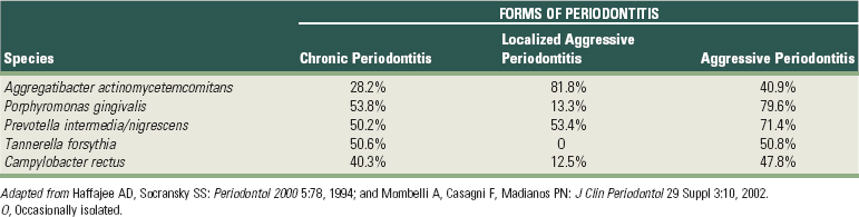
243 Mombelli A, Casagni F, Madianos PN. Can presence or absence of periodontal pathogens distinguish between subjects with chronic and aggressive periodontitis? A systematic review. J Clin Periodontol. 2002;29(Suppl 3):10.
244 Mombelli A, Lang NP, Burgin WB, et al. Microbial changes associated with the development of puberty gingivitis. J Periodontal Res. 1990;25:331.
245 Mombelli A, Lehmann B, Tonetti M, et al. Clinical response to local delivery of tetracycline in relation to overall and local periodontal conditions. J Clin Periodontol. 1997;24:470.
246 Mombelli A, Marxer M, Gaberthuel T, et al. The microbiota of osseointegrated implants in patients with a history of periodontal disease. J Clin Periodontol. 1995;22:124.
247 Moore WE. Microbiology of periodontal disease. J Periodontal Res. 1987;22:335.
248 Moore WE, Holdeman LV. Special problems associated with the isolation and identification of intestinal bacteria in fecal flora studies. Am J Clin Nutr. 1974;27:1450.
249 Moore WE, Holdeman LV, Cato EP, et al. Comparative bacteriology of juvenile periodontitis. Infect Immun. 1985;48:507.
250 Moore WE, Moore LV. The bacteria of periodontal diseases. Periodontol 2000. 1994;5:66.
251 Mouton C, Reynolds HS, Genco RJ. Characterization of tufted streptococci isolated from the “corn cob” configuration of human dental plaque. Infect Immun. 1980;27:235.
252 Nelson KE, Fleischmann RD, DeBoy RT, et al. Complete genome sequence of the oral pathogenic Bacterium porphyromonas gingivalis strain W83. J Bacteriol. 2003;185:5591.
253 Newman HN. The approximal apical border of plaque on children’s teeth. 1. Morphology, structure and cell content. J Periodontol. 1979;50:561.
254 Newman MG, Socransky SS. Predominant cultivable microbiota in periodontosis. J Periodontal Res. 1977;12:120.
255 Newman MG, Socransky SS, Savitt ED, et al. Studies of the microbiology of periodontosis. J Periodontol. 1976;47:373.
256 Nishiyama S, Murakami Y, Nagata H, et al. Involvement of minor components associated with the FimA fimbriae of Porphyromonas gingivalis in adhesive functions. Microbiology. 2007;153:1916.
257 Nowzari H, MacDonald ES, Flynn J, et al. The dynamics of microbial colonization of barrier membranes for guided tissue regeneration. J Periodontol. 1996;67:694.
258 Nyvad B, Fejerskov O. Scanning electron microscopy of early microbial colonization of human enamel and root surfaces in vivo. Scand J Dent Res. 1987;95:287.
259 Nyvad B, Kilian M. Microflora associated with experimental root surface caries in humans. Infect Immun. 1990;58:1628.
260 O’Brien-Simpson NM, Veith PD, Dashper SG, et al. Antigens of bacteria associated with periodontitis. Periodontol 2000. 2004;35:101.
261 Oberste MS, Maher K, Kilpatrick DR, et al. Molecular evolution of the human enteroviruses: correlation of serotype with VP1 sequence and application to picornavirus classification. J Virol. 1999;73:1941.
262 Ofek I, Doyle RJ, editors. Bacterial adhesion to cells and tissues. London: Chapman and Hall, 1994.
263 Ong ES, Newman HN, Wilson M, et al. The occurrence of periodontitis-related microorganisms in relation to titanium implants. J Periodontol. 1992;63:200.
264 Ouverney CC, Armitage GC, Relman DA. Single-cell enumeration of an uncultivated TM7 subgroup in the human subgingival crevice. Appl Environ Microbiol. 2003;69:6294.
265 Pallansch M, Roos R. Enteroviruses: polioviruses, coxsackieviruses, echoviruses, and newer enteroviruses. In: Knipe DM, Howley PM, editors. Fields virology. Philadelphia: Lippincott, Williams and Wilkins; 2007:840.
266 Palmer RJJr, Gordon SM, Cisar JO, et al. Coaggregation-mediated interactions of streptococci and actinomyces detected in initial human dental plaque. J Bacteriol. 2003;185:3400.
267 Papaioannou W, Quirynen M, van Steenberghe D. The influence of periodontitis on the subgingival flora around implants in partially edentulous patients. Clin Oral Implants Res. 1996;7:405.
268 Park Y, James CE, Yoshimura F, et al. Expression of the short fimbriae of Porphyromonas gingivalis is regulated in oral bacterial consortia. FEMS Microbiol Lett. 2006;262:65.
269 Park Y, Simionato MR, Sekiya K, et al. Short fimbriae of Porphyromonas gingivalis and their role in coadhesion with Streptococcus gordonii. Infect Immun. 2005;73:3983.
270 Patton LL, Phelan JA, Ramos-Gomez FJ, et al. Prevalence and classification of HIV-associated oral lesions. Oral Dis. 2002;8(Suppl 2):98.
271 Pearce C, Bowden GH, Evans M, et al. Identification of pioneer viridans streptococci in the oral cavity of human neonates. Journal of Medical Microbiology. 1995;42:67.
272 Pearce MA, Devine DA, Dixon RA, et al. Genetic heterogeneity in Prevotella intermedia, Prevotella nigrescens, Prevotella corporis and related species isolated from oral and nonoral sites. Oral Microbiol Immunol. 2000;15:89.
273 Pellett PE, Roizman B. The family Herpesviridae: a brief introduction. In: Knipe DM, Howley PM, editors. Fields virology. Philadelphia: Lippincott, Williams and Wilkins; 2007:2479.
274 Perez BA, Planet PJ, Kachlany SC, et al. Genetic analysis of the requirement for flp-2, tadV, and rcpB in Actinobacillus actinomycetemcomitans biofilm formation. J Bacteriol. 2006;188:6361.
275 Perry JA, Jones MB, Peterson SN, et al. Peptide alarmone signalling triggers an auto-active bacteriocin necessary for genetic competence. Mol Microbiol. 2009.
276 Petersilka GJ, Ehmke B, Flemmig TF. Antimicrobial effects of mechanical debridement. Periodontol 2000. 2002;28:56.
277 Pfaller MA. Nosocomial candidiasis: emerging species, reservoirs, and modes of transmission. Clin Infect Dis. 1996;22(Suppl 2):S89.
278 Planet PJ, Kachlany SC, Fine DH, et al. The widespread colonization island of Actinobacillus actinomycetemcomitans. Nat Genet. 2003;34:193.
279 Porter SR, Luker J, Scully C, et al. Orofacial manifestations of a group of British patients infected with HIV-1. J Oral Pathol Med. 1989;18:47.
280 Potempa J, Banbula A, Travis J. Role of bacterial proteinases in matrix destruction and modulation of host responses. Periodontol 2000. 2000;24:153.
281 Potempa J, Pike R, Travis J. Titration and mapping of the active site of cysteine proteinases from Porphyromonas gingivalis (gingipains) using peptidyl chloromethanes. Biol Chem. 1997;378:223.
282 Potempa M, Potempa J, Kantyka T, et al. Interpain A, a cysteine proteinase from Prevotella intermedia, inhibits complement by degrading complement factor C3. PLoS Pathog. 2009;5:e1000316.
283 Prakobphol A, Xu F, Hoang VM, et al. Salivary agglutinin, which binds Streptococcus mutans and Helicobacter pylori, is the lung scavenger receptor cysteine-rich protein gp-340. J Biol Chem. 2000;275:39860.
284 Pratten J, Barnett P, Wilson M. Composition and susceptibility to chlorhexidine of multispecies biofilms of oral bacteria. Appl Environ Microbiol. 1998;64:3515.
285 Preber H, Bergstrom J, Linder LE. Occurrence of periopathogens in smoker and non-smoker patients. J Clin Periodontol. 1992;19:667.
286 Qandil R, Sandhu HS, Matthews DC. Tobacco smoking and periodontal diseases. J Can Dent Assoc. 1997;63:187.
287 Quinn SM, Zhang JB, Gunsolley JC, et al. Influence of smoking and race on immunoglobulin G subclass concentrations in early-onset periodontitis patients. Infect Immun. 1996;64:2500.
288 Quirynen M, Anatomical and inflammatory factors influence bacterial plaque growth and retention in man, 1986
289 Quirynen M, Bollen CM. The influence of surface roughness and surface-free energy on supra- and subgingival plaque formation in man. A review of the literature. J Clin Periodontol. 1995;22:1.
290 Quirynen M, Bollen CM. The influence of surface roughness and surface-free energy on supra- and subgingival plaque formation in man. A review of the literature. J Clin Periodontol. 1995;22:1.
291 Quirynen M, Bollen CM, Papaioannou W, et al. The influence of titanium abutment surface roughness on plaque accumulation and gingivitis: short-term observations. Int J Oral Maxillofac Implants. 1996;11:169.
292 Quirynen M, Bollen CM, Vandekerckhove BN, et al. Full- vs. partial-mouth disinfection in the treatment of periodontal infections: short-term clinical and microbiological observations. J Dent Res. 1995;74:1459.
293 Quirynen M, De Soete M, Dierickx K, et al. The intra-oral translocation of periodontopathogens jeopardises the outcome of periodontal therapy. A review of the literature. J Clin Periodontol. 2001;28:499.
294 Quirynen M, Dekeyser C, van Steenberghe D. The influence of gingival inflammation, tooth type, and timing on the rate of plaque formation. J Periodontol. 1991;62:219.
295 Quirynen M, Gijbels F, Jacobs R. An infected jawbone site compromising successful osseointegration. Periodontol 2000. 2003;33:129.
296 Quirynen M, Gizani S, Mongardini C, et al. The effect of periodontal therapy on the number of cariogenic bacteria in different intra-oral niches. J Clin Periodontol. 1999;26:322.
297 Quirynen M, Listgarten MA. Distribution of bacterial morphotypes around natural teeth and titanium implants ad modum Branemark. Clin Oral Implants Res. 1990;1:8.
298 Quirynen M, Marechal M, Busscher HJ, et al. The influence of surface free-energy on planimetric plaque growth in man. J Dent Res. 1989;68:796.
299 Quirynen M, Papaioannou W, van Steenbergen TJ, et al. Adhesion of Porphyromonas gingivalis strains to cultured epithelial cells from patients with a history of chronic adult periodontitis or from patients less susceptible to periodontitis. J Periodontol. 2001;72:626.
300 Quirynen M, Teughels W, De Soete M, et al. Topical antiseptics and antibiotics in the initial therapy of chronic adult periodontitis: microbiological aspects. Periodontol 2000. 2002;28:72.
301 Quirynen M, van der Mei HC, Bollen CM, et al. An in vivo study of the influence of the surface roughness of implants on the microbiology of supra- and subgingival plaque. J Dent Res. 1993;72:1304.
302 Quirynen M, van Steenberghe D. Is early plaque growth rate constant with time? J Clin Periodontol. 1989;16:278.
303 Quirynen M, Vogels R, Pauwels M, et al. Initial subgingival colonization of pristine pockets in an established environment. J Dent Res. 2005. submitted
304 Quirynen M, Vogels R, Peeters W, et al. Dynamics of initial subgingival colonization of peri-implant pockets. Clin Oral Implants Res. 2005. submitted
305 Quirynen M, Vogels R, Peeters W, et al. Dynamics of initial subgingival colonization of “pristine” peri-implant pockets. Clin Oral Implants Res. 2006;17:25.
306 Ramberg P, Axelsson P, Lindhe J. Plaque formation at healthy and inflamed gingival sites in young individuals. J Clin Periodontol. 1995;22:85.
307 Ramberg P, Lindhe J, Dahlen G, et al. The influence of gingival inflammation on de novo plaque formation. J Clin Periodontol. 1994;21:51.
308 Rams TE, Feik D, Slots J. Staphylococci in human periodontal diseases. Oral Microbiol Immunol. 1990;5:29.
309 Ramsey MM, Whiteley M. Polymicrobial interactions stimulate resistance to host innate immunity through metabolite perception. Proc Natl Acad Sci U S A. 2009;106:1578.
310 Reed WP, Williams RC. Bacterial adherence: first step in pathogenesis of certain infections. J Chronic Dis. 1978;31:67.
311 Renvert S, Dahlen G, Wikstrom M. Treatment of periodontal disease based on microbiological diagnosis. Relation between microbiological and clinical parameters during 5 years. J Periodontol. 1996;67:562.
312 Renvert S, Dahlen G, Wikstrom M. The clinical and microbiological effects of non-surgical periodontal therapy in smokers and non-smokers. J Clin Periodontol. 1998;25:153.
313 Renvert S, Wikstrom M, Dahlen G, et al. Effect of root debridement on the elimination of Actinobacillus actinomycetemcomitans and Bacteroides gingivalis from periodontal pockets. J Clin Periodontol. 1990;17:345.
314 Renvert S, Wikstrom M, Dahlen G, et al. On the inability of root debridement and periodontal surgery to eliminate Actinobacillus actinomycetemcomitans from periodontal pockets. J Clin Periodontol. 1990;17:351.
315 Rickard AH, Palmer RJJr, Blehert DS, et al. Autoinducer 2: a concentration-dependent signal for mutualistic bacterial biofilm growth. Mol Microbiol. 2006;60:1446.
316 Rimondini L, Fare S, Brambilla E, et al. The effect of surface roughness on early in vivo plaque colonization on titanium. J Periodontol. 1997;68:556.
317 Roberts FA, Darveau RP. Beneficial bacteria of the periodontium. Periodontol 2000. 2002;30:40.
318 Rosen G, Genzler T, Sela MN. Coaggregation of Treponema denticola with Porphyromonas gingivalis and Fusobacterium nucleatum is mediated by the major outer sheath protein of Treponema denticola. FEMS Microbiol Lett. 2008;289:59.
319 Rotimi VO, Duerden BI. The development of the bacterial flora in normal neonates. Journal of Medical Microbiology. 1981;14:51.
320 Rudney JD, Chen R, Sedgewick GJ. Intracellular Actinobacillus actinomycetemcomitans and Porphyromonas gingivalis in buccal epithelial cells collected from human subjects. Infect Immun. 2001;69:2700.
321 Rudney JD, Chen R, Sedgewick GJ. Actinobacillus actinomycetemcomitans, Porphyromonas gingivalis, and Tannerella forsythensis are components of a polymicrobial intracellular flora within human buccal cells. J Dent Res. 2005;84:59.
322 Russel AL. Epidemiology of periodontal disease. Int Dent J. 1967;17:282.
323 Rutter PR, Vincent B. Physicochemical interactions of the substratum, microorganisms and the fluid phase. In: Marshall KC, editor. Microbial adhesion and aggregation. Berlin: Springer Verlag; 1984:21.
324 Ryder MI. An update on HIV and periodontal disease. J Periodontol. 2002;73:1071.
325 Saadi AT, Blackwell CC, Essery SD, et al. Developmental and environmental factors that enhance binding of Bordatella pertussis to human epithelial cells in relation to sudden infant death syndrome (SIDS). FEMS Immunol Med Microbiol. 1996;16:51.
326 Saglie FR, Carranza FAJr, Newman MG, et al. Identification of tissue-invading bacteria in human periodontal disease. J Periodontal Res. 1982;17:452.
327 Saglie FR, Marfany A, Camargo P. Intragingival occurrence of Actinobacillus actinomycetemcomitans and Bacteroides gingivalis in active destructive periodontal lesions. J Periodontol. 1988;59:259.
328 Saito A, Inagaki S, Kimizuka R, et al. Fusobacterium nucleatum enhances invasion of human gingival epithelial and aortic endothelial cells by Porphyromonas gingivalis. FEMS Immunol Med Microbiol. 2008;54:349.
329 Samaranayake LP, Keung LW, Jin L. Oral mucosal fungal infections. Periodontol 2000. 2009;49:39.
330 Samonis G, Mantadakis E, Maraki S. Orofacial viral infections in the immunocompromised host. Oncol Rep. 2000;7:1389.
331 Sanford BA, Shelokov A, Ramsay MA. Bacterial adherence to virus-infected cells: a cell culture model of bacterial superinfection. J Infect Dis. 1978;137:176.
332 Sanz M, Quirynen M. Advances in the aetiology of periodontitis. Group A consensus report of the 5th European Workshop in Periodontology. J Clin Periodontol. 2005;32(Suppl 6):54.
333 Sbordone L, Barone A, Ciaglia RN, et al. Longitudinal study of dental implants in a periodontally compromised population. J Periodontol. 1999;70:1322.
334 Scannapieco FA. Saliva-bacterium interactions in oral microbial ecology. Crit Rev Oral Biol Med. 1994;5:203.
335 Schaeffer LM, Schmidt ML, Demuth DR. Induction of Aggregatibacter actinomycetemcomitans leukotoxin expression by IS1301 and orfA. Microbiology. 2008;154:528.
336 Schei O, Waerhaug J, Loval A. Alveolar bone loss as related to oral hygiene and age. J Periodontol. 1959;30:7.
337 Schneider LC, Schneider AE. Diagnosis of oral ulcers. Mt Sinai J Med. 1998;65:383.
338 Schreiner HC, Sinatra K, Kaplan JB, et al. Tight-adherence genes of Actinobacillus actinomycetemcomitans are required for virulence in a rat model. Proc Natl Acad Sci U S A. 2003;100:7295.
339 Schroeder HE, De Boever J. The structure of microbial dental plaque. In: McHugh WD, editor. Dental plaque. Edinburgh: Livingstone; 1970:49.
340 Schuman P, Ohmit SE, Sobel JD, et al. Oral lesions among women living with or at risk for HIV infection. HIV Epidemiology Research Study (HERS) Group. Am J Med. 1998;104:559.
341 Scully C, Felix DH. Oral medicine–update for the dental practitioner. Aphthous and other common ulcers. Br Dent J. 2005;199:259.
342 Sheiham A. Periodontal disease and oral cleanliness in tobacco smokers. J Periodontol. 1971;42:259.
343 Shibli JA, Melo L, Ferrari DS, et al. Composition of supra- and subgingival biofilm of subjects with healthy and diseased implants. Clin Oral Implants Res. 2008;19:975.
344 Simonsson T. Aspects of dental plaque formation with special reference to colloid-chemical phenomena. Swed Dent J Suppl. 1989;58:1.
345 Simonsson T, Ronstrom A, Rundegren J, et al. Rate of plaque formation–some clinical and biochemical characteristics of “heavy” and “light” plaque formers. Scand J Dent Res. 1987;95:97.
346 Siqueira WL, Oppenheim FG. Small molecular weight proteins/peptides present in the in vivo formed human acquired enamel pellicle. Arch Oral Biol. 2009;54:437.
347 Siqueira WL, Zhang W, Helmerhorst EJ, et al. Identification of protein components in in vivo human acquired enamel pellicle using LC-ESI-MS/MS. J Proteome Res. 2007;6:2152.
348 Sjodin B, Crossner CG, Unell L, et al. A retrospective radiographic study of alveolar bone loss in the primary dentition in patients with localized juvenile periodontitis. J Clin Periodontol. 1989;16:124.
349 Skopek RJ, Liljemark WF, Bloomquist CG, et al. Dental plaque development on defined streptococcal surfaces. Oral Microbiol Immunol. 1993;8:16.
350 Slaney JM, Gallagher A, duse-Opoku J, et al. Mechanisms of resistance of Porphyromonas gingivalis to killing by serum complement. Infect Immun. 2006;74:5352.
351 Sliepen I, Hofkens J, Van EM, et al. Aggregatibacter actinomycetemcomitans adhesion inhibited in a flow cell. Oral Microbiol Immunol. 2008;23:520.
352 Sliepen I, Van Damme J, Van Essche M, et al. Microbial interactions influence inflammatory host cell responses. J Dent Res. 2009.
353 Slots J. The predominant cultivable microflora of advanced periodontitis. Scand J Dent Res. 1977;85:114.
354 Slots J. Subgingival microflora and periodontal disease. J Clin Periodontol. 1979;6:351.
355 Slots J. Herpesviruses in periodontal diseases. Periodontol 2000. 2005;38:33.
356 Slots J. Oral viral infections of adults. Periodontol 2000. 2009;49:60.
357 Slots J, Gibbons RJ. Attachment of Bacteroides melaninogenicus subsp. asaccharolyticus to oral surfaces and its possible role in colonization of the mouth and of periodontal pockets. Infect Immun. 1978;19:254.
358 Slots J, Listgarten MA. Bacteroides gingivalis, Bacteroides intermedius and Actinobacillus actinomycetemcomitans in human periodontal diseases. J Clin Periodontol. 1988;15:85.
359 Slots J, Rams TE. New views on periodontal microbiota in special patient categories. J Clin Periodontol. 1991;18:411.
360 Socransky SS, Gibbons RJ, Dale AC, et al. The microbiota of the gingival crevice area of man. I. Total microscopic and viable counts of specific microorganisms. Arch Oral Biol. 1953:275.
361 Socransky SS, Haffajee AD. Microbial mechanisms in the pathogenesis of destructive periodontal diseases: a critical assessment. J Periodontal Res. 1991;26:195.
362 Socransky SS, Haffajee AD. The bacterial etiology of destructive periodontal disease: current concepts. J Periodontol. 1992;63:322.
363 Socransky SS, Haffajee AD. The nature of periodontal diseases. Ann Periodontol. 1997;2:3.
364 Socransky SS, Haffajee AD, Cugini MA, et al. Microbial complexes in subgingival plaque. J Clin Periodontol. 1998;25:134.
365 Socransky SS, Haffajee AD, Goodson JM, et al. New concepts of destructive periodontal disease. J Clin Periodontol. 1984;11:21.
366 Socransky SS, Haffajee AD, Smith GL, et al. Difficulties encountered in the search for the etiologic agents of destructive periodontal diseases. J Clin Periodontol. 1987;14:588.
367 Socransky SS, Manganiello AD, Propas D, et al. Bacteriological studies of developing supragingival dental plaque. J Periodontal Res. 1977;12:90.
368 Socransky SS, Manganiello SD. The oral microbiota of man from birth to senility. J Periodontol. 1971;42:485.
369 Socransky SS, Smith C, Haffajee AD. Subgingival microbial profiles in refractory periodontal disease. J Clin Periodontol. 2002;29:260.
370 Stenderup A. Oral mycology. Acta Odontol Scand. 1990;48:3.
371 Stingu CS, Eschrich K, Rodloff AC, et al. Periodontitis is associated with a loss of colonization by Streptococcus sanguinis. J of Med Microbiol. 2008;57:495.
372 Stoltenberg JL, Osborn JB, Pihlstrom BL, et al. Association between cigarette smoking, bacterial pathogens, and periodontal status. J Periodontol. 1993;64:1225.
373 Sudbery P, Gow N, Berman J. The distinct morphogenic states of Candida albicans. Trends Microbiol. 2004;12:317.
374 Sumida S, Ishihara K, Kishi M, et al. Transmission of periodontal disease-associated bacteria from teeth to osseointegrated implant regions. Int J Oral Maxillofac Implants. 2002;17:696.
375 Syed SA, Loesche WJ. Bacteriology of human experimental gingivitis: effect of plaque age. Infect Immun. 1978;21:821.
376 Sztajer H, Lemme A, Vilchez R, et al. Autoinducer-2-regulated genes in Streptococcus mutans UA159 and global metabolic effect of the luxS mutation. J Bacteriol. 2008;190:401.
377 Takashiba S, Naruishi K. Gene polymorphisms in periodontal health and disease. Periodontol 2000. 2006;40:94.
378 Takenaka S, Trivedi HM, Corbin A, et al. Direct visualization of spatial and temporal patterns of antimicrobial action within model oral biofilms. Appl Environ Microbiol. 2008;74:1869.
379 Tanner AC, Haffer C, Bratthall GT, et al. A study of the bacteria associated with advancing periodontitis in man. J Clin Periodontol. 1979;6:278.
380 Tappuni AR, Challacombe SJ. Distribution and isolation frequency of eight streptococcal species in saliva from predentate and dentate children and adults. J Dent Res. 1993;72:31.
381 Taylor DC, Clancy RL, Cripps AW, et al. An alteration in the host-parasite relationship in subjects with chronic bronchitis prone to recurrent episodes of acute bronchitis. Immunol Cell Biol. 1994;72:143.
382 Teles RP, Haffajee AD, Socransky SS. Microbiological goals of periodontal therapy. Periodontol 2000. 2006;42:180.
383 Teughels W, Kinder Haake SA, Sliepen I, et al. Bacteria interfere with A. actinomycetemcomitans colonization. J Dent Res. 2007;87:611.
384 Teughels W, Newman MG, Coucke W, et al. Guiding periodontal pocket recolonization: a proof of concept. J Dent Res. 2007;86:1078.
385 Teughels W, Sliepen I, Quirynen M, et al. Human cytomegalovirus enhances A. actinomycetemcomitans adherence to cells. J Dent Res. 86, 2007.
386 Teughels W, Van EM, Sliepen I, et al. Probiotics and oral healthcare. Periodontol 2000. 2008;48:111.
387 Theilade E, Wright WH, Jensen SB, et al. Experimental gingivitis in man. II. A longitudinal clinical and bacteriological investigation. J Periodontal Res. 1966;1:1.
388 Ting M, Contreras A, Slots J. Herpesvirus in localized juvenile periodontitis. J Periodontal Res. 2000;35:17.
389 Tonetti MS, Pini-Prato G, Cortellini P. Effect of cigarette smoking on periodontal healing following GTR in infrabony defects. A preliminary retrospective study. J Clin Periodontol. 1995;22:229.
390 Tong H, Chen W, Merritt J, et al. Streptococcus oligofermentans inhibits Streptococcus mutans through conversion of lactic acid into inhibitory H2O2: a possible counteroffensive strategy for interspecies competition. Mol Microbiol. 2007;63:872.
391 Truper HG, De Clari L. Taxonomic note: necessary correction of specific epithets formed as substantives (nouns) “in apposition,”. Int J Syst Bacteriol. 1997;47:908.
392 Umeda M, Chen C, Bakker I, et al. Risk indicators for harboring periodontal pathogens. J Periodontol. 1998;69:1111.
393 Vaahtoniemi LH, Raisanen S, Stenfors LE. Attachment of bacteria to oral epithelial cells in vivo: a possible correlation to gingival health status. J Periodontal Res. 1993;28:308.
394 van der Mei HC, Rustema-Abbing M, Bruinsma GM, et al. Influence of weight on removal of co-adhering bacteria from salivary pellicles by different modes of brushing. Caries Res. 2004;38:85.
395 van der Reijden WA, Dellemijn-Kippuw N, Stijne-van Nes AM, et al. Mutans streptococci in subgingival plaque of treated and untreated patients with periodontitis. J Clin Periodontol. 2001;28:686.
396 Van der Velden U. Purpose and problems of periodontal disease classification. Periodontol 2000. 2005;39:13.
397 Van Hoogmoed CG, Geertsema-Doornbusch G, Teughels W, et al. Reduction of periodontal pathogens adhesion by antagonistic strains. Oral Microbiol Immunol. 2007.
398 van Loosdrecht MC, Norde W, Zehnder AJ. Physical chemical description of bacterial adhesion. J Biomater Appl. 1990;5:91.
399 van Steenberghe D, Jacobs R, Desnyder M, et al. The relative impact of local and endogenous patient-related factors on implant failure up to the abutment stage. Clin Oral Implants Res. 2002;13:617.
400 van Winkelhoff AJ, Bosch-Tijhof CJ, Winkel EG, et al. Smoking affects the subgingival microflora in periodontitis. J Periodontol. 2001;72:666.
401 van Winkelhoff AJ, Boutaga K. Transmission of periodontal bacteria and models of infection. J Clin Periodontol. 2005;32(Suppl 6):16.
402 van Winkelhoff AJ, Herrera D, Oteo A, et al. Antimicrobial profiles of periodontal pathogens isolated from periodontitis patients in The Netherlands and Spain. J Clin Periodontol. 2005;32:893.
403 van Winkelhoff AJ, Loos BG, van der Reijden WA, et al. Porphyromonas gingivalis, Bacteroides forsythus and other putative periodontal pathogens in subjects with and without periodontal destruction. J Clin Periodontol. 2002;29:1023.
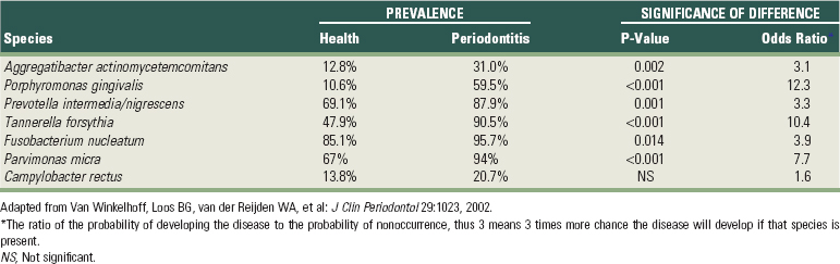
404 Vartoukian SR, Palmer RM, Wade WG. Diversity and morphology of members of the phylum “synergistetes” in periodontal health and disease. Appl Environ Microbiol. 2009;75:3777.
405 Vianna ME, Conrads G, Gomes BP, et al. Identification and quantification of archaea involved in primary endodontic infections. J Clin Microbiol. 2006;44:1274.
406 Vickerman MM, Brossard KA, Funk DB, et al. Phylogenetic analysis of bacterial and archaeal species in symptomatic and asymptomatic endodontic infections. J Med Microbiol. 2007;56:110.
407 Wade WG, Addy M. In vitro activity of a chlorhexidine-containing mouthwash against subgingival bacteria. J Periodontol. 1989;60:521.
408 Walden WC, Hentges DJ. Differential effects of oxygen and oxidation-reduction potential on the multiplication of three species of anaerobic intestinal bacteria. Appl Microbiol. 1975;30:781.
409 Walker C, Sedlacek MJ. An in vitro biofilm model of subgingival plaque. Oral Microbiol Immunol. 2007;22:152.
410 Walker CB. Selected antimicrobial agents: mechanisms of action, side effects and drug interactions. Periodontol 2000. 1996;10:12.
411 Wan AK, Seow WK, Purdie DM, et al. Oral colonization of Streptococcus mutans in six-month-old predentate infants. J Dent Res. 2001;80:2060.
412 Wan AK, Seow WK, Purdie DM, et al. A longitudinal study of Streptococcus mutans colonization in infants after tooth eruption. J Dent Res. 2003;82:504.
413 Wan AK, Seow WK, Walsh LJ, et al. Association of Streptococcus mutans infection and oral developmental nodules in pre-dentate infants. J Dent Res. 2001;80:1945.
414 Wang Y, Chen C. Mutation analysis of the flp operon in Actinobacillus actinomycetemcomitans. Gene. 2005;351:61.
415 Weiger R, Netuschil L, von Ohle C, et al. Microbial generation time during the early phases of supragingival dental plaque formation. Oral Microbiol Immunol. 1995;10:93.
416 Weiss EI, Eli I, Shenitzki B, et al. Identification of the rhamnose-sensitive adhesin of Capnocytophaga ochracea ATCC 33596. Arch Oral Biol. 1990;35(Suppl):127S.
417 Weiss EI, London J, Kolenbrander PE, et al. Characterization of monoclonal antibodies to fimbria-associated adhesins of Bacteroides loescheii PK1295. Infect Immun. 1988;56:219.
418 Weiss EI, London J, Kolenbrander PE, et al. Localization and enumeration of fimbria-associated adhesins of Bacteroides loescheii. J Bacteriol. 1988;170:1123.
419 Wennstrom JL, Heijl L, Dahlen G, et al. Periodic subgingival antimicrobial irrigation of periodontal pockets (I). Clinical observations. J Clin Periodontol. 1987;14:541.
420 Weyant RJ, Burt BA. An assessment of survival rates and within-patient clustering of failures for endosseous oral implants. J Dent Res. 1993;72:2.
421 Whittaker CJ, Clemans DL, Kolenbrander PE. Insertional inactivation of an intrageneric coaggregation-relevant adhesin locus from Streptococcus gordonii DL1 (Challis). Infect Immun. 1996;64:4137.
422 Wickstrom C, Svensater G. Salivary gel-forming mucin MUC5B–a nutrient for dental plaque bacteria. Oral Microbiol Immunol. 2008;23:177.
423 Williams P. Role of the cell envelope in bacterial adaptation to growth in vivo in infections. Biochimie. 1988;70:987.
424 Wilson M. An introduction to the human-microbe symbiosis. In: Wilson M, editor. Microbial inhabitants of humans. New York: Cambridge University Press; 2005:1.
425 Winkel EG, Abbas F, van d V, et al. Experimental gingivitis in relation to age in individuals not susceptible to periodontal destruction. J Clin Periodontol. 1987;14:499.
426 Wohl DA, Zeng D, Stewart P, et al. Cytomegalovirus viremia, mortality, and end-organ disease among patients with AIDS receiving potent antiretroviral therapies. J Acquir Immune Defic Syndr. 2005;38:538.
427 Wolff L, Dahlen G, Aeppli D. Bacteria as risk markers for periodontitis. J Periodontol. 1994;65:498.
428 Wood SR, Kirkham J, Marsh PD, et al. Architecture of intact natural human plaque biofilms studied by confocal laser scanning microscopy. J Dent Res. 2000;79:21.
429 Ximenez-Fyvie LA, maguer-Flores A, Jacobo-Soto V, et al. Subgingival microbiota of periodontally untreated Mexican subjects with generalized aggressive periodontitis. J Clin Periodontol. 2006;33:869.
430 Ximenez-Fyvie LA, maguer-Flores A, Jacobo-Soto V, et al. Description of the subgingival microbiota of periodontally untreated Mexican subjects: chronic periodontitis and periodontal health. J Periodontol. 2006;77:460.
431 Xu KD, McFeters GA, Stewart PS. Biofilm resistance to antimicrobial agents. Microbiology. 2000;146(Pt 3):547.
432 Yao Y, Berg EA, Costello CE, et al. Identification of protein components in human acquired enamel pellicle and whole saliva using novel proteomics approaches. J Biol Chem. 2003;278:5300.
433 Yokoyama M, Hinode D, Yoshioka M, et al. Relationship between Campylobacter rectus and periodontal status during pregnancy. Oral Microbiol Immunol. 2008;23:55.
434 Zambon JJ. Periodontal diseases: microbial factors. Ann Periodontol. 1996;1:879.
435 Zambon JJ, Grossi SG, Machtei EE, et al. Cigarette smoking increases the risk for subgingival infection with periodontal pathogens. J Periodontol. 1996;67:1050.
436 Zee KY, Samaranayake LP, Attstrom R. Predominant cultivable supragingival plaque in Chinese “rapid” and “slow” plaque formers. J Clin Periodontol. 1996;23:1025.
437 Zee KY, Samaranayake LP, Attstrom R. Scanning electron microscopy of microbial colonization of ‘rapid’ and ‘slow’ dental-plaque formers in vivo. Arch Oral Biol. 1997;42:735.
438 Zhang G, Chen R, Rudney JD. Streptococcus cristatus attenuates Fusobacterium nucleatum-induced interleukin-8 expression in oral epithelial cells. J Periodontal Res. 2008;43:408.
* References 13, 149, 150, 167, 169, 198, 202, 235, 246, 267, 297, and 333.
* References 2, 5, 16, 151, 154, 342, 344, and 345.
* References 27, 42, 118, 291, 301, and 316.
* References 13, 149, 150, 167, 169, 198, 202, 235, 246, 267, 297, 333.
* References 28, 123, 285, 372, 392, 400, and 435.
* References 183, 189, 217, 250, 354, 361, 362, 379, 382, 429, and 430.