Four in vitro studies100,164,279,282 were identified investigating the bactericidal effect of ozone compared to 2.5% NaOCl, the standard irrigation solution in endodontics. The results of these studies164,279,282 are controversial. The hazards of ozone when used in endodontics have not been investigated. Care should be taken with regard to the patient’s and the dentist’s exposure to the gas. Fig. 8-60 shows ozone equipment with a special tip for intracanal application.
Superoxidized water,152 also called electrochemically activated water241,365 or oxidative potential water,162,344 is saline that has been electrolyzed to form superoxidized water, hypochlorous acid, and free chlorine radicals. It is available as Sterilox (Sterilox Technologies, Radnor, PA). This solution is nontoxic to biologic tissues yet able to kill microorganisms. The solution is generated by electrolyzing saline solution, a process no different than that used in the commercial production of NaOCl.116 The difference, however, is that the solution accumulating at the anode is harvested as the anolyte and that at the cathode as the catholyte. These solutions display properties that are dependent upon the strength of the initial saline solution, the applied potential difference, and the rate of generation. The technology that allows harvesting of the respective solutions resides in the design of the anode and the cathode and originates either in Russia (electrochemically activated water) or Japan (oxidative potential water).240,241 Although the solutions bear different names, the principles in the manufacturing process are probably the same.
The use of superoxidized water is sparse in the endodontic literature but shows early promise. The solutions from both technologies have been tested for their ability to débride root canals,162,241 remove smear layer,344,365 and kill bacteria172 and bacterial spores.234 Results are favorable, while also showing biocompatibility with vital systems.183
Anolyte and catholyte solutions generated from one such technology (Radical Waters Halfway House 1685, S. Africa) have shown promise as antibacterial agents against laboratory-grown, single-species biofilm models.132 Such solutions have been recommended as suitable for removing biofilms in dental unit water lines240 and have even been marketed for this purpose. Cautious clinicians may prefer to wait for more studies to demonstrate safety and efficacy under ordinary clinical setting conditions before adopting newer, less tested irrigating solutions.
Photodynamic therapy (PDT) or light-activated therapy (LAT) may have endodontic applications because of its antimicrobial effectiveness.159 In principle, the strategy utilizes a selectively accumulated or endogenously produced photosensitizer (photoactive compound). The activation of photosensitizer molecules by a specific wavelength of light produces deleterious chemical entities that kill bacteria. The strong oxidizer generated during the process can act on multiple targets in a microbial cell, resulting in membrane damage, enzyme inactivation, and genomic and plasmid DNA damage, resulting in instantaneous death. In addition, the light-activated killing of bacteria has a wide spectrum of antimicrobial activity with a minimal chance of giving rise to a resistant microbial population, making it an ideal disinfectant. Fighting biofilm-mediated localized human infections is one of the potential medical applications of LAT.410 The principal agent involved in bacterial killing is the oxygen-based free radical.159 Unlike systemic therapeutic agents, LAT has the advantage of dual selectivity. Only those cells which have selectively accumulated the photosensitizer or possessing endogenously produced photosensitizers will be affected upon irradiation. Separately, the low-intensity light and the nontoxic photosensitizer generally used in LAT are harmless. In addition, the use of an optic fiber helps in directing the irradiation to the intended site of application. Studies have highlighted the possibility of killing antibiotic-resistant strains of pathogenic bacteria using LAT.409,411 Because of the wide spectrum of antimicrobial activity, instant bacterial killing, and ability to target biofilm bacteria, the application of LAT-based regimens has great promise in root canal disinfection.400 However, a thorough knowledge about the behavior of the photosensitizer and the physicochemical characteristics of the microenvironment prevailing in the root canal system is necessary for disinfection to be effectively achieved. In advanced stages of infection with mineralized biofilms, it may be necessary to do a minimal mechanical preparation of the root canal prior to LAT to achieve maximum disinfection.
Ultraviolet (UV) light is widely used for disinfection purposes. IntraLight (InterLight, Or-Yehuda, Israel) consists of an intracanal UV illuminator shaped with dimensions of an endodontic file, which allows uniform circumferential illumination of the root canal walls with 254 nm UV light. In a recent in vitro study,255 root canals were infected with E. faecalis bacteria that were allowed to grow and establish a biofilm on the root canal walls. Treatment with 5% NaOCl for 10 minutes failed to result in negative cultures in 53% of the canals. Similar results were reported by others.255,257 An additional illumination of the canal walls with 254 nm UV light (300 mJ/cm2) rendered 96% of the canals free of cultivable bacteria. A wide range of bacteria are sensitive to 254 nm UV light, including those that are resistant to Ca(OH)2.257 The device is used as a supplemental disinfection after NaOCl irrigation in the canal. Otherwise, bacteria in the inner layers of thick biofilms may be protected from the UV light, which is absorbed by bacteria in the outer layers.257
After the root canal system has been appropriately prepared, it must be obturated with a material capable of completely preventing communication between the oral cavity and the periapical tissue wound. Materials used for this purpose should be compatible with healing. This is achieved by attempting to seal the root canal system at both its coronal and apical ends, as well as throughout the canal system, to seal the openings of accessory canals. Apical and intracanal obturation blocks the exit to the periradicular tissues from organisms that have survived in the root canal after cleaning and shaping. Coronal obturation prevents reinfection of the pulp space from the oral environment. The required physical and biologic properties make the selection of obturation material critical. The materials commonly used for root canal fillings normally can be divided into a solid phase and a cementing medium (i.e., a sealer).
Gutta-percha is the most commonly used root canal filling material. It is a linear crystalline polymer that melts at a set temperature, with a random but distinct change in structure resulting. (See Chapter 10 for further discussion of the physical properties of gutta-percha.) It occurs naturally as 1,4-polyisoprene and is harder, more brittle, and less elastic than natural rubber.
The crystalline phase has two forms, the alpha phase and the beta phase. The forms differ only in the molecular repeat distance and single-bond form. The alpha form is the material that comes from the natural tree product. The processed, or beta, form is used in gutta-percha for root fillings.333 When heated, gutta-percha undergoes phase transitions. The transition from beta phase to alpha phase occurs at around 115° F (46° C). An amorphous phase develops at around 130° F to 140° F (54° C to 60° C). When cooled very slowly (i.e., 1° F per hour), gutta-percha crystallizes to the alpha phase. Normal cooling returns the gutta-percha to the beta phase. Gutta-percha cones soften at a temperature above 147° F (64° C).139,333 These cones can easily be dissolved in chloroform and halothane and dissolve less in turpentine or xylene.
Modern gutta-percha cones that are used for root canal fillings contain only about 20% gutta-percha (Box 8-4). The major component is zinc oxide (60% to 75%). The remaining 5% to 10% consists of various resins, waxes, and metal sulfates. Antiseptic gutta-percha with various antimicrobial agents has been suggested, and several studies are available concerning the effect of these additives (see Medicated Gutta-Percha).
Because gutta-percha cannot be heat sterilized, other decontamination methods must be used. The most practical method is to disinfect the gutta-percha in NaOCl before use. This can be done in 1 minute if the cone is submerged in a 5% solution of NaCOl.342 However, after this disinfection and before its use for obturation, it is imperative that the gutta-percha be rinsed in ethyl alcohol to remove crystallized NaOCl; such crystals may impair the obturation seal.349
Obturation with gutta-percha normally requires some form of compaction pressure, but real compression of gutta-percha is practically impossible.333 Pressure applied during root canal filling procedures does not compress gutta-percha, but rather compacts the gutta-percha cones to obtain a more three-dimensionally complete fill of the root canal system.
After heating, while cooling, there is a slight shrinkage of approximately 1% to 2% when the gutta-percha has solidified. Prevention of shrinkage is practically impossible in vertical warm compaction.139,334 Careful control of the temperature during warm compaction is crucial to prevent focal areas of unnecessarily high temperatures. Better temperature control is available with electrically controlled heating devices as the Touch ’n Heat, System B, Elements units (SybronEndo, Orange, CA), or the recently introduced vibrating heat carrier, DownPack67 (Hu-Frieday, Chicago, IL), or HotTip (Discus Dental, Culver City, CA), all of which are more commonly used for this purpose (see Figs. 8-41 through 8-44).
Gutta-percha oxidizes with exposure to air and light and eventually becomes brittle.198,242 It should be stored in a cool, dry place (e.g., a refrigerator) for better shelf life. Methods of “rejuvenating” aged gutta-percha have been suggested.189
Gutta-percha cannot be used as the sole filling material; it lacks the adherent properties necessary to seal the root canal space. Therefore, a sealer (cement) is always needed for the final seal. (See the following sections on sealers.)
Manufacturers now supply gutta-perch cones in tapers matching the larger tapered rotary instruments (#.02, #.04, and #.06) (Fig. 8-61). An international standard has been accepted for gutta-percha cones, based on similar size and taper standards set forth for the endodontic files (ANSI No.78) (see Table 8-1). The tolerance is much less stringent for gutta-percha compared to files. An endodontic file must be manufactured with a tolerance of +0.05 mm. Consequently, with the same size instrument and gutta-percha cone, a difference in diameter of 0.07 mm (more than one file size) is possible (see Table 8-2 for gutta-percha cone sizes).
Gutta-percha has been extensively investigated as a root canal filling material in animals and has been proven biocompatible. Compared with sealers used for root canal obturation, it clearly has the lowest tissue toxicity. When compared to Resilon, both materials were equally biocompatible.38 After subcutaneous implantation, gutta-percha normally becomes surrounded by a defined capsule rich in cells but without a significant number of inflammatory cells, although some macrophages are present.37,288,362 Results from more sensitive assays in vitro support the in vivo results, suggesting that gutta-percha used for root canal fillings has a low toxicity.295 Nevertheless, gutta-percha in the form of very small particles induces an intensive foreign body reaction, with massive accumulation of mononucleated and multinucleated macrophages.362 This is not surprising, since materials that are normally considered inert (e.g., Teflon) cause similar reactions when presented to the tissues as an irregular surface or particle.50
Resilon (Pentron Clinical Technologies, Wallingford, CT), a thermoplastic, synthetic, polymer-based root canal filling material, was developed in an attempt to create an adhesive bond between the solid-core material and the sealer. It is designed to be used with Epiphany (Pentron Clinical Technologies), a new resin sealer with a bonding capacity to dentin. Resilon can be supplied in the same ISO sizes and shapes (cones and pellets) as gutta-percha. The manufacturer has stated that it can be used with any current root canal obturation technique (lateral compaction, thermoplasticized, carrier, injection). When manufactured in cones, Resilon’s flexibility is similar to that of gutta-percha. Based on polyester polymers, Resilon contains bioactive glass and radiopaque fillers (bismuth oxychloride and barium sulfate) with a filler content of approximately 65%. It can be softened with heat or dissolved with solvents such as chloroform. This characteristic allows the use of current retreatment techniques for nonhealing cases. Because it is a resin-based system, it is compatible with current restorative techniques in which cores and posts are placed with resin bonding agents (Figs. 8-62 to 8-64).348
FIG. 8-62 A, Resilon/Epiphany cone root canal system obturation. B, Recall radiograph taken 8 months after initial treatment shows complete healing.
(Courtesy Dr. Richard Rubenstein, Farmington Hills, MI.)
Gutta-percha is now available that may achieve bonding between the solid core and a resin sealer. The uniform layer is placed on the gutta-percha cone by the manufacturer. When the material comes in contact with the resin sealer, a resin bond is formed. The manufacturer claims that this will inhibit leakage between the solid core and the sealer. The technique calls for use of EndoRez sealer (Ultradent, South Jordan, UT) with this new solid-core material (Fig. 8-65). The material with a resin sealer was compared with gutta-percha for microleakage. The results indicated a bonding between core and sealer resulting in far less microleakage than gutta-percha.348 Another study tested the apical sealer of methacrylate-coated gutta-percha, and it caused the least amount of leakage.445
FIG. 8-65 A, EndoRez environmental scanning electron microscope (ESEM) image under hydrated conditions. When a gap appears, one can be certain that it is true, not an artifact. ESEM taken at relative humidity of 95%. D, Radicular dentin from coronal third of root canal wall; G, gutta-percha; S, EndoRez sealer; asterisk, proprietary coat in EndoRez GP cones; open arrowhead, true (not artifact) gap between root filling material and sealer; pointer: true gap along sealer-dentin interface; pointer: a gap-free region along sealer-dentin interface (×350). B, EndoRez ESEM, higher magnification of (A), labels are similar. Pointer, Hybrid layer probably caused by demineralization of radicular dentin with 0.5M EDTA; open arrowhead, between sealer and gutta-percha (×1000).
(Courtesy Dr. Franklin Tay, Medical College of Georgia, Augusta, GA.)
The melding of an antibacterial substance to a gutta-percha cone or other solid-core obturation materials may have utility in preventing root canal therapy failures due to coronal or apical microleakage. In an early study, Ca(OH)2 and CHX combined with zinc oxide and eugenol were found effective against the test organisms studied.305 It is interesting to note that gutta-percha and Resilon appear to have no antibacterial or antifungal effects in and of themselves. When tested against five common organisms found in necrotic root canal systems, both materials proved not to be effective.
Several studies have attempted to examine gutta-percha cones which were formed with various antibacterial and antifungal preparations. In one study,42 gutta-percha was impregnated with either CHX or Ca(OH)2 and tested against eight organisms commonly found in infected root canal systems. CHX demonstrated inhibition against all organisms studied, while Ca(OH)2 did not inhibit any of the same organisms. The antimicrobial effect of gutta-percha cones containing a mixture of both CHX and Ca(OH)2 was found superior when compared the antimicrobial effectiveness of either substance when tested separately.291 Another study testing CHX or Ca(OH)2 with gutta-percha found CHX to be significantly more effective when tested in cultures of gingival fibroblasts.383 When epithelial tumor cells and gingival fibroblasts were used to test gutta-percha cones containing either CHX or Ca(OH)2, CHX samples showed significantly lower protein synthesis. Both tested materials induced cell growth–specific alterations.422
Early studies of medicated gutta-percha examined the use of iodoform in the solid-core material. While inhibitory against some pathogens, it was not inhibitory for all organisms.351 A later study showed no differences between iodoform and regular gutta-percha samples in delaying microleakage of E. faecalis.59 Gutta-percha discs impregnated with iodoform or other iodine combinations were tested against usual root canal system pathogens. Medicated gutta-percha inhibited all bacterial growth for 24 hours and was statistically more effective than regular gutta-percha.35
When tetracycline impregnation was compared to iodoform-medicated gutta-percha cones and Resilon and tetracycline discs, the impregnated cones inhibited growth for all species tested.251 Tetracycline integrated with gutta-percha inhibited all test organisms when compared to conventional gutta-percha.36
Although effective in the cited studies, medicated gutta-percha cones are not used on a regular basis. More testing must be done to investigate their toxicity, antibacterial and antifungal potentials, and potential for allergic reactions.
The sealer plays an important role in the root canal filling. It fills all the space the solid-core material is unable to fill because of the solid core’s physical limitations. A good sealer adheres strongly to the dentin and the core material. The sealer also must have cohesive strength to hold the obturation material together. Sealers ideally should be antimicrobial, which would be an important role in the success of root canal therapy. Sealers usually are made of a mixture that hardens through a chemical reaction. This reaction normally includes the release of toxic material, which makes the sealer less biocompatible. In general, the sealer is the critical component when the toxicity of material is assessed.
The sealer must have some degree of radiopacity to be visible on adequately exposed radiographs. Additives used to enhance radiopacity are silver, lead, iodine, barium, and bismuth. Compared with gutta-percha cones, most sealers have a slightly lower radiopacity and can be distinguished from gutta-percha alone or in combination with the sealer. A variety of sealers are available, and the clinician must be careful to evaluate all characteristics of a sealer before selecting one (Fig. 8-66, A-F).
FIG. 8-66 Scanning electron microscope replicas comparing Resilon/Epiphany and Thermafil/AH Plus in variously shaped root canal systems. Impressions of the sectioned canals were taken with an ultraflow viscosity polyvinylsiloxane (PVS), and the replicas were directly examined. They are negative impressions of gaps, with PVS impression material flowing into the gaps. When the impression material was removed, a gap would appear as a flap of PVS material sticking out of the impression. All leaflets of PVS gaps appear blunt and stick out from the regions that represent root-filling material (arrows). Two views of each canal morphology are shown. A, Thermafil/AH Plus (×269), round, narrow canal shape. B, Resilon/Epiphany (×351), round, narrow canal shape. C, Thermafil/AH Plus (×198), wider, irregular canal shape. D, Resilon/Epiphany (×241), wider, irregular canal shape. E, Thermafil/AH Plus (×198), narrow, ribbon-like (rotary instrument) shape. F, Resilon/Epiphany (×241), narrow, ribbon-like, (rotary instrument) shape.
(Courtesy Dr. Franklin Tay, Medical College of Georgia, Augusta, GA.)
Many endodontic sealers are simply zinc oxide eugenol (ZnOE) cements that have been modified for endodontic use. The mixing vehicle for these materials is mostly eugenol. The powder contains zinc oxide that has been finely sifted to enhance the flow of the cement. The setting time is adjusted to allow for adequate working time. These cements easily lend themselves to the addition of chemicals; paraformaldehyde was sometimes added for antimicrobial and mummifying effects, germicides for antiseptic action, rosin or Canada balsam for greater dentin adhesion, and occasionally corticosteroids for suppression of inflammatory reactions. However, the use of formaldehyde is completely unacceptable, given the possibility of its circulation to other body tissues and organs. Corticosteroids are generally not included in sealers in the United States; it appears that their effect is limited by the amount contained in the preparation.176
Zinc oxide is a valuable component of the sealer. It is effective as an antimicrobial agent. The incorporation of rosins containing resin acids in sealers initially may have been for the adhesive properties.148 Rosins (i.e., colophony), which are derived from a variety of conifers, are composed of approximately 90% resin acid. Resin acids are amphophilic, with the carbon group being lipophilic, affecting the lipids in cell membranes. Thus the resin acids have a strong antimicrobial effect that in mammalian cells is expressed as cytotoxicity. The resin acids work similarly to quaternary ammonium compounds by increasing the cell membrane permeability of affected cells. Although toxic, the combination of zinc oxide and resin acids overall may be beneficial. Under certain conditions, resin acids may react with zinc, forming a resin acid salt (i.e., resinate). This matrix-stabilized zinc resinate is only slightly soluble in water,249 so ZnOE cements with resin components are less soluble than regular ZnOE cements.
The antimicrobial action of zinc oxide in both gutta-percha cones and many sealers create a low-level but long-lasting antimicrobial effect. In one study, a ZnOE sealer had a statistically significant larger mean zone of inhibition than three Ca(OH)2 sealers.264 When a new preparation of ZnOE sealer (Fill Canal) was tested, it showed large zones of inhibition against all microorganisms tested.355
The setting of ZnOE cements is a chemical process combined with physical embedding of zinc oxide in a matrix of zinc eugenolate. The particle size of the zinc oxide, pH, and the presence of water regulate the setting and other additives that might be included in special formulas. The formation of eugenolate constitutes hardening of the cement, and Ca(OH)2 accelerates this action, so canal systems containing Ca(OH)2 must be well irrigated before obturation. Free eugenol always remains in the mass and acts as an irritant.
Several of the companies producing ZnOE root canal cements have been replaced by new companies, resulting in new names for older preparations such as Rickerts sealer, Procosol, and Wach’s sealer. Fill Canal (Ligas Odontologicas, Sao Paulo, Brazil), Tubli-Seal (SybronEndo), and Pulp Canal Sealer (SybronEndo) are three successors to the original ZnOE cements and have been recently investigated. E. faecalis is a highly resistant organism found in necrotic root canal systems. That organism was exposed to two ZnOE sealers, Roth’s 811 and Kerr EWT. Roth’s exhibited the largest zone of inhibition.263 Sultan, a ZnOE sealer, was one of the three most potent bacterial-growth inhibitors (E. faecalis).61,304,353 When Candida albicans was used as the test organism, a ZnOE preparation demonstrated the largest zone of inhibition.269 A direct contact test determined that the virulence of endodontic pathogens determine response to sealers.298
Historically, formaldehyde was commonly mixed into endodontic sealers, but formaldehyde is a dangerous additive to any sealer; it adds to the already toxic effect of eugenol and prevents or delays healing. Chapters 11 and 27 further discuss why potential major liabilities exist for any dentist who unwisely chooses to use formaldehyde or paraformaldehyde during endodontic therapy.
Chloropercha was another type of sealer used for many years. It is made by mixing white gutta-percha (i.e., alba) with chloroform. This allowed gutta-percha root filling to fit better in the canal. However, chloropercha has no adhesive properties and is no longer used as an obturation material in root canal therapy. The use of chloroform has been sharply curtailed for many years because of its demonstrated toxicity.436 In endodontics, the amounts normally used are insignificant and cause no detectable health hazard.39,60,338 Nevertheless, the clinician must take prudent steps to reduce vaporization during use, because chloroform is highly volatile. When used to soften gutta-percha during revision of old root fillings, the chloroform should be dispensed through a syringe and hypodermic needle and only passively expressed into the area of the pulp chamber. For other uses, the exposure time, amount used, and chloroform surface exposed should be kept to a minimum.
Some chloroform substitutes, such as halothane and turpentine, are in use. Compared to chloroform, halothane is less effective at softening gutta-percha, is similarly hepatotoxic, and has a higher local toxicity (Table 8-4). For these reasons, halothane is not a good substitute. Turpentine is not carcinogenic but is reported to cause allergic reactions.52,395 It has a high local toxicity and dissolves gutta-percha poorly. Therefore, no good substitute exists for the use of chloroform in endodontic procedures. With careful workplace hygiene, little risk is associated with the occasional use of minute amounts of chloroform in preparing customized master cones or when performing endodontic retreatments, provided no federal or state laws are violated.21,244
Several Ca(OH)2-based sealers are now commercially available, such as Sealapex (Sybron Endo), RealSeal (Sybron Endo) Apexit and Apexit Plus (Ivoclar Vivadent). These sealers are promoted as having therapeutic effects because of their Ca(OH)2 content (Box 8-5). The antimicrobial effect of Ca(OH)2 is thought to occur because of its ability to release hydroxyl ions and by having a high pH.297 Short-term direct contact tests have shown Sealapex and Apexit to be mildly effective antimicrobial agents.
BOX 8-5 Composition of Calcium Hydroxide Sealers: Sealapex, CRCS, and Apexit*
* Proportions are unavailable for CRCS and incomplete for Sealapex.
These sealers also have poor cohesive strength.325 No evidence supports the contention that a Ca(OH)2 sealer provides any advantage for root canal obturations or has any of the desirable biologic effects of Ca(OH)2 paste. In a study of diffusion of hydroxyl ions into surrounding dentin after root filling with Sealapex and Apexit, no traces were found in teeth filled with Apexit. Some hydroxyl ions could be detected in the dentin close to the root filling with Sealapex.378 In vivo studies of Sealapex and CRCS (Coltene Whaledent, Altstätten, Switzerland) have demonstrated that these sealers easily disintegrate in the tissue, and both cause chronic inflammation.
Many studies have been conducted on various classes of root canal sealers, including Ca(OH)2. The majority of these studies report that all sealers leak.85 Ca(OH)2 appears to be effective in formation of dentin bridges when used as a direct capping material,4,107 which may indicate its ability to encourage formation of root-end hard tissue. It also appears to demonstrate acceptable antimicrobial activity. Success was seen at the same levels observed for mineral trioxide aggregate.387
Despite the favorable responses seen in the cited study, the results must be considered ambivalent and variable. Adhesion of the sealer to the prepared dentin wall and the solid-core obturation material also is a consideration. There is no creditable information that states that Ca(OH)2 sealers in and of themselves provide adhesive forces. One study does state that use of an erbium-doped yttrium aluminum garnet (Er:YAG) laser does increase the adhesiveness of Ca(OH)2 sealers.27
Polymers are macromolecules composed of 10,000 or more atoms. Rubber has a polymeric structure based on a repeating isoprene (monomer). Polymeric molecules, discovered in nature (rubber), have led to synthetic analogs with a variety of properties and applications, such as adhesives (root canal sealers). While it is more convenient to discuss polymer sealers according to their classification or type, it may be more meaningful to describe them according to their ability to act in a particular manner based on what or how each group of sealers’ characteristics are when used, such as their adhesiveness, sealing ability, and similar properties.
When considering the adhesiveness of a sealer, it must have the ability to adhere to both the prepared dentin wall and the solid-core obturation material used. As such, adhesiveness becomes very important in the material’s ability to seal the prepared root canal system from coronally and apically, since the preparation procedures appear unable to sterilize the root canal space and at most may only disinfect the system.
Other significant sealer considerations include their biocompatibility, radiopacity, cytotoxicity, and bond strength.
Most of the newer sealers are polymers, including AH26 and AH Plus (DENTSPLY Caulk), Epiphany (Pentron Clinical Technologies), and EndoRez (Ultradent).
AH26 is an epoxy resin that was initially developed as a single obturation material. Because of its positive handling characteristics, it has been extensively used as a sealer. It has a good flow, seals well to dentin walls, and has sufficient working time.224 Like most sealers, AH26 is very toxic when freshly prepared.295 Earlier research indicated that AH26 toxicity was limited and principally caused by release of small traces of formaldehyde.34,176 However, recent studies using more modern methods of analysis indicate that the sealer’s mechanism for induced cytotoxicity may be due to activation of COX-2 in RNA gene expression,175 leading to decreases in iNOS protein expression,215 and the synergistic effects of root canal sealers on LPS induces COX-2 expression by macrophage cells.214 Toxicity may decline rapidly during setting, and after 24 hours the cement may have one of the lowest toxicities of endodontic sealers.187 A new formulation of AH26 is AH Plus. This is a two-paste mixing system that assures a better mixture and does not release formaldehyde upon setting.63,218 It is more radiopaque and has a shorter setting time (approximately 8 hours), lower solubility, and a better flow compared with AH26. One study demonstrated that AH Plus had a lower short- and long-term toxicity level and was less genotoxic than AH26 (Figs. 8-67 and 8-68).16,222
FIG. 8-67 AH26 implant in the mandible of a guinea pig 12 weeks after implantation. Bone has grown up and integrated with the implant. Remnants of the implant, which was lost during histologic preparation, can be seen as a black area at the top of the image. Complete healing was observed without signs of inflammation.
FIG. 8-68 Comparison of dye penetration after various obturation methods using AH26 sealer. The dye was delivered with a vacuum method, and penetration was measured as millimeters of leakage. The single cone technique and use of ThermaFil resulted in the least leakage, followed by lateral compaction, vertical compaction, and use of UltraFil. The standard deviation is high, as is common for leakage studies, reflecting the variability in the obturation method. The less complicated the obturation method, the lower the standard deviation.69
Epiphany is a dual curable dental resin composite sealer composed of BisGMA, ethoxylated BisGMA, UDMA, and hydrophilic difunctional methacrylates with fillers of Ca(OH)2, barium sulfate, barium glass, and silica. The total filler content of the sealer is approximately 70% by weight. Biocompatibility has been demonstrated both in vitro and in vivo, resulting in approval by the U.S. Food and Drug Administration. Epiphany was designed for use with Resilon instead of gutta-percha, although it can also be used with either core material. Unlike other resin sealers, this system’s sealer requires a self-etch primer before placement of the resin sealer.348 The newest iteration of the sealer utilizes a self-etching injectable paste that bonds to the prepared dentin walls and the solid-core material. Although not without controversy, used with Resilon cones, the subsequent obturated canal system may be fracture resistant.182,423
EndoRez is a UDMA resin-based root canal sealer with hydrophilic properties that allow good penetration into dentinal tubules. In addition to its biocompatibility,442,445 it offers good radiopacity. EndoRez is as radiopaque as gutta-percha, which simplifies radiographic interpretation (Fig. 8-69).444 The system includes a sealer, solid cores (resin-coated gutta-percha) and an accelerator and does not rely on heat to soften the material or pluggers to exert pressure to move the core apically. Much like Resilon/Epiphany, the bonding to both the dentin and solid core is thought to make the completed obturation resistant to fracture.160
FIG. 8-69 Transmission electron microscope image illustrates the application of EndoRez sealer to smear layer–covered dentin (created by Profile nickel-titanium instruments) following use of 6.15% NaOCl initial rinse and sterile distilled water as final rinse. (Manufacturer recommends use of EDTA as final rinse.) A, A gap (asterisk) seen between the sealer (S) and radicular dentin (D). Smear plugs (open arrowheads) retained along orifices of dentin tubules (T). The smear layer was originally loosely attached to the canal wall dentin surface. EndoRez was applied to canal walls after NaOCl and distilled water irrigation, the resin portion of EndoRez (fillers) did not penetrate the loosely arranged smear layer particles. B, During polymerization of EndoRez, shrinkage stresses pulled the smear layer (pointer) away from the dentin, resulting in the smear layer being attached to sealer (S). Gap filled with epoxy resin used for embedding.
(Courtesy Dr. Franklin Tay, Medical College of Georgia, Augusta, GA.)
Glass ionomer cements have been introduced as endodontic sealers (Ketac-Endo [ESOE]) and are known to cause little tissue irration,443 with low toxicity in vitro. Questions remain about the quality of the seal achieved with Ketac-Endo, because dentin and sealer adhesive failures have been observed.77 Some investigators have expressed concern about glass ionomers’ solubility compared with other sealers.331
RoekoSeal (Roeko, Langenau, Germany) is a polydimethylsiloxane-based root canal sealer. One in vitro study comparing it with eugenol-based Sealapex or Ca(OH)2-based Kerr’s Pulp Canal Sealer concluded that RoekoSeal was less cytotoxic.8 It has adequate solubility properties.331 When its resistance to coronal and apical leakage was compared with that of AH26, no significant difference was observed.61 With both sealers, removal of the smear layer improved the coronal and apical seal. Roeko recently introduced another version of its silicone sealer, GuttaFlow. It comes in a unidose capsule and is injected after mixing. The silicone is mixed with gutta-percha powder to form what the company calls a “two-in-one” cold filling system. GuttaFlow and RoekoSeal have recently been examined for spreadability on dentin and gutta-percha surfaces and compared to Roth 801 and AH26 sealers. Use of a sufficient load during lateral compaction is necessary for RoekoSeal and GuttaFlow to satisfactorily wet gutta-percha and dentin.200 Microleakage and sealing ability also have been tested. When RoekoSeal and GuttaFlow were compared to other sealers, leakage patterns were found to be similar to other sealers.75 GuttaFlow showed similar leakage results when used with lateral compaction or System B techniques, but it demonstrated expanding capacity over time compared to compacted gutta-percha and AH26 over a 12-month period.200 When GuttaFlow was compared to two single-cone obturation systems, it was found that the apical seal was as effective as AH Plus used with vertical compaction.274 However, in a dual-chamber leakage model with Streptococcus mutans as a biologic marker, thermoplasticized gutta-percha with AH Plus using vertical compaction was more effective in minimizing bacterial leakage.273 Evaluating the long-term sealing ability of GuttaFlow compared to Epiphany, PCS, and AH Plus, GuttaFlow and Epiphany allowed less fluid movement.41
A large group of endodontic sealers and cements had substantial paraformaldehyde additives. Some of the more common brands were Endomethasone, Riebler’s paste (Amubarut; Wera Karl, Biensingen, Germany), and N2 (Indrag-Agsa, Bologne, Italy). All of these sealers have essentially the same toxicity. N2 is also known as RC-2B or the Sargenti technique. Historically, it has been heavily commercialized. There are no creditable studies available as to establish this material’s use clinically. (Chapter 11 presents a more comprehensive discussion.) There are no clinical conditions to justify the use of these pastes.
N2 is basically a ZnOE sealer. Its composition has varied extensively over the years. The significant amount of lead oxide and smaller amount of organic mercury that were formerly major toxic components are often missing in more recent formulations. However, N2 still contains unacceptable amounts of formaldehyde.44 Because it contains 6% to 8% paraformaldehyde (and sometimes hydrocortisone and prednisolone), it loses substantial volume when exposed to fluid. It also absorbs more than 2% of fluid during the first week in situ.
N2 is extremely toxic in experiments in vitro and in animal experiments. The tissue reaction normally observed is a coagulation necrosis within a very short time. The coagulated tissue is altered to such an extent that it cannot undergo any repair for months because it is impregnated with formaldehyde. With time, the formaldehyde may be washed out of the necrotic tissue, allowing either bacteria to be established in the necrosis or, if the blood supply is adequate, repair to take place.
The Interstate Commerce Commission of the U.S. Government has banned the transport of N2 across state lines, effectively halting the sale of these types of sealers. Please read Chapters 11 and 27 to gain a fuller understanding of the legal consequences of using these unacceptable pastes in unwary patients.
Endodontic sealers are listed under ANSI/ADA specification number 57, Endodontic Sealing Material 2000 (reaffirmed 2006). This specification is for material used in endodontics within the tooth to seal the root canal space. This specification in an adoption of ISO6876:2001. ANSI/ADA Standard 57 outlines various test methods for evaluation of the physical properties of endodontic sealer-filling materials. Sealers are classified into two categories, depending on the intended use: type I materials are intended to be used with core materials; type II materials are intended for use with or without core material or sealer. Type I materials are divided into three classes. Class 1 includes materials in the form of powder and liquid that set through a nonpolymerizing process; class 2 includes materials in the form of two pastes that set through a nonpolymerizing process; class 3 includes polymer and resin systems that set through polymerization. The subclasses for type II materials are the same as for type 1 materials, except that metal amalgams are also included. ANSI/ADA No. 57 describes testing methods for working time, setting time, flow, film thickness, solubility, and disintegration; it also establishes a specific requirement for radiodensity. The tests for endodontic core materials include determinations of the material’s dimensions, brittleness, and flow. A separate ANSI/ADA standard (No. 78) examines prefabricated metallic or polymeric-based cones used in obturation procedures. Despite the standard’s often detailed requirements, significant disagreement exists on the ideal properties of endodontic sealers and fillers, so most of these expectations are guidelines that have not significantly affected the industry.
ANSI/ADA No. 41 recommends protocols for biologic evaluation of dental materials. This document covers recommended standard practices for biologic evaluation of the safety of materials used in dentistry and is not intended for use in the evaluation of pharmacologically active medicaments. This document outlines recommended test protocols for various dental materials, including certain guidelines for endodontic filling materials. These methods include general toxicity assessments (LD50), cytotoxicity assessments in vitro, sensitization assays, mutagenicity assays, implantation tests, and usage tests. It was last updated in 2006. Several test methods are presented for each of these factors, depending on the type of material.
Root canal filling materials generally are toxic, and none fulfills the expectations set forth in ANSI/ADA No. 41. However, the methods described in this specification can be used to distinguish more toxic materials. Less toxic materials produce a less intense or shorter chemical insult to the remaining apical pulp or apical periodontium. If the wound area is free of bacteria when the initial chemical necrosis occurs, as results from sealers containing paraformaldehyde, tissue repair should occur as the initial irritant declines in intensity. Some tissue irritation may occur as a result of phagocytosis of particles of the material, but an expanding lesion would not develop.362
Endodontic sealers other than those that contain paraformaldehyde should not be implicated as the cause of a periradicular bone lesion. If the tissue in the apical root area is not sterile due to infection within the root canal system, presence of sealer should not cause a chemically induced periradicular necrosis. Thus, materials that cause extensive tissue necrosis inside the root canal may become vehicles for the development of failure of endodontic treatment. This supports the idea that treatment should focus on the proper application of asepsis and antisepsis, and that materials that are as biocompatible as possible should be used.
Sealers and cements are the very toxic component of gutta-percha root filling techniques. Therefore, the clinician must take great care in selecting materials and must have an understanding of what each material contributes to a disease process. ZnOE cements have a significant drawback in their release of free eugenol and loss of volume during the hydrolysis that takes place after setting. This is somewhat mitigated by its change into a relatively inert eugenolate substance within 24 to 48 hours. Several of the polymer materials have high toxicity during the polymerization phase but may become practically inert when polymerized. Sealers with inclusions of dissolvable components, such as Ca(OH)2, lose these components in the tissue, resulting in compromise of the integrity of the obturation.
The search for a more simplified and proficient obturation technique has led to the development of many new hybrid materials. Differences focus on alternative systems for introducing the solid-core material into the root canal system while maintaining length control. The newer methods currently in use vary considerably. Most hybrid obturation methods require modification of the outline of the root canal preparation. One hybrid preparation technique involves the use of a rotating instrument that spins in the opposite direction of normal rotations and feeds the obturation material into the canal system instead of moving it out of the system. For optimal results, the clinician must consider technique variations before attempting a hybrid obturation method. A comparison of the more commonly practiced obturation methods, using an objective and sensitive vacuum dye penetration method, failed to show any major difference in the quality of the obturation.69 However, dye penetration techniques have come into question in recent years. Other evaluation methods, such as dye extraction and fluid filtration techniques, seem to produce more consistent and credible data.51
The introduction of new materials, techniques, and delivery systems is challenging basic beliefs and concepts previously thought to be standard. For example, use of a single cone of gutta-percha with sealer historically has been considered unacceptable. However, with the introduction of new technology for cleaning and shaping and the development of innovative sealer delivery systems, a paradigm shift may be occurring toward improved canal preparation to receive more flowable sealers and a single insertion of a core material. To validate the long-term success of any of the obturation techniques, clinical studies to support evidence-based practice must be one of the goals of endodontic research.
ThermaFil, ThermaFil Plus, ProTaper, and GT Obturators (DENTSPLY Tulsa Dental, Tulsa, OK) are obturation systems in which alpha-phase gutta-percha is preapplied to a flexible solid central carrier (Fig. 8-70). The gutta-percha obturator is heated in a proprietary heater (ThermaPrep Plus Oven, DENTSPLY Tulsa Dental) (Fig. 8-71) to the appropriate softness, and after the placement of canal sealer, the obturator is placed into the canal. These devices are available with a plastic, stainless steel, or titanium carrier core. In one study, ThermaFil demonstrated a greater percentage of canal obturation than either System B compaction or lateral compaction.76 If necessary, the plastic core material can be softened with chloroform or heated using a System B428 for easy removal. These obturators offer an alternative method of obturation with gutta-percha.
FIG. 8-70 ThermaFill obturators. Currently, obturators are provided to fit many of the individual types of files used in the canal. In this case: ProTaper instruments sizes F3 and F4 with their corresponding obturators.
(All rights owned by and used with permission from DENTSPLY International, Inc.)
FIG. 8-71 ThermaPrep Plus oven.
(All rights owned by and used with permission from DENTSPLY International, Inc.)
SimpliFill (Discus, Culver City, CA) (Fig. 8-72) is an obturation system designed to be used with the LightSpeed instrumentation system. The technique is based on a presized plug (5 mm long) of gutta-percha or Resilon which is attached to the end of a metal carrier. After sealer has been placed, the appropriate size SimpliFill is placed to working length, and the carrier is removed, leaving the apical segment obturated and the coronal segment open. Sealer is then injected with a syringe into the coronal segment, and a single cone (gutta-percha or Resilon) or post is inserted. In a study of coronal microbial leakage, an apical plug of SimpliFill in combination with FiberFill (Pentron Clinical Technologies, Wallingford, CT) in the coronal segment of canals was found to have significantly less leakage than lateral, vertical, and Obtura III (SybronEndo, Orange, CA) compaction techniques.348 A later study found that SimpliFill samples demonstrated the most complete coverage in both coronal and apical sections when compared to vertical compaction, Obtura III, System B and ThermaFil Plus.177
Several techniques have been described for introducing gutta-percha into the root canal system after the gutta-percha has been plasticized with heat. The Obtura system (Fig. 8-73) is currently the most commonly used injection delivery system for gutta-percha.385 It dispenses a heavy form of gutta-percha heated to a high temperature. Although the temperature of the gutta-percha in the Obtura III injection gun is as high as 302° F to 338° F (150° C to 170° C), the temperature of the extruded material may range from 140° F (60° C) to 280° F (138° C).89 When the Obtura III was compared to the System B heat source using different heat points, neither exceeded an increase of 10° C at any root level monitored.382 Intracanal temperatures after delivery, measured with intracanal thermocouples, have ranged from 107.6° F to 192.2° F (42° C to 89° C). Periodontal injuries have been reported after excessive heat during endodontic treatment of teeth in the ferret and the dog.329 Recent studies testing the Obtura III found a 10° C increase in the mandibular anterior teeth in vitro,229 less temperature rise than when a gutta-percha carrier was combined with an injectable gutta-percha.230 Shrinkage of gutta-percha in these injectable gutta-percha systems does not appear to be different from the shrinkage of normally heated gutta-percha. When plasticized by heat, gutta-percha has a volume loss of approximately 2% after cooling.145
Frictional heat also can be used to soften gutta-percha. This was first suggested with the introduction of the McSpadden Compactor device (DENTSPLY Maillefer). This technique has undergone many variations, with the development of several compaction instruments. The heat generated may exceed safe levels compared to other heat compaction techniques.329 One such device, Quickfil (J.S. Dental, Ridgefield, CT), has the ability to obturate root canals comparable to lateral compaction.62 Volumetric changes in the gutta-percha after friction heat compaction are similar to other types of gutta-percha compactions using heat. The major observed problem was the inability of the heated gutta-percha to fill the entire canal system due to the presence of sealer in the apical third of the preparation. Rotary techniques, although still used by practitioners, have become less popular in many instances with the introduction of the new solid-core resin polymer sealers.
Although not injectable systems, Touch ’n Heat and System B (SybronEndo, Orange, CA), are involved with the placement and removal of gutta-percha from root canal systems using heat. Touch ’n Heat delivers a heated, pointed probe into a mass of gutta-percha, removing the material in small increments and down packing with pluggers after each removal. The gutta-percha is warmed approximately 5 mm ahead of the heating probe, and at a distance of 5 mm from the measured end of the master cone, a final down packing is made to move the remaining mass to the apex. The System B device is an outgrowth of the Touch ’n Heat and attempts to remove the greater bulk of gutta-percha to within 5 mm of the apex in one heated mass. These techniques are often combined with an injectable technique of obturation.
If endodontic therapy cannot be completed in one visit, the pulp space must be closed with a temporary restoration. The same is true whenever a root canal procedure is completed. This restoration must provide a satisfactory seal to prevent bacteria and fluids in the oral cavity from contaminating the pulp space. The restoration must have enough structural strength to withstand the masticatory forces and retain the occlusal seal. A literature search identified 28 materials that have been tested between 2000 and 2009 (Box 8-6). The most common such materials are IRM (DENTSPLY Caulk), TERM (DENTSPLY Caulk), Cavit (3M ESPE St. Paul, MN.) and Coltosol (Coltene Whaledent). IRM is a reinforced zinc oxide cement available as a powder-liquid mix in single-dose mixing capsules. Cavit is a premixed material composed of zinc oxide, calcium sulfate, glycol, and polyvinyl acetate, polyvinyl chloride, and triethanolamine. It sets upon contact with water. TERM is a filled composite resin that is light activated. When Cavit, IRM, TERM and Fermit (Ivoclar Vivadent, Schaan, Lichtenstein) (the latter two being light-cured preparations) were compared over a 21-day period, Cavit was found to be more resistant to leakage at 7 days.19,88 Coltosol and IRM were tested against two resins, and results showed they sealed statistically better than the resins. Coltosol is a non-eugenol preparation delivered by syringe.435 In another study,442 coronal microleakage was evaluated after thermocycling, comparing Cavit, IRM, and a polycarboxylate cement (Ultratemp Firm). There was no statistically significant marginal microleakage between the three; IRM also absorbed the dye into the bulk of the material. IRM has extensive marginal leakage of fluid, whereas Cavit seems to absorb fluid into the entire body of the restoration.18,209 These findings are surprising; however, because of its eugenol content, IRM may provide a bacterial barrier but allow leakage of other liquid substances (Figs. 8-74 and 8-75). Therefore, if Cavit or other types of relatively soft temporary cements are used, they must be placed at a thickness of at least 4 to 5 mm.419 If a more robust temporary restoration is required for longer than 1 week, the soft cement must be covered with a harder cement such as IRM, glass ionomer cement, or resin.
FIG. 8-74 Temporary filling of IRM (white I). The dye has penetrated the margins and dyed the content of the pulp chamber (P). The dentin (D) has been stained by the dye penetrating the margins.
FIG. 8-75 Temporary filling with Cavit. Dye has been absorbed into the body of the restoration (C) but has not penetrated to the pulp chamber (P), which is filled with white cotton. The inner part of the Cavit is unstained.
The temporary coronal seal of endodontically treated teeth will not prevent coronal leakage if left for long time periods.19 The interval between sealing the canal system and placing the core buildup is possibly more important than the material used. In a Colleagues for Excellence newsletter, the American Association of Endodontists stated that “restoration … should commence as soon as possible after RCT.”65 Whenever possible, the bonded core buildup should be placed at the obturation appointment.
Lasers are among the alternative methods used in endodontic treatment. Various laser wavelengths have properties that may be useful when applied to access cavity preparation, cleaning and shaping of root canal systems, and three-dimensional obturation procedures. The use of lasers for endodontics is becoming more predictable with the development of different wavelengths and radially emitting probes. One of the greatest strengths in using laser irradiation is its ability to cause a change in the hard tissue of the root canal space dentin. The change occurs in the morphology and physical aspects of dentin by melting and resolidifying dentin in a manner that closes the orifices of the dentinal tubules, which may harbor microorganisms and other organic pulpal remnants. However, this strength also turns out to be laser irradiation’s greatest weakness. This occurs because of the dentin’s location, confined to a space that is curved and cylindrical that cannot be viewed in a manner where the changes can be identified or visualized, such as can occur in cavity preparation procedures. A further weakness is the amount of energy needed to effect change of internal morphology. Intertubular dentin surfaces are in the shape of calcospherites rather than the relatively flat surfaces seen in extratubular coronal dentin, which can be easily visualized and affected by the laser. The amount of energy required for intertubular procedures generates increased temperatures that may cause harm to the attachment apparatus (cementum, periodontal ligament, and alveolar bone).
Dental lasers currently in use operate in several areas of the electromagnetic spectrum, including the infrared, visible, and ultraviolet ranges.374,375 Each laser wavelength in a particular range allows the device to target various tissues and carry out different procedures. Carbon dioxide (CO2) is highly absorbed by tissues with high water content, and neodymium-doped yttrium aluminum garnet (Nd:YAG) can be absorbed by vascular tissues, having greater effect on gingiva and dental pulp tissue, respectively.
Early attempts using lasers to carry out cleaning and shaping functions were less than successful. The Nd:YAG laser used a contact probe that simulated a hand instrument, but it was composed of a fused silica (glass); it could not be precurved and could not be seen radiographically if it separated within the root canal system. In addition, the probe emitted energy at its end rather than radially over a 360-degree range. The result was a preparation that was neither fully clean nor shaped. Other lasers used noncontact probes that emitted the laser beams through a system of mirrors in an articulated arm (wave guide with CO2). Their lack of contact with the dentin walls of the root canal system precluded a sense of touch available to the operator.194,248
Several laser systems are now available that appear to be useful in root canal system preparation. Necessary to the success of these procedures is an understanding of the effects of irradiation on the morphology of the inner dentin walls. Nd:YAG lasers have been extensively studied in the past several years. A study showed that at increased power, the system produced globular formations and melting and glazing typical of earlier studies with this wavelength.326 The same group found that root canal systems treated with NaOCl and EDTA with Nd:YAG irradiation demonstrated partial obliteration of dentin tubules and removal of smear layer.106 When an Nd:YAG laser was compared to a diode laser, scanning electron microscope (SEM) images showed fusion and resolidification of the dentin surface, more evident with the Nd:YAG group, but the diode group had more uniform changes.79 The erbium/chromium-doped yttrium scandium gallium garnet (Er,Cr:YSGG) laser has also been extensively studied over the past several years since its inception, but to date, the studies have not demonstrated the same results seen with older laser systems. Improvement in the use of these systems will lead to a device that accomplishes all the requirements of a well-cleaned and shaped system and aid in three-dimensional obturation.294
More recent investigations have indicated that lasers are effective when used in infected root canal systems. When patients were treated using a step-back technique, half the teeth were treated with the Nd:YAG laser using NaOCl and H2O2 irrigants. Tenderness to percussion was less at 1 week and 3 months in the laser-treated groups compared to controls.196
Removal of filling materials and broken files in retreatment cases is often difficult but effective if temperatures are controlled.434 A recent study compared the removal of gutta-percha using two solvents with an Nd:YAG system, which raised root surface temperatures 4° C.407 Laser energy alone was found to be effective, whereas solvents did not appreciably improve removal.
Dentin exposed to the Nd:YAG laser was evaluated using five different irrigation solutions. The surfaces of the teeth were examined using a SEM, while dispersive x-ray fluorescence spectrometry assessed mineral content. Calcium levels and calcium/phosphorus ratios decreased significantly with NaOCl solutions, suggesting changes at the molecular level. Surface examination showed that the lowest scores for root cleaning occurred with the use of EDTA and the Nd:YAG laser.154 The use of the Nd:YAG laser has been suggested as a more efficient way of handling this procedure. A study using the Nd:YAG laser using a flexible quartz fiber probe which is able to be concentrated in a specific area was used to remove gutta-percha and Sealapex or AH26 and was compared to use of Gates-Glidden drills and K-files. Results showed that the use of laser ablation was significantly shorter than conventional methods.11
Together with removal of obturation materials, lasers have been used in attempts to alter the dentin surfaces of root canal systems to create greater sealer adhesion. Single-rooted teeth were tested using an Nd:YAG laser. Black ink was used in one test group as an initiator, with two groups lased without ink. Controls were not exposed to laser energy. Results demonstrated leakage in all groups at both coronal and apical test areas.82 Using an Er:YAG laser, extracted teeth were tested to determine whether increased adhesion occurred between ZnOE and resin sealers. The resin-based sealer (Sealer 26) adhered better to dentin prepared with and without laser exposure than ZnOE-based sealers.367 Increasing the frequency and use of EDTA increased adhesion when a resin sealer was used with either an Nd:YAG laser or Er:YAG laser.368 When a 980-nm diode laser was tested, bond strength again increased for the resin sealer (AH26) but did not affect adhesion of Epiphany sealer.9
When India ink was used to absorb energy to cause an effect, the Nd:YAG laser was found to remove smear layer and reduce apical leakage in extracted, obturated human teeth.144 Others found the same results, showing that this laser wavelength was useful for reducing apical leakage in obturated root canal systems.55,236 The Nd:YAG laser was also used to seal the dentin surface after apicoectomy ex vivo.143,376,377
The use of laser energy in endodontics receiving the most attention relates to cleaning and shaping (instrumentation) and laser-assisted endodontic procedures involving the use of laser irradiation within the confines of the root canal system. As noted earlier, this may cause increased temperatures extending to the outer root surface of the tooth and lead to damage to the attachment apparatus. An Er,Cr:YSGG laser used to instrument the root canal system walls for 7 seconds found average rises of temperature on the outer root surfaces were less than 8° C. An 810-nm diode laser recorded temperatures at less than 7° C when energy input was alternated with periods of no energy input. A 910-nm diode laser recorded differing temperatures depending on what area of the root canal system was being affected. Highest temperatures were found in the apical third but were judged not to be in the range harmful to the attachment apparatus. An 810-nm diode laser was able to melt dentin, which resolidified and closed tubular orifices. Temperature rise was reported to be in the range of 1.6° C to 8.6° C, below those temperatures that would adversely affect the PDL.74
The permeability of root dentin is an important consideration in endodontic therapy, and the laser has been suggested as a means of removing smear layer and melting and solidifying the walls of the preparation. Comparison of the Nd:YAG device to an Er:YAG laser showed that the former had lessened radicular dentin permeability with various irrigation regimens.45 An in vitro study using extracted teeth that were inoculated with E. coli and E. faecalis found that Nd:YAG, Ho:YAG (holmium-doped yttrium aluminum garnet), and Er:YAG lasers eliminated 99% of the organisms without causing unfavorable temperature increases at the settings used.110
Temperature rise is an important consideration in the use of any laser energy. One study used 20 extracted human teeth and an Er:YAG laser at two irradiation intensities with water spray.193 A temperature increase of less than 6° C was recorded at the apical third and less than 4° C in the midportion of the root, suggesting that the thermal effect on periodontal tissues may be minimal.
Although no widespread agreement exists on the need to remove the smear layer in root canal preparations, the Er:YAG laser has proved effective at accomplishing this task. An Er:YAG laser was compared with a CO2 laser for ability to remove the smear layer.384 A comparison was also made with NaOCl, H2O2, and EDTA irrigation regimens. The Er:YAG laser was found to be the most effective of these methods at removing the smear layer. A more recent study, using radially emitting probes with Er:YAG and Er,Cr:YSGG lasers, increased the action of EDTA in removing thick, artificially created smear layers.131 An associated study found that the use of a radial-firing tip and Er,Cr:YSGG laser was a suitable tool for the elimination of microorganisms and removal of smear layer from root canal systems. A study in modification of root canal dentin found that by varying the electromagnetic wavelength, Er:YAG energy induced different modifications to the root canal surface, which may have some utility in preparation procedures.91
Sterilization of root canal systems has continued to be an aim of successful endodontic therapy. Chemical and other liquid medicaments have been utilized alone or in combination in an attempt to sterilize these systems, with no lasting effect. The idea of removing smear layer appears to be necessary in sterilization, and lasers have been suggested for that use and altering the intracanal dentin through melting and resolidification, causing closure of dentinal tubule orifices. An Er:YAG laser was used in posterior dogs’ teeth that were necrotic with chronic periradicular lesions. Group 1 was treated by biochemical preparation, and group 2 was also bioprepared but with laser irradiation. Teeth were sampled for anaerobes, aerobes, and total colony-forming units (CFU) 7 days post treatment. Results showed increased CFU for all microorganisms and significant reduction of anaerobes in group 1 and anaerobes and total CFU in group 2.219 Using an Er:YAG laser, other investigators404 found statistically similar results between laser-treated and conventionally treated groups. One study found that treatment with an Er:YAG laser showed effective antimicrobial action against E. coli and Staphylococcus aureus.337 The results were significantly better than those achieved with NaOCl. More recently, lasers were found to be effective against Candida albicans and four other test organisms. However, 70% of specimens lased 3 mm short of the apex remained infected.299
New laser systems using diodes are now available. In the presence of methylene blue or other dyes, diode lasers activate the dyes and cause photochemical reactions that produce reactive oxygen species. The process is termed photoactivated disinfection (PAD) and has the potential to eradicate persistent endodontic infections in cases where conventional methods may be unsuccessful. When used against four common root canal microorganisms (Fusobacterium nucleatum, Peptostreptococcus micros, P. intermedia, Streptococcus intermedius), significantly higher numbers were killed. When pathogens in planktonic phase and E. coli biofilms were sensitized with methylene blue and treated with red light, all organisms were eliminated. When energy levels were raised, 97% of E. faecalis was also eliminated. Other studies resulted in the conclusion that the use of photoactivated disinfection resulted in maximum elimination of microorganisms, especially E. faecalis.*
However, when comparing teeth treated conventionally, using rotary instrument NaOCl and EDTA versus the added use of Er:YAG laser system, neither method eliminated infection.185 An in vitro study using the Er:YAG laser system found that it was effective against six of the bacterial species examined. The numbers of viable cells in the biofilms were significantly decreased with atrophic changes in bacterial cells and biofilm density.285 When an Nd:YAG laser was used to kill microorganisms seeded on dentin discs with three endodontic pathogens, bacterial eradication occurred. However, when microcolonies were found in biofilms, bacterial eradication was reduced.
The Nd:YAG system was found to be more effective than the Er,Cr:YSGG laser, with the former demonstrating a reduction of 97% and 98% of organisms.417 Researchers94 found the Er,Cr:YSGG system to be less effective than a 3% solution of NaOCl, with the latter inhibiting growth of E. faecalis in all root canal systems tested. A later study found that the same system eliminated bacteria and smear layer debris in root canals, with only moderate elevation of temperature on the outer root surfaces.336 Others have demonstrated the same results when comparing the Er,Cr:YSGG system to NaOCl, using a radically emitting laser probe for 2 minutes; 99.7% bacterial reduction occurred with the laser.142 A high-powered 810-nm diode laser was used to examine the ability to achieve disinfection in deep layers of dentin. A group of 10 extracted teeth were treated with rotary files, 0.5% NaOCl, 17% EDTA, and irradiated, with a second group treated the same but without laser exposure. A third group was treated only with saline irrigation. Results showed 100% and 98.3% disinfection when compared with the control group.86 However, few of the investigators cited were willing to call the effects sterilization as opposed to disinfection. There are also differences in results that appear to be dependent on wavelength. Therefore, the use of lasers still remains somewhat controversial for use in sterilization of root canal systems.
Laser irradiation has been used in the preparation of root-end restorations. The root-end areas prepared with burs expose dentinal tubules, which may allow leakage of microorganisms and their byproducts. When preparations using an Er:YAG laser were compared to an Nd:YAG laser and high-speed burs, the lasers demonstrated lower infiltration indices than high-speed burs.287 In a similar study, results determined that a transversely excited atmospheric pressure (TEA) CO2 laser and an Er:YAG laser showed clean, smooth root-end preparation surfaces with sealed tubules and a reduction of permeability to methylene blue dye.143 An Er:YAG laser was compared to root-end ultrasonic preparation using three filling materials (MTA, Super EBA, IRM). The laser-treated areas demonstrated lower microleakage values for all three materials.188 When comparing use of a low-speed bur and an Er,Cr:YSGG laser for reverse root-end preparations using cyanoacrylate and MTA as filling materials, MTA and laser samples presented significantly higher permeability rates.426 Alternating Er:YAG and Nd:YAG irradiation preparations with high-speed hand piece preparation showed no differences between either group exposed to lasers and no differences between bur preparations in microleakage percentages.13 Diode lasers are available at much lower costs than other systems and can also be used for root-end preparations. Groups treated with a 980-nm laser were compared to nonlaser groups, demonstrating absence of smear layer and reduced leakage when compared with nonlased teeth.418 However, when used in combination with MTA for reverse filling, the laser radiation did not improve apical sealing.87
Laser energy may prove useful for treating diseases of the root canal system and periradicular regions, but years of successful endodontic care have demonstrated the value of several already proven methods. Until independently supported, reproducible studies are published in peer-reviewed journals—wherein findings match present marketing claims—the reader is advised to exercise prudent caution before embracing these newer laser technologies.
An explosion in knowledge and technology has created an exciting time in the specialty of endodontics. New instruments and materials seem to appear faster than clinicians can learn about the preceding versions. This has created an educational challenge for practitioners, universities, and manufacturers, requiring a greater degree of cooperation among these groups than ever before. Clinicians should only use those instruments and materials that have been shown safe and effective by independent studies.
1. Abbott PV, Hetthersay GS, Hume WR. Release and diffusion through human tooth roots in vitro of corticosteroid and tetracycline trace molecules from Ledennix® paste. Endod Dent Traumatol. 1988;4:55-62.
2. Abbott PV, Hume WR, Hetthersay GS. Barriers to diffusion of Ledennix® paste in radicular dentine. Endod Dent Traumatol. 1989;5:98.
3. Abbott PV, Hume WR, Hetthersay GS. Effects of combining Ledennix® and calcium hydroxide pastes on the diffusion of corticosteroid and tetracycline through human tooth roots in vitro. Endod Dent Traumatol. 1989;5:188.
4. Accorinte ML, Loguercio AD, Reis A, Costa CA. Response of human pulps capped with different self-etch adhesive systems. Clin Oral Invest. 2008;12:119.
5. Ahmad M. An analysis of breakage of ultrasonic files during root canal instrumentation. Endod Dent Traumatol. 1989;5:78.
6. Ahmad M, Pitt-Ford TR, Crum LA. Ultrasonic debridement of root canals: acoustic streaming and its possible role. J Endod. 1987;13:490.
7. Ahmad M, Roy RA, Ghanikamarudin A, Safar M. The vibratory pattern of ultrasonic files driven piezoelectrically. Int Endod J. 1992;26:120.
8. Al-Awadhi S, Spears R, Gutmann JL, Opperman LA. Cultured primary odontoblast viability and apoptosis in the presence of root canal sealers. J Endod. 2004;30:527.
9. Alfredo E, Silva SR, Ozbrio JE, Souza-Neto MD, Brugnera-Junior A, Silva-Sousa YT. Bond strength of AH Plus and Epiphany. Int Endod J. 2008;41:733.
10. Andersen M, Lund A, Andreasen JO, Andreasen FM. In vitro solubility of human pulp tissue in calcium hydroxide and sodium hypochlorite. Endod Dent Traumatol. 1992;8:104.
11. Anjo T, Ebihara A, Takeda A, Takashima M, Sunakawa M, Suda H. Removal of two types of root canal filling material using pulsed Nd:YAG laser irradiation. Photomed Laser Surg. 2004;22:470.
12. Archer R, Reader A, Nist R, Beck M, Meyers WJ. An in vivo evaluation of the efficacy of ultrasound after step-back preparation in mandibular molars. J Endod. 1992;18:549.
13. Arisu HD, Sadik B, Bala O, Turkoz E. Computer assisted evaluation of microleakage after apical resection with laser and conventional techniques. Laser Med Sci. 2008;23:415.
14. Asboe-Jorgensen V, Attstrom R, Lang NP, Löe H. Effect of a chlorhexidine dressing on the healing after periodontal surgery. J Periodontol. 1974;45:13.
15. Autegarden JE, Pecquet C, Huet S, Bayrou O, Leynadier F. Anaphylactic shock after application of chlorhexidine to unbroken skin. Contact Dermatitis. 1999;40:215.
16. Azar NG, Heidari M, Bahrami ZS, Shokri F. In vitro cytotoxicty of a new epoxy resin root canal sealer. J Endod. 2000;26:462.
17. Azarpazhooh A, Limeback H. The application of ozone in dentistry: A systematic review of literature. J Dent. 2008;36:104.
18. Balto H. An assessment of microbial coronal leakage of temporary filling material in endodontically treated teeth. J Endod. 2002;28:762.
19. Balto H, Al-Nazha N, Al-Mansour K, Al-Otaibi M, Siddiqu Y. Microbial leakage of Cavit, IRM, and Temp Bond in post-prepared root canals using two methods of gutta-percha removal: an invitro study. J Contemp Dent Pract. 2005;6:53.
20. Barbosa CA, Goncalves RB, Siqueira JFJr., De Uzeda M. Evaluation of the antibacterial activities of calcium hydroxide, chlorhexidine, and camphorated paramonochlorophenol as intracanal medicament. A clinical and laboratory study. J Endod. 1997;23:297.
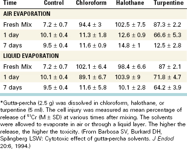
21. Barbosa SV, Burkard DH, Spångberg SW. Cytotoxic effects of gutta-percha solvents. Pediatr Dent. 1994;16:102.
22. Barkhordar RA, Kempler D. A comparison between xeroradiography and conventional radiography in the diagnosis of endodontic lesions. Oral Surg Oral Med Oral Pathol. 1988;66:489.
23. Barthel CR, Zimmer S, West G, Roulet JF. Bacterial leakage in obturated root canals following the use of different intracanal medicaments. Endod Dent Traumatol. 2000;16:282.
24. Basrani B, Ghanem A, Tjäderhane L. Physical and chemical properties of chlorhexidine and calcium hydroxide-containing medications. J Endod. 2004;30:413.
25. Basrani B, Lemonie C. Chlorhexidine gluconate. Aust Endod J. 2005;31:48.
26. Basrani B, Manek S, Sodhi R, Fillery E, Manzur A. Interaction between sodium hypochlorite and chlorhexidine gluconate. J Endod. 2007;33:966.
27. Basrani B, Santos JM, Tjäderhane L, et al. Substantive antimicrobial activity in chlorhexidine-treated human root dentin. Oral Surg Oral Med Oral Pathol Oral Radiol Endod. 2002;94:240.
28. Basrani B, Tjäderhane L, Santos JM, et al. Efficacy of chlorhexidine- and calcium hydroxide-containing medicaments against Enterococcus faecalis in vitro. Oral Surg Oral Med Oral Pathol Oral Radiol Endod. 2003;96:618.
29. Baumgartner JC, Johal S, Marshall JG. Comparison of the antimicrobial efficacy of 1.3% NaOCl/Biopure MTAD to 5.25% NaOCl/15% EDTA for root canal irrigation. J Endod. 2007;33:48.
30. Beach CW, Bramwell JD, Hutter JW. Use of an electronic apex locator on a cardiac pacemaker patient. J Endod. 1996;22:182.
31. Beltz RE, Torabinejad M, Pouresmail M. Quantitative analysis of the solubilizing action of MTAD, sodium hypochlorite, and EDTA on bovine pulp and dentin. J Endod. 2003;29:334.
32. Bender IB, Landau MA, Fonsecca S, Trowbridge HO. The optimum placement-site of the electrode in electric pulp testing of the 12 anterior teeth. J Am Dent Assoc. 1989;118:305-310.
33. Bergmans L, Moisiadis P, Huybrechts B, Van Meerbeek B, Quirynen M, Lambrechts P. Effect of photo-activated disinfection on endodontic pathogens ex vivo. Int Endod J. 2008;41:227.
34. Bernath M, Szabo J. Tissue reaction initiated by different sealers. Int Endod J. 2003;36:256.
35. Bodrumlu E, Alacam T. Evaluation of anitmicrobial effects of idoform-integrating gutta-percha. J Can Dent Assoc. 2006;72:733.
36. Bodrumlu E., Alacam T, Semiz M. The antimicrobial and antifungal efficacy of tetracycline-integrated gutta-percha. Indian J Dent Res. 2008;19:112.
37. Bodrumlu E, Muglali M, Sumer M, Guvenc T. The response of subcutaneous connective tissue to a new filling material. J Biomed Mater Res B Appl Biomater. 2008;84:463.
38. Bodrumlu E, Tonga U. The apical sealing ability of a new root canal filling material. Am J Dent. 2007;20:295.
39. Bodrumlu E, Uzam O, Topuz O, Semiz M. Efficacy of 3 techniques in removing root canal filling material. J Can Dent Assoc. 2008;74:721.
40. Bortnick KL, Steiman HR, Ruskin A. Comparison of nickel-titanium file distortion using electric and air-driven handpieces. J Endod. 2001;27:57.
41. Bouillaguet S, Shaw L, Barthelemy J, Krejci I, Wataha JC. Long-term sealing ability of Pulp Canal Sealer, AH-Plus, GuttaFlow and Epiphany. Int Endod J. 2008;41:219.
42. Bozza FL, Molgatini SL, Perez SB, Tejerin DP, Perez Tito RI, Kaplan AE. Antimicrobial effect in vitro of chlorhexidine and calcium hydroxide impregnated gutta-percha points. Acta Odontol Latinoam. 2005;18:51.
43. Briseňo-Marroquin B, El-Syed MAA, Willershausen-Zonnchen B. Morphology of the physiological foramen: I. Maxillary and mandibular molars. J Endod. 2004;30:321.
44. Brown BDK, Kafrawy AH, Patterson SS. Studies of Sargenti technique of endodontics: autoradiographic and scanning electron microscope studies. J Endod. 1979;5:14.
45. Brugnera A, Zanin F, Barbin EL, Spano JC, Santana R, Pecora JD. Effect of Er:YAG and Nd:YAG laser irradiation on radicular dentine permeability using different irrigating solutions. Lasers Surg Med. 2003;33:256.
46. Byström A, Claesson R, Sundqvist G. The antimicrobial effect of camphorated paramono-chlorphenol, camphorated phenol, and calcium hydroxide in the treatment of infected root canals. Endod Dent Traumatol. 1985;1:170.
47. Byström A, Sundqvist G. Bacteriologic evaluation of the effect of 0.5 percent sodium hypochlorite in endodontic therapy. Oral Surg Oral Med Oral Pathol. 1983;55:307.
48. Byström A, Sundqvist G. The antibacterial action of sodium hypochlorite and EDTA in 60 cases of endodontic therapy. Int Endod J. 1985;18:35.
49. Cabral CT, Fernandes MH. In vitro comparison of chlorhexidine and povidone-iodine on the long-term proliferation and functional activity of human alveolar bone cells. Clin Oral Invest. 2007;11:155.
50. Calan J. The use of inert plastic material in reconstructive surgery. Br J Plast Surg. 1963;16:1.
51. Camps J, Pashley D. Reliability of the dye penetration studies. J Endod. 2003;29:592.
52. Canadian Center for Occupational Health and Safety, ChemInfo, Record 728, 1994.
53. Carrilho MR, Carvalho RM, De Goes MF, et al. Chlorhexidine preserves dentine bond in vitro. J Dent Res. 2007;86:90.
54. Carrilho MR, Carvalho RM, Tay FR, Yiu C, Pashley DH. Durability of resin-dentin bonds related to water and oil storage. Am J Dent. 2005;18:315.
55. Carvalho CA, Valera MC, Gown-Soares S, dePaula Eduardo C. Effects of Nd:YAG and Er:YAG lasers on the sealing of root canal fillings. J Clin Laser Med Surg. 2002;20:215.
56. Caso A, Hung LK, Beirne OR. Prevention of alveolar osteitis with chlorhexidine: a meta-analytic review. Oral Surg Oral Med Oral Pathol Oral Radiol Endod. 2005;99:155.
57. Chen H, Teixeira FB, Ritter AL, Levin L, Trope M. The effect of intracanal anti-inflammatory medicaments on external root resorption of replanted dog teeth after extended extra-oral dry time. Dent Traumatol. 2008;24:74.
58. Cheung GS, Stock CJ. In vitro cleaning ability of root canal irrigants with and without endosonics. Int Endod J. 1993;26:334.
59. Chogle S, Mickel AK, Huffaker SK, Neibaur B. An in vitro assessment of iodoform gutta-percha. J Endod. 2005;31:814.
60. Chutich MJ, Kaminski EJ, Miller DA, Lautenschlager EP. Risk management of the toxicity of gutta-percha used in endodontic retreatment. J Endod. 1998;24:213.
61. Cobankara FK, Altinoz HC, Ergani O, Kav K, Belli S. In vitro antibacterial activities of root canal sealers using two different methods. J Endod. 2004;30:57.
62. Cohen BD, Combe ED, Lilley JD. Effects of thermal placement techniques on some physical properties of gutta-percha. Int Endod J. 1992;25:292.
63. Cohen BI, Pagnillo MK, Musikant BL, Deutsch AS. Formaldehyde evaluation from endodontic materials. Oral Health. 1998;88(12):37-39.
64. Cooley RL, Robison SF. Variables associated with electric pulp testing. Oral Surg Oral Med Oral Pathol. 1980;50:66.
65. Coronal Leakage: Clinical and Biological Implications, Endodontics: Colleagues for Excellence, American Association of Endodontics, Fall/Winter 2002.
66. Cotti E, Campisi G, Garau V, Puddu G. A new technique for the study of peripical bone lesions: Ultrasound real-time imaging. Int Endod J. 2002;35:142.
67. Cotton TP, Geisler TM, Holden DT, Schwartz SA, Schindler WG. Endodontic applications of cone 381 beam volumetric tomography. J Endod. 2007;33:1121.
68. Cunningham WT, Martin H. A scanning electron microscope evaluation of root canal debridement with the endosonic ultrasonic synergistic system. Oral Surg Oral Med Oral Pathol. 1982;53:527.
69. Dalat DM, Spångberg LSW. Comparison of apical leakage in root canals obturated with various gutta-percha techniques using a dye vacuum tracing method. J Endod. 1994;20:315.
70. Dammaschke T, Schneider U, Stratmann U, Yoo JM, Schafer E. Effect of root canal dressings on the regeneration of inflamed periapical tissue. Acta Odontol Scand. 2005;63:143.
71. Daugherty DW, Gound TG, Comer TL. Comparison of fracture rate, deformation rate, and efficiency between rotary endodontic instruments driven at 150 rpm and 350 rpm. J Endod. 2001;27:93.
72. Davies A. The mode of action of chlorhexidine. J Periodontal Res. 1973;12(Suppl):68.
73. Davis JM, Maki J, Bahcall JK. An in vitro comparison of the antimicrobial effects of various endodontic medicaments on Enterococcus faecalis. J Endod. 2007;33:567.
74. Decosta Rebeiro A, Nogueira GE, Antoniazzi JH, Moritz A, Zezell DM. Effects of diode lasers (810 nm) irradiation on root canal walls: Thermographic and morphological studies. J Endod. 2007;33:252.
75. De-Deus G, Brandao MC, Fidel RA, Fidel SR. The sealing ability of GuttaFlow in oval-shaped canals: an ex vivo study using polymicrobial leakage model. Int Endod J. 2007;40:794.
76. De-Deus G, Gurgel-Filho ED, Magalhaes KM, Coutinho-Filho T. A laboratory analysis of gutta-percha filled area obtained using Thermafil, System B and lateral condensation. Int Endod J. 2006;39:378.
77. De Gee AJ, Wu MK, Wesselink PR. Sealing properties of a Ketac-Endon glass ionomer cement an AH26 root canal sealer. Int Endod J. 1994;27:239.
78. Delany GM, Patterson SS, Miller CH, Newton CW. The effect of chlorhexidine gluconate irrigation on the root canal flora of freshly extracted necrotic teeth. Oral Surg Oral Med Oral Pathol. 1982;53:518.
79. deMoura-Netto C, deMoura AA, Davidowicz H, Aun CE, Antonio MP. Morphologic changes and removal of debris on apical dentin surfaces after Nd:YAG laser and diode laser irradiation. Photomed Laser Surg. 2008;26:263.
80. De Munck J, Van Landuyt K, Peumans M, et al. A critical review of the durability of adhesion to tooth tissue: methods and results. J Dent Res. 2005;84:118.
81. Denton G Chlorhexidine Block SS, editor. Disinfection, sterilization and preservation, 4th ed, Philadelphia: Lea & Febiger, 1991.
82. Depraet FJ, DeBruyne MA, DeMoor RJ. The sealing ability of an epoxy root canal sealer of Nd:YAG laser irradiation. Int Endod J. 2005;38:302.
83. De Rossi A, Silva LA, Leonardo MR, Rocha LB, Rossi MA. Effect of rotary or manual instrumentation, with or without a calcium hydroxide, 1% chlorhexidine intracanal dressing, on the healing of experimentally induced chronic periapical lesions. Oral Surg Oral Med Oral Pathol Oral Radiol Endod. 2005;99:628.
84. Desai P, Himel V. Comparative safety of various intracanal irrigation systems. J Endod. 2009;35:545.
85. Desai S, Chandler N. Calcium hydroxide-based root canal sealers: a review. J Endod. 2009;35:475.
86. DeSouza EB, Cai S, Simionato MR, Lage-Marques JL. High power diode laser in a disinfection in depth of the root canal dentin. Oral Surg Oral Med Oral Pathol. 2008;106:e68.
87. DeSouza EB, deAmorim CV, Marques JL. Effect of diode laser irradiation on the apical sealing of MTA retrofillings. Braz Oral Res. 2006;20:231.
88. Deveaux E, Hildebert P, Neut C, Romond C. Bacterial microleakage of Cavit, IRM, TERM, and Fermit: a 21-day in vitro study. J Endod. 1999;25:653.
89. Donley DL, Weller RN, Kulild JC, Jucak JJ. In vitro intra-canal temperatures produced by low- and high temperature thermoplasticized injectible gutta-percha. J Endod. 1991;17:307.
90. Dove SB, McDavid WD, Hamilton KE. Analysis of sensitivity and specificity of a new digital conventional radiography and computed tomography. Oral Surg Oral Med Oral Pathol Oral Radiol Endod. 2001;92:682.
91. Ebihara A, Majaron B, Liaw LL, Krasieva TB, Wilder-Smith P. Er:YAG laser modification of root canal dentine: influence of pulse duration, repetitive irradiation and water spray. Laser Med Sci. 2002;17:198.
92. Ebo DG, Stevens WJ, Bridts CH, Matthieu L. Contact allergic dermatitis and life-threatening anaphylaxis to chlorhexidine. J Allergy Clin Immunol. 1998;101:128.
93. Ehrmann EH. Pulp testers and pulp testing with particular reference to the use of dry ice. Aust Dent J. 1977;22:272.
94. Eideniz AU, Ozer F, Hadimli HH, Erganis O. Bacterial efficacy of Er, Cr:YSGG laser irradiation against Enterococcus facecalis compared with NaOCl irrigation: an ex vivo pilot study. Int Endod J. 2007;40:112.
95. El Ayouti A, Weiger R, Löst C. The ability of Root ZX apex locator to reduce the frequency of overestimated radiographic working length. J Endod. 2002;28:116.
96. Ellerbruch ES, Murphy RA. Antimicrobial activity of root canal medicament vapors. J Endod. 1977;3:189.
97. Emilson CG, Ericson T, Heyden G, Magnusson BC. Uptake of chlorhexidine to hydroxyapatite. J Periodontal Res. 1973;12(Suppl):17.
98. Ercan E, Dalli M, Duulgergil CT, Yaman F. Effect of intracanal medication with calcium hydroxide and 1% chlorhexidine in endodontic retreatment cases with periapical lesions: an in vivo study. J Formos Med Assoc. 2007;106:217.
99. Ercan E, Ozekinci T, Atakul F, Gul K. Antibacterial activity of 2% chlorhexidine gluconate and 5.25% sodium hypochlorite in infected root canal: in vivo study. J Endod. 2004;30:84.
100. Estrela C, Estrela CRA, Decurcio DA, Hollanda ACB, Silva JA. Antimicrobial efficacy of ozonated water, gaseous ozone, sodium hypochlorite and chlorhexidine In infected human root canals. Int Endod J. 2007;40:85.
101. Eun HC, Lee AY, Lee YS. Sodium hypochlorite dermatitis. Contact Dermatitis. 1984;11:45.
102. Evans MD, Baumgartner JC, Khemaleelakul SU, Xia T. Efficacy of calcium hydroxide: chlorhexidine paste as an intracanal medication in bovine dentin. J Endod. 2003;29:338.
103. Fanibunda KB. The feasibility of temperature measurement as a diagnostic procedure in human teeth. J Dent. 1986;14:126.
104. Fardal O, Turnbull RS. A review of the literature on use of chlorhexidine in dentistry. J Am Dent Assoc. 1986;112:863.
105. Faria G, Celes MR, De Rossi A, Silva LA, Silva JS, Rossi MA. Evaluation of chlorhexidine toxicity injected in the paw of mice and added to cultured L929 fibroblasts. J Endod. 2007;33:715.
106. Faria MI, Souza-Gabriel Ae, Marchesan MA, Sousa-Neto MD, Silva-Sousa YT. Ultrastructural evaluation of radicular dentin after Nd:YAG irradiation combined with different chemical substances. Gen Dent. 2008;56:641.
107. Fernandes AM, Silva GA, Lopes NJr., Napimoga MH, Benatti BB, Alves JB. Direct capping of human pulps with a dentin bonding system and calcium hydroxide: an immunohistochemical analysis. Oral Surg Oral Med Oral Pathol Oral Radiol Endod. 2008;105:385.
108. Fimple JL, Fontana CR, Foschi F, et al. Photodynamic treatment of endodontic polymicrobial infection in vivo. J Endod. 2008;34:728.
109. Folk RB, Thorpe JR, McClanahan SB, Johnson JD, Strother JM. Comparison of two different direct digital radiography systems for the ability to detect artificially prepared periapical lesions. J Endod. 2005;31:304.
110. Folwaczny M, Mehl A, Jordan C, Hickel R. Antibacterial effect of pulsed Nd:YAG laser radiation at different energy settings in the root canals. J Endod. 2002;28:24.
111. Fonseca MB, Junior PO, Pallota RC, et al. Photodynamic therapy for root canals in fected with Enterococcus faecalis. Photomed Laser Surg. 2008;26:209.
112. Foschi F, Fontana CR, Ruggeiro K, et al. Photodynamic inactivation of Enterococcus faecalis in dental root canals in vitro. Las Surg Med. 2007;39:782.
113. Fouad AF, Hopson JR, Martins JB, Krell KV, Drews TA, Jensen ME. Effects of selected electronic dental instruments on patients with cardiac pacemakers. J Endod. 1990;16:188.
114. Fouad AF, Krell KV, McKendry DJ, Koorbusch GF, Olson RA. A clinical evaluation of five electronic root canal length measuring instruments. J Endod. 1990;16:446.
115. Fouad AF, Reid LC. Effect of using electronic apex locators on selected endodontic treatment parameters. J Endod. 2000;26:364.
116. Frais S, Ng Y-L, Gulabivala K. Some factors affecting the concentration of commercially available sources of sodium hypochlorite. Int Endod J. 2001;34:206.
117. Frankenberger R, Pashley DH, Reich SM, Lohbauer U, Petschelt A, Tay FR. Characterisation of resin-dentine interfaces by compressive cyclic loading. Biomaterials. 2005;26:2043.
118. Friedman J, Marcus MI. Transillumination of the oral cavity with use of fiber optics. J Am Dent Assoc. 1970;80:801.
119. Fukumoto Y. Intracanal aspiration technique for root canal irrigation: evaluation of smear layer removal (in Japanese). Kokubyo Gakkai Zasshi. 2005;72:13.
120. Fukumoto Y, Kikuchi I, Yoshioka T, Kobayashi C, Suda H. An ex vivo evaluation of a new root canal irrigation technique with intracanal aspiration. Int Endod J. 2006;39:93.
121. Fuss Z, Trowbridge H, Bender IB, Rickoff B, Sorin S. Assessment of reliability of electrical and thermal pulp testing agents. J Endod. 1986;12:301.
122. Gabel WP, Hoen M, Steiman HR, Pink FE, Dietz R. Effect of rotational speed on nickel-titanium file distortion. J Endod. 1999;25:752.
123. Gambarini G, Pongione G, Rizzo F, Testarelli L, Cavalleri G, Gerosa R. Bending properties of nickel-titanium instruments: a comparative study. Minerva Stomatol. 2008;57:393.
124. Garcez AS, Nunez SC, Hamblin MR, Ribeiro MS. Antimicrobial effects of photodynamic therapy on patients with necrotic pulps and periapical lesions. J Endod. 2008;34:138.
125. Garcez AS, Ribeiro MS, Trope MS, et al. Antimicrobial therapy combined with conventional endodontic treatment to eliminate root canal biofilm infection. Lasers Surg Med. 2007;39:59.
126. Garofalo RR, Ede EN, Dorn SO, Kuttler S. Effect of electronic apex locators on cardiac pacemaker function. J Endod. 2002;28:831.
127. Garvey LH, Roed-Petersen J, Husum B. Anaphylactic reactions in anaesthetised patients—four cases of chlorhexidine allergy. Acta Anaesthesiol Scand. 2001;45:1290.
128. Gatewood RS, Himel VT, Dorn SO. Treatment of the endodontic emergency: a decade later. J Endod. 1990;16:284.
129. Gazelius B, Edwall B, Olgart L, Lundberg JM, Hokfelt T, Fisher JA. Vasodilatory effects and coexistence of calcitonin gene-related peptide (CGRP) and substance P in sensory nerves of cat dental pulp. Acta Physiol Scand. 1987;130:33.
130. Gazelius B, Olgart L, Edwall B, Edwall L. Non-invasive recording of blood flow in human dental pulp. Endod Dent Traumatol. 1986;2:219.
131. George R, Meyers IA, Walsh LJ. Laser activation of endodontic irrigants with improved conical laser fiber tips for removing smear layer with apical third of the root canal. J Endod. 2008;34:1524.
132. Ghori S, Gulabivala K, Premdas C, Spratt DA. Evaluation of the antimicrobial efficacy of electrochemically activated water on selected isolates from the root canal. Int Endod J. 2002;35:85.
133. Giardino L, Ambu E, Becce C, Rimondini L, Morra M. Surface tension comparison of four common root canal irrigants and two new irrigants containing antibiotic. J Endod. 2006;32:1091.
134. Giardino L, Ambu E, Savoldi E, Rimondini R, Cassanelli C, Debbia EA. Comparative evaluation of antimicrobial efficacy of sodium hypochlorite, MTAD, and tetraclean against Enterococcus faecalis biofilm. J Endod. 2007;33:852.
135. Goldberg F, De Silvio AC, Manfré S, Nastri N. In vitro measurement accuracy of an electronic apex locator in teeth with simulated apical root resorption. J Endod. 2002;28:461.
136. Goldman M, Kronman JH, Goldman LB, Clausen H, Grady J. New method of irrigation during endodontic treatment. J Endod. 1976;2:257.
137. Gomes BP, Ferraz CC, Vianna ME, Berber VB, Teixeira FB, Souza-Filho FJ. In vitro antimicrobial activity of several concentrations of sodium hypochlorite and chlorhexidine gluconate in the elimination of Enterococcus faecalis. Int Endod J. 2001;34:424.
138. Gomes BP, Sato E, Ferraz CC, Teixeira FB, Zaia AA, Souza-Filho FJ. Evaluation of time required for recontamination of coronally sealed canals medicated with calcium hydroxide and chlorhexidine. Int Endod J. 2003;36:604-609.
139. Goodman A, Schilder H, Aldrich W. The thermomechanical properties of gutta-percha II. Oral Surg Oral Med Oral Pathol. 1974;37:954.
140. Gopikrishna V, Pradeep G, Venkateshbabu N. Assessment of pulp vitality: a review. Int J Paediatr Dent. 2009;19:3.
141. Gordon MP, Chandler NP. Electronic apex locators. Int Endod J. 2004;37:425.
142. Gordon W, Atabakhsh VA, Meza F, et al. The antimicrobial efficacy of the erbium, chromium: yttrium-scandium-gallium-garnet laser with radial emitting tips on root canal dentin walls infected with Enterococcus faecalis. J Am Dent Assoc. 2007;138:992.
143. Gouw-Soares S, Staboltz A, Lage-Marques JL, Zezell DM, Groth EB, Edwardo CP. Comparative study of dentine permeability after apicoectomy and surface treatment with 9.6 micron TEA CO2 and Er:YAG laser irradiation. J Clin Laser Med Surg. 2004;22:129.
144. Goya C, Yamazaki R, Tomita Y, Kimura Y, Matsumoto K. Effects of pulsed Nd:YAG irradiation on smear layer at the apical stop and apical leakage after obturation. Int Endod J. 2000;33:266.
145. Grassi MD, Plazek DJ, Michanowicz AE, Chay IC. Changes in the physical properties of the ultrafil low temperature (70 degrees C) thermoplasticized gutta-percha system. J Endod. 1989;15:517.
146. Grawehr M, Sener B, Waltimo T, Zehnder M. Interactions of ethylenediamine tetraacetic acid with sodium hypochlorite in aqueous solutions. Int Endod J. 2003;36:411.
147. Greenstein G, Berman C, Jaffin R. Chlorhexidine. an adjunct to periodontal therapy. J Periodontol. 1986;57:370.
148. Grossman LI. Solibility of root canal cements. J Dent Res. 1978;57:927.
149. Grossman LI Clinical diagnostic methods Grossman LI, editor Endodontic practice 10th ed 1981 Lea & Febiger Philadelphia, PA 17-22
150. Grossman LI, Meiman BW. Solution of pulp tissue by chemical agent. J Am Dent Assoc. 1941;28:223.
151. Gulabivala K, personal communication, 2009.
152. Gulabivala K, Stock CJ, Lewsey JD, Ghori S, Ng YL, Spratt DA. Effectiveness of electrochemically activated water as an irrigant in an infected tooth model. Int Endod J. 2004;37:624.
153. Gundappa M, Ng SY, Whaites EJ. Comparison of ultrasound, digital and conventional radiography in the differentiation of periapical lesions. Dentomaxillofac Radiol. 2006;35:326.
154. Gurbuz T, Ozdemir Y, Kara N, Zehir C, Kurudirek M. Evaluation of root canal dentin after Nd:YAG laser irradiation and treatment with five different irrigation solutions: a preliminary study. J Endod. 2008;30:318.
155. Haapasalo HK, Siren EK, Waltimo TM, Orstavik D, Haapasalo MP. Inactivation of local root canal medicaments by dentine: An in vitro study. Int Endod J. 2000;33:126.
156. Habets JM, Geursen-Reitsma AM, Stolz E, Van Joost T. Sensitization to sodium hypochlorite causing hand dermatitis. Contact Dermatitis. 1986;15:140.
157. Haenni S, Schmidlin PR, Mueller B, Sener B, Zehnder M. Chemical and antimicrobial properties of calcium hydroxide mixed with irrigating solutions. Int Endod J. 2003;36:100.
158. Haikel Y, Gasser P, Allemann C. Dynamic fracture of hybrid endodontic hand instruments compared with traditional files. J Endod. 1991;17:217.
159. Hamblin MR, Hasan T. Photodynamic therapy: A new antimicrobial approach to infectious disease. Photochem Photobiol Sciences. 2004;3:436.
160. Hammad M, Qualtrough A, Silikas N. Effect of new obturating materials on vertical root fracture resistance of endodontically treated teeth. J Endod. 2007 Jun;33(6):732-736. (Epub 2007 Apr 16)
161. Hasselgren G, Olsson B, Cvek M. Effects of calcium hydroxide and sodium hypochlorite on the dissolution of necrotic porcine muscle tissue. J Endod. 1988;14:125.
162. Hata G, Hayami S, Weine FS, Toda T. Effectiveness of oxidative potential water as a root canal irrigant. Int Endod J. 2001;34:308.
163. Haznedaroglu F, Ersev H. Tetracycline HCl solution as a root canal irrigant. J Endod. 2001;27:738.
164. Hems RS, Gulabivala K, Ng YL, Ready D, Spratt DA. An in vitro evaluation of the ability of ozone to kill a strain of Enterococcus faecalis. Int Endod J. 2005;38:22.
165. Hennessey TS. Some antibacterial properties of chlorhexidine. J Periodontal Res. 1973;12(Suppl):61.
166. Hermann BW. Calciumhydroxyd als mittel zum behandel und füllen von zahnwurzelkanälen, Dissertation. Germany: University of Würzburg; 1920. Med Diss V
167. Hermann BW. Dentin obliteration der wurzelkanäle nach behandlung mit calcium. Zahnärtzl Rundschau. 1930;39:888.
168. Hidalgo E, Dominguez C. Mechanisms underlying chlorhexidine-induced cytotoxicity. Toxicol In Vitro. 2001;15:271.
169. Hjeljord LG, Rolla G, Bonesvoll P. Chlorhexidine-protein interactions. J Periodontal Res. 1973;12(Suppl):11.
170. Holland G, Walton R. Diagnosis and treatment planning. In Torabinejad M, Walton R, editors: Endodontics: principles and practice, ed 4, St. Louis: Saunders, 2009.
171. Holloway GA. Laser doppler measurement of cutaneous blood flow. In: Rolfe P, editor. Non-invasive physiological measurements. London: Academic Press Inc.; 1983:11.
172. Horiba N, Hiratsuka K, Onoe T, et al. Bactericidal effect of electrolysed neutral water on bacteria isolated from infected root canals. Oral Surg Oral Med Oral Pathol Oral Radiol Endod. 1999;87:83.
173. Howell RM, Duell RC, Mullaney TP. The determination of pulp vitality by thermographic means using cholesteric liquid crystals. Oral Surg Oral Med Oral Pathol. 1970;29:763.
174. Hsieh YD, Gau CH, Kung Wu SF, Shen EC, Hsu PW, Fu E. Dynamic recording of irrigating fluid distribution in root canals using thermal image analysis. Int Endod J. 2007;40:11.
175. Huang FM, Chang YC. Prevention of the epoxy resin-based root canal sealer-infused cyclooxygenase-2 expression and cytotoxicity of human oseteoblast cells by various antioxidants. Biomaterials. 2005;26:1849.
176. Huang FM, Tai KW, Chou MY, Chang YC. Cytotixicty of resin-, zinc oxide-eugenol-, and calcium hydroxide-based root canal sealers on human periodontal ligament cells and permanent V79 cells. Int Endod J. 2002;35:153.
177. Hugh CL, Walton RE, Facer SR. Evaluation of intracanal sealer distribution with 5 different obturation techniques. Quintessence Int. 2005;36:712.
178. Hugo WB, Longworth AR. The effect of chlorhexidine on the electrophoretic mobility, cytoplasmic constituents, dehydrogenase activity and cell walls of Excherichia coli and Staphylococcus aureus. J Pharm Pharmacol. 1966;18:569.
179. Hugo WB, Longworth ARL. Some aspects of the mode of action of chlorhexidine. J Pharm Pharmacol. 1964;16:655.
180. Hülsmann M, Pieper K. Use of an electronic apex locator in the treatment of teeth with incomplete root formation. Endod Dent Traumatol. 1989;5:238.
181. Hülsmann M, Rödig T, Nordmeyer S. Complications during root canal irrigation. Endod Topics. 2007;16:27.
182. Iatha H, Sandeep M, Kulkarni S, Yakub SS. Evaluation of fracture resistance in simulated immature teeth using Resilon and Ribbond as root reinforcements—an in vitro study. Dent Traumatol. 2009;25:433-438.
183. Ichikawa K, Nakamura HK, Ogawa N, Sakimura T, Kuroda M. R&D of long-term life-support system by using electrochemically activated biofilm reactor of aquatic animals for space examinations. Biological Science in Space. 1999;13:348.
184. Jeansonne MJ, White RR. A comparison of 2.0% chlorhexidine gluconate and 5.25% sodium hypochlorite as antimicrobial endodontic irrigants. J Endod. 1994;20:276.
185. Jha D, Guerrero A, Ngo T, Helfer A, Hasselgren G. Inability of laser and rotary instrumentation to eliminate root canal infection. J Am Dent Assoc. 2006;137:67.
186. Johnson E, Lloyd A, Kuttler S, Namerow K. Comparison between a novel nickel-titanium alloy and 508 Nitinol on the cyclic fatigue life of ProFile 25/.04 rotary instruments. J Endod. 2008;34:1406.
187. Kaplan AE, Ormaechea MF, PIcca M, Canzobre MC, Ubios AM. Rheological properties and biocompatibility of endodontic sealers. Int Endod J. 2003;36:527.
188. Karlovic Z, Pezelj-Ribaric S, Miletic I, Jukic S, Grgurevic J, Anic I. Erbium:YAG laser versus ultrasonic in preparation of root-end cavities. J Endod. 2005;31:821.
189. Katz A, Tagger M, Tamse A. Rejuvenation of brittle gutta-percha cones – a universal technique? J Endod. 1987;13:65.
190. Keate KC, Wong M. A comparison of endodontic file tip quality. J Endod. 1990;16:486.
191. Kim S, Dorscher-Kim J, Liu MT, Trowbridge H. Biphasic pulp blood flow response to substance P in the dog as measured with radiolabeled, microsphere injection method. Arch Oral Biol. 1988;33:305.
192. Kim S, Schuessler G, Chien S. Measurement of blood flow in the dental pulp of dogs with Xe-133 washout method. Arch Oral Biol. 1983;28:501.
193. Kimura Y, Onaga K, Yokoyama K, Kinoshita J, Ogata Y, Matsumoto K. Root surface temperature increase during Er:YAG laser irradiation on root canals. J Endod. 2002;28:76.
194. Kimura Y, Wilder-Smaith P, Matsumoto K. Lasers in endodontics: a review. Int Endod J. 2000;33:173.
195. Klotz MD, Gerstein HS, Bahn HN. Bacteraemia after topical use of prednisolone in infected pulps. J Am Dent Assoc. 1965;71:871.
196. Koba K, Kimura Y, Matsumoto K, et al. Post-operative symptoms and healing after endodontic treatment of infected teeth using pulsed Nd:YAG laser. Endod Dent Traumatol. 1999;15:68.
197. Kobayashi C, Suda H. New electronic canal measuring device based on the ratio method. J Endod. 1994;20:111.
198. Kolokuris I, Avanitoyannis I, Robinson C, Blanshard JM. Effect of moisture and aging on gutta-percha. J Endod. 1992;18:583.
199. Komorowski R, Grad H, Yu-Wu X, Friedman S. Antimicrobial substantivity of chlorhexidine-treated bovine root dentin. J Endod. 2000;26:315.
200. Kontakiotis EG, Tzanetakis GN, Loizides AL. A 12 month longitudinal in vitro leakage study on a new silicon-based root canal filling material (Gutta-Flow). Oral Surg Oral Med Oral Pathol Oral Radiol Endod. 2007;103:854.
201. Krause TA, Liewehr FR, Hahn CL. The antimicrobial effect of MTAD, sodium hypochlorite, doxycycline, and citric acid on Enterococcus faecalis. J Endod. 2007;33:28.
202. Krautheim AB, Jermann TH, Bircher AJ. Chlorhexidine anaphylaxis: case report and review of the literature. Contact Dermatitis. 2004;50:113.
203. Krell KV. The effects of CO2 ice on PFM restorations. J Endod. 1985;11:51.
204. Krell KV, Johnson RJ, Madison S. Irrigation patterns during ultrasonic canal instrumentation. Part I: K-type files. J Endod. 1988;14:65.
205. Krupp JD, Brantley WA, Gerstein H. An investigation of the torsional and bending properties of seven brands of endodontic files. J Endod. 1984;10:372.
206. Kulid J. Diagnostic testing. In: Ingle JI, Bakland L, Baumgartner C, editors. Endodontics. ed 6. Hamilton: BC Decker Inc.; 2008:542-543.
207. Kullendorff B, Petersson K, Rohlin M. Direct digital radiography for the detection of periapical bone lesions: a clinical study. Endod Dent Traumatol. 1997;13:183.
208. Kuruvilla JR, Kamath MP. Antimicrobial activity of 2.5% sodium hypochlorite and 0.2% chlorhexidine gluconate separately and combined, as endodontic irrigants. J Endod. 1998;24:472.
209. Lai YY, Pai L, Chen cp. Marginal leakage of different temporary restorations in standardized complex endodontic access preparations. J Endod. 2007;33:875.
210. Lambrianidis T, Kosti E, Boutsioukis C, Mazinis M. Removal efficacy of various calcium hydroxide/chlorhexidine medicaments from the root canal. Int Endod J. 2006;39:55-61.
211. Lambrianidis T, Margelos J, Beltes P. Removal efficiency of calcium hydroxide dressing from the root canal. J Endod. 1999;25:85.
212. Larsen CM, Watanabe I, Glickman GN, He J. Cyclic fatigue analysis of a new generation of nickel titanium rotary instruments. J Endod. 2009;35:401.
213. Lautenschlager EP, Jacobs JJ, Marshall GW, Heuer MA. Brittle and ductile torsional failures of endodontic instruments. J Endod. 1977;3:175.
214. Lee DH, Kim NR, Lim BS, Lee YK, Hwang KK, Yang HC. Effects of root canal sealers on lipopolysaccharide expression of cyclooxygenase-2 in murine macrophage cells. J Endod. 2007;33:1329.
215. Lee DH, Lim BS, Lee YK, Kim NR, Yang HC. Inhibitory effects of root canal sealers on the expression of inducible nitric oxide synthase in lipopolysaccharide-stimulated murine macrophage cells. J Biomed Mater Res. 2007;83:91. B
216. Lee DH, Park B, Saxena A, Serene TP. Enhanced surface hardness by boron implantation in nitinol alloy. J Endod. 1996;22:543.
217. Lenet BJ, Komorowski R, Wu XY, et al. Antimicrobial substantivity of bovine root dentin exposed to different chlorhexidine delivery vehicles. J Endod. 2000;26:652.
218. Leonardo MR, Bezerra da Silva LA, Filho MT, Santana da Silva R. Release of formaldehyde by 4 endodontic sealers. Oral Surg Oral Med Oral Pathol Oral Radiol Endod. 1999;88:221-225.
219. Leonardo MR, Guillen-Carlas MG, Pecora JD, Ito IY, Silva LA. Er:YAG laser antimicrobial effects in the root canals of dogs’ teeth with pulp necrosis and chronic periapical lesions. Photomed Laser Surg. 2005;38:295.
220. Leonardo MR, Tanomaru Filho M, Silva LA, Nelson Filho P, Bonifacio KC, Ito IY. In vivo antimicrobial activity of 2% chlorhexidine used as a root canal irrigating solution. J Endod. 1999;25:167.
221. Lewis BB, Chester SB. Formaldehyde in dentistry: a review of mutagenic and carcinogenic potential. J Am Dent Assoc. 1981;103:429.
222. Leyhausen G, Heil J, Reifferscheid G, Waldmann P, Geurtsen W. Genotoxicity of the epoxy resin-based root canal sealer AH Plus. J Endod. 1999;25:109.
223. Lima KC, Fava LR, Siqueira JFJr. Susceptibilities of Enterococcus faecalis biofilms to some antimicrobial medications. J Endod. 2001;27:616.
224. Limkangwalmongkol S, Burtscher P, Abbot PV, Sandler AB, Bishop BM. A comparative study of the apical leakage of four root canal sealers and laterally condensed gutta-percha. J Endod. 1991;17:495.
225. Lin J, Chandler NP, Purton D, Monteith B. Appropriate electrode placement site for electric pulp testing first molar teeth. J Endod. 2007;33:1296.
226. Lin S, Zuckerman O, Weiss EI, Fuss Z. Antibacterial efficacy of a new chlorhexidine slow-releasing device to disinfect dentinal tubules. J Endod. 2003;29:416.
227. Lindberg LG, Tamura T, Öberg PÅ. Photoplethysmography. Part 1: comparison with laser doppler flowmetry. Med Biol Eng Comput. 1991;29:40.
228. Lindskog S, Pierce AM, Blomlof L. Chlorhexidine as a root canal medicament for treating inflammatory lesions in the periodontal space. Endod Dent Traumatol. 1998;14:186.
229. Lipski M. In vitro infrared thermographic assessment of root surface temperatures generated by high-temperature thermo-plasticized injectible gutta-percha obturation technique. J Endod. 2006;32:438.
230. Lipski M. Root surface temperature rises in vitro during root canal obturation with thermoplasticized gutta-percha on a carrier or by injection. J Endod. 2004;30:441.
231. Löe H. Does chlorhexidine have a place in the prophylaxis of dental diseases? J Periodontal Res. 1973;12(Suppl):93.
232. Löe H, Schiott CR. The effect of mouthrinses and topical application of chlorexidine on the development of dental plaque and gingivitis in man. J Periodontal Res. 1970;5(Suppl):79.
233. Lohbaur U, Dahl U, Dasch W, Petschelt A. Calcium release and pH of gutta-percha points containing calcium hydroxide. J Dent Res. 2001;80:272.
234. Loshon CA, Melly E, Setlow B, Setlow P. Analysis of the killing of spores of Bacillus subtilis by a new disinfectant, Sterilox®. J Appl Microbiol. 2001;91:1051.
235. Lumley PJ, Walmsley AD. Effect of precurving on the performing of endosonic K files. J Endod. 1992;18:232.
236. Maden M, Gorgul G, Tinaz AC. Evaluation of apical leakage of root canals obturated with Nd:YAG laser-softened gutta-percha, System B, and lateral condensation techniques. J Contemp Dent Pract. 2002;3:16.
237. Magnusson B, Heyden G. Autoradiographic studies of 14C-chlorhexidine given orally in mice. J Periodontal Res. 1973;Suppl 12(Suppl):49.
238. Malkhassian G, Manzur AJ, Legner M, et al. Antibacterial efficacy of MTAD final rinse and two percent chlorhexidine gel medication in teeth with apical periodontitis: a randomized double-blinded clinical trial. J Endod. 2009;35:1483-1490.
239. Manzur A, Gonzalez AM, Pozos A, Silva-Herzog D, Friedman S. Bacterial quantification in teeth with apical periodontitis related to instrumentation and different intracanal medications: a randomized clinical trial. J Endod. 2007;33:114.
240. Marais JT, Brozel VS. Electro-chemically activated water in dental unit water lines. Br Dent J. 1999;187:154.
241. Marais JT, Williams WP. Antimicrobial effectiveness of electro-chemically activated water as an endodontic irrigation solution. Int Endod J. 2001;34:237.
242. Marciano J, Michailesco PM. Dental gutta-percha: chemical composition, x-ray identification, enthalpic studies, and clinical implications. J Endod. 1989;15:149.
243. Margelos J, Eliades G, Verdelis C, Palaghias G. Interaction of calcium hydroxide with zinc oxide-eugenol type sealers: a potential clinical problem. J Endod. 1997;23:43.
244. Margelos J, Verdelis K, Eliades G. Chloroform uptake by gutta-percha and assessment of its concentration in air during the chloroform-dip technique. J Endod. 1996;22:547.
245. Margolis HC, Moreno EC, Murphy BJ. Importance of high pKa acids in cariogenic potential of plaque. J Dent Res. 1985;64:786-792.
246. Marmary Y, Koter T, Heling I. The effect of periapical rarefying osteitis on cortical and cancellous bone. A study comparing conventional radiographs with computed tomography. Dentomaxillofac Radiol. 1999;28:267.
247. Martin H, Ferris C, Mazzella W. An evaluation of media used in electric pulp testing. Oral Surg Oral Med Oral Pathol. 1969;27:374.
248. Matsumoto K. Lasers in endodontics. Dent Clin North Am. 2000;44:889.
249. Matsuya Y, Matsuya S. Effect of abietic acid and poly-methyl methacrylate on the dissolution process of zinc oxide-eugenol cement. Biomaterials. 1994;15:307.
250. Mayeda DL, Simon JH, Aimar DF, Finley K. In vivo measurement accuracy in vital and necrotic canals with the Endex apex locator. J Endod. 1993;19:545.
251. Melker KB, Vertucci FJ, Rojas MF, Progulske-Fox A, Belanger M. Antimicrobial efficacy of medicated root canal filling materials. J Endod. 2006;32:148.
252. Magnifying the surgical field with an OM. In: Merino EM, editor. Endodontic Microsurgery. London: Quintessence Pub Co; 2009:5-9.
253. Mesaros SV, Trope M. Revascularization of traumatized teeth assessed by laser doppler flowmetry: case report. Endod Dent Traumatol. 1997;13:24.
254. Messer HH, Feigal RJ. A Comparison of the antibacterial and cytotoxic effects of parachlorophenol. J Dent Res. 1985;64:818.
255. Metzger Z, Better H, Abramovitz I. Immediate root canal disinfection with ultraviolet light: an ex vivo feasibility study. Oral Surg Oral Med Oral Pathol Oral Radiol Endod. 2007;104:425.
256. Metzger Z, Teperovich E, Cohen R, Zary R, Paué F, Hülsmann M. Removal of apical smear layer by the Self Adjusting File (SAF): a scanning electron microscope study. J Endod. 2009. Submitted for publication
257. Metzger Z, Dotan M, Better H, Abramovitz I. Sensitivity of oral bacteria to 254 nm ultraviolet light. Int Endod J. 2007;40:120.
258. Metzger Z, Huber R, Slavescu D, Dragomirescu D, Tobis I, Better H. Healing kinetics of periapical lesions enhanced by the Apexum procedure: a clinical trial. J Endod. 2009;35:153.
259. Metzger Z, Huber R, Tobis I, Better H. Enhancement of healing kinetics of periapical lesions in dogs by the Apexum procedure. J Endod. 2009;35:40.
260. Metzger Z, Teperovich E, Zary R, Cohen R, Hof R. Respecting the root canal: a new concept of a Self Adjusting File (SAF). J Endod. 2009. Submitted for publication
261. Michaelson RE, Seidberg BH, Guttuso J. An in vivo evaluation of interface media used with the electric pulp tester. J Am Dent Assoc. 1975;91:118.
262. Mickel AK, Lindquist KAD, Chogle S, Jones JJ, Curd F. Electric pulp tester conductance through various interface media. J Endod. 2006;32:1178.
263. Mickel AK, Nguyen TH, Chogle S. Antimicrobial activity of endodontic sealers on Enterococcus faecalis. J Endod. 2003;29:257.
264. Mickel AK, Wright ER. Growth inhibition of Stretococcus anginosus (Milleri) by three calcium hydroxide sealer and one zinc oxide-eugenol sealer. J Endod. 1999;25:34.
265. Mikrogeorgis G, Lyroudia K, Moyvdas I, Nikolaidis N, Pitas I. Digital radiograph registration and subtraction: a useful tool for the evaluation of the progress of chronic apical periodontitis. J Endod. 2004;30:513.
266. Miserendino LJ, Moser JB, Heuer MA, Osetek EM. Cutting efficiency of endodontic instruments. I. A quantitative comparison of the tip and fluted region. J Endod. 1985;11:435.
267. Miserendino LJ, Moser JB, Heuer MA, Osetek EM. Cutting efficiency of endodontic instruments. II. An analysis of the design of the tip. J Endod. 1986;12:8.
268. Miwa Z, Ikawa M, Iijima H, Saito M, Takagi Y. Pulpal blood flow in vital and nonvital young permanent teeth measured by transmitted-light photoplethysmography: a pilot study. Pediatr Dent. 2002;24:594.
269. Miyagak DC, de Carvalho EM, Robazza CR, Chavasco JK, Levorato GL. In vitro evaluation of the antimicrobial activity of endodontic sealers. Braz Oral Res. 2006;20:303.
270. Mjör IA, Moorhead JE, Dahl JE. Reasons for replacement of restorations in permanent teeth in general dental practice. Int Dent J. 2000;50:361.
271. Mohammadi Z, Khademi AA, Davari AR. Evaluation of the antibacterial substantivity of three concentrations of chlorhexidine in bovine root dentine. Iranian Endod J. 2008;2:113.
272. Möller ÅJR. Microbiological examination of root canals and periapical tissues of human teeth. Odontol Tidskr. 1966;74:1. (special issue)
273. Monticelli F, Sadek FT, Schuster CG, et al. Efficacy of two contemporary single-cone filling techniques in preventing bacterial leakage. J Endod. 2007;33:310.
274. Monticelli F, Sword J, Martin RL, et al. Sealing properties of two contemporary single-cone obturation systems. Int Endod J. 2007;40:374.
275. Mora MA, Mol A, Tyndall DA, Rivera EM. In vitro assessment of local computed tomography for the detection of longitudinal tooth fractures. Oral Surg Oral Med Oral Pathol Oral Radiol Endod. 2007;103:825.
276. Moser JB, Heuer MA. Forces and efficacy in endodontic irrigation systems. Oral Surg Oral Med Oral Pathol. 1982;53:425.
277. Moshonov J, Slutzky-Goldberg I. Apex locators: an update and prospects for the future. Int J Comput Dent. 2004;7:359.
278. Moshonov J, Slutzky-Goldberg I. Apex locators: update and prospects for the future. Int J Comp Dent. 2004;7(4):359.
279. Muller P, Guggenheim B, Schmidlin PR. Efficacy of gasiform ozone and photodynamic therapy on a multispecies oral biofilm in vitro. Eur J Oral Sci. 2007;115:77.
280. Mumford JM. Evaluation of gutta percha and ethyl chloride in pulp-testing. Br Dent J. 1964;116:338.
281. Narhi MVO, Virtanen A, Kuhta J, Huopaniemi T. Electrical stimulation of teeth with a pulp tester in the cat. Scand J Dent Res. 1979;87:32.
282. Nagayoshi M, Kitamura C, Fukuizumi T, Nishihara T, Terashita M. Antimicrobial effect of ozonated water on bacteria invading dentinal tubules. J Endod. 2004;30:778.
283. Nielsen BA, Craig BJ. Comparison of the Endovac System to needle irrigation of root canals. J Endod. 2007;33:611.
284. Nissan R, Trope M, Zheng C-D, Chance B. Dual wavelength spectrophotometry as a diagnostic test of the pulp chamber contents. Oral Surg Oral Med Oral Pathol. 1992;74:508.
285. Noiri Y, Katsumoto T, Azakami H, Ebisu S. Effects of Er:YAG laser radiation on biofilm-forming bacteria associated with endodontic pathogens in vitro. J Endod. 2008;34:826.
286. Okano M, Nomura M, Hata S, et al. Anaphylactic symptoms due to chlorhexidine gluconate. Arch Dermatol. 1989;125:50.
287. Oliveira RG, Gouw-Soares S, Baldochi SL, Edwardo CP. Scanning electron microscopy: effect of Er:YAG and Nd:YAG lasers on apical seals after apicoectomy and retrofill. Photomed Laser Surg. 2004;23:533.
288. Onay EO, Unger M, Ozdemir BH. In vivo evaluation of the biocompatibility of a new resin-based obturation material. Oral Surg Oral Med Oral Pathol. 2007;104:e60.
289. Oncag O, Hosgor M, Hilmioglu S, Zekioglu O, Eronat C, Burhanoglu D. Comparison of antibacterial and toxic effects of various root canal irrigants. Int Endod J. 2003;36:423.
290. Ørstavik D, Haapasalo M. Disinfection by endodontic irrigants and dressings of experimentally infected dentinal tubules. Endod Dent Traumatol. 1990;6:142.
291. Oztan MD, Kiyan M, Gerceker D. Antimicrobial effect, in vitro, of gutta-percha points containing root canal medications against yeasts and Enterococcus faecalis. Oral surg Oral Med Oral Pathol Oral Radiol Endod. 2006;102:410.
292. Pantera EA, Anderson RW, Pantera CT. Reliability of electric pulp testing after pulpal testing with dichlorodi-fluoromethane. J Endod. 1993;19:312.
293. Paquette L, Legner M, Fillery ED, Friedman S. Antibacterial efficacy of chlorhexidine gluconate intracanal medication in vivo. J Endod. 2007;33:788.
294. Park DS, Lee HJ, Yoo HM, Oh TS. Effect of Nd:YAG laser irradiation of the apical leakage of obturated root canals: an electrochemical study. Int Endod J. 2001;34:318.
295. Pascon E, Spångberg LS. In vitro cytotoxicity of root canal filling materials. Part I. Gutta-percha. J Endod. 1990;16:429.
296. Paurazas SB, Geist JR, Pink FE, Hoen MM, Steiman HR. Comparison of diagnostic accuracy of digital imaging by using CCD and CMOS-APS sensors with E-speed film in the detection of periapical bony lesions. Oral Surg Oral Med Oral Pathol Oral Radiol Endod. 2000;89:356.
297. Pawinska M, Skrzydlewska E. Release of hyroxcyl ions from calcium hydroxide preparations used in endodontic treatment. Rocz Akad Med Bialymst. 2003;48:145.
298. Perez SB, Tejerina DP, Perez Tito RI, Bozza FL, Kaplan AE, Molgatini SL. Endodontic microorganisms susceptabilty by direct contact test. Acta Odontol Latinoam. 2008;21:169.
299. Perin FM, Franca SC, Silva-Sousa YT, et al. Evaluation of the microbial effect of Er:YAG laser irradiation versus 1% sodium hypochlorite irrigation for root canal disinfection. Aust Endod J. 2004;30:20.
300. Peters DD. Evaluation of the effects of carbon dioxide used as a pulp test. Part III: in vivo effect on human enamel. J Endod. 1986;12:13.
301. Peters LB, Van Winkelhoff AJ, Buijs JF, Wesselink PR. Effects of instrumentation, irrigation and dressing with calcium hydroxide on infection in pulpless teeth with periapical bone lesions. Int Endod J. 2002;35:13.
302. Peters OA. Current challenges and concepts in the preparation of root canal systems: a review. J Endod. 2004;30:559.
303. Pham NH, Weiner JM, Reisner GS, Baldo BA. Anaphylaxis to chlorhexidine. Case report. Implication of immunoglobulin E antibodies and identification of an allergenic determinant. Clin Exp Allergy. 2000;30:1001.
304. Pizzo G, Giammanco GM, Cumbo E, Nicolosi G, Gallina G. In vitro antibacterial activity of endodontic sealers. J Dent. 2006;34:35.
305. Podbielski A, Spahr A, Haller B. Additive anitmiccrobial activity of calcium hydroxide and chlorhexidine on common endodontic bacterial pathogens. J Endod. 2003;29:340.
306. Popescu IG, Popescu M, Man D. Drug allergy: incidence in terms of age and some drug allergens. Med Intern. 1984;22:195.
307. Portenier I, Waltimo T, Orstavik D, Haapasalo M. Killing of Enterococcus faecalis by MTAD and chlorhexidine digluconate with or without cetrimide in the presence or absence of dentine powder or BSA. J Endod. 2006;32:138.
308. Powell SE, Simon JHS, Maze B. A comparison of the effect of modified and nonmodified instrument tips on apical canal configuration, Part 1. J Endod. 1986;12:293.
309. Powell SE, Wong PD, Simon JHS. A comparison of the effect of modified and nonmodified instrument tips on apical canal configuration. Part 2. J Endod. 1988;14:224.
310. Rapisarda E, Bonaccorso A, Tripi TR, Condorelli GG, Torrisi L. Wear of nickel-titanium endodontic instruments evaluated by scanning electron microscopy: effect of ion implantation. J Endod. 2001;27:588.
311. Rasimick BJ, Nekich M, Hladek MM, Musikant BL, Deutsch AS. Interaction between chlorhexidine digluconate and EDTA. J Endod. 2008;34:1521.
312. Richman MJ. The use of ultrasonics in root canal therapy and root resection. J Med. 1957;12:12.
313. Rickard GD, Richardson R, Johnson T, Mccoll D, Hooper L: Ozone therapy for the treatment of dental caries. Cochrane Database Syst Rev, CD004153, 2004.
314. Rickoff B, Trowbridge H, Baker J, Fuss Z, Bender IB. Effects of thermal vitality tests on human dental pulp. J Endod. 1988;14:482.
315. Ringel AM, Patterson SS, Newton CW, Miller CH, Mulhern JM. In vivo evaluation of chlorhexidine gluconate solution and sodium hypochlorite solution as root canal irrigants. J Endod. 1982;8:200.
316. Roane JB, Sabala CL, Duncanson MG. The “balanced force” concept for instrumentation of curved canals. J Endod. 1985;11:203.
317. Rölla G, Löe H, Schiott CR. The affinity of chlorhexidine for hydroxyapatite and salivary mucins. J Periodont Res. 1970;5(Suppl):90.
318. Roy RA, Ahmad M, Crum LA. Physical mechanisms governing the hydrodynamic response of an oscillating ultrasonic file. Int Endod J. 1994;27:197.
319. Rowe AHR, Pitt Ford TR. The assessment of pulpal vitality. Int Endod J. 1990;23:77.
320. Ruddle CJ. The ProTaper endodontic system: geometries, features and guidelines for use. Dent Today. 2001;20:60.
321. Sabala CL, Roane JB, Southard LZ. Instrumentation of curved canals using a modified tipped instrument: a comparison study. J Endod. 1988;14:59.
322. Safavi KE, Nichols FC. Alteration of biological properties of bacterial lipopolysaccharide by calcium hydroxide treatment. J Endod. 1994;20:127.
323. Safavi KE, Nichols FC. Effect of calcium hydroxide on bacterial lipopolysaccharide. J Endod. 1993;19:76.
324. Safavi KE, Spångberg L, Langeland K. Root canal dentinal tubule disinfection. J Endod. 1990;16:207.
325. Saleh IM, Ruyter IE, Haapasalo M, Ørstavik D. The effects of dentin pretreatment on the adhesion of root canal sealers. Int Endod J. 2002;35:859.
326. Santos C, Sousa-Neto MD, Alfredo E, Guerisoli DM, Percora JD, Comelli Lea RF. Morphologic evaluation of the radicular dentin irradiated with Nd:YAG laser under different parameters and angles of incidence. Photomed Laser Surg. 2005;23:590.
327. Sathorn C, Parashos P, Messer HH. Effectiveness of single- versus multiple-visit endodontic treatment of teeth with apical periodontitis: a systematic review and meta-analysis. Int Endod J. 2005;38:347.
328. Sato I, Ando-Kurihara N, Kota K, Iwaku M, Hoshino E. Sterilization of infected root-canal dentine by topical application of a mixture of ciprofloxacin, metronidazole and minocycline in situ. Int Endod J. 1996;29:118.
329. Saunders EM. In vivo findings associated with heat generation during thermomechanical compaction of gutta-percha. 1. Temperature levels at the external surface of the root. Int Endod J. 1990;23:263.
330. Schäfer E. Irrigation of the root canal. ENDO. 2007;1:11-28.
331. Schäfer E, Zandbiglari T. Solubility of root canal sealers in water and artificial saliva. Int Endod J. 2003;36:660.
332. Schilder H, Goodman A, Aldrich W. The thermomechanical properties of gutta-percha I. The compressibility of gutta-percha. Oral Surg Oral Med Oral Pathol. 1974;37:946.
333. Schilder H, Goodman A, Aldrich W. The thermomechanical properties of gutta-percha, III. Determination of phase transition temperatures for gutta-percha. Oral Surg Oral Med Oral Pathol. 1974;38:109.
334. Schilder H, Goodman A, Aldrich W. The thermomechanical properties of gutta-percha, V. Volume changes in bulk gutta-percha as a function of temperature and its relationship to molecular phase transformation. Oral Surg Oral Med Oral Pathol. 1985;59:285.
335. Schnettler JM, Wallace JA. Pulse oximetry as a diagnostic tool of pulpal vitality. J Endod. 1991;17:488.
336. Schoop U, Goharkhay K, Klimscha J, et al. The use of erbium, chromium:yttrium-scandium-gallium-garnet laser in endodontic treatment: the results of an in vitro study. J Am Dent Assoc. 2007;138:949.
337. Schoop U, Moritz A, Kluger W, et al. The Er:YAG laser in endodontics: results of an in vitro study. Lasers Surg Med. 2002;30:360.
338. Schuur AH, Moorer WR, Wesselink PR. Solvents for the removal of gutta-percha from root canals. 2. Side effects of Chloroform, halothane and xylene. Ned Tijdschr Tandheelkd. 2004;111:303.
339. Schroeder A. Ledermix 1962–Ledermix today. Evaluation after 13 years of experience. Zahnarztl Prax. 1975;26(9):195-196. May 2
340. Scully C, Ng YL, Gulabivala K. Systemic complications due to endodontic manipulations. Endod Topics. 2003;4:60.
341. Seltzer S, Bender IB, Ziontz M. The dynamics of pulpal inflammation: correlation between diagnostic data and actual histological findings in the pulp. Oral Surg Oral Med Oral Pathol. 1963;16:973.
342. Senia ES, Marraro RV, Mitchell JL, Lewis AG, Thomas L. Rapid sterilization of gutta-percha cones with 5.25% sodium hypochlorite. J Endod. 1975;1:136.
343. Senia ES, Marshal FJ, Rosen S. The solvent action of sodium hypochlorite on pulp tissue of extracted teeth. Oral SurgOral Med Oral Pathol. 1971;31:96.
344. Serper A, Calt S, Dogan AL, Gue D, Ozcelik B, Kuraner T. Comparison of the cytotoxic effects and smear layer removing capacity of oxidative potential water, NaOCl, EDTA. J Oral Sci. 2001;43:233.
345. Seto BG, Nicholls JI, Harrington GW. Torsional properties of twisted and machined endodontic files. J Endod. 1990;16:355.
346. Shabahang S, Pouresmail M, Torabinejad M. In vitro antimicrobial efficacy of MTAD and sodium hypochlorite. J Endod. 2003;29:450.
347. Shabahang S, Torabinejad M. Effect of MTAD on Enterococcus faecalis-contaminated root canals of extracted human teeth. J Endod. 2003;29:576.
348. Shipper G, Ørstavik D, Teixeira F, Trope M. An evaluation of microbial leakage in roots filled with a thermoplastic synthetic polymer-based root canal filling material (Resilon). J Endod. 2004;30:342.
349. Short RD, Dorn SO, Kutter S. The crystallization of sodium hypochlorite on gutta-percha cones after the rapid-sterilization technique: an SEM study. J Endod. 2003;29:670.
350. Shuping GB, Ørstavik D, Sigurdsson A, Trope M. Reduction of intracanal bacteria using nickel-titanium rotary instrumentation and various medications. J Endod. 2000;26:751.
351. Shur Al, Sedgley CM, Fenno JC. The antimicrobial efficacy of ‘MPG’ gutta-percha in vitro. Int Endod J. 2003;36:616.
352. Sigurdsson A. Pulpal diagnosis. Endod Topics. 2003;5:12.
353. Sipert CR, Hussne RP, Nishiyama CK, Torres SA. In vitro antimicrobial activity of Fill Canal, Sealarex, Mineral Trioxide Aggregate, Portland Cement and EndoRez. Int Endod J. 2005;38:539.
354. Siqueira JFJr, De Uzeda M. Intracanal medicaments: evaluation of the antibacterial effects of chlorhexidine, metronidazole, and calcium hydroxide associated with three vehicles. J Endod. 1997;23:167.
355. Siqueira JFJr, Favieri A, Gahyva SM, Moraes SR, Lima KC, Lopes HP. Antimicrobial activity and flow rate or newer and established root canal sealers. J Endod. 2000;26:274.
356. Siqueira JFJr, Paiva SS, Rocas IN. Reduction in the cultivable bacterial populations in infected root canals by a chlorhexidine-based antimicrobial protocol. J Endod. 2007;33:541.
357. Siqueira JFJr, Rocas IN, Paiva SS, Guimaraes-Pinto T, Magalhaes KM, Lima KC. Bacteriologic investigation of the effects of sodium hypochlorite and chlorhexidine during the endodontic treatment of teeth with apical periodontitis. Oral Surg Oral Med Oral Pathol Oral Radiol Endod. 2007;104:122.
358. Siren EK, Lavonious E, Kontakiotis E. Effects of Ca(OH)2 gutta-percha points on bacteria in root canals. J Dent Res. 2000;79:543.
359. Sirtes G, Waltimo T, Schaetzle M, Zehnder M. The effects of temperature on sodium hypochlorite short-term stability, pulp dissolution capacity, and antimicrobial efficacy. J Endod. 2005;31:669-671.
360. Sjögren U, Figdor D, Spångberg LSW, Sundqvist G. The antimicrobial effect of calcium hydroxide as a short-term intracanal dressing. Int Endod J. 1991;24:119.
361. Sjögren U, Sundqvist G. Bacteriologic evaluation of ultrasonic root canal instrumentation. Oral Surg Oral Med Oral Pathol. 1987;63:366.
362. Sjögren U, Sundquist G, Nair PNR. Tissue reaction to gutta-percha particles of various sizes when implanted subcutaneously in guinea pigs. Eur J Oral Sci. 1995;103:313.
363. Snellman E, Rantanen T. Severe anaphylaxis after a chlorhexidine bath. J Am Acad Dermatol. 1999;40:771.
364. Soares JA, Leonardo MR, Da Silva LA, Tanomaru Filho M, Ito IY. Effect of rotary instrumentation and of the association of calcium hydroxide and chlorhexidine on the antisepsis of the root canal system in dogs. Braz Oral Res. 2006;20:120.
365. Solovyeva AM, Dummer PMH. Cleaning effectiveness of root canal irrigation with electrochemically activated anolyte and catholyte solutions: a pilot study. Int Endod J. 2000;33:494.
366. Soukos NS, Chen PS, Morris JT, et al. Photodynamic therapy for endodontic disinfection. J Endod. 2006;32:979.
367. Sousa-Neto MD, Marchesan MA, Pecora JD, Junior AB, Silva-Souse YT, Saquy PC. Effect of Er:YAG laser on adhesion of root canal sealers. J Endod. 2002;28:185.
368. Sousa-Neto MD, Silva Coelho FI, Marchesan MA, Alfredo E, Silva-Sousa YT. Ex vivo study of the adhesion of an epoxy based sealer to human dentin submitted to irradiation with Er:YAG and Nd:YAG lasers. Int Endod J. 2005;38:866.
369. Southard SR, Drisko CL, Killoy WJ, Cobb CM, Tira DE. The effect of 2% chlorhexidine digluconate irrigation on clinical parameters and the level of Bacteroides gingivalis in periodontal pockets. J Periodontol. 1989;60:302.
370. Spångberg L, Engström B, Langeland K. Biologic effects of dental materials. 3. Toxicity and antimicrobial effect of endodontic antiseptics in vitro. Oral Surg Oral Med Oral Pathol. 1973;36:856.
371. Spångberg L, Rutberg M, Rydinge E. Biological effects of endodontic antimicrobial agents. J Endod. 1979;5:166.
372. Spångberg LSW, Barbosa SV, Lavigne GD. AH26 releases formaldehyde. J Endod. 1993;19:596.
373. Spratt DA, Pratten J, Wilson M, Gulabivala K. An in vitro evaluation of the antimicrobial efficacy of irrigants on biofilms of root canal isolates. Int Endod J. 2001;34:300.
374. Stabholtz A, Sahar-Helft S, Moshonov J. Laser in endodontics. Dent Clin North Am. 2004;48:809.
375. Stabholz A, Moshonov J, Sahar-Helft S, Rocca JP. Lasers in endodontics. In Ingle JI, Bakland LK, Baumgartener JC, editors: Ingle’s endodontics, ed 6, Hamilton, ON, Canada: BC Decker Inc, 2008.
376. Stabholz A, Khayat A, Ravanshad SH, McCarthy DWD, Neev J, Torabinejad M. Effects of Nd:YAG laser on apical seal of teeth after apicoectomy and retrofill. J Endod. 1992;18:371.
377. Stabholz A, Khayat A, Weeks DA, Neev J, Torabinejad M. A scanning electron microscopic study of the apical dentine surfaces lased with Nd:YAG laser following apicectomy and retrofill. Int Endod J. 1992;25:288.
378. Staehle HJ, Spiess V, Heinecke A, Muller HP. Effect of root canal filling materials containing calcium hydroxide and the alkalinity of root dentin. Endod Dent Traumatol. 1995;11:163.
379. Stenman E, Spångberg LSW. Machining efficiency of Flex-R, K-Flex, Trio-Cut, and S-Files. J Endod. 1990;16:575.
380. Stock CJR. Current status of the use of ultrasound in endodontics. Int Dent J. 1991;41:175.
381. Sunada I. New method for measuring the length of the root canal. J Dent Res. 1962;41:375.
382. Sweatman TL, Baumgartner JC, Sakaguchi RL. Radicular temperatures associated with thermoplasticized gutta-percha. J Endod. 2001;27:512.
383. Szep S, Grumann L, Ronge K, Schriever A, Schultz M, Heidemann D. In vitro cytoxicity of medicated and nonmedicated gutta-percha points in cultures of gingival fibroblasts. J Endod. 2003;29:36.
384. Takeda FH, Harashima T, Kimura Y, Matsumoto K. A comparative study of the removal of smear layer by three endodontic irrigants and two types of lasers. Int Endod J. 1999;32:32.
385. Tani-Ishii N, Teranara T. Clinical and radiographic evaluation of root canal obturation with Obtura II. J Endod. 2003;29:739.
386. Tanomaru Filho M, Leonardo MR, Da Silva LA. Effect of irrigating solution and calcium hydroxide root canal dressing on the repair of apical and periapical tissues of teeth with periapical lesion. J Endod. 2002;28:295.
387. Tanomaru-Filho M, Tanomaru JM, Barros DB, Wantanabe E, Ito IY. In vitro antimicrobial activity of endodontic sealers, MTA-based cements and Portland cement. J Oral Sci. 2007;49:41.
388. Tasman F, Cehreli ZC, Ogan C, Etikan I. Surface tension of root canal irrigants. J Endod. 2000;26:586.
389. Tay FR, Hosoya Y, Loushine RJ, Pashley DH, Weller RN, Low DC. Ultrastructure of intraradicular dentin after irrigation with biopure MTAD. II. The consequence of obturation with an epoxy resin-based sealer. J Endod. 2006;32:473.
390. Tay FR, Pashley DH, Loushine RJ, et al. Ultrastructure of smear layer-covered intraradicular dentin after irrigation with biopure MTAD. J Endod. 2006;32:218.
391. Thibodeau B, Teixeira F, Yamauchi M, Caplan DJ, Trope M. Pulp revascularization of immature dog teeth with apical periodontitis. J Endod. 2007;33:680.
392. Torabinejad M, Johnson WB (Inventors, Assignee): Irrigation solution and methods for use, US Patent and Trademark Office, December 25, 2003.
393. Torabinejad M, Shabahang S, Aprecio RM, Kettering JD. The antimicrobial effect of MTAD: an in vitro investigation. J Endod. 2003;29:400.
394. Torabinejad M, Shabahang S, Bahjri K. Effect of MTAD on postoperative discomfort: a randomized clinical trial. J Endod. 2005;31:171.
395. Treudler R, Richter G, Geier J, Schnuch A, Orfanos CE, Tebbe B. Increse in sensitization to oil of turpentime: recent data from a Multicenter Study on 45,005 patients from the German-Austrian Information Network of Departments of Dermatology (IVDK). Contact Dermatitis. 2001;42:68.
396. Trope M, Jaggi J, Barnett F, Tronstad L. Vitality testing of teeth with a radiation probe using 133 xenon radioisotope. Endod Dent Traumatol. 1986;2:215.
397. Trope M, Sigurdsson A. Clinical manifestations and diagnosis. In: Ørstavik D, Pitt Ford TR, editors. Essential endodontology: prevention and treatment of apical periodontitis. Oxford: Blackwell Science, 1998.
398. Turesky S, Warner V, Lin PS, Soloway B. Prolongation of antibacterial activity of chlorhexidine adsorbed to teeth. Effect of sulfates. J Periodontol. 1977;48:646.
399. Tyndall DA, Kapa SF, Bagnell CP. Digital subtraction radiography for detecting cortical and cancellous bone changes in the periapical region. J Endod. 1990;16:173.
400. Usacheva MN, Teichert MC, Biel MA. Comparison of the methylene blue and toluidine blue photobactericidal efficacy against gram-positive and gram-negative microorganisms. Lasers Surg Med. 2001;29:165.
401. Vahdaty TR, Ford Pitt, Wilson RF. Efficacy of chlorhexidine in disinfecting dentinal tubules in vitro. Endod Dent Traumatol. 1993;9:243-248.
402. van der Sluis LWM, Wu MK, Wesselink PR. A comparison between a smooth wire and a K-file in theremoval of artificially placed dentine debris in plastic blocks. Int Endod J. 2005;38:593.
403. van der Sluis LWM, Wu MK, Wesselink PR. The efficacy of ultrasonic irrigation to remove artificially placed dentine debris from human root canals prepared using instruments of varying taper. Int Endod J. 2005;38:764.
404. Vezzani MS, Pietro R, Silva-Sousa YT, Brugnera-Junior A, Sousa-Neto MD. Disinfection of root canals using Er:YAG laser at different frequencies. Photomed Laser Surg. 2006;24:499.
405. Vianna ME, Horz HP, Gomes BP, Conrads G. In vivo evaluation of microbial reduction after chemo-mechanical preparation of human root canals containing necrotic pulp tissue. Int Endod J. 2006;39:484.
406. Villalobos RL, Moser JB, Heuer MA. A method to determine the cutting efficiency of root canal instruments in rotary motion. J Endod. 1980;6:667.
407. Virducic D, Jukietc S, Karlovic Z, Miletic L, Anic I. Removal of gutta-percha from root canals using Nd:YAG laser. Int Endod J. 2003;36:670.
408. Vivacqua-Gomes N, Ferraz CC, Gomes BP, Zaia AA, Teixeira FB, Souza-Filho FJ. Influence of irrigants on the coronal microleakage of laterally condensed gutta-percha root fillings. Int Endod J. 2002;35:791-795.
409. Wainwright M. Photodynamic antimicrobial chemotherapy (PACT). J Antimicrob Chemother. 1998;42:13.
410. Wainwright M, Crossley KB. Photosensitising agents—circumventing resistance and breaking down biofilms: a review. Int Biodeterioration Biodegradation. 2004;53:119.
411. Wainwright M, Phoenix DA, Laycock SL, Wareing DR, Wright PA. Photobactericidal activity of phenothiazinium dyes against methicillin-resistant strains of Staphylococcus aureus. FEMS Microbiol Lett. 1998;160:177.
412. Walia H, Brantley WA, Gerstein H. An initial investigation of the bending and torsional properties of nitinol root canal files. J Endod. 1988;14:246.
413. Walmsley AD. Ultrasound and root canal treatment: the need for scientific evaluation. Int Endod J. 1987;20:105.
414. Walmsley AD, Williams AR. Effects of constraint on the oscillatory pattern of endosonic files. J Endod. 1989;15:189.
415. Waltimo T, Trope M, Haapasalo M, Ørstavik D. Clinical efficacy of treatment procedures in endodontic infection control and one year follow-up of periapical healing. J Endod. 2005;31:863.
416. Waltimo TM, Ørstavik D, Siren EK, Haapasalo MP. In vitro susceptibility of Candida albicans to four disinfectants and their combinations. Int Endod J. 1999;32:421.
417. Wang QQ, Zhang CF, Yim XZ. Evaluation of the bacteriocidal effect of Er,Cr:YSGG and Nd:YAG lasers in experimentally infected root canals. J Endod. 2007;33:630.
418. Wang X, Sun Y, Kimura Y, Kinoshita J, Ishizaki NT, Matsumo T. Effects of diode laser irradiation on smear layer removal from root canal walls and apical leakage after obturation. Photomed Laser Surg. 2005;23:575.
419. Webber RT, del Rio CE, Brady JM, Segall RO. Sealing quality of a temporary filling material. Oral Surg Oral Med Oral Pathol. 1978;46:123.
420. Weller RN, Brady JM, Bernier WE. Efficacy of ultrasonic cleaning. J Endod. 1980;6:740.
421. Wilder-Smith PE. A new method for the non invasive recording of blood flow in human dental pulp. Int Endod J. 1988;21:307.
422. Willershausen B, Hagedorm B, Tekyatan H, Briseno Marroquin B. Effect of calcium hydroxide and chlorhexidine based gutta-percha points on gingival fibroblasts and epithelial tumor cells. Eur J Med Res. 2004;9:345.
423. Wilkinson K, Beeson T, Kirkpatrick T. Fracture resistance of simulated immature teeth filled with Resilon, gutta-percha, or composite. J Endod. 2007;33:480-483.
424. Wilson BL, Baumgartner JC. Comparison of spreader penetration during lateral compaction of .04 and .02 tapered gutta-percha. J Endod. 2003;29:828.
425. Windley W3rd, Teixeira F, Levin L, Sigurdsson A, Trope M. Disinfection of immature teeth with a triple antibiotic paste. J Endod. 2005;31:439.
426. Winik R, Araki AT, Negrao JA, Bello-Silva MS, Lage-Marques JL. Sealer penetration and marginal permability after apicoectomy varying retrocavity preparation and retrofilling material. Braz Dent J. 2006;17:323.
427. Winrow MJ. Metabolic studies with radiolabelled chlorhexidine in animals and man. J Periodontal Res. 1973;12(Suppl):45.
428. Wolcott JF, Himel VT, Hicks ML. Thermafil retreatment using a new “System B” technique or a solvent. J Endod. 1999;25:761.
429. Wu MK, Van Der Sluis LWM, Wesselink PR. The capability of two hand instrumentation techniques to remove the inner layer of dentine in oval canals. Int Endod J. 2003;36:218.
430. Wuerch RM, Apicella MJ, Mines P, Yancich PJ, Pashley DH. Effect of 2% chlorhexidine gel as an intracanal medication on the apical seal of the root-canal system. J Endod. 2004;30:788.
431. Yamashita JC, Tanomaru Filho M, Leonardo MR, Rossi MA, Silva LA. Scanning electron microscopic study of the cleaning ability of chlorhexidine as a root-canal irrigant. Int Endod J. 2003;36:391.
432. Yesilsoy C, Whitaker E, Cleveland D, Phillips E, Trope M. Antimicrobial and toxic effects of established and potential root canal irrigants. J Endod. 1995;21:513.
433. Yoshioka T, Kobayashi C, Suda H, Sasaki T. An observation of the healing process of periapical lesions by digital subtraction radiography. J Endod. 2002;28:589.
434. Yu DG, Kimura Y, Tomita Y, Nakamura Y, Watanabe H, Matsumoto K. Study on removal effects of filling materials and broken files from root canals using pulsed Nd:YAG laser. J Clin Laser Med Surg. 2000;18:23.
435. Zaia AA, Nakagawa R, Quadros I, et al. An in vitro evaluation of four materials as barriers to coronal microleakage in root-filled teeth. Int Endod J. 2002;35:729.
436. Zakariasen KL, Brayton SM, Collison DM. Efficient root canal placement without chloroform. J Can Dent Assoc. 1990;56:509.
437. Zamany A, Safavi K, Spångberg LSW. The effect of chlorhexidine as an endodontic disinfectant. Oral Surg Oral Med Oral Pathol Oral Radiol Endod. 2003;96:578.
438. Zehnder M, Grawehr M, Hasselgren G, Waltimo T. Tissue-dissolution capacity and dentin-disinfecting potential of calcium hydroxide mixed with irrigating solutions. Oral Surg Oral Med Oral Pathol Oral Radiol Endod. 2003;96:608.
439. Zehnder M. Root canal irrigants. J Endod. 2006;32:389.
440. Zehnder M, Söderling E, Salonen J, Waltimo T. Preliminary evaluation of bioactive glass S53p4 as an endodontic medication in vitro. J Endod. 2004;30:220.
441. Zerella JA, Fouad AF, Spångberg LS. Effectiveness of a calcium hydroxide and chlorhexidine digluconate mixture as disinfectant during retreatment of failed endodontic cases. Oral Surg Oral Med Oral Pathol Oral Radiol Endod. 2005;100:756.
442. Zmener O, Banegas G, Pameijer CH. Coronal micro-leakage of three temporary restorative materials: an in vitro study. J Endod. 2004;30:582.
443. Zmener O, Dominquez FV. Tissue response to a glass ionomer used as an endondontic cement. Oral Surg Oral Med Oral Pathol. 1983;56:198.
444. Zmener O, Pameijer CH. Clinical and radiographic evaluation of a resin-based root canal sealer. Am J Dent. 2004;17:19.
445. Zmener O, Pameijer CH, Serrano SA, Vidueira M, Macchi RL. Significance of moist root canal dentin with the use of methacrylate-based endodonitc sealers: an in vitro coronal dye leakage study. J Endod. 2008;34:76.
Addy M. Hibitane in the treatment of aphthous ulceration. J Clin Periodontol. 1977;4:108-116.
Attin R, Thon C, Schlagenhauf U, et al. Recolonization of mutans steptococci on teeth with orthodontic appliances after antimicrobial therapy. Eur J Orthod. 2005;27:489.
Bergmans L, Moisiadis P, Tenghels W, VanMeerbeek B, Quiryneu M, Lambrechts P. Bacteriocidal effect of Nd:YAG laser irradiation on some endodontic pathogens ex vivo. Int Endod J. 2006;39:547.
Cameron B. The synergistic relationship between ultrasound and sodium hypochlorite: a SEM evaluation. J Endod. 1987;13:41.
Cohen S, Berman LH. Obturation with heat and vibration: the DownPak device. Gen Dent. 2008;56:194.
Cooke C, Rowbotham TC. The closure of open apices in non-vital immature incisor teeth. Br Dent J. 1988;165:420.
Coolidge E. The diagnosis and treatment of conditions resulting from diseased dental pulps. J Natl Dent Assoc. 1919;6:337.
Dakin HD. On the use of certain antiseptic substances in the treatment of infected wounds. Br Med J. 1915;2:318.
Dezan E. Reaction of dogs’ teeth to root canal filling with mineral trioxide or a glass-ionomer sealer. J Endod. 1999;25:728.
Emilson CG, Fornell J. Effect of toothbrushing with chlorhexidine gel on salivary microflora, oral hygiene, and caries. Scand J Dent Res. 1976;84:308.
Estrela C, Bueno MR, Leles CR, Azevedo B, Azevedo JR. Accuracy of cone beam computed tomography and panoramic and periapical radiography for detection of apical periodontitis. J Endod. 2008;34:273.
Fouad AF, Rivera EM, Krell KV. Accuracy of the Endex with variations in canal irrigants and foramen size. J Endod. 1993;19:63.
Hauser V, Braun A, Frentzen M. Penetration depth of a dye marker into dentine using a novel hydrodynamic system (Rinsendo). Int Endod J. 2007;40:644.
Hoshino E, Kurihara-Ando N, Sato I, et al. In-vitro antibacterial susceptibility of bacteria taken from infected root dentine to a mixture of ciprofloxacin, metronidazole and minocycline. Int Endod J. 1996;29:125.
Kazemi RB, Stenman E, Spångberg LSW. A comparison of stainless steel and nickel-titanium H-type instruments of identical design: torsional and bending tests. Oral Surg Oral Med Oral Pathol. 2000;90:500.
Lauper R, Lutz F, Barbakow F. An in vivo comparison of gradient and absolute impedance electronic apex locators. J Endod. 1996;22:260.
Lin YH, Mickel AK, Chogle S. Effectiveness of selected materials against Enterococcus faecalis: Part 3. The antibacterial effect of calcium hydroxide and chlorhexidine on Enterococcus faecalis. J Endod. 2003;29:565.
Martell B, Chandler NP. Electrical and dye leakage comparison of three root-end restorative materials. Quintessence Int. 2002;33:30.
Moorer WR, Wesselink PR. Factors promoting the tissue dissolving capability of sodium hypochlorite. Int Endod J. 1982;15:187.
O’Driscoll, Dowker S, Anderson P, Wilson R, Gulabivala K. Effects of sodium hypochlorite solution on root dentine composition. J Mater Sci Mater Med. 2002;13:219.
Picoli F, Brugnera-Junior A, Saquy PC, Guerisoli DM, Pecora JD. Effect of Er:YAG laser and EDTAC on the adhesiveness to dentin of different sealers containing calcium hydroxide. Int Endod J. 2003;36:472.
Pogrel MA, Yen CK, Taylor R. Studies in tooth crown temperature gradients with the use of infrared thermography. Oral Surg Oral Med Oral Pathol. 1989;67:583.
Pommel L, About I, Pashley D, Camps J. Apical leakage of four endodntic sealers. J Endod. 2003;29:208.
Saunders EM. In vitro finding associated with heat generation during thermomechanical compaction of gutta-percha. 2. Histological response to temperature elevation on the external root surface of the root. Endod Dent Traumatol. 1995;11:181.
Schoop U, Barylyak A, Goharkhay K, Beer F, et al. The impact of an erbium, chromium-scandium-gallium-garnet laser with radial-firing tips on endodontic treatment. Laser Surg Med. 2009;24:59.
Soares I, Goldberg F, Massone EJ, Soares IM. Periapical tissue response to two calcium hydroxide-containing endodontic sealers. J Endod. 1990;16:166.
Torabinejad M, Handysides R, Khademi AA, Bakland LK. Clinical implications of the smear layer in endodontics: a review. Oral Surg Oral Med Oral Pathol Oral Radiol Endod. 2002;94:658.
Trope M, Pettigrew J, Petras J, Barnett F, Tronstad L. Differentiation of radicular cyst and granulomas using computerized tomography. Endod Dent Traumatol. 1989;5:69.
White RR, Hays GL, Janer LR. Residual antimicrobial activity after canal irrigation with chlorhexidine. J Endod. 1997;23:229.
Wilson M. Susceptibility of oral bacterial biofilms to antimicrobial agents. J Med Microbiol. 1996;44:79.