Taking a history
The truism that ‘there is no substitute for a good history’ is just as applicable in dermatology as in any other branch of medicine. The time needed to take a history depends on the complaint. For example, the history in a patient with hand warts can usually be completed quickly, but more time and detailed questioning are required for the patient with generalized itching.
History taking in dermatology can be divided into five basic investigations: the presenting complaint, past medical history, social and occupational history, family history, and drug history.
Presenting complaint
Before any diagnosis, it is essential to find out when, where and how the problem started, what the initial lesions looked like and how they evolved and extended. Symptoms, particularly itching, the prime dermatological complaint, must be recorded along with any aggravating or exacerbating factors, such as sunlight. It is useful to gauge the effect of the eruption on the patient’s ability to perform everyday tasks. For chronic conditions, it is helpful to assess the effect on the patient’s quality of life and mental wellbeing. Specific scoring systems can record these effects, e.g. the Dermatology Life Quality Index (DLQI).
Case history 1
An 18-year-old male bank clerk developed a scaly erythematous plaque on the left elbow (Fig. 1) 6 months before presentation. It spread to involve the other elbow and both knees, but was not itchy. He developed scaliness in the scalp and nail dystrophy. His mother once had a similar rash.
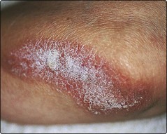
Diagnosis: psoriasis (p. 28).
Past medical history
Patients must be asked about any previous skin disease or atopic symptoms, such as hay fever, asthma or childhood eczema. Internal medical disorders may be relevant; these can involve the skin directly or may be associated with certain skin diseases. Prescribed or self-administered drugs may also cause an eruption. Dietary history is occasionally important, e.g. in some patients with atopic eczema (p. 36), but diet is often erroneously blamed for skin disease.
Case history 2
A 29-year-old woman was referred from the department of respiratory medicine where she had recently been diagnosed as having pulmonary sarcoidosis. Three weeks previously, she had developed tender, warm erythematous nodules (Fig. 2) on the shins. She was on no medication. An incisional biopsy confirmed the clinical impression.
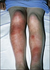
Fig. 2 Erythema nodosum on the lower legs.
From Weller R, Hunter JAA, Savin J, Dahl, M 2009 Clinical Dermatology, 4th Edn, Wiley-Blackwell, with permission.
Diagnosis: erythema nodosum (p. 83).
Social and occupational history
Many social factors can cause, influence or be influenced by a patient’s skin complaint. Occupational factors can induce contact dermatitis or other skin changes, and it is often necessary to ask the patient to explain exactly what he or she does. If the eruption improves when the patient is away from work, occupational factors should be suspected. Hobbies may also involve contact with objects or chemicals that could produce contact dermatitis.
Knowledge of the patient’s living conditions and home background can be helpful in understanding a problem and deciding on treatment. Alcohol intake should be noted (especially if the use of potentially hepatotoxic drugs is being considered), as well as other factors. Living or travelling in warm climates potentially exposes an individual to a wide range of tropical and subtropical infections, and to strong sunlight.
Case history 3
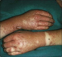
A 45-year-old male printer engineer gave a 6-month history of hand dermatitis (Fig. 3). A few months previously, he had started to use the solvent trichloroethylene in his job. Patch testing was negative. On substituting a different solvent, the eruption cleared.
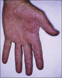
Diagnosis: irritant contact dermatitis (p. 34).
Family history
A full family history is essential. Some disorders with prominent skin signs are genetically inherited, e.g. tuberous sclerosis. Others, such as psoriasis or atopic eczema, have a strong hereditary component. In addition to genetic syndromes, a family history may reveal that other family members have had a recent onset of an eruption similar to that of the patient, suggesting an infection or infestation. It is sometimes also necessary to enquire about sexual contacts.
Case history 4
An 18-year-old male student gave a 3-month history of an intensely itchy papular eruption affecting the hands, wrists and penis (Table 1). Several lesions were excoriated (Fig. 4). Treatment with a potent topical steroid was of little benefit. His girlfriend had also recently developed itchy lesions. Close examination showed burrows in the skin.
Table 1 Itchy eruption: diagnosis
| Symptom | Intensely itchy eruption |
|---|---|
| Possible diagnosis |
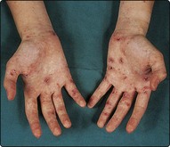
Diagnosis: scabies (p. 63).
Case history 5
A 25-year-old female shop assistant complained of brownish macules over her back (Fig. 5) and chest, which had first appeared in childhood and had gradually increased in number and size. During her teens, she had developed several soft pinkish, painless nodules on the trunk, some of which had become pedunculated. Her father had developed a few similar nodules in later life, and one of her two brothers had brown patches on his skin.
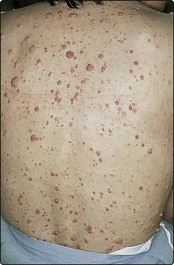
Diagnosis: von Recklinghausen’s neurofibromatosis (NF-1; p. 92).
Drug history
Both prescribed and self-administered medicaments can result in a ‘drug eruption’. Almost all patients try an over-the-counter topical preparation (or a friend or relative’s ointment) on rashes, and many have had a variety of treatments prescribed that may be inappropriate or may cause irritant or allergic reactions. It is important to quiz the patient about all medicament use, including the use of over-the-counter tablets or creams that the patient may well not think relevant.
Cosmetics, cleansing wipes and moisturizing creams can cause dermatitis, and it is often necessary to ask specifically about their use.
Case history 6
A 68-year-old woman had a minor irritating eruption on her forehead. She applied an antihistamine-containing cream that she bought in a pharmacy. Within 24 hours of applying it, her face became severely swollen (Fig. 6). Patch testing carried out later showed an allergic reaction to the cream.
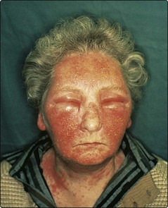
Diagnosis: medicament dermatitis (p. 35).
Case history 7
An 18-year-old female secretary was given griseofulvin for a fungal infection. She went sunbathing and, 12 hours later, developed an eruption with a distribution in light-exposed areas (Fig. 7).
Diagnosis: phototoxic drug eruption (p. 47).
Taking a history
 Elicit the nature and temporal course of the eruption or lesion.
Enquire about atopic symptoms, general medical conditions and foreign travel.
Take a social, occupational and family history – it may be relevant. Ask about eczema or psoriasis in relatives.
Identify any influence of the illness on day-to-day functions including work.
Record the recent use of drugs and medications, including topical agents.
Ask about the use of cosmetics and, in relevant cases, exposure to sun or ultraviolet radiation (e.g. sunbeds).