Fungal infections
Fungal infection in humans is common and mainly due to two groups of fungi:
These are usually confined to the stratum corneum, but deep mycoses invade other tissues (p. 61). Pityriasis versicolor, due to the yeast Malassezia (previously Pityrosporum ovale) is described on page 42.
Dermatophyte infections
Dermatophyte fungi reproduce by spore formation. They infect the stratum corneum, nail and hair, and induce inflammation by delayed hypersensitivity or by metabolic effects. There are three asexual genera:
Thirty species are pathogenic in humans. Zoophilic species (transmitted to humans from animals), e.g. Trichophyton verrucosum (Fig. 1), produce more inflammation than anthropophilic (human only) species.
Pathology
Dermatophytes inhabit keratin as branching hyphae, identifiable on microscopy. Skin scrapings, placed on a slide with 10% aqueous potassium hydroxide (to separate the keratinocytes) and a coverslip, are examined microscopically for hyphae (p. 20). The dermatophyte is identified by culturing the scrapings on medium (e.g. Sabouraud’s) for 3 weeks.
Clinical presentation
Tinea (Latin: worm) denotes a fungal skin infection which is often annular. The exact features depend on the site. The various presentations include the following:
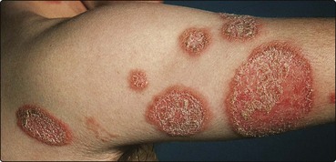
 Tinea corporis (trunk and limbs). Single or multiple plaques, with scaling and erythema especially at the edges, characterize this presentation. The lesions enlarge slowly, with central clearing, leaving a ring pattern, hence ‘ringworm’ (Figs 1 and 2). Pustules or vesicles may be seen.
Tinea cruris (groin). This is more common in men and is often seen in athletes (‘jock itch’), who may also have tinea pedis. It spreads to the upper thigh but rarely involves the scrotum. The advancing edge may be scaly, pustular or vesicular. Causative organisms are shown in Table 1.
Tinea incognito. Fungal infection can be modified in appearance and spread by the anti-inflammatory effect of a topical steroid.
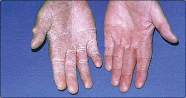
Tinea manuum (hand). Typically, this appears as a unilateral, diffuse powdery scaling of the palm (Fig. 3). Trichophyton rubrum is often the cause. Tinea pedis may coexist.
Tinea capitis (scalp/hair). See pages 66 and 114.
Tinea unguium (nails). See page 68.
Table 1 Superficial mycoses: causative organisms and differential diagnosis
| Area | Commonest organism | Differential diagnosis |
|---|---|---|
| Body/limbs (corporis) | T. verrucosum, M. canis, T. rubrum | Discoid eczema, psoriasis, pityriasis rosea |
| Feet (pedis) | T. rubrum, T. interdigitale, E. floccosum | Contact dermatitis, psoriasis, pompholyx, erythrasma |
| Groin (cruris) | T. rubrum, E. floccosum, T. interdigitale | Intertrigo, candidiasis, erythrasma |
| Hand (manuum) | T. rubrum | Chronic eczema, psoriasis, granuloma annulare |
| Nail (unguium) | T. rubrum, T. interdigitale | Psoriasis, trauma, candidiasis |
| Scalp (capitis) | M. canis, M. audouinii, T. tonsurans, T. schoenleinii | Alopecia areata, psoriasis, seborrhoeic eczema, furunculosis |
Athlete’s foot (p. 114) is common in adults (especially young men), rare in children and predisposed to by communal washing, swimming baths, occlusive footwear and hot weather. Itchy interdigital maceration, usually of the fourth/fifth toeweb space, is most frequent, but diffuse ‘moccasin’ involvement is seen. Recurrent vesicles also occur, sometimes with pompholyx as an id reaction. The commonest organisms are T. rubrum, T. mentagrophytes var. interdigitale and Epidermophyton floccosum.
The differential diagnoses of superficial mycoses are shown Table 1. Microscopy and culture of skin scrapings are often helpful. Wood’s ultraviolet light examination is used for tinea capitis, especially for screening during outbreaks. Hair infected by Microsporum audouinii and M. canis fluoresces green, but Trichophyton tonsurans does not fluoresce.
Management
Humid and sweaty conditions, including occlusive footwear, should be minimized, and dusting powder may help to keep the feet or body folds dry. Minor fungal infections respond to topical treatments, but widespread involvement or diseases of the nails or scalp requires systemic therapy.
Topical therapy
Tinea corporis, tinea pedis and tinea cruris respond to topical imidazole (e.g. clotrimazole and miconazole) creams, sprays or powders. Terbinafine cream once daily is often effective. Amorolfine, applied once weekly, produces a 40–50% cure for tinea unguium of one or two nails; similarly tioconazole may be used topically.
Systemic therapy
Tinea capitis, tinea manuum, tinea unguium and extensive tinea corporis often require systemic treatment. Griseofulvin is still the licensed choice for tinea capitis in children (10 mg/kg daily for 1–2 months) but, for other indications, it has largely been superseded by the newer antifungals, terbinafine (Lamisil, often used off-licence) and itraconazole (Sporanox), which show greater efficacy, have fewer side-effects and require shorter treatments.
Terbinafine 250 mg daily or itraconazole 100 mg daily may be used for tinea capitis, corporis, cruris, manuum and pedis, given for 2–4 weeks. In tinea unguium, terbinafine (250 mg daily for 6–12 weeks) is the drug of choice; itraconazole (200 mg daily for 12 weeks or as ‘pulsed’ courses) is an alternative. In the elderly, uncomplicated fungal toenail infection may not require any therapy. Itraconazole can potentially cause hepatotoxicity and requires cautious use in heart failure.
Ketoconazole (Nizoral) by mouth, although effective, is limited in use by hepatotoxicity.
Candida albicans infection
Candida albicans is a ubiquitous commensal of the mouth and gastrointestinal tract that can produce opportunistic infection. Predisposing factors include:
immunosuppression (p. 57)
Clinical presentation
In infection, hyphal forms of C. albicans are seen in the stratum corneum. Infection may present as the following:
Genital. Thrush commonly appears as an itchy, sore vulvovaginitis. White plaques adhere to inflamed mucous membranes, and a white vaginal discharge may occur. Males develop similar changes on the penis. It can be spread by sexual intercourse.
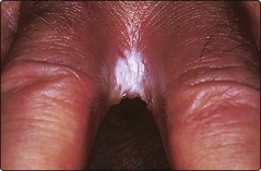
Intertrigo. Superinfection with C. albicans, and often also with bacteria, gives a moist, glazed and macerated appearance to the submammary, axillary or inguinal body folds. The interdigital clefts are involved (Fig. 4) in wet workers who do not dry their hands properly.
Mucocutaneous candidiasis. This rare, sometimes inherited disorder of immune deficiency starts in infancy. Chronic C. albicans intertrigo with nail and mouth infections is seen.
Oral. White plaques adhere to an erythematous buccal mucosa. Broad-spectrum antibiotics, false teeth and poor oral hygiene predispose. Angular stomatitis may coexist.
Paronychia. See page 68.
Systemic. Systemic candidiasis can occur in immunosuppressed patients. Red nodules are seen in the skin.
Management
Candida albicans infections must be differentiated from other conditions (Table 2). General measures are important. Body folds are separated and kept dry with dusting powder. Hands are dried carefully (p. 38) and oral hygiene improved. Systemic antibiotics may need to be stopped. Specific agents against Candida are used topically and systemically.
Table 2 Differential diagnosis: C. albicans infections
| Variant | Differential diagnosis |
|---|---|
| Genital | Psoriasis, lichen planus, lichen sclerosus |
| Intertrigo | Psoriasis, seborrhoeic dermatitis, bacterial secondary infection |
| Oral | Lichen planus, epithelial dysplasia |
| Paronychia | Bacterial infection, chronic eczema |
Topical therapy
Imidazoles are effective and available as creams, powders, pessaries and lotions. For oral candida, use amphotericin, nystatin or miconazole as lozenges, suspension or gels.
Systemic therapy
Bowel carriage may be reduced in recurrent candidiasis by oral nystatin. Itraconazole 100 mg daily or fluconazole 50 mg daily, but not griseofulvin, can be given as a short course for persistent C. albicans infections and in the long term for mucocutaneous candidiasis. Vaginal candidiasis is treated by a single dose of 500 mg clotrimazole or 150 mg econazole as a pessary, or with itraconazole or fluconazole by mouth. C. glabrata is increasingly identified and is frequently resistant to fluconazole.
Fungal infections
Dermatophytes infect the feet, groin, body, nails, hands and scalp. The commonest dermatophyte pathogens are Trichophyton rubrum, T. mentagrophytes var. interdigitale and Epidermophyton floccosum.
Topical imidazoles and oral terbinafine or itraconazole are effective for most dermatophyte infections.
C. albicans produces opportunistic infection of the body folds, mouth, genitals and nail fold. These are predisposed to by humidity, obesity, diabetes and oral antibiotic therapy.