Cutaneous T cell lymphomas and malignant dermal tumours
Cutaneous T cell lymphoma (CTA) is the most common type of skin lymphoma, with an incidence of 0.6 per 100 000. B cell lymphoma of the skin is rare. Malignant tumours of the dermis are infrequent. The commonest are secondary deposits (p. 88) Kaposi’s sarcoma (p. 56) and a malignancy of dermal fibroblasts (dermatofibrosarcoma).
Cutaneous T cell lymphoma (mycosis fungoides)
CTCL describes a lymphoma that evolves in the skin, although extracutaneous T cell tumours often produce secondary skin deposits. CTCL is a slowly progressive tumour of epidermotropic CD3+, CD4+ T lymphocytes that becomes systemic only in its terminal stage.
Clinical presentation
The course is usually protracted, although it is occasionally more rapidly progressive. The diagnosis is often delayed for some years as, in its initial stages, CTCL may resemble eczema or ‘chronic superficial dermatitis’ (p. 43). CTCL can be regarded as having four stages:
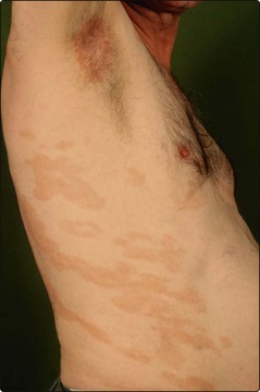
1. Patch stage. Describes small, scaly, slightly raised erythematous patches, typically on the trunk, that can resemble eczema (Fig. 1). It may persist for 10 years or more. Occasionally, the skin becomes atrophic, pigmented and telangiectatic (poikiloderma).
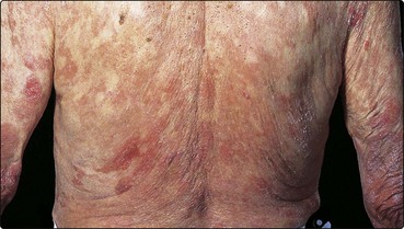
2. Infiltrated plaques. Fixed plaques develop, usually on the trunk but sometimes more widely distributed (Fig. 2). This stage may last for years.
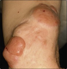
3. Tumour stage. This later phase, characterized by tumorous nodules or ulcers within the plaques, has a 5-year survival of 40–65% (Fig. 3).
4. Systemic disease. Involvement of lymph nodes or internal organs is a late finding. The Sézary syndrome (p. 44) is a variant.
Differential diagnosis
Chronic superficial dermatitis, undifferentiated eczema and psoriasis are the main differential diagnoses. Those cases of chronic superficial dermatitis that show larger patches are said to be more likely to progress to CTCL. CD30+ lymphoproliferative disorders represent a subgroup of skin lymphomas. They present either as multiple nodules that heal with scarring and follow a protracted course (lymphomatoid papulosis) or as large cell lymphoma, in which ulcerated nodules appear on the trunk.
Management
Diagnosis relies on matching the clinical and pathological appearances T cell receptor gene analysis demonstrates clonality of the lymphocytic infiltrate. Current treatment is not curative but aimed at controlling the lymphoma. The patch-stage lesions often improve with moderately potent topical steroids and ultraviolet (UV) B therapy. More infiltrated plaques require PUVA or topical nitrogen mustard. Localized lesions respond to conventional radiotherapy. Advanced CTCL can be treated with extracorporal photopheresis (p. 114), electron beam therapy, oral bexarotene (p. 114), or combined chemotherapy.
Cutaneous T cell lymphomas and malignant dermal tumours
 Definition: CTCL is an uncommon tumour resulting from infiltration of the skin by malignant clonal T lymphocytes.
Stages: CTCL progresses through patch stage to indurated plaques, and then to tumours and systemic involvement.
Treatment: therapy depends on the stage and extent of the disease. Localized patch stage CTCL responds to topical steroids and phototherapy. In more advanced disease, phototherapy, radiotherapy and oral bexarotene or chemotherapy may be prescribed.
Malignant dermal tumors are uncommon. Secondary deposits, Kaposi’s sarcoma and other sarcomas can be found.