Paediatric dermatology
Some conditions are almost exclusive to childhood (e.g. napkin dermatitis and juvenile plantar dermatosis), and others are more common in children (e.g. atopic eczema or viral exanthems). The common childhood dermatoses not mentioned elsewhere are detailed here along with some rare but important disorders.
Childhood eczemas and related disorders
Forms of eczema found in childhood include:
 infantile seborrhoeic dermatitis
napkin psoriasis (p. 29)
atopic eczema (p. 36)
pityriasis alba (p. 43).
Napkin (diaper) dermatitis
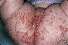
Napkin dermatitis is the commonest type of napkin eruption. It is usually seen in infants who are only a few weeks old, and is rare after the age of 12 months. It is an irritant dermatitis due to the macerating effect of prolonged contact of the skin with faeces and urine. A glazed erythema is seen in the napkin area, sparing the skin folds. Erosions or ulceration may follow (Fig. 1), and hypopigmentation is a complication in pigmented skin. Secondary bacterial or Candida albicans infection is frequent, and the latter may account for the development of erythematous papules or pustules.
The differential diagnosis is from infantile seborrhoeic eczema and candidiasis, both of which tend to affect the flexures. The treatment of napkin dermatitis is aimed at keeping the area dry. The use of disposable super-absorbent nappies helps, as may more frequent changes. A bland white soft paraffin is used, with aqueous cream as a soap substitute, and a silicone-based cream (e.g. Drapolene) may have a protective action. Topical 1% hydrocortisone, with an antifungal (e.g. Daktacort or Canesten-HC), is also effective.
Infantile seborrhoeic eczema
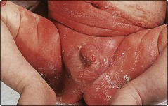
Infantile seborrhoeic eczema starts in the first few weeks of life and tends to affect the body folds, including the axillae, groin and neck, but it also may involve the face and scalp. Flexural lesions present as moist, shiny, well-demarcated scaly erythema (Fig. 2), but a yellowish crust is often found on the scalp. The condition can usually be differentiated from napkin dermatitis (which spares the flexures), candidiasis (which is usually pustular) and atopic eczema (which is more pruritic, although differentiation can be difficult in some cases). Infantile seborrhoeic eczema is treated by emollients and 1% hydrocortisone ointment, or with a hydrocortisone–antifungal combination. Scalp lesions respond to 2% ketoconazole shampoo. Olive oil will help to soften the scalp scales of cradle cap.
Candidiasis
Infection with C. albicans is relatively common in the neonatal period. The organism can also secondarily complicate infantile seborrhoeic eczema or napkin dermatitis. Erythema, scaling and pustules are seen, often involving the flexures, and there may be satellite lesions. Treatment is with a topical anticandidal agent, e.g. 2% ketoconazole cream, and 2% miconazole gel orally.
Juvenile plantar dermatosis
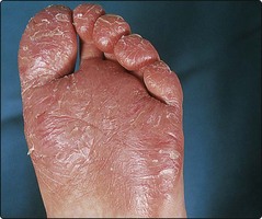
Juvenile plantar dermatosis, first recognized in 1968, presents with red, dry, fissured and glazed skin, principally over the forefeet but sometimes involving the whole sole (Fig. 3). It usually starts in the primary school years and resolves spontaneously in the early to mid teens. The condition is thought to be linked to the wearing of socks and shoes made from synthetic materials, although it may be a manifestation of atopy in some children. It is usual to advise cotton socks and less occlusive footwear, preferably made of leather. Topical steroids are ineffective but emollients help.
Other childhood dermatoses
Some uncommon but characteristic eruptions are found in childhood. These include:
Kawasaki disease and other viral infections (p. 53)
ichthyosis (p. 90)
epidermolysis bullosa (p. 91).
Urticaria pigmentosa
Urticaria pigmentosa is characterized by multiple reddish–brown macules or papules on the trunk and limbs of an infant. The lesions may become red, swollen and itchy after a bath or when rubbed, and blistering may occur. Histologically, there are accumulations of mast cells in the dermis. The disorder normally resolves spontaneously before adolescence. There is a form with a later onset, usually beginning in adolescence or adult life, which rarely resolves and may involve internal organs – something that is rare in the childhood variety.
Langerhans cell histiocytosis (histiocytosis X)
Langerhans cell histiocytosis is a rare and serious condition that normally involves internal organs. The skin signs are common, variable and include a seborrhoeic-like dermatitis, papules or pustules on the trunk and ulceration, particularly of the flexures. The skin, abdominal organs, lungs and bones are infiltrated by clonal Langerhans cells, which may behave in a malignant fashion, although the condition is believed to be reactive and not a true malignancy. Skin biopsy is usually diagnostic. The prognosis is poorer when the onset is before 2 years of age.
Vascular naevi
Vascular naevi are common and are present at birth or develop soon after. Superficial lesions are due to capillary networks in the upper or mid dermis, but larger angiomas show multiple vascular channels in the lower dermis and subcutis.
Clinical presentation
There are four main clinical pictures, which are described below.
Salmon patch
This is the commonest vascular naevus, seen in 20–60% of neonates. Patches at the upper eyelid fade quickly, but the ‘stork mark’ at the posterior neck persists in 20–30% of cases. Parents should be reassured: no investigation or treatment is required.
Port wine stain naevus
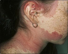
Present at birth, the port wine stain (or naevus flammeus) is an irregular red or purple macule that often affects one side of the face (Fig. 4), although other sites can be affected. Lesions vary from millimetres to centimetres in diameter. In middle age, it can darken and become lumpy. A port wine stain involving the ophthalmic division of the trigeminal nerve may have an associated intracranial vascular malformation (the Sturge–Weber syndrome). Port wine stains near the eye can be associated with glaucoma.
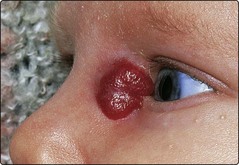
Infantile haemangioma
Infantile haemangiomas appear shortly after birth. Most occur on the head or neck and grow to reach a maximum in the first 12 months (Fig. 5). They remain static for the next 6–12 months and then involute. Most cases will have regressed by the age of 5–7 years, leaving an area of atrophy. Infantile haemangiomas are classified as superficial (e.g. strawberry naevus), deep or mixed, and can be localized or segmental. The deep type, e.g. cavernous haemangioma, is composed of larger and deeper vascular channels and presents as a nodular bluish swelling. The overlying skin may be normal or show a superficial vascular component (i.e. be mixed). Regression is not as complete as in the superficial type. Ulceration with bleeding and secondary infection can develop. Large haemangiomas may trap platelets and cause thrombocytopenia (the Kasabach–Merritt syndrome). In segmental haemangiomas, abnormalities in the underlying organs should be suspected.
Management
Port wine stains may be covered with camouflage cosmetics (p. 108), but treatment is now available with the flashlamp-pulsed dye laser (p. 112), which obliterates the abnormal dermal vessels and improves the appearance. A child with a facial port wine stain needs neurological and ophthalmic assessment. Infantile haemagiomas should be allowed to involute unless they compromise vital structures such as the eye or airway. In this case, a short course of propranolol, prednisolone or even emergency surgery is needed. The Kasabach–Merritt syndrome is treated in a similar fashion. Haemangiomas at the lower back may have associated tethering of the spinal cord; a neurological assessment and imaging are indicated. An AVM requires the opinion of a vascular surgeon.
Paediatric dermatology
| Disorder | Age at onset | Clinical features |
|---|---|---|
| Napkin dermatitis | First few weeks to 12 months | Glazed erythema that spares body folds |
| Erosions may occur | ||
| Infantile seborrhoeic eczema | First few weeks | Moist scaly erythema |
| Flexures and scalp affected | ||
| Candidiasis | Infancy | Erythema, with scaling and pustules |
| Flexures affected | ||
| Secondary infection found | ||
| Juvenile plantar dermatosis | School age to mid teens | Glazed red fissured skin on the forefeet and soles |
| Urticaria pigmentosa | Mostly at 3–9 months | Reddish–brown macules or papules on trunk, which urticate when rubbed |
| Langerhans cell histiocytosis | All ages (different types) | Seborrhoeic-like dermatitis, papules/pustules, ulceration |
| Vascular naevi | At birth, in first few weeks | Salmon patch on neck, port wine naevus (e.g. on face), strawberry naevus |