Chapter 18 Fusobacteria, Leptotrichia and spirochaetes
Fusobacteria are non-sporing, anaerobic, non-motile, non- or weakly fermentative, spindle-shaped bacilli (with fused ends: hence the name). They are normal inhabitants of the oral cavity, colon and female genital tract and are sometimes isolated from pulmonary and pelvic abscesses. Fusospirochaetal infections, which they cause in combination with spirochaetes, are noteworthy. Fusobacterium nucleatum (the type species), Fusobacterium periodontium and Fusobacterium simiae are isolated mainly from periodontal disease sites, and others such as Fusobacterium alocis and Fusobacterium sulci are sometimes found in the healthy gingival sulcus. Non-oral species include Fusobacterium gonidiaformans, Fusobacterium russii and Fusobacterium ulcerans.
Fusobacteria
Fusobacterium nucleatum
Habitat and transmission
Several subspecies of F. nucleatum have been identified in different habitats. These include F. nucleatum subsp. polymorphum, found in the healthy gingival crevice, and F. nucleatum subsp. nucleatum, recovered mainly from periodontal pockets. A third subspecies is F. nucleatum subsp. vincentii. Infections are almost invariably endogenous.
Characteristics
Gram-negative, strictly anaerobic, cigar-shaped bacilli with pointed ends (Fig. 18.1). Cells often have a central swelling. A Gram-stained smear of deep gingival debris obtained from a lesion of acute ulcerative gingivitis is a simple method of demonstrating the characteristic fusobacteria, together with spirochaetes and polymorphonuclear leukocytes (Fig. 18.2). These, together with the clinical picture, confirm a clinical diagnosis of acute ulcerative gingivitis.
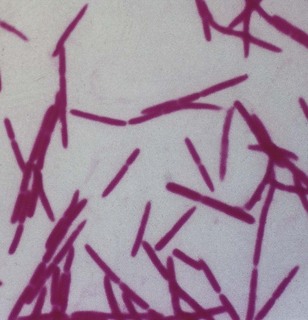
Fig. 18.1 A photomicrograph of fusobacteria showing characteristic Gram-negative, cigar-shaped cells with pointed ends.
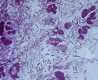
Fig. 18.2 A Gram-stained smear obtained from deep gingival plaque of a patient with acute ulcerative gingivitis (see also Fig. 33.6) showing the fusospirochaetal complex. Note: the large cells are polymorphs.
Culture and identification
Grows on blood agar as dull, granular colonies with an irregular, rhizoid edge. Biochemical reactions and the acidic end products of carbohydrate metabolism help identification. As fusobacteria can remove sulphur from cysteine and methionine to produce odoriferous hydrogen sulphide and methylmercaptan, they are thought to be associated with halitosis.
Pathogenicity
The endotoxin of the organism appears to be involved in the pathogenesis of periodontal disease. It possesses remarkable adherence properties and the fusobacterium adhesin A (FadA), which confers this property has recently been isolated. F. nucleatum is usually isolated from polymicrobial infections; it is rarely the sole pathogen. Thus, in combination with oral spirochaetes (Treponema vincentii and others), it causes the classic fusospirochaetal infections. These are:
As fusobacteria coaggregate with most other oral bacteria, they are believed to be important bridging organisms between early and late colonizers during plaque formation (see Fig. 31.3).
Leptotrichia
Leptotrichia spp. are oral commensals previously thought to belong to the genus Fusobacterium. They are Gram-negative, strictly anaerobic, slender, filamentous bacilli, usually with one pointed end. Leptotrichia buccalis, present in low proportions in dental plaque, is the sole representative of this genus.
Spirochaetes
Spirochaetes are a diverse group of spiral, motile organisms comprising five genera. Of these, three genera are human pathogens:
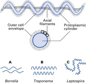
Spirochaetes are helical organisms with a central protoplasmic cylinder surrounded by a cytoplasmic membrane (Fig. 18.3). The cell wall is similar to Gram-negative bacteria but stains poorly with the Gram stain. Underneath the cell wall run three to five axial filaments that are fixed to the extremities of the organism. Contractions of these filaments distort the bacterial cell body to give it its helical shape. The organism moves either by rotation along the long axis or by flexion of cells. Because of their weak refractile nature, dark-ground microscopy is used to visualize these organisms in the laboratory, although immunofluorescence is more useful for identification purposes. All spirochaetes are strictly anaerobic or microaerophilic.
Treponema
The coils of Treponema are regular, with a longer wavelength than that of Leptospira (Fig. 18.3). A number of species and subspecies are recognized, some of which are important systemic pathogens, while others are oral inhabitants implicated in periodontal disease.
Treponema pallidum
Characteristics
Slender, corkscrew-shaped cells with 6–12 evenly spaced coils, 6–14 × 0.2 µm; too slender to visualize by light microscopy but can be seen by silver impregnation or immunofluorescent techniques; strictly anaerobic and extremely sensitive to drying and heat, hence dies rapidly outside the body.
Culture and identification
Cannot be cultured in vitro, but can be propagated in the testes of rabbits; Treponema pallidum thus harvested can be used as antigens to detect specific antibody in the patient’s serum.
Dark-ground microscopy of tissue fluid from primary and secondary clinical lesions helps identification, but serological tests are the mainstay of diagnosis.
Pathogenicity
Causes syphilis, a sexually transmitted disease with protean manifestations (see Chapter 27). The virulence factors of T. pallidum are not well characterized. Immunopathology plays a significant role in disease manifestations, especially in the late (tertiary and quaternary) stages of the disease.
Treponema pallidum subsp. pertenue
The agent of yaws, characterized by chronic, ulcerative, granulomatous lesions of skin, mucosae and bone. The disease, widespread in the tropics, is spread by direct contact.
Treponema carateum
The agent of pinta, a non-venereal skin infection characterized by depigmented and hyperkeratotic skin. The disease affects mainly dark-skinned natives of Central and South America and the West Indies.
Oral treponemes
All oral spirochaetes are classified in the genus Treponema. Although many species have been described, only four have been cultivated and maintained reliably: Treponema denticola, Treponema vincentii, Treponema pectinovarum and Treponema socranskii. In another classification, they are categorized according to cell size as small, medium and large spirochaetes.
Habitat and transmission
Predominantly, the oral cavity of humans and primates, at the gingival margin and crevice in particular. Transmission routes are unknown. Infections are endogenous.
Characteristics
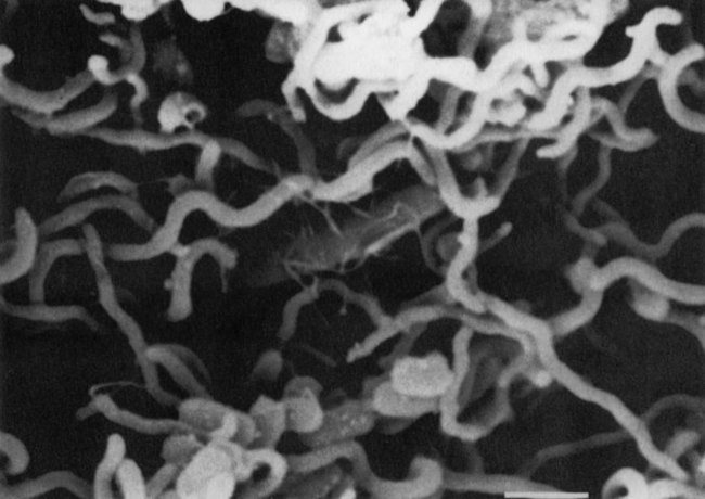
Motile, helical rods, 5–15 × 0.5 µm, with irregular (three to eight) spirals, which are less tightly coiled than, for instance, T. pallidum (Figs 18.3 and 18.4). Cell walls are Gram-negative but stain poorly. The size is variable and can be used as a basis for classification (large, medium or small).
Culture and identification
In contrast to T. pallidum, oral spirochaetes can be grown in vitro. They are strict anaerobes, slow-growing in oral treponema isolation (OTI) medium. Subspecies can be differentiated by fermentation reactions and serology (agglutination).
Suspect lesions of acute necrotizing ulcerative gingivitis or advanced periodontitis can be examined by obtaining a Gram-stained smear of deep gingival plaque and visualizing the characteristic fusospirochaetal complex under light microscopy (see Fig. 18.2); alternatively, dark-ground microscopy may be used.
Pathogenicity
These organisms are a component of the fusospirochaetal complex of acute necrotizing ulcerative gingivitis and Vincent’s angina, and are a coagent of advanced periodontal disease. The ability to travel through viscous environments enables oral spirochaetes to migrate within the gingival crevicular fluid and to penetrate sulcular epithelial linings as well as gingival connective tissue. Virulence factors are little known; endotoxin is possibly contributory to disease. T. denticola is more proteolytic than other species and degrades collagen and dentine.
Borrelia
Borrelia burgdorferi
Habitat and transmission
Found in ticks and small mammals, particularly deer. Transmission is by a tick vector.
Characteristics
This species is a helical spirochaete, 0.18–0.25 × 4.3 µm. Gram-negative, it grows under microaerophilic conditions at 34°C. Identification is by serology and immunofluorescence or enzyme-linked immunosorbent assay (ELISA).
Leptospira
Leptospira biflexa and Leptospira interrogans are the recognized species, each of which comprises a number of serogroups.
These organisms are found in damp environments such as stagnant water and wet soil. The kidneys of some rodents and domestic animals act as a reservoir for L. interrogans. The urine of these animals serves as a vehicle of transmission of human leptospirosis, the symptoms of which vary from mild febrile illness to fatal attacks of jaundice and renal failure.
Key facts
Bolstad A.I., Jensen H.B., Bakken V. Taxonomy, biology and periodontal aspects of Fusobacterium nucleatum. Clinical Microbiology Reviews. 1996;9:55-71.
Duerden B.I., Drasar B.S., editors. Anaerobes in human disease. London: Edward Arnold, 1991.
Greenwood D., Slack R., Peutherer J., editors. Medical microbiology, 16th ed., Edinburgh: Churchill Livingstone, 2003. Chs 37 and 38
Review questions (answers on p. 353)
Please indicate which answers are true, and which are false.