Chapter 110The Racing Quarter Horse
History and Description of the Sport
Racing Quarter Horses (QHs) are the sprinters of the racing world, competing at distances of 220 yards (one furlong, 201 meters) to 870 yards (795 meters). The explosive power exerted while leaving the starting gate and the ability to attain speeds of more than 50 mph (80 km/h) to run 440 yards (402 meters) in less than 21 seconds distinguishes the QH from other racing breeds. The American QH originates from colonial Virginia in the 1600s where imported English Thoroughbreds (TBs) were crossed with “native” breeds of Spanish descent to produce a compact, heavily muscled horse that excelled at running short distances. They were known as Quarter Pathers or Quarter Milers, named after the quarter-mile distance at which they excelled. The first race records came from Enrico County, Virginia, in 1674, where match races were run down village streets and in small level fields. Gambling on races was popular at the time, with plantations changing hands over lost bets.1
When settlers moved west with their horses, racing grew along with the popularity of the breed recognized for its versatility for ranch work and innate “cow sense.” The first official QH racetrack was Rillito Park in Tucson, Arizona, opening in 1943. The American Quarter Horse Association (AQHA) was formed in 1940 to register and preserve the breed that presently represents the largest breed association in the world with more than 3 million members. QH racing is now nationwide and spans over five countries on three different continents. In North America alone, $123 million in purse money was paid out in 2007.1 Many racing QH owners, trainers, and jockeys raise horses or participate in Western performance events (roping, cutting, reining, showing, and rodeo events), and they take deep pride in the accomplishments of their horses. Winning the All American Futurity, held each year on Labor Day at Ruidoso Downs, Ruidoso, New Mexico and featuring a $2 million purse ($1 million going to the winner), has been the dream of most everyone in the industry. Los Alamitos Racetrack, a predominantly QH track in southern California, offers at least five races during the year with high dollar purses, with the Los Alamitos Two Million Futurity being the richest. The Champion of Champions race held at the end of the racing year is the most prestigious race for older horses, representing winners of the top races during the year.
Emphasis in the sport is on 2-year-old racing (Futurities), requiring qualifying heats where horses with the 10 fastest times compete in a final 2 weeks later. Horses must be nominated to Futurities with periodic payments to maintain eligibility. The same applies to Derbies held for 3-year-olds, but the purse money is less than for 2-year-olds. Yearling sale prices are driven by precocious, nicely conformed, and well-bred individuals to compete in these lucrative Futurities at 2 years of age and then Derbies the following year.
The AQHA Racing Challenge is a program that was designed to provide more racing opportunities for older American QHs. At this time, 59 races take place in 11 regions across the United States, Canada, Mexico, and South America throughout the year. The horses compete in one of six different types of races, depending on age and ability. The series culminates with a championship night held in different locations.1
The AQHA stud book has remained open to the breeding of TBs ever since the QH breed was formally established. Breeding to TBs is a useful outcross to expand an otherwise small gene pool and maintain the classic quarter-mile distance of 440 yards because some QHs run best at even shorter distances of 300 to 350 yards. Unlike the Jockey Club, artificial insemination is the norm for breeding, and embryo transfer is popular.
Training the Racing Quarter Horse
Horses are usually broken in the latter part of the yearling year to prepare for racing as 2-year-olds. They are not allowed to race before March of the 2-year-old year, and they are restricted from racing 440 yards until later in the year.2 The early races are very short (220 to 300 yards), and the training schedule is light compared with the racing TB. Most of the 2-year-old QHs are very precocious, big bodied, and naturally fast. They can perform well with a low level of fitness, possibly a risk factor for injury. Once they are fit, they gallop fewer days than does a TB, and fitness in many older racehorses is maintained using a mechanical horse walker at the barn. To race for the first time, the horse must have two satisfactory morning works (qualifying). They generally work from the gate in “sets” of two horses at a time for a specific distance.
Lameness Related to Track Surface
QHs race on varying track surfaces around the country, but trainers prefer a firmer surface when possible because a loose or sandy surface poses problems with breaking at speed from the starting gate and getting enough traction for sprinting at high speeds. A new set of injuries is seen when racing on a sandier type of track, especially superficial digital flexor (SDF) tendonitis. Hindlimb lameness, muscle strains, back soreness, and suspensory injuries are also increased. The firmer track is favored, but it may lead to the high incidence of joint and bone injury seen as a result of greater concussive forces. No statistics are available at this time for QH injuries racing on the newer synthetic tracks.
Conformation Relating to Lameness
Conformational factors that may contribute to lameness in the forelimb are relative large body mass, poor carpal conformation (back at the knee and bench kneed), upright pasterns, and small feet. Major hindlimb conformational defects (sickle hocks, cow hocks, too straight in the stifles) are undesirable in the QH racehorse because breaking sharply from the starting gate is necessary to be competitive. Any serious hindlimb lameness may limit the horse’s usefulness.
Lameness Examination
There is no set procedure for lameness examination, but a systematic approach is essential for completeness. An efficient approach is required, however, because many trainers like to examine every horse before it is entered to race. Soundness is imperative for optimum racing performance in the QH breed because races are so short and therefore won or lost by photo finishes on a regular basis. Horses with lameness may be fractious in the starting gate, and those with hindlimb lameness may be slow to break from the gate. Close lameness monitoring is required to maintain QHs in peak condition, and intraarticular therapy is frequently used because of the high level of joint trauma associated with the speed and concussion in these elite athletes.
Watching the horse walk out of the stall and down the shed row is useful and a good time to obtain a history from the trainer or barn foreman. A QH with a sore carpus may be noted immediately by a characteristic wide placement of the limb while walking. History is very useful, such as “the horse was getting hotter than usual at the track this morning,” or “he is digging a hole in the stall and standing in it.” Large barns often keep records of lameness work on each horse, and it is always useful to quickly review past radiographs and joint injections. Observing the horse walk and jog on a hard level surface may accentuate the lameness and aid in diagnosis. Hoof testers should always be used to define foot pain. Joint flexion and palpation are particularly useful owing to the high prevalence of joint problems.
When flexing the carpus, if the limb is raised forward and upward so that the radius is in the horizontal position, the veterinarian may observe an immediate tell-tale withdrawal response of the neck and shoulder muscles as a response to pain. Relaxing the limb, the joints are palpated by placing the thumbs along the individual dorsal borders of the carpal bones while the fingers apply pressure behind the joint to further localize the pain causing lameness. Moving in a distal direction, the dorsal aspect of the metacarpal region, suspensory ligament (SL), digital flexor tendons, and any splints should be palpated for a painful response. The fetlock is flexed, and the suspensory branches are palpated. The distal interphalangeal (DIP) joint is palpated for heat and excessive joint effusion. The digital pulse amplitudes should always be checked because they frequently will be elevated in horses with acute problems of the foot. Excessive synovial effusion, tenderness to palpation, and heat or filling in a specific area of the limb should be noted. The horse’s back, loin, and gluteal muscles are briefly palpated and observed for asymmetry, and then the separate compartments of the stifle are palpated. The medial femorotibial joint is the most common area of soreness in the stifle and may or may not have effusion. A positive Churchill test may be indicative of hock soreness and is quickly performed before palpating the distal aspect of the limb. The history of a poor performance (especially leaving the gate) or recognition of hindlimb lameness at the initial jog usually prompts a more complete examination, with proximal and distal limb flexion tests.
Diagnostic blocks are used when necessary to localize the lameness (see Chapter 10).
Imaging Considerations
Because of the high incidence of joint injury, digital radiography is the most frequently used modality. The carpus and fetlock are the joints most commonly examined radiologically. Ultrasonographic examination is used for definition of suspensory, tendon, and joint injuries. Ultrasonographic examination is generally unrewarding for detection of proximal suspensory injuries in the QH. SDF tendonitis occurs with varying frequency, depending on the characteristics of the racing surface, and lesions vary widely in severity. Nuclear scintigraphy is used less than in the TB, but it is especially helpful in the diagnosis of tibial stress fractures.
Shoeing
The most serious shoeing problems in the QH are the same as in the TB: long toe, low excessively sloping heel, and medial-to-lateral hoof imbalance. Most racehorses are shod close to or on race day. Therefore corrective shoeing is not used as often as needed in many horses because of the risk of sore feet on race day. Horses race in aluminum shoes, and various kinds of pads are used for foot sore horses, including rim pads, wedge pads, full plastic pads, and even full aluminum pads with an assortment of different hoof packings. The most controversial topic at the moment is the use of toe grabs. Toe grabs have been associated with catastrophic injury in the racing TB, and use of these shoe additives is restricted in some states. Historically, many QH racehorses have raced with high toe grabs (>6 mm) to prevent stumbling and slipping from the starting gate and to maintain traction at high speeds. Now many QHs race without a toe grab or a 2-mm toe grab in front. Some leading trainers of QHs believe that the injury rate has been greater with the recent change to smaller toe grabs at the shorter sprinting distances.
A racetrack study including TBs, QHs, and other breeds showed that the prevalence of underrun heels was a significant risk factor for suspensory apparatus failure. The length of toe grabs was not shown to be significant in this study.3 Further investigation of toe grabs in relation to racing surfaces is necessary.
Specific Lameness Conditions
The following lameness conditions are the most relevant for the QH racing breed. Most of the topics are covered extensively in other chapters, and therefore this section is meant as a review of QH injuries and differences from those of the TB.
Synovitis of the Carpal and Metacarpophalangeal Joints
Synovitis of the carpus is the most frequent condition seen in the young racing QH. Back-at-the-knee conformation is common, and this predisposes to carpal injury during hyperextension of the joint while running. Many 2-year-olds have a large body mass and are inherently fast sprinters, reaching very fast speeds without much conditioning. Synovitis is characterized by heat and synovial effusion of the affected joints, with the absence of radiological changes. Lameness may be present but is generally not severe. Carpal flexion and palpation are used to localize the affected joints. Symptomatic treatment includes the use of ice, leg sweats or poultice, and nonsteroidal antiinflammatory drugs (NSAIDs). Intravenous hyaluronan (Legend, Bayer HealthCare, Animal Health Division, Shawnee Mission, Kansas, United States) or intramuscular polysulfated glycosaminoglycans (PSGAGs) (Adequan, Luitpold Pharmaceuticals Inc., Animal Health Division, Shirley, New York, United States) are often used as systemic treatment. Intraarticular therapy is very effective, with a good response from corticosteroids with or without hyaluronan.
The metacarpophalangeal joint of the forelimb is another frequent site of pain causing lameness in QHs. Heat and synovial effusion are the first signs of synovitis, along with a varying degree of lameness. The condition is often bilateral, and radiological examination is negative. In my opinion, capsulitis does not occur with the same frequency as in the TB because QHs in training do far less galloping than TBs; therefore much less stress is placed on the soft tissue structures of the metacarpophalangeal joint. The treatment is the same as for carpal synovitis. If the condition does not resolve with intraarticular therapy, or if it recurs rapidly, the training program should be altered, or the risk of further joint damage is likely, with osteoarthritis (OA) the end result. Many 2-year-olds are entered in numerous Futurities, so training revolves around these races. Trainers try to keep horses on schedule for these race dates, without sustaining injuries that jeopardize the racing careers or require extended layup periods.
Arthrosis of the Distal Interphalangeal Joint and Problems Associated with the Foot
DIP joint synovitis is a clinically significant cause of lameness in the QH.4 The breed is well known for having undersized feet in relation to the body size, and this, coupled with the tendency for racehorses to develop a long toe and an excessively sloping heel, probably leads to greater stresses to the foot than in other breeds. QHs also have short upright pasterns, and they race at higher speeds on a harder track surface. Bilateral forelimb lameness is seen that can be accentuated by jogging on a hard surface. The stride is shortened with a transfer of weight to the hindlimbs. Horses typically respond to the application of hoof testers over the central third of the frog (as in navicular syndrome). Increased digital pulse amplitude is usually evident, and DIP effusion may be palpated above the coronet in many horses. Younger horses (2- and 3-year-olds) with synovitis show a greater degree of localizing inflammatory signs than older horses with chronic OA.5
Intraarticular analgesia may be used to localize the pain causing lameness, and I use intraarticular analgesia in combination with intraarticular medication when necessary to confirm the diagnosis. Radiographs are frequently normal but may show some degree of radiolucent changes along the margin of the distal phalanx and, rarely, radiological changes associated with navicular syndrome. OA of the DIP joint occasionally will be evidenced by the presence of osteophytes involving the distal aspect of the middle phalanx or the extensor process of the distal phalanx. A generalized suspensory soreness, as well as soreness in the area of the bicipital bursa, is often palpated secondary to inflammation of the DIP joint. Back pain may also be associated with the presence of sore feet and can be detrimental to racing performance because of the horse’s reluctance to break sharply and extend its stride.4 These secondary clinical signs usually disappear after resolution of the foot soreness.
Antiinflammatory medications and corrective shoeing are used to treat DIP joint synovitis. The shoeing is in accordance to individual needs, most commonly backing up the shoe as much as possible and protecting the sole. A wide variety of pads are used, including rim pads, full pads, and aluminum pads with various sole packings. Wedge pads or shoes may be used to correct the low heel conformation, but care must be taken because often the heel pain is exacerbated. Some horses are trained in bar shoes until they are ready to race. NSAIDs (usually phenylbutazone) are used, and the feet are iced twice daily during the acute stage.
Intraarticular corticosteroids are effective in relieving pain. Betamethasone esters or triamcinolone acetonide, with or without hyaluronan, is preferred especially if frequent joint injection is necessary. In my experience, frequent use of methylprednisolone acetate (Depo-Medrol, Pharmacia and Upjohn Co., Kalamazoo, Michigan, United States) in the DIP joint will produce severe OA over time.
Other important common problems of the foot include bruises, abscesses, grabbed quarters, and quarter cracks, which are discussed in Chapter 28.
Dorsal Metacarpal Disease
Bucked shins and stress fractures of the dorsal aspect of the third metacarpal bone (McIII) are mainly problems of the 2-year-old QH racehorse but are occasionally seen in a 3-year-old. Dorsal metacarpal disease is basically a bone remodeling phenomenon of the dorsal aspect of the McIII along the lines of stress, resulting in various degrees of periostitis and osteoporosis.6 The incidence is less now that trainers understand this bone remodeling process as it relates to exercise. There are four categories of this syndrome in QHs5:
The treatments are variable, depending on the owner, trainer, and the horse’s racing schedule. Extracorporeal shock wave therapy is a popular treatment, but some of the older methods such as electrical hyfercation, pin firing, and periosteal scraping are still used. They are all used with varying periods of rest depending on disease severity.
Osteochondral Fragmentation of the Carpus
The incidence of osteochondral fragmentation (chip fractures) of the carpus is very high in the racing QH. Numerous chip fractures are often seen, and many times they are bilateral. The distal aspect of the radial carpal bone is the most common site for a chip fracture, followed by the proximal aspect of the intermediate carpal bone. It is not uncommon to have distal radial carpal chip fractures in both middle carpal joints and proximal intermediate carpal bone chip fractures in both antebrachiocarpal joints in the same horse.7 Most are the result of cyclic trauma leading to alteration in bone structure.8
The diagnosis is generally made by physical examination and radiological assessment to confirm the location of the fragments. The lameness is fairly obvious; most horses have a characteristic wide placement or circumduction of the involved limb at the walk. Most horses are sensitive to flexion of the carpus and palpation of the dorsal aspect of the carpal bones. Heat and synovial effusion are often present. Arthroscopic surgery is the treatment of choice, although some less valuable claiming horses are injected with corticosteroids and raced. Distal radial carpal fractures in particular are associated with progressive cartilage damage and OA if the horse continues to race with a chip fracture. Preoperative radiographs of the contralateral carpus often reveal chip fractures. Many QHs have numerous surgeries because of the high incidence of osteochondral chip fragmentation.
Occasionally tearing of the medial palmar intercarpal ligament is seen with or without a chip fracture. The condition is suspected when intraarticular analgesia eliminates lameness, but there are no obvious radiological abnormalities, or only a small fragment can be seen. The horse shows a disproportionate degree of lameness compared with the radiological findings. The diagnosis of tearing of the medial palmar intercarpal ligament can only be confirmed by arthroscopic surgery.9
Osteochondral Fragmentation of the Metacarpophalangeal Joint
Intraarticular chip fractures of the dorsal, proximal aspect of the proximal phalanx are commonly seen in the forelimb of racing QHs (but less commonly than carpal chip fractures). These fractures are considered to be traumatic hyperextension injuries. They occur primarily on the medial aspect but may also occur laterally.10 Horses usually exhibit lameness and synovial effusion and are positive to flexion of the fetlock joint. In some horses there are large fragments (especially compared with those fragments occurring in TBs), with a long frontal component. It may be necessary to use a flat blade knife to surgically remove the fragment from the joint capsule during arthroscopic surgery (Figure 110-1).
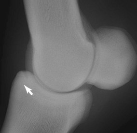
Fig. 110-1 Lateromedial digital radiographic image of a metacarpophalangeal joint in a racing Quarter Horse with a large osteochondral fracture (arrow) of the dorsal, proximal aspect of the proximal phalanx. Large dorsal plane fractures are common in the racing Quarter Horse.
(Courtesy Dr. C.W. McIlwraith, Ft. Collins, CO, United States.)
OA may be seen in association with chip fractures of the proximal aspect of the proximal phalanx especially in older horses, manifested as wear lines and erosions on the distal articular surface of the McIII.10 Defects on the palmar distal aspect of the McIII are far less common than in the TB, but they do occur in older QHs, and the prognosis is similarly poor.
Fragments of the palmar and plantar aspects of the proximal phalanx are seen less frequently than are dorsal fragments and are not always a source of lameness.11 They are removed arthroscopically when clinically relevant.
Fractures of the proximal sesamoid bones (PSBs)—apical, abaxial, and basilar—are relatively common in the QH, despite such fractures being considered fatigue-related injuries.
Distal Hock Joint Pain
Lameness associated with the pain arising from the distal hock joints, as well as other hindlimb lameness, is associated with a failure to break sharply from the starting gate. Proximal limb flexion tests may be equivocal, but a positive Churchill test (see page 60) along with a history of poor performance may be indicative of distal hock joint pain, although a negative test does not rule this out. The condition is usually bilateral, and the horse may track closely behind or a hindlimb may cross axially during protraction when observed from behind. Horses that wear patches behind to protect from scalping or horses with laceration marks seen on the medial aspect of the hock are highly suspect for distal hock joint pain. Radiographs are negative in many instances, or subtle abnormalities may be seen. Intraarticular corticosteroids are effective at relieving pain and/or improving performance. Even though the tarsometatarsal and centrodistal joints are low motion joints, maintaining articular cartilage is important because these joints rarely fuse spontaneously. Betamethasone esters and triamcinolone acetonide have been shown to have fewer deleterious effects on cartilage than methylprednisolone acetate.12,13 If methylprednisolone acetate is used, low doses should be considered (20 to 40 mg).
Stifle Lameness
The most common site of stifle pain is the medial femorotibial joint, and this is the same as in the racing TB. Stifle pain can cause poor performance. The horse may be positive to proximal limb flexion or palpation, but usually intraarticular analgesia is required to localize the pain causing lameness. It is not always clear whether the condition is synovitis or early OA. Radiological examination is useful to assess the joint and rule out certain conditions. Ultrasonographic examination is useful to identify soft tissue lesions.14 Most horses with stifle pain respond well to intraarticular therapy, but if lameness persists diagnostic arthroscopy is necessary to make a definitive diagnosis. Lameness associated with the femoropatellar joint is often accompanied by joint effusion. In recent years most osteochondritis dissecans lesions are removed surgically before a horse starts racing. Upward fixation of the patella may be a problem in immature horses in early training.
Subchondral bone cysts of the medial femoral condyle can be an important problem. Arthroscopic surgery with injection of triamcinolone acetonide into numerous areas of the cyst lining has been recently reported as successful and is now the preferred treatment.15
Proximal Suspensory Desmitis
Forelimb proximal suspensory desmitis (PSD) is usually seen as an acute, profound lameness (grade 4 of 5) the day after a race and often affects the fastest horse in the trainer’s barn. The horse may walk on the toe, without dropping the heel to contact the ground. Such severe lameness associated with PSD rapidly subsides, similar to that seen in QHs with a stress fracture of the third carpal bone. If digital palpation is unrewarding, perineural analgesia of the lateral palmar nerve may be necessary to localize the pain causing lameness without blocking the middle carpal joint. Radiography and ultrasonography should be performed. Nuclear scintigraphy may be used but is not usually necessary for the diagnosis. QHs with acute PSD usually respond well to shock wave treatment and altering the training schedule as necessary.
Tibial Stress Fractures
Stress fractures of the tibia are seen in young horses about the time of their second qualifying work or first race. Diagnosis has become more common with access to nuclear scintigraphy. The lameness is unilateral and severe; the left hindlimb is affected most commonly. This may be because horses are pulling up quickly from a high rate of speed before entering a left-hand turn on the racetrack. Unlike in the TB, in which tibial stress fractures occur most frequently in the caudolateral cortex, the most common location of tibial stress fractures in the QH is the distal medial aspect of the tibia.16 Nuclear scintigraphy is the most accurate means of diagnosis; digital radiography performed 7 to 10 days after injury may identify a lesion. Rest is required for at least 90 days before resuming training.
Miscellaneous Fractures of Importance
The three most common fractures resulting in catastrophic injury in the racing QH and TB are those of the PSB, the McIII, and the humerus.17 Although most often associated with racing and fatigue in TBs, biabaxial and comminuted PSB fractures occur in the QH with resulting disruption of the suspensory apparatus and the integrity of the MCP joint. Fractures of the McIII and the humerus are a cause of catastrophic injury in the QH. Horses generally are able to walk after shoulder injury, but the definitive diagnosis is usually made the next day when the pain and swelling are localized.
Another important injury is severe carpal bone fractures (slab fractures and comminuted fractures), many of which are amenable to surgery. Whenever pain causing severe lameness is localized to the carpus, a thorough radiographic examination, including skyline images, is necessary to ensure that degenerative lesions, incomplete sagittal fractures, and any nondisplaced fractures may be seen before a catastrophic injury occurs. All the recognized third carpal bone fractures can be seen in the QH, but a higher percentage of large dorsal plane slab fractures involve both the radial and intermediate facets than in the TB (Figure 110-2). The slab fracture is often displaced, with the distal margin of the radial carpal bone collapsing into the proximal aspect of the third carpal bone fracture site (Figure 110-3). Internal fixation using screws placed in lag fashion is necessary for proper healing and to prevent further collapse of the joint.
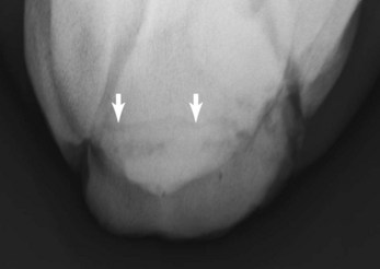
Fig. 110-2 Dorsoproximal-dorsodistal (skyline) digital radiographic image of the distal row of carpal bones in a racing Quarter Horse with a large dorsal plane slab fracture (arrows) of the third carpal that spans the entire radial and intermediate fossae.
(Courtesy Dr. C.W. McIlwraith, Ft. Collins, CO, United States.)
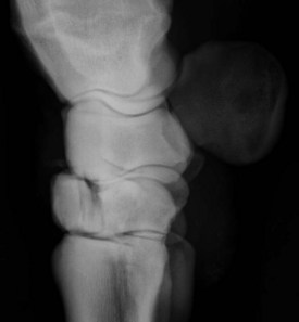
Fig. 110-3 Lateromedial digital radiographic image of a racing Quarter Horse with a large, displaced dorsal plane slab fracture of the third carpal bone (C3). With dorsal displacement of C3 there is distal movement of the overlying radial carpal bone causing carpal instability. Prognosis for future racing is guarded, and surgical repair is necessary.
(Courtesy Dr. C.W. McIlwraith, Ft. Collins, CO, United States.)
Comminuted carpal fractures caused by slab fractures involving both rows of carpal bones are catastrophic injuries seen in QHs. Challenging, salvage surgical procedures, such as partial arthrodesis or panarthrodesis using numerous bone plates, are necessary.
Although less frequent, fractures of the lumbosacral vertebrae are particularly devastating because they not only result in the death of the horse but as a result of the horse falling on the jockey, they often also result in severe injury to the rider.