CHAPTER 63 True pelvis, pelvic floor and perineum
TRUE PELVIS AND PELVIC FLOOR
The true pelvis is a bowl-shaped structure formed from the sacrum, pubis, ilium, ischium, the ligaments which interconnect these bones and the muscles which line their inner surfaces. The true pelvis is considered to start at the level of the plane passing through the promontory of the sacrum, the arcuate line on the ilium, the iliopectineal line and the posterior surface of the pubic crest. This plane, or ‘inlet’ lies at an angle of between 35 and 50° up from the horizontal and above this the bony structures are sometimes referred to as the false pelvis. They form part of the walls of the lower abdomen. The floor or ‘outlet’ of the true pelvis is formed by the muscles of levator ani. Although the floor is gutter shaped, it generally lies in a plane between 5 and 15° up from the horizontal. This difference between the planes of the inlet and outlet is the reason why the true pelvis is said to have an axis (lying perpendicular to the plane of both inlet and outlet) which progressively changes through the pelvis from above downwards. The details of the topography of the bony and ligamentous pelvis are considered fully on page 1358.
MUSCLES AND FASCIAE OF THE PELVIS
Pelvic muscles
The muscles arising within the pelvis form two groups. Piriformis and obturator internus, although forming part of the walls of the pelvis, are considered as primarily muscles of the lower limb (Fig. 63.1). Levator ani and coccygeus form the pelvic diaphragm and delineate the lower limit of the true pelvis (Fig. 63.2). The fasciae investing the muscles are continuous with visceral pelvic fascia above, perineal fascia below and obturator fascia laterally.
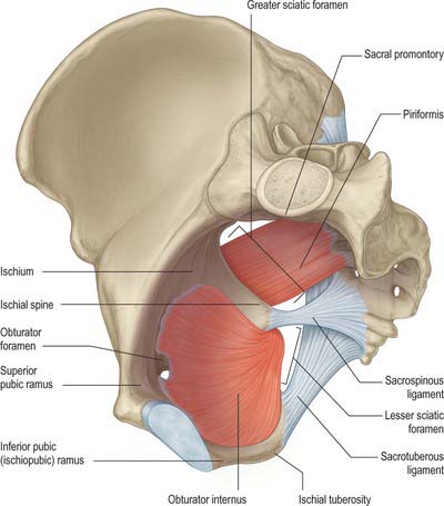
Fig. 63.1 Piriformis, obturator internus and the ligaments of the pelvis. Those muscles relating only to the pelvis or perineum have been omitted for clarity.
(Adapted from Drake, Vogl and Mitchell 2005.)
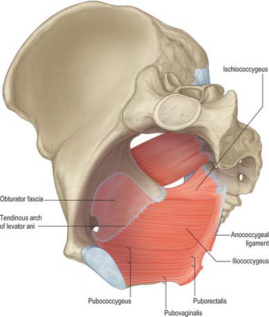
Fig. 63.2 Muscles of the female pelvis. The superior gluteal and obturator vessels and nerves as well as the pelvic viscera have been omitted for clarity.
(Adapted from Drake, Vogl and Mitchell 2005.)
Piriformis
The complete description of piriformis is in Chapter 80.
Piriformis (see p. 1371) forms part of the posterolateral wall of the true pelvis and is attached to the anterior surface of the sacrum, the gluteal surface of the ilium near the posterior inferior iliac spine, the capsule of the adjacent sacroiliac joint and sometimes to the upper part of the pelvic surface of the sacrotuberous ligament. It passes out of the pelvis through the greater sciatic foramen. Within the pelvis, the anterior surface of piriformis is related to the rectum (especially on the left), the sacral plexus of nerves and branches of the internal iliac vessels. The posterior surface lies against the sacrum.
Obturator internus
Obturator internus (see p. 1371) and the fascia over its upper inner (pelvic) surface form part of the anterolateral wall of the true pelvis. It is attached to the structures surrounding the obturator foramen, the inferior ramus of the pubis, the ischial ramus, the pelvic surface of the hip bone below and behind the pelvic brim, and the upper part of the greater sciatic foramen. It also attaches to the medial part of the pelvic surface of the obturator membrane. The muscle is covered by a thick fascial layer and the fibres themselves cannot be seen directly from within the pelvis. This fascia gives attachment to some of the fibres of levator ani and thus only the upper portion of the muscle lies lateral to the contents of the true pelvis, whilst the lower portion forms part of the boundaries of the ischio-anal fossa. In the male, the upper portion lies lateral to the bladder, the obturator and vesical vessels, and the obturator nerve. In the female, the attachments of the broad ligament of the uterus, the ovarian end of the uterine tubes, and the uterine vessels, also lie medial to obturator internus and its fascia.
Levator ani (ischiococcygeus, iliococcygeus, pubococcygeus)
Levator ani is a broad muscular sheet of variable thickness which is attached to the internal surface of the true pelvis and forms a large portion of the pelvic floor (Fig. 63.3). The muscle is subdivided into named portions according to their attachments and the pelvic viscera to which they are related. These parts are often referred to as separate muscles, but the boundaries between each part cannot be easily distinguished and they perform many similar physiological functions. The separate parts are referred to as ischiococcygeus, iliococcygeus and pubococcygeus. Pubococcygeus is often subdivided into separate parts according to the pelvic viscera to which they relate (puboperinealis, puboprostaticus or pubovaginalis, puboanalis, puborectalis). Levator ani arises from each side of the walls of the pelvis along the condensation of the obturator fascia (the tendinous arch of levator ani; see below). Fibres from ischiococcygeus attach to the sacrum and coccyx but the remaining parts of the muscle converge in the midline. The fibres of iliococcygeus join by a partly fibrous intersection and form a raphe posterior to the anorectal junction. Closer to the anorectal junction and elsewhere in the pelvic floor, the fibres are more nearly continuous with those of the opposite side and the muscle forms a sling (puborectalis and pubovaginalis or pubourethralis).
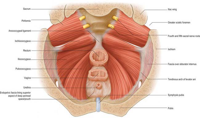
Fig. 63.3 Muscles of the female pelvis viewed from above. The sacral nerve roots have been divided close to the sacral foramina. The anorectal junction, vagina and urethra have been divided at the level of the pelvic floor.
The attachments for the ischiococcygeus, iliococcygeus and pubococcygeus parts are as follows.
The ischiococcygeal part may be referred to as a separate muscle, sometimes named coccygeus. It lies as the most posterosuperior portion of levator ani and arises as a triangular musculotendinous sheet with its apex attached to the pelvic surface and tip of the ischial spine. The base of the muscle is attached to the lateral margins of the coccyx and the fifth sacral segment. Ischiococcygeus is rarely absent, but may be nearly completely tendinous rather than muscular. It lies on the pelvic aspect of the sacrospinous ligament and may be fused with it, particularly if it is mostly tendinous. The sacrospinous ligament may represent a degenerate part or an aponeurosis of the muscle since the muscle and ligament are coextensive.
The iliococcygeal part is attached to the inner surface of the ischial spine below and anterior to the attachment of ischiococcygeus and to the tendinous arch as far forward as the obturator canal (Fig. 63.2). The most posterior fibres are attached to the tip of the sacrum and coccyx but most join with fibres from the opposite side to form a raphe. This raphe is effectively continuous with the fibroelastic anococcygeal ligament, which is closely applied to its inferior surface and some muscle fibres may attach into the ligament. The raphe provides a strong attachment for the pelvic floor posteriorly and must be divided to allow wide excisions of the anorectal canal during abdominoperineal excisions for malignancy. An accessory slip may arise from the most posterior part and is sometimes referred to as iliosacralis.
The pubococcygeal part is attached to the back of the body of the pubis and passes back almost horizontally. The most medial fibres run directly lateral to the urethra and its sphincter as it passes through the pelvic floor; here the muscle is correctly called the puboperinealis, although due to its close relationship to the upper half of the urethra in both sexes it is often referred to as pubourethralis. The muscle fibres from both sides form part of the urethral sphincter complex together with the intrinsic striated and smooth musculature of the urethra; fibres decussate across the midline directly behind the urethra. In males some of these fibres lie lateral and inferior to the prostate and are referred to as puboprostaticus (levator prostatae). In females, some fibres form the pubourethralis, others run further back to form a sling around the posterior wall of the vagina where they are referred to as pubovaginalis. In both sexes, fibres from this part of pubococcygeus attach to the perineal body; a few elements also attach to the anorectal junction. Some of these fibres, sometimes called puboanalis, decussate and blend with the longitudinal rectal muscle and fascial elements to contribute to the conjoint longitudinal coat of the anal canal. Behind the rectum, some fibres of pubococcygeus form a tendinous intersection as part of the levator raphe, and a thick muscular sling, puborectalis, wraps around the anorectal junction. Some fibres blend with those of the external anal sphincter. Pubourethralis, pubovaginalis/puboprostaticus and puboanalis are sometimes collectively referred to as ‘pubovisceralis’.
The superior, pelvic surface of levator ani is separated only by fascia (superior pelvic diaphragmatic, visceral and extraperitoneal) from the urinary bladder, prostate or uterus and vagina, rectum and peritoneum. Its inferior, perineal, surface forms the medial wall of the ischio-anal fossa and the superior wall of the anterior recess of the fossa, both being covered by inferior pelvic diaphragmatic fascia. The posterior border is separated from the coccyx by areolar tissue. The medial borders of the two levator muscles are separated by the visceral outlet, through which pass the urethra, vagina, and anorectum.
Levator ani is supplied by branches of the inferior gluteal, inferior vesical and pudendal arteries.
Fibres which originate mainly in the second, third and fourth sacral spinal segments reach levator ani from below and above by a variety of routes (Wendell-Smith & Wilson 1991). Most commonly, pubococcygeus is supplied by second and third sacral spinal segments via the pudendal nerve, and ischiococcygeus and iliococcygeus by direct branches of the sacral plexus from the third and fourth sacral spinal segments.
Pubococcygeus is a lateral compressor of the various visceral canals which cross the pelvic floor. Puborectalis also reinforces the external anal sphincter, helps to create the anorectal angle, and reduces the anteroposterior dimension of the ano-urogenital hiatus. Iliococcygeus and, to a lesser extent, the less muscular ischiococcygeus, assist puborectalis in contributing to anorectal and urinary continence.
Levator ani must relax appropriately to permit expulsion of urine and particularly faeces, and it contracts with abdominal muscles and the abdominothoracic diaphragm to raise intra-abdominal pressure. It forms much of the basin-shaped muscular pelvic diaphragm, which supports the pelvic viscera. Like the abdominothoracic diaphragm, but unlike abdominal muscles, levator ani is also active in the inspiratory phase of quiet respiration. In the pregnant female, the shape of the pelvic floor may help to direct the fetal head into the anteroposterior diameter of the pelvic outlet.
Pelvic fasciae
The pelvic fasciae may be conveniently divided into the parietal pelvic fascia, which mainly forms the coverings of the pelvic muscles, and the visceral pelvic fascia, which forms the coverings of the pelvic viscera and their vessels and nerves (Fig. 63.4).
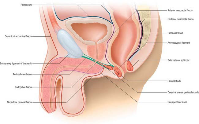
Fig. 63.4 Fasciae of the pelvis and perineum. Median sagittal section in the male. The parietal pelvic fascia is shown in black, the mesorectal fascia in brown, the fascial boundaries of the deep perineal space/pouch in green, the peritoneum in blue, the deep abdominal fascia in purple and the subcutaneous abdominal and perineal fascia in red. The visceral perietal fasciae have been omitted for clarity.
Parietal pelvic fascia
The parietal pelvic fascia consists of the obturator fascia, the fasciae over piriformis, and over levator ani (the pelvic diaphragm), and the presacral fascia.
Obturator fascia
The parietal pelvic fascia on the pelvic (medial) surface of obturator internus is well differentiated. Although in humans it is derived from the degenerate upper portion of the attachment of levator ani, it is usually referred to as the obturator fascia. Above, it is connected to the posterior part of the arcuate line of the ilium, and is continuous with iliac fascia. Anterior to this, as it follows the line of origin of obturator internus, it is gradually separated from the attachment of the iliac fascia and a portion of the periosteum of the ilium and pubis spans between them. It arches below the obturator vessels and nerve, investing the obturator canal, and is attached anteriorly to the back of the pubis. Behind the obturator canal, the fascia is markedly aponeurotic and gives a firm attachment to levator ani, usually called the tendinous arch of levator ani (arcus tendineus musculi levatoris ani) (see Figs 63.3, 63.13, 63.14). Below the attachment of levator ani, the fascia is thin and is effectively composed only of the epimysium of the muscle and overlying connective tissue; posteriorly it forms part of the lateral wall of the ischio-anal fossa in the perineum, and anteriorly it merges with the fasciae of the muscles of the deep perineal space (see Fig. 63.14), which is continuous with the ischio-anal fossa. The obturator fascia is continuous with the pelvic periosteum and thus the fascia over piriformis.
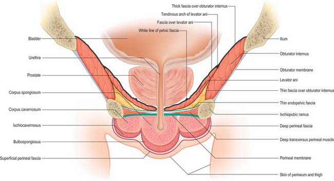
Fig. 63.13 Muscles and fasciae of the male perineum – coronal view. The section passes through the bulb of the penis at the level of the urethra. The deep perineal space is continuous with the ischio-anal fossa posteriorly. The parietal pelvic fascia is shown in black, fascial boundaries of the deep perineal space/pouch in green, the deep perineal fascia in purple and the superficial perineal fascia in red. The visceral and parietal fasciae have been omitted for clarity. The pelvic fascia over the ‘pelvic’ aspect of the deep transverse perinei is very thin and does not form a distinct layer as such in places blending with the parietal pelvic fascia over the inferior aspect of levator ani.
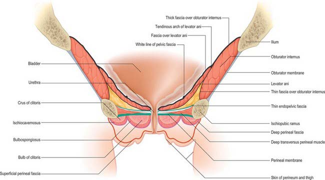
Fig. 63.14 Muscles and fasciae of the female perineum – coronal view. The section passes through the bulb of the clitoris at the level of the urethra. The deep perineal space is continuous with the ischio-anal fossa posteriorly. The parietal pelvic fascia is shown in black, fascial boundaries of the deep perineal space/pouch in green, the deep perineal fascia in purple and the superficial perineal fascia in red. The visceral and parietal fasciae have been omitted for clarity. The pelvic fascia over the ‘pelvic’ aspect of the deep transverse perinei is very thin and does not form a distinct layer as such in places blending with the parietal pelvic fascia over the inferior aspect of levator ani.
Fascia over piriformis
The fascia over the inner aspect of piriformis is very thin, and fuses with the periosteum on the front of the sacrum at the margins of the anterior sacral foramina. It ensheathes the sacral anterior primary rami which emerge from these foramina: the nerves are often described as lying behind the fascia. The internal iliac vessels lie in front of the fascia over piriformis; their branches draw out sheaths of the fascia and extraperitoneal tissue into the gluteal region, above and below piriformis.
Fascia over levator ani (pelvic diaphragm)
The fascia over levator ani covers both of the surfaces of the pelvic diaphragm. On the lower surface, the thin inferior fascia is continuous with the obturator fascia below the tendinous arch of levator ani laterally. It covers the medial wall of the ischio-anal fossa and blends below with fasciae on the urethral sphincter and the external anal sphincter. On the upper surface, the superior fascia of the pelvic diaphragm is markedly thicker than that over the inferior surface. It is attached anteriorly to the back of the body of the pubis, approximately 2 cm above its lower border, and extends laterally across the superior ramus of the pubis, blending with the obturator fascia and continuing along an irregular line to the spine of the ischium. It is continuous posteriorly with the fascia over piriformis and the anterior sacrococcygeal ligament. Medially, the superior fascia of the pelvic diaphragm blends with the visceral pelvic fascia forming part of the endopelvic fascia.
Arcus tendineus fascia pelvis/white line of the parietal pelvic fascia
Low on the superomedial aspect of the upper fascia over levator ani, a thick white band of condensed connective tissue extends from the lower part of the symphysis pubis to the inferior margin of the spine of the ischium (see Figs 63.4, 63.13, 63.14). Although often referred to as the tendinous arch of the pelvic fascia (arcus tendineus fasciae pelvis), it is really the remnant of the degenerate tendon of iliococcygeus in humans and is best referred to as the white line of the parietal pelvic fascia (Smith 1908). It provides attachment for the condensations of visceral pelvic fascia which provide support to the urethra, bladder and vagina in females (see below).
Presacral fascia
The presacral fascia forms a hammock-like structure posterior to the posterior portion of the mesorectal fascia. Laterally it extends to the origin of the fascia over piriformis and the fascia over levator ani (superior pelvic diaphragmatic fascia) with which it blends, and more inferiorly it extends between the white line of the parietal pelvic fascia on either side. Inferiorly, it extends to the anorectal junction, where it fuses with the posterior aspect of the mesorectal fascia and the anococcygeal ligament at the level of the anorectal junction. Superiorly, it can be traced to the origin of the superior hypogastric plexus, where it becomes progressively thinner over the promontory of the sacrum and becomes continuous with the retroperitoneal tissues. The right and left hypogastric nerves and inferior hypogastric plexuses lie on its surface and the presacral veins lie immediately posterior to it. It forms a distinct layer which can be seen both on magnetic resonance images of the pelvis and during surgery. The presacral fascia provides an important landmark because extension of rectal tumours through it significantly reduces the possibility of curative resectional surgery. Dissection in the plane posterior to the fascia may result in bleeding from the presacral veins: because the adventitia of the veins is partly attached to the posterior surface of the fascia, the haemorrhage may be severe (since the veins are unable to contract properly).
Visceral pelvic fascia
The visceral pelvic fascia is formed from condensations of connective tissue which are closely associated with the pelvic viscera to which it relates and with the neurovascular structures supplying or running near those organs. In its most inferior and lateral extent, the visceral pelvic fascia is closely related to, and effectively derived from, the superior fascia over the attachment of levator ani (see below), whereas more superiorly and posteriorly, it is derived from part of the fascia over piriformis. The condensations where these fasciae meet are sometimes collectively referred to as the ‘endopelvic fascia’. Further accounts are given of the paravisceral portions of the visceral pelvic fascia (e.g. parametrium, paracolpium) in the chapters describing the organs to which the fascia relates.
Several different condensations of endopelvic fascia are recognized both at operation and by MRI imaging. They provide important support to the pelvic viscera and play a role in continence in both sexes.
In the female, pubourethral ligaments form from periurethral connective tissue and attach to the white line of the parietal pelvic fascia close to its pubic end. A little more laterally, similar tissue exists which may be termed the urethropelvic ligaments, and which insert into the white line over levator ani. These condensations are effectively continuous with connective tissue which runs from the paravaginal tissue to the white line, and is sometimes referred to as the vaginolevator support or attachment. Since the normal angle of the urethra during standing is nearly 45° to the vertical, these two structures combined can be viewed as providing a ‘hammock-like’ support to the midurethra (DeLancey 1994); some fibres may even decussate across the midline between the urethra and the anterior vaginal wall. The upper portion of the pubourethral ligament blends with the pubovesical and vesicopelvic ligaments (connective tissue around the neck of the urinary bladder), and may contain cholinergically innervated smooth muscle fibres, sometimes referred to as pubovesicalis. In males, pubourethral structures are augmented by the puboprostatic ligament/puboprostaticus. In both sexes, these muscles may have a role in opening the upper urethra and bladder neck during micturition.
There is much less condensation of connective tissue around the rectum, and no similar rectopelvic ligaments exist. The connective tissue over the longitudinal muscles of the rectum is thickened just above the anal hiatus in levator ani and it fuses with the endopelvic fascia and the anococcygeal ligament, sometimes referred to as a rectosacral ligament.
VASCULAR SUPPLY AND LYMPHATIC DRAINAGE OF THE PELVIS
The true pelvis contains the internal iliac arteries and veins and the lymphatics which drain the majority of the pelvic viscera. The common and external iliac vessels and the lymphatics which drain the lower limb lie along the pelvic brim and in the lower retroperitoneum, but are conveniently discussed together with the vessels of the true pelvis.
Arteries of the pelvis
Common iliac arteries
The abdominal aorta bifurcates into the right and left common iliac arteries anterolateral to the left side of the fourth lumbar vertebral body. These arteries diverge as they descend and divide at the level of the sacroiliac joint into external and internal iliac arteries. The external iliac artery is the principal artery of the lower limb. The internal iliac artery provides the principal supply to the walls and viscera of the pelvis, the perineum and the gluteal region.
The right common iliac artery is approximately 5 cm long. It passes obliquely across part of the fourth and the fifth lumbar vertebral bodies, and is crossed anteriorly by the sympathetic rami to the pelvic plexus and, at its division into internal and external iliac arteries, by the ureter. It is covered by the parietal peritoneum, which separates it from the coils of the small intestine. Posteriorly, it is separated from the fourth and fifth lumbar vertebral bodies and their intervening disc by the right sympathetic trunk, the terminal parts of the common iliac veins and the start of the inferior vena cava, the obturator nerve, lumbosacral trunk and iliolumbar artery. Laterally, the inferior vena cava and the right common iliac vein lie superiorly and the right psoas major lies inferiorly. The left common iliac vein is medial to the upper part of the right common iliac artery.
The left common iliac artery is shorter than the right and is approximately 4 cm long. Lying anterior to it are the sympathetic rami to the pelvic plexus, the superior rectal artery and, at its terminal bifurcation, the ureter. The sympathetic trunk, the fourth and fifth lumbar vertebral bodies and intervening disc, the obturator nerve, lumbosacral trunk and iliolumbar artery are all posterior. The left common iliac vein is posteromedial, and the left psoas major lateral, to the left common iliac artery.
In addition to the external iliac and internal iliac branches, each common iliac artery also gives small branches to the peritoneum, psoas major, ureter, adjacent nerves and surrounding areolar tissue. The common iliac artery occasionally gives rise to the iliolumbar artery and accessory or replaced renal arteries if the kidney is low lying.
Internal iliac arteries
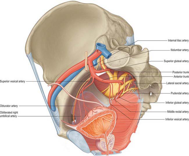
Each internal iliac artery, approximately 4 cm long, begins at the common iliac bifurcation, level with the lumbosacral intervertebral disc and anterior to the sacroiliac joint. It descends posteriorly to the superior margin of the greater sciatic foramen where it divides into an anterior trunk, which continues in the same line towards the ischial spine, and a posterior trunk, which passes back to the greater sciatic foramen. Anterior to the artery are the ureter and, in females, the ovary and fimbriated end of the uterine tube. The internal iliac vein, lumbosacral trunk and sacroiliac joint are posterior. Lateral is the external iliac vein, between the artery and psoas major, and the obturator nerve lying inferior to the vein. The parietal peritoneum is medial, separating it from the terminal ileum on the right and the sigmoid colon on the left. Tributaries of the internal iliac vein are also medial (Fig. 63.5).
In the fetus, the internal iliac artery is twice the size of the external iliac artery and is the direct continuation of the common iliac artery. The main trunk ascends on the anterior abdominal wall to the umbilicus, converging on the contralateral artery, and the two arteries run through the umbilicus to enter the umbilical cord as the umbilical arteries. At birth, when placental circulation ceases, only the pelvic segment remains patent as the internal iliac artery and part of the superior vesical artery; the remainder becomes a fibrous medial umbilical ligament. In males, the patent part (usually the superior vesical artery) usually gives off an artery to the vas deferens.
Posterior trunk branches
The branches of the posterior trunk of the internal iliac artery are the iliolumbar, lateral sacral and superior gluteal arteries.
The iliolumbar artery is the first branch of the posterior trunk and ascends laterally anterior to the sacroiliac joint and lumbosacral nerve trunk. It lies posterior to the obturator nerve and external iliac vessels and reaches the medial border of psoas major, dividing behind it into the lumbar and iliac branches. The lumbar branch supplies psoas major and quadratus lumborum and anastomoses with the fourth lumbar artery. It sends a small spinal branch through the intervertebral foramen between the fifth lumbar and first sacral vertebrae, to supply the cauda equina. The iliac branch supplies iliacus; between the muscle and bone it anastomoses with the iliac branches of the obturator artery. A large nutrient branch enters an oblique canal in the ilium. Other branches runs around the iliac crest, contribute to the supply of the gluteal and abdominal muscles, and anastomose with the superior gluteal, circumflex iliac and lateral circumflex femoral arteries.
The lateral sacral arteries are usually double, or if single, divide rapidly into superior and inferior branches. The superior and larger artery passes medially into the first or second anterior sacral foramen, supplies the sacral vertebrae and contents of the sacral canal, and then leaves the sacrum via the corresponding dorsal foramen to supply the skin and muscles dorsal to the sacrum. The inferior or lateral sacral artery crosses obliquely anterior to piriformis and the sacral anterior spinal rami, then descends lateral to the sympathetic trunk to anastomose with its fellow and the median sacral artery anterior to the coccyx. Its branches enter the anterior sacral foramina and are distributed in the same way as the branches of the superior artery.
The superior gluteal artery is the largest branch of the internal iliac artery and effectively forms the main continuation of its posterior trunk. It runs posteriorly between the lumbosacral trunk and the first sacral ramus or between the first and second rami, then turns slightly inferiorly, leaving the pelvis by the greater sciatic foramen above piriformis and dividing into superficial and deep branches. In the pelvis it supplies piriformis, obturator internus and a nutrient artery to the ilium. The superficial branch enters the deep surface of gluteus maximus. Its numerous branches supply the muscle and anastomose with the inferior gluteal branches while others perforate the tendinous medial attachment of the muscle to supply the skin over the sacrum, where they anastomose with the posterior branches of the lateral sacral arteries. The deep branch of the superior gluteal artery passes between gluteus medius and the bone, soon dividing into superior and inferior branches. The superior branch skirts the superior border of gluteus minimus to the anterior superior iliac spine and anastomoses with the deep circumflex iliac artery and the ascending branch of the lateral circumflex femoral artery. The inferior branch runs through gluteus minimus obliquely, supplies it and gluteus medius and anastomoses with the lateral circumflex femoral artery. A branch enters the trochanteric fossa to join the inferior gluteal artery and ascending branch of the medial circumflex femoral artery; other branches run through gluteus minimus to supply the hip joint.
The superior gluteal artery occasionally arises directly from the internal iliac artery with the inferior gluteal artery and sometimes from the internal pudendal artery.
Anterior trunk branches
The branches of the anterior trunk of the internal iliac artery are the superior and inferior vesical, middle rectal, vaginal, obturator, uterine, internal pudendal and inferior gluteal.
The superior vesical arter is the first large branch of the anterior trunk. It lies on the lateral wall of the pelvis just below the brim and runs anteroinferiorly medial to the periosteum of the posterior surface of the pubis. It supplies the distal end of the ureter, the bladder, the proximal end of the vas deferens and the seminal vesicles. It also gives origin to the umbilical artery in the foetus, which remains as a fibrous cord, the medial umbilical ligament, in the adult. This vessel occasionally remains patent as a small artery supplying the umbilicus.
The inferior vesical artery may arise as a common branch with the middle rectal artery. In the female it is often replaced by the vaginal artery. It supplies the bladder, the prostate, the seminal vesicles and the vas deferens.
The middle rectal artery is often multiple and may be small. It runs into the lateral fascial coverings of the mesorectum. It occasionally arises close to or in common with the origin of the inferior vesical artery in males.
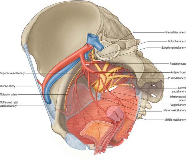
In females the vaginal artery may replace the inferior vesical artery. It may arise from the uterine artery close to its origin (Fig. 63.6).
The obturator artery runs anteroinferiorly from the anterior trunk on the lateral pelvic wall to the upper part of the obturator foramen. It leaves the pelvis via the obturator canal and divides into anterior and posterior branches. In the pelvis it is related laterally to the fascia over obturator internus and is crossed on its medial aspect by the ureter and, in the male, by the vas deferens. In the nulliparous female the ovary lies medial to it. The obturator nerve is above the artery, the obturator vein below it. In the pelvis, the obturator artery provides iliac branches to the iliac fossa which supply the bone and iliacus and anastomose with the iliolumbar artery. A vesical branch runs medially to the bladder and sometimes replaces the inferior vesical branch of the internal iliac artery. A pubic branch usually arises just before the obturator artery leaves the pelvis, and ascends over the pubis to anastomose with the contralateral artery and the pubic branch of the inferior epigastric artery.
Outside the pelvis, the anterior and posterior terminal branches encircle the foramen between obturator externus and the obturator membrane. The anterior branch curves anteriorly on the membrane and then inferiorly along its anterior margin to supply branches to obturator externus, pectineus, the femoral adductors and gracilis. It anastomoses with the posterior branch and the medial circumflex femoral artery. The posterior branch follows the posterior margin of the foramen and turns anteriorly on the ischial part to anastomose with the anterior branch. It supplies the muscles attached to the ischial tuberosity and anastomoses with the inferior gluteal artery. An acetabular branch enters the hip joint at the acetabular notch, ramifies in the fat of the acetabular fossa and sends a branch along the ligament of the femoral head.
Occasionally the obturator artery is replaced by an enlarged pubic branch of the inferior epigastric artery which descends almost vertically to the obturator foramen. It usually lies near the external iliac vein, lateral to the femoral ring, and is rarely injured during inguinal or femoral hernia surgery. Sometimes it curves along the edge of the lacunar part of the inguinal ligament, partly encircling the neck of a hernial sac, and may be inadvertently cut during enlargement of the femoral ring in reducing a femoral hernia.
The uterine artery is an additional branch in females. It is a large artery which arises below the obturator artery on the lateral wall of the pelvis and runs inferomedially into the broad ligament of the uterus.
Internal pudendal artery (in the pelvis)
The internal pudendal artery arises just below the origin of the obturator artery. It descends laterally to the inferior rim of the greater sciatic foramen, where it leaves the pelvis between piriformis and ischiococcygeus to enter the gluteal region. It then curves around the dorsum of the ischial spine to enter the perineum by the lesser sciatic foramen. This course effectively allows the nerve to wrap around the posterior limit of levator ani at its attachment to the ischial spine and so gain access to the perineum. In the pelvis the internal pudendal artery crosses anterior to piriformis, the sacral plexus and the inferior gluteal artery. Behind the ischial spine it is covered by gluteus maximus, with the pudendal nerve medial and the nerve to obturator internus lateral to it. The internal pudendal artery gives off several muscular branches in the pelvis and gluteal region which supply adjacent muscles and nerves.
The inferior gluteal artery is the larger terminal branch of the anterior internal iliac trunk and principally supplies the buttock and thigh. It descends posteriorly, anterior to the sacral plexus and piriformis but posterior to the internal pudendal artery. It passes between the first and second, or second and third, sacral anterior spinal nerve rami, then between piriformis and ischiococcygeus and runs through the lower part of the greater sciatic foramen to reach the gluteal region. The artery runs inferiorly between the greater trochanter and ischial tuberosity with the sciatic and posterior femoral cutaneous nerves, deep to gluteus maximus. It continues down the thigh, supplying the skin and anastomosing with branches of the perforating arteries. The inferior gluteal and internal pudendal arteries often arise as a common stem from the internal iliac artery, sometimes with the superior gluteal artery. Inside the pelvis the inferior gluteal artery gives branches to piriformis, ischiococcygeus and iliococcygeus and occasionally contributes to the middle rectal arterial supply. In the male, it may supply vessels to the seminal vesicles and prostate.
External iliac arteries
The external iliac arteries are of larger calibre than the internal iliac arteries. Each artery descends laterally along the medial border of psoas major, from the common iliac bifurcation to a point midway between the anterior superior iliac spine and the symphysis pubis, and enters the thigh posterior to the inguinal ligament to become the femoral artery.
The parietal peritoneum and extraperitoneal tissue separate the right external iliac artery from the terminal ileum and, usually, the appendix, and the left external iliac artery from the sigmoid colon and coils of the small intestine anteromedially. At its origin, the external iliac artery may be crossed by the ureter and is also crossed by the gonadal vessels, the genital branch of the genitofemoral nerve, the deep circumflex iliac vein and the vas deferens (male) or round ligament (female). Posteriorly, the artery is separated from the medial border of psoas major by the iliac fascia. The external iliac vein lies partly posterior to its upper part, but is more medial below. Laterally, it is related to psoas major, which is covered by the iliac and psoas fascia. Numerous lymph vessels and nodes lie on its front and sides.
The external iliac artery is principally the artery of the lower limb and as such has few branches in the pelvis. Apart from very small vessels to psoas major and neighbouring lymph nodes, the artery has no branches until it gives off the deep circumflex iliac and inferior epigastric arteries, which arise near to the point at which it passes under the inguinal ligament.
The deep circumflex iliac artery branches laterally from the external iliac artery almost opposite the origin of the inferior epigastric artery. It ascends and runs laterally to the anterior superior iliac spine behind the inguinal ligament in a sheath formed by the union of the transversalis and iliac fasciae. There it anastomoses with the ascending branch of the lateral circumflex femoral artery, pierces the transversalis fascia and skirts the internal lip of the iliac crest. About halfway along the iliac crest it runs through transversus abdominis and then between transversus abdominis and internal oblique to anastomose with the iliolumbar and superior gluteal arteries. It gives off a large ascending branch at the anterior superior iliac spine which runs between internal oblique and transversus abdominis, supplies both muscles, and anastomoses with the lumbar and inferior epigastric arteries.
The inferior epigastric artery originates from the external iliac artery posterior to the inguinal ligament. It curves forwards in the anterior extraperitoneal tissue and ascends obliquely along the medial margin of the deep inguinal ring whence it continues as an artery of the anterior abdominal wall.
Veins of the pelvis
The true pelvis contains a large number of veins which drain the pelvic walls and most of the viscera contained within the pelvis, and which carry venous blood from the gluteal region, hip and thigh. The external iliac veins, which lie close to the brim of the pelvis, carry the venous drainage from most of the lower limb. There is considerable variation in the venous drainage of the pelvis: although the major veins frequently follow their named arterial counterparts, the small tributaries exhibit a great deal of variation between individuals.
Common iliac veins
The common iliac vein is formed by the union of the external and internal iliac veins, anterior to the sacroiliac joints. It ascends obliquely to end at the right side of the fifth lumbar vertebra, uniting at an acute angle with the contralateral vessel to form the inferior vena cava. The right common iliac vein is shorter and more nearly vertical, lying posterior and then lateral to its artery. The right obturator nerve passes posteriorly. The left common iliac vein is longer and more oblique, and lies first medial, then posterior, to its artery. It is crossed anteriorly by the attachment of the sigmoid mesocolon and superior rectal vessels. Each vein receives iliolumbar and sometimes lateral sacral veins. The left common iliac vein usually drains the median sacral vein. There are no valves in these veins.
The left common iliac vein occasionally ascends to the left of the aorta to the level of the kidney, where it receives the left renal vein and crosses anterior to the aorta to join the inferior vena cava: this vessel represents the persistent caudal half of the left postcardinal or supracardinal vein.
The right and left medial sacral veins accompany the corresponding arteries anterior to the sacrum, where they unite to form a single vein which usually ends in the left common iliac vein, but which sometimes ends at the common iliac junction.
Internal iliac vein
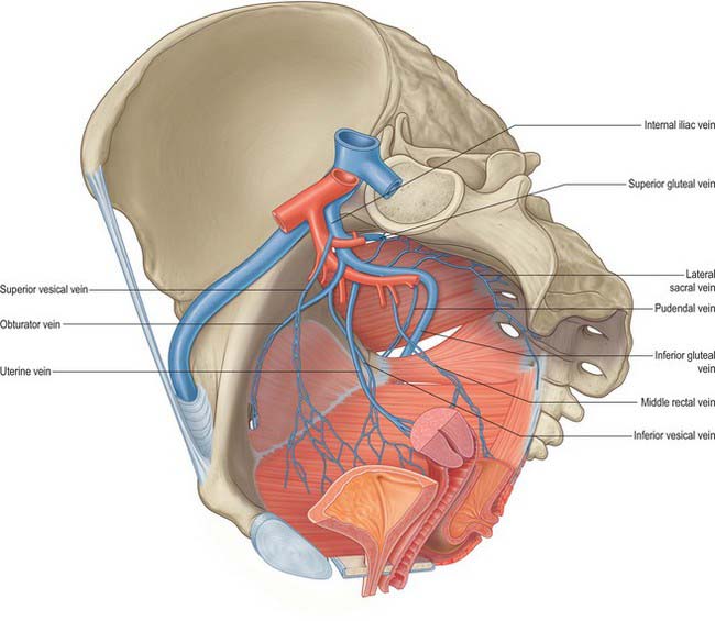
The internal iliac vein is formed by the convergence of several veins above the greater sciatic foramen. It does not have the predictable trunks and branches of the internal iliac artery but its tributaries drain the same territories. It ascends posteromedial to the internal iliac artery to join the external iliac vein, forming the common iliac vein at the pelvic brim, anterior to the lower part of the sacroiliac joint. It is covered anteromedially by parietal peritoneum. Its tributaries are the gluteal, internal pudendal and obturator veins, which originate outside the pelvis; the lateral sacral veins, which run from the anterior surface of the sacrum; and the middle rectal, vesical, uterine and vaginal veins, which originate in the venous plexuses of the pelvic viscera (Fig. 63.7).
The venous drainage of the leg may be blocked by thrombosis involving the external iliac systems and the inferior vena cava. Under these circumstances, the pelvic veins, particularly the internal iliac tributaries, enlarge and provide a major avenue of venous return from the femoral system. Surgical interference with these veins may seriously compromise venous drainage and precipitate oedema of one or both legs.
The superior gluteal veins are the venae comitantes of the superior gluteal artery. They receive tributaries which correspond to the branches of the superior gluteal artery and enter the pelvis via the greater sciatic foramen, above piriformis. They join the internal iliac vein, frequently as a single trunk.
The inferior gluteal veins are venae comitantes of the inferior gluteal artery. They begin proximally and posterior in the thigh, where they anastomose with the medial circumflex femoral and first perforating veins, and enter the pelvis low in the greater sciatic foramen, joining to form a vessel opening into the distal (lower) part of the internal iliac vein. The inferior gluteal and superficial gluteal veins connect by perforating veins (Doyle 1970) which are analogous to the sural perforating veins. The gluteal veins probably have a venous ‘pumping’ role, and provide collaterals between the femoral and internal iliac veins.
The obturator vein begins in the proximal adductor region and enters the pelvis via the obturator foramen. It runs posteriorly and superiorly on the lateral pelvic wall below the obturator artery and between the ureter and internal iliac artery and ends in the internal iliac vein. It is sometimes replaced by an enlarged pubic vein, which joins the external iliac vein.
The lateral sacral veins accompany the lateral sacral arteries, and are interconnected by a sacral venous plexus.
The middle rectal vein begins in the rectal venous plexus and drains the rectum and mesorectum. Variable in size, it runs laterally on the pelvic surface of levator ani to end in the internal iliac vein. The middle rectal vein often receives tributaries from the bladder and either the prostate and seminal vesicle (males) or the posterior aspect of the vagina (females).
External iliac vein
The external iliac vein is the proximal continuation of the femoral vein. It begins posterior to the inguinal ligament, ascends along the pelvic brim and ends anterior to the sacroiliac joint by joining the internal iliac vein to form the common iliac vein. On the right it lies medial to the external iliac artery, gradually inclining behind it as it ascends. On the left it is wholly medial. Disease of the external iliac artery may cause it to adhere closely to the vein at the point where it is in contact, and, particularly on the right side, the walls of the vessels may become fused, making dissection hazardous. The external iliac vein is crossed medially by the ureter and internal iliac artery. In males it is crossed by the vas deferens, in females by the round ligament and ovarian vessels. Psoas major lies laterally, except where the artery intervenes. The vein is usually valveless, but may contain a single valve. It tributaries are the inferior epigastric, deep circumflex iliac and pubic veins.
One or two inferior epigastric veins accompany the inferior epigastric artery and drain into the external iliac vein a little above the inguinal ligament.
Lymphatic drainage of the pelvis
The lymph nodes in the pelvis are grouped around the common, external and internal iliac vessels, and are named accordingly.
Common iliac nodes
The common iliac nodes receive the entire lymphatic drainage of the lower limb because they drain both internal and external iliac nodes. They usually lie in medial, lateral and anterior chains around the common iliac artery, the lateral being the main route: one or two lie inferior to the aortic bifurcation and anterior to the fifth lumbar vertebra or sacral promontory. The common iliac nodes connect to the lateral aortic nodes.
External iliac nodes
The external iliac nodes usually form three subgroups which are lateral, medial and anterior to the external iliac vessels. The medial nodes are considered the main channel of drainage, collecting lymph from the lower limb via the inguinal nodes, the deeper layers of the infra-umbilical abdominal wall, the adductor region of the thigh, the glans penis or clitoris, the membranous urethra, prostate, fundus of the bladder, uterine cervix and upper vagina. Their efferents pass to the common iliac nodes.
Internal iliac nodes
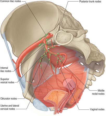
The internal iliac nodes surround the branches of the internal iliac vessels. They receive afferents from most of the pelvic viscera (with the exception of the gonads and the majority of the rectum), the deeper parts of the perineum and the gluteal and posterior femoral muscles, and drain to the common iliac nodes. The individual groups are considered in the description of the viscera. There are frequent connections between the right and left groups particularly when they lie close to the anterior and posterior midlines (Fig. 63.8).
INNERVATION OF THE PELVIS
The pelvis contains the lumbosacral nerve trunk, the sacral and coccygeal plexuses and the pelvic parts of the sympathetic and parasympathetic systems. Collectively, these nerves carry the somatic and autonomic innervation to the majority of the pelvic visceral organs, the pelvic floor and perineum, the gluteal region and the lower limb.
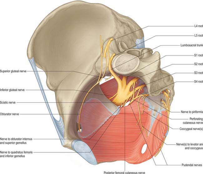
The ventral rami of the sacral and coccygeal spinal nerves form the sacral and coccygeal plexuses (Fig. 63.9). The upper four sacral ventral rami enter the pelvis by the anterior sacral foramina, the fifth enters between the sacrum and coccyx, and the ventral ramus of the coccygeal nerve curves forwards below the rudimentary transverse process of the first coccygeal segment. The first and second sacral ventral rami are large, the third to fifth diminish progressively, and the coccygeal is the smallest. Each receives a grey ramus communicans from a corresponding sympathetic ganglion. Visceral efferent rami leave the second to fourth sacral rami as the pelvic splanchnic nerves, containing parasympathetic fibres to minute ganglia in the walls of the pelvic viscera.
Lumbosacral trunk and sacral plexus
The sacral plexus is formed by the lumbosacral trunk, the first to third sacral ventral rami and part of the fourth sacral ventral ramus (the remainder of the fourth sacral ventral ramus joins the coccygeal plexus).
The lumbar part of the lumbosacral trunk contains part of the fourth and all the fifth lumbar ventral rami; it appears at the medial margin of psoas major, and descends over the pelvic brim anterior to the sacroiliac joint to join the first sacral ramus. The greater part of the second and third sacral rami converge on the inferomedial aspect of the lumbosacral trunk in the greater sciatic foramen to form the sciatic nerve. The ventral and dorsal divisions of the nerves do not separate physically from each other, but their fibres remain separate within the rami; the ventral and dorsal divisions of each contributing root join within the sciatic nerve. The fibres of the dorsal divisions will go on to form the common fibular nerve and the fibres of the ventral division form the tibial nerve. The sciatic nerve occasionally divides into common fibular and tibial nerves inside the pelvis. In these cases the common fibular nerve usually runs through piriformis.
The sacral plexus lies against the posterior pelvic wall anterior to piriformis, posterior to the internal iliac vessels and ureter, and behind the sigmoid colon on the left. The superior gluteal vessels run either between the lumbosacral trunk and first sacral ventral ramus or between the first and second sacral rami, while the inferior gluteal vessels lie between either the first and second or second and third sacral rami (Fig. 63.9).
The sacral plexus is not commonly involved in malignant tumours of the pelvis because it lies behind the relatively dense presacral fascia, which resists all but locally very advanced malignant infiltration. When it occurs, there is intractable pain in the distribution of the branches of the plexus which may be very difficult to treat. The plexus may also be involved in the reticuloses or be affected by plexiform neuromas.
Branches of the sacral plexus
The branches of the sacral plexus are:
| Ventral divisions | Dorsal divisions | |
|---|---|---|
| Nerve to quadratus femoris and gemellus inferior | L4,5, S1 | |
| Nerve to obturator internus and gemellus superior | L5, S1,2 | |
| Nerve to piriformis | S2 (S1) | |
| Superior gluteal nerve | L4,5, S1 | |
| Inferior gluteal nerve | L5, S1,2 | |
| Posterior femoral cutaneous nerve | S2,3 | S1,2 |
| Tibial (sciatic) nerve | L4,5, S1,2,3 | |
| Common fibular (sciatic) nerve | L4,5, S1,2 | |
| Perforating cutaneous nerve | S2,3 | |
| Pudendal nerve | S2,3,4 | |
| Nerves to levator ani and external anal sphincter | S4 | |
| Pelvic splanchnic nerves | S2,3 (S4) |
The course and distribution of most of the branches of the sacral plexus are covered fully on page 1384.
Pudendal nerve (in the pelvis)
The pudendal nerve arises from the ventral divisions of the second, third and fourth sacral ventral rami and is formed just above the superior border of the sacrotuberous ligament and the upper fibres of ischiococcygeus. It leaves the pelvis via the greater sciatic foramen between piriformis and ischiococcygeus, enters the gluteal region and crosses the sacrospinous ligament close to its attachment to the ischial spine, where it lies medial to the internal pudendal vessels. It accompanies the internal pudendal artery through the lesser sciatic foramen into the pudendal (Alcock’s) canal on the lateral wall of the ischio-anal fossa. In the posterior part of the canal it gives rise to the inferior rectal nerve, the perineal nerve and the dorsal nerve of the penis or clitoris.
Sacral visceral branches
Visceral branches, the pelvic splanchnic nerves, arise from the second to fourth sacral ventral rami and innervate the pelvic viscera.
Sacral muscular branches
Several muscular branches arise from the fourth sacral ventral ramus to supply the superior surface of levator ani and the upper part of the external anal sphincter. The branches to levator ani enter the superior (pelvic) surface of the muscle whilst the branch to the external anal sphincter (also referred to as the perineal branch of the fourth sacral nerve) reaches the ischio-anal fossa by running either through ischiococcygeus or between ischiococcygeus and iliococcygeus. It supplies the skin between the anus and coccyx via its cutaneous branches.
Coccygeal plexus
The coccygeal plexus is formed by a small descending branch from the fourth sacral ramus and by the fifth sacral and coccygeal ventral rami. The fifth sacral ventral ramus emerges from the sacral hiatus, curves round the lateral margin of the sacrum below its cornu and pierces ischiococcygeus from below to reach its upper, pelvic surface. Here it is joined by a descending branch of the fourth sacral ventral ramus; the small trunk so formed descends on the pelvic surface of ischiococcygeus and joins the minute coccygeal ventral ramus which emerges from the sacral hiatus and curves round the lateral coccygeal margin to pierce coccygeus to reach the pelvis. The small trunk that is formed in this way is the coccygeal plexus. Anococcygeal nerves arise from it and form a few fine filaments which pierce the sacrotuberous ligament to supply the adjacent skin.
Pelvic part of the sympathetic system
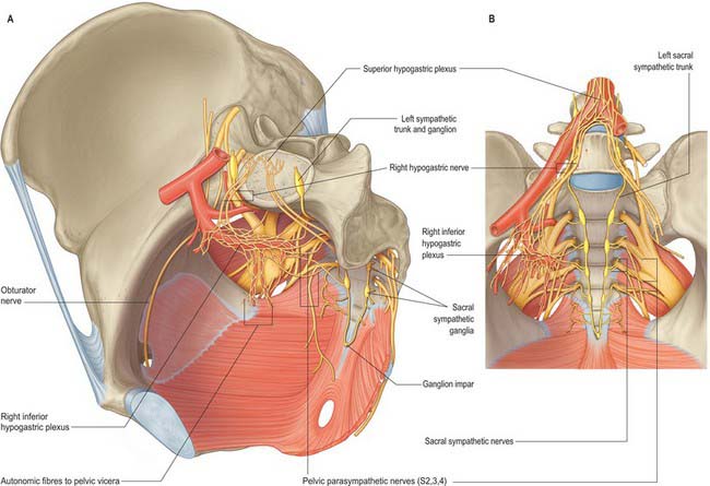
The pelvic sympathetic trunk lies in the extraperitoneal tissue anterior to the sacrum beneath the presacral fascia (Fig. 63.10). It lies medial or anterior to the anterior sacral foramina and has four or five interconnected ganglia. Above, it is continuous with the lumbar sympathetic trunk. The right and left trunks converge below the lowest ganglia and unite in the small ganglion impar anterior to the coccyx. Grey rami communicantes pass from the ganglia to sacral and coccygeal spinal nerves but there are no white rami communicantes. Medial branches connect across the midline and twigs from the first two ganglia join the inferior hypogastric plexus or the hypogastric ‘nerve’. Other branches form a plexus on the median sacral artery.
Vascular branches
Preganglionic fibres for the vessels supplying the pelvis and lower limb are derived from the lower three thoracic and upper two or three lumbar spinal segments. They reach the lower thoracic and upper lumbar ganglia through white rami communicantes and descend through the sympathetic trunk to synapse in the lumbar ganglia. Postganglionic fibres pass from these ganglia via grey rami communicantes to the femoral nerve which carries them to the femoral artery and its branches. Some fibres descend through the lumbar ganglia to synapse in the upper two or three sacral ganglia, from which postganglionic axons pass through grey rami communicantes to the roots of the sacral plexus. Those in the pudendal and superior and inferior gluteal nerves accompany the arteries of the same name to the gluteal and perineal tissues; branches may also supply the pelvic lymph nodes. Those joining the tibial nerve are carried to the popliteal artery and distributed via its branches to the leg and foot.
Sympathetic denervation of vessels in the lower limb can be effected by removing or ablating the upper three lumbar ganglia and the intervening parts of the sympathetic trunk, which is used rarely in treating vascular insufficiency of the lower limb.
PERINEUM
MUSCLES AND FASCIAE OF THE PERINEUM
The perineum is an approximately diamond-shaped region which lies below the pelvic floor, between the inner aspects of the thighs and anterior to the sacrum and coccyx. It is usually described as if from the position of an individual lying supine with the hip joints in abduction and partial flexion. The surface projection of the perineum and the form of the skin covering it varies considerably depending on the position of the thighs whereas the deep tissues themselves occupy relatively fixed positions. The perineum is bounded anteriorly by the pubic symphysis and its arcuate ligament, posteriorly by the coccyx, anterolaterally by the ischiopubic rami and the ischial tuberosities, and posterolaterally by the sacrotuberous ligaments. The deep limit of the perineum is the inferior surface of the pelvic diaphragm and its superficial limit is the skin which is continuous with that over the medial aspect of the thighs and the lower abdominal wall. An arbitrary line joining the ischial tuberosities (the interischial line) divides the perineum into an anterior urogenital triangle and a posterior anal triangle. The urogenital triangle faces downwards and forwards, whereas the anal triangle faces downwards and backwards.
The male urogenital triangle contains the bulb and attachments of the penis (Fig. 63.11) (Ch. 76) and the female urogenital triangle contains the mons pubis, the labia majora, the labia minora, the clitoris and the vaginal and urethral orifices (Ch. 77).
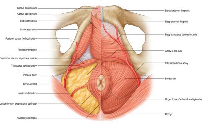
Fig. 63.11 Muscles and fasciae of the male perineum. On the left side the skin and superficial fascia of the perineum only have been removed. The posterior scrotal (perineal) artery has been shown as it runs forward into the scrotal tissues. On the right side, the corpora cavernosa and corpus spongiosum and their associated muscles, the superficial perineal muscles and perineal membrane have been removed to reveal the underlying deep muscles and arteries of the perineum. All veins and nerves have been omitted for clarity.
Anal triangle
The structure of the anal triangle is similar in males and females, the main difference reflecting the wider transverse dimension of the triangle in females as a result of the larger size of the pelvic outlet. The anal triangle contains the anal canal and its sphincters, and the ischio-anal fossa and its contained nerves and vessels. It is lined by superficial and deep fascia.
Superficial fascia of the anal triangle
The superficial fascia of the region is thin and is continuous with the superficial/subcutaneous fascia of the skin of the perineum, thighs and buttocks (see p. 1095).
Ischio-anal fossa
The ischio-anal fossa is an approximately horse-shoe shaped region filling the majority of the anal triangle: although often referred to as a space, it is filled with loose adipose tissue and occasional blood vessels. The ‘arms’ of the horseshoe are triangular in cross section because levator ani slopes downwards towards the anorectal junction (see Fig. 67.43). The anal canal and its sphincters lie in the centre of the horseshoe. Above them the deep medial limit of the fossa is formed by the deep fascia over levator ani. The outer boundary of the fossa is formed anterolaterally by the deep fascia over obturator internus deeply and the periosteum of the ischial tuberosities more superficially. Posterolaterally, the outer boundary is formed by the lower border of gluteus maximus and the sacrotuberous ligament. Anteriorly, the superficial boundary of the fossa is formed by the posterior aspect of the transverse perineal muscles and the deep perineal pouch. Deep to this there is no fascial boundary between the fossa and the tissues deep to the perineal membrane as far anteriorly as the posterior surface of the pubis below the attachment of levator ani. Posteriorly, the fossa contains the attachment of the external anal sphincter to the tip of the coccyx: above and below this, the adipose tissue of the fossa is uninterrupted across the midline. These continuations of the ischio-anal fossa mean that infections, tumours and fluid collections within may not only enlarge relatively freely to the side of the anal canal, but may also spread with little resistance to the opposite side and deep to the perineal membrane. The internal pudendal vessels and accompanying nerves lie in the lateral wall of the ischio-anal fossa, enclosed in fascia forming the pudendal canal. The inferior rectal vessels and nerves cross the fossa from the pudendal canal and often branch within it.
The ischio-anal fossa is an important surgical plane during resections of the anal canal and anorectal junction for malignancy. It provides an easy, relatively bloodless, plane of dissection which encompasses all of the muscular structures of the anal canal and leads to the inferior surface of levator ani through which the dissection is carried.
External anal sphincter
The external anal sphincter is an oval tube of striated muscle which surrounds the lowest part of the anal canal. The uppermost (deepest) fibres blend with the lowest fibres of puborectalis; the two are seen to be continuous on endoanal ultrasound and magnetic resonance imaging. Anteriorly, some of these upper fibres decussate into the superficial transverse perineal muscles. Posteriorly, some fibres are attached to the anococcygeal raphe. The majority of the middle fibres of the external anal sphincter surround the lower part of the internal sphincter and are attached anteriorly to the perineal body and posteriorly to the coccyx via the anococcygeal ligament. Some fibres from each side of the sphincter decussate in these areas to form a sort of commissure in the anterior and posterior midline. The anterior and posterior attachments of the external anal sphincter give the muscular tube an oval profile lying anteroposteriorly. The lower fibres lie below the level of the internal anal sphincter and are separated from the lowest anal epithelium only by submucosa.
Anococcygeal ligament
The anococcygeal ligament is a layered musculotendinous structure running between the middle portion of the external anal sphincter and the coccyx. The lowest portion of the presacral fascia lies above the deep part of the ligament and the most posterior fibres of the raphe of iliococcygeus lie between the two: these three structures together are sometimes referred to as the postanal plate. Division of the anococcygeal raphe may cause descent of the anal canal and a lowering of the posterior part of the anal triangle, but does not demonstrably interfere with the process of defecation.
Urogenital triangle
The urogenital triangle is bounded posteriorly by the inter-ischial line which usually overlies the posterior border of the transverse perineal muscles. Anteriorly and laterally it is bounded deeply by the symphysis pubis and ischiopubic rami. In males, the urogenital triangle extends superficially to encompass the scrotum and the root of the penis. In females, it extends to the lower limit of the labia and mons pubis. The urogenital triangle is divided into two parts by a strong perineal membrane: the deep perineal space lies above the membrane and the superficial perineal space lies below it.
The female urogenital triangle includes muscles, fasciae and spaces similar to those in the male. There are some differences in size and disposition caused by the presence of the vagina and female external genitalia.
Deep perineal pouch (space)
The deep perineal pouch was long regarded as an anatomical region between the urogenital diaphragm and the perineal membrane containing the urethra and urethral sphincter. However, the urogenital diaphragm does not exist and the urethral sphincter, previously thought to be the principle content of the deep perineal space (where it was described as surrounding the urethra), is now recognized to be contained within the urethra. The deep perineal pouch is bounded deeply by the endopelvic fascia of the pelvic floor and superficially by the perineal membrane. Between these two fascial layers lie the deep transverse perinei, superficial to the urethral sphincter mechanism and pubourethralis, and in females superficial to the compressor urethrae and sphincter urethrovaginalis. These muscles do not form a true diaphragmatic sheet as such because fibres from several parts extend through the visceral outlet in the pelvic floor into the lower reaches of the pelvic cavity.
Perineal membrane (previously the inferior fascia of the urogenital diaphragm)
The perineal membrane is a triangular membrane which stretches almost horizontally across the deep perineal pouch. It is attached laterally to the periosteum of the ischiopubic rami and its apex is attached to the arcuate ligament of the pubis. It is particularly thick in this area and is here referred to as the transverse perineal ligament. The posterior border is fused with the deep part of the perineal body and is continuous with the fascia over the deep transverse perinei.
In the male, the perineal membrane is crossed by the urethra, 2–3 cm behind the inferior border of the symphysis pubis; the vessels and nerves to the bulb of the penis; the ducts of the bulbourethral glands, posterolateral to the urethral orifice; the deep dorsal vessels and dorsal nerves of the penis, behind the pubic arch in the midline; and the posterior scrotal vessels and nerves, anterior to the transverse perinei.
In the female, the perineal membrane is less well defined than in the male. It is divided almost into two halves by the vagina and urethra such that it forms a triangle on each side of these structures; the pubourethral ligament (the female equivalent of the transverse perineal ligament in males) links the two sides anteriorly behind the pubic arch. It is crossed by the urethra, 2–3 cm behind the inferior border of the symphysis pubis; the vagina, centrally; the ducts of Bartholin’s glands, posterolateral to the urethral orifice; the deep dorsal vessels and dorsal nerves of the clitoris, behind the pubic arch in the midline; the posterior labial vessels and nerves, anterior to the transverse perinei.
Deep transverse perinei
The deep transverse perinei form an incomplete sheet of muscle extending across the deep perineal pouch from the medial aspects of the ischiopubic rami. Posteriorly the sheet is attached to the perineal body where its fibres decussate with those of the opposite side. Anteriorly, the muscles are deficient and the visceral structures pass across the endopelvic fascia and the perineal membrane. Some fibres pass to the deep part of the external anal sphincter posteriorly and sphincter urethrae anteriorly. Together with the superficial transverse perinei, the muscles act to tether the perineal body in the median plane and may help to support the visceral canals which pass through them. They are supplied by the perineal branches of the pudendal vessels and nerves.
Urethral sphincter mechanism
The urethral sphincter mechanism consists of the intrinsic striated and smooth muscle of the urethra and the puboperinealis (pubourethralis) component of levator ani which surrounds the urethra at the point of maximum concentration of those muscles. It surrounds the membranous urethra in the male and the middle and lower thirds of the urethra in the female. In the male, fibres also reach up to the lowest part of the neck of the bladder and, between the two, lie on the surface of the prostate. The bulk of the fibres surround the membranous urethra and some fibres are attached to the inner surface of the ischiopubic ramus. In the female, the sphincter mechanism surrounds more than the middle third of the urethra. It blends above with the smooth muscle of the bladder neck and below with the smooth muscle of the lower urethra and vagina.
The urethral sphincter mechanism compresses the urethra, particularly when the bladder contains fluid. Its location around the region of highest urethral closing pressure suggests that it plays an important role in the continence of urine. Like bulbospongiosus, it is relaxed during micturition, but it contracts to expel final drops of urine, or of semen in the male, from the bulbar urethra. It may be stimulated via a vaginal tampon electrode, as has been used in the treatment of stress incontinence of urine in females.
The urethral sphincter mechanism extends from the perineum through the urogenital hiatus into the pelvic cavity. It probably receives innervation via the perineal branch of the pudendal nerve from below and direct branches from the sacral plexus and the pelvic splanchnic nerves from above (Wendell-Smith & Wilson 1991). All these nerves originate in the second, third and fourth sacral spinal segments.
Compressor urethrae
Compressor urethrae exists in females and arises from the ischiopubic rami of each side by a small tendon. Fibres pass anteriorly to meet their contralateral counterparts in a flat band which lies anterior to the urethra, below sphincter urethrae (Fig. 63.12). A variable number of fibres from the same origin fan medially to reach the lower walls of the vagina, and may rarely reach as far posteriorly as the perineal body.
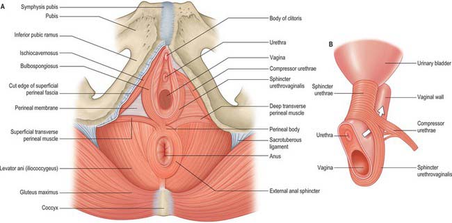
Fig. 63.12 Muscles of the female perineum. A, On the right side, the membranous layer of superficial fascia has been removed (note the cut edge). On the left side, superficial perineal muscles and overlying fascia have been removed to show the deep perineal muscles. B, The continuity of the deep perineal muscles with sphincter urethrae.
Superficial and deep perineal fasciae
Superficial perineal fascia
The tissue commonly referred to as the superficial fascia of the perineum (Colles’ fascia) forms a clear, surgically recognizable plane beneath the skin of the anterior perineum. It is firmly attached posteriorly to the fascia over the superficial transverse perinei and the posterior limit of the perineal membrane. Laterally, it is attached to the margins of the ischiopubic rami as far back as the ischial tuberosities. From here it runs more superficially to the skin of the urogenital triangle, lining the skin of the external genitalia; in the male it is also continuous with the fascial layer in the skin of the scrotum which contains the dartos muscle. In females, the fascia follows the same limits but is much less extensive in the labia majora. This layer runs anteriorly and superiorly into the skin of the lower abdominal wall where it is continuous with the membranous fascia (Scarpa’s fascia). These tissue layers are referred to in English as a ‘superficial fascia’ but this is really a misnomer since they are correctly referred to as the membranous layer of the subcutaneous tissue (the tela subcutanea).
Deep perineal fascia (investing fascia of the superficial perineal muscles)
The tissue commonly referred to as the deep perineal fascia is a layer of fascia overlying the superficial muscles of the perineum (bulbospongiosus, ischiocavernosus, superficial transverse perinei). It is in effect the investing fascia of the superficial perineal muscles and as such is firmly attached to the borders of the muscles at attachments to the ischiopubic rami, posterior margin of the perineal membrane and perineal body. In front it fuses with the suspensory ligament of the penis or clitoris and the fasciae of external oblique and the rectus sheath.
Superficial and subcutaneous perineal pouches
Subcutaneous perineal pouch
The subcutaneous perineal pouch lies between the deep perineal fascia and the superficial perineal fascia. Under normal circumstances these two layers are only separated by relatively thin subcutaneous connective tissue and the skin of the anterior perineum and genitals is relatively mobile over the deeper structures. This pouch is, however, capable of expanding considerably in the presence of fluid accumulation; blood, urine or fluid collecting in the subcutaneous pouch due to trauma or surgery on the urogenital triangle will spread throughout the tissues of the triangle including the scrotum or labia majora, but cannot pass posteriorly into the anal triangle or laterally into the medial thigh due to the firm tethering of the posterior attachments of the subcutaneous fascia. Since the superficial perineal fascia is in continuity with the fascia of the anterior abdominal wall, fluid, blood or pus may also track freely between the subcutaneous tissues of the anterior abdominal wall and the subcutaneous perineal pouch (e.g. post surgical haematomas of the abdominal wall readily cause discolouration of the perineal and genital skin).
Superficial perineal pouch
The superficial perineal pouch lies below the perineal membrane and is limited superficially by the deep perineal fascia (investing fascia of the superficial perineal muscles) (Fig. 63.13). It contains the corpora cavernosa and corpus spongiosum, ischiocavernosus, bulbospongiosus and the superficial transverse perinei, and branches of the pudendal vessels and nerves. In the female, it is crossed by the urethra and vagina and contains the clitoris. In the male, it contains the urethra as it runs in the root of the penis. It is a fully confined space; injuries to the contents of the space (such as bleeding into the urethra in the penile root) do not communicate with the deep or subcutaneous pouches unless the fascial coverings are also lacerated or breached.
Perineal body
The perineal body is a poorly defined aggregation of fibromuscular tissue located in the midline at the junction between the anal and urogenital triangles. It is attached to many structures in both the deep and superficial urogenital spaces. Posteriorly, it merges with fibres from the middle part of the external anal sphincter and the conjoint longitudinal coat. Superiorly, it is continuous with the rectoprostatic or rectovaginal septum, including fibres from levator ani (puborectalis or pubovaginalis). Anteriorly, it receives a contribution from the deep transverse perinei, the superficial transverse perinei and bulbospongiosus (Fig. 63.4). The perineal body is continuous with the perineal membrane and the superficial perineal fascia. Since the superficial perineal fascia runs forward into the skin of the perineum, the perineal body is tethered to the central perineal skin, which is often puckered over it. In males, this is continuous with the perineal raphe in the skin of the scrotum. In females, the perineal body lies directly posterior to, and is attached to, the posterior commissure of the labia majora and the introitus of the vagina.
The anus can be surgically detached from the perineal body without any clinical consequences. However, spontaneous lacerations of the body sustained during childbirth are often associated with damage to the anterior fibres of the external anal sphincter. The deliberate division of the perineal body to facilitate delivery (episiotomy) is angled laterally to avoid such injuries. The perineal body is often used for the positioning of radiological markers used to determine the amount the perineum descends during straining in order to assess pelvic floor dysfunction.
Superficial transverse perinei
The superficial transverse perinei are narrow strips of muscle which run more or less transversely across the superficial perineal space anterior to the anus. The muscles are occasionally small and rarely absent. On each side the muscle is attached to the medial and anterior aspects of the ischial tuberosity. Medially, the fibres mostly run into the perineal body although some may also pass into the ipsilateral bulbospongiosus or external anal sphincter.
Bulbospongiosus
Bulbospongiosus differs between the sexes. In the male, it lies in the midline, anterior to the perineal body. It consists of two symmetrical parts united by a median fibrous raphe. The fibres attach to the perineal body, in which they decussate, and to the transverse superficial perinei and the external anal sphincter; they diverge like the sides of a feather from the median raphe. A thin layer of posterior fibres joins the posterior portion of the perineal membrane. The majority of the middle fibres encircle the bulb of the penis and adjacent corpus spongiosum and attach to an aponeurosis on the dorsal surfaces. The anterior fibres spread out over the sides of the corpora cavernosa, ending partly in them, anterior to ischiocavernosus, and partly in a tendinous expansion which covers the dorsal vessels of the penis. In the female, bulbospongiosus also attaches to the perineal body, but the muscle on each side is separate and covers the superficial parts of the vestibular bulbs and greater vestibular glands. Fibres run anteriorly on either side of the vagina to attach to the corpora cavernosa clitoridis, and a few fibres cross over the dorsum of the body of the clitoris.
Bulbospongiosus helps to empty the urethra of urine after the bladder has emptied. In the male, it may assist in the final stage of erection as the middle fibres compress the erectile tissue of the bulb and the anterior fibres contribute by compressing the deep dorsal vein of the penis. It contracts six or seven times during ejaculation, assisting in the expulsion of semen. In the female, bulbospongiosus acts to constrict the vaginal orifice and express the secretions of the greater vestibular glands. Anterior fibres contribute to erection of the clitoris by compressing its deep dorsal vein.
Ischiocavernosus
In the male, ischiocavernosus covers the crus penis. It is attached by tendinous and muscular fibres to the medial aspect of the ischial tuberosity behind, and to the ischial ramus on both sides of the crus. These fibres end in an aponeurosis which is attached to the sides and under surface of the crus penis. In the female, ischiocavernosus is related to a smaller crus clitoris and has a much smaller attachment to the ischiopubic ramus, but is otherwise similar to the corresponding muscle in the male (Figs 63.14, 63.15).
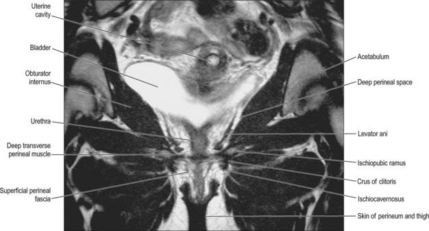
Fig. 63.15 Muscles and fasciae of the female perineum – coronal T2-weighted MRI.
(By courtesy of Dr J Lee and Ms K Wimpey, Chelsea and Westminster Hospital, London.)
Ischiocavernosus compresses the crus penis in males and may help to maintain penile erection. The muscles form a triangle on each side of the midline with bulbospongiosus medially and the superficial transverse perinei posteriorly, attached to the perineal membrane: when contracted, the two ischiocavernosi act together to stabilize the erect penis. In the female, ischiocavernosus may help to promote clitoral erection.
VASCULAR SUPPLY AND LYMPHATIC DRAINAGE OF THE PERINEUM
Arteries of the perineum
Internal pudendal artery (in the perineum)
The internal pudendal artery enters the perineum around the posterior aspect of the ischial spine and runs on the lateral wall of the ischio-anal fossa in the pudendal (Alcock’s) canal with the pudendal veins and the pudendal nerve. The canal is formed by the connective tissue binding the vessels and nerve to the perineal surface of the obturator internus fascia and lies about 4 cm above the lower limit of the ischial tuberosity. As the artery approaches the margin of the ischial ramus, it proceeds above or below the perineal membrane, along the medial margin of the inferior pubic ramus and ends behind the inferior pubic ligament.
In the male, the internal pudendal artery distal to the perineal artery gives a branch to the bulb of the penis before it divides into the deep (cavernosal) and dorsal arteries of the penis (Fig. 63.11). Given its distribution, the internal pudendal artery distal to its perineal branch has been named the artery of the penis. The artery to the bulb supplies the corpus spongiosum and the deep artery to the penis supplies the corpus cavernosa on each side. The dorsal artery runs on the dorsal aspect of the penis, and supplies circumflex branches to the corpora cavernosa and corpus spongiosum which end by anastomosing in the coronal sulcus and supplying the glans penis and its overlying skin.
In the female, the artery to the bulb is distributed to the erectile tissue of the vestibular bulb and vagina. The cavernosal artery is much smaller and supplies the corpora cavernosa of the clitoris; the dorsal artery supplies the glans and prepuce of the clitoris (Fig. 63.11).
Branches of the internal pudendal artery are sometimes derived from an accessory pudendal artery, which is usually a branch of the pudendal artery before its exit from the pelvis; effectively the artery is double before it leaves the pelvis.
Inferior rectal artery
The inferior rectal artery arises just after the internal pudendal artery enters the canal on the lateral wall of the ischio-anal fossa and runs anteromedially through the adipose tissue of the ischio-anal fossa to reach the deep portion of the external anal sphincter. It often branches before reaching the sphincter. During dissections of the anal canal, particularly during perineal excisions of the anorectum, the inferior rectal vessels are encountered in the ischio-anal fossa and must be secured before division or they tend to retract laterally to the canal, where they can cause troublesome bleeding.
Perineal artery
The perineal artery is a branch of the internal pudendal artery near the anterior end of the pudendal canal, and runs through the perineal membrane. In the male, it approaches the scrotum in the superficial perineal space, between bulbospongiosus and ischiocavernosus. A small transverse branch passes medially, inferior to the superficial transverse perineal muscle, to anastomose with the contralateral artery and with the posterior scrotal and inferior rectal arteries. It supplies the transverse perinei, the perineal body and the posterior attachment of the bulb of the penis. The posterior scrotal arteries are usually terminal branches of the perineal artery but may also arise from its transverse branch. They are distributed to the scrotal skin and dartos muscle in the male and supply the perineal muscles. In the female, the perineal artery runs an almost identical course to that in the male, and gives off similar branches; the posterior scrotal arteries are replaced by posterior labial arteries.
Veins of the perineum: internal pudendal veins
The internal pudendal veins are venae comitantes of the internal pudendal artery and unite as a single vessel ending in the internal iliac vein. The perineal tributaries receive veins from the penile bulb and the scrotum (males) or clitoris and labia (females) and the inferior rectal veins join towards the posterior end of the pudendal canal.
Lymphatic drainage of the perineum
The lymphatics from the skin of the penis and scrotum (male) or skin of the clitoris and labia (female) drain together with lymphatics from the perineal skin to the superficial inguinal nodes and thence to the deep inguinal nodes. The glans, corpora cavernosa and corpus spongiosum of the penis or clitoris drain directly to the deep inguinal nodes.
INNERVATION OF THE PERINEUM: PUDENDAL NERVE (IN THE PERINEUM)
The pudendal nerve gives rise to the inferior rectal and perineal nerves and the dorsal nerves of the penis or clitoris.
The pudendal nerve occupies a very constant position over the ischial spine and is readily found. It may be ‘blocked’ by infiltration with a local anaesthetic applied via a needle passed through the lateral wall of the vagina to numb the perineal and anal skin. It may also be palpated over the ischial spine through the lateral wall of the rectum and motor terminal latencies measured.
Inferior rectal nerve
The inferior rectal nerve runs through the medial wall of the pudendal canal with the inferior rectal vessels. It crosses the ischio-anal fossa to supply the external anal sphincter, the lining of the lower part of the anal canal, and the circumanal skin. It frequently breaks into terminal branches just before reaching the lateral border of the sphincter. Its cutaneous branches distributed around the anus overlap the cutaneous branches of the perineal branch of the posterior femoral cutaneous nerve and of the scrotal or labial nerves. The inferior rectal nerve occasionally arises directly from the sacral plexus and crosses the sacrospinous ligament or reconnects with the pudendal nerve. In females, the inferior rectal nerve may supply sensory branches to the lower part of the vagina.
Perineal nerve
The perineal nerve is the inferior and larger terminal branch of the pudendal nerve in the pudendal canal. It runs forwards below the internal pudendal artery and accompanies the perineal artery, dividing into posterior scrotal or labial and muscular branches. The posterior scrotal or labial nerves are usually double and have medial and lateral branches which run over the perineal membrane and pass forwards in the lateral part of the urogenital triangle with the scrotal or labial branches of the perineal artery. They supply the skin of the scrotum or labia majora, overlapping the distribution of the perineal branch of the posterior femoral cutaneous and inferior rectal nerves. In females, the posterior labial branches also supply sensory fibres to the skin of the lower vagina.
Muscular branches arise directly from the pudendal nerve to supply the superficial transverse perinei, bulbospongiosus, ischiocavernosus, deep transverse perinei, sphincter urethrae and the anterior parts of the external anal sphincter and levator ani. In males, a nerve to the bulb of the urethra leaves the nerve to the bulbospongiosus, pierces this muscle to supply the corpus spongiosum penis and ends in the urethral mucosa.
Dorsal nerve of the penis or clitoris
The dorsal nerve of the penis or clitoris runs anteriorly above the internal pudendal artery along the ischiopubic ramus deep to the perineal membrane. It supplies the corpus cavernosum and accompanies the dorsal artery of the penis or clitoris between the layers of the suspensory ligament. In males, the dorsal nerve of the penis runs on the dorsum of the penis to end in the glans. In females the dorsal nerve of the clitoris is very small.
Doyle JF. The perforating veins of the gluteus maximus. Ir J Med Sci. 1970;3:285-288.
Smith GE. Studies in the anatomy of the human pelvis, with special reference to the fasciae and visceral supports. Part I. J Anat Physiol. 1908;42:198-218.
DeLancey JO. Structural support of the urethra as it relates to stress urinary incontinence: the hammock hypothesis. Am J Obstet Gynecol. 1994;170:1713-1723.
Wendell-Smith CP, Wilson PM. The vulva, vagina and urethra and the musculature of the pelvic floor. In: Philipp E, Setchell M, Ginsburg J. Scientific Foundations of Obstetrics and Gynaecology. Oxford: Butterworth-Heinemann; 1991:84-100.