Prevention of phosphatic calculi requires adjustment of the dietary calcium/phosphorus ratio to a level of 2:1 or greater.72 The magnesium content of the ration should be maintained at recommended levels. Abandoning pelleted feeds and increasing the quantity of long-stem forage in the ration may increase salivary flow and fecal phosphate excretion.9
Addition of salt to feedlot rations has proved effective in several studies. Sodium chloride, fed at a level of 3% to 5%, reduces the incidence of urolithiasis without adverse effects on feed intake or weight gain.64 Cattle consuming such rations show variable increases in water intake and urine output, implying that some of the beneficial effect of salt feeding results from diuresis.57,82 Other studies show minimal effect of this level of salt supplementation on urine volume, and one investigator has suggested that the preventive effect is caused by interruption of crystal development by chloride ion in urine.66 Nonetheless, it is prudent to anticipate increased water intake after salt supplementation is initiated.
Ammonium chloride supplementation, fed at a level of 0.5% to 1% of ration dry matter, also reduces the incidence of struvite urolithiasis.82,83 Ammonium chloride may increase the solubility of magnesium ammonium phosphate crystals through a modest reduction in urinary pH. The pH of the urine is likely to be influenced by the relative concentrations of strong cations (sodium and potassium) and strong anions (chloride and sulfate) of the entire ration, a relationship termed the dietary cation-anion difference.84 Thus the efficacy of ammonium chloride in reducing urinary pH and therefore reducing struvite urolithiasis may vary among different livestock operations because the concentration of these cations and anions may vary among rations.81
Restriction of dietary silica intake in ruminants grazing native grasses is not feasible; thus dietary management is limited to salt (sodium chloride) supplementation. However, loose salt or lick salt is unlikely to be ingested in sufficient quantities to affect water intake.85 Sodium chloride supplementation of palatable creep feeds, at a level of 15% of dry matter, is an effective measure for range calves.57 Creep feeding should begin at 4 months of age or earlier. Initially, lower salt concentrations may be required for young calves to become accustomed to the feed. The feeders should be located near a reliable source of palatable water.
Ammonium chloride supplementation (1% of dry matter) has been demonstrated to significantly reduce silica urolith development in lambs.78 In the same study, reduction of the dietary calcium/phosphorus ratio from 2:1 to almost 1:1 resulted in a trend toward reduced silica calculi formation. The role of copper and zinc deficiencies in ruminant silica urolithiasis remains to be determined.
This type of urolith has been frequently recovered from small ruminants fed alfalfa hay.81 Because of this and the risk factors identified in Australian sheep, reduction of dietary calcium levels could be beneficial. This may not be possible for sheep grazing legume pastures. Ammonium chloride supplementation may be an effective preventive measure because calcium carbonate is more soluble in acidic solutions.69
Maximizing water intake is an important aspect of urolithiasis prevention, regardless of the urolith type involved. Cleaning of water containers should be a regular practice. Water palatability may also be improved through provision of shade for water containers during the summer. Dark liners (with sun exposure) or heaters will warm water during the winter. Automatic waterers should be checked regularly for proper function. Shallow containers capable of rapid refilling provide higher rates of water turnover, resulting in less stagnation.
In operations involving multiple animals or large pastures, placement of multiple watering sites might allow for more frequent intake. This is especially true for sheep, whose banding instinct usually prevents individuals from traveling alone to distant watering sites.
Abnormalities of the umbilicus and umbilical remnants, including the urachus, are frequently encountered disease conditions of neonatal calves. Infections of the umbilicus, urachus, and umbilical vein or arteries (navel ill) are often associated with localized or systemic bacterial infections acquired from environmental contamination at birth. The association of umbilical abnormalities with calfhood morbidity and mortality has been established.86 Of the umbilical structures, the urachus is most frequently involved.87,88 Occasionally, more insidious and chronic conditions of the urachal remnant are observed in older calves and mature cattle. Internal abscessation, adhesions, sepsis, peritonitis, uroperitoneum, cystitis, and intestinal strangulation have all been reported.89-94 Urachal fistulas or acquired patent urachus, although often observed in foals, is infrequently reported in cattle.95
Subsequent to bacterial infections of the urachus of the neonate, the inflammatory response within the abdomen precipitates development of fibrinous adhesions between the urachus and surrounding viscera, including the rumen, intestine, uterus, bladder, or ovaries. Abscesses may form in single or multiple locations in the urachal stalk; although uncommon, abscesses involving the apex of the urinary bladder result in concurrent cystitis.91,94 Urachal fibrosis and adhesions may serve as a means of mechanical interference of bladder emptying, resulting in secondary cystitis from chronic urine retention.94 Infrequently, the urachal abscess perforates, with subsequent peritonitis, sepsis, or uroperitoneum.89,90 Most urachal infections involve Arcanobacterium pyogenes or Eschericia coli; other skin or enteric organisms may be present as well.96
Alternatively, the urachus may fail to regress completely, creating a persistent communication between the bladder apex and the pouchlike urachal remnant. The volume of urine retained in the urachal remnant is variable. Animals with this problem may remain asymptomatic for life; however, retention of urine in the urachal “pouch” can predispose the animal to urinary tract infection. Further, rupture of the urachal remnant can occur, resulting in uroabdomen and, if cystitis is also present, septic peritonitis.89
Calves with internal urachal abscesses, adhesions, and other sequelae are usually older than 4 weeks of age. A history of umbilical infection during the neonatal period may or may not exist, and external umbilical abnormalities may not be present. Clinical signs may be nonspecific, including fever, lethargy, poor body condition, and poor growth or productivity. Dysuria, pollakiuria, stranguria, and colic may be evident on examination or may be included in the medical history. External palpation of the umbilicus and abdomen in smaller calves may reveal pain and enlarged umbilical remnants, particularly in the caudoventral abdomen along a line from the umbilicus to the pelvic brim. Diarrhea, abdominal distention, and an ached-back stance may be evident in animals with concurrent peritonitis. Complications from associated septicemia, such as lameness and joint distention, pneumonia, hypopion, and meningitis, are occasionally seen. When infection extends into the bladder lumen, hematuria and pyuria are reliably present.91,94
In older animals, spontaneous rupture of the infected urachal stalk may result in the acute onset of peritonitis. If the urachal remnant communicates with the bladder lumen, rupture of the urachus results in uroabdomen. Rupture may occur spontaneously or after abdominal trauma or parturition. Transrectal palpation may allow detection of abnormal bladder size, position, or shape resulting from adhesion of the bladder apex to the urachal remnant. Distention or displacement of bowel may be detected in cases of urachovisceral adhesions.
A detailed ultrasonographic examination of the interior of the umbilical remnant is warranted. Transrectal and transabdominal ultrasonographic examination of the caudal and ventral abdomen may reveal structures compatible with urachal abscesses. The bladder and other viscera may assume abnormal shape or position as a result of urachal adhesions.91,97 The luminal contents of the urachal remnant and bladder may appear flocculent in cases complicated by concurrent cystitis. Uroabdomen is characterized by accumulation of echolucent fluid in the abdominal cavity, with variable amounts of fibrin deposition evident.
In cases complicated by urachal infection or peritonitis, laboratory findings of an inflammatory hemogram, including leukocytosis, neutrophilia, hyperfibrinogenemia, and hyperglobulinemia are expected. Urinalysis may be normal or indicative of cystitis if the urinary bladder is involved. Abdominocentesis findings are variable and depend on the presence and extent of peritonitis associated with the urachal lesion. Voluminous, blood-tinged abdominal fluid suggests uroperitoneum, and analysis of the fluid and serum creatinine concentration is indicated (see Urolithiasis).
Because of the variable nature of urachal problems, the differential diagnosis will vary according to the organ(s) involved. The general clinical picture may suggest acute, subacute, or chronic infection with involvement of the abdomen and lower urinary tract. Urachal adhesions to the intestine or uterus may result in colic, signs of intestinal obstruction, and postpartum peritonitis or uroabdomen. Concurrent signs involving the lower urinary system might indicate the potential for primary ascending infections, such as cystitis or pyelonephritis. In cases of uroabdomen in males, obstructive urolithiasis is an important differential. Urolithiasis, urethritis, or neurologic disease may be included in differential diagnoses of dysuric animals. Nephritis of hematogenous origin is worthy of consideration in animals showing ill-thrift, colic or an arched back, or abnormal urine.
Ventral midline celiotomy, paramedian celiotomy, or laparoscopy under general anesthesia is recommended to enable complete evaluation of the abdominal cavity.98,99 Resection of the urachal remnants with careful dissection of adhesions is often required for complete resolution. Resection of the apex of the urinary bladder is necessary if the urachus incorporates or communicates with the bladder. Perioperative and postoperative antibiotic therapy is essential, and appropriate antibiotic selection may be based on culture and sensitivity of the abscess(es) or urine.
Outcomes of the surgical management are often satisfactory, but recurrence of adhesions should always be considered as a potential outcome. A guarded to poor prognosis is warranted for severe peritonitis or extensive adhesions. Without surgery, medical management with long-term antibacterial therapy would be expected to provide limited success because the structural problems created by adhesions and abscesses are not directly addressed.
Eversion of the bladder is an uncommon event that occurs during or shortly after parturition in cows.100-104 Forceful straining moves the bladder fundus caudally, eventually turning the bladder inside-out. The bladder is then forced out of the urethral orifice. Prolapse of the bladder is also a rare periparturient event in cattle, more frequently associated with dystocia.100,101 In this condition a full-thickness tear of the vaginal wall occurs during delivery, allowing the bladder (and possibly other viscera) to be displaced from the abdominal cavity into the vagina.
Cows with either eversion or prolapse of the bladder have a smooth, spherical mass within the vagina, usually protruding from the vulva. An affected cow may be alert and ambulatory, but if concurrent hypocalcemia, exhaustion, or peritonitis exists, the cow may be recumbent and depressed. Careful vaginal examination is required to differentiate these two conditions from each other and from vaginal prolapse, vaginal polyps, fat protrusion from a vaginal tear, vaginal neoplasia, fetal membranes, and uterine prolapse. Prevention of straining through epidural anesthesia is essential because expulsive efforts can cause herniation of other viscera through the urethra (bladder eversion) or through the vaginal tear (bladder prolapse).100 Epidural anesthesia also facilitates cleaning of the area because straining may result in continual fecal contamination of the perineum and vestibule.
The mucosal surface of the bladder is exposed. The ureteral openings may be visible on its dorsal aspect, although these may be occluded and difficult to see if the wall of the bladder is edematous.103 Vaginal palpation reveals that the protruding tissue originates from the urethral orifice. With time, constriction of the everted bladder by the narrow urethra may cause venous congestion, edema, thrombosis, and eventual necrosis.100 Palpation and ultrasonographic examination are required for detection of herniation of other viscera through the urethral orifice and into the interior of the everted bladder. Strangulation of incarcerated bowel may occur.101,102 Careful fine-needle aspiration of the interior of the eversion has been used to differentiate eversion from prolapse of the bladder. With bladder eversion, aspiration may yield peritoneal fluid, but laceration of herniated bowel is a concern.100
The serosal surface of the bladder is exposed. Careful palpation of the vagina reveals that the bladder protrudes from a full-thickness tear in the floor or lateral wall of the vagina. Other viscera may be present in the vagina as well. Fine-needle aspiration yields urine from the bladder lumen.
Manual reduction of the everted bladder may not be possible if it is edematous or if other viscera have herniated into its interior. The dorsal aspect of the urethra may be incised to widen the route through which the bladder is to be replaced.100,101 Laparotomy is required for assessment of the viability of herniated bowel, and subtotal cystectomy may be performed if extensive bladder trauma or necrosis has occurred.101 The viability of the involved structures is of primary concern for prognosis. Bladder paralysis and rupture are potential sequelae to ischemic damage that develops during eversion.100 Cystitis and pyelonephritis may develop as well, and antibiotic therapy is warranted if repair is attempted. Animals with chronic conditions may develop hydroureter, hydronephrosis, and renal failure.104
A flexible catheter may be passed into the urethra to remove urine from the bladder, thereby confirming the diagnosis and facilitating bladder replacement. After catheterization and removal of urine, the bladder can be replaced into the abdominal cavity and the vaginal tear can be sutured. Severe contamination of the peritoneal cavity may render attempts at treatment unjustified. Antibiotic therapy is indicated for surgical candidates.
In pelvic entrapment of the bladder, the apex and fundus of the bladder are displaced caudodorsally into the pelvic cavity, resulting in impaired urine outflow. In one report the condition was diagnosed in two Holstein cows a few days after parturition had occurred, suggesting a potential role of delivery or postpartum straining in bladder displacement.105 Entrapment of the bladder in a perineal hernia106 or within a vaginal prolapse107 may also occur.
Bladder emptying is impaired, and the presence of the bladder in the pelvic inlet induces straining. An affected cow may show tenesmus, pollakiuria, and stranguria. On rectal examination a soft, fluctuant mass may be detected beside the vagina.105 In calves, radiography has been used to demonstrate the pelvic position of the bladder.108
Differential diagnoses include proctitis, vaginitis, retained placenta, bladder paralysis, cystitis, and perivaginal abscess. Needle aspiration of the bladder per vaginum has been used to make a definitive diagnosis,105 although the danger of uterine puncture should be considered.107 Ultrasonographic examination can also be useful for definitive diagnosis.107 Draining the bladder through catheterization or needle aspiration facilitates replacement of the bladder by manipulation per vaginum. However, laparotomy was necessary in one cow to reset the bladder because of the development of fibrinous adhesions between the bladder and vagina.105 In cases of bladder entrapment within a vaginal prolapse, complete correction of the prolapse usually restores the bladder to its normal position.105
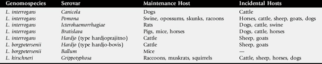
Enzootic hematuria is a disease of chronic or intermittent hematuria in cattle and sheep and is associated with chronic ingestion of bracken fern (Pteridium aquilinum)109-115 (Fig. 34-21). A different fern species, Cheilanthes sieberi, may induce this disease in Australian cattle.109,111 Hemorrhagic cystitis is the initial consequence of exposure to the toxic compound(s) in the plant. With continued ingestion of bracken fern, cattle develop bladder neoplasms of epithelial, mesenchymal, or mixed origin. Bladder infection with bovine papillomavirus type 2 is involved in carcinogenesis.
In most cases, several animals are affected in a group that is grazing a particular pasture or being fed a particular type or cutting of hay. Protracted, possibly intermittent, hematuria is the first clinical sign detected in most animals.109 Blood clots may be voided on occasion. Chronic blood loss eventually results in tachycardia, tachypnea, exercise intolerance, pale mucous membranes, and a decline in productivity and body condition. Bladder wall thickening and bladder tumors may be palpated per rectum. Proliferative changes or overt neoplasia of the bladder may cause dysuria, pollakiuria, and rarely, obstruction of the bladder trigone. Occasionally, blood clots may cause urethral obstruction. Depending on the magnitude and duration of bracken fern ingestion, hematuria may last for months to years before severe debilitation or death occurs. Ultrasonographic examination of the urinary bladder of affected animals may reveal bladder thickening and an irregularly shaped bladder wall. In an Indian study, affected cattle showed a bladder wall thickness of 4 to 5 mm (normal, 1 to 2 mm).110
The syndrome of enzootic hematuria is quite different from acute bracken poisoning, which occurs after ingestion of large quantities of bracken fern (approximating the animal’s body weight), usually over 1 to 3 months.111,112 Acute bracken poisoning manifests as an acute coagulopathy or fulminant septicemic crisis associated with severe bone marrow suppression. Clinical signs include fever, profound weakness, epistaxis, hyphema, dysentery, and petechial hemorrhages of the mucosal surfaces and sclera.111 Acute bracken poisoning is further described in Chapter 54.
Severe anemia is often seen on hematologic examination of cattle and sheep with enzootic hematuria. Evidence of a regenerative response may not be present if bone marrow suppression is severe. The platelet, segmented neutrophil, and lymphocyte counts may be reduced.111-113 Urinalysis reveals hematuria, proteinuria, and variable pyuria.109
Examination of serum for evidence of hemolysis and analysis of sediment from a freshly voided urine sample for intact red blood cells allow for differentiation of hematuria from the hemoglobinuria found in hemolytic diseases. Icterus, also characteristic of ruminant hemolytic disorders, is not found in cases of enzootic hematuria. Hematuria may be evident in urinary tract infection, but pyuria and bacteriuria are marked, and anemia, if present, is usually mild. Simultaneous involvement of several animals is uncommon with urinary tract infection but common in enzootic hematuria. Hematuria may be evident in cattle affected by malignant catarrhal fever (MCF). Protracted, severe hematuria is rare in cases of urolithiasis, and anemia is also not expected. Without necropsy, a diagnosis of enzootic hematuria requires documentation of access to bracken fern in animals with characteristic clinical signs and laboratory data.
Enzootic hematuria has a wide geographic distribution, with cases reported in North and South America, the United Kingdom, Australia, and several European countries.109 Bracken fern is found in all areas of the United States except the Great Plains, with most livestock poisonings occurring in the Pacific Northwest and upper Midwest.112 The plant grows best in well-drained, fertile soils and is often localized in open areas of forests.114 Sheep and cattle are poisoned by grazing the plant or consuming contaminated hay.114
Enzootic hematuria is primarily seen in adult sheep and cattle. In field cases, cattle grazing infested pastures develop hematuria by 2 to 3 years of age.109,115 Feeding adult cattle 1 to 2 kg bracken fern/head/day led to hematuria within 10 to 15 months in one trial.109 Papillomas of the bladder occur as early as 1 year after bracken feeding begins, with invasive carcinomas arising 2 to 6 years later.109,115
All parts of the plant are toxic to sheep and cattle.111,112 Several compounds in bracken fern possess irritant, mutagenic, immunosuppressive, or carcinogenic activities.109,113 These include ptaquiloside (aquilide A), quercetin, and a-ecdysone.116-119 The carcinogenic principles are present in the milk of cows grazing bracken fern.120,121 Bracken fern compounds may cause recrudescence of latent bovine papillomavirus 2 (BPV-2) infections through immunosuppression. Bladder infection with BPV-2 follows, and mutagenic compounds in bracken fern interact with BPV-2 in the bladder to induce local neoplasia.109,113,116 Growth of the resultant neoplastic tissue may be enhanced by further exposure to mutagenic bracken compounds.109,113,116 Similarly, BPV-4 and mutagens from bracken fern may act in synergy to induce neoplasia of the mucosa of the upper GI tract.116,118,119
In multiple species, cyclooxygenase 2 (COX-2) is overexpressed in epithelial neoplasms, including those of the urinary bladder origin. Certain types of tumors of urinary epithelium may coexpress COX-1 and COX-2. Bladder carcinomas from cows with enzootic hematuria have been shown to express both COX-1 and COX-2 at a high level relative to normal controls, when evaluated by immunohistochemical methods.122 The efficacy of COX-2 inhibitor drugs on cancer prevention or treatment in cattle at risk for bladder tumors remains to be investigated.
Immunosuppression results from reduction in circulating neutrophil and lymphocyte counts. Neutropenia appears to be a reversible phenomenon that results from bone marrow suppression. Neutrophil counts may normalize within 1 to 2 weeks of cessation of bracken feeding. Lymphopenia persists during periods of low-level ingestion.113
Tissue pallor from anemia is often appreciated. The bladder wall is thickened and the mucosa hemorrhagic and ulcerated. Microscopic examination of the bladder wall reveals capillary engorgement, intramural hemorrhage, and metaplasia of the bladder epithelium.111 Several types of bladder tumors and mixed-origin neoplasms may be present. Metastasis of epithelial neoplasms to the regional lymph nodes or other organs can occur.123 Pharyngeal, esophageal, or ruminal papillomas may be found as well, and carcinomas may develop in these same locations in cattle exposed to bracken fern over several years.115
Treatment of enzootic hematuria is limited to reduction or elimination of bracken fern in the diet. Wooded areas that support growth of bracken fern can be fenced off, and forage improvement may help to limit incorporation of the plant into hay. If such measures are not feasible, a program of early culling may help to avoid low productivity from anemia and neoplasia. Hematuria will cease if bracken feeding is discontinued before the onset of tumor formation.
Cystitis, ureteritis, and pyelonephritis in ruminants most often result from ascending urinary tract infection (UTI) with Corynebacterium renale or Escherichia coli.124 Less common causative organisms include various coliform species125 and other members of the C. renale group.126 Renal infection via the hematogenous route (suppurative embolic nephritis) is much less common but may result from bacteremia with such agents as Salmonella species, Actinomyces pyogenes, or in small ruminants, Corynebacterium pseudotuberculosis.127
Cystitis in cattle is typified by dysuria and pollakiuria, with or without gross hematuria and pyuria. During urination the flow rate is often decreased. An affected cow may tread or swish the tail and retain an arched stance after voiding has ceased. Blood, purulent debris, or crystalline material may occasionally be found on the hairs of the ventral commissure of the vulva. Rectal palpation may reveal a thickened, painful bladder. If UTI is limited to the bladder, an affected cow usually does not show generalized signs of infectious disease (e.g., fever, anorexia, depression).124
In contrast, cattle with acute pyelonephritis often have a history of an abrupt reduction in feed intake and milk production. Fever, depression, ruminal stasis, scleral injection, and occasional episodes of mild colic accompany the signs of cystitis previously described.124 With bilateral or left-sided pyelonephritis, renal enlargement, pain, and a loss of normal lobulation of the left kidney may be evident on rectal examination. Transabdominal ultrasonographic examination is useful for evaluation of the right kidney, because the right kidney usually cannot be reached during rectal examination unless it is greatly enlarged.124 In an adult cow, ultrasound evaluation of the right kidney can be performed using a 3.5-MHz transducer at the twelfth intercostal space. This transducer can also be used to view the left kidney at the dorsocranial aspect of the right paralumbar fossa.128 Alternatively, the left kidney can be imaged transrectally with a linear-array transducer. Dilation of renal calyces; echogenic, flocculent material within the renal pelvis; abnormal renal shape; and renal enlargement are ultrasonographic findings suggestive of pyelonephritis. Vaginal palpation is usually necessary to detect ureteritis124; the ureters may be enlarged and painful when palpated through the vaginal wall.
The clinical signs of chronic pyelonephritis are relatively vague and inconsistent.124 Weight loss or poor growth rate, anorexia, and reduced milk production are common presenting complaints. Posture and behavior during urination may be normal, although affected calves have been noted to dribble urine, thereby developing phosphatic calculi adherent to the vulvar hairs and urine scald of the perineum and hindlegs. Ulcerative vulvovaginitis may be present as well.129 Polyuria without gross urine abnormalities may be found in some cases. Diarrhea and pale mucous membranes may also be seen during physical examination. Rectal and vaginal examination findings for chronic pyelonephritis are similar to those of the acute form, although the involved structures may not be painful or enlarged when palpated.
Urinalysis with chemical reagent strips appears to be a sensitive ancillary test for both acute and chronic pyelonephritis. In a study of 15 cases of bovine pyelonephritis, clinical signs suggestive of urinary tract disease were found in only three cows. However, evidence of hematuria or proteinuria was found through routine reagent strip urinalysis in all 15 cases.124 Further examination and testing of the urinary tract confirmed a diagnosis of pyelonephritis.
Mild colic may result from a variety of GI disorders, but urinalysis findings are normal in these conditions. Enzootic hematuria usually affects multiple animals in a particular locale, and access to bracken fern is usually demonstrable. Although dysuria and hematuria may be evident in enzootic hematuria, anemia is profound, whereas pyuria and bacteriuria are mild or nonexistent. Urolithiasis may induce colic, dysuria, and hematuria. However, urolithiasis is almost exclusively a disease of male ruminants, and UTI is more common in females. Bladder distention is a common finding in urolithiasis but rarely occurs in UTI. Other conditions that may cause dysuria include vaginitis, vulvar trauma, perivaginal abscesses, and pelvic entrapment of the bladder. Careful assessment of the neurologic system is warranted in cases of UTI to determine if the underlying cause is bladder paresis or incomplete voiding.
Neutrophilic leukocytosis and hyperfibrinogenemia are evident on hematologic evaluation of cattle with pyelonephritis. Hyperglobulinemia may develop if the infection is established for several days. Severe, protracted proteinuria may cause hypoalbuminemia, and the resultant low plasma oncotic pressure may contribute to the development of diarrhea in occasional cases. In chronic pyelonephritis, anemia may result from reduced erythropoietin production, chronic inflammatory disease, and blood loss through the urine.124
If azotemia is found on serum chemistry analysis, the clinician must consider renal and prerenal causes before formulation of a prognosis. Pyelonephritis with azotemia and isosthenuria would indicate bilateral renal involvement, lowering the chances for successful treatment.124
Urinalysis is required for definitive diagnosis of UTI, but careful collection technique is important for valid conclusions to be made. Concurrent metritis, vaginitis, or posthitis may result in contamination of urine with blood, bacteria, and inflammatory cells, particularly if the rate of voiding is slow. A midstream or end-stream catch is likely to provide the most accurate culture results.130 Tentative identification of the organism may be obtained through Gram stain of the urine.
Urinary tract infection consistently produces hematuria, proteinuria, and bacteriuria on urinalysis. Quantitative culture of a urine sample allows for confirmation of the diagnosis and identification of the causative organism. Although not present in all cases, leukocyte casts provide definitive evidence of pyelonephritis.131
Factors involved in ascending UTI include the dose and virulence of the bacterial challenge, the presence of urogenital trauma (e.g., from calving injuries) or abnormal vulvar conformation, obstetric manipulation, bladder catheterization, and urine retention (as occurs with bladder paralysis or urethral obstruction). After cystitis is established, alterations in the contractility and thickness of the bladder wall may promote vesicoureteral reflux, spreading infection into one or both ureters.132 Hemorrhage, fibrin deposition, and epithelial necrosis may result in intermittent ureteral or renal obstruction, which may be responsible for the episodic signs of colic occasionally seen in affected cows. Once pyelonephritis is established, necrosis of papillary and tubular epithelium leads to accumulation of necrotic debris in the renal pelvis, loss of functional nephron mass, abscess formation, fibrosis, and distortion of renal shape. Renal calculi, particularly struvite uroliths, may occasionally develop in cases of pyelonephritis. Crystal deposition on necrotic debris and the high local pH caused by bacterial urease activity may contribute to calculogenesis.
Corynebacterium renale is a large, pleomorphic, club-shaped bacillus that is aerobic, ureolytic, nonmotile, and gram positive.126 Pyelonephritis caused by C. renale has been reported in a sheep132 and induced experimentally in goats.133C. renale is adapted to and maintained in the bovine and ovine urinary tract and is unlikely to be maintained in the external environment for prolonged periods.126 Subclinical carriers and diseased animals transmit the organism through direct vulvar contact or by splashing urine droplets onto the vulvas of susceptible cows. Iatrogenic transmission through contaminated obstetric instruments or urinary catheters is also possible. Venereal transmission of C. cystitidis126 and C. renale134,135 from infected bulls may also occur.
Adherence of C. renale to urinary tract epithelium appears to be mediated by pili136 in a pH-dependent manner.137 Adherence is enhanced under alkaline conditions and inhibited by acidic conditions. This may explain the clinical improvements reported in infected cattle fed salts that promote urinary acidification.134 Through ureolysis and ammonia production, the organism maintains urine alkalinity, thereby facilitating colonization of the epithelial surface. A serum antibody response develops after renal infection is established, but this response is rarely curative124 and does not appear to impart resistance to reinfection with C. renale.138
Ruminant UTI is also frequently caused by Escherichia coli, a ubiquitous, gram-negative coliform bacterium.124 The serotype(s) and virulence factors of E. coli involved in bovine pyelonephritis have not been identified. Clinical evidence suggests that UTI results from fecal contamination of the urogenital tract or loss of normal urinary tract defenses.
Congenital defects such as ectopic ureter occasionally result in UTI, presumably from ascending infection of an abnormally positioned ureter. Impairment of bladder emptying, as might occur with bladder adhesions, urachal remnant infection, or diseases of the spinal cord, may promote ascending UTI. Urethral trauma caused by urolithiasis, breeding injury, urogenital papillomas, or catheterization of the urethra may also be conducive to infection.
An Israeli study found that the prevalence of pyelonephritis on a per-farm basis varied between 0.3% and 2.7%.139 UTI is much more common in female ruminants than in males because of the relatively short urethral length in females140 and the potential for urinary tract contamination and trauma during parturition. Seventy-three percent of pyelonephritis cases developed within the first 90 days after calving, suggesting that the postpartum period is a critical time for initiation of UTI.139 Another study identified reproductive tract abnormalities such as pneumovagina, metritis, and poor perineal conformation in 7 of 15 cows with pyelonephritis.124
In the past, C. renale has been regarded as the most common causative organism for bovine pyelonephritis125; however, in recent studies from Israel, E. coli has been the most frequent cause.129,141 Once C. renale infection exists in a herd, the number of subclinically infected cows increases over time, and the infection becomes difficult to eradicate.126 Through increased frequency of contact, overcrowded cattle may experience more rapid transmission of infection.
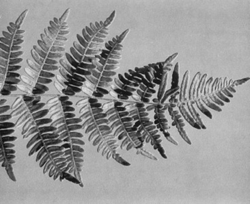
Hemorrhage, ulceration, and fibrin deposition are evident on the epithelium of the bladder and urethra. With chronic infection, polypoid growths may develop in the bladder mucosa; these masses grossly resemble tumors and must be definitively identified by histopathologic examination.125 One or both ureters may be enlarged, with purulent debris occasionally occluding the ureteral lumen. Pyelonephritis cases may show gross renal enlargement in acute to subacute cases (Fig. 34-22). On sagittal sectioning of the kidney, viscous, gray, odorless exudate is found within the renal pelvis and extending into the medulla and cortex.126 A Gram stain of the exudate is useful for differentiation of C. renale from E. coli infection. Renal abscesses, with gross distortion of renal size and shape, may be seen in cases of chronic pyelonephritis.
Aggressive antibiotic therapy is essential for successful treatment of UTI. Penicillin is the treatment of choice for C. renale infection; recommended dosage regimen includes procaine penicillin G (22,000 to 44,000 IU/kg IM twice daily) or ampicillin trihydrate (11 mg/kg IM twice daily).124 For valuable animals, higher serum and urinary concentrations of penicillin may be achieved with IV administration of sodium or potassium penicillin (22,000 to 44,000 IU/kg every 6 hours) or sodium ampicillin (10 to 50 mg/kg every 8 hours). Treatment should be continued for a minimum of 3 weeks. Subcutaneous injection of antimicrobial drugs may be necessary to limit muscle pain and swelling during the course of treatment. Urinalysis and urine culture should be repeated 1 week after treatment is discontinued to ensure complete resolution. After prolonged therapy with these extralabel dosages of antibiotics, residue withdrawal times for meat and milk must be extended appropriately. In addition, induction of diuresis through oral or parenteral fluid therapy may aid in removing necrotic debris and bacteria from the lumen of the urinary tract.
Urinary tract infection with E. coli or other coliforms may also be successfully treated with high doses of penicillin or ampicillin.124 Achieving high urinary concentrations of these antibiotics may render them effective against many coliforms, even those that show in vitro resistance to the expected serum concentrations of the antibiotic.124 Repeated assessment of appetite, attitude, rectal temperature, and reagent strip urinalysis is recommended for monitoring cows with coliform UTI that are receiving penicillin or ampicillin therapy. If these parameters do not improve after 96 hours of treatment, another antibiotic should be chosen.124 Gentamicin (2.2 mg/kg IM twice daily) has been used to successfully treat refractory coliform UTI in a cow, but the nephrotoxicity of the drug and the current prolonged slaughter withdrawal period are important considerations.124 Trimethoprim-sulfadiazine (15 mg/kg IV once daily)125 and ceftiofur (3 mg/kg IV twice daily)142 have also been used with success.
The prognosis for UTI in ruminants depends on the duration of infection, the extent of UTI (cystitis alone vs. unilateral or bilateral ureteritis and pyelonephritis), and the remaining renal function. The chances for successful treatment are improved if treatment is initiated early in the course of infection. In recent reports the combined case fatality and cull rate for pyelonephritis in dairy cattle varied between 18%124 and 33%139 for treated cases; however, antibiotic dose and duration varied greatly between these two studies. Cows with pyelonephritis and marked azotemia (BUN >100 mg/dL) were found to be at much greater risk for culling (odds ratio = 60) than nonazotemic cows with pyelonephritis.139
Isolation of animals infected with C. renale is recommended to limit spread of the organism, and disinfection of heavily contaminated areas is advised. Aseptic technique during urogenital procedures and disinfection of obstetric and surgical equipment will limit iatrogenic transmission. In herds using natural service, venereal transmission by subclinically infected bulls may be difficult to control over the long term. An artificial insemination or mass treatment program may be required to prevent further losses from UTI.
Amyloidosis in cattle is caused by deposition of insoluble protein fibrils in the kidney, GI tract, liver, and adrenal glands. Renal amyloidosis in cattle is characterized as a sporadic, chronic wasting disease. Amyloid deposition in the kidney disrupts the normal glomerular structure, resulting in a protein-losing nephropathy.
The most common clinical signs of amyloidosis include chronic diarrhea, weight loss, and poor productivity in mature animals.143,144 Generalized or ventral edema may be present as a result of hypoproteinemia. Alterations in appetite and attitude may be present, although this may be caused by concurrent disease. Enlargement of the left kidney may be palpated during rectal examination. The enlarged kidneys generally are not painful and maintain normal lobular patterns. Urine may develops stable foam after hitting the ground or being collected and shaken in a container, a result of high urine protein concentration.
Cattle with renal amyloidosis consistently develop marked proteinuria and hypolbuminemia.143 Serum creatinine and BUN levels may be elevated if renal damage is advanced. In cases of chronic, active inflammatory disease, hyperfibrinogenemia and hyperglobulinemia may occur.143,144 Polarized light microscopy and electron microscopy have been used to examine urine sediment for the presence of amyloid protein in urine.145
Amyloidosis must be differentiated from other diseases causing chronic diarrhea, hypoproteinemia, weight loss, and poor productivity. Diseases to consider include Johne’s disease, copper deficiency, salmonellosis, bovine viral diarrhea, GI parasitism, and glomerulonephritis. Other than amyloidosis, glomerulonephritis is the only other differential diagnosis routinely displaying prolonged proteinuria. Renal biopsy can be performed to differentiate glomerulonephritis from amyloidosis in the live animal.
Amyloidosis of cattle is classified as the reactive (AA) type,146,147 which is frequently associated with chronic inflammatory disease in domestic animals and humans.148 Concurrent inflammatory disease, such as traumatic reticuloperitonitis, pneumonia, mastitis, and metritis, have been found in some, but not all, cattle with amyloidosis.143,144 Serum amyloid A protein (SAA) is synthesized in the liver and is a precursor of amyloid A (AA) fibril in tissues.149 SAA concentrations increase dramatically in disorders such as trauma, neoplasia, and inflammatory disease. An elevation in SAA is apparently required for an animal to develop active amyloidosis.148 Elevations in SAA as a result of abnormal catabolism by the reticuloendothelial system may also increase AA fibril formation.148,150 AA fibrils are resistant to proteolysis, allowing for their accumulation in tissues over time.146 Accumulation of amyloid in the glomerulus alters glomerular filtration. A resultant hypoalbuminemia develops, which in turn decreases intravascular oncotic pressure. Diarrhea develops as a result of edema or amyloid deposition in the GI tract.144 The protein-losing nephropathy and diarrhea result in weight loss. Glomerular filtration rate will be reduced if the glomeruli are obliterated by amyloid deposition. Renal or pulmonary thrombosis may develop as a result of the loss of low—molecular-weight anticoagulants through the compromised kidney.151
Renal enlargement with yellow-tan to white discoloration is frequently present. A waxy quality of the renal parenchyma may be appreciated on cut surface of the kidney.143 Generalized edema resulting from hypoalbuminemia may be present. Some animals will have renal or pulmonary thrombosis.144 Other inflammatory lesions may be found in other sites. Histologic examination of the kidney may reveal amyloid deposition in the glomerulus, interstitium, and tubule lumen. Immunohistochemical tests using antihuman AA monoclonal antibody can be used for specific demonstration of amyloid in bovine kidney specimens.151
Because the lesions of amyloidosis are irreversible, the prognosis for affected cattle is poor. The resilient nature of the amyloid protein results in its persistence in tissues, even if the underlying cause of inflammatory disease is treated successfully. Specific treatment for amyloidosis has not been reported in cattle.
Glomerulonephritis (GN) is a rare clinical disorder of ruminants that may result from deposition of antigen-antibody complexes in the glomerular basement membrane or from binding of antibody to intrinsic or foreign antigens in the glomerulus. Glomerular injury occurs subsequent to targeting of glomerular tissues by the immune system. Nonimmune mechanisms may be involved in certain forms of the disease.
Cattle with GN may have a history of weight loss, poor productivity, and chronic diarrhea.152,153 Lethargy and generalized edema may be detected on physical examination. Rectal palpation may reveal a mildly enlarged but nonpainful left kidney.153 GN may be clinically occult in cattle persistently infected with bovine viral diarrhea (BVD) virus154 and in cattle with fascioliasis.155 GN has been associated with pregnancy toxemia in ewes; affected animals tend to show clinical signs typical of pregnancy toxemia156 (see Chapter 33).
Mesangiocapillary GN has been described in Finnish Landrace lambs of specific lineage in Scotland and Canada.157 The disease is heritable, but the exact mode of inheritance remains unknown. Clinical signs of this disease begin within hours after birth to 3 months of age. Affected lambs may be dull, ataxic, and appear blind. Fine muscle tremors, colic, and convulsions may also be seen.
The differential diagnosis for cattle with GN is similar to that for amyloidosis (see preceding section).
Heavy proteinuria, mild anemia, and hypoalbuminemia have been reported in cattle with GN.152,153 Granular casts, red blood cells, and leukocytes were found in the urine sediment of one affected cow.152 Azotemia, proteinuria, and ketonuria are found in ewes with GN associated with pregnancy toxemia. Mesangiocapillary GN in Finnish Landrace lambs is characterized by uremia, hypoalbuminemia, proteinuria, hypocalcemia, and hyperphosphatemia.157
In humans, GN may result from a variety of infectious, toxic, or autoimmune disorders, all of which induce eventual immunologic injury to the glomerulus.152,158 Antibodies may be directed against host or foreign antigens located in the vascular endothelium, mesangial cells, or basement membrane. In addition, circulating immune complexes may deposit in the glomerulus. The ultimate consequences of antigen-antibody interaction in the glomerulus are activation of complement and chemotaxis of leukocytes, both of which result in direct glomerular injury and increased glomerular permeability.158
Filtration of plasma albumin through the damaged glomerulus results in chronic albuminuria, eventually leading to reduced plasma oncotic pressure and generalized edema. Passage of antithrombin III through the damaged glomerulus and into the urine may result in a hypercoagulable state.159
Immunohistochemical data suggest involvement of immune-mediated mechanisms for spontaneous GN in cattle152,153 and sheep,157 GN associated with persistent BVD infection and fascioliasis in cattle,154,155 and mesangiocapillary GN of Finnish Landrace lambs.157 In the last condition a heritable deficiency of the third component of complement has been documented, but the role of this deficiency in GN remains unclear.156 Glomerulonephritis may also be an incidental histologic finding in animals with acute septic disease.157
The clinical and histopathologic characteristics of GN of pregnancy toxemia in ewes resemble those of the preeclampsia syndrome of women.156 Enlarged glomeruli with reduced blood content in glomerular capillaries are found throughout the renal cortex of affected ewes. The renal lesion in preeclampsic women may result from endothelial injury during disseminated intravascular coagulation (DIC) or an excessive glomerular vasomotor response to angiotensin.160 The lesion can be reversible in women, but the consequences of this condition in ewes have not been described.
Treatment of GN in ruminants has not been described. Because most cases of GN are advanced at diagnosis, the prognosis is poor. Mesangiocapillary GN in Finnish Landrace sheep is not invariably lethal, and some affected lambs may survive until adulthood.156
Hemolytic uremic syndrome (HUS) is classified within the group of thrombotic microangiopathy syndromes.161 HUS is a set of symptoms characterized clinically by acquired, nonimmune hemolytic anemia, thrombocytopenia, and acute renal failure (ARF).162 Histologically, HUS is characterized by renal thrombotic microangiopathy.163 HUS is the most common cause of ARF in young children and infants, with prodromal diarrhea occurring in approximately 90% of cases.164 Evidence indicates that almost all the postdiarrheal human cases of HUS are caused by enterohemorrhagic (EHEC) or verotoxigenic Escherichia coli infections, and that the majority of the cases in the United States are caused by the EHEC serotype O157:H7.164 Although three horses165,166 and a heifer167 have been reported with clinical syndromes similar to HUS, an etiologic agent was not identified in any of these four animals.
Horses with clinical signs indicative of HUS have exhibited fever, diarrhea, hematuria, hemoglobinuria, profound azotemia, oliguria, and ventral edema.165,166 Hematologic findings included leukocytosis, anemia, and evidence of hemolysis.165,166 The blood smear of one horse revealed the presence of poikilocytes and schistocytes.166 All three horses were euthanized after unsuccessful treatment of anuria and azotemia with fluid therapy and diuretics. The reported case in a heifer was fatal postparturient HUS demonstrated by severe progressive anuric renal failure, acute hemolytic anemia, and consumptive thrombocytopenia.167
In humans, HUS presents as pallor, oligoanuria, edema, seizures (rarely), or generalized hemorrhagic diathesis. In prodromal human cases, this syndrome develops on average 1 week after onset of diarrhea.162 The classical clinical syndrome of HUS in people includes ARF, hemolysis, thrombocytopenia, and manifestations of DIC.163 For treatment of HUS in humans, evidence is insufficient to support use of specific therapies, and some treatments, such as certain antibiotics and motility-modifying agents, may be detrimental.162 Supportive therapy, including fluid/plasma therapy and dialysis, can be of paramount importance.
In humans, HUS is the most common life-threatening complication of hemorrhagic colitis (HC) from EHEC infection. The EHEC strains produce the exotoxins Shiga toxin 1 (Stx1) and Shiga toxin 2 (Stx2), also referred to as verotoxin 1 and verotoxin 2.168 Although E. coli O157:H7 is the most widely publicized, as many as 100 different serotypes of E. coli (as well as Shigella and other Enterobacteriaceae) can carry the genes for Stx1 or Stx2 and are capable of causing disease.161
The EHEC organisms, including serotype O157:H7, are noninvasive but attach to the intestinal mucosa and produce characteristic histologic attaching and effacing lesions. The Shiga toxins (Stxs) released from the bacteria are believed to translocate across the mucosa, where they access the systemic circulation. The Stxs bind to specific glycolipid receptors on the surface of vascular endothelial cells, are internalized by endocytosis, and induce cell death through inhibition of protein synthesis.161 Activation of the coagulation cascade after exposure of subendothelial collagen may result in thrombosis of small vessels in the kidney and other organs. Oliguric or anuric ARF can result from fibrin/platelet thrombi in renal vessels and glomeruli, fibrinoid necrosis of vessel walls, congestion of glomeruli, and tubular ischemia. Because the coagulation events may be localized to certain organs, the results of laboratory tests of coagulation (e.g., PT, PTT) are not consistently abnormal.169
Cattle feces are considered to be the major source of EHEC; however, these bacteria have been isolated from the feces of many other asymptomatic species, including humans. EHEC organisms do not generally cause illness in cattle, although they do colonize the bowel. Fecal contamination of ground beef, other food sources, and water is thought to be the primary mode of transmission of the organisms. However, the small infectious dose makes person-to-person transmission a significant problem, especially in day care, nursing home, and outbreak situations.161
In the four large animals reported with HUS, the inciting cause was uncertain, and the isolation of EHEC or other Stx-producing organisms was not attempted.165-167 The heifer had a necrotizing endometritis, but during postmortem examination of the three horses, no focus of infection was identified. Renomegaly, renal infarcts, and scattered petechial and ecchymotic hemorrhages within the renal parenchyma were apparent on gross necropsy. Acute tubular necrosis and fibrin thrombi within the glomerular capillaries were evident on histologic examination.
Although the etiology was not determined, the clinical signs and pathologic lesions reported in the four large animal cases are compatible with a diagnosis of HUS. Based on these reports, the pathogenesis of HUS in large animal animals shares some features of the disease in humans.
Tubular necrosis (TN), or tubular nephrosis, is the disease condition that results from a variety of toxic, infectious, or hemodynamic insults to the kidneys. Compounds identified as nephrotoxins for ruminants are listed in Box 34-2. Hemodynamic causes of TN include diseases that reduce renal perfusion (blood loss, endotoxic shock) or that occlude the renal vasculature (DIC, renal vein thrombosis). Bilateral bacterial infection of the kidneys may result in ARF or chronic renal failure (CRF) as a result of destruction of nephrons by bacterial toxins and the host inflammatory response. Renal infection may be established by ascending UTI or by hematogenous infection of the kidneys. Depending on the nature and duration of the primary insult, widespread dysfunction or necrosis of tubular epithelial cells may produce reversible renal injury, ARF, or CRF.
The clinical signs of ARF in ruminants are nonspecific and usually are not indicative of overt urinary tract dysfunction. Depending on the inciting cause, anuria, oliguria, or polyuria may exist. Cattle with ARF frequently are presented for evaluation of poor appetite, diarrhea, or epistaxis.170 Depression, nasal discharge, ileus, melena, and mild free-gas bloat may also be present. If a concurrent septic condition exists, fever, tachycardia, and scleral injection may be present. The saliva may have a strong ammonia smell. Muscular weakness, even recumbency, may result from the acid-base and electrolyte imbalances and intravascular volume depletion that occur with severe acute TN. Rectal palpation findings are usually unremarkable, although renal enlargement and perirenal edema may be found in occasional cases. Table 34-2 lists clinical criteria that may facilitate diagnosis of TN caused by nephrotoxins. If untreated, CRF may ensue, usually producing weight loss in addition to the signs just listed. In such cases a reduction in size of the left kidney may be appreciated on rectal palpation of affected cattle.
Table 34-2 Clinical Characteristics of Acute Toxic Nephrosis Caused by Common Nephrotoxins
| Nephrotoxin | Clinical Findings* |
|---|---|
| Aminoglycosides | Usually nonoliguric, ototoxicosis possible; hematuria, glucosuria, proteinuria, increased serum trough concentration of drug |
| Tetracycline | Hematuria, glucosuria, proteinuria; possible hepatocellular enzyme elevation |
| Ionophores | Diarrhea, dark urine, dyspnea, cardiac dysrhythmias; elevated CK, AST, and indirect bilirubin |
| Ethylene glycol | Anuria or oliguria, tachypnea, ataxia, weakness; hemolysis, increased anion gap, increased serum osmolality, increased osmolar gap, acidosis, calcium oxalate crystalluria |
| Oxalate-containing plants, vitamin C | Hindlimb ataxia or paresis, apprehension, salivation; hypocalcemia, calcium oxalate crystalluria, aciduria, increased renal cortical echogenicity on ultrasonogram |
| Oak, acorns | Hemorrhagic diarrhea, ascites, hydrothorax, subcutaneous and perirenal edema; hyperfibrinogenemia, increased hepatic enzymes |
| Myoglobin | Muscle stiffness and weakness, dark urine; elevated serum CK and AST, positive reactions for blood and protein on urine chemistry strip, hemolyzed serum not present |
| Hemoglobin/methemoglobin | Icteric to pale mucous membranes, tachycardia, tachypnea, red or brown urine; positive blood and protein reactions on urine chemistry strip, anemia, elevated serum total protein, hemolyzed serum (hemoglobin only) |
| Arsenic | Colic, hemorrhagic diarrhea, ataxia; elevated blood and liver arsenic levels |
CK, Creatine kinase; AST, aspartate aminotransferase.
* Many general clinical characteristics of acute tubular necrosis are also present. See Clinical Signs and Clinical Pathology.
Renal (uremic) encephalopathy is a syndrome of brain dysfunction associated with renal disease. It is characterized by signs of intracranial disease, such as altered behavior or sensorium, weakness, motor dysfunction, and convulsions, which may resolve on restoration of normal renal function or improvement of clinical parameters of renal disease. The pathogenesis of renal encephalopathy is complex and incompletely understood; neurologic function appears to be impaired by alterations in the extracellular fluid content of acids, phosphorus, amino acids, or certain hormones. Alteration in neurotransmitter balance or release within the central nervous system (CNS) may also be involved. Renal encephalopathy appears to be a rare complication of renal failure in ruminants, as reported in two cows and a goat.171-173
Formulation of a differential diagnosis for a ruminant with TN may be difficult because of the nonspecific nature of the clinical signs and the variety of primary disease conditions that may predispose cattle to secondary TN. Coagulopathies and pulmonary abscesses are common causes of epistaxis in ruminants. The differential diagnoses for diarrhea are listed in Chapter 7. Female ruminants with advanced pregnancy toxemia may be depressed, azotemic, inappetent, and recumbent. Recumbent cattle should be evaluated for musculoskeletal injury, mastitis, metritis, peritonitis, spinal cord disease, and metabolic diseases. Cattle with TN are frequently misdiagnosed with milk fever because a temporary improvement in muscular strength may be seen in cattle with TN after treatment with calcium salts.
Elevation in BUN and creatinine levels occurs with clinical TN, and the azotemia is confirmed to be of renal origin by detection of isosthenuria on measurement of urine specific gravity. Proteinuria, hematuria, and granular casts may be found on urinalysis. Hypochloremia and metabolic alkalosis, resulting from abomasal atony or chloride loss in the urine, are often found in ruminants with ARF.170,174 Hyponatremia occurs after sodium loss in the urine. Because the kidney is the primary organ controlling magnesium excretion in ruminants, hypermagnesemia may occur in TN, particularly under conditions of high magnesium intake.170,175 Hyperphosphatemia results from reduced phosphorus excretion in saliva during anorexia, reduced urinary phosphorus excretion, and tissue hypoxia.176,177 Hypocalcemia is also common in TN in ruminants because of reduced calcium intake, GI stasis, urinary losses, and the competitive effect of hyperphosphatemia.170 Metabolic acidosis may develop in juvenile ruminants with TN and concurrent diarrhea.177,178
Fractional clearance (fractional excretion [FE]) of sodium has been used to help document renal failure in cattle.178 Values of 0% to 4% have been described in normal cattle; age, ration, and metabolic status may affect FE values.178 When applying this test, it is prudent to compare the patient’s FE value for sodium to that of an age-matched herdmate in a similar physiologic state and on a similar ration. The normal values for several urinary diagnostic indices in healthy calves have been reported.179
Reduced blood flow to the kidneys most often occurs during generalized loss of vascular volume, as seen with marked blood loss, septicemia, endotoxemia, or severe dehydration. Oliguria or anuria is seen initially, and urine output varies after IV fluid therapy. These conditions may also cause infarction of the renal cortex and renal vein thrombosis.170 Severe ruminal gas distention may impair renal perfusion.170 Prolonged, severe ischemia may destroy the tubular basement membrane, thereby preventing tubular epithelial cell regeneration.180
The high metabolic demands of renal tubular epithelial cells render them susceptible to toxins that disrupt cellular enzymes. The injury caused by most nephrotoxins is compounded by dehydration, which concentrates the toxin in the tubular filtrate, slows toxin clearance, and if severe, reduces renal perfusion. Because some nephrotoxins are therapeutic agents, it is vital that the veterinarian monitors appetite, body weight, water intake, urine output, routine urine chemistry, serum drug concentration, and serum creatinine concentration during administration of these agents. Young or elderly patients, patients with preexisting renal insufficiency or sepsis, those receiving other potentially nephrotoxic drugs, and patients on prolonged or high-dose therapy with these agents warrant the closest attention.181
In cases of toxic nephroses, the animal should first be removed from the toxin source, or treatment with a nephrotoxic drug should be discontinued. Rumenotomy, with removal of toxic material, is most beneficial if performed soon (within 24 hours) after the animal has ingested a nephrotoxin. Activated charcoal (2 to 4 g/kg PO) may bind the agent in the gut lumen. The use of magnesium sulfate or other magnesium-containing laxatives should be avoided in such cases because severe hypermagnesemia may result in animals with concurrent compromise of renal function. If an animal is exposed to a potentially harmful quantity of a nephrotoxin, prophylactic diuresis through fluid therapy is warranted. In such cases, if the veterinarian were to wait for azotemia to appear before initiating fluid therapy, significant (>75%) loss of nephron function would occur before medical intervention.
The cornerstone of treatment of TN is restoration of adequate renal perfusion and urine production. This is most effectively achieved through IV administration of isotonic, sodium-containing fluids, with calcium and potassium supplementation as indicated. If cost or facilities make IV fluid therapy impractical, repeated administration of water and electrolytes by stomach tube is an option. A small-bore stomach tube can be passed through the nasal cavity into the rumen and secured to the animal’s halter to allow one person to administer fluids repeatedly without the need for repeated tube passage. Placement of a small rumenostomy or securing in place a nasogastric tube allows one person to administer fluids repeatedly with relative ease. Administration of IV or oral fluids at 1.5 to 2 times the adult maintenance level of 60 mL/kg/day may be adequate to induce diuresis. The patient should be monitored for chemosis or labored or rapid respiration, which may be indicative of overhydration. Fluid therapy should be continued until azotemia resolves, at which time the patient’s voluntary fluid intake can be assessed. Oral supplementation of potassium and calcium salts may be necessary in some cases, because it is often not possible to add adequate yet safe levels of these salts to IV fluids in cases of refractory hypokalemia and hypocalcemia, respectively.
Restoration of urine production is necessary in anuric or oliguric animals. If fluid therapy does not promote diuresis, furosemide (1 mg/kg IV or IM) may be administered. Repeated administration (every 1 to 2 hours) may be necessary to induce urine production in oliguric or anuric patients. With repeated use of furosemide, the patient’s serum sodium and potassium concentrations must be monitored. Mannitol (0.25 g/kg IV) or dopamine (2 to 5 μg/kg/min IV) may be required to initiate urine flow if the previous measures are unsuccessful.
Lesions that occlude tubular blood flow (renal vein thrombosis, DIC) warrant a poor prognosis, whereas renal failure resulting from toxic causes carries a more favorable prognosis with early diagnosis and aggressive therapy. Return of appetite and progressive reduction in BUN and serum creatinine levels are positive prognostic indicators.182 Prolonged supportive treatment (2 to 3 weeks) may be necessary to allow for regeneration of tubular epithelium in cases of acute TN.
Leptospirosis is a complex disease of both animals and humans caused by pathogenic species of Leptospira.183-189 Pathogenic Leptospira species persist as chronic infections of the renal tubules of the maintenance host species, often causing little or no disease. Transmission to incidental hosts results from direct contact with urine from an infected maintenance host or through environmental and feed contamination with infected urine. Infection in the incidental host can cause acute disease in multiple organ systems, including the kidney, liver, and CNS, as well as result in abortion or reproductive failure. This discussion focuses on Leptospira infection and disease of the renal system of ruminants.
Leptospira is a diverse genus of motile, gram-negative, obligate aerobic, tightly coiled spirochetes approximately 0.1 to 0.3 μm in diameter and 6 to 20 μm in length.183-185 The bacteria can survive in the environment for up to 6 months.183,184,190 Leptospira species prefer a warm, moist environment with a pH of 7.2 to 8.0. Survival is short under dry conditions or at temperatures below 10°C.183 Leptospira does not survive freezing in the envrironment.191
Before 1989, Leptospira was divided into the pathogenic species, L. interrogans, and the nonpathogenic saprophytic species, L. biflexa. The current taxonomy and classification of Leptospira use a complex system of both serologic and genetic characteristics.184,185 Serologic classification is based on antigenic grouping of the lipopolysaccharide (LPS) and other outer surface antigens using the cross-agglutinin adsorption test (CAAT). More than 240 serovars are characterized. Antigenically related serovars are combined into larger serogroups, and serovars may also be further characterized into serotypes. Seventeen genomospecies have been identified based on DNA sequence heterogeneity.184,185
Leptospirosis is predominantly observed under conditions where livestock come in direct or indirect contact with urine from an infected maintenance host (Table 34-3).183-185190 The prevalence of infection within a maintenance host population tends to be high (30% to 50%); in such populations, infection is often spread between animals by direct contact.183 Transmission to incidental hosts is generally by contact with the environment, feed, or water that is contaminated with urine from an infected maintenance host. Transmission can also occur from contact with an infected fetus or uterine discharge. Survival of the bacteria in the environment and the incidence of infection in animals are increased in regions with warm, humid climatic conditions. The seasonal incidence is higher during the summer or fall in temperate regions and during the rainy season in warm-climate regions.184 Environmental conditions that contribute to moist surroundings and foot abrasions may contribute to Leptospira transmission, particularly in housed dairy cattle.
Table 34-3 Current Nomenclature, Maintenance Host, and Incidental Hosts for Common Leptospira Isolates in Ruminants
Cattle are the primary maintenance host reservoir of L. interrogans serovar hardjo (type hardjopragitno) and L. borgpetersenii serovar hardjo (type hardjo-bovis). L. interrogans serovar hardjo (type hardjopragitno) is isolated primarily from cattle in the United Kingdom, and L. borgpetersenii serovar hardjo (type hardjo-bovis) is observed worldwide. Cattle can also serve as maintenance or incidental hosts for L. interrogans serovar pomona and L. interrogans serovar grippotyphosa.
Serovars hardjo, pomona, and grippotyphosa are most often implicated in renal infection of cattle.192 Although data are scarce, renal disease caused by leptospirosis in small ruminants appears to be uncommon.191,193 Sheep may serve as subclinical carriers of serovar hardjo.187,194 Some serovars of Leptospira have zoonotic potential, and humans are always considered an incidental host.183-185188
An abattoir study of more than 5000 cattle in the United States identified approximately 2% renal carriers of L. interrogans.192 Serovar hardjo was the most common renal isolate, followed by serovar pomona and serovar grippotyphosa. A study of Texas slaughterhouse cattle detected Leptospira species in 36% of urine samples by PCR. The seroprevalence for serovars pomona and hardjo was 22% and 15%, respectively.195 A national survey showed 49% seroprevalence for L. interrogans serovars in cattle, with the highest seroprevalence found in cattle from southeastern, south-central, and Pacific Coast states.196 Because contact with urine from infected animals is a means of transmission within cattle populations, high stocking density or confinement may increase the rate of infection in a herd.
In general, infection of cattle with the host-adapted serovar hardjo rarely results in acute, severe disease. If present, signs of disease are usually mild in acutely infected cattle and may simply present as cases of undifferentiated fever. Persistent, latent urogenital infection usually follows acute infection, with most overt losses attributable to adverse effects on reproduction.188,197-199
Acute, severe renal disease is more characteristic of incidental (also termed “accidental”) infection of cattle, particularly calves, with a non—host-adapted serovar of Leptospira. However, exceptions to this generalization do occur because host immunity and virulence of the organism are variable.187,188
Serovar hardjo is host-adapted to cattle, and many infections are asymptomatic or result in nonspecific reproductive failure or abortion. Leptospira serovar hardjo infection of cattle may produce chronic interstitial nephritis of variable severity, but overt renal dysfunction is rarely observed.200 Chronic infection of the genital tract of cows and bulls is common.189,201 Protracted shedding of the organism in the urine often results, possibly lasting for the life of the animal.200,201 Infertility, stillbirth, abortion, and birth of weak calves are typical clinical manifestations of infection with serovar hardjo in cows.189,198,202,203 The fetus can be infected in utero, and if it survives the acute infection, it may be born persistently infected.183 Fever, agalactia, and mastitis may occasionally occur, and the resulting syndrome has been termed the “milk-drop syndrome”187 or “flabby udder.”188 The udder is uniformly soft, and the milk may be yellow- or red-tinged and thick.
In contrast, infection with the non—host-adapted serovars can result in severe systemic disease, hemolytic anemia, hepatitis, interstitial nephritis, and tubular nephrosis in calves and less often in adult cattle.189,200 Meningitis is a rare manifestation.193 Agalactia and mastitis often occur in lactating cows, and pregnant cows may abort. Urine shedding of non—host-adapted serovars by infected cattle can persist for weeks to months.188,189,203 Renal lesions result from direct damage to the vascular endothelium during leptospiremia, hypoxia from endothelial damage and hemolysis, tubular epithelial damage from hemoglobin, and interstitial nephritis.183,184,200 Some Leptospira serovars, particularly pomona, produce hemolysins that can cause acute intravascular hemolysis and anemia in cattle.
Clinical signs associated with acute infection may include fever, anorexia, lethargy, decreased milk production, petechiation, hemolytic anemia, and hemoglobinuria. Oliguria may be seen with interstitial nephritis or hemoglobinuric nephrosis. Elevated creatinine caused by pre-renal or renal causes may be observed on serum chemistry analysis. Examination of the urine may show proteinuria, pyuria, and cellular or granular casts in cases of nephritis. Hemoglobinemia and hemoglobinuria may be observed in patients with leptospire-induced hemolysis and can occasionally result in hemoglobinuric nephrosis.
Contaminated feed and surface water, wildlife, rodents, and domestic animals are potential sources of pathogenic serovars for cattle.183,184,187,188 Leptospires penetrate external mucosal surfaces and scarified or macerated skin. The bacteria multiply locally during an incubation period that can last 2 to 20 days.183,184 After the incubation phase, the organism enters the bloodstream through the lymphatics or by direct penetration into the blood vessel. Leptospiremia results in dissemination throughout the body and infection of multiple organs. This bacteremic phase lasts 4 to 7 days. Fever and other systemic signs are often present in clinically affected animals. Humoral antibodies can be detected at the end of the bacteremic phase. Opsonizing antibodies are generated and aid in clearing infection from most tissues in the host.
During the convalescent phase, leptospires may become localized in the mammary gland, kidney, or genital tract, where they appear to be protected from the immune response.183,204 Depending on the virulence of the serovar involved, chronic renal infection may create few histologic changes, mild interstitial nephritis, or diffuse, severe, lymphocytic interstitial nephritis with fibrosis.193 Nephritis may persist long after the host immune response has cleared the organism. Chronic infection of the kidney or reproductive tract allows for transmission of the organism in urine, uterine and vaginal secretions, placenta, fetal tissues, and semen.187,189,201,205 Shedding in the urine may last for weeks to months with non—host-adapted infections. Renal shedding of host-adapted serovars can persist for months to years. The bacteria reside in the lumen of the renal proximal tubules, where they are protected from phagocytes and humoral antibodies. The bacteria do not stimulate a systemic immune response while localized in the proximal tubule lumen, and thus serum antibody titers can decline and become negative even though the kidney is infected and shedding bacteria.
Multiple potential virulence factors may contribute to leptospirosis.184,185 Leptospiral LPS and outer membrane proteins are believed to contribute to the development of interstitial nephritis. However, leptospiral LPS has different biochemical properties that make it less toxic than other gram-negative lipopolysaccharides. Motility is an important pathogenic mechanism and contributes to invasion and dissemination of the bacteria. As many as 50 genes are related to leptospiral motility.185 Pathogenic Leptospira strains produce chemotaxis proteins, and some strains exhibit chemotaxis toward hemoglobin. Adherence to cells is in part conferred by fibronectin-binding protein present on the surface of pathogenic strains but not on nonpathogenic strains. Leptospiral immunoglobulin-like protein A (LigA) may also be involved in attachment and invasion. Additional proteins that may contribute to virulence include hemolysins, sphingomyelinase C, sphingomyelinase H, and hemolysis-associated protein 1 (HAP-1).184,185
The microscopic agglutination test (MAT) is the most widely used serologic test for the diagnosis of leptospirosis in cattle. The MAT detects antibodies to specific serovars, but cross-reactivity occurs between related serovars, particularly within the same serogroup. Thus, an infection with one strain may result in increased MAT titer to multiple serovars. Serum antibody enzyme-linked immunosorbent assay (ELISA) tests have been developed for research but have not yet been adopted in the routine clinical diagnostic setting for animals.206-209
An elevated serum antibody titer is observed after the bacteremic phase and is suggestive of Leptospira infection when associated with concurrent clinical signs. However, interpretation of a single titer is problematic in vaccinated animals or when endemic exposure is suspected.189,204 A fourfold increase in MAT titer between acute and convalescent serum samples, or conversion from a negative titer to a titer of 1/100 or greater, supports a diagnosis for both host-adapted and non—host-adapted serovars. However, vaccinated animals may have a diminished serologic response after challenge and renal colonization with L. borgpetersenii serovar hardjo.210 Serologic detection of persistent infection with serovar hardjo can be difficult because paired serum titers may be increasing, static, decreasing, or undetectable at examination (e.g., at abortion).189,204 It is generally recommended to consider the serovar with the highest titer as being the infecting strain. However, recent studies demonstrate that serologic antibody titers may not accurately predict the infecting strain in humans because of cross-reaction between different serovars within the same serogroup.211 Consultation with a clinical immunologist affiliated with the laboratory performing the MAT is recommended for accurate interpretation of results.
Leptospira shedding in urine and semen can be detected by multiple tests, including urine culture, phase-contrast microscopy, darkfield microscopy, fluorescent antibody (FA), polymerase chain reaction (PCR), nucleic acid hybridization, and immunoblot.192,204,205,212-214 Urine cultures are often unrewarding because of the fastidious nature of the organism, and conclusive results may not be obtained for up to 6 months.205 Both FA and PCR assays are typically used by veterinary diagnostic laboratories to identify Leptospira species from urine samples. Neither of these tests will determine the infecting serovar. The sensitivity of detecting Leptospira shedding can be improved by performing two tests on a single sample.205,214 Evaluation of serovar-specific antibody titers from positive animals may aid in identification of the infecting serovar.
Second-voiding urine samples collected after the administration of IV furosemide are recommended for urine testing.191,215 Furosemide is administered at 0.5 to 1.0 mg/kg IV or IM, and the first-voided urine is discarded. A second urine sample is then collected after cleaning the vulva of gross debris. Approximately 10 mL of urine should be collected and stored on ice (not frozen) for transport to the diagnostic laboratory. Urine from 10 to 15 adult animals should be tested when evaluating a herd for endemic Leptospira serovar hardjo infection.
Biopsy or necropsy samples of renal tissue may be treated with Warthin-Starry or Levaditi silver stains for microscopic examination.193 Immunoperoxidase staining was shown to be more sensitive for identifying Leptospira species in kidney and liver of naturally infected cattle than Levaditi silver stain.216
Treatment of acute leptospirosis caused by non—host-adapted serovars should focus on the elimination of the bacteria, systemic support, and diuresis if renal involvement is observed. In vitro susceptibility has been demonstrated for several antibiotics that could be used in food-producing ruminants, including ampicillin, amoxicillin, penicillin G, erythromycin, tetracycline, tylosin, and tilmicosin.203,217-219 Oxytetracycline (10 to 15 mg/kg IM twice daily) and dihydrostreptomycin (12.5 mg/kg IM twice daily) have been recommended for treatment of acutely infected cattle.191 Nephrotoxicosis is a potential concern with this dosage of oxytetracycline in cattle with preexisting renal disease, and dihydrostreptomycin is not currently available for use in cattle in the United States. Penicillin (25,000 IU/kg IM twice daily) and sodium ampicillin (20 mg/kg IM twice daily) have been suggested for treatment, but the efficacy of these regimens in cattle remains unproved.191 In humans, doxycycline (100 mg every 12 hours for 7 days) has been shown to reduce the duration and severity of illness.184
Intravenous or oral fluids and nonsteroidal antiinflammatory drugs (NSAIDs) may be indicated for systemic support. In animals with severe hemolysis, blood transfusion may be considered. Renal diuresis should be established in animals with hemoglobinuria or evidence of ARF from pigment nephrosis or interstitial nephritis. The prognosis for renal disease caused by leptospirosis is influenced by the virulence of the serovar involved, host immunity, and the extent of renal lesions. Cases with renal azotemia warrant a guarded prognosis because more than 75% of nephrons are affected in such cases, and chronic interstitial nephritis and fibrosis may occur after treatment of the acute disease.
Chronic renal infection with Leptospira serovar pomona in cattle can be eliminated with a single injection of dihydrostreptomycin (25 mg/kg IM), although spontaneous clearance of this serovar often occurs in cattle.191,203 Conflicting data exist on the efficacy of this treatment in clearing renal infection with serovar hardjo.220,221 Long-acting oxytetracycline (20 mg/kg IM or SC, two doses 10 days apart) has been recommended to treat chronic leptospiral infections or reduce the risk of introduction of infected animals into a herd.187 Oxytetracycline (20 mg/kg IM once), ceftiofur (2.2 mg/kg IM every 24 hours for five treatments), and tilmicosin (10 mg/kg SC once) have been shown effective in clearing renal shedding of L. borgpetersenii serovar hardjo from experimentally infected cattle.222
Draining or fencing off standing water may reduce transmission. Maintaining a dry, clean environment may also help reduce exposure and infection. Limiting rodent and wildlife contact with cattle and their feed and water is often difficult to accomplish, but it reduces the potential for transmission of non—host-adapted leptospires.189 For host-adapted Leptospira serovar hardjo, it is necessary to prevent or eliminate the renal carrier state in infected cattle in order to reduce transmission.
Vaccination is regarded as an effective means of preventing losses from leptospirosis. Conventional pentavalent (L. canicola, grippotyphosa, hardjo, icterohaemorrhagiae, and pomona), whole-cell, inactivated leptospiral vaccines are recommended in calves and adult cattle as an aid to prevent clinical disease caused by Leptospira species. However, these conventional pentavalent vaccines may not consistently prevent renal colonization and shedding after challenge with host-adapted L. borgpetersenii serovar hardjo (type hardjo-bovis).223,224
A monovalent L. borgpetersenii serovar hardjo (type hardjo-bovis) vaccine (Spirovac, Pfizer Animal Health, New York) is licensed for the prevention of Leptospira serovar hardjo infection, including reproductive and renal tract colonization, and urinary shedding for up to 12 months. This vaccine stimulates an immune response characterized by IgG1 and IgG2 agglutinating antibodies, as well as antigen-specific T-cell production of interferon gamma (IFN-γ).210,225-227 The strong IFN-γ response is consistent with induction of a type 1 immune response and may be responsible for antibody class switching to IgG2, which is a more potent opsonin than IgG1.225 Combination pentavalent Leptospira vaccines have been developed that also promote high L. hardjo antibody titers, in addition to the other serovars contained in the vaccine (Vira Shield 6+L5HB, Novartis Animal Health US, Greensboro, NC; Vista 5 L5 SQ, Intervet, Millsboro, Del). Because Leptospira species are largely extracellular pathogens, the relevant protective immune response appears to be antibody-mediated neutralization and opsonization during leptospiremia.184,185 There is no indication that cytotoxic immunity is a relevant immune response for protection or resolution of leptospirosis.
Although these vaccines help prevent colonization and shedding of leptospira serovar hardjo from the kidney, they are not effective in resolving current renal infection and elimination of the carrier state. Thus, vaccination at an early age followed by annual boosters is recommended to prevent initial renal colonization. Renal shedding in previously infected animals can be resolved by antibiotic treatment, and vaccination can be used to prevent future colonization.
Most severe congenital defects of the urinary tract of ruminants manifest at an early age, although occasionally defects remain clinically occult until adulthood.228 Congenital defects of the urinary system should be considered in the differential diagnosis for a young animal with renal disease or abnormal urination. However, tubular necrosis caused by severe volume depletion, nephrotoxins, and infectious diseases are much more common. If a congenital defect of the urinary tract is identified, a careful examination of other body systems should be performed; 73% of lambs with urogenital defects were found to have one or more defects in other organ systems.229 Urogenital defects in ruminants are described in Chapter 51.
Renal cysts are considered to be a common bovine renal defect230 and have been described in sheep229 and a goat.231 These fluid-filled cavities in the renal parenchyma are usually of no clinical significance unless they are large or numerous (polycystic kidneys).228,230 Renal agenesis was found to be the most common renal defect in lambs, with hydronephrosis and renal dysgenesis occurring less frequently.229 A retroperitoneal, perirenal pseudocyst (a fluid sac lacking an epithelial layer) has been reported in a ram with acute abdominal pain.232 Pseudocysts typically develop as outpocketings of the renal capsule and can develop as a result of renal trauma, urinary tract obstruction, or vascular or lymphatic anomalies.
Renal oxalosis is a metabolic disease of beefmaster calves that is suspected to have an inherited basis. The calves show weakness, lethargy, and anorexia within days to several weeks of age. Alopecia over the head, neck, and extremities; dehydration; and diarrhea may also be seen. Evidence of renal failure is found on serum chemistry analysis. An inherited abnormality of glycine or glyoxalate metabolism may generate high levels of endogenous oxalate, which readily complexes with calcium. As a result, calcium oxalate crystals accumulate in the renal tubules, obstructing outflow of the tubular filtrate. Exposure to oxalate-containing plants and ethylene glycol must be ruled out in all cases of renal oxalosis.233
Ectopic ureter is a rare congenital defect in which one or both ureters terminate in an abnormal location. An ectopic ureter may terminate in the urethra, vagina, or cervix or caudal to the bladder trigone in females. In males the ectopic ureter usually terminates in the urethra, vas deferens, or seminal vesicles. Urinary incontinence is the most common presenting complaint for affected animals.234 Urine dribbling results in scalding of the perineum and medial surfaces of the hindlimb in heifers, and scalding in males is usually located on the prepuce and ventral abdomen. Occasional episodes of normal micturition may be seen. UTI, polycystic kidney(s), and hydronephrosis may exist concurrent to ectopic ureter(s).
Definitive diagnosis requires IV contrast urography or endoscopic examination of the urinary tract. Options for surgical correction include transposition of the ectopic ureter(s) or, in the case of unilateral involvement, ipsilateral nephrectomy. The latter option is valid only if the contralateral kidney is functional, as determined by blood chemistry, and structurally normal or near normal, as determined by ultrasonography or IV pyelography. The use of affected animals for breeding should be discouraged.
Other than those associated with ingestion of bracken fern, primary neoplasms of the urinary tract of ruminants are rare. In a North American abbatoir survey, primary tumors of the bladder were detected in 0.05% of more than 21,000 cattle examined, with cases of papillomas, adenomas, and transitional cell carcinomas identified in the sample population.235
Renal carcinoma has been described in cattle,236,237 and nephroblastoma has been documented in a ewe and an aborted lamb.238 Renal involvement may be seen in occasional cases of lymphosarcoma in ruminants. Single cases of lymphoma involving the urinary bladder of a cow239 and a goat240 have been reported.
1 Humes HD, et al. Clinical and pathophysiologic aspects of aminoglycoside nephrotoxicity. Am J Kidney Dis. 1982;11:5.
2 Bartol JM, Divers TJ, Perkins GA. Nephrotoxicant-induced acute renal failure in five horses. Compend Cont Educ (Pract Vet). 2000;22:870.
3 Riviere JE, Traver DS, Coppoe GL. Gentamicin toxic nephropathy in horses with disseminated bacterial infection. J Am Vet Med Assoc. 1982;180:648.
4 Hinchcliff KW, McGuirk SM, MacWilliams PS. Gentamicin nephrotoxicity. Proceedings of the Thirty-third Annual Convention of the American Association of Equine Practitioners, New Orleans. 1987:65.
5 Sojka JE, Brown SA. Pharmacokinectic adjustment of gentamicin dosing in horses with sepsis. J Am Vet Med Assoc. 1986;189:784.
6 Geor RJ, Brashier MK. Rational approach to gentamicin dosing in neonatal foals. Proceedings of the Thirty-seventh Annual Convention of the American Association of Equine Practitioners, San Francisco. 1991:603.
7 Divers TJ. Urine production, renal function, and drug monitoring in the equine intensive-care unit. Clin Tech Equine Pract. 2003;2(2):188.
8 Schumacher J, et al. Effect of diet on gentamicin-induced nephrotoxicosis in horses. Am J Vet Res. 1991;52:1274.
9 Hewitt WL. Reflections on the clinical pharmacology of gentamicin. Acta Pathol Microb Scand. 1977;241(suppl 1381):151.
10 Gilbert DN. Once-daily aminoglycoside therapy. Antimicrob Agents Chemother. 1991;35:399.
11 Godbar LM, et al. Pharmacokinetics, nephrotoxicosis, and in vitro antibacterial activity associated with single versus multiple (three times) daily gentamicin treatments in horses. Am J Vet Res. 1995;56:613.
12 Magdesian KG, et al. Pharmacokinetics of a high dose of gentamicin administered intravenously or intramuscularly to horses. J Am Vet Med Assoc. 1998;213:1007.
13 Tudor RA, Papich MG, Redding WR. Drug disposition and dosage determination of once daily administration of gentamicin sulfate in horses after abdominal surgery. J Am Vet Med Assoc. 1999;215:503.
14 Adams R, et al. Evaluation of a technique for measurement of γ-glutamyltranspeptidase in equine urine. Am J Vet Res. 1985;46:147.
15 Adams R, Brown M, Gronwall R. In defense of gentamicin. Vet Rec. 1987;120:277.
16 Sweeney RW, et al. Kinetics of gentamicin elimination in two horses with acute renal failure. Equine Vet J. 1988;20:182.
17 Sprayberry KA, et al. Renal failure, laminitis, and colitis following severe rhabdomyolysis in a draft horse-cross with polysaccharide storage myopathy. Can Vet J. 1998;39:500.
18 Long PH. Red maple—associated pulmonary thrombosis in a horse. J Am Vet Med Assoc. 1984;184:977.
19 Alward A, Corriher CA, Barton MH, et al. Red maple (Acer rubrum) leaf toxicosis in horses: a retrospective study of 32 cases. J Vet Intern Med. 2006;20:1197.
20 Oh MS. Does serum creatinine rise faster in rhabdomyolysis? Nephron. 1993;63:255.
21 Rebhun WC, et al. Vitamin K3-induced renal toxicosis in the horse. J Am Vet Med Assoc. 1984;184:1237.
22 MacKay RJ, French TW, Nguyen HT, et al. Effects of large doses of phenylbutazone administration to horses. Am J Vet Res. 1983;44:774.
23 MacAllister CG, et al. Comparison of adverse effects of phenylbutazone, flunixin meglumine, and ketoprofen in horses. J Am Vet Med Assoc. 1993;202:71.
24 Read WK. Renal medullary crest necrosis associated with phenylbutazone therapy in horses. Vet Pathol. 1983;20:662.
25 Gunson DE. Renal papillary necrosis in horses. J Am Vet Med Assoc. 1983;182:263.
26 Behm RJ, Berg IE. Hematuria caused by renal medullary crest necrosis in a horse. Compend Cont Educ (Pract Vet). 1987;9:698.
27 Ehnen SJ, et al. Obstructive nephrolithiasis and ureterolithiasis associated with chronic renal failure in horses: eight cases (1981-1987). J Am Vet Med Assoc. 1990;197:249.
28 Dunn MJ, Zambraski EJ. Renal effects of drugs that inhibit prostaglandin synthesis. Kidney Int. 1980;18:609.
29 Clive DM, Stoff JS. Renal syndromes associated with nonsteroidal antiinflammatory drugs. N Engl J Med. 1984;310:563.
30 Gunson DE, Soma LR. Renal papillary necrosis in horses after phenylbutazone and water deprivation. Vet Pathol. 1983;20:603.
31 Krook L, et al. Hypercalcemia and calcinosis in Florida horses: implication of the shrub Cestrum diurnium as the causative agent. Cornell Vet. 1975;65:26.
32 Harrington DD, Page EH. Acute vitamin D3 toxicosis in horses: case reports and experimental studies of the comparative toxicity of vitamins D2 and D3. J Am Vet Med Assoc. 1983;182:1358.
33 McClure JJ, et al. Vitamin D intoxication in a thoroughbred racehorse. Compend Cont Educ (Pract Vet). 1987;9:573.
34 Roberts MC, Seawright AA. The effects of prolonged daily low-level mercuric chloride dosing in a horse. Vet Human Toxicol. 1978;20:410.
35 Bayly WM, et al. A reproducible means of studying acute renal failure in the horse. Cornell Vet. 1986;76:287.
36 Markel MD, et al. Acute renal failure associated with application of a mercuric blister in a horse. J Am Vet Med Assoc. 1984;185:92.
37 Schuh JCL, et al. Concurrent mercuric blister and dimethyl sulphoxide (DMSO) application as a cause of mercury toxicity in two horses. Equine Vet J. 1988;20:68.
38 Gunson DE, et al. Environmental zinc and cadmium pollution associated with generalized osteochondrosis, osteoporosis, and nephrocalcinosis in horses. J Am Vet Med Assoc. 1982;180:295.
39 Anderson GA, et al. Fatal acorn poisoning in a horse: pathologic findings and diagnostic considerations. J Am Vet Med Assoc. 1983;182:1105.
40 Schmitz DG. Toxic nephropathy in horses. Compend Cont Educ (Pract Vet). 1988;10:104.
41 Vivrette S, et al. Hemodialysis for treatment of oxytetracycline-induced acute renal failure in a neonatal foal. J Am Vet Med Assoc. 1993;203:105.
42 Barton MH. Use of polymixin B for treatment endotoxemia in horses. Compend Cont Educ (Pract Vet). 2000;22:1056.
43 Schmitz DG. Cantharidin toxicosis in horses. J Vet Intern Med. 1989;3:208.
44 Shibahara T, Takai H, Shimizu C, et al. Equine renal granuloma caused by Halicephalobus species. Vet Rec. 2002;151:672.
45 Divers TJ, et al. Acute renal failure in six horses resulting from hemodynamic causes. Equine Vet J. 1987;19:178.
46 Roberts MC, Kelly WR. Renal dysfunction in a case of purpura haemorrhagica in a horse. Vet Rec. 1982;110:144.
47 Morris CF, et al. Hemolytic uremic-like syndrome in two horses. J Am Vet Med Assoc. 1987;191:1453.
48 Dolente BA, Seco OM, Lewis ML. Streptococcal toxic shock in a horse. J Am Vet Med Assoc. 2000;217:64.
49 Divers TJ, Byars TD, Shin SJ. Renal dysfunction associated with infection of Leptospira interrogans in a horse. J Am Vet Med Assoc. 1992;201:1391.
50 Bernard WV, et al. Hematuria and leptospiruria in a foal. J Am Vet Med Assoc. 1993;203:276.
51 Hogan PM, et al. Acute renal disease due to Leptospira interrogans in a weanling. Equine Vet J. 1996;28:331.
52 Frazer ML. Acute renal failure from leptospirosis in a foal. Aust Vet J. 1999;77:499.
53 Kiper ML, Traub-Dargatz JL, Wrigley RH. Renal ultrasonography in horses. Compend Cont Educ (Pract Vet). 1990;12:993.
54 Hoffman KL, Wood AKW, McCarthy PH. Sonographic-anatomic correlation and imaging protocol for the kidneys of horses. Am J Vet Res. 1995;56:1403.
55 Divers TJ, Yeager AE. The value of ultrasonographic examination in the diagnosis and management of renal diseases in horses. Equine Vet Educ. 1997;7:334.
56 Roussel AJ, et al. Urinary indices of horses after intravenous administration of crystalloid solutions. J Vet Int Med. 1993;7:241.
57 Matthews HK, et al. Measuring renal function in horses. Vet Med. 1993;88:349.
58 Barratt-Boyes SM, et al. Ultrasound localization and guidance for renal biopsy in the horse. Vet Radiol. 1991;32:121.
59 Lieberthal W, Levisky NG. Treatment of acute tubular necrosis. Semin Nephrol. 1990;10:571.
60 Divers TJ. Treatment of acute renal failure in the horse. Proceedings of the Thirty-first Annual Convention of American Association of Equine Practitioners, Toronto. 1985:673.
61 Divers TJ. Acute renal failure in the horse. Proceedings of the 10th Bain Fallon Memorial Lectures, Australia, 1988, Adelaide. 1988:93.
62 Lindner A. Synergism of dopamine and furosemide in oliguric acute renal failure. Nephron. 1987;33:121.
63 Johansson AM, Gardner SY, Levine JF, et al. Furosemide continuous rate infusion in the horse: evaluation of enhanced efficacy and reduced side effects. J Vet Intern Med. 2003;17:887.
64 Power DA, Duggan J, Brady HR. Renal-dose (low-dose) dopamine for the treatment of sepsis-related and other forms of acute renal failure: ineffective and probably dangerous. Clin Exp Pharmacol Physiol Suppl. 1999;26:S23.
65 Groover ES, Woolums AR, Cole DJ, et al. Risk factors associated with renal insufficiency in horses with primary gastrointestinal disease: 26 cases (2000–2003). J Am Vet Med Assoc. 2006;228:572.
66 Thornhill JA, et al. Hemodialysis in a horse with acute renal failure as induced by rhabdomyolysis. Proceedings of the Conference of Research Workers in Animal Disease Chicago. 1983:16.
67 Gallatin LL, Couetil LL, Ash SR. Use of continuous-flow peritoneal dialysis for the treatment of acute renal failure in an adult horse. J Am Vet Med Assoc. 2005;226:756.
68 Divers TJ. Chronic renal failure in horses. Compend Cont Educ (Pract Vet). 1983;5:S310.
69 Schott HC, et al. Chronic renal failure in 99 horses. Proceedings of the Forty-third Annual Convention of the American Association of Equine Practitioners. 1997:345.
70 Banks KL, Henson JB. Immunologically mediated glomerulitis of horses. II. Antiglomerular basement membrane antibody and other mechanisms in spontaneous disease. Lab Invest. 1972;26:708.
71 Roberts MC, Kelly WR. Renal dysfunction in a case of purpura haemorrhagica in a horse. Vet Rec. 1982;110:144.
72 Divers TJ, et al. Equine glomerulonephritis and renal failure associated with complexes of group C streptococcal antigen and IgG antibody. Vet Immunol Immunopathol. 1992;32:93.
73 Banks KL, Henson JB, McGuire TC. Immunologically mediated glomerulitis of horses. I. Pathogenesis in persistent infection by equine infectious anemia virus. Lab Invest. 1972;26:701.
74 Sabnis SG, Gunson DE, Antonovych TT. Some unusual features of mesangioproliferative glomerulonephritis in horses. Vet Pathol. 1984;21:574.
75 Ehnen SJ, et al. Obstructive nephrolithiasis and ureterolithiasis associated with chronic renal failure in horses: eight cases (1981–1987). J Am Vet Med Assoc. 1990;197:249.
76 Andrews FM, et al. Bilateral renal hypoplasia in four young horses. J Am Vet Med Assoc. 1986;189:209.
77 Ronen N, et al. Renal dysplasia in two adult horses: clinical and pathological aspects. Vet Rec. 1993;132:269.
78 Bertone JJ, et al. Monitoring the progression of renal failure in a horse with polycystic kidney disease: use of the reciprocal of serum creatinine concentration and sodium sulfanilate clearance half-time. J Am Vet Med Assoc. 1987;191:565.
79 Held JP, Wright B, Henton JE. Pyelonephritis associated with renal failure in a horse. J Am Vet Med Assoc. 1986;189:688.
80 Carrick JB, Pollitt CC. Chronic pyelonephritis in a brood mare. Aust Vet J. 1987;64:252.
81 Sloet van Oldruitenborgh-Oosterbaan MM, Klabec HC. Ureteropyelonephritis in a Fresian mare. Vet Rec. 1988;122:609.
82 Jakob W. Spontaneous amyloidosis of mammals. Vet Pathol. 1971;8:292.
83 Haschek WM, King JM, Tennant BC. Primary renal cell carcinoma in two horses. J Am Vet Med Assoc. 1981;179:992.
84 Wimberly HC, Antonovych TT, Lewis RM. Focal glomerulosclerosis-like disease with nephrotic syndrome in a horse. Vet Pathol. 1981;18:692.
85 Roberts MC, Seiler RJ. Renal failure in a horse with chronic glomerulonephritis and renal oxalosis. J Equine Med Surg. 1979;3:278.
86 Chandler KJ, Johnston HM, Murphy DM. Polycystic kidney disease in an aged pony. Vet Rec. 2003;153:754.
87 Brown C. Equine nephrology. Vet Annu. 1986;26:1.
88 Macharg MA, et al. Two methods for the treatment of ureterolithiasis in a mare. Vet Surg. 1984;13:95.
89 Tennant B, et al. Chronic renal failure in the horse. Proceedings of the Twenty-fourth Annual Convention of the American Association of Equine Practitioners. 1978:293.
90 Koterba AM, Coffman JR. Acute and chronic renal disease in the horse. Compend Cont Educ (Pract Vet). 1981;3:S461.
91 Brobst DF, et al. Parathyroid hormone evaluation in normal horses and horses with renal failure. Equine Vet Sci. 1982;2:150.
92 Tennant B, et al. Hypercalcemia and hypophosphatemia in ponies following bilateral nephrectomy. Exp Biol Med. 1981;167:365.
93 Naylor JM, Kronfeld DS, Acland H. Hyperlipemia in horses: effects of undernutrition and disease. Am J Vet Res. 1980;41:899.
94 Schott HC, Hodgson DR, Bayly WM. Haematuria, pigmenturia and proteinuria in exercising horses. Equine Vet J. 1995;27:67.
95 Kohn CW, Strasser SL. 24-hour renal clearance and excretion of endogenous substances in the mare. Am J Vet Res. 1986;47:1332.
96 Kiper ML, Traub-Dargatz JL, Wrigley RH. Renal ultrasonography in horses. Compend Cont Educ (Pract Vet). 1990;12:993.
97 Hoffman KL, Wood AKW, McCarthy PH. Sonographic-anatomic correlation and imaging protocol for the kidneys of horses. Am J Vet Res. 1995;56:1403.
98 Divers TJ, Yeager AE. The value of ultrasonographic examination in the diagnosis and management of renal diseases in horses. Equine Vet Educ. 1997;7:334.
99 Matthews HK, et al. Measuring renal function in horses. Vet Med. 1993;88:349.
100 Morris DD, Divers TJ, Whitlock RH. Renal clearance and fractional excretion of electrolytes over a 24 hour period. Am J Vet Res. 1984;45:2431.
101 Brewer BD, et al. A comparison of inulin, para-aminohippuric acid and endogenous creatinine clearance as measures of renal function in neonatal foals. J Vet Intern Med. 1990;4:301.
102 Matthews HK, et al. Comparison of standard and radionuclide methods for measurement of glomerular filtration rate and effective renal blood flow in female horses. Am J Vet Res. 1992;53:1612.
103 Walsh DM, Royal HD. Evaluation of a single injection of 99mTc-labeled diethylenetriaminepentaacetic acid for measuring glomerular filtration rate in horses. Am J Vet Res. 1992;53:776.
104 Schott HC, et al. Nuclear scintigraphy as a diagnostic aid in the evaluation of renal disease in horses. Proceedings of Thirty-ninth Annual Convention of the American Association of Equine Practitioners. 1993:251.
105 Divers TJ. Management of chronic renal failure in the horse. Proceedings of the Thirty-first Annual Convention of the American Association of Equine Practitioners. 1985:679.
106 Cianciaruso B, et al. Salt intake and renal outcome in patients with progressive renal disease. Miner Electrolyte Metab. 1998;24:296.
107 Erslev AJ. Erythropoietin. N Engl J Med. 1991;324:1339.
108 Watson TDG, et al. Selective measurement of lipoprotein and hepatic triglyceride lipase in heparinized horses. Am J Vet Res. 1992;53:771.
109 Scharschmidt LA, et al. Effects of dietary fish oil on renal insufficiency in rats with subtotal nephrectomy. Kidney Int. 1987;32:700.
110 Cappeli P, et al. Lipids in the progression of chronic renal failure. Nephron. 1992;62:31.
111 Klahr S, et al. The effects of dietary protein restriction and blood-pressure control on the progression of chronic renal disease. N Engl J Med. 1994;330:877.
112 Ikizler TA, Hakim RM. Nutrition in end-stage renal disease. Kidney Int. 1996;50:343.
113 Bretzel RG. Protecting the residual renal function: which drugs of choice? Am J Hypertens. 1997;10:159S.
114 Porush JG. Hypertension and chronic renal failure: the use of ACE inhibitors. Am J Kidney Dis. 1998;31:177.
115 Müller GA, et al. Prevention of progression of renal fibrosis: how far are we? Kidney Int. 1996;49(suppl 54):S75.
116 Frye MA. Pathophysiology, diagnosis and management of urinary tract infection in the horse. Vet Clin Equine Pract. 2006;22:497.
117 Rubin RH, Tolkoff-Rubin NE, Cotran RS. Urinary tract infection, pyelonephritis, and reflux nephropathy. Brenner BM, Rector FC, editors. The kidney, ed 4, vol II. Saunders: Philadelphia, 1991;1369.
118 Mulholland SG, et al. Effect of hormonal deprivation on the bladder defense mechanism. J Urol. 1982;127:1010.
119 Adams LG, et al. Cystitis and ataxia associated with sorghum ingestion. J Am Vet Med Assoc. 1969;155:518.
120 Roberts MC. Ascending urinary tract infection in ponies. Aust Vet J. 1979;55:191.
121 Kisthardt KK, Schumacher J, Finn-Bodner ST, et al. Severe renal hemorrhage caused by pyelonephritis in 7 horses: clinical and ultrasonographic evaluation. Can Vet J. 1999;40:571.
122 Farrar WE. Infections of the urinary tract: symposium on infections in office practice. Med Clin North Am. 1983;67:187.
123 Stamm WE. Measurement of pyuria and its relation to bacteriuria. Am J Med. 1983;28:53.
124 Kiper ML, Traub-Dargatz JL, Wrigley RH. Renal ultrasonography in horses. Compend Cont Educ (Pract Vet). 1990;12:993.
125 Hoffman KL, Wood AKW, McCarthy PH. Sonographic-anatomic correlation and imaging protocol for the kidneys of horses. Am J Vet Res. 1995;56:1403.
126 Divers TJ, Yeager AE. The value of ultrasonographic examination in the diagnosis and management of renal diseases in horses. Equine Vet Educ. 1997;7:334.
127 Sullins KE, Traub-Dargatz JL. Endoscopic anatomy of the equine urinary tract. Compend Cont Educ. 1984;11:S663.
128 Traub-Dargatz JL, McKinnon AO. Adjunctive methods of examination of the urogenital tract. Vet Clin North Am Equine Pract. 1988;4:339.
129 Schott HC, Hodgson DR, Bayly WM. Ureteral catheterisation in the horse. Equine Vet Educ. 1990;2:140.
130 Murray BE. Vancomycin-resistant Enterococcal infections. N Engl J Med. 2000;342:710.
131 Ling GV. Clinical use experience. In: Proceedings of the Symposium on Trimethoprim/sulfadiazine. Research Triangle Park, NC: Burroughs-Wellcome; 1978:61.
132 Tolkoff-Rubin NE, Weber D, Fang LS, et al. Single-dose therapy with trimethoprim/sulfamethoxazole for urinary tract infections in women. Rev Infect Dis. 1982;4:444.
133 Divers TJ, et al. Experimental induction of Proteus mirabilis cystitis in the pony and evaluation of therapy with trimethoprim/sulfadiazine. Am J Vet Res. 1981;42:1203.
134 Keys TF. Antimicrobials commonly used for urinary tract infections. Mayo Clin Proc. 1977;52:680.
135 Bach MC, et al. Susceptibility of recently isolated pathogenic bacteria to trimethoprim and sulfamethoxazole separately and combined. J Infect Dis. 1973;128(suppl):408.
136 Nouws JFM, et al. Pharmacokinetics and renal clearance of sulfamethazine, sulfamerazine, and sulfadiazine and their N4-acetyl and hydroxy metabolites in horses. Am J Vet Res. 1987;48:392.
137 Divers TJ. Urinary tract: horse, cow. In: Johnston D, editor. Bristol veterinary handbook of antimicrobial therapy. Princeton, NJ: Veterinary Learning Systems; 1982:84.
138 Sidor TA, Resnick MI. Urinary tract infections. In: Speck WT, Blumer JL, editors. Pediatric clinics of North America. Philadelphia: Saunders; 1983:322.
139 Cummings LE, et al. Pharmacokinetics of gentamicin in newborn to 30-day-old foals. Am J Vet Res. 1990;51:1988.
140 Sweeney RW, Divers TJ, Rossier Y. Disposition of gentamicin administered intravenously to horses with sepsis. J Am Vet Med Assoc. 1992;200:503.
141 Divers TJ. Serum levels of chloramphenicol after oral dosing and nasogastric administration in horses with acute Salmonella enteritis. Proceedings of the Equine Colic Research Symposium, Athens, Ga. 1982:194.
142 Horspool LJI, McKellar QA. Disposition of oxytetracycline in horses, ponies and donkeys after intravenous administration. Equine Vet J. 1990;22:284.
143 Pringle JK, Ducharme NG, Baird JD. Ectopic ureter in the horse: three cases and a review of the literature. Can Vet J. 1990;31:26.
144 Blikslager AT, Green EM. Ectopic ureters in horses. Compend Cont Educ (Pract Vet). 1992;14:802.
145 Johnson PJ, et al. Treatment of two mares with obstructive (vaginal) urinary outflow incontinence. J Am Vet Med Assoc. 1987;191:973.
146 Sponseller BA, et al. Frontal septation of the bladder in a mare. J Vet Intern Med. 1998;12:313.
147 Watanabe T, et al. High incidence of occult neurogenic bladder dysfunction in neurologically intact patients with thoracolumbar spinal injuries. J Urol. 1998;159:965.
148 Holt PE, Mair TS. Ten cases of bladder paralysis associated with sabulous urolithiasis in horses. Vet Rec. 1990;127:108.
149 Madison JB. Estrogen-responsive urinary incontinence in an aged pony mare. Compend Cont Educ (Pract Vet). 1984;6:S390.
150 Watson ED, et al. Oestrogen responsive urinary incontinence in two mares. Equine Vet Educ. 1997;9:81.
151 Kay AD, Lavoie JP. Urethral pressure profilometry in mares. J Am Vet Med Assoc. 1987;191:212.
152 Clark ES, et al. Cystometrography and urethral pressure profiles in healthy horse and pony mares. Am J Vet Res. 1987;48:552.
153 Ronen N. Measurements of urethral pressure profiles in the male horse. Equine Vet J. 1994;26:55.
154 Pringle JK, Ducharme NG, Baird JD. Ectopic ureter in the horse: three cases and a review of the literature. Can Vet J. 31(26), 1990.
155 Blikslager AT, Green EM. Ectopic ureters in horses. Compend Cont Educ (Pract Vet). 14(802), 1992.
156 Squire KRE, Adams SB. Bilateral ureterocystostomy in a 450-kg horse with ectopic ureters. J Am Vet Med Assoc. 201(1213), 1992.
157 Modransky PD, Wagoner PC, Robinette JD. Surgical correction of bilateral ectopic ureters in two foals. Vet Surg. 12(141), 1983.
158 Blikslager AT, et al. Ultrasonography and excretory urography in the diagnosis of bilateral ectopic ureters in a foal. Vet Radiol. 33(41), 1992.
159 Brown PJ, Holt PE. Primary renal cell carcinoma in four horses. Equine Vet J. 1985;17:473.
160 Fischer AT, et al. Neoplasia of the equine urinary bladder as a cause of hematuria. J Am Vet Med Assoc. 1985;186:1294.
161 Traub JL, et al. Intraabdominal neoplasia as a cause of chronic weight loss in the horse. Compend Cont Educ (Pract Vet). 1983;5:S526.
162 Matsuda K, Kousaka Y, Nagamine N, et al. Papillary renal adenoma of distal nephron differentiation in a horse. J Vet Med Sci. 2007;69(7):763.
163 Murray RC, Brodbelt DC. Hematuria due to renal hemangiosarcoma in a donkey. Equine Pract. 1999;21:14.
164 Sweeney RW, Hamir AN, Fisher RR. Lymphosarcoma with urinary bladder infiltration in a horse. J Am Vet Med Assoc. 1991;199:1177.
165 Rhind SM, Sturgeon B. Papillary renal carcinoma with an unusual clinical presentation in a horse. Equine Vet Educ. 1999;11(4):171.
166 Rhind SM, Hawe C, Dixon PM, et al. Oral metastasis of renal cell carcinoma in a horse. J Comp Pathol. 1999;120:97.
167 Rumbaugh ML, Latimer FG, Porthouse KP, et al. Renal carcinoma with osseous and pulmonary metastases in an Arabian gelding. Equine Vet J. 2003;35(1):107.
168 Barton MH, Sharma P, LeRoy BE, et al. Hypercalcemia and high serum parathyroid hormone-related protein concentration in a horse with multiple myeloma. J Am Vet Med Assoc. 2004;225(3):409.
169 Swain JM, Pirie RS, Hudson NPH, et al. Insulin-like growth factors and recurrent hypoglycemia associated with renal cell carcinoma in a horse. J Vet Intern Med. 2005;19:613.
170 Macharg MA, et al. Two methods for the treatment of ureterolithiasis in a mare. Vet Surg. 1984;13:95.
171 Laverty S, et al. Urolithiasis in 68 horses. Vet Surg. 1992;21:56.
172 Ehnen SJ, et al. Obstructive nephrolithiasis and ureterolithiasis associated with chronic renal failure in horses: eight cases (1981–1987). J Am Vet Med Assoc. 1990;197:249.
173 Wooldridge AA, Seahorn TL, Williams J, et al. Chronic renal failure associated with nephrolithiasis, ureterolithiasis, and renal dysplasia in a 2-year-old quarter horse gelding. Vet Radiol Ultrasound. 1999;40:361.
174 Kiper ML, Traub-Dargatz JL, Wrigley RH. Renal ultrasonography in horses. Compend Cont Educ (Pract Vet). 1990;12:993.
175 Hoffman KL, Wood AKW, McCarthy PH. Sonographic-anatomic correlation and imaging protocol for the kidneys of horses. Am J Vet Res. 1995;56:1403.
176 Divers TJ, Yeager AE. The value of ultrasonographic examination in the diagnosis and management of renal diseases in horses. Equine Vet Educ. 1997;7:334.
177 Rodger LD, Carlson GP, Moran ME, et al. Resolution of a left ureteral stone using electrohydraulic lithotripsy in a thoroughbred colt. J Vet Intern Med. 1995;9:280.
178 Neumann RD, Ruby AL, Ling GV, et al. Ultrastructure and mineral composition of urinary calculi from horses. Am J Vet Res. 1994;55:1357.
179 Juzwiak JS, Bain FT, Slone DE, et al. Unilateral nephrectomy for treatment of chronic hematuria due to nephrolithiasis in a colt. Can Vet J. 1988;29:931.
180 DeBowes RM, Nyrop KA, Boulton CH. Cystic calculi in the horse. Compend Cont Educ (Pract Vet). 1984;6:S268.
181 Holt PE, Pearson H. Urolithiasis in the horse: a review of 13 cases. Equine Vet J. 1984;16:31.
182 DeBowes RM. Surgical management of urolithiasis. Vet Clin North Am Equine Pract. 1988;4:461.
183 Holt PE, Mair TS. Ten cases of bladder paralysis associated with sabulous urolithiasis in horses. Vet Rec. 1990;127:108.
184 Kaneps AJ, Shires GMH, Watrous BJ. Cystic calculi in two horses. J Am Vet Med Assoc. 1985;187:737.
185 Mair TS, Holt PE. The aetiology and treatment of equine urolithiasis. Equine Vet Educ. 1994;6:189.
186 MacHarg MA, et al. Electrohydraulic lithotripsy for treatment of a cystic calculus in a mare. Vet Surg. 1985;14:325.
187 Eustace RA, Hunt JM. Electrohydraulic lithotripsy for treatment of cystic calculus in two geldings. Equine Vet J. 1988;20:221.
188 Howard RD, Pleasant RS, May KA. Pulsed dye laser lithotripsy for treatment of urolithiasis in two geldings. J Am Vet Med Assoc. 1998;212:1600.
189 Sertich PL, et al. Medical management of urinary calculi in a stallion with breeding dysfunction. J Am Vet Med Assoc. 1998;213:843.
190 Johnson PJ, Crenshaw KL. The treatment of cystic and urethral calculi in a gelding. Vet Med. 1990;85:891.
191 Remillard RL, et al. Dietary management of cystic calculi in a horse. J Equine Vet Sci. 1992;12:359.
192 Trotter GW, Bennett DG, Behm RJ. Urethral calculi in five horses. Vet Surg. 1981;10:159.
193 Mair TS, Walmsley JP, Phillips TJ. Surgical treatment of 45 horses affected by squamous cell carcinoma of the penis and prepuce. Equine Vet J. 2000;32:406.
194 Schott HC, Hines MT. Severe urinary tract hemorrhage in two horses. J Am Vet Med Assoc. 1994;204:1320. (letter)
195 Latimer FG, Magnus R, Duncan RB. Arterioureteral fistula in a colt. Equine Vet J. 1991;23:483.
196 Schott HC, et al. Renal arteriovenous malformation in a quarter horse foal. J Vet Intern Med. 1996;10:204.
197 Pardo V, Berian MG, Levi DF, Strauss J. Benign primary hematuria: clinicopathologic study of 65 patients. Am J Med. 1979;67:817.
198 Hughes JH, et al. Massive nontraumatic hematuria: a challenging demand demanding immediate action. Postgrad Med. 1990;67:97.
199 Stone EA, DeNovo RC, Rawlings CA. Massive hematuria of nontraumatic renal origin in dogs. J Am Vet Med Assoc. 1983;183:868.
200 Holt PE, Lucke VM, Pearson H. Idiopathic renal hemorrhage in the dog. J Small Anim Pract. 1987;28:253.
201 Kaufman AC, Barsanti JA, Selcer BA. Benign essential hematuria in dogs. Compend Contin Educ (Pract Vet). 1994;16:1317.
202 Roberts MC, Kelly WR. Renal dysfunction in a case of purpura haemorrhagica in a horse. Vet Rec. 1982;110:144.
203 Morris CF, et al. Hemolytic uremic-like syndrome in two horses. J Am Vet Med Assoc. 1987;191:1453.
204 Dolente BA, Seco OM, Lewis ML. Streptococcal toxic shock in a horse. J Am Vet Med Assoc. 2000;217:64.
205 Bernard WV, et al. Hematuria and leptospiruria in a foal. J Am Vet Med Assoc. 1993;203:276.
206 Kisthardt KK, et al. Severe renal hemorrhage caused by pyelonephritis in 7 horses: clinical and ultrasonographic evaluation. Can Vet J. 1999;40:571.
207 Sullins KE, et al. Treatment of hemospermia in stallions: a discussion of 18 cases. Compend Cont Educ (Pract Vet). 1988;10:1396.
208 Lloyd KC, et al. Ulceration in the proximal portion of the urethra as a cause of hematuria in horses: four cases (1978–1985). J Am Vet Med Assoc. 1989;194:1324.
209 Schumacher J, et al. Urethral defects in geldings with hematuria and stallions with hemospermia. Vet Surg. 1995;24:250.
210 Dunn MJ, Zambraski EJ. Renal effects of drugs that inhibit prostaglandin synthesis. Kidney Int. 1980;18:609.
211 Gunson DE, Soma LR. Renal papillary necrosis in horses after phenylbutazone and water deprivation. Vet Pathol. 1983;20:603.
212 Hughes D. Polyuria and polydipsia. Compend Cont Educ (Pract Vet). 1992;14:1161.
213 Knotterbelt DC. Polyuria-polydipsia in the horse. Equine Vet Educ AE. 2000;2:237.
214 Sufit E, Houpt KA, Sweeting M. Physiological stimuli of thirst and drinking patterns of ponies. Equine Vet J. 1985;17:12.
215 Houpt KA. Thirst in horses: the physiological and psychological causes. Equine Pract. 1987;9:28.
216 Cymbaluk NF. Water balance of horses fed various diets. Equine Pract. 1989;11:19.
217 Love S. Equine Cushing’s disease. Br Vet J. 1993;149:139.
218 Hillyer MH, Taylor FGR, Mair TS. Diagnosis of hyperadrenocorticism in the horse. Equine Vet Educ. 1992;4:131.
219 Van der Kolk JH, et al. Equine pituitary neoplasia: a clinical report of 21 cases (1990–1992). Vet Rec. 1993;133:594.
220 Link RP. Glucose tolerance in horses. J Am Vet Med Assoc. 1940;97:261.
221 Green EM, Hunt EL. Hypophyseal neoplasia in a pony. Compend Cont Educ (Pract Vet). 1985;7:S249.
222 Browning AP. Polydipsia and polyuria in two horses caused by psychogenic polydipsia. Equine Vet Educ AE. 2000;2:231.
223 Brobst DF, Bayly WM. Responses of horses to a water deprivation test. Equine Vet Sci. 1982;2:51.
224 Genetzky RM, Loparco FV, Ledet AE. Clinical pathological alterations in horses during a water deprivation test. Am J Vet Res. 1987;48:1007.
225 Buntain BJ, Coffman JR. Polyuria and polydypsia in a horse induced by psychogenic salt consumption. Equine Vet J. 1981;13:266.
226 Schryver HF, et al. Salt consumption and the effect of salt on mineral metabolism in horses. Cornell Vet. 1987;77:122.
227 Robertson GL. Differential diagnosis of polyuria. Annu Rev Med. 1988;39:425.
228 Breukink HJ, Van Wegen P, Schotman AJH. Idiopathic diabetes insipidus in a Welsh pony. Equine Vet J. 1983;15:284.
229 Filar J, Ziolo T, Szalecki J. Diabetes insipidus in the course of encephalitis in the horse. Med Weterynaryjna. 1971;27:205.
230 Schott HCII, et al. Nephrogenic diabetes insipidus in sibling colts. J Vet Intern Med. 1993;7:68.
231 Harb MF, et al. Central diabetes insipidus in dogs: 20 cases (1986–1995). J Am Vet Med Assoc. 1996;209:1884.
232 Takemura N. Successful long-term treatment of congenital nephrogenic diabetes insipidus in a dog. J Small Anim Pract. 1998;39:592.
233 Muylle E, et al. Non-insulin dependent diabetes mellitus in a horse. Equine Vet J. 1986;18:145.
234 Baker JR, Richie HE. Diabetes mellitus in the horse: a case report and review of the literature. Equine Vet J. 1974;6:7.
235 Ruoff WW, et al. Type II diabetes mellitus in a horse. Equine Vet J. 1986;18:143.
236 Traver DS, et al. Peritonitis in a horse: a cause of acute abdominal distress and polyuria-polydipsia. J Equine Med Surg. 1977;1:36.
237 Kinter LB, Huffman WF, Stassen FL. Antagonists of the antidiuretic activity of vasopressin. Am J Physiol. 1988;254:F165.
238 Hardy RM, Osborne CA. Canine pyometra: pathophysiology, diagnosis and treatment of uterine and extra-uterine lesions. J Am Anim Hosp Assoc. 1974;10:245.
239 Thurmon JC, et al. Xylazine causes transient dose-related hyperglycemia and increased urine volume in mares. Am J Vet Res. 1984;45:224.
240 Gellai M. Modulation of vasopressin antidiuretic action by renal α2-adrenoceptors. Am J Physiol. 1990;259:F1.
241 Battle D. Renal tubular acidosis. Med Clin North Am. 1983;67:859.
242 Rocher LL, Tannen RL. The clinical spectrum of renal tubular acidosis. Annu Rev Med. 1986;37:319.
243 Mueller DL, Jergens AE. Renal tubular acidosis. Compend Contin Educ (Pract Vet). 1991;13:435.
244 Hansen TO. Renal tubular acidosis in a mare. Compend Cont Educ (Pract Vet). 1986;8:864.
245 Trotter GW, et al. Type II renal tubular acidosis in a mare. J Am Vet Med Assoc. 1986;188:1050.
246 Ziemer EL, et al. Clinical features and treatment of renal tubular acidosis in two horses. J Am Vet Med Assoc. 1987;190:294.
247 Van der Kolk JH, Kalsbeek HC. Renal tubular acidosis in a mare. Vet Rec. 1993;133:43.
248 MacLeay JM, Wilson JH. Type-II renal tubular acidosis and ventricular tachycardia in a horse. J Am Vet Med Assoc. 1998;212:1597.
249 Ziemer EL, et al. Renal tubular acidosis in two horses: diagnostic studies. J Am Vet Med Assoc. 1987;190:289.
250 Halperin ML, et al. Urine ammonium: the key to the diagnosis of distal renal tubular acidosis. Nephron. 1988;50:1.
251 Nyrop KA, et al. Rupture of the urinary bladder in two post-parturient mares. Compend Cont Educ (Pract Vet). 1984;6:S510.
252 Gibson KT, Trotter GW, Gustafson SB. Conservative management of uroperitoneum in a gelding. J Am Vet Med Assoc. 1992;200:1692.
253 Richardson DW, Kohn CW. Uroperitoneum in the foal. J Am Vet Med Assoc. 1983;182:267.
254 Hackett RP. Rupture of the urinary bladder in neonatal foals. Compend Cont Educ. 1984;6:S488.
255 Hardy J. Uroabdomen in foals. Equine Vet Educ. 1998;10:21.
256 Behr MJ, et al. Metabolic abnormalities associated with rupture of the urinary bladder in neonatal foals. J Am Vet Med Assoc. 1981;178:263.
257 Adams R, et al. Exploratory celiotomy for suspected urinary tract disruption in neonatal foals: a review of 18 cases. Equine Vet J. 1988;20:13.
258 Kablack KA, et al. Uroperitoneum in the hospitalised equine neonate: retrospective study of 31 cases, 1988-1997. Equine Vet J. 2000;32:505.
259 Lees MJ, et al. Subcutaneous rupture of the urachus, its diagnosis and surgical management in three foals. Equine Vet J. 1989;21:462.
260 Robertson JT, et al. Repair of a ureteral defect in a foal. J Am Vet Med Assoc. 1983;183:799.
261 Divers TJ, Byars TD, Spirito M. Correction of bilateral ureteral defects in a foal. J Am Vet Med Assoc. 1988;192:384.
262 Jean D, Marcoux M, Louf CF. Congenital bilateral distal defect of the ureters in a foal. Equine Vet Educ. 1998;10:17.
263 Cutler TJ, et al. Bilateral ureteral tears in a foal. Aust Vet J. 1997;75:413.
264 Hoffmann KL, Wood AK, McCarthy PH. Ultrasonography of the equine neonatal kidney. Equine Vet J. 2000;32:109.
265 Kawashima A, et al. Ureteropelvic junction injuries secondary to blunt abdominal trauma. Radiology. 1997;205:487.
266 Bauer JE. Normal blood chemistry. In: Koterba AM, Drummond WH, Kosch PC, editors. Equine clinical neonatology. Philadelphia: Lea & Febiger; 1990:608.
267 Schott HC, Mansmann RA. Biochemical profiles of normal equine amniotic fluid at parturition. Equine Vet J Suppl. 1988;5:52.
268 Edwards DJ, Brownlow MA, Hutchins DR. Indices of renal function: values in eight normal foals from birth to 56 days. Aust Vet J. 1990;67:251.
269 Geor RJ, Brashier MK. Rational approach to gentamicin dosing in neonatal foals. Proceedings of the Thirty-seventh Annual Convention of the American Association of Equine Practitioners, San Francisco. 1991:603.
1 Kimberling CV, Arnold KS. Diseases of the urinary system of sheep and goats. Vet Clin North Am Large Anim Pract. 1983;5:637.
2 Scott DW. Large animal dermatology. Philadelphia: Saunders, 1988;102.
3 Southcott WH. Etiology of ovine posthitis: description of a causal organism. Aust Vet J. 1965;41:193.
4 Brook AH, Southcott WH, Stacy BD. Etiology of ovine posthitis: relationship between ovine posthitis and a causal organism. Aust Vet J. 1966;42:9.
5 Southcott WH. Epidemiology and control of ovine posthitis and vulvitis. Aust Vet J. 1965;41:225.
6 Uzal FA, Woods L, Stillian M, et al. Abortion and ulcerative posthitis associated with caprine herpesvirus-1 infection in goats in California. J Vet Diag Invest. 2004;16:478.
7 Shelton M, Livingston CW. Posthitis in angora wethers. J Am Vet Med Assoc. 1975;167:154.
8 Dent CHR. Ulcerative vulvitis and posthitis in Australian sheep and cattle. Vet Bull. 1971;41:719.
9 Hay L. Prevention and treatment of urolithiasis in sheep. In Pract. 1990;12:87.
10 Gasthuys F, Martens A, DeMoor A. Surgical treatment of urethral dilatation in seven cattle. Vet Rec. 1996;138:17.
11 Bailey CB. Siliceous urinary calculi in bulls, steers, and partial castrates. Can J Anim Sci. 1975;55:187.
12 Wolfe DF, Moll HD, May KA. Urolithiasis. In: Wolfe DF, Moll HD, editors. Large animal urogenital surgery. Baltimore: Williams & Wilkins; 1999:349.
13 Oehme FW, Tillman H. Diagnosis and treatment of ruminant urolithiasis. J Am Vet Med Assoc. 1965;147:1331.
14 Hofmeyr CFB. Ruminant urogenital surgery. Ames: Iowa State University Press, 1987;15.
15 VanMetre DC, Smith BP. Clinical management of urolithiasis in small ruminants. Proc ACVIM. 1991;9:555.
16 Benson GJ, Thurmon JC. Regional anesthesia. In: Howard JL, editor. Current veterinary therapy 3: food animal practice. Philadelphia: Saunders; 1992:77.
17 House JK G, et al. Assessment of ruminant digestive system. Vet Clin North Am Food Anim Pract. 1992;8:189.
18 Todhunter P, Baird AN, Wolfe DW. Erection failure secondary to obstructive urolithiasis in a male goat. J Am Vet Med Assoc. 1996;209:650.
19 Smith JA, Divers TJ, Lamp TM. Ruptured urinary bladder in a postparturient cow. Cornell Vet. 1983;73:3.
20 Carr EA, et al. Ruptured urinary bladder after dystocia in a cow. J Am Vet Med Assoc. 1993;202:631.
21 Baxter GM, Zamos DT, Mueller PO. Uroperitoneum attributable to ruptured urachus in a yearling bull. J Am Vet Med Assoc. 1992;200:517.
22 Gera KL, Nigam JM. Urolithiasis in bovines: a report of 193 clinical cases. Indian Vet J. 1979;56:417.
23 Sockett DC, et al. Metabolic changes due to experimentally induced rupture of the bovine urinary bladder. Cornell Vet. 1986;76:198.
24 Aldridge BM, Garry FB. Chronic partial obstructive urolithiasis causing hydronephrosis and chronic renal failure in a steer. Cornell Vet. 1992;82:311.
25 Blood DC, Radostits OM. Veterinary medicine, ed 7, London: Balli豄 Tindall; 1989:402.
26 Divers TJ, Reef VB, Roby KA. Nephrolithiasis resulting in intermittent ureteral obstruction in a cow. Cornell Vet. 1989;79:143.
27 Ziemer EL, Smith BP. Pyelonephritis, glomerulonephritis, and urolithiasis in a Holstein bull with diarrhea. Compend Cont Educ (Pract Vet). 1988;10:82.
28 Braun U, Schefer U, Fohn J. Urinary tract ultrasonography in normal rams and in rams with obstructive urolithiasis. Can Vet J. 1992;33:654.
29 Palmer JL, et al. Contrast radiography of the lower urinary tract in the management of obstructive urolithiasis in small ruminants and swine. Vet Radiol. 1998;39:175.
30 Scott P. Ultrasonography of the urinary tract in male sheep with urethral obstruction. In Pract. 2000;22:329.
31 Scott P, Rhind S. Ultrasonography in the differential diagnosis of azotaemia in an adult Cheviot ram. Vet Rec. 2002;150:150.
32 Haven ML, et al. Surgical management of urolithiasis in small ruminants. Cornell Vet. 1993;83:47.
33 Donecker JM, Bellamy JEC. Blood chemical abnormalities in cattle with ruptured bladders and ruptured urethras. Can Vet J. 1982;23:355.
34 Harris CL, Krawiec DR. The pathophysiology of uremic bleeding. Compend Cont Educ (Pract Vet). 1990;12:1294.
35 Divers TJ, et al. Acute renal disorders in cattle: a retrospective study of 22 cases. J Am Vet Med Assoc. 1982;181:694.
36 Larson BL. Identifying, treating, and preventing bovine urolithiasis. Vet Med. 1996;91:366.
37 Rakestraw PC, et al. Tube cystostomy for treatment of urolithiasis in small ruminants. Vet Surg. 1995;24:498.
38 Surawicz B. Relationship between electrocardiogram and electrolytes. Am Heart J. 1967;73:814.
39 Vivrette S, et al. Hemodialysis for treatment of oxytetracycline-induced acute renal failure in a neonatal foal. J Am Vet Med Assoc. 1993;203:105.
40 Murray MJ. Urolithiasis in a ram. Compend Cont Educ (Pract Vet). 1985;7(suppl):269.
41 Garett PD. Urethral recess in male goats, sheep, cattle, and swine. J Am Vet Med Assoc. 1987;191:689.
42 Smith MC. The reproductive anatomy and physiology of the male goat. In: Morrow DA, editor. Current therapy in theriogenology 2. Philadelphia: Saunders; 1986:616.
43 Van Weeren PR, Klein WR, Voorhout G. Urolithiasis in small ruminants. I. A retrospective evaluation of urethrostomy. Vet Q. 1987;9:76.
44 Winter RB, et al. Catheterization: an effective method of treating bovine urethral calculi. Vet Med. 1987;82:1261.
45 Stone WC, et al. Prepubic urethrostomy for relief of urethral obstruction in a sheep and a goat. J Am Vet Med Assoc. 1997;210:939.
46 Pearce SG, Dearo AC, Howard BE, Brisson BA. Management of obstructive urolithiasis and concurrent urethral rupture in a goat. Aust Vet J. 2003;81:268.
47 Ewoldt JM, Anderson DE, Miesner MD, Saville WJ. Short- and long-term outcome and factors predicting survival after surgical tube cystostomy for treatment of obstructive urolithiasis in small ruminants. Vet Surg. 2006;35:417.
48 Streeter R, Washburn K, McCauley C. Percutaneous tube cystostomy and vesicular irrigation for treatment of obstructive urolithiasis in a goat. J Am Vet Med Assoc. 2002;221:546.
49 Fortier L, Gregg A, Erb H, Fubini SL. Caprine obstructive urolithiasis: requirement for 2nd surgical intervention and mortality after percutaneous tube cystostomy, surgical tube cystostomy, or urinary bladder marsupialization. Vet Surg. 2004;33:661.
50 Tulleners EP, Hamilton GF, Farrow CS. Surgical repair of ruptured urinary bladder in a ram. J Am Vet Med Assoc. 1980;177:708.
51 Noordsy JL. Food animal surgery, ed 3, Trenton: Veterinary Learning Systems; 1994:501.
52 Hastings DH. Retention catheters for treatment of steers with ruptured bladders. J Am Vet Med Assoc. 1965;147:1329.
53 May KA, et al. Urinary bladder marsupialization for treatment of obstructive urolithiasis in male goats. Vet Surg. 1998;27:583.
54 Weaver AD. Nephrectomy and cystotomy in a bull. Vet Rec. 1964;76:191.
55 Tulleners EP, et al. Indications for unilateral bovine nephrectomy: a report of four cases. J Am Vet Med Assoc. 1981;179:696.
56 Klahr S. Pathophysiology of obstructive nephropathy. Kidney Int. 1983;23:414.
57 Bailey CB. Silica metabolism and silica urolithiasis in ruminants: a review. Can J Anim Sci. 1981;61:219.
58 Maes D, Vrielinck J, Millet S, Janssens G, Deprez P. Urolithiasis in finishing pigs. Vet J. 2004;168:209.
59 Urinary calculi of beef cattle, Annual Report 1961, Montana Veterinary Research Laboratory, Montana Experimental Station, Bozeman, Mont, 1961
60 Salman MD, et al. Rates of diseases and their associated costs in two Colorado sheep feedlots (1985-1986). J Am Vet Med Assoc. 1988;193:1518.
61 Jensen R, Mackey DR. Diseases of feedlot cattle, ed 3, Philadelphia: Lea & Febiger; 1979:262.
62 Osborne CA, Klausner JS. Calcium oxalate urolithiasis. In: Kirk RW, editor. Current veterinary therapy VII. Philadelphia: Saunders; 1988:45.
63 Romanowski RD. Biochemistry of urolith formation. J Am Vet Med Assoc. 1965;147:1324.
64 Bailey CB. The precipitation of polymerized silicic acid by urine protein: a possible mechanism in the etiology of silica urolithiasis. Can J Anim Sci. 1972;50:305.
65 Jubb KVF, Kennedy PC, Palmer N. Pathology of domestic animals, ed 4, San Diego: Academic; 1993:525.
66 Udall RH, Seger CL, Chen-Chow F. Studies on urolithiasis. VI. The mechanism of action of sodium chloride in the control of urinary calculi. Cornell Vet. 1965;55:198.
67 Hoar DW, Emerick RJ, Embry LB. Influence of calcium source, phosphorus level, and acid-forming effects of the diet on feedlot performance in lambs. J Anim Sci. 1970;31:118.
68 McIntosh GH. Urolithiasis in animals. Aust Vet J. 1978;54:267.
69 Manning RA, Blaney BJ. Epidemiological aspects of urolithiasis in domestic animals in Queensland. Aust Vet J. 1986;63:423.
70 Bailey CB. Effects of ammonium chloride on formation of siliceous urinary calculi in calves. Can J Anim Sci. 1976;56:359.
71 Stewart SR, Emerick RJ, Pritchard RH. Effects of dietary ammonium chloride and variations in calcium to phosphorus ratio on silica urolithiasis in sheep. J Anim Sci. 1991;69:2225.
72 Bushman DH, Emerick RJ, Embry LB. Experimentally induced ovine phosphatic urolithiasis: relationships involving dietary calcium, phosphorus, and magnesium. J Nutr. 1965;87:499.
73 Hoar DW, Emerick RJ, Embry LB. Potassium, phosphorus, and calcium interrelationships influencing feedlot performance and phosphatic urolithiasis in lambs. J Anim Sci. 1970;30:597.
74 Kallfelz FA, et al. Dietary magnesium and urolithiasis in growing calves. Cornell Vet. 1987;77:33.
75 Loretti A, DeOliveira L, Cruz C, Driemeier D. Clinical and pathological study of an outbreak of obstructive urolithiasis in feedlot cattle in southern Brazil. Pesquisa Vet Brasil. 2003;23:61.
76 Villar D, et al. Case report: obstructive urolithiasis in a feedlot steer. Bovine Pract. 2003;37:74.
77 Stewart SR, Emerick RJ, Kayongo-Male H. Silicon-zinc interactions and potential roles for dietary zinc and copper in minimizing silica urolithiasis. J Anim Sci. 1993;71:946.
78 Stewart SR, Emerick RJ, Pritchard RH. High dietary calcium to phosphorus ratio and alkali-forming potential as factors promoting silica urolithiasis in sheep. J Anim Sci. 1990;68:498.
79 Angus KW. Nephropathy in young lambs. Vet Rec. 1990;126:525.
80 Adair HS, Adams WH. Ascorbic acid as suspected cause of oxalate nephrotoxicosis in a goat. J Am Vet Med Assoc. 1990;197:1626.
81 Van Metre DC, et al. Obstructive urolithiasis in ruminants: surgical management and prevention. Compend Cont Educ (Pract Vet). 1996;18:S275.
82 Bushman DH, Emerick RJ, Embry LB. Effect of various chlorides and calcium carbonate calcium, phosphorus, sodium, potassium, and chloride balance and their relationship to urinary calculi in lambs. J Anim Sci. 1968;27:490.
83 Crookshank HR, et al. Effect of chemical and enzymatic agents on the formation of urinary calculi in fattening steers. J Anim Sci. 1960;19:595.
84 Jardon PW. Using urine pH to measure anionic salt diets. Compend Cont Educ (Pract Vet). 1995;17:860.
85 Bailey CB. Formation of siliceous urinary calculi in calves given supplements containing large amounts of sodium chloride. Can J Anim Sci. 1973;53:55.
86 Virtala AM, Mechor GD, Grohn YT, et al. Morbidity from nonrespiratory diseases and mortality in dairy heifers during the first three months of life. J Am Vet Med Assoc. 1996;208:2043.
87 Bouckaert JH, deMoor A. Surgical treatment of umbilical infections in calves. Vet Rec. 1965;77:771.
88 Trent AM, Smith DF. Surgical management of umbilical masses with associated umbilical cord remnant infections in calves. J Am Vet Med Assoc. 1984;185:1531.
89 Baxter GM, Zamos DT, Mueller PO. Uroperitoneum attributable to ruptured urachus in a yearling bull. J Am Vet Med Assoc. 1992;200:517.
90 Bell GJ, Macrae AI, Milne EM, et al. Extensive uroperitoneum and pleural effusion associated with necrotic urachal remnant in a bull calf. Vet Rec. 2004;154:508.
91 Hassel DM, Tyler JW, Tucker RL, et al. Clinical vignette: urachal abscess and cystitis in a calf. J Vet Intern Med. 1995;9:286.
92 Hylton WE, Rousseaux CG. Intestinal strangulation: association with omphaloarteritis in a calf. J Am Vet Med Assoc. 1985;186:1099.
93 Mesaric M, Modic T. Strangulation of the small intestine in a cow by a persistent urachal remnant. Vet Rec. 2003;153:688.
94 Trent AM, Smith DF. Pollakiuria due to urachal abscesses in two heifers. J Am Vet Med Assoc. 1984;184:984.
95 Madigan JE, House JK. Patent urachus, omphalitis and other umbilical abnormalities. In: Smith BP, editor. Large animal internal medicine. ed 3. St Louis: Mosby; 2002:368.
96 Baxter GM. Umbilical masses in calves: diagnosis, treatment, and complications. Compend Cont Educ (Pract Vet). 1989;11:505.
97 Staller GS, Tulleners EP, Reef VB, et al. Concordance of ultrasonographic and physical findings in cattle with an umbilical mass or suspected to have infection of the umbilical cord remnants; 32 cases (1987-1989). J Am Vet Med Assoc. 1995;206:77.
98 Rings DM. Umbilical hernias, umbilical abscesses, and urachal fistulas: surgical considerations. Vet Clin North Am Food Anim Pract. 1995;11:137.
99 Boure L, Foster RA, Palmer M, et al. Use of an endoscopic suturing device for laparoscopic resection of the apex of the bladder and umbilical structures in normal neonatal calves. Vet Surg. 2001;30:319.
100 Ducharme NG, Stem ES. Eversion of the urinary bladder in a cow. J Am Vet Med Assoc. 1981;179:996.
101 Peter AT, Arighi M, Gaines JD. Herniation of distal jejunum into the partially everted urinary bladder of a cow. Can Vet J. 1989;30:830.
102 Frazier GS. Uterine torsion followed by jejunal incarceration in a partially everted urinary bladder in a cow. Aust Vet J. 1988;65:24.
103 Hojbjerg A. Eversion of the bovine bladder. Bovine Pract. 1990;25:120.
104 Friesen CH, Theoret CL, Barber SM. Urinary bladder eversion with hydronephrosis and renal failure in a beef cow. Can Vet J. 1995;36:710.
105 Gaines JD. Postparturient pelvic entrapment of the bladder in two cows. J Am Vet Med Assoc. 1988;193:222.
106 Campbell SG, Fubini SL. External lumps on sheep and goats. Vet Clin North Am Large Anim Pract. 1983;5:457.
107 Scott PR, Gessert ME. Ultrasonographic examination of 12 ovine vaginal prolapses. Vet J. 1998;155:323.
108 Divers TJ. Assessment of the urinary system. Vet Clin North Am Food Anim Pract. 1992;8:373.
109 Hopkins NCG. Aetiology of enzootic hematuria. Vet Rec. 1986;118:715.
110 Hoque M, Somvanashi R, Singh GR, et al. Ultrasonographic evaluation of urinary bladder in normal, fern fed, and enzootic hematuria-affected cattle. J Vet Med Physiol Pathol Clin Med. 2002;49:403.
111 Humphreys DJ. Veterinary toxicology, ed 3, London: Bailli豄 Tindall; 1988:258.
112 Osweiler GD, Ruhr P. Plants affecting blood coagulation. In: Howard JL, editor. Current veterinary therapy 3: food animal medicine. Philadelphia: Saunders; 1986:404.
113 Campo MS, et al. Association of bovine papillomavirus type 2 and bracken fern with bladder cancer in cattle. Cancer Res. 1992;52:6898.
114 Chick BF, Quin C, McCleary BV. Pteridophyte intoxication of livestock in Australia. In: Seawright AA, et al, editors. Plant toxicology. Melbourne: Dominion; 1985:453.
115 Madewell BR, Theilen GH. Tumors of the urinary tract. In: Theilen GH, Madewell BR, editors. Veterinary cancer medicine. ed 2. Philadelphia: Lea & Febiger; 1987:567.
116 Campo MS. Bovine papillomavirus and cancer. Vet J. 1997;154:175.
117 Bringuier P, et al. Bracken fern—induced bladder tumors in guinea pigs. Am J Pathol. 1995;147:858.
118 Pennie WD, Campo MS. Synergism between bovine papillomavirus type 4 and the flavonoid quercetin in cell transformation in vitro. Virology. 1992;190:861.
119 Anderson RA, et al. Viral proteins of bovine papillomavirus type 4 during the development of alimentary canal tumors. Vet J. 1997;154:69.
120 Panuckcu AM, et al. Carcinogenic and mutagenic activities of milk from cows fed bracken fern (Pteridium aquilinium). Cancer Res. 1978;38:1556.
121 Alonso-Amelot ME, et al. Bracken ptaquiloside in milk. Nature. 1996;382:587.
122 Borzacchiello G, Galati AP, Roperto F. Cyclooxygenase-1 and -2 expression in urothelial carcinomas of the urinary bladder in cows. Vet Pathol. 2003;40:455.
123 Jubb KVF, Kennedy PC, Palmer N. Pathology of domestic animals, ed 4, San Diego: Academic; 1993:534.
124 Rebhun WC, et al. Pyelonephritis in cows: 15 cases. J Am Vet Med Assoc. 1989;194:953.
125 Mills-Wallace LL, et al. Polypoid cystitis, pyelonephritis, and obstructive nephropathy in a cow. J Am Vet Med Assoc. 1990;197:1181.
126 Timoney JF, et al. Hagan and Bruner’s infectious diseases of domestic animals, ed 8, Ithaca, NY: Cornell University Press; 1988:247.
127 Angus KW. Nephropathy in young lambs. Vet Rec. 1990;126:525.
128 Hayashi H, et al. Ultrasonographic diagnosis of pyelonephritis in a cow. J Am Vet Med Assoc. 1994;205:736.
129 Yeruham I, Elad D, Avidar Y, et al. A herd level analysis of urinary tract infection in dairy cattle. Vet J. 2006;171:172.
130 Divers TJ. Assessment of the urinary system. Vet Clin North Am Food Anim Pract. 1992;8:373.
131 Cotran RS, Kumar V, Collins T. Robbins’ pathologic basis of disease, ed 6, Philadelphia: Saunders; 1999:931.
132 Higgins RJ, Weaver CR. Corynebacterium renale pyelonephritis and cystitis in a sheep. Vet Rec. 1981;109:256.
133 Elias S, Abbas B, El San-Ousi SM. The goat as a model for Corynebacterium renale pyelonephritis. Br Vet J. 1993;149:485.
134 Blood DC, Radostits OM. Veterinary medicine, ed 7. London: Baillière Tindall, 1989. p 574
135 Sheldon IM. Suspected venereal transmission of Corynebacterium renale. Vet Rec. 1995;137:100.
136 Honda E, Yanagawa R. Attachment of Corynebacterium renale to tissue culture cells by the pili. Am J Vet Res. 1975;36:1663.
137 Takai S, Yanagawa R, Kitamura Y. pH-dependent adhesion of piliated Corynebacterium renale to bovine bladder epithelial cells. Infect Immun. 1980;28:669.
138 Campbell SG. Personal communication. 1994.
139 Markusfeld O, et al. Observations on bovine pyelonephritis. Br Vet J. 1989;45:573.
140 Monoghan MLM, Hannan J. Abbatoir survey of bovine kidney disease. Vet Rec. 1983;113:55.
141 Yeruham I, Elad D, Avidar Y, et al. Four-year survey of urinary tract infections in Israel. Vet Rec. 2004;154:2046.
142 Tyler JW, Ruffin DC, Yu A. Probable ceftiofur-induced cutaneous drug reaction in a cow. Can Vet J. 1998;39:296.
144 Murray M, Rushton A, Selman I. Bovine renal amyloidosis: a clinico-pathologic study. Vet Rec. 1972;90:210.
145 Kim DH, Ono K, et al. Urinary amyloids in bovine amyloidosis. Jpn J Vet Sci. 1985;47:129.
146 Gruys E, Timmermans HJF. Diagnosis of secondary amyloid in bovine renal amyloidosis. Vet Sci Commun. 1979;3:21.
147 Senturk S, Ozygit O. Renal AA amyloidosis in a dairy cow in Turkey. Vet Rec. 2006;158:448.
148 DiBartola SP, Benson MD. The pathogenesis of reactive systemic amyloidosis. J Vet Intern Med. 1989;3:31.
149 Kawahara E, Shiroo M, Nakanishi I, Migita S. The role of fibronectin in the development of experimental amyloidosis. Am J Pathol. 1989;134:1305.
150 Cotran RS, Kumar V, Robbins SL. Amyloidosis. In Robbins’ pathologic basis of disease, ed 6, Philadelphia: Saunders; 1999:251.
151 Cotran RS, Kumar V, Robbins SL. The kidney. In Robbins’ pathologic basis of disease, ed 4, Philadelphia: Saunders; 1989:1033.
152 White MR, Crowell WA, Blue JA. A nephrotic-like syndrome with an associated mesangio-proliferative glomerulopathy in a cow. Vet Pathol. 1986;23:439.
153 Wiseman A, Spencer A, Petrie L. The nephrotic syndrome in a heifer due to glomerulonephritis. Res Vet Sci. 1980;28:325.
154 Cutlip RC, McClurkin AW, Coria MF. Lesions in healthy cattle persistently infected with the virus of bovine viral diarrhea: glomerulonephritis and encephalitis. Am J Vet Res. 1980;41:1938.
155 Marques ST, Scroferneker ML, Edelweiss MI. Kidney pathology in cattle naturally infected by Fasciola hepatica. Isr J Vet Med. 2005;60:23.
156 Ferris TF, et al. Toxemia of pregnancy in sheep: a clinical, physiological, and pathological study. J Clin Invest. 1969;48:1643.
157 Angus KW. Nephropathy in young lambs. Vet Rec. 1990;126:525.
158 Cotran RS, Kumar V, Collins T. Robbins’ pathologic basis of disease, ed 6, Philadelphia: Saunders; 1999:931.
159 Pusterla N, et al. Antithrombin-III activity in plasma of healthy and sick cattle. Vet Rec. 1997;140:17.
160 Stratta P, et al. Acute renal failure in pre-eclampsia-eclampsia. Gynecol Obstet Invest. 1987;24:225.
161 Keusch GT, Acheson DWK. Thrombotic thrombocytopenic purpura associated with Shiga toxins. Semin Hematol. 1997;34:106.
162 Tarr PI. Escherichia coli O157:H7: clinical, diagnostic, and epidemiological aspects of human infection. Clin Infect Dis. 1995;20:1.
163 Cotran RS, Kumar V, Robbins SL. Thrombotic microangiopathies. In Robbins’ pathologic basis of disease, ed 6, Philadelphia: Saunders; 1999:985.
164 Siegler RL. The hemolytic uremic syndrome. Pediatr Clin North Am. 1995;42:1505.
165 Morris CF, et al. Hemolytic uremic-like syndrome in two horses. J Am Vet Med Assoc. 1987;191:1453.
166 MacLachlan NJ, Divers TJ. Hemolytic anemia and fibrinoid change of renal vessels in a horse. J Am Vet Med Assoc. 1982;181:716.
167 Roby KW, Bloom JC, Becht JL. Postpartum hemolytic-uremic syndrome in a cow. J Am Vet Med Assoc. 1987;190:187.
168 Buchanan RL, Doyle MP. Foodborne disease significance of Escherichia coli O157:H7 and other enterohemorrhagic E. coli. Food Technol. 1997;51:69.
169 Cotran RS, Kumar V, Collins T. Thrombotic microangiopathies: thrombotic thrombocytopenic purpura (TTP) and hemolytic-uremic syndrome (HUS). In Robbins’ pathologic basis of disease, ed 6, Philadelphia: Saunders; 1999:636.
170 Divers TJ, et al. Acute renal disorders in cattle: a retrospective study of 22 cases. J Am Vet Med Assoc. 1982;181:694.
171 Dunigan C, Tyler J, Valdez R, et al. Apparent renal encephalopathy in a cow. J Vet Intern Med. 1996;10:39.
172 Summers BA, Smith CA. Renal encephalopathy in a cow. Cornell Vet. 1985;75:524.
173 Radi ZA, Thomsen BV, Summers BA. Renal (uremic) encephalopathy in a goat. J Vet Med A Physiol Pathol Clin Med. 2005;52:397.
174 Fetcher A. Renal disease in cattle. II. Clinical signs, diagnosis, and treatment. Compend Cont Educ (Pract Vet). 1986;8(suppl):338.
175 Kasari TR, Woodbury AH, Morcom-Kasari E. Adverse effects of orally administered magnesium hydroxide on serum magnesium concentration and systemic acid-base balance in adult cattle. J Am Vet Med Assoc. 1990;196:735.
176 Carr EA, et al. Ruptured urinary bladder after dystocia in a cow. J Am Vet Med Assoc. 1993;202:631.
177 Brobst DF, et al. Azotemia in cattle. J Am Vet Med Assoc. 1978;173:481.
178 Mechor GD, Cebra C, Blue J. Renal failure in a calf secondary to chronic enteritis. Cornell Vet. 1993;83:325.
179 Sommerdahl C, et al. Urinary diagnostic indices in calves. J Am Vet Med Assoc. 1997;211:212.
180 Cotran RS, Kumar V, Collins T. Robbins’ pathologic basis of disease, ed 6, Philadelphia: Saunders; 1999:931.
181 Hinchcliff KW, Shaftoe S, Dubielzig RR. Gentamicin-induced nephrotoxicosis in a cow. J Am Vet Med Assoc. 1988;192:923.
182 Spier SJ, et al. Oak toxicosis in northern California: clinical and pathologic findings. J Am Vet Med Assoc. 1987;191:958.
183 Bolin C. Leptospirosis. In: Brown C, Bolin C, editors. Emerging diseases of animals. Washington, DC: ASM Press; 2000:185.
184 Levett PN. Leptospirosis. Clin Microbiol Rev. 2001;14:296.
185 Bharti AR, Nally JE, Ricaldi JN, et al. Leptospirosis: a zoonotic disease of global importance. Lancet Infect Dis. 2003;3:757.
186 McBride AJ, Athanazio DA, Reis MG, et al. Leptospirosis. Curr Opin Infect Dis. 2005;18:376.
187 Bolin C, Prescott J. Leptospirosis. In: Howard J, Smith R, editors. Current veterinary therapy 4: food animal practice. ed 4. Philadelphia: Saunders; 1999:352.
188 Heath SE, Johnson R. Leptospirosis. J Am Vet Med Assoc. 1994;205:1518.
189 Songer JG, Thiermann AB. Leptospirosis. J Am Vet Med Assoc. 1988;193:1250.
190 Radostits OM, Gay CC, Blood DC, et al. Diseases caused by Leptospira spp. In Veterinary medicine, ed 9, New York: Saunders; 2000:971.
191 Thiermann AB. Leptospirosis: current developments and trends. J Am Vet Med Assoc. 1984;184:722.
192 Miller DA, Wilson MA, Beran GW. Survey to estimate prevalence of Leptospira interrogans infection in mature cattle in the United States. Am J Vet Res. 1991;52:1761.
193 Jubb K, Kennedy P, Palmer N. Pathology of domestic animals, ed 4, San Diego: Academic; 1993:503.
194 Ellis GR, Partington DL, Hindmarsh M, et al. Seroprevalence to Leptospira interrogans serovar hardjo in Merino stud rams in South Australia. Aust Vet J. 1994;71:203.
195 Talpada MD, Garvey N, Sprowls R, et al. Prevalence of leptospiral infection in Texas cattle: implications for transmission to humans. Vector Borne Zoonotic Dis. 2003;3:141.
196 Miller DA, Wilson MA, Beran GW. Relationships between prevalence of Leptospira interrogans in cattle, and regional, climatic, and seasonal factors. Am J Vet Res. 1991;52:1766.
197 Givens MD. A clinical, evidence-based approach to infectious causes of infertility in beef cattle. Theriogenology. 2006;66:648.
198 Grooms DL. Reproductive losses caused by bovine viral diarrhea virus and leptospirosis. Theriogenology. 2006;66:624.
199 Grooms DL, Bolin CA. Diagnosis of fetal loss caused by bovine viral diarrhea virus and Leptospira spp. Vet Clin North Am Food Anim Pract. 2005;21:463.
200 Fetcher A. Renal disease in cattle. I. Causative agents. Compend Cont Educ (Pract Vet). 1985;7:701.
201 Leonard FC, Quinn PJ, Ellis WA, et al. Duration of urinary excretion of leptospires by cattle naturally or experimentally infected with Leptospira interrogans serovar hardjo. Vet Rec. 1992;131:435.
202 Ellis WA. Leptospirosis as a cause of reproductive failure. Vet Clin North Am Food Anim Pract. 1994;10:463.
203 Prescott J. Treatment of leptospirosis. Cornell Vet. 1991;81:7.
204 Smith CR, Ketterer PJ, McGowan MR, et al. A review of laboratory techniques and their use in the diagnosis of Leptospira interrogans serovar hardjo infection in cattle. Aust Vet J. 1994;71:290.
205 Bolin CA, Zuerner RL, Trueba G. Comparison of three techniques to detect Leptospira interrogans serovar hardjo type hardjo-bovis in bovine urine. Am J Vet Res. 1989;50:1001.
206 Bomfim MR, Ko A, Koury MC. Evaluation of the recombinant LipL32 in enzyme-linked immunosorbent assay for the serodiagnosis of bovine leptospirosis. Vet Microbiol. 2005;109:89.
207 Surujballi O, Elmgren C. Monoclonal antibodies suitable for incorporation into a competitive enzyme-linked immunosorbent assay (ELISA) for detection of specific antibodies to Leptospira interrogans serovarpomona. Vet Microbiol. 2000;71:149.
208 Surujballi O, Mallory M. Competitive enzyme-linked immunosorbent assay for detection of Leptospira interrogans serovar pomona antibodies in bovine sera. Clin Diagn Lab Immunol. 2001;8:40.
209 Surujballi O, Mallory M. An indirect enzyme-linked immunosorbent assay for the detection of bovine antibodies to multiple Leptospira serovars. Can J Vet Res. 2004;68:1.
210 Bolin CA, Alt DP. Use of a monovalent leptospiral vaccine to prevent renal colonization and urinary shedding in cattle exposed to Leptospira borgpetersenii serovar hardjo. Am J Vet Res. 2001;62:995.
211 Levett PN. Usefulness of serologic analysis as a predictor of the infecting serovar in patients with severe leptospirosis. Clin Infect Dis. 2003;36:447.
212 Heinemann MB, Garcia JF, Nunes CM, et al. Detection and differentiation of Leptospira spp. serovars in bovine semen by polymerase chain reaction and restriction fragment length polymorphism. Vet Microbiol. 2000;73:261.
213 Suwimonteerabutr J, Chaicumpa W, Saengjaruk P, et al. Evaluation of a monoclonal antibody-based dot-blot ELISA for detection of Leptospira spp in bovine urine samples. Am J Vet Res. 2005;66:762.
214 Wagenaar J, Zuerner RL, Alt D, et al. Comparison of polymerase chain reaction assays with bacteriologic culture, immunofluorescence, and nucleic acid hybridization for detection of Leptospira borgpetersenii serovar hardjo in urine of cattle. Am J Vet Res. 2000;61:316.
215 Nervig RM, Garrett LA. Use of furosemide to obtain bovine urine samples for leptospiral isolation. Am J Vet Res. 1979;40:1197.
216 Saglam YS, Temur A, Aslan A. Detection of leptospiral antigens in kidney and liver of cattle. Dtsch Tierarztl Wochenschr. 2003;110:75.
217 Hospenthal DR, Murray CK. In vitro susceptibilities of seven Leptospira species to traditional and newer antibiotics. Antimicrob Agents Chemother. 2003;47:2646.
218 Kim D, Kordick D, Divers T, et al. In vitro susceptibilities of Leptospiraspp. and Borrelia burgdorferi isolates to amoxicillin, tilmicosin, and enrofloxacin. J Vet Sci. 2006;7:355.
219 Murray CK, Hospenthal DR. Determination of susceptibilities of 26 Leptospira sp. serovars to 24 antimicrobial agents by a broth microdilution technique. Antimicrob Agents Chemother. 2004;48:4002.
220 Ellis WA, Montgomery J, Cassells JA. Dihydrostreptomycin treatment of bovine carriers of Leptospira interrogans serovar hardjo. Res Vet Sci. 1985;39:292.
221 Gerritsen MJ, Koopmans MJ, Dekker TC, et al. Effective treatment with dihydrostreptomycin of naturally infected cows shedding Leptospira interrogans serovar hardjo subtype hardjobovis. Am J Vet Res. 1994;55:339.
222 Alt DP, Zuerner RL, Bolin CA. Evaluation of antibiotics for treatment of cattle infected with Leptospira borgpetersenii serovar hardjo. J Am Vet Med Assoc. 2001;219:636.
223 Bolin CA, Thiermann AB, Handsaker AL, et al. Effect of vaccination with a pentavalent leptospiral vaccine on Leptospira interrogans serovar hardjotype hardjo-bovisinfection of pregnant cattle. Am J Vet Res. 1989;50:161.
224 Bolin CA, Zuerner RL, Trueba G. Effect of vaccination with a pentavalent leptospiral vaccine containing Leptospira interrogans serovar hardjo type hardjo-bovis on type hardjo-bovis infection of cattle. Am J Vet Res. 1989;50:2004.
225 Brown RA, Blumerman S, Gay C, et al. Comparison of three different leptospiral vaccines for induction of a type 1 immune response to Leptospira borgpetersenii serovar hardjo. Vaccine. 2003;21:4448.
226 Naiman BM, Alt D, Bolin CA, et al. Protective killed Leptospira borgpetersenii vaccine induces potent Th1 immunity comprising responses by CD4 and gammadelta T lymphocytes. Infect Immun. 2001;69:7550.
227 Naiman BM, Blumerman S, Alt D, et al. Evaluation of type 1 immune response in naive and vaccinated animals following challenge with Leptospira borgpetersenii serovar hardjo: involvement of WC1(+) gammadelta and CD4 T cells. Infect Immun. 2002;70:6147.
228 Deem DA, Whitlock RH. Renal cysts in a cow with anorexia, hypocalcemia, and abdominal pain. Cornell Vet. 1982;72:36.
229 Dennis SM. Urogenital defects in sheep. Vet Rec. 1979;105:344.
230 Fetcher A. Renal disease in cattle. I. Causative agents. Compend Cont Educ (Pract Vet). 1985;7:S702.
231 Krotec K, et al. Congenital cystic disease of the liver, pancreas, and kidney in a Nubian goat (Capra hircus). Vet Pathol. 1996;33:708.
232 Mutinelli F, et al. Retroperitoneal perirenal pseudocyst in a Massese breed ram. J Vet Diagn Invest. 2005;17:288.
233 Rhyan JC, et al. Severe renal oxalosis in five young beefmaster calves. J Am Vet Med Assoc. 1992;201:1907.
234 Hammer EJ, et al. Nephrectomy for treatment of ectopic ureter in a Holstein calf. Bovine Pract. 2000;34:101.
235 Herenda D, Dukes TW, Feltmate TE. An abbatoir survey of bladder lesions in cattle. Can Vet J. 1990;31:515.
236 Sato S, et al. Renal cell carcinoma in a cow. Jpn J Vet Sci. 1986;48:1007.
237 Tateishi S, Kobayashi Y, Kurosawa S, et al. Bovine renal cell carcinoma with marked connective tissue proliferation. J Jpn Vet Med Assoc. 2005;58:676.
238 Raperto F, Damiano S. Nephroblastoma in an ovine fetus. Zendralbl Vet Med. 1981;28:504. (abstract)
239 Braun U, Rutten M, Hogger R, et al. Colic in a cow caused by lymphoma of the urinary bladder. Vet Rec. 2004;155:777.
240 Timurkaan N, et al. Leiomyoma of the urinary bladder in a goat. Aust Vet J. 2001;79:708.
Kimberling CV, Arnold KS. Diseases of the urinary system of sheep and goats. Vet Clin North Am Large Anim Pract. 1983;5:637.
Braun U, Schefer U, Fohn J. Urinary tract ultrasonography in normal rams and in rams with obstructive urolithiasis. Can Vet J. 1992;33:654.
Ewoldt JM, Anderson DE, Miesner MD, Saville WJ. Short- and long-term outcome and factors predicting survival after surgical tube cystostomy for treatment of obstructive urolithiasis in small ruminants. Vet Surg. 2006;35:417.
Sockett DC, et al. Metabolic changes due to experimentally induced rupture of the bovine urinary bladder. Cornell Vet. 1986;76:198.
Wolfe DF, Moll HD, May KA. Urolithiasis. In: Wolfe DF, Moll HD, editors. Large animal urogenital surgery. Baltimore: Williams & Wilkins; 1999:349.
Baxter GM. Umbilical masses in calves: diagnosis, treatment, and complications. Compend Cont Educ (Pract Vet). 1989;11:505.
Trent AM, Smith DF. Surgical management of umbilical masses with associated umbilical cord remnant infections in calves. J Am Vet Med Assoc. 1984;185:1531.
Ducharme NG, Stem ES. Eversion of the urinary bladder in a cow. J Am Vet Med Assoc. 1981;179:996.
Hojbjerg A. Eversion of the bovine bladder. Bovine Pract. 1990;25:120.
Gaines JD. Postparturient pelvic entrapment of the bladder in two cows. J Am Vet Med Assoc. 1988;193:222.
Campo MS. Bovine papillomavirus and cancer. Vet J. 1997;154:175.
Hopkins NCG. Aetiology of enzootic hematuria. Vet Rec. 1986;118:715.
Hoque M, Somvanashi R, Singh GR, et al. Ultrasonographic evaluation of urinary bladder in normal, fern fed, and enzootic hematuria-affected cattle. J Vet Med Physiol Pathol Clin Med. 2002;49:403.
Rebhun WC, et al. Pyelonephritis in cows: 15 cases. J Am Vet Med Assoc. 1989;194:953.
Yeruham I, Elad D, Avidar Y, et al. Four-year survey of urinary tract infections in Israel. Vet Rec. 2004;154:204.
Yeruham I, Elad D, Avidar Y, et al. A herd level analysis of urinary tract infection in dairy cattle. Vet J. 2006;171:172.
Cotran RS, Kumar V, Robbins SL. Amyloidosis. In Robbins’ pathologic basis of disease, ed 6, Philadelphia: Saunders; 1999:251.
DiBartola SP, Benson MD. The pathogenesis of reactive systemic amyloidosis. J Vet Intern Med. 1989;3:31.
Johnson R, Jamison K. Amyloidosis in six dairy cows. J Am Vet Med Assoc. 1984;185:1538.
143 Johnson R, Jamison K. Amyloidosis in six dairy cows. J Am Vet Med Assoc. 1984;185:1538.
Cutlip RC, McClurkin AW, Coria MF. Lesions in healthy cattle persistently infected with the virus of bovine viral diarrhea: glomerulonephritis and encephalitis. Am J Vet Res. 1980;41:1938.
White MR, Crowell WA, Blue JA. A nephrotic-like syndrome with an associated mesangio-proliferative glomerulopathy in a cow. Vet Pathol. 1986;23:439.
Buchanan RL, Doyle MP. Foodborne disease significance of Escherichia coliO157:H7 and other enterohemorrhagic E. coli, Food Technol. 1997;51:69.
Cotran RS, Kumar V, Collins T. Thrombotic microangiopathies: thrombotic thrombocytopenic purpura (TTP) and hemolytic-uremic syndrome (HUS). In Robbins’ pathologic basis of disease, ed 6, Philadelphia: Saunders; 1999:636.
Tarr PI. Escherichia coli O157:H7: clinical, diagnostic, and epidemiological aspects of human infection. Clin Infect Dis. 1995;20:1.
Divers TJ, et al. Acute renal disorders in cattle: a retrospective study of 22 cases. J Am Vet Med Assoc. 1982;181:694.
Hinchcliff KW, Shaftoe S, Dubielzig RR. Gentamicin-induced nephrotoxicosis in a cow. J Am Vet Med Assoc. 1988;192:923.
Kasari TR, Woodbury AH, Morcom-Kasari E. Adverse effects of orally administered magnesium hydroxide on serum magnesium concentration and systemic acid-base balance in adult cattle. J Am Vet Med Assoc. 1990;196:735.
Spier SJ, et al. Oak toxicosis in northern California: clinical and pathologic findings. J Am Vet Med Assoc. 1987;191:958.
Bharti AR, Nally JE, Ricaldi JN, et al. Leptospirosis: a zoonotic disease of global importance. Lancet Infect Dis. 2003;3:757.
Bolin C. Leptospirosis. In: Brown C, Bolin C, editors. Emerging diseases of animals. Washington, DC: ASM Press; 2000:185.
Brown RA, Blumerman S, Gay C, et al. Comparison of three different leptospiral vaccines for induction of a type 1 immune response to Leptospira borgpetersenii serovar hardjo. Vaccine. 2003;21:4448.
Givens MD. A clinical, evidence-based approach to infectious causes of infertility in beef cattle. Theriogenology. 2006;66:648.
Talpada MD, Garvey N, Sprowls R, et al. Prevalence of leptospiral infection in Texas cattle: implications for transmission to humans. Vector Borne Zoonotic Dis. 2003;3:141.
Deem DA, Whitlock RH. Renal cysts in a cow with anorexia, hypocalcemia, and abdominal pain. Cornell Vet. 1982;72:36.
Dennis SM. Urogenital defects in sheep. Vet Rec. 1979;105:344.
Hammer EJ, et al. Nephrectomy for treatment of ectopic ureter in a Holstein calf. Bovine Pract. 2000;34:101.
Braun U, Rutten M, Hogger R, et al. Colic in a cow caused by lymphoma of the urinary bladder. Vet Rec. 2004;155:777.
Herenda D, Dukes TW, Feltmate TE. An abbatoir survey of bladder lesions in cattle. Can Vet J. 1990;31:515.