Chapter 7 Appendix

image

Tibiofemoral Rotation Syndrome

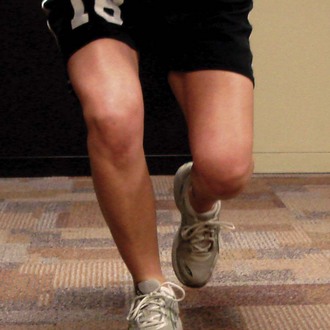
The principal movement impairment in tibiofemoral rotation (TFR) syndrome is knee joint pain associated with impaired rotation of the tibiofemoral joint (lateral rotation of the tibia and/or medial rotation of the femur). Correction of impairment often decreases symptoms. The subcategories of TFR syndrome are TFR with valgus (TFRVal) syndrome: Valgus knee during static/dynamic activities and TFR with varus (TFRVar) syndrome: Varus knee during static/dynamic activities.

image

Treatment

Emphasis of treatment is decreasing excessive rotation between the tibia on the femur.

Patient Education

The goal of patient education is correction of impaired postural habits and movements.

I. Alignment
A. Improve alignment between femur and tibia.
1. Relax/unlock knees to reduce knee hyperextension if present.
2 Ideally, align knees over feet with neutral rotation of hips and tibias by decreasing MR of femur and LR of tibia.
3 note: If structural tibial torsion or femoral anteversion or retroversion is present, ideal alignment will not be possible. Instruct patient in proper alignment that accommodates these structural impairments. For example, if the person has the following:
a. Tibial torsion: Allow appearance lateral deviation of the foot or “turn out” of the tibia and the foot.
b Femoral anteversion (torsion): Allow the appearance of MR of femur.
B. Valgus: Individuals often stand with foot aligned laterally to the hip. The individual may or may not be able to correct entirely. Must accommodate for structural variations such as structural valgus, excessive soft tissue of thigh.
II Functional activities that contribute to the movement impairment must be addressed.
A. Gait
1. Avoid hip MR and knee hyperextension during stance phase of gait cycle.
2 Encourage proper heel-to-toe gait pattern (common fault is decreased push-off).
a. If patient has the impairment of hyperextension, cue the patient to “lift the heel” to discourage recruitment of the hamstrings.
b If patient demonstrates MR of the femur without hyperextension, cue the patient to contract the gluteal muscles to control femoral MR. If the patient has difficulty contracting the gluteals on command an exercise such as weight shifting may be useful to teach proper contraction. Gait can then be attempted to see if contraction ability improves.
c TFRVar: In severe cases, the patient may be instructed to walk with a slight toe-out gait. Instruct the patient to rotate laterally at the hip and avoid lateral rotation at the tibiofemoral joint.
d Patient may require an assistive device to decrease the forces through the affected knee.
(1) Cane is used in the opposite hand of the impaired lower extremity.
(2) exception: If patient has a varus alignment and using the cane in the opposite hand does not reduce symptoms, the cane may be placed on the same side of the affected knee. Observe gait to determine if desired effect of reducing varus alignment is being achieved. Also use the patient’s pain response to determine the proper side of cane placement.
B. Sit-to-stand; stand-to-sit
1. Slide forward in chair.
2 Feet hip-width apart and aligned behind knees.
3 Use quadriceps and gluteus maximus muscles to lift body up and forward out of chair.
a. Ensure that the tibia advances over the foot with performance (shifts weight forward).
b Avoid hip MR.
c Avoid pulling knees back to meet body.
C. Stairs
1. Instruct in use of rail to decrease weight bearing on the involved limb.
2 Ascending stairs.
a. Use quadriceps and gluteus maximus muscles to lift body up and forward.
(1) Ensure that the tibia advances over the foot with performance (shifts weight forward).
b Avoid hip MR.
c Avoid pulling knee back to meet body.
3 Descending stairs.
a. Avoid hip MR.
b If difficult to perform without pain during initial visit, may need to instruct in step-to pattern leading with the involved extremity.
c If significantly limited, patient may need to descend stairs backward.
D. Personal activities (work, school, leisure activities)
1. Address activities that patients are performing throughout the day that may contribute to the movement impairment. These may include prolonged sitting, driving, and getting in and out of a car.
2 Address fitness activities early to maintain patient’s routine. Modifications or alternative activities may need to be provided. Modify intensity of activities to decrease stress to injured tissues.
a. Running
(1) Interval training is recommended.
(a) Begin with walking program and gradually mix in short bouts of running. Gradually increase the time running and decrease the time walking.
(2) Modify surface of training if indicated.
(a) Instruct patient to initiate running with surfaces that reduce the ground reaction force on the lower extremities. A track or chip trail is better than asphalt, and asphalt is better than concrete. Concrete should be avoided if possible.
(b) Running on a street with a camber may contribute to common knee problems such as ITB friction syndrome. Runners should be encouraged to either avoid the camber or alternate the direction of their run.
(3) Modify activities that may encourage TFL-ITB recruitment over gluteus medius/maximus recruitment.
(a) Biking: Use of toe clips can encourage overrecruitment of the TFL-ITB. Patient should be encouraged to focus more on the pushing phase of the cycle and less on the pulling phase.
(b) Running: Patients often run with their body weight shifted posteriorly (referred to as chasing their center of gravity). Cue the patient to shift body weight slightly forward to encourage better recruitment of the gluteal muscles. Sometimes, use of a small incline will assist patients in shifting the weight forward.

Home Exercise Program

Patients should be instructed that they should not feel an increase in their symptoms during the performance of their exercises. In addition to monitoring for symptoms, they should not experience a “pressure” in the knee during exercises. If either pain or pressure occurs, they should review the instructions to the exercise to be sure that they are performing it correctly and try again. If they still experience pain or pressure, they should discontinue this exercise until they return for their next visit.

I. Improve muscle performance
A. Intrinsic hip lateral rotators and posterior gluteus medius muscles
1. Strengthening: Progressing from easiest to most difficult.
a. Prone hip lateral rotation isometrics (prone foot pushes).
b Prone hip abduction.
c Sidelying hip abduction with lateral rotation (level 1, 2, or 3).
(1) Monitor to be sure patient feels the contraction in the “seat” region; the therapist must palpate to be sure that the patient is recruiting the correct muscles. Common cues for improve performance of the hip lateral rotators include the following:
(a) Positioning: The pelvis may be rotated posteriorly too far. Ask the patient to roll the pelvis anteriorly.
(b) Positioning: Place a pillow between the knees.
(c) Spin the thigh around an axis longitudinally through the femur.
(d) caution: Do not use foot as a guide for lateral rotation of the hip.
d Hip lateral rotation against resistance.
(1) Sitting: Ligaments of the knee are most lax when the knee is in 90 degrees of flexion. This exercise should be monitored closely to be sure the patient is able to stabilize the tibia while performing this exercise.
(2) Standing.
e Lunges.
(1) Perform with lower extremity in good alignment; initially without weight or resistance.
(2) Progression.
(a) Resisted: Using an elastic band around proximal thigh, the therapist pulls in the direction of MR and adduction.
(b) Patients can hold weights in their hands.
2 Recruitment.
a. Weight shifting with gluteal squeeze on the stance lower extremity.
(1) Progress to standing on one leg with correct alignment.
(2) Progress to resisted activities of the opposite leg while standing on the affected leg.
B. Gluteus maximus muscles
1. Prone hip extension with the knee flexed.
a. Positioning: Patient’s that have short hip flexors with require a pillow under the pelvis.
b Patient must be able to control the tibial positioning during prone knee flexion to begin this exercise.
2 Lunges, squats.
C. Abdominal muscles (if appropriate)
1. Strengthening: Lower abdominal progression as described by Sahrmann.2
2 Recruitment: Encourage patient to pull in abdominals with functional activities.
D. Quadriceps muscle
1. Functional activities.
a. Sit to stand, stand to sit, step-ups/step-downs as tolerated.
b Lunges, wall sits as tolerated.

SPECIAL NOTE: Knees with Malalignment or Excessive Varus or Valgus

It is thought that the quadriceps muscles provide some shock absorption to the knee; however, recent studies show that increased quadriceps activity can actually increase the progression of OA in knees with malalignment. One must consider the compression forces that the quadriceps can add to a joint before administering quadriceps strengthening activities. Quadriceps performance should be enhanced only through the proper performance of functional activities, and therapeutic exercises to hypertrophy the quadriceps should be avoided in patients with malalignment of the knees.

II Improve extensibility
A. TFL-ITB and rectus femoris muscles (listed in the order of least aggressive to most aggressive)
1. During all stretching exercises, be sure that the patient has good abdominal support to avoid pelvic anterior tilt or rotation.
a. Prone bilateral knee flexion: With knees and feet together, flex both knees at the same time, monitor tibial position and avoid LR of tibia.
2 May need to begin with femurs in an abducted position and gradually adduct the hips as the patient improves.
3 Prone hip lateral rotation: Monitor tibial position and avoid LR of tibia.
4 Hip flexor length test position: Allow the hip to abduct as it extends, then actively adduct with the tibia in neutral (foot pointing forward or slightly inward).
5 Ober test position: Hip in LR and tibia in neutral to slight MR (level 3 of posterior gluteus medius progression).
B. Gastrocnemius and hamstring muscles
1. Active sitting knee extension with dorsiflexion in neutral hip rotation.
2 Hamstring muscles: Prolonged passive stretches in supine.
3 Gastrocnemius muscles: Standing runners stretch.
a. Patients with excessive pronation must have their arch supported during this stretch (shoes on).
III Other
A. Taping
1. Posterior knee X taping may be helpful.
a. To control rotation during taping, the patient should be positioned so that the lower extremity is in the desired position before applying the tape. The patient is asked to contract the gluteal muscles to laterally rotate the thigh.
b ITB friction syndrome: Tape placed along the ITB to support the tissue.
B. Bracing
1. Although bracing may not be the first choice in treatment for this patient population, it may be an option for patients who are not achieving adequate pain control with their activities.
a. One theoretical benefit of bracing is that it can increase proprioception of the knee.3-5
b Unloader bracing has seemed to be beneficial in patients with OA and malalignment.6,7 Must consider patient goals, motivation, and anthropomorphics.
C. Orthotics
1. Temporary orthotics may be tried to determine if they will be helpful; then custom orthotics may be ordered if indicated.
a. Valgus.
(1) For pronation that is flexible: Orthotics to assist in controlling motion at the foot.
b Varus.
(1) For a rigid supinated foot: Cushioned insert to improve shock absorption.
D. Pain control
1. Modalities.
a. Ice as often as needed.
2 Modify activities to reduce stress to injured tissues.
a. Daily activities.
b Fitness activities.
E. Neuromuscular training (see Box 7-2)

Tibiofemoral Hypomobility Syndrome

The principal movement impairment in tibiofemoral hypomobility (TFHypo) syndrome is associated with a limitation in the physiological motion of the knee. This limitation may result from degenerative changes in the joint or the effects of prolonged immobilization. In type I TFHypo syndrome, the potential for recovery of ROM is good; whereas the potential for recovery in type II TFHypo syndrome is poor.

image

Treatment

Type I Tibiofemoral Hypomobility Syndrome

The potential for recovery of ROM is good in patients with type I TFHypo syndrome. These individuals demonstrate a limitation in ROM; however, duration of limitation is not long. End-feel to PROM may be stiff: however, some extensibility is noted. The therapist should monitor progression of ROM over time to determine if the classification of type I is appropriate. If ROM does not improve in 3 to 4 weeks and all treatment strategies have been investigated, type II should be considered.

Treatment emphasis is on improving ROM, strength, and conditioning, without increasing pain and swelling. In the treatment of the knee with degeneration, consider that rotation may be contributing to the symptoms.

Patient Education

The goal of patient education is correction of impaired postural habits and movements.

I. Alignment
A. Correct standing alignment as appropriate.
B. If structural impairment is present, ideal alignment may not be possible.
II Functional activities that contribute to the movement impairment must be addressed.
A. Gait
1. Avoid hip MR during stance phase of gait cycle.
2 Encourage a “rolling” heel-to-toe gait pattern (common fault is decreased push-off).
a. This is to improve the shock absorption value of the foot, as well as encourage proper gait pattern.
b Patient may require an assistive device to decrease the forces through the affected knee.
(1) Cane is used in the opposite hand of the impaired lower extremity.
(2) exception: If patient has a varus alignment and using the cane in the opposite hand does not reduce symptoms, the cane may be placed on the same side of the affected knee. Observe gait to determine if the desired effect of reducing varus alignment is being achieved. Also, use the patient’s pain response to determine the proper side of cane placement.
B. Sit-to-stand; stand-to-sit
1. Slide forward in chair.
2 Feet hip-width apart and aligned behind knees.
3 Use quadriceps and gluteus maximus muscles to lift body up and forward out of chair.
a. Ensure that the tibia advances over the foot with performance (shifts weight forward).
4 Avoid hip MR.
5 Avoid pulling knees back to push against chair behind them.
C. Stairs
1. Instruct in use of rail to decrease weight bearing on the involved limb.
2 Ascending stairs.
a. Use quadriceps and gluteus maximus muscles to lift body up and forward.
(1) Ensure that the tibia advances over the foot with performance (shifts weight forward).
b Avoid hip MR.
c Avoid pulling knee back to meet body.
3 Descending stairs.
a. Avoid hip MR.
b If difficult to perform without pain during initial visit, may need to instruct in step-to pattern leading with the involved extremity.
c If significantly limited, patient may need to descend stairs backward.
D. Personal activities (work, school, leisure activities)
1. Address activities that patients are performing throughout the day that may contribute to the movement impairment, including prolonged sitting, driving, and getting in and out of a car.
2 Fitness activities.
a. Address fitness early to maintain patient’s routine. Modifications or alternative activities may need to be provided.
(a) Degeneration: While initiating exercises, it is safer to begin with high repetitions of relatively low weight.
b Modify intensity of activities to decrease stress to injured tissues.
(a) Reduce weight bearing during initial phases and gradually increase weight bearing as tolerated by the patient.
(i) Swimming, stationary biking
(ii) Stair master, elliptical
(iii) Treadmill
III Education
A. Degeneration: Arthritis and joint protection education
1. Risk factors for progression of OA.
a. Previous injury to the meniscus or ligament.
b Manual labor with prolonged positioning of knee flexion.
c Obesity.
d Laxity of the knee.
e Malalignment of the knee.

Home Exercise Program

Degeneration

Patients should be instructed that they should not feel an increase in their symptoms during the performance of their exercises. In addition to monitoring for their symptoms, they should not experience a “pressure” in their knee during their exercises. If either pain or pressure occurs, they should review the instructions to the exercise to be sure that they are performing it correctly and try again. If they still experience pain or pressure, they should discontinue this exercise until they return for their next visit.

Immobilization

Patients should be instructed that they will experience some discomfort (often described as pain or pressure) with their exercises to improve ROM. Patients should be encouraged to continue with the exercises as tolerated. Pain medications should be timed so that they are at maximum level during exercises.

I. Improve muscle performance
A. Intrinsic hip lateral rotators and posterior gluteus medius muscle
1. Strengthening
a. Prone hip lateral rotation isometrics (prone foot pushes).
b Prone hip abduction.
c Sidelying hip abduction with lateral rotation (level 1, 2, or 3).
(1) Monitor to be sure patient feels the contraction in the “seat” region; the therapist must palpate to be sure that the patient is recruiting the correct muscles. Common cues for improve performance of the hip lateral rotators include the following:
(a) Positioning: The pelvis may be rotated posteriorly too far. Ask the patient to roll the pelvis anteriorly.
(b) Positioning: Place a pillow between the knees.
(c) Spin the thigh around an axis longitudinally through the femur.
(d) caution: Do not use foot as a guide for lateral rotation of the hip.
d Lunges.
(1) Perform with lower extremity is good alignment; initially without weight or resistance.
(2) Progression.
(a) Resisted: Using an elastic band around proximal thigh, the therapist pulls in the direction of medial rotation and adduction.
(b) Patients can hold weights in their hands.
2 Recruitment
a. Weight shifting with gluteal squeeze on the stance lower extremity
(1) Progress to standing on 1 leg with correct alignment
(2) Progress to resisted activities of the opposite leg while standing on the affected leg
B. Gluteus maximus muscle
1. Prone hip extension with the knee flexed
a. Positioning: Patient’s that have short hip flexors with require a pillow under the pelvis.
b Patient must be able to control the tibial positioning during prone knee flexion in order to begin this exercise.
2 Lunges, squats
a. caution: Aggressive exercise. Must be sure patient is able to perform without difficulty
C. Gastrocnemius muscle
1. Elastic band resistance
2 Standing heel raises, bilateral to unilateral
D. Abdominal muscles (if appropriate)
1. Strengthening: Lower abdominal progression as described by Sahrmann2
2 Recruitment: Encourage patient to pull in abdominals with functional activities
E. Quadriceps muscle
1. Strengthen with functional activities only
a. Sit to stand, stand to sit, step-ups/step-downs as tolerated.

Knees with Degenerative Changes and Malalignment, Excessive Varus or Valgus

It is thought that the quadriceps muscles provide some shock absorption to the knee; however, recent studies show that increased quadriceps strength can actually increase the progression of OA in knees that have malalignment. One must consider the compression forces that the quadriceps can add to a joint before administering quadriceps strengthening activities. The quadriceps performance should be enhanced only through the proper performance of functional activities, and therapeutic exercises to hypertrophy the quadriceps should be avoided in patients with malalignment of the knees.

II Improve extensibility
A. Hip flexors (listed in the order of least aggressive to most aggressive)
1. During all stretching exercises, be sure that the patient has good abdominal support to avoid pelvic anterior tilt or rotation.
2 Supine hip and knee extension (heel slide).
3 Prone bilateral knee flexion: With knees and feet together, flex both knees at the same time; monitor tibial position, avoid LR of tibia.
a. May need to begin with femurs in and abducted position and gradually adduct the hips as the patient improves.
4 Prone hip lateral rotation: Monitor tibial position, avoid LR of tibia.
B. Gastrocnemius and hamstrings muscles
1. Active sitting knee extension with dorsiflexion in neutral hip rotation.
III Other
A. Mobilization
1. Accessory mobilization
a. If patient is having pain at rest (e.g., in non–weight-bearing positions such as supine), distraction mobilization can be taught to the patient. A trial of gentle distraction should be performed to determine if this technique will be appropriate.
(1) Home program: Patient in sitting position with the foot dangling (a towel may be placed under the distal thigh to raise the thigh). A lightweight shoe may be applied (1 lb). Patient allows the leg to dangle up to 10 minutes to help relieve discomfort. This may be performed as often as needed to relieve pain.
(2) caution: This activity is used only to relieve pain at rest.
b Joint mobilization.
(1) Pain relief: Oscillatory I, II; sustained I, II.
(2) Increasing ROM: Oscillatory III, IV; sustained III.
2 Physiological mobilization
a. Assisted active ROM.
(1) Supine hip and knee flexion (heel slides).
(a) A towel or sheet may used by the patient to provide gentle overpressure.
(2) Sitting knee flexion/extension.
(3) Prone knee flexion as previously described.
(4) Stationary biking.
b Passive ROM.
(1) Knee extension: Supine or prone.
(2) Proprioceptive neuromuscular facilitation (PNF) techniques, such as contract/relax and hold/relax, may be useful.
B. Edema and pain control
1. Modalities
a. Ice (if tolerated)
b Compression wrap for swelling
c Ultrasound
d Moist heat pack
e Electrical stimulation
C. Bracing
1. Degeneration
a. Bracing should be considered for patients who continue to have symptoms that are functionally limiting.
b Unloader bracing has seemed to be beneficial in patients with OA and malalignment.6,7 Must consider patient goals, motivation, and anthropomorphics.
2 Immobilization
a. Dynamic splinting/bracing may used to provide a low load, long duration stretch.
D. Neuromuscular training (see Box 7-2)

Type II Tibiofemoral Hypomobility Syndrome

The potential for recovery of ROM is poor in patients with type II TFHypo syndrome. These individuals may report a long duration of loss of ROM, either through immobilization or long-standing OA. End-feel to PROM to the joint are very stiff, with the soft tissues demonstrating very little extensibility. When in doubt, it is best to classify the individual with type I for a trial period and assess the patient’s progress appropriately. Emphasis is on educating the patient in modifications of functional activities to accommodate for loss of ROM.

I. Treatment concepts are similar to the treatment of type I TFHypo syndrome; however, modifications must be made for the limited ROM of the knee.
A. Emphasis of treatment should be placed on functional activities specific to the patient. See treatment suggestions for type I TFHypo syndrome for specifics.
B. Proper footwear to provide shock absorption and proper support to the foot.
C. Independence in home exercise program to maintain current ROM of the knee.
D. Independence in home exercise program to maintain proper movement/alignment of the adjacent joints such as the hip, back, and ankle/foot.
E. If limitation significantly affects functional activities, an occupational therapy consult may be advised to address more specific modifications.

Knee Extension Syndrome and Knee Extension with Patellar Superior Glide Syndrome

The principal movement impairment in knee extension (Kext) syndrome is knee pain associated with dominance of quadriceps muscles that results in excessive pull on the patella, patellar tendon/ligament, or tibial tubercle. The Kext syndrome has a subcategory of patellar superior glide (KextSG).

image

Treatment

Treatment emphasis in Kext syndrome is on decreasing the activity of the quadriceps while improving the performance of the hip extensors. In KextSG syndrome, treatment emphasis is similar to Kext syndrome; however, methods to stabilize the patellar glide may need to be implemented.

Patient Education

The following treatment is appropriate for both Kext and KextSG syndromes unless otherwise noted. The goal of patient education is correction of impaired postural habits and movements.

I. Alignment
A. Correct alignment as appropriate
II Functional activities that contribute to the movement impairment must be addressed.
A. Gait
1. Encourage proper heel-to-toe gait pattern (common fault is decreased push-off).
2 Cue the patient to push off with the toes. Patient may also benefit from cues to shift weight slightly forward.
B. Sit-to-stand; stand-to-sit
1. Slide forward in chair.
2 Feet hip-width apart and aligned behind knees.
3 Use quadriceps and gluteus maximus muscles to lift body up and forward out of chair.
a. Ensure that the tibia advances over the foot with performance (shifts weight forward).
C. Sitting
1. Patient should avoid prolonged periods of increased knee flexion (≥90 degrees).
a. Take standing/walking breaks every 30 minutes.
b When unable to take breaks, use sitting knee extension to decrease time spent in knee flexion.
c Patients with short rectus femoris muscles may need to sit with the knee in relatively less flexion initially. As symptoms improve, they should be instructed to gradually flex the knee until they are at a normal position.
D. Stairs
1. Instruct in use of rail to decrease weight bearing on the involved limb.
2 Ascending stairs.
a. Emphasize the use of the gluteus maximus muscle to lift body up and forward.
(1) Ensure that the tibia advances over the foot with performance (shifts weight forward).
3 Descending stairs.
(1) If difficult to perform without pain during initial visit, may need to instruct in step-to pattern, leading with the involved extremity.
b. If significantly limited, patient may need to descend stairs backward.
E. Personal activities (work, school, leisure activities)
1. Address activities that patients perform throughout the day that may contribute to the movement impairment. These may include prolonged standing or sitting.
2 Fitness activities.
a. Address fitness early to maintain patient’s routine. Modifications or alternative activities may need to be provided.
b Modify intensity of activities to decrease stress to injured tissues.
(1) Running
(a) Interval training is recommended.
i. Begin with walking program and gradually mix in short bouts of running. Gradually increase the time running and decrease the time walking.
(b) Modify surface of training if indicated.
ii Instruct patient to initiate running with surfaces that reduce the ground reaction force on the lower extremities. A track or chip trail is better than asphalt and asphalt is better than concrete. Concrete should be avoided if possible.
(c) Modify strength training.
i. Patients should be discouraged from performing resistance activities to increase the hypertrophy of the quadriceps muscles.
(d) Modify activities to encourage gluteus medius/maximus muscle recruitment.
i. Biking: Patient should be encouraged to focus more on the pushing phase of the cycle (extension of hip = gluts) and less on the pulling phase (knee flexion = hams).
ii Running: Patients often run with their body weight shifted posteriorly (referred to as chasing their center of gravity in Sahrmann2). Cue the patient to shift body weight slightly forward to encourage better recruitment of the gluteal muscles. Sometimes, use of a small incline will assist the patient in shifting the weight forward.

Home Exercise Program

The patient should be instructed that they should not feel an increase in their symptoms during the performance of their exercises. If this occurs, they should review the instructions to the exercise to be sure that they are performing it correctly and try again. If they still experience pain, they should discontinue this exercise until they return for their next visit.

I. Improve muscle performance
A. Gluteus maximus muscle
1. Prone hip extension with the knee flexed.
a. Patient may require a pillow under the pelvis if short hip flexors.
2 Weight shifting with gluteal squeeze on the stance lower extremity.
a. Progress to standing on 1 leg with correct alignment.
b Progress to resisted activities of the opposite leg while standing on the affected leg.
3 Lunges, squats.
a. caution: In patients who have knee extension, the quadriceps muscles place excessive pull on the patellar tendon and tibial tubercle. Quadriceps strengthening would be contraindicated in these patients.
II Improve extensibility
A. Quadriceps muscles (listed in the order of least aggressive to most aggressive)
1. During all stretching exercises, be sure that the patient has good abdominal support to avoid pelvic anterior tilt or rotation.
a. KextSG: During performance of exercises that stretch the quadriceps, patellar taping to reduce patellar glide may be required if the patient cannot perform the exercises without an increase in symptoms.
b Prone knee flexion.
(1) May need to begin with pillows under pelvis if patient also has short hip flexors.
(2) Perform within range that does not increase symptoms.
c Prone hip lateral rotation.
d Hip flexor length test position: Instruct the patient to flex the knee while the hip is in neutral rotation and neutral hip abduction/adduction.
III Other
A. Taping or bracing
1. KextSG
a. Horseshoe taping to discourage superior glide.
b Cho-pat strap across the patellar tendon.
2 Patellar joint mobilization
a. KextSG
(1) Inferior glides to patella.

a. Initially may need to perform least aggressive grades (I and II) but should be able to progress to more aggressive grades quickly. Grades III and IV are usually tolerated well. Patient may be instructed in proper performance for home exercise program.
(2) Mobilization with movement.
b Sitting knee flexion: The patient allows the knee to flex from the fully extended position. As the knee flexes, the patient performs an inferior glide of the patella.
3 Pain control
a. Modalities.
(1) Ice as often as needed.
4 Neuromuscular training (see Box 7-2)

Knee Hyperextension Syndrome

The principal movement impairment in knee hyperextension (Khext) syndrome is knee pain associated with impaired knee extensor mechanism. Dominance of hamstrings and poor functional performance of gluteus maximus and quadriceps muscles result in hyperextension of the knee placing excessive stresses on the structures of the knee.

image

Treatment

Treatment emphasis in knee hyperextension syndrome is to decrease hyperextension of the knee.

Patient Education

The goal of patient education is correction of impaired postural habits and movements.

I. Alignment
A. Improve alignment between femur and tibia.
1. Relax/unlock knees to reduce hyperextension of knee.
a. Improve alignment of pelvis if applicable.
II Functional activities that contribute to the movement impairment must be addressed
A. Gait
1. Encourage proper heel-to-toe gait pattern.
a. Knee hyperextension.
(1) Avoid knee hyperextension during stance phase of gait cycle.
(2) Cue the patient to “lift the heel” to discourage overrecruitment of the hamstrings.
(3) Cue to land softly on the heel at heel strike.
B. Sit-to-stand; stand-to-sit
1. Slide forward in chair.
2 Feet hip-width apart and aligned behind knees.
3 Use quadriceps and gluteus maximus muscles to lift body up and forward out of chair.
a. Ensure that the tibia advances over the foot with performance.
4 Avoid pulling knees back to meet body; final position of knee should be relaxed knee (not hyperextension).
C. Stairs
1. Instruct in use of rail to decrease weight bearing on the involved limb.
2 Ascending stairs.
a. Use quadriceps and gluteus maximus muscles to lift body up and forward.
(1) Ensure that the tibia advances over the foot with performance.
b Avoid pulling knee back to meet body.
3 Descending stairs.
a. If patient is unable to perform without pain during initial visit, may need to instruct in step-to pattern leading with the involved extremity.
b If significantly limited, patient may need to descend stairs backward.
D. Personal activities (work, school, leisure activities)
1. Address activities that patients perform throughout the day that may contribute to the movement impairment. These may include prolonged standing.
2 Fitness activities.
a. Address fitness early to maintain patient’s routine. Modifications or alternative activities may need to be provided.
b Modify intensity of activities to decrease stress to injured tissues.
(1) Running
(a) Interval training is recommended.
i. Begin with walking program and gradually mix in short bouts of running. Gradually increase the time running and decrease the time walking.
(b) Modify surface of training if indicated.
i. Instruct patient to initiate running with surfaces that reduce the ground reaction force on the lower extremities. A track or chip trail is better than asphalt and asphalt is better than concrete. Concrete should be avoided if possible.
ii Running on a street with a camber may contribute to common knee problems such as ITB friction syndrome. Runners should be encouraged to either avoid the camber or alternate the direction of their run.
c Modify activities to encourage gluteus medius/maximus muscle recruitment.
(1) Biking: Patients should be encouraged to focus more on the pushing phase of the cycle (extension of hip = gluts) and less on the pulling phase (knee flexion = hams).
(2) Running: Patients often run with their body weight shifted posteriorly (referred to as chasing the center of gravity in Sahrmann2). Cue the patient to shift body weight slightly forward to encourage better recruitment of the gluteal muscles. Sometimes, use of a small incline will assist the patient in shifting the weight forward.
d Kickboxing: Educate patients to decrease speed of kicks to improve control of limb.
e Swimming: Educate patients to decrease intensity of wall push-off with turning.

Home Exercise Program

Patients should be instructed that they should not feel an increase in their symptoms during the performance of their exercises. If this occurs, they should review the instructions to the exercise to be sure that they are performing it correctly and try again. If they still experience pain, they should discontinue this exercise until they return for their next visit.

I. Improve muscle performance
A. Gluteus maximus
1. Prone hip extension with the knee flexed.
a. Patient may require a pillow under the pelvis if short hip flexors.
2 Weight shifting with gluteal squeeze on the stance lower extremity.
a. Progress to standing on 1 leg with correct alignment
b Progress to resisted activities of the opposite leg while standing on the affected leg
3 Lunges, squats.
B. Abdominals (if appropriate)
1. Strengthening: Lower abdominal progression as described by Sahrmann.2
2 Recruitment: Encourage patient to pull in abdominals with functional activities.
C. Quadriceps
1. Progress quadriceps strengthening according to pain and results of resisted testing and functional testing such as stairs.
2 Quadriceps: Sit to stand, wall sits, step-ups/step downs, lunges (emphasize correct hip and tibial alignment).
II Improve extensibility
A. Gastrocnemius muscles
1. Active sitting knee extension with dorsiflexion in neutral hip rotation.
2 Standing runners stretch.
B. Hamstrings
1. Prolonged hamstring stretch, maintain proper spinal alignment.
III Other
A. Taping or bracing
1. Knee hyperextension.
a. Posterior knee X taping to decrease hyperextension of the knee
b Unloader V taping to unload the fat pad
B. Pain control
1. Modalities.
a. Ice as often as needed.
C. Neuromuscular training (see Box 7-2)

Patellar Lateral Glide Syndrome

The principal movement impairment in patellar lateral glide syndrome is knee pain as a result of an impaired patellar relationship within the trochlear groove. Often a secondary diagnosis and therefore the movement impairments of tibiofemoral rotation (TFR) or knee hyperextension (Khext) should be considered. Correction of impairment often decreases symptoms.

image

Treatment

Treatment emphasis in patellar lateral glide syndrome is to address the impairment of patellar tracking. If patellar lateral glide syndrome is given as a secondary diagnosis, please refer to the treatment described for the primary diagnosis (tibiofemoral rotation or knee hyperextension) in addition to the treatment described below.

Patient Education

The goal of patient education is correction of impaired postural habits and movements.

I. Alignment
A. Correct alignment as appropriate
II Functional activities that contribute to the movement impairment must be addressed
A. If a primary diagnosis of tibiofemoral rotation or knee hyperextension is determined, see treatment for the primary diagnosis.
B. Stairs
1. Instruct in use of rail to decrease weight bearing on the involved limb.
2 Ascending stairs.
a. Use quadriceps and gluteus maximus to lift body up and forward.
3 Descending stairs.
a. If difficult to perform without pain during initial visit, may need to instruct in step-to pattern leading with the involved extremity.
b If significantly limited, patient may need to descend stairs backwards.
C. Sitting
1. Patient should avoid prolonged periods of increased knee flexion (≥90 degrees).
a. Take standing/walking breaks every 30 minutes.
b When unable to take breaks, use sitting knee extension to decrease time spent in knee flexion.
c In patients with short TFL-ITB, they may need to sit with the thighs slightly abducted initially. As symptoms improve, they should be instructed to gradually adduct the thigh until they are at a normal position.
D. Personal activities (work, school, leisure activities)
1. Address activities that patients are performing throughout the day which are pain provoking for the patient.
2 Fitness activities.

Home Exercise Program

The patient should be instructed that they should not feel an increase in their symptoms during the performance of their exercises. In addition to monitoring for their symptoms, they should not experience a “pressure” in their knee during their exercises. If either pain or pressure occurs, they should review the instructions to the exercise to be sure that they are performing it correctly and try again. If they still experience pain or pressure, they should discontinue this exercise until they return for their next visit.

I. Improve muscle performance
A. If a primary diagnosis of tibiofemoral rotation or hyperextension is determined, see treatment for the primary diagnosis.
1. Quadriceps
a. Recommend use of functional activities to improve quadriceps performance (listed in order of difficulty beginning with least aggressive)
(1) Sit to stand transfers
(2) Step-ups may progress to step downs as patient’s symptoms improve
(3) Lunges
(4) Squats
(5) Biofeedback may be beneficial for improving possible timing or recruitment of the vastus medialis oblique (VMO) muscle
II Improve extensibility
A. TFL-ITB
1. Listed in the order of least aggressive to most aggressive.
2 During all stretching exercises, be sure that the patient has good abdominal support to avoid pelvic anterior tilt or rotation.
3 During performance of exercises that stretch the TFL-ITB, patellar taping to reduce patellar glide may be required if the patient cannot perform the exercises without an increase in symptoms.
a. Prone knee flexion
(1) May need to begin with femurs in an abducted position and gradually adduct the hips as the patient improves.
b Prone hip lateral rotation
c Hip flexor length test position: Allow the hip to abduct as it extends then actively adduct with the tibia in neutral
d Ober test position: Hip in lateral rotation and tibia in neutral to slight medial rotation (level 3 of post. glut. med. progression)
B. Other muscles (as needed)
1. Lateral patellar retinaculum
2 Gluteus maximus/ITB
C. Other
1. Patellar taping/bracing
a. McConnell taping for medial glide
2 Patellar joint mobilization
a. Medial glide to patella
b Initially may need to perform least aggressive grades (I, II), but should be able to progress to more aggressive grades quickly. Grades III, IVs are usually tolerated well. Patient may be instructed in proper performance for home exercise program.
3 Pain control
a. Modalities
(1) Ice as often as needed
4 Neuromuscular training (see Box 7-2)

Knee Impairment

Knee impairment is the classification given in the absence of a specific movement impairment diagnosis or when a diagnosis cannot not be determined because of pain, physician-imposed restrictions, or both. If possible, the physical therapist should determine the pathoanatomical structure involved, as identified by the physician; the procedure performed, if any; and the stage for rehabilitation.

Factors that affect the physical stress of tissue and/or thresholds of tissue adaptation and injury10 include the following:

I. Physiological factors
A. Tissue factors specific to the knee
1. Bone
a. Tibial plateau
(1) Cancellous bone and poor vascular supply.
(2) Often non–weight-bearing for 2 to 3 months.
b Patella fracture: Quad contraction often contraindicated
2 Cartilage
a. Meniscus
(1) Red zone: Peripheral one-third vascular, with good healing potential.
(2) Pink zone: Middle one-third vascularity, with variable healing potential.
(3) White zone: Inner one-third avascular, with poor healing.
(4) Increased compression of meniscus at 90 degrees of knee flexion.
(5) Aggressive hamstring exercise is contraindicated, especially if injury to posterior horn.
(6) Change in weight-bearing surfaces with removal of meniscus.
3 Muscle
a. Quad atrophy common postsurgery or trauma to knee
4 Tendon
a. Quad tendon rupture
(1) Quad contraction contraindicated.
(2) May have flexion ROM restrictions.
5 Ligament
a. ACL
(1) New graft is weakest from 4 to 12 weeks.
(2) Avoid anterior tibial translation.
(3) Avoid open-chain resisted knee extension in early rehabilitation phase.
b PCL
(1) Avoid posterior tibial translation.
(2) Avoid active hamstring contraction.
c MCL
(1) Adolescent: Separation of distal femoral epiphysis can mimic MCL sprain. Radiographs must be performed.
d Patellar ligament rupture
(1) Quad contraction contraindicated.
(2) May have flexion ROM restrictions.
6 Skin
7 Nerve
a. Fibular (peroneal) nerve injury possible with surgery or trauma to knee
B. Types of surgeries (indications)
1. Stabilization
2 Osteotomy (malalignment or osteosarcoma)
a. Femoral
b Tibial
3 Arthroplasty (DJD, arthritis, joint destruction)
a. Total/unicompartment
(1) Cemented/uncemented
4 Debridement (tear, degeneration)
a. Meniscal
b Patellar ligament
c Patella
5 Repair
a. Ligament reconstruction (ACL, PCL)
b Meniscal repair
c Cartilage repair
(1) Microfracture
(2) Mosaicplasty, osteochondral autograft transplant (OATS)
6 Meniscal transplant
7 Soft tissue release (short tissues or spastic muscles)
(1) ITB
(2) Hamstrings
(3) Hip adductors
C. Medical complications
1. Baker’s cyst
2 Peroneal nerve neurapraxia
3 Leg length changes (total knee replacement)
II Movement and alignment factors
A. Variations
B. Standing alignment
1. May demonstrate protective stance or rotational impairments
C. Underlying movement impairment syndromes
1. TFR syndrome
a. TFR syndrome with valgus
b TFR syndrome with varus
2 TFhypo syndrome
3 Kext syndrome
a. Kext syndrome with patellar superior glide
4 Khext syndrome
5 PLG syndrome
6 TFAH syndrome

Treatment for Knee Impairment

Emphasis of treatment is to restore ROM of the knee and strength of the lower extremity without adding excessive stresses to the injured tissues. Underlying movement impairments should be addressed during rehabilitation and functional activities to ensure optimal stresses to the healing tissues.

Impairments (Body Functions and Structures)

Pain

Be sure to clarify the location, quality, and intensity.

Stage 1

Surgical: Within the first 2 weeks of the postoperative period, some pain will be associated with exercises. Gradually, over the next few weeks, pain associated with the exercise should lessen. Sharp, stabbing pain should be avoided. Mild aching is expected after exercises but should be tolerable for the patient. This postexercise discomfort should decrease within 1 to 2 hours of the rehabilitation. Complaints of increasing pain, pain that is not decreasing with treatment, or burning pain are all “red flag” indicators that treatment is too aggressive or there is a disruption in the usual course of healing. Coordinating the use of analgesics with exercise sessions is important. Splinting, bracing, and/or assistive devices may be used during this period to protect the injured tissue.

Acute Injury: Despite discomfort, tests may need to be performed to rule out serious injury. Modalities and taping/bracing may be helpful to decrease pain. The patient may also require the use of an assistive device in the early phases of healing.

Stage 2 to 3

Surgical/Acute Injury: Pain associated with the specific tissue that was involved in the surgery should be significantly decreased by weeks 4 to 6. Precautions may be lifted during or by postoperative weeks 4 to 6. As activity level of the patient is progressed, the patient may report increased pain/discomfort with new activities such as returning to daily activities and fitness. Pain/discomfort location should be monitored closely. Muscle soreness is expected, similar to the response of muscle to overload stimulus (e.g., weight training). General muscle soreness should be allowed to resolve, usually 1 to 2 days before repeating the bout of activity. Pain described as stabbing should always be avoided.

Edema

Stage 1

Surgical/Acute Injury: Edema is quite common in the knee s/p surgery or injury. Edema has also been implicated in the inhibition of the quadriceps and therefore should be treated aggressively.11-13 The patient should be educated in use of edema controlling techniques:

Active ROM (AROM)
Ice14
Elevation
Compression: Ace wraps, stockings

Patients should be encouraged to keep the lower extremity elevated as much as possible particularly in the early phases (1 to 3 weeks), without keeping the knee in a flexed position. Application of ice after exercise is recommended. Other methods to control edema in the knee include electrical stimulation or compression pumps. Edema should be measured at each visit. A sudden increase in edema may indicate that the rehabilitation program is too aggressive or a possible infection.

Stage 2 to 3

Surgical/Acute Injury: Time until swelling is resolved is variable among patients and surgical procedures. As patients increase the time spent on their feet, in regular daily activities, or doing more weight-bearing exercises, the patient may experience a slight increase in edema. This is to be expected; however, the patient should be further encouraged to use techniques stated previously to manage the edema.

Appearance

Stage 1

Surgical: Infection should be suspected if the area around the incision or the involved joint appears to be red, hot, and swollen. The physician should be consulted immediately if infection is suspected. It is common to observe bruising after surgery. This should be monitored continuously for any changes; an increase in bruising during the rehabilitation phases may indicate infection. Changes in hair growth, perspiration, or color may indicate some disturbance to the sympathetic nervous function, especially if in combination with the complaint of excessive pain. Stitches are typically removed in 7 to 14 days.

Stage 2 to 3

Surgical: Incision should be well healed. Bruising may still be present as far as 3 to 4 weeks after surgery but should be diminishing. Signs of increased bruising are a red flag and should be immediately referred to their physician.

Range of Motion

Stage 1

Surgical/Acute Injury: Refer to physician’s precautions and specific protocols for guidelines regarding progression of the exercises. The most conservative, common ROM precautions include the following:

ACL reconstruction: Flexion <120 degrees.
Meniscal repair: Flexion <90 degrees.
Collateral ligament repair: Avoid full extension.
Patellar fractures and quadriceps tendon repairs: Restrictions can be varied for the amount of knee flexion allowed and/or when ROM exercises can begin.
Mobilizations to the joint may be contraindicated.

Patellar mobilizations should begin as soon as possible after surgery. Common time frames to begin patellar mobilizations include the following:

ACL reconstruction: Immediately after surgery.
Meniscal repair: Immediately after surgery.
Patellar fractures and quad tendon repairs: Within 1 week, however, consult with physician before initiation.

Tibiofemoral mobilizations after ACL reconstruction, meniscal repair or debridement, collateral ligament repair:

There is little information in the literature that describes the “safe time” that mobilizations can begin. These mobilizations are not recommended until the initial healing phases are complete. If mobilizations are indicated, consult with physician before initiating.
For mobilizations after meniscal debridement, it is recommended that distraction be added before performing glides to reduce shear to the meniscus.

Stage 2 to 3

Surgical/Acute Injury: Precautions are typically lifted by the time the patient reaches this stage. ROM should be approaching normal. Exercises may need to be progressed using passive force. To increase knee extension, prone knee extension, patients can be instructed to hang the limb off the edge of mat with weight on ankle. Patients should be advised to build up tolerance gradually and break up prolonged hang with knee flexion. For knee flexion the patient may raise knee toward the chest and use hands to add overpressure. Patient should be instructed that a stretching discomfort is expected; however, sharp pain should be avoided.

Mobilizations to the tibiofemoral joint may be indicated in later stages of rehabilitation to improve ROM. Consult with the physician before initiating joint mobilization after surgery of the knee.

Strength

Stage 1

Surgical/Acute Injury: Strengthening with overload often begins after the initial phase of healing (4 to 6 weeks). Isometrics and active movement within precautions may be started sooner. At times less than 4 weeks, emphasis should be placed on proper movement patterns in preparation for strengthening activities. After 4 weeks, strengthening may be gradually incorporated. Progression to resistive exercise is based on the patient’s ability to perform ROM with a good movement pattern and without significant increase in pain.

Quadriceps muscles are most commonly affected with surgery or injury to the knee; however, others may be involved such as the hamstrings or gastrocnemius muscles. If the patient is having difficulty recruiting the quadriceps, the following cues are helpful:

Have patient try to pull the knee cap up toward the hip.
Have patient perform an isometric on the uninvolved side first.
With the patient in short sitting, the clinician raises the knee passively into extension. Then the patient attempts to hold the leg straight as you gradually remove the support of your hands. Be careful not to “drop” the limb. Only remove the amount of assistance that allows the patient to perform successfully.
Light tapping of the fingers on the quadriceps: Be careful of incisions.
When performing quadriceps isometrics in long sitting, monitor for compensation of the hamstrings. Patients actually use hamstrings to pull the tibia posteriorly to extend the knee. Be sure you see the quadriceps change shape. If these cues do not work, ask patients to reduce their effort. Often, they pull harder trying to recruit the quadriceps, but it only increases the activity of the hamstrings.

Electrical stimulation or biofeedback may be used to improve strengthening (see the following “Medications/Modalities” section).

The patient may also have strengthening precautions per the physician. Common examples of these precautions include the following:

ACL reconstruction: No resisted extension during open-chain exercises.
Meniscus repair: Restrictions of hamstring strengthening.
Patellar fractures and quadriceps tendon repairs: Restrictions of quadriceps strengthening.

note: Caution should be used in single-leg raise in patients >55 years of age and patients with history of low back pain.

Stage 2 to 3

Surgical/Acute Injury: At this stage, precautions are typically lifted; however, with surgical procedures, such as ACL reconstruction/injury, some restrictions on open-chain resisted extension may still be in place. Strength activities can be progressed as tolerated by the patient. Common functional activities that can be considered strengthening activities include wall slides, lunges, and step-downs/step-ups. A common compensation is to shift weight away from the involved limb. Be sure that the patient maintains the appropriate amount of weight bearing during closed-chain activities.

Proprioception/Balance15

Stage 1

Surgical/Acute Injury: Activities to improve proprioception of the knee joint should be incorporated as soon as possible. Early in treatment, these activities include weight shifting, progressive increases in weight bearing on the involved lower extremity, and eventually unilateral stance. As the patient can take full weight on the involved knee, activities are progressed to use of a balance board and closed-chained activities such as wall sits, lunges, and single-leg stance.

Stage 2 to 3

Surgical/Acute Injury: In this stage, precautions are typically lifted. Activities should be progressed to prepare patient to return to daily activities, fitness routines, and work or sporting activities. As the patient progresses, proprioception can be challenged by asking the patient to stand on unstable surfaces (pillows, trampoline, or BOSU ball), perturbations can be applied through having the patient catch a ball being thrown to him or her while standing on one leg. Sliding board activities have been shown to be beneficial to patients after surgery.16 See Box 7-2 for higher level neuromuscular training (Stage 3).

Cardiovascular and Muscular Endurance

Stage 1

Surgical/Acute Injury: Early in rehabilitation, if the patient does not have adequate knee ROM to complete a full revolution on a stationary bike, unilateral cycling can be performed with the uninvolved extremity. The involved extremity is supported on a stationary surface, while the patient pedals with the uninvolved extremity. Water walking and swimming are good substitutes for full weight-bearing activities. For swimming, if kicking against the resistance is contraindicated, the patient may participate in swim drills that mainly challenge the upper extremities for conditioning. Low resistance stationary cycling can begin when knee flexion ROM is approximately 110 degrees. As strength improves, resistance may be increased.

Stage 2 to 3

Surgical/Acute Injury: The patient may then be progressed to activities such as water walking → walking on the treadmill → elliptical machine → Nordic ski machine → StairMaster → running when appropriate. The patient should be given specific instruction in gradual progression of these activities. See Box 7-1 for progression of running.

Patient Education

Stages 1 to 3

Surgical/Acute Injury: Educate the patient in the structures and tissues involved and the specific medical precautions when indicated. Patients should also be taught schedule for use, and how to don and doff their brace/splint. Educate the patient in the timeline to return to activity, often driven by physician’s guidelines and educate the patient in maintaining precautions during various functional activities such as ambulation, stairs, and transfers.

Scarring

Stage 1

Surgical: Scarring, although a normal process of healing, must be managed well. Exercise, massage, compression, silicone gel sheets, and vibration are used to manage scars. The use of silicone gel is best supported by evidence in the literature. However, clinical experts also commonly use the other methods of scar management. Further research is needed to determine the efficacy of these other methods. The gradual application of stress to the scar/incision helps the scar remodel so that it allows the necessary gliding between structures. A dry incision that has been closed and reopens because of the stresses applied with scar massage indicates that the scar massage is too aggressive. Scars may be classified according to type. Linear scars that are immature are confined to the area of the incision. They may be raised and pink or reddish in the remodeling phase. As they mature they become whitish and flatten. A hypersensitive scar requires desensitization. See Chapter 5 for the examination of the hand and general treatment guidelines and Box 5-3 for more treatment suggestions on managing scar.

Stage 2 to 3

A scar may continue to remodel for up to 2 years. Scar management techniques may be effective until the scar matures, although they are probably most effective early in the healing process.

Changes in Status

Stages 1 to 3

Surgical/Acute Injury: Consider carefully patient reports of increased pain or edema, decreased strength, or significant change in ROM, especially in combination. The patient should be questioned regarding precipitating events such as time of onset, or the activity. If the integrity of the surgery is in doubt, contact the physician promptly. If the patient has fever and erythema spreading from the incision, the physician should be contacted because of the possibility of an infection.

Function (Activity Limitations/Participation Restrictions)

Mobility

Stage 1

Surgical/Acute Injury: While following medical precautions, patients should be instructed in mobility, as follows:

Sit-to-stand: The patient should be instructed in the proper use of an assistive device if a device is indicated.
Ambulation: The patient may have weight-bearing precautions. The patient should be instructed in the proper use of an assistive device and proper gait pattern. Emphasis should be placed on normalizing the patient’s gait pattern. If the patient is given partial or toe-touch weight-bearing restrictions, the patient should be instructed in using a heel-to-toe pattern while restricting the amount of weight that is accepted by the lower extremity. The patient should not place his or her weight on the ball of the foot only.
Stairs: The patient should be instructed in the proper stair ambulation with use of an assistive device (if indicated). In the early phases of healing (after surgery or acute injury) the patient should be instructed to use a step to cadence, lead with the involved lower extremity when descending stairs and lead with the uninvolved lower extremity when ascending stairs.

Stage 2 to 3

Surgical/Acute Injury: Instructions in mobility should be continued while following medical precautions.

All Mobility: As weight-bearing precautions are lifted, the patient should be instructed to gradually reduce the level or type of assistive device required. Progression away from the device depends on the ability of the patient to achieve a normal gait pattern. If the patient demonstrates a significant gait deviation secondary to pain or weakness, the patient should continue to use the device. This may prevent the adaptation of movement impairment and other pain problems in the future. A progression may be: walker → crutches → one crutch → cane → no assistive device.

Stairs: As the patient progresses through the healing stages and can accept more weight onto the involved leg, he or she should be instructed in normal stair ambulation.

Work/School/Higher Level Activities

Stage 1

Surgical/Acute Injury: The patient may be off work or school in the immediate postoperative period or after acute injury. When they are cleared to return to work or school, patients should be instructed in gradual resumption of activities. Emphasis should also be placed on edema control, particularly elevation and compression.

Stage 2 to 3

Surgical/Acute Injury: The patient should be prepared to return to their previous activities. Suggestions for improving proprioception and balance are provided in the preceding “Proprioception/Balance” section. In preparation to return to sports, sport-specific activities should be added. The initial phases of these activities will include straight plane activities at a slow pace and then gradually increase the level of difficulty. See Box 7-2 for more detail.

Sleeping

Stage 1 to 3

Surgical/Acute Injury: Sleeping is often disrupted in the immediate postoperative period or after acute injury. The lower extremity should be slightly elevated (foot higher than the knee and knee higher than the hip) to minimize edema. Avoid placing pillows so that the knee is held in the flexed position throughout the night.

Support

Stage 1

Surgical: A brace may be used to protect the surgical site, depending on the procedure or type of fracture. The brace should fit comfortably. The patient should be educated in the timeline for wearing the brace. Consult with physician if the wearing time is not clear.

It is common for a patient to complain of patellofemoral pain with rehabilitation after surgery. Taping can be helpful in the postoperative period. When applying tape, consider the underlying movement impairment (e.g., tibiofemoral rotation, patellar glide).

Acute injury: Taping may help decrease symptoms in a patient with acute knee injury. When applying tape, consider the underlying movement impairment (e.g., tibiofemoral rotation, knee hyperextension).

Stage 2 to 3

Surgical: The recommendations concerning the need for bracing long term are varied. Communication among the team (patient, physician, and physical therapist) is necessary. Functional bracing is recommended if the patient wishes to return to high level sporting activities and demonstrates either of the following:

1 Laxity in the joint
2. Performs poorly on functional tests17

Acute Injury: For injuries to the ACL that are not repaired or reconstructed, if the patient returns to sport, functional bracing is recommended.18

Medications/Modalities

Medications

Surgical: During the acute stage, physical therapy treatments should be timed with analgesics, typically 30 minutes after administration of oral medication. If medication is given intravenously, therapy often can occur immediately after administration. Communication with nurses and physicians is critical to provide optimal pain relief for the patient.

Acute injury: The patient’s medications should be reviewed to ensure that they are taking the medications appropriately.

Aquatic Therapy

Surgical/Acute Injury: Aquatic therapy to decrease weight bearing during ambulation may be helpful in the rehabilitation of patients after fracture or surgical procedures. Often, this medium is not available but should be considered if the patient’s progress is slowed secondary to pain or difficulty maintaining weight-bearing precautions. Incisions should be healed before aquatic therapy is initiated; however, materials to cover the incision may be used to allow patients to get into the water sooner.

Thermal Modalities

Surgical/Acute Injury: Instruct the patient in proper home use of thermal modalities to decrease pain. Ice has been shown to be beneficial, particularly in the immediate postoperative phases.14

Electrical Stimulation

Stage 1/Progression

Surgical/Acute Injury: Electrical stimulation can be used for three purposes: Pain relief, edema control, and strengthening. Interferential current has been shown to be helpful in decreasing pain and edema.19-21 Sensory level transcutaneous electrical nerve stimulation (TENS) can assist in decreasing pain. Currently, no definitive answer exists for electrical stimulation for quadriceps strengthening. It was once believed that electrical stimulation did not provide a distinct advantage over high-intensity exercise training.22-23 However, more recent studies support the use of stimulation to improve motor recruitment and strength.23-26 Be sure to check for contraindications. Avoid areas where metal is in close approximation to the skin (e.g., wires/screws to fix patellar fracture). Electrical stimulation for quadriceps strengthening can be used in patients with total knee arthroplasty once staples have been removed.25

Biofeedback

Stage 1/Progression

Surgical/Acute Injury: Biofeedback has been shown to be an effective adjunct to exercise for strengthening the quadriceps in early postoperative phases.27

Discharge Planning

Stage 1

Surgical: Equipment, such as the following, may be needed, depending on the patient’s abilities, precautions, and home environment.

Assistive devices: Walker, crutches, cane
Reacher
Tub bench and hand-held shower

Therapy: Assess the need for physical therapy after discharge from the acute phase of recovery or from the following:

Skilled nursing facility
Rehabilitation facility
Home health
Outpatient physical therapy

After the acute phase of recovery, the patient should be reassessed to determine whether a movement impairment diagnosis exists. Supply the patient with documentation for consistency of care. Documentation should include the following:

Physician protocol along with precautions and progression of activities
Progress of patient during physical therapy
Expected outcomes

References

1 Muellner T, Weinstabl R, Schabus R, et al. The diagnosis of meniscal tears in athletes: a comparison of clinical and magnetic resonance imaging investigations. Am J Sports Med. 1997;25(1):7-12.

2 Sahrmann SA. Diagnosis and treatment of movement impairment syndromes. St Louis: Mosby; 2002.

3 Birmingham TB, Kramer JF, Kirkley A, et al. Knee bracing after ACL reconstruction: effects on postural control and proprioception. Med Sci Sports Exerc. 2001;33(8):1253-1258.

4 Birmingham TB, Kramer JF, Kirkley A, et al. Knee bracing for medial compartment osteoarthritis: effects on proprioception and postural control. Rheumatology. 2001;40(3):285-289.

5 Wu GK, Ng GY, Mak AF. Effects of knee bracing on the sensorimotor function of subjects with anterior cruciate ligament reconstruction. Am J Sports Med. 2001;29(5):641-645.

6 Lindenfeld TN, Hewett TE, Andriacchi TP. Joint loading with valgus bracing in patients with varus gonarthrosis. Clin Orthop Relat Res. 1997;344:290-297.

7 Draper ER, Cable JM, Sanchez-Ballester J, et al. Improvement in function after valgus bracing of the knee. An analysis of gait symmetry. J Bone Joint Surg Br. 2000;82(7):1001-1005.

8 Ramesh R, Von Arx O, Azzopardi T, et al. The risk of anterior cruciate ligament rupture with generalised joint laxity. J Bone Joint Surg Br. 2005;87(6):800-803.

9 Perry M, Morrissey M, Morrissey D, et al. Knee extensors kinetic chain training in anterior cruciate ligament deficiency. Knee Surg Sports Traumatol Arthrosc. 2005;13(8):638-648.

10 Mueller MJ, Maluf KS. Tissue adaptations to physical stress: a proposed “Physical Stress Theory” to guide physical therapist practice, education and research. Phys Ther. 2002;82(4):383-403.

11 Young A, Stokes M, Iles JF. Effects of joint pathology on muscle. Clin Orthop. 1987;219:21-27.

12 Delitto A, Lehman RC. Rehabilitation of the athlete with a knee injury. Clin Sports Med. 1989;8(4):805-839.

13 DeAndrade JR, Grant C, Dixon SJ. Joint distention an reflex muscle inhibition in the knee. J Bone Joint Surg Am. 1965;47:313-322.

14 Lessard L, Scudds R, Amendola A, et al. The efficacy of cryotherapy following arthroscopic knee surgery. J Orthop Sports Phys Ther. 1997;26(1):14-22.

15 Hewett TE, Paterno MV, Myer GD. Strategies for enhancing proprioception and neuromuscular control of the knee. Clin Orthop. 2002;1(402):76-94.

16 Blanpied P, Carroll R, Douglas T, et al. Effectiveness of lateral slide exercise in an anterior cruciate ligament reconstruction rehabilitation home exercise program. Phys Ther. 2000;30(10):609-611.

17 Fitzgerald GK, Axe MJ, Snyder-Mackler L. A decision-making scheme for returning patients to high-level activity with nonoperative treatment after anterior cruciate ligament rupture. Knee Surg Sports Traumatol Arthrosc. 2000;8(2):76-82.

18 Fitzgerald GK, Axe MJ, Snyder-Mackler L. Proposed practice guidelines for nonoperative anterior cruciate ligament rehabilitation of physically active individuals. J Orthop Sports Phys Ther. 2000;30(4):194-203.

19 Christie AD, Willoughby GL. The effect of interferential therapy on swelling following open reduction and internal fixation of ankle fractures. Physiother Theory Pract. 1990;6:3-7.

20 Johnson MI, Wilson H. The analgesic effects of different swing patterns of interferential currents of cold-induced pain. Physiotherapy. 1997;83:461-467.

21 Young SL, Woodbury MG, Fryday-Field K. Efficacy of interferential current stimulation alone for pain reduction in patients with osteoarthritis of the knee: a randomized placebo control clinical trial. Phys Ther. 1991;71:252.

22 Lieber RL, Silva PD, Daniel DM. Equal effectiveness of electrical and volitional strength training for quadriceps femoris muscles after anterior cruciate ligament surgery. J Orthop Res. 1996;14(1):131-138.

23 Van Swearingen J. Electrical stimulation for improving muscle performance. In Nelson RM, Hayes KW, Currier DP, editors: Clinical electrotherapy, ed 3, Stamford, CT: Appleton & Lange, 1999.

24 Delitto A, Rose SJ, Lehman RC, et al. Electrical stimulation versus voluntary exercise in strengthening the thigh musculature after anterior cruciate ligament surgery. Phys Ther. 1988;68:660-663.

25 Stevens JE, Mizner RL, Snyder-Mackler L. Neuromuscular electrical stimulation for quadriceps muscle strengthening after bilateral total knee arthroplasty: a case series. J Orthop Sports Phys Ther. 2004;34(1):21-29.

26 Fitzgerald GK, Piva SR, Irrgang JJ. A modified neuromuscular electrical stimulation protocol for quadriceps strength training following anterior cruciate ligament reconstruction 1. J Orthop Sports Phys Ther. 2003;33(9):492-501.

27 Krebs DE. Clinical electromyographic feedback following menisectomy. A multiple regression experimental analysis. Phys Ther. 1983;61:1017-1021.