Chapter 3 Introduction to the Nervous System
1. The neuron is the major functional unit of the nervous system.
2. The mammalian nervous system has two major subdivisions: the central nervous system and the peripheral nervous system.
3. The central nervous system can be divided into six anatomical regions.
4. The central nervous system is protected by the meninges and cerebrospinal fluid.
5. The nervous system collects and integrates sensory information, formulates a response plan, and produces a motor output.
The nervous system is the first multicellular system described in this book because it is one of the major coordinating systems of the body, and because many concepts that concern the nervous system must be clarified to understand other systems of the body.
Most clinical signs in veterinary neurology involve abnormal movement (e.g., seizures, paralysis); therefore the physiology of posture and locomotion is emphasized in the following chapters. Because veterinary ophthalmology has become an extensive subspecialty, the physiology of vision is also emphasized. Other sensory systems that can produce easily recognizable clinical signs (e.g., vestibular system, hearing) are discussed in Section II as well. Understanding the autonomic nervous system is essential for understanding pharmacology and the involuntary control of many of the body’s most critical functions. Similarly, understanding the blood-brain barrier and the cerebrospinal fluid system (Chapter 15) is essential to understanding the results of the diagnostic cerebrospinal fluid tap and the homeostasis of the cellular microenvironment of the central nervous system. The electroencephalogram and sensory-evoked potentials (Chapter 16) are described because of their clinical importance in veterinary medicine. Because of space limitations, only the basic physiological concepts essential to understanding the mechanisms of disease and the practice of veterinary medicine are emphasized. For a more expansive study of neurophysiology, the reader should refer to the texts listed in the chapter bibliographies.
The Neuron Is the Major Functional Unit of the Nervous System
The major functional unit of the nervous system is the neuron, a cell type whose shape varies considerably with its location in the nervous system. Almost all neurons have an information-receiving area of the cell membrane, usually called the dendrite; a cell body, or soma, containing the organelles for most cell metabolic activity; an information-carrying extension of the cell membrane, called an axon; and a presynaptic terminal at the end of the axon to transmit information to other cells. The axon is often covered with a fatty coating called the myelin sheath that enhances the speed of information transfer along the axon’s length.
The other cell type in the nervous system is the glial cell (Greek for “glue”), originally thought to primarily provide structural support. Glial cells do not produce action potentials, but growing evidence indicates that they can indirectly monitor the electrical activity of neurons and use this information to modulate the effectiveness of neural communication. In addition, glial cells play important roles in producing the myelin sheaths of axons, modulating the growth of developing or damaged neurons, buffering extracellular concentrations of potassium and neurotransmitters, and participating in certain immune responses of the nervous system.
The Mammalian Nervous System Has Two Major Subdivisions: the Central Nervous System and the Peripheral Nervous System
The central nervous system (CNS) is divided into the brain and spinal cord (Box 3-1). A series of protective bones surround the entire CNS. The brain is surrounded by the skull, and the spinal cord is surrounded by a series of cervical, thoracic, and lumbar vertebrae and ligaments. These vertebrae are aligned so that they form a functional canal, or conduit, through which the spinal cord passes; some degree of flexion is possible between vertebrae.
The peripheral nervous system (PNS) is composed of the spinal and cranial nerves that carry electrical signals, called action potentials, away from or toward the CNS. These nerves are bundles of PNS axons. The axons carrying action potentials toward the CNS are called afferents, and those carrying such signals away are efferents. One way to group the elements of the PNS functionally is into sensory and motor subsystems. The elements of spinal and cranial nerves that serve a motor function are (1) axons of somatic efferent neurons, which carry action potential commands from the CNS to junctions, called synapses, at skeletal muscles, and (2) axons of visceral efferent neurons, which carry action potentials toward synapses at smooth muscle, cardiac muscle, and some exocrine glands. PNS components serving a sensory function are axons of afferent neurons that bring action potential messages to the CNS from peripheral sensory receptors. These receptors are directly or indirectly responsible for transducing energy from the body’s external or internal environment into action potentials that travel to the CNS. The intensity of this energy’s stimulation of the receptor is encoded by changing the frequency of action potentials as the intensity of stimulation changes.
Spinal and cranial nerve sensory components are axons of (1) somatic afferent neurons and (2) visceral afferent neurons. Somatic afferent axons carry action potentials resulting from stimulation of receptors such as the photoreceptors of the eye, auditory receptors of the ear, and stretch receptors of the skeletal muscle. Action potentials generated by stretch receptors or chemoreceptors (e.g., O2, CO2) located within visceral organs of the chest and abdomen are carried to the CNS along visceral afferent axons. Visceral efferent and afferent axons are part of the autonomic nervous system; the portions of the PNS and CNS responsible for involuntary control of smooth muscle, cardiac muscle, some endocrine glands, and many physiological life support functions (e.g., heart rate, blood pressure, digestion).
Peripheral nerve axons converge to form a single spinal nerve at each of the intervertebral foramina. Within the spinal canal, afferent sensory and efferent motor axons are separated; afferent sensory axons enter the spinal cord through the dorsal roots, whereas the efferent motor axons exit the spinal cord through the ventral roots (Figure 3-1).
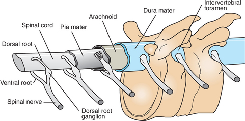
FIGURE 3-1 Spinal cord and the three layers of the meninges within the vertebral canal. Action potentials generated on sensory afferents enter the spinal cord along axons in the dorsal roots. Those generated on motor efferents exit the spinal cord along axons in the ventral roots.
(Redrawn from Gardner E: Fundamentals of neurology, ed 3, Philadelphia, 1959, Saunders.)
The PNS and CNS differ in the regenerative ability of their neural axons following physical injury. Peripheral nerve axons can slowly regrow and reconnect to their peripheral targets. Damaged CNS axons do not effectively regenerate, possibly resulting from inhibitory features of their local environment. Experimental manipulations of this environment have been shown to improve CNS axonal regrowth.
The Central Nervous System Can Be Divided into Six Anatomical Regions
The CNS has a longitudinal organization, with the phylogenetically oldest parts lying more caudal and the newest portions lying rostral. The CNS can be divided into six major regions (Figure 3-2): the spinal cord and five major brain regions. From caudal to rostral, these brain regions are the medulla, pons, midbrain, diencephalon, and telencephalon. (The cerebellum, a brain structure that lies dorsal to portions of the pons and medulla, is sometimes named as a seventh major region of the CNS.) The medulla, pons, and midbrain form the brainstem; the diencephalon and telencephalon form the forebrain.
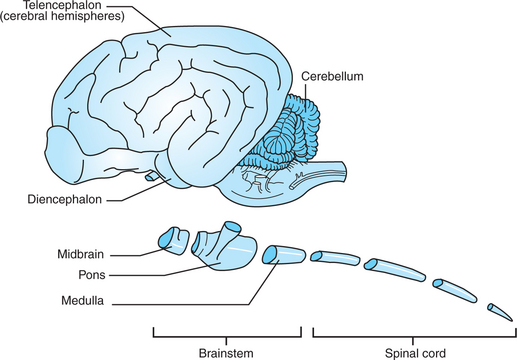
FIGURE 3-2 Central nervous system (CNS) has longitudinal organization in which the phylogenetically oldest parts are caudal and the newest parts are rostral. The CNS can be divided into six major regions: the spinal cord, medulla, pons, midbrain, diencephalon, and telencephalon (cerebral hemispheres).
In general, the spinal cord, brainstem, and forebrain represent a hierarchy of functional organization. The spinal cord receives sensory input from and supplies motor output to the trunk and limbs; the brainstem performs these functions for the face and head. Sensory information entering the brainstem is passed to the forebrain, where the most sophisticated forms of information processing take place. Sensory information entering the spinal cord is relayed to the forebrain by way of the brainstem. The forebrain also formulates the most sophisticated forms of motor output. This output is sent to the brainstem for executing movement of the face and head or for relay to the spinal cord to execute trunk and limb movement. The forebrain is also capable of sending motor commands directly to the spinal cord.
Each of the six CNS regions has distinctive anatomical and functional characteristics. Some of these include the following:
1. The spinal cord is the most caudal region in the CNS. As previously noted, it receives action potentials along sensory dorsal root axons from receptors in the skin, muscles, tendons, joints, and visceral organs. It contains the cell bodies and dendrites of motor neurons whose axons exit through the ventral roots either to reach skeletal muscles or to reach out toward smooth muscle. The spinal cord also contains tracts of axons carrying sensory information to the brain and motor commands from the brain to the motor neurons. The isolated spinal cord can control simple reflexes, such as muscle stretch reflexes and limb withdrawal to painful stimuli.
2. The medulla lies rostral to the spinal cord and resembles it in many ways. By way of cranial nerves, the medulla too receives information from the body’s external and internal sensory receptors and sends motor commands out to skeletal and smooth muscle. Large populations of these receptors and muscles lie in the head and neck region. The cell bodies of medullary neurons that receive the sensory input from cranial nerves or that send the motor output are respectively collected in aggregates called sensory or motor cranial nerve nuclei. The cranial nerve nuclei of the medulla play a critical role in life support functions of the respiratory and cardiovascular systems and in aspects of feeding (e.g., taste, tongue movement, swallowing, digestion) and vocalization.
3. The pons lies rostral to the medulla and contains the cell bodies of large numbers of neurons in a two-neuron chain that relays information from the cerebral cortex to the cerebellum. The cerebellum is not a part of the brainstem but is often described along with the pons because of a similar embryological origin. The cerebellum is important for smooth, accurate, coordinated movement and for motor learning. Cranial nerve nuclei of the pons play important roles in receiving sensory information about facial touch and in the motor control of chewing.
4. The midbrain, or mesencephalon, lies rostral to the pons and contains the superior and inferior colliculi, which are important in processing and relaying visual and auditory information that has entered the brain at other levels. The midbrain also contains cranial nerve nuclei that directly control eye movement and regions that coordinate some eye movement reflexes.
Each region of the brainstem contains axon tracts carrying action potentials to or from the forebrain, as well as tracts that carry action potentials to or from the spinal cord. Each brainstem region also contains a portion of the reticular formation, a netlike complex of many small clusters of cell bodies (nuclei) and loosely organized axonal projections, located near the midline. The reticular formation plays important roles in modulating consciousness and arousal, pain perception, and spinal reflexes, as well as in movement.
5. The diencephalon contains the thalamus and the hypothalamus, both of which are large structures consisting of several subnuclei. The thalamus is a relay station for and a modulator of information being passed to the cerebral cortex from sensory systems and other brain regions. The hypothalamus regulates the autonomic nervous system, controls hormone secretion of the pituitary gland, and plays a major role in physiological and behavioral aspects of homeostasis (e.g., maintenance of temperature and blood pressure; feeding).
6. The telencephalon, also commonly referred to as the cerebral hemispheres, is made up of the cerebral cortex and a small number of prominent subcortical structures, such as the basal ganglia and hippocampus. The cerebral cortex mediates the most complex forms of sensory integration and conscious sensory perception. It also formulates and executes sequences of voluntary movement. The basal ganglia are a collection of nuclei that modulate the motor functions of cerebral cortex, and the hippocampus plays an important role in memory and spatial learning.
The Central Nervous System Is Protected by the Meninges and Cerebrospinal Fluid
The entire CNS is surrounded by three protective layers called meninges: the pia mater, arachnoid, and dura mater (see Figure 3-1). The innermost layer, lying next to the CNS, is the pia mater, which is a single layer of fibroblast cells joined to the outer surface of the brain and spinal cord. The middle layer, the arachnoid, so named because of its spiderweb appearance, is a thin layer of fibroblast cells that traps cerebrospinal fluid between it and the pia mater (in the subarachnoid space). The outermost meningeal layer, the dura mater, is a much thicker layer of fibroblast cells that protects the CNS. Within the brain cavity of the skull, the dura mater is often fused with the inner surface of the bone.
Cerebrospinal fluid (CSF) is a clear, colorless fluid found within the subarachnoid space, the central canal of the spinal cord and the ventricular system of the brain (see Chapter 15). CSF is produced primarily in the ventricles of the brain, flows down a pressure gradient from the ventricles to the subarachnoid space, where it bathes the surface of the CNS, and from the subarachnoid space eventually passes into the venous system. It is a dynamic fluid, being replaced several times daily. Because CSF can exchange freely with the extracellular fluid of the CNS, it is an important determinant of the neuronal microenvironment, both carrying away metabolic waste and providing certain micronutrients. CSF also serves as a shock absorber for the CNS during abrupt body movement.
The Nervous System Collects and Integrates Sensory Information, Formulates a Response Plan, and Produces a Motor Output
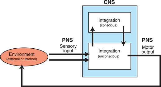
In simplest terms, the nervous system (1) collects sensory information from its external or internal environment, (2) consciously or unconsciously integrates these various inputs to formulate a response plan, and (3) produces a final motor output that can either change the environment (external or internal) or keep it constant (Figure 3-3). Collecting sensory information and executing the final motor output are the primary responsibilities of the PNS, whereas integration is primarily performed by the CNS. As discussed in Chapter 4, these same functions occur at the level of the individual neuron, which is the principal building block of the nervous system.
CLINICAL CORRELATIONS
Neurologic Disease in a Horse
History.
A client calls and asks you to look at a 4-month-old Arab filly. The owners have had her since birth, and she has always seemed a little clumsy compared with other foals. They think she is getting worse, however, and say she stumbles in the field. She falls over at times when she is playing with the other foals, and she seems very stiff, almost stabbing at the ground when she is walking.
Clinical Examination.
The filly is bright and alert. Her temperature, pulse, and respiration are normal. Abnormalities are limited to your neurological examination. She is weak (paresis) in both the hind and the front limbs (grade II), with the hind limbs being worse (grade III). When you assess her conscious proprioception (ataxia), she is also greatly delayed (grade III hind limbs, grade II front limbs). When she walks, the filly seems to slap at the ground (hypermetria), and she drags her toes forward across the ground. You detect no other neurological deficits.
Comment.
This filly has equine degenerative myeloencephalopathy. An antemortem diagnosis is difficult. Exclusion of other causes is important. Serum vitamin E levels are often, but not exclusively, low. A definitive diagnosis is made at necropsy.
The pathogenesis of the disease is not clear, but risk factors include diets low in vitamin E, use of insecticides, keeping animals on dirt lots, and exposure to wood preservatives. On histopathology, significant changes occur in the medulla and spinal cord. There is diffuse neuronal degeneration of the white matter. Astrocytosis and lipofuscin-like pigment accumulate in affected areas. Demyelination is marked.
Animals with this disease have loss of functional neurons as well as the myelin sheath that surrounds them. As a result, the ability to conduct impulses is greatly affected. Clinically, this affects the animal’s ability to respond to external stimuli as well as initiate conscious responses.
Treatment.
Supportive treatment is the only therapy that can be given. Keeping horses on green pasture has been shown to be somewhat protective. Supplementing with vitamin E can improve some horses’ condition and slow the progression of disease. There are some familial tendencies in Arabs, Appaloosas, thoroughbreds, and paso finos.
Behan M. Organization of the nervous system. Reece WO, ed. Duke’s physiology of domestic animals, ed 12, Ithaca, NY: Comstock Cornell University Press, 2004.
Boron WF, Boulpaep EL. Medical physiology: a cellular and molecular approach, updated-edition. Philadelphia: Saunders, 2005.
Fields RD. The other half of the brain. Sci Am. 2004;290:54.
Fields RD, Stevens-Graham B. New insights into neuron-glia communication. Science. 2002;298:556.
Guyton AC, Hall JE. Textbook of medical physiology, ed 11. Philadelphia: Saunders, 2006.
Kandel ER, Schwartz JH, Jessell TM. Principles of neural science, ed 4, New York: McGraw-Hill, 2000.
Kitchell RL. Introduction to the nervous system. Evans HE, ed. Miller’s anatomy of the dog, ed 6, Philadelphia: Saunders, 1993.
Kitchell RL, Evans HE. The spinal nerves. Evans HE, ed. Miller’s anatomy of the dog, ed 6, Philadelphia: Saunders, 1993.
Matthews GG. Neurobiology: molecules, cells, and systems, ed 2. Malden: Blackwell Science, 2001.
Matthews HK. Spinal cord, vertebral and intracranial trauma. In: Reed SM, Bayly WM. Equine internal medicine. Philadelphia: Saunders, 1998.
PRACTICE QUESTIONS
1. Which part of a neuron is primarily characterized as the information-receiving component?
2. Which of the following is not characteristic of glial cells?
3. The elements of spinal and cranial nerves that carry action potential commands from the CNS to synapses at skeletal muscles are:
4. The thalamus and hypothalamus are components of which major brain division?